Proliferation of parenchymal epithelial cells enhanced in chronic pancreatitis
5
J. Pathol. 186: 104–108 (1998) PROLIFERATION OF PARENCHYMAL EPITHELIAL CELLS ENHANCED IN CHRONIC PANCREATITIS . . 1 **, . . . 2 . . 3* 1 Department of Histopathology, Royal Postgraduate Medical School, Hammersmith Hospital, Du Cane Road, London W12 0HS, U.K. 2 Department of Surgery, Royal Postgraduate Medical School, Hammersmith Hospital, Du Cane Road, London W12 0HS, U.K. 3 Department of Cellular and Molecular Pathology, Duncan Building, University of Liverpool, PO Box 147, Liverpool L69 3GA, U.K. SUMMARY This study was performed to determine whether pancreatic parenchymal epithelial cells in human chronic pancreatitis tissues retain a biologically significant capability to proliferate and, if so, within which epithelial compartment proliferation occurs. The techniques of immediate per-operative in vitro labelling with bromodeoxyuridine (BrdU) and conventional immunohistochemistry for Ki-67 antigen expression were used to identify proliferating cells. Concordance between the two techniques was confirmed in all tissues examined. In normal pancreas, proliferation was restricted to acinar epithelial cells, with no activity in the ductules. In chronic pancreatitis of both chronic obstructive and chronic calcifying types, the number of proliferating cells in the acini was significantly increased. A small population of proliferating cells was also apparent within ductules in chronic calcifying pancreatitis, but not in chronic obstructive pancreatitis. This investigation has shown that loss of parenchymal epithelium occurring in chronic pancreatitis is not caused by a primary failure of pancreatic ‘stem-cell’ proliferation, but is due to disproportionate attrition of differentiated parenchymal epithelial cells by a mechanism, possibly stromal in origin, which remains hitherto unidentified. The presence of proliferating ductular cells in chronic calcifying pancreatitis, but not chronic obstructive pancreatitis, suggests that distinct pathogenic processes may be operating in the former condition, which is classically regarded as secondary to ductal obstruction by stones, and in this single respect might be considered to be identical to chronic obstructive pancreatitis. Preservation of ‘stem-cell’ function supports the belief that regeneration of pancreatic parenchymal tissue could be a feasible proposition if biologically appropriate management strategies were developed to treat chronic calcifying pancreatitis. ? 1998 John Wiley & Sons, Ltd. KEY WORDS—chronic pancreatitis; stem cells; proliferation INTRODUCTION Chronic pancreatitis is an inexorably progressive dis- ease defined as ‘the presence of chronic inflammatory lesions initially characterized by selective eradication of the exocrine parenchyma, together with fibrosis, but ultimately including destruction of the endocrine paren- chyma’. 1 In the U.K., approximately 1500 new cases are reported each year, 2 with the incidence rising over the past 20 years. 3 Two major aetiological categories, ‘chronic obstructive pancreatitis’ and ‘chronic calcifying pancreatitis’, are recognized. 4 The first is due to proxi- mal obstruction of duct drainage by a variety of neoplastic and non-neoplastic lesions. The second encompasses several subtypes, all believed to arise as a result of intraductal stone formation, although the aetio- pathogenesis remains elusive. In Western countries, alcohol abuse, 5 genetic predisposition, 6 diets deficient in micronutrients and protein, together with abnormal handling of xenobiotics, smoking, and drug toxicity have all been proposed as aetiological agents. 5–8 Autoimmune rejection of the pancreas has also been postulated. 9 Between 9 and 41 per cent of cases remain idiopathic. 2 Nevertheless, the precise aetiopathological factors contributing to the progressive loss of parenchy- mal elements, and their replacement by dense fibrous connective tissue, have not yet been incontrovertibly identified. 10 The pancreatic parenchyma is believed to exhibit continuous but conditional renewal. In rodents, com- plete regeneration follows partial pancreatic resection, whereas in the human, there is only limited evidence of compensatory regeneration following loss of pancreatic tissue. 11 Depletion of the exocrine epithelial component characterizes all types of chronic pancreatitis, although it is not known whether this is due to excess destruction of fully-differentiated epithelial cells, including those with residual proliferative capacity, or a failure of pancreatic stem cells to divide and to repopulate the organ. The present study was performed to determine whether a biologically significant proliferative capacity might be demonstrated in pancreatic tissues affected by both forms of chronic pancreatitis. Two independent markers of cell cycle parameters have been examined: bromodeoxyuridine (BrdU), a dynamic marker of proliferation 12 incorporated into the cell cycle during S-phase of cell division, was detected immunohisto- chemically following intraoperative incubation of pancreatic tissue samples. Expression of Ki-67, a *Correspondence to: Professor C. S. Foster, Department of Cellular and Molecular Pathology, Duncan Building, University of Liverpool, PO Box 147, Liverpool L69 3GA. **Current address: Department of Histopathology, University College Hospital Medical School, Rockefeller Building, University Street, London WC1E 6JJ, U.K. Contract grant sponsors: British Digestive Foundation; Kancatak (Carbofab Research). CCC 0022–3417/98/010104–05 $17.50 ? 1998 John Wiley & Sons, Ltd. Received 11 July 1997 Accepted 25 March 1998
Transcript of Proliferation of parenchymal epithelial cells enhanced in chronic pancreatitis

J. Pathol. 186: 104–108 (1998)
PROLIFERATION OF PARENCHYMAL EPITHELIALCELLS ENHANCED IN CHRONIC PANCREATITIS
. . 1**, . . . 2 . . 3*
1Department of Histopathology, Royal Postgraduate Medical School, Hammersmith Hospital, Du Cane Road,London W12 0HS, U.K.
2Department of Surgery, Royal Postgraduate Medical School, Hammersmith Hospital, Du Cane Road, London W12 0HS, U.K.3Department of Cellular and Molecular Pathology, Duncan Building, University of Liverpool, PO Box 147,
Liverpool L69 3GA, U.K.
SUMMARY
This study was performed to determine whether pancreatic parenchymal epithelial cells in human chronic pancreatitis tissues retaina biologically significant capability to proliferate and, if so, within which epithelial compartment proliferation occurs. The techniques ofimmediate per-operative in vitro labelling with bromodeoxyuridine (BrdU) and conventional immunohistochemistry for Ki-67 antigenexpression were used to identify proliferating cells. Concordance between the two techniques was confirmed in all tissues examined. Innormal pancreas, proliferation was restricted to acinar epithelial cells, with no activity in the ductules. In chronic pancreatitis of bothchronic obstructive and chronic calcifying types, the number of proliferating cells in the acini was significantly increased. A smallpopulation of proliferating cells was also apparent within ductules in chronic calcifying pancreatitis, but not in chronic obstructivepancreatitis. This investigation has shown that loss of parenchymal epithelium occurring in chronic pancreatitis is not caused by aprimary failure of pancreatic ‘stem-cell’ proliferation, but is due to disproportionate attrition of differentiated parenchymal epithelialcells by a mechanism, possibly stromal in origin, which remains hitherto unidentified. The presence of proliferating ductular cells inchronic calcifying pancreatitis, but not chronic obstructive pancreatitis, suggests that distinct pathogenic processes may be operating inthe former condition, which is classically regarded as secondary to ductal obstruction by stones, and in this single respect might beconsidered to be identical to chronic obstructive pancreatitis. Preservation of ‘stem-cell’ function supports the belief that regeneration ofpancreatic parenchymal tissue could be a feasible proposition if biologically appropriate management strategies were developed to treatchronic calcifying pancreatitis. ? 1998 John Wiley & Sons, Ltd.
KEY WORDS—chronic pancreatitis; stem cells; proliferation
INTRODUCTION
Chronic pancreatitis is an inexorably progressive dis-ease defined as ‘the presence of chronic inflammatorylesions initially characterized by selective eradication ofthe exocrine parenchyma, together with fibrosis, butultimately including destruction of the endocrine paren-chyma’.1 In the U.K., approximately 1500 new cases arereported each year,2 with the incidence rising over thepast 20 years.3 Two major aetiological categories,‘chronic obstructive pancreatitis’ and ‘chronic calcifyingpancreatitis’, are recognized.4 The first is due to proxi-mal obstruction of duct drainage by a variety ofneoplastic and non-neoplastic lesions. The secondencompasses several subtypes, all believed to arise as aresult of intraductal stone formation, although the aetio-pathogenesis remains elusive. In Western countries,alcohol abuse,5 genetic predisposition,6 diets deficient inmicronutrients and protein, together with abnormalhandling of xenobiotics, smoking, and drug toxicityhave all been proposed as aetiological agents.5–8
*Correspondence to: Professor C. S. Foster, Department of Cellularand Molecular Pathology, Duncan Building, University of Liverpool,PO Box 147, Liverpool L69 3GA.
**Current address: Department of Histopathology, UniversityCollege Hospital Medical School, Rockefeller Building, UniversityStreet, London WC1E 6JJ, U.K.
Contract grant sponsors: British Digestive Foundation; Kancatak(Carbofab Research).
CCC 0022–3417/98/010104–05 $17.50? 1998 John Wiley & Sons, Ltd.
Autoimmune rejection of the pancreas has also beenpostulated.9 Between 9 and 41 per cent of cases remainidiopathic.2 Nevertheless, the precise aetiopathologicalfactors contributing to the progressive loss of parenchy-mal elements, and their replacement by dense fibrousconnective tissue, have not yet been incontrovertiblyidentified.10
The pancreatic parenchyma is believed to exhibitcontinuous but conditional renewal. In rodents, com-plete regeneration follows partial pancreatic resection,whereas in the human, there is only limited evidence ofcompensatory regeneration following loss of pancreatictissue.11 Depletion of the exocrine epithelial componentcharacterizes all types of chronic pancreatitis, althoughit is not known whether this is due to excess destructionof fully-differentiated epithelial cells, including thosewith residual proliferative capacity, or a failure ofpancreatic stem cells to divide and to repopulate theorgan.
The present study was performed to determinewhether a biologically significant proliferative capacitymight be demonstrated in pancreatic tissues affected byboth forms of chronic pancreatitis. Two independentmarkers of cell cycle parameters have been examined:bromodeoxyuridine (BrdU), a dynamic marker ofproliferation12 incorporated into the cell cycle duringS-phase of cell division, was detected immunohisto-chemically following intraoperative incubation ofpancreatic tissue samples. Expression of Ki-67, a
Received 11 July 1997Accepted 25 March 1998

105PROLIFERATION OF PARENCHYMAL EPITHELIAL CELLS IN CHRONIC PANCREATITIS
proliferation-association protein tightly bound by chro-matin in all cells as they progress from S into G2 phaseof the cell cycle, was also detected immunohistochemi-cally.13 The findings of this study have confirmed thatpancreatic epithelia in chronic pancreatitis maintain anenhanced proliferative capacity, which has yet to beexploited in the management of patients with this groupof diseases.
MATERIALS AND METHODS
Case material
Fresh pancreatic specimens were collected from 12partial pancreatectomy cases performed betweenDecember 1993 and September 1994 at the Hammer-smith Hospital, London. Of these 12, four comprisedcontrol specimens obtained from patients undergoingresection for non-obstructing, small neuroendocrinetumours and benign cystic lesions. Pathological tissueswere obtained from four cases of chronic obstructivepancreatitis and four cases of chronic calcifying pan-creatitis, the latter being of probable alcoholic origin.The total complement of tissues examined comprisedfive females and seven males, with a mean age of 58years and an age range of 24–74 years.10,14
Tissue collection
Tissues were collected intraoperatively and three rep-resentative slices were taken from each at least 1 cmfrom the surgical resection margin, according to theprotocol described in our previous publications.10,14
Each piece of tissue was diced into several cubes(1–3 mm diameter) and incubated in air-bufferedLeibowtiz medium, pre-warmed to 37)C.
BrdU incorporation technique
Tissue samples were processed within 30 min accord-ing to the BrdU in vitro techniques.15 For each specimen,three incubation chambers were set up. In each chamber,three pieces of pancreatic tissue, each approximately1·5 mm3, were incubated in 1·5 ml of HEPES-bufferedLeibovtiz medium containing 0·15 per cent (w/v) BrdUunder hyperbaric conditions, and the tubes closed withperforated stoppers. Incubation was carried out for 1 hat 37)C. Thereafter, the screw of the apparatus wasgently released under water to allow excess oxygen toescape. Tissues were then processed individually intoparaffin wax.
Tissue preparation
Paraffin sections—Sections cut at 5 ìm thickness weremounted onto poly--lysine-coated slides for the BrdUsections and onto Superfrost slides to improve adhesionduring microwaving for the Ki-67-stained sections. Afterdrying overnight, sections were dewaxed in xylene andrehydrated using graded alcohols. Consecutive sectionswere used for the two investigative procedures.
Immunohistochemistry
Control tissues—Sections of rat liver from animalsflash-labelled with BrdU (50 mg/kg body weight)
? 1998 John Wiley & Sons, Ltd.
for 1 h prior to killing were used as controls for thein vitro BrdU-labelled human tissues. Sections ofhuman small intestine were used as controls for Ki-67immunohistochemical staining.
Pretreatment requirements prior toimmunohistochemistry
Trypsinization/hydrochloric acid pretreatment of BrdUsections—After blocking endogenous peroxidase byimmersion in 0·1 per cent hydrogen peroxide in metha-nol for 30 min at room temperature, sections werewashed in pre-warmed 37)C distilled water; pre-digestedin freshly prepared trypsin (Sigma, U.K.) solution, 1 mg/ml, in calcium chloride solution (0·66 g/500 ml), pH 7·8,for 13 min at 37)C; rewashed and heated in normalhydrochloric acid, for 15 min, at 60)C to improvenuclear penetration; and finally washed thoroughly indistilled water and Tris-buffered saline (TBS, pH 7·6)before immunohistochemistry.
Microwave pretreatment of Ki-67 sections—The Ki-67sections were microwave-treated at 700 W for 10 minand immersed in pre-warmed 10 mmol citrate buffersolution, pH 6·0 (2·1 g of citric acid in 1·0 l of distilledwater). Endogenous peroxidase was then blocked using0·3 per cent H2O2 in water for 30 min before rinsing inphosphate-buffered saline (PBS, pH 7·4).
Immunohistochemical technique
The BrdU sections were washed throughout in TBSand the Ki-67 sections in PBS. Non-specific proteinbinding was inhibited with normal rabbit serum at adilution of 1:20. Sections were incubated with primaryantibody in a moist chamber, at room temperature, for2 h. The anti-BrdU (Dako, Denmark) antibody wasdiluted to 1:30 in 1 per cent human A-B serum in TBSand the anti-Ki-67 antibody (Dako, Denmark) wasdiluted to 1:50 in PBS. As a negative control, theprimary antibody was replaced by buffer. BrdU-labelledsections were then washed and incubated with bioti-nylated rabbit anti-mouse immunoglobulin (Dako,Denmark; diluted 1:300 in 1 per cent human serum inPBS) for 1 h. After washing, sections were incubatedwith ABC complex (Dako, Denmark) for 45 min. Anti-Ki-67 antibody-labelled sections were incubated in swine(anti-rabbit) immunoglobulin (Dako, Denmark) at adilution of 1:100 in PBS for 1 h and then washed beforefurther incubation for 1 h with rabbit peroxidase anti-peroxidase antibody (Dako, dilution 1:300 in PBS). Allsections were then developed by immersion in a 0·5 percent (v/v) solution of 3,3*-diaminobenzidine hydrochlo-ride (DAB; Aldrich, Gillingham, U.K.) in PBS contain-ing 0·03 per cent (v/v) H2O2 after final washing. Nucleiwere lightly counterstained with haematoxylin beforemounting.
Stained sections were scored for BrdU and Ki-67labelling by light microscopy at a magnification of#400. Both antibodies produced stained positive nuclei.The total number of epithelial nuclei, together withpositively staining nuclei, in ten randomly selected fields
J. Pathol. 186: 104–108 (1998)
106 S. D. SLATER ET AL.
was recorded for the three specimens taken in each case.The location of positive-staining nuclei was recorded asductal or acinar.
RESULTS
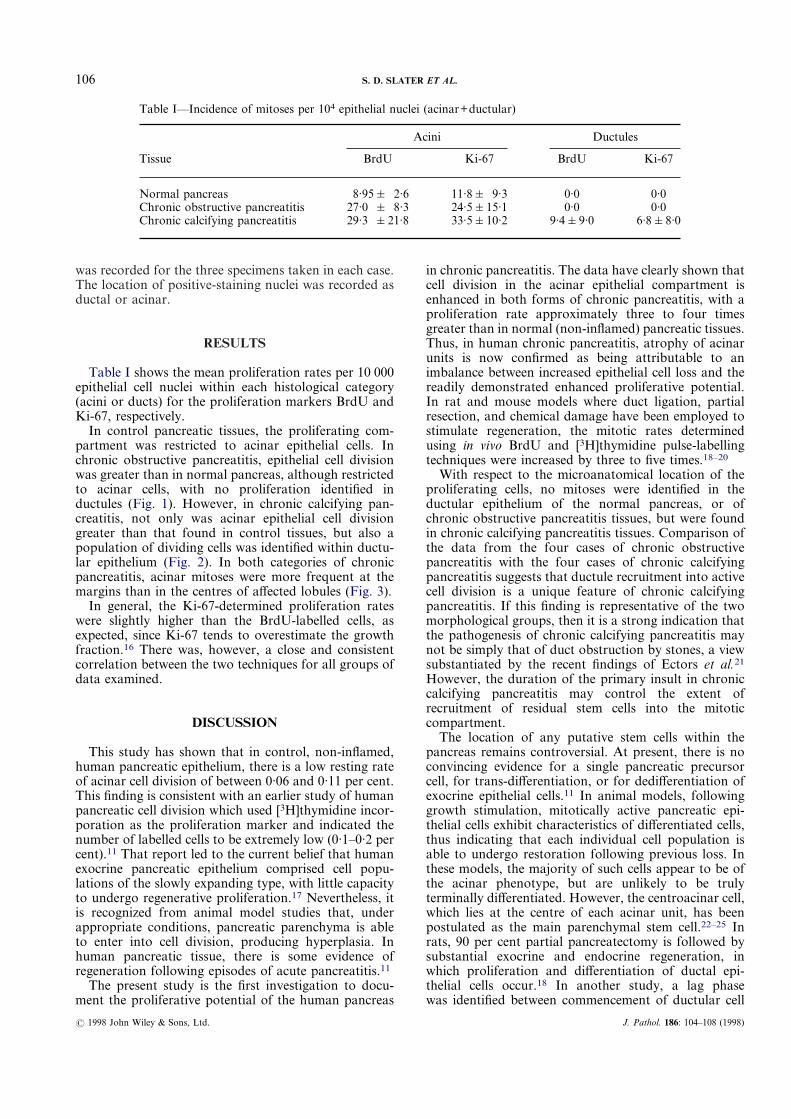
Table I shows the mean proliferation rates per 10 000epithelial cell nuclei within each histological category(acini or ducts) for the proliferation markers BrdU andKi-67, respectively.
In control pancreatic tissues, the proliferating com-partment was restricted to acinar epithelial cells. Inchronic obstructive pancreatitis, epithelial cell divisionwas greater than in normal pancreas, although restrictedto acinar cells, with no proliferation identified inductules (Fig. 1). However, in chronic calcifying pan-creatitis, not only was acinar epithelial cell divisiongreater than that found in control tissues, but also apopulation of dividing cells was identified within ductu-lar epithelium (Fig. 2). In both categories of chronicpancreatitis, acinar mitoses were more frequent at themargins than in the centres of affected lobules (Fig. 3).
In general, the Ki-67-determined proliferation rateswere slightly higher than the BrdU-labelled cells, asexpected, since Ki-67 tends to overestimate the growthfraction.16 There was, however, a close and consistentcorrelation between the two techniques for all groups ofdata examined.
DISCUSSION
This study has shown that in control, non-inflamed,human pancreatic epithelium, there is a low resting rateof acinar cell division of between 0·06 and 0·11 per cent.This finding is consistent with an earlier study of humanpancreatic cell division which used [3H]thymidine incor-poration as the proliferation marker and indicated thenumber of labelled cells to be extremely low (0·1–0·2 percent).11 That report led to the current belief that humanexocrine pancreatic epithelium comprised cell popu-lations of the slowly expanding type, with little capacityto undergo regenerative proliferation.17 Nevertheless, itis recognized from animal model studies that, underappropriate conditions, pancreatic parenchyma is ableto enter into cell division, producing hyperplasia. Inhuman pancreatic tissue, there is some evidence ofregeneration following episodes of acute pancreatitis.11
The present study is the first investigation to docu-ment the proliferative potential of the human pancreas
? 1998 John Wiley & Sons, Ltd.
in chronic pancreatitis. The data have clearly shown thatcell division in the acinar epithelial compartment isenhanced in both forms of chronic pancreatitis, with aproliferation rate approximately three to four timesgreater than in normal (non-inflamed) pancreatic tissues.Thus, in human chronic pancreatitis, atrophy of acinarunits is now confirmed as being attributable to animbalance between increased epithelial cell loss and thereadily demonstrated enhanced proliferative potential.In rat and mouse models where duct ligation, partialresection, and chemical damage have been employed tostimulate regeneration, the mitotic rates determinedusing in vivo BrdU and [3H]thymidine pulse-labellingtechniques were increased by three to five times.18–20
With respect to the microanatomical location of theproliferating cells, no mitoses were identified in theductular epithelium of the normal pancreas, or ofchronic obstructive pancreatitis tissues, but were foundin chronic calcifying pancreatitis tissues. Comparison ofthe data from the four cases of chronic obstructivepancreatitis with the four cases of chronic calcifyingpancreatitis suggests that ductule recruitment into activecell division is a unique feature of chronic calcifyingpancreatitis. If this finding is representative of the twomorphological groups, then it is a strong indication thatthe pathogenesis of chronic calcifying pancreatitis maynot be simply that of duct obstruction by stones, a viewsubstantiated by the recent findings of Ectors et al.21
However, the duration of the primary insult in chroniccalcifying pancreatitis may control the extent ofrecruitment of residual stem cells into the mitoticcompartment.
The location of any putative stem cells within thepancreas remains controversial. At present, there is noconvincing evidence for a single pancreatic precursorcell, for trans-differentiation, or for dedifferentiation ofexocrine epithelial cells.11 In animal models, followinggrowth stimulation, mitotically active pancreatic epi-thelial cells exhibit characteristics of differentiated cells,thus indicating that each individual cell population isable to undergo restoration following previous loss. Inthese models, the majority of such cells appear to be ofthe acinar phenotype, but are unlikely to be trulyterminally differentiated. However, the centroacinar cell,which lies at the centre of each acinar unit, has beenpostulated as the main parenchymal stem cell.22–25 Inrats, 90 per cent partial pancreatectomy is followed bysubstantial exocrine and endocrine regeneration, inwhich proliferation and differentiation of ductal epi-thelial cells occur.18 In another study, a lag phasewas identified between commencement of ductular cell
Table I—Incidence of mitoses per 104 epithelial nuclei (acinar+ductular)
Tissue
Acini Ductules
BrdU Ki-67 BrdU Ki-67
Normal pancreas 8·95& 2·6 11·8& 9·3 0·0 0·0Chronic obstructive pancreatitis 27·0 & 8·3 24·5&15·1 0·0 0·0Chronic calcifying pancreatitis 29·3 &21·8 33·5&10·2 9·4&9·0 6·8&8·0
J. Pathol. 186: 104–108 (1998)

107PROLIFERATION OF PARENCHYMAL EPITHELIAL CELLS IN CHRONIC PANCREATITIS
division and onset of acinar mitosis.17 It was suggestedthat the acinar cells which divide are, most probably,those cells from the ductular compartment which havebecome differentiated and it was hypothesized that the
? 1998 John Wiley & Sons, Ltd.
pancreas is composed of an integrated series of prolif-erative units, each comprising acinar cells, centroacinarcells, and ductules, and reacting as an integrated unitwhenever acinar cell loss occurs.17
Fig. 1—(a) Early chronic obstructive pancreatitis in which pancreatic lobular architecture remains well preserved. A small collection ofchronic inflammatory cells is present (left) where they are associated with focal epithelial cell drop-out. Proliferatively active cell nuclei areclearly labelled with BrdU. #250. (b) Early chronic obstructive pancreatitis in which lobular architecture is preserved but in whichintralobular fibrous connective tissue is increased. Cell proliferation within the acinar epithelial compartment is clearly demonstrated byimmunohistochemical localization of Ki-67 protein. #250
Fig. 2—(a) Established chronic calcifying pancreatitis in which nuclei in residual second-order duct epithelial cells are clearly labelled withBrdU. Within the centre of a lobular structure, no local acinar epithelial cells were similarly labelled. #250. (b) Late-stage chronic calcifyingpancreatitis in which nuclei of individual residual acinar and ductular epithelial cells both express immunohistochemically reactive Ki-67protein. #250
Fig. 3—(a) Periphery of lobule in chronic calcifying pancreatitis. Multiple nuclei, particularly of acinar epithelial origin, are strongly labelledwith BrdU. #250. (b) In many lobules, proliferating nuclei were particularly located around the peripheral rim of acini when compared withthe relatively quiescent central acini. A star identifies the terminal ductular structure at the centre of the acinus #250.
J. Pathol. 186: 104–108 (1998)
108 S. D. SLATER ET AL.
In the present study, the principal site of epithelial cellproliferation was identified at the margins of lobulesadjacent to the fibrous bands characteristic of chronicpancreatitis. We suggest that an interaction betweenresidual epithelial cells retaining a proliferative capacityand the adjacent specialized pancreatic stroma maybe an important factor in pancreatic regeneration.Mesenchyme is essential for normal pancreatic growth,possibly by binding growth factors to the mesenchymalproteoglycans and thus presenting these to the epithelialcells via a basement membrane.26 Connective tissueappears to support the adult differentiated state byinhibiting the formation of acinar cells and localizingonly around large ducts in the normal pancreas.11 Theremay also be a role for transforming growth factor beta(TGF-â1), which we have shown to be enhanced withinthe ductules in chronic pancreatitis of both types.14
TGF-â1 is known to promote fibrosis and to limit cellproliferation,14 possibly acting as an autocrine regulatorof proliferation.27
In summary, the present study has confirmed that inchronic obstructive pancreatitis, and in chronic calcify-ing pancreatitis, the proliferative potential of acinar cellsis not only preserved but also enhanced, when comparedwith non-inflamed pancreatic tissues, as assessed by theindependent techniques of in vitro BrdU labelling andexpression of Ki-67 protein. Furthermore, in chroniccalcifying pancreatitis, ductular epithelial cells alsobecome recruited into the proliferating compartment. Incontrast, large ductal epithelial cells do not appear tobecome recruited or to participate in proliferation.These data not only suggest that the pathogenesis of thetwo types of chronic pancreatitis is likely to be distinct,thus supporting the recent opinion expressed by Ectorset al.,21 but also suggest that in both disease groups, lossof pancreatic exocrine parenchyma is not due to aprimary failure of pancreatic epithelial cell proliferationbut to another (possibly stromal) mechanism which hasyet to be identified. Whether or not the cells derivedfrom these proliferating epithelial compartments mightrecapitulate the full spectrum of parenchymal functionsdepends on a variety of factors, including the compo-sition and interaction of the adjacent residual stroma.Nevertheless, despite these obvious caveats, the poten-tial remains for developing a more biologicallyappropriate strategy to manage patients with chronicpancreatitis, rather than the non-selective ablativesurgical approach currently employed.28
ACKNOWLEDGEMENTS
Dr S. D. Slater gratefully acknowledges the BritishDigestive Foundation in continuing its support of thiswork. Additional generous funding to allow this workto be completed was also received from Kancatak(Carbofab Research). Professor Foster also thanks Mr
? 1998 John Wiley & Sons, Ltd.
Alan Williams for his photographic assistance and MrsJill Gosney for typing and editing the manuscript.
REFERENCES1. Singer MV, Gyr K, Sarles H. Revised classification of pancreatitis. Report
of the Second International Symposium on the Classification of Pancreatitisin Marseille, France. Gastroenterology 1985; 89: 683–690.
2. Jalleh RP, Gilbertson JA, Williamson RCN, Slater SD, Foster CS. Expres-sion of major histocompatibility antigens in human chronic pancreatitis.Gut 1993; 34: 1452–1457.
3. Kloppel G, Maillet B. Pathology of acute and chronic pancreatitis. Pancreas1993; 8: 659–670.
4. Singh S, Reber H. The pathology of chronic pancreatitis. World J Surg1990; 1: 2–10.
5. Muench R. Etiology and natural history of chronic pancreatitis. Digest Dis1992; 10: 335–344.
6. Sarner M. Update in the classification of chronic pancreatitis. In: Berger H,Buchler M, Ditschuneit H, Malfertheiner P, eds. Chronic Pancreatitis.Berlin: Springer-Verlag, 1990; 3–7.
7. Sandilands D, Jeffrey IJM, Haboubi NY, Maclennan IAM, Braganza JM.Abnormal drug metabolism in chronic pancreatitis—treatment with anti-oxidants. Gastroenterology 1990; 98: 766–772.
8. Cavallini G, Talamini G, Vaona B, et al. Effect of alcohol and smoking onpancreatic lithogenesis in the course of chronic pancreatitis. Pancreas 1994;9: 42–46.
9. Cavallini G. Is chronic pancreatitis a primary disease of the pancreaticducts? A new hypothesis. Ital J Gastroenterol 1993; 25: 391–396.
10. Slater SD, Jalleh RP, Gilberton JA, Lampert IA, Williamson RCN, FosterCS. Expression of heat shock proteins in chronic pancreatitis: protective orpathogenic? Lab Invest 1997; 76: 533–545.
11. Elsasser HP, Adler G, Kern HF. Replication and regeneration of thepancreas. In: Go V, Dimanjo E, Gardner J, eds. The Pancreas. Biology,Pathobiology and Diseases. New York: Raven Press, 1992; 75–86.
12. Lynch D, Clarke A, Jackson P. Comparison of labelling by bromodeoxyu-ridine, MIB1 and proliferating cell nuclear antigen in gastric mucosal biopsyspecimen. J Clin Pathol 1994; 47: 122–125.
13. Sawhney N, Hall PA. Ki-67 structure, function and new antibodies. JPathol 1992; 168: 161–162.
14. Slater SD, Williamson RCN, Foster CS. Expression of transforming growthfactor-beta 1 in chronic pancreatitis. Digestion 1995; 56: 237–241.
15. Britto M, Filipe M, Morris R. Cell proliferation study on gastric carcinomaand non-involved gastric mucosa using a bromodeoxyuridine labellingtechnique. Eur J Cancer Prev 1992; 1: 429–435.
16. Scott RJ, Hall PA, Haldane JS. A comparison of immunohistochemicalmarkers of cell proliferation with experimentally determined growth factors.J Pathol 1991; 165: 173–178.
17. Walker NI, Pound AW. An autoradiographic study of the cell proliferationduring involution of the rat pancreas. J Pathol 1983; 139: 407–418.
18. Bonner-Weir S, Baxter LA, Schuppin GT, Smith F. A second pathway forregeneration of adult exocrine and endocrine pancreas. Diabetes 1993; 42:1715–1720.
19. Gasslander T, Chu M, Smeds S, Ihse I. Proliferative response of differentexocrine pancreatic cells after surgical pancreaticobiliary diversion in therat. Scand J Gastroenterol 1991; 26: 399–404.
20. Gasslander T, Smeds S, Blomqvist L, Ihse I. Proliferative response ofdifferent exocrine pancreatic cell types to hormonal stimuli. Scand JGastroenterol 1990; 25: 1103–1110.
21. Ectors N, Maillet B, Aerts R, et al. Non-alcoholic duct destructive chronicpancreatitis. Gut 1997; 41: 263–268.
22. Gasslander T, Smeds S, Ihse I. Importance of the centroacinar region incerulein-induced mouse pancreatic growth. Scand J Gastroenterol 1992; 27:564–570.
23. Fitzgerald PJ. The problem of the precursor cell of regenerating pancreaticacinar epithelium. Lab Invest 1960; 9: 67–85.
24. Alder G, Hupp T, Kern HF. Course and spontaneous regression of acutepancreatitis in the rat. Virchows Arch Pathol Anat 1979; 382: 31–47.
25. Pour P. Pancreatic centroacinar cells. Int J Pancreatol 1994; 15: 51–64.26. Lee PC, Lebenthal E. Prenatal and postnatal development of the human
exocrine pancreas. In: Go V, Dimanjo E, Gardner J, eds. The Pancreas.Biology, Pathobiology and Diseases. New York: Raven Press, 1992; 57–74.
27. Sporn MB, Roberts AB. What is TGF-Beta? Clinical Applications ofTGF-Beta. Ciba Foundation Symposium, Chichester: John Wiley, 1991:1–6.
28. Jalleh RP, N Wiliamson RCN. Pancreatic exocrine and endocrine functionafter operations for chronic pancreatitis. Ann Surg 1992; 216: 656–662.
J. Pathol. 186: 104–108 (1998)