Prokaryotic and Eukaryotic Cells
18
PROKARYOT IC AND EUKARYOTI C CELLS Difference between prokaryotic and eukaryotic cells:
-
Upload
salahuddinmd5935 -
Category
Documents
-
view
19 -
download
0
description
ppt
Transcript of Prokaryotic and Eukaryotic Cells

PROKARYOTIC AND EUKARYOTIC CELLSDifference between prokaryotic and eukaryotic cells:
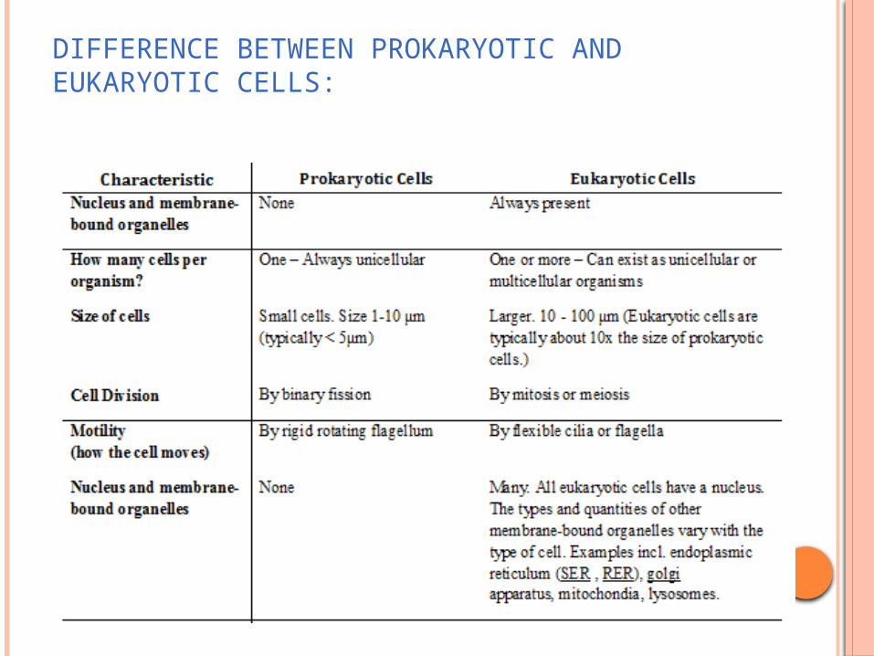
DIFFERENCE BETWEEN PROKARYOTIC AND EUKARYOTIC CELLS:

WHAT PROKARYOTIC AND EUKARYOTIC CELLS HAVE IN COMMON?
Both have DNA as their genetic material (it’s DNA that tells cells what kind of cells they should be).
Both are covered by a cell membrane.
Both contain RNA.
Both are made from the same basic chemicals: carbohydrates, proteins, nucleic acid, minerals, fats and vitamins.
Both have ribosomes (the structures on which proteins are made).
Both regulate the flow of the nutrients and wastes that enter and leave them.
Both have similar basic metabolism (life processes) like photosynthesis and reproduction.
Both require a supply of energy.
Both are highly regulated by elaborate sensing systems ("chemical noses”) that make them aware of the reactions within them and the environment around them
Describe the ultra structure of an animal cell (eukaryotic) and recognise these organelles from EM(electron microscope) images:

THE NUCLEUS The nucleus Usually the largest organelle in the cell
(10-20μm) and can be seen with the light microscope.
Electron microscopes portray how the nucleus, which is normally spherical, is surrounded by a double nuclear membrane containing nuclear pores.
Chemicals pass in and out of these pores so that it can control the events in the cytoplasm.
The nuclear envelope (membrane) encloses two main substances: nucleic acids and proteins. Acids are (DNA and RNA).
When the DNA is not dividing, the DNA is bonded to the protein to form chromatin, which looks like tiny granules.
The nucleus contains at least one nucleolus – an extra dense area of almost pure DNA and protein.
The nucleolus is involved in the production of ribosomes, cell growth and division.

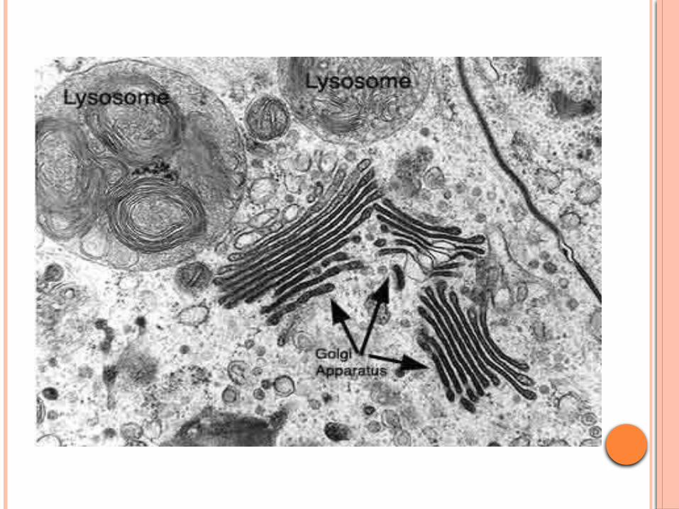
LYSOSOME They appear as dark, spherical bodies in the cytoplasm
containing many digestive enzymes. These are small in size, single membrane. Produced by Golgi apparatus. They frequently fuse with each other and with a
membrane bound vacuole containing either food or a foreign organelle.
Function: The function of these organelles is to break down any of
the organelles that have been worn out. The enzymes break down the contents into molecules
that can be reused. Lysosomes may fuse with outer cell membrane to
release enzymes outside the cell for destruction of bacteria or in, digestion.
They can also self-destruct – they may rupture if the cell is worn out (apoptosis).
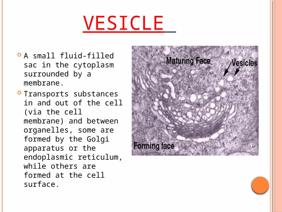
VESICLE A small fluid-filled sac
in the cytoplasm surrounded by a membrane.
Transports substances in and out of the cell (via the cell membrane) and between organelles, some are formed by the Golgi apparatus or the endoplasmic reticulum, while others are formed at the cell surface.
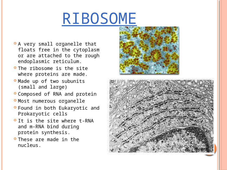
RIBOSOME A very small organelle that
floats free in the cytoplasm or are attached to the rough endoplasmic reticulum.
The ribosome is the site where proteins are made.
Made up of two subunits (small and large)
Composed of RNA and protein Most numerous organelle Found in both Eukaryotic and
Prokaryotic cells It is the site where t-RNA and
m-RNA bind during protein synthesis.
These are made in the nucleus.
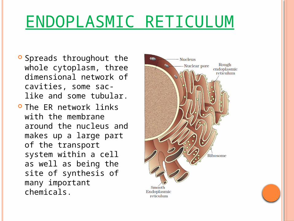
ENDOPLASMIC RETICULUM
Spreads throughout the whole cytoplasm, three dimensional network of cavities, some sac-like and some tubular.
The ER network links with the membrane around the nucleus and makes up a large part of the transport system within a cell as well as being the site of synthesis of many important chemicals.
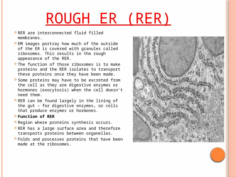
ROUGH ER (RER) RER are interconnected fluid filled
membranes. EM images portray how much of the outside
of the ER is covered with granules called ribosomes. This results in the rough appearance of the RER.
The function of those ribosomes is to make proteins and the RER isolates to transport these proteins once they have been made.
Some proteins may have to be excreted from the cell as they are digestive enzymes or hormones (exocytosis) when the cell doesn’t need them.
RER can be found largely in the lining of the gut – for digestive enzymes, or cells that produce enzymes or hormones.
Function of RER Region where proteins synthesis occurs. RER has a large surface area and therefore
transports proteins between organelles. Folds and processes proteins that have
been made at the ribosomes.
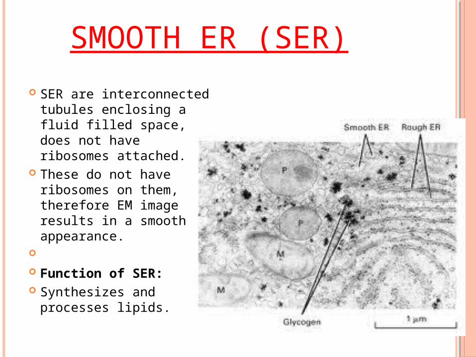
SMOOTH ER (SER)
SER are interconnected tubules enclosing a fluid filled space, does not have ribosomes attached.
These do not have ribosomes on them, therefore EM image results in a smooth appearance.
Function of SER: Synthesizes and
processes lipids.
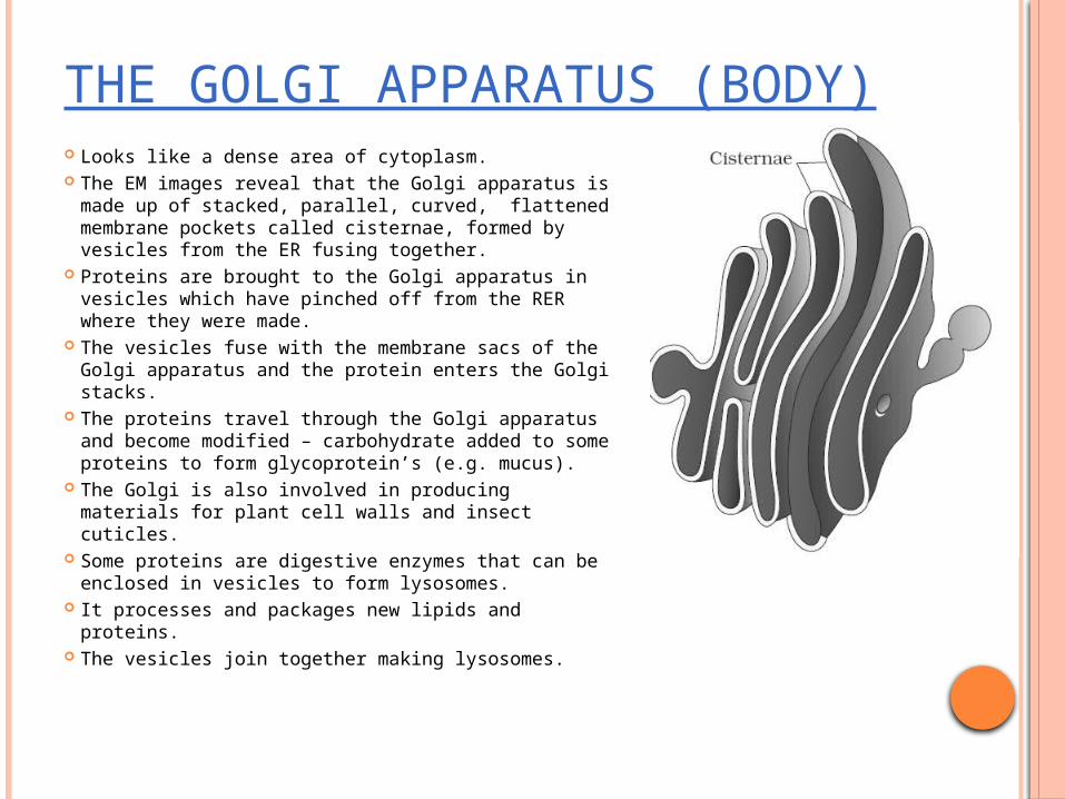
THE GOLGI APPARATUS (BODY) Looks like a dense area of cytoplasm. The EM images reveal that the Golgi apparatus is
made up of stacked, parallel, curved, flattened membrane pockets called cisternae, formed by vesicles from the ER fusing together.
Proteins are brought to the Golgi apparatus in vesicles which have pinched off from the RER where they were made.
The vesicles fuse with the membrane sacs of the Golgi apparatus and the protein enters the Golgi stacks.
The proteins travel through the Golgi apparatus and become modified – carbohydrate added to some proteins to form glycoprotein’s (e.g. mucus).
The Golgi is also involved in producing materials for plant cell walls and insect cuticles.
Some proteins are digestive enzymes that can be enclosed in vesicles to form lysosomes.
It processes and packages new lipids and proteins.
The vesicles join together making lysosomes.
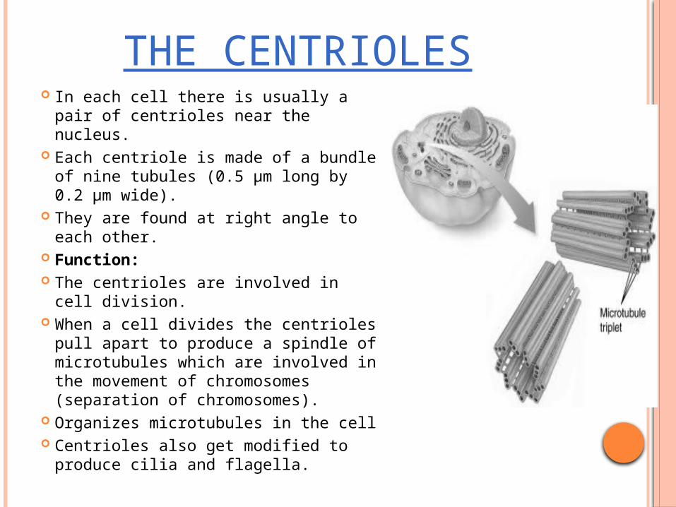
THE CENTRIOLES In each cell there is usually a pair of
centrioles near the nucleus. Each centriole is made of a bundle of
nine tubules (0.5 μm long by 0.2 μm wide).
They are found at right angle to each other.
Function: The centrioles are involved in cell
division. When a cell divides the centrioles pull
apart to produce a spindle of microtubules which are involved in the movement of chromosomes (separation of chromosomes).
Organizes microtubules in the cell Centrioles also get modified to
produce cilia and flagella.
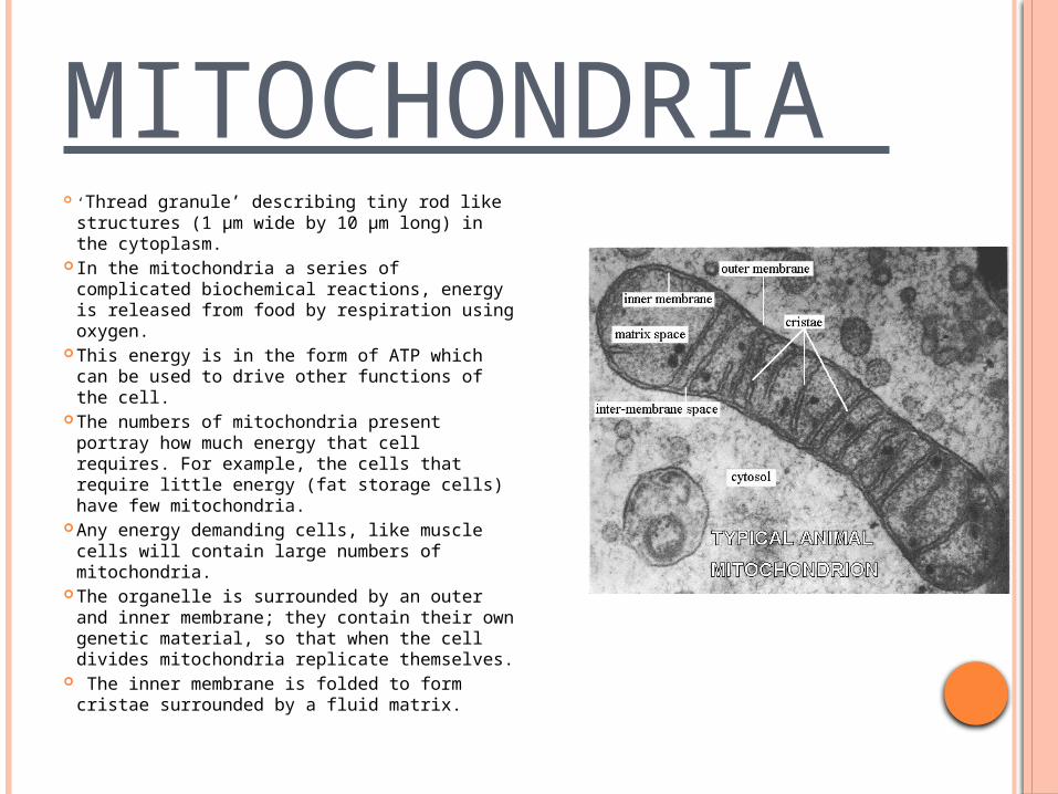
MITOCHONDRIA ‘Thread granule’ describing tiny rod like
structures (1 μm wide by 10 μm long) in the cytoplasm.
In the mitochondria a series of complicated biochemical reactions, energy is released from food by respiration using oxygen.
This energy is in the form of ATP which can be used to drive other functions of the cell.
The numbers of mitochondria present portray how much energy that cell requires. For example, the cells that require little energy (fat storage cells) have few mitochondria.
Any energy demanding cells, like muscle cells will contain large numbers of mitochondria.
The organelle is surrounded by an outer and inner membrane; they contain their own genetic material, so that when the cell divides mitochondria replicate themselves.
The inner membrane is folded to form cristae surrounded by a fluid matrix.

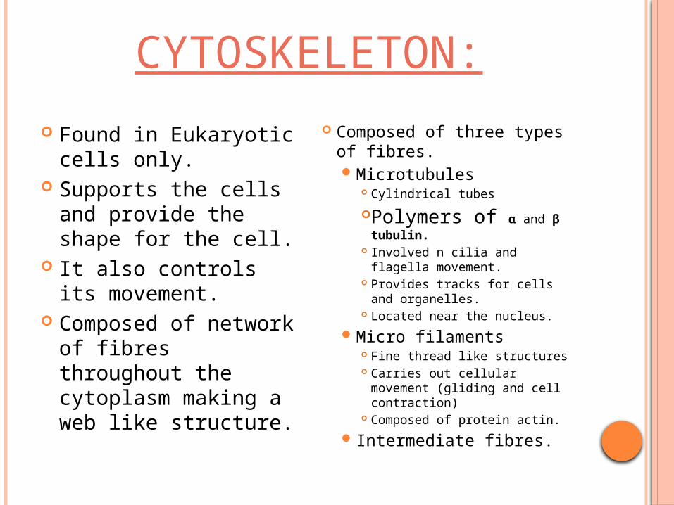
CYTOSKELETON:
Found in Eukaryotic cells only.
Supports the cells and provide the shape for the cell.
It also controls its movement.
Composed of network of fibres throughout the cytoplasm making a web like structure.
Composed of three types of fibres. Microtubules
Cylindrical tubes
Polymers of α and β tubulin.
Involved n cilia and flagella movement.
Provides tracks for cells and organelles.
Located near the nucleus.
Micro filaments Fine thread like structures Carries out cellular movement
(gliding and cell contraction) Composed of protein actin.
Intermediate fibres.