
Project Status The full amount of the requested funds ... · Project Status The full amount of the...
22
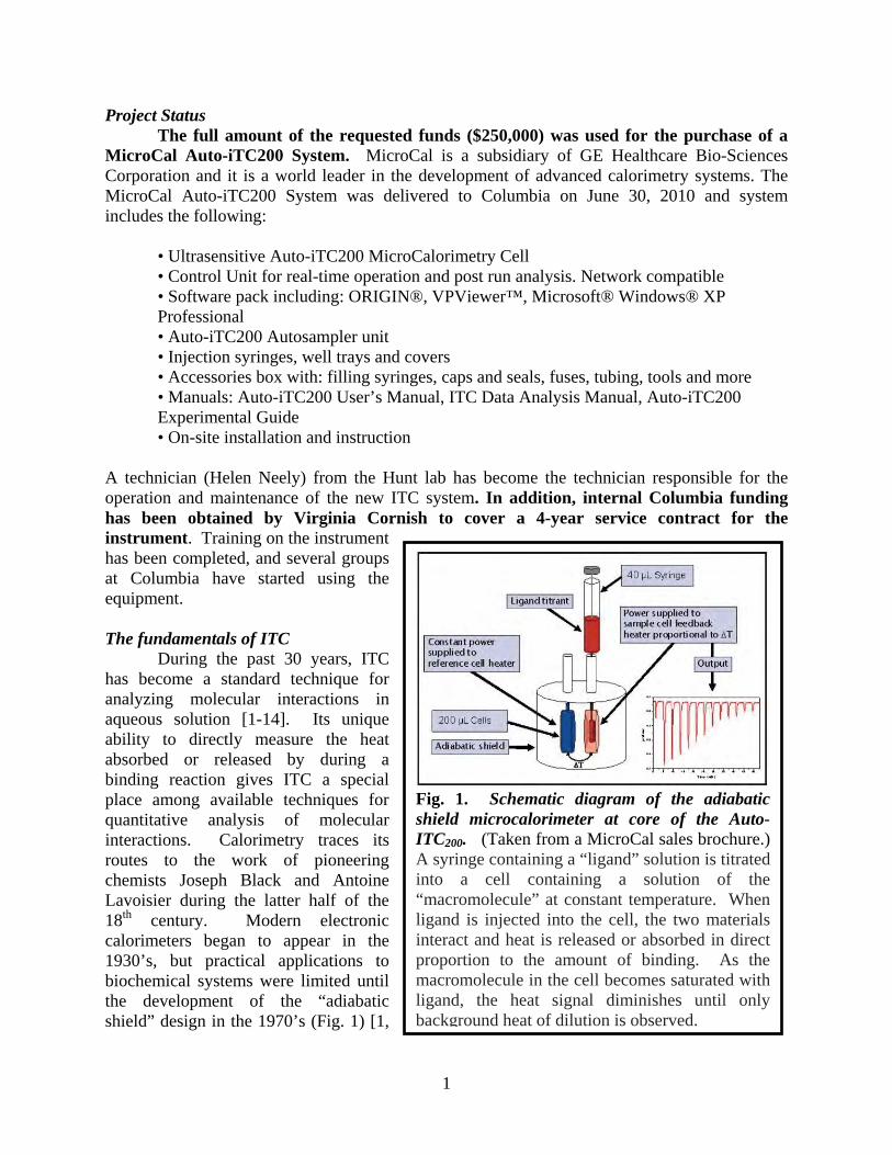
1 Project Status The full amount of the requested funds ($250,000) was used for the purchase of a MicroCal Auto-iTC200 System. MicroCal is a subsidiary of GE Healthcare Bio-Sciences Corporation and it is a world leader in the development of advanced calorimetry systems. The MicroCal Auto-iTC200 System was delivered to Columbia on June 30, 2010 and system includes the following: • Ultrasensitive Auto-iTC200 MicroCalorimetry Cell • Control Unit for real-time operation and post run analysis. Network compatible • Software pack including: ORIGIN®, VPViewer™, Microsoft® Windows® XP Professional • Auto-iTC200 Autosampler unit • Injection syringes, well trays and covers • Accessories box with: filling syringes, caps and seals, fuses, tubing, tools and more • Manuals: Auto-iTC200 User’s Manual, ITC Data Analysis Manual, Auto-iTC200 Experimental Guide • On-site installation and instruction A technician (Helen Neely) from the Hunt lab has become the technician responsible for the operation and maintenance of the new ITC system. In addition, internal Columbia funding has been obtained by Virginia Cornish to cover a 4-year service contract for the instrument. Training on the instrument has been completed, and several groups at Columbia have started using the equipment. The fundamentals of ITC During the past 30 years, ITC has become a standard technique for analyzing molecular interactions in aqueous solution [1-14]. Its unique ability to directly measure the heat absorbed or released by during a binding reaction gives ITC a special place among available techniques for quantitative analysis of molecular interactions. Calorimetry traces its routes to the work of pioneering chemists Joseph Black and Antoine Lavoisier during the latter half of the 18 th century. Modern electronic calorimeters began to appear in the 1930’s, but practical applications to biochemical systems were limited until the development of the “adiabatic shield” design in the 1970’s (Fig. 1) [1, Fig. 1. Schematic diagram of the adiabatic shield microcalorimeter at core of the Auto- ITC 200 . (Taken from a MicroCal sales brochure.) A syringe containing a “ligand” solution is titrated into a cell containing a solution of the “macromolecule” at constant temperature. When ligand is injected into the cell, the two materials interact and heat is released or absorbed in direct proportion to the amount of binding. As the macromolecule in the cell becomes saturated with ligand, the heat signal diminishes until only background heat of dilution is observed.
Transcript of Project Status The full amount of the requested funds ... · Project Status The full amount of the...
1
Project Status The full amount of the requested funds ($250,000) was used for the purchase of a
MicroCal Auto-iTC200 System. MicroCal is a subsidiary of GE Healthcare Bio-Sciences Corporation and it is a world leader in the development of advanced calorimetry systems. The MicroCal Auto-iTC200 System was delivered to Columbia on June 30, 2010 and system includes the following:
• Ultrasensitive Auto-iTC200 MicroCalorimetry Cell • Control Unit for real-time operation and post run analysis. Network compatible • Software pack including: ORIGIN®, VPViewer™, Microsoft® Windows® XP Professional • Auto-iTC200 Autosampler unit • Injection syringes, well trays and covers • Accessories box with: filling syringes, caps and seals, fuses, tubing, tools and more • Manuals: Auto-iTC200 User’s Manual, ITC Data Analysis Manual, Auto-iTC200 Experimental Guide • On-site installation and instruction
A technician (Helen Neely) from the Hunt lab has become the technician responsible for the operation and maintenance of the new ITC system. In addition, internal Columbia funding has been obtained by Virginia Cornish to cover a 4-year service contract for the instrument. Training on the instrument has been completed, and several groups at Columbia have started using the equipment. The fundamentals of ITC
During the past 30 years, ITC has become a standard technique for analyzing molecular interactions in aqueous solution [1-14]. Its unique ability to directly measure the heat absorbed or released by during a binding reaction gives ITC a special place among available techniques for quantitative analysis of molecular interactions. Calorimetry traces its routes to the work of pioneering chemists Joseph Black and Antoine Lavoisier during the latter half of the 18th century. Modern electronic calorimeters began to appear in the 1930’s, but practical applications to biochemical systems were limited until the development of the “adiabatic shield” design in the 1970’s (Fig. 1) [1,
Fig. 1. Schematic diagram of the adiabatic shield microcalorimeter at core of the Auto-ITC200. (Taken from a MicroCal sales brochure.) A syringe containing a “ligand” solution is titrated into a cell containing a solution of the “macromolecule” at constant temperature. When ligand is injected into the cell, the two materials interact and heat is released or absorbed in direct proportion to the amount of binding. As the macromolecule in the cell becomes saturated with ligand, the heat signal diminishes until only background heat of dilution is observed.

Report Documentation Page Form ApprovedOMB No. 0704-0188
Public reporting burden for the collection of information is estimated to average 1 hour per response, including the time for reviewing instructions, searching existing data sources, gathering andmaintaining the data needed, and completing and reviewing the collection of information. Send comments regarding this burden estimate or any other aspect of this collection of information,including suggestions for reducing this burden, to Washington Headquarters Services, Directorate for Information Operations and Reports, 1215 Jefferson Davis Highway, Suite 1204, ArlingtonVA 22202-4302. Respondents should be aware that notwithstanding any other provision of law, no person shall be subject to a penalty for failing to comply with a collection of information if itdoes not display a currently valid OMB control number.
1. REPORT DATE 12 MAY 2011 2. REPORT TYPE
3. DATES COVERED 15-07-2010 to 14-07-2011
4. TITLE AND SUBTITLE (DURIP-10) Isothermal Titration Calorimeter For ResearchAnd Education
5a. CONTRACT NUMBER
5b. GRANT NUMBER
5c. PROGRAM ELEMENT NUMBER
6. AUTHOR(S) 5d. PROJECT NUMBER
5e. TASK NUMBER
5f. WORK UNIT NUMBER
7. PERFORMING ORGANIZATION NAME(S) AND ADDRESS(ES) The Trustees of Columbia University in the City of NewYork,Research Administration 2960 Broadway 211 LowLibrary,New York,NY,10027-
8. PERFORMING ORGANIZATION REPORT NUMBER
9. SPONSORING/MONITORING AGENCY NAME(S) AND ADDRESS(ES) 10. SPONSOR/MONITOR’S ACRONYM(S) AFRL-OSR-VA-TR-2012-0015
11. SPONSOR/MONITOR’S REPORT NUMBER(S)
12. DISTRIBUTION/AVAILABILITY STATEMENT Approved for public release; distribution unlimited
13. SUPPLEMENTARY NOTES
14. ABSTRACT We have purchased a state-of-the-art MicroCal Auto-iTC200 Isothermal Titration Calorimetry (ITC)System. ITC is the gold standard for measuring biomolecular interactions. ITC simultaneously determinesall binding parameters (n, K, ΔH and ΔS) in a single experiment which is information thatcannot be obtained from any other single method. The system will dramatically enhance the activities inseveral interdisciplinary projects at Columbia University that have been funded by, or will be submittedfor funding to, the Department of Defense.
15. SUBJECT TERMS
16. SECURITY CLASSIFICATION OF: 17. LIMITATIONOF ABSTRACT
Same asReport (SAR)
18.NUMBEROF PAGES
22
19a. NAME OF RESPONSIBLE PERSON
a. REPORT unclassified
b. ABSTRACT unclassified
c. THIS PAGE unclassified
Standard Form 298 (Rev. 8-98) Prescribed by ANSI Std Z39-18

2
15]. This design involves placing carefully matched sample and reference cells in a sealed thermal bath at a fixed temperature. Thermistors embedded in the cells maintain both of them at an identical fixed temperature somewhat above that of the bath. The energy required to maintain the temperature differential between these cells and the bath is provided by electricity running through the thermistors, which can be measured with extremely high precision. An isothermal titration calorimeter is configured to sequentially deliver precisely metered small aliquots of a titrant solution into the sample cell while the contents of the reference cell remain unchanged. Release or absorption of heat upon mixing of the titrant with the solution in the sample cell causes a reduction or an increase, respectively, in the amount of heat energy that needs to be delivered by the thermistors on that cell compared to those on the reference cell while keeping them locked at exactly the same temperature. This difference in the electric energy delivered to the two cells matches the enthalpy change (i.e., the heat absorption/release) in the course of any chemical or physical reactions occurring when the titrant solution is mixed with the solution in the sample cell. Today’s isothermal titration microcalorimeters combine this ingenious design with sophisticated solid-state electronics to achieve ultrasensitive measurement of the enthalpy changes occurring when solutions of different chemical composition are mixed together.
ITC is routinely used to rigorously characterize the thermodynamics of many different kinds of chemical and physical processes occurring in liquids [2, 4, 6, 7, 9-11, 14, 16-20]. It is immediately applicable to studying the enthalpy of solvation as well as the thermodynamics of solute self-association. A classic application in this area involves characterizing the thermodynamics of detergent micelle formation [18-21]. In this case, the titrant contains a high concentration of the detergent above its critical micelle concentration (cmc), so most detergent molecules are present in the form of micelles. When this solution is diluted into a sample cell containing a low concentration of detergent below the cmc, the micelles spontaneously dissociate, and the enthalpy difference between the solvated and micelle-bound detergent molecules is measured. As the titration continues and the detergent concentration n the sample cell reaches the cmc, the micelles no longer dissociate upon dilution into the sample cell and much smaller enthalpy changes are observed related to micelle self-association processes. This generally dramatic change in the enthalpy of dilution enables ITC to be used to determine detergent cmc in a very simple way [18-20]. Moreover, such experiments have provided novel insight into detergent physical chemistry. Most detergents show intermediate enthalpies of dilution in a relatively broad window near the cmc. Such intermediates enthalpies are inconsistent with solvated detergent monomers being in equilibrium with a single homogeneous micellar species, and they indicate the formation of variant (probably smaller) “pre-micellar” detergent aggregates near the critical micelle concentration. The Hunt lab (Co-PI) has published ITC experiments of this kind and used them to demonstrate that a conformational change in SecA, an ATP-driven polypeptide pump, is driven by pre-micellar detergent aggregates rather than fully formed micelles [21].
The most common biochemical application of ITC involves measuring the enthalpy (DH) of a binding reaction [1, 2, 7, 9, 14]. If saturation is achieved in the course of the titration experiment, mathematical modeling of the data yields estimates of the dissociation constant (Kd) for the interaction as well as the binding stoichiometry (n). Using standard thermodynamic relationships, the equilibrium constant gives the Gibbs Free Energy of the binding reaction:
DG = RT•ln(Kd) In this equation, R is the ideal gas (1.98 cal/mole-degree), and T is the absolute
temperature. Knowing DH from direct measurement and DG from curve-fitting allows
3
calculation of the entropy (DS) of the reaction from the definition of the Gibbs Free Energy: DS = DG – DH
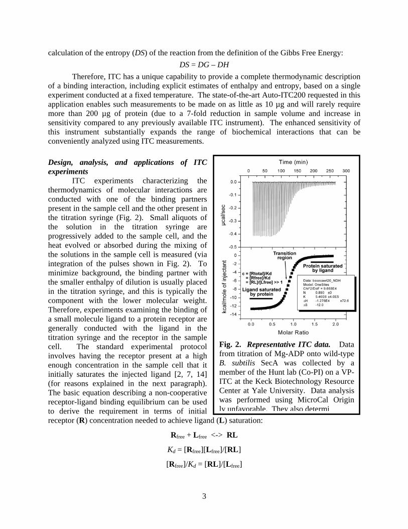
Therefore, ITC has a unique capability to provide a complete thermodynamic description of a binding interaction, including explicit estimates of enthalpy and entropy, based on a single experiment conducted at a fixed temperature. The state-of-the-art Auto-ITC200 requested in this application enables such measurements to be made on as little as 10 µg and will rarely require more than 200 µg of protein (due to a 7-fold reduction in sample volume and increase in sensitivity compared to any previously available ITC instrument). The enhanced sensitivity of this instrument substantially expands the range of biochemical interactions that can be conveniently analyzed using ITC measurements. Design, analysis, and applications of ITC experiments
ITC experiments characterizing the thermodynamics of molecular interactions are conducted with one of the binding partners present in the sample cell and the other present in the titration syringe (Fig. 2). Small aliquots of the solution in the titration syringe are progressively added to the sample cell, and the heat evolved or absorbed during the mixing of the solutions in the sample cell is measured (via integration of the pulses shown in Fig. 2). To minimize background, the binding partner with the smaller enthalpy of dilution is usually placed in the titration syringe, and this is typically the component with the lower molecular weight. Therefore, experiments examining the binding of a small molecule ligand to a protein receptor are generally conducted with the ligand in the titration syringe and the receptor in the sample cell. The standard experimental protocol involves having the receptor present at a high enough concentration in the sample cell that it initially saturates the injected ligand [2, 7, 14] (for reasons explained in the next paragraph). The basic equation describing a non-cooperative receptor-ligand binding equilibrium can be used to derive the requirement in terms of initial receptor (R) concentration needed to achieve ligand (L) saturation:
Rfree + Lfree <-> RL
Kd = [Rfree][Lfree]/[RL]
[Rfree]/Kd = [RL]/[Lfree]
Fig. 2. Representative ITC data. Data from titration of Mg-ADP onto wild-type B. subtilis SecA was collected by a member of the Hunt lab (Co-PI) on a VP-ITC at the Keck Biotechnology Resource Center at Yale University. Data analysis was performed using MicroCal Origin ly unfavorable. They also determi

4
[RL]/[Lfree] >> 1 => [Rfree]/Kd >> 1
The parameter c is used to represent the ratio of total receptor concentration ([Rtotal]) to Kd, which will equal the ratio of free receptor concentration ([Rfree]) to Kd at the start of the titration when minimal amounts of the receptor are bound by ligand:
c = [Rtotal]/Kd = [Rfree]/Kd = [RL]/[Lfree] >> 1
This simple derivation shows that the value of this parameter c determines the degree of ligand binding by the receptor at the outset of a titration experiment. Achieving the desired saturation of the injected ligand by the receptor requires c >> 1.
The reason for designing the experiment so that the injected ligand is initially saturated with receptor is that this regime provides a straightforward and robust measurement of DH, the enthalpy of ligand binding (Fig. 2) [2, 7, 14]. Under these conditions where all of the ligand is immediately bound to the receptor, DH is given directly by the amount of heat released or absorbed divided by the total amount of ligand introduced into the sample cell in the injection. Accurate estimation of this parameter anchors the fit of the remainder of the binding data because it enables the heat released in all subsequent objections to be used to determine the number of ligand molecules bound during that injection. Later in the titration, as the receptor becomes progressively saturated with ligand, the free receptor concentration declines, and less of the newly introduced ligand is bound during each injection. The exact shape of the function in this transition region depends on the dissociation constant, the binding stoichiometry, and the enthalpy of the reaction. If the last parameter is constrained (i.e., by the data from the initial phase of the titration), robust estimation of both of the other parameters is generally possible from the data in the transition. However, coupled variations in the values of the three parameters prevent accurate estimation of all of them exclusively based on data in the transition region [2, 7, 14]. Therefore, parameter values cannot be estimated reliably if the titration occurs entirely in the transition region and lacks a phase where saturation of the injected ligand by the receptor provides an independent estimate of DH. (It is possible to get reasonable estimates of the dissociation constant and enthalpy if binding stoichiometry is fixed and the protein concentration is accurately known, but there is no way to way to assess the reliability of such estimates, meaning that gross errors are possible if the binding stoichiometry misestimated or the protein precipitates during the experiment.)
A straightforward extension of the mathematical methods used for ITC data analysis enables quantitative modeling and complete thermodynamic characterization of some more complex reactions mechanisms involving sequential binding events [2, 14, 22]. However, very tight (i.e., sub-nanomolar) ligand binding can prevent accurate measurement of affinity in ITC experiments even for simple non-cooperative binding reactions [9]. This problem arises because saturation of the receptor with ligand occurs so abruptly that the transition region becomes a step-function. While accurate measurement of binding enthalpy and estimation of binding stoichiometry are still possible in this situation, determination of binding affinity depends on modeling the shape of the heat release function in the transition region, which cannot be done robustly if the transition region is too small. However, elegant competitive displacement protocols have been developed to enable reliable estimation of binding affinity even of ligands with picomolar affinity [9]. A similar competitive displacement approach can also be used to characterize binding interactions involving low enthalpy changes (although in this case the competing ligand must have appreciable binding enthalpy). Combining all of these sophisticated methods, ITC is applicable to rigorous thermodynamic characterization of an immense number

5
of biochemical binding reactions. Importance of ITC for research activities at Columbia
A vast number of research groups at Columbia University are interested in investigating protein-protein interactions, protein-small molecule interactions, and protein-material interactions. The PI and the 3 co-PIs on this proposal have ongoing research projects in their various groups with application range from drug development, to diagnostics development, to bioenergy applications, to materials development, to fundamental biomedical investigations. All current ITC usage involves travel to other institutions, typically to the Keck Biotechnology Resource Center at Yale University. Due to the inconvenience of this arrangement, ITC has only been used when no viable alternative assays can be found that can be performed locally. Much more extensive use of ITC is likely now that an ITC instrument is located on the Columbia campus.
In all of these cases, the MicroCal Auto-ITC200 instrument that we have acquired will serve a crucial role in the advancement of these projects which are relevant to the Department of Defense. The MicroCal Auto-iTC200 is the gold standard for measuring biomolecular interactions. ITC simultaneously determines all binding parameters (n, K, ∆H and ∆S) in a single experiment which is information that cannot be obtained from any other single method. The acquisition of this system will greatly enhance the ability of researchers in three Departments at Columbia University to monitor critical biomolecular interactions. The specific applications of the ITC to some of these projects in the labs of the PI and the 3 co-PIs is outlined below: Research projects in the Banta lab (PI), Department of Chemical Engineering
Professor Scott Banta’s group focuses on the application of protein engineering and metabolic engineering tools and techniques to study and solve important problems in bioengineering and medicine. One of the primary themes of the research group is the design, engineering, and application of peptides with unique structures and functional responses to environmental conditions [23-25]. There are several ongoing projects funded by the Department of Defense within the Banta group that will dramatically benefit from access to the new ITC system.
AFOSR MURI Grant: Fundamentals and Bioengineering of Enzymatic Fuel Cells
The ability to extract energy from the surrounding environment is a central hallmark of all life forms, and new technologies that can mimic this process will undoubtedly be of great use to the military, including the Air Force. Enzyme-based biofuel cells represent a significant advancement towards this goal, as the natural selectivity, efficiency and activity of enzymes can be exploited to extract electrical energy from both simple and complex carbohydrates. If engineered carefully, these devices will be compact
Fig. 3 A schematic diagram of a compartment-less enzymatic biofuel cell. Biofuels such as glucose are oxidized on the anode using enzymes and after passing through an external load, the electrons are used to reduce dioxygen to water on the cathode side of the biofuel cell.

6
and robust power sources that exhibit a high energy density and broad fuel source flexibility. The PI on this DURIP proposal is a co-PI and Thrust Leader on a funded Multi
University Research Initiative grant from AFOSR (FA-9550-06-1-0264, Program Officer Dr. Walt Kozumbo). The other Thrust Leaders on the grant are Plamen Atanassov, University of New Mexico, Shelley Minteer, Saint Louis University, and Scott Calabrese-Barton, Michigan State University. The overall goals of the MURI project are the elucidation of valuable details for the design, construction, and operation of biofuel cells that use ambient carbohydrate fuels. Robust mathematical models of these systems will be created that will allow for the engineering of novel devices that will aim to meet new and unmet energy demands. This project will advance our understanding of the mechanistic underpinnings of biofuel cell performance, which will enable the production of optimal devices. The PI is involved in several aspects of the MURI grant, and the MicroCal Auto-iTC200 will significantly augment two of the specific goals of the project. Directed Evolution of Dehydrogenase Enzymes to Use Alternative Cofactors and Substrates
Fuel cells offer significant advantages over other power sources in that they have the potential to convert chemical energy directly to electrical energy with minimal environmental impact. A standard fuel cell consists of an anode and a cathode, such that electrons are extracted from the fuel (such as hydrogen) using a catalyst on the anodic side, and after passing through an external load, they are combined with oxygen using a catalyst on the cathodic side, resulting in the production of water (Fig. 3). The most specific and active catalysts known are natural enzymes, but these biological molecules do not have the stability of metal catalysts. Enzymes have already been naturally evolved to promote the transfer of electrons between substrates with an exquisite level of specificity, and therefore it has been possible to demonstrate the use of enzymes to make biofuel cells [26]. But, enzymes have not been evolved to operate in biofuel cells, and thus they will need to be further engineered in order to perform optimally in this artificial environment. The overall goal of the MURI team is to produce fundamental insight and new tools and techniques for the development of next-generation biofuel cells that will provide compact and
Fig. 4 ITC used to investigate the interactions of the E. coli ketopantoate reductase with NADP+ and the pantoate substrate. Similar experiments will be performed in the Banta lab to characterize the interactions between biomimetic cofactors and mutant dehydrogenases for the development of advanced biofuel cell anodes.

7
distributed power from ambient fuel sources. The goal of the PI’s Thrust Area is the protein engineering of new enzymes for biofuel cell anodes and cathodes.
There has been a good amount of interest in the development of anodes capable of the multi-step oxidation of biofuels. The Minteer group at Saint Louis University (SLU) has immobilized multiple native enzymes together to create metabolic pathways on electrodes in order to create anodes capable of multi-step oxidations that mimic natural metabolism [27, 28]. These systems generally use dehydrogenase enzymes, and the nicotinamide cofactors (NAD(P)(H)) are thought to limit these systems due to their low stability, low redox potentials, and slow diffusion in a the polymeric film used to immobilize the enzymes on the bioelectrode.
Therefore, we are using protein engineering to engineer thermostable dehydrogenase enzymes that are able to use alternative biomimetic cofactors such as nicotinamide mononucleotide and N-benzyl-1,4-dihydronicotinamide [29]. The use of these alternative cofactors in the development of new anodes could have a tremendous impact on the power and efficiency of biofuel cells, but the dehydrogenase enzymes did not evolve to use these molecules. We are developing a novel directed evolution system to engineer the enzymes to use these new biomimetic cofactors. There are many well-established methods for examining the interactions of enzymes with cofactors including titration of fluorescence that can be used when energy transfer takes place between tryptophan side chains in the cofactor binding pocket and the cofactor molecules. But, when non-natural cofactors are used, these analyses are not possible which makes the study of the interactions of mutant enzymes with the biomimetic cofactors difficult. ITC has previously been used by many groups to study the interactions of dehydrogenases with nicotinamide cofactors [30] (Fig. 4), and we will use this method as well to better characterize our new enzymes as we develop improved biofuel cell anodes. Engineering of self-assembling protein-based hydrogels for electrodes and sensors
Another Thrust Area in the MURI grant from the AFOSR is aimed at improving the architectures of mediated biofuel cell electrodes. Generally, electrocatalytic enzymes are combined with a synthetic polymer that contains osmium-based redox centers. High current densities have been achieved using this method, but it suffers from complex synthesis, poor reproducibility, and a difficulty in producing homogenous dispersions of enzyme and mediator.
The PI of this DURIP proposal is developing self-assembling protein-based hydrogels that can be used for the construction of biofuel cell electrodes [31-33]. Peptides have been created by David Tirrell’s group at Caltech that are composed of soluble regions flanked by alpha-helical regions. Upon changes in pH these helical regions can form bundles that cross-link the monomers which results in the formation of hydrogels.[34-36] This monomer is the starting point for the development of self-assembling, enzymatically active protein hydrogels for use in novel electrode architectures.
Under the MURI-funded biofuel project we have developed enzymatically active protein hydrogels by fusing the gel forming helices of the protein to a bacterially expressed laccase [37] for use at the biofuel cell cathode [31, 38]. Currently, we are developing novel gel forming electrode architectures to include alcohol dehydrogenase enzymes [33]. Gel formation requires relatively high concentrations of gel forming monomeric fusion proteins and the interactions between the helical domains appended to the fusion proteins that mediate gel formation are difficult to study. The new ITC system will allow us to better understand the thermodynamics of gel formation and dissolution. This will be critical as these hydrogels

8
are implemented in actual biofuel cell electrodes as additional cross-linking groups will likely be needed to develop robust architectures. ERDC-CERL Agreement: In Silico Biomimetic Sensor for Rapid Testing of Water Resources The United States Army needs to be prepared to deploy and operate anywhere on Earth. This level of flexibility and readiness requires continual training, advanced installation operations, and the ultimate in personal protection. Currently, there is no method to conduct rapid, portable field analysis for toxins in water for the war fighter or at Army installations. Current methods for field testing are slow and require laboratory analysis. This process costs, on average, $2000 per sample and can take up to two weeks. Rapid toxicity sensors would not only eliminate soldier sickness and death from consumption of contaminated drinking water, but would also reduce or avoid the need for costly and time consuming laboratory analysis. The PI on this DURIP proposal, is a contributing researcher on a funded Construction Engineering Research Laboratory of the Engineering Research and Development Center (CERL-ERDC) of the Army Corps of Engineers (Agreement number W9132T-07-2-0008). The PI of the cooperative team is Dr. Donald M. Cropek, U.S. Army Corps of Engineers, Construction Engineering Research Laboratory, University of Illinois. The other team members involved in this agreement are Dr. Paul Bohn, University of Notre Dame, Dr. Charles Henry, Colorado State University, Dr. Ali Khademhosseini, Harvard University, Dr. Robert Langer, Massachusetts Institute of Technology, and Dr. Michael Shuler, Cornell University. Other participating members include Dr. William van der Schalie, U.S. Army Center for Environmental Health Research, Ft. Detrick, MD, Mr. Hany Zaghloul, U.S. Army Corps of Engineers, Construction Engineering Research Laboratory, and Dr. William Severinghaus, U.S. Army Corps of Engineers, Construction Engineering Research Laboratory.
The overall goal of the ERDC-CERL project is to create a “human-on-a-chip” sensor which reproduces human physiology on a microfluidic device and allows for the detection of human cell and tissue responses to toxins in water (Fig. 5). The microfluidic device guides the sample water, potentially containing harmful toxins, through a series of channels that allow for chemical interaction between toxins and human cells or model tissues. Signaling mechanisms at different steps dependent upon the chemistry will provide feedback on the toxic response to the analytes. The signals can be combined to inform the user with a simple binary response; an alarm if toxic, no alarm if not. The PI is involved in several aspects of this grant and the ITC system will significantly augment this project, as described below.
Directed evolution of peptides for the capture and detection of cytokines in a microfluidic device The cellular inflammatory response often involves the release of cytokines that are involved in inter-cell communication. For example, tissue necrosis factor (TNF)α is produced in
Fig. 5. A “human on a chip” sensor will be used to assess toxicity in field water samples. Cell culture modules will be exposed to toxins and stress responses in the cells will be monitored using various biosensors.

9
a variety of cell types and is involved in the inflammatory toxicology response [39]. The electrochemical detection of cytokines is challenging, as the catalytic activity of the targets may not be readily measurable. Therefore, binding motifs are used, and impedance spectroscopy can be used to detect the presence of increased mass on electrode surface following the binding event [40] Recently, phage display libraries have been used to isolate peptides that can specifically bind Botulinum neurotoxin serotype A [41] and BLyS, which is a TNF-α family member and a regulator of B-cells [42]. These and other results suggest that polypeptides can be engineered to bind most protein targets, and these new peptide aptamers will be well-suited for incorporation into impedance-based bioelectrodes. Therefore, we will use Directed Evolution to create peptide sequences that bind to cytokines of interest that will be secreted from cells in the microfluidic device during the toxicological response. Once identified, these peptides purified and the biomolecular interactions will be characterized away from the phage particles in order to further engineer them for surface attachment and incorporation into biosensors. The ITC system will be ideal for measuring and characterizing the biomolecular interactions between the evolved peptides the protein-based targets. These peptides will be used to make novel impedance-based biosensors for use in the biomimetic toxicology sensors. DTRA Grant: Beta-Roll Peptide Structures for Allosterically Controlled Biomolecular Recognition and Decontamination Biomolecular recognition using antibodies is increasingly being exploited for a variety of important defense and military applications. But, these proteins have evolved only to bind their target molecules. Biological recognition could be better exploited in many applications if the binding events could be controlled by external stimuli. The PI on this proposal is the co-PI on a recently funded grant to support the DTRA initiative Topic E: Biomolecular Recognition and Catalysis, aimed at advancing the state of the science and body of knowledge for Protection and Countermeasures. The goal of this project is to utilize two different natural phenomena and combine them to create a novel controllable detection element. Specifically, we will use a unique peptide scaffold from a metalloprotease enzyme that exhibits natural stimulus-responsive conformational behavior. The β-roll domain from the serralysin protease is allosterically regulated by calcium [43]. Using Directed Evolution techniques, we will engineer the β-roll scaffold to incorporate a molecular recognition functionality that will result in the creation of new allosterically regulated binding proteins that can specifically and conditionally recognize new targets. These new peptides will then be immobilized on a surface for controlled binding and release. As an example, the peptides will be immobilized on photocatalyst nanoparticles, which would enable the light-
Fig. 6. Evolved peptides for biosensor development. Conditional specific biological recognition peptides could be used in a biosensor to capture, purify and concentrate a target molecule in a sample (A). An effector molecule can then be used to release the target molecule from the peptide for subsequent detection (B).

10
Fig. 7 (a) Cartoon image of polymer decorated nanoparticles. (b) TEM of 14 nm silica particles functionalized with a polystyrene brush with molecular mass and grafting density as shown in the figure. These particles are placed in a polystyrene matrix with molecular mass of 142 kg/mol. The samples, with 5 mass% silica, were annealed for 5 days at 1500 C.
triggered destruction of bound target molecules. The fundamental characterization of the new allosteric peptides will be used to better understand this new class of evolved allosteric binding modules, which will be used in the creation of new binding scaffolds to additional targets. Initially, we are engineering the peptides to associate with the protective antigen protein from Bacillus anthracis [44]. This protein is required for disease progression, as after proteolytic cleavage, it heptamerizes and forms a channel in the endosome which enables the transport of other lethal proteins into the cell. The specific inhibition of binding of the lethal proteins to the protective antigen heptamer by an engineered peptide has been shown to inhibit toxin action in cell culture and in an animal model [45]. The triggered binding and release of this protein could facilitate diagnostic efforts (Fig. 6), and the binding and specific photocatalytic destruction of the protein may prevent the spread of this disease. Other pathogen-associated proteins will also be explored as targets including the ricin toxin B, cholera toxin B, and botulinum neurotoxin type E. The binding affinities of the novel β-roll scaffolds that will be engineered in this project will need to be characterized with and without calcium, and the new MicroCal Auto-iTC200 will be ideal for this application. Research projects in the Kumar lab (Co-PI), Department of Chemical Engineering Professor Kumar’s group focuses on complex fluids, concentrating on novel uses of synthetic polymer-based materials, including polymer nanocomposites and polymers used in fuel cells. His group is also interested in biopolymer systems, working on self-assembly of proteins and developing better methods for separating biological macromolecules. His group also has interests in systems with ultra-slow dynamics, particularly glass and reversibly gelling materials such as gelatin.
Professor Kumar has submitted a proposal to the AFOSR Polymer Matrix Composites Division (Program Officer Dr. Charles Y-C Lee) to create novel nanocomposites. It is now well accepted that the addition of nanoparticles to an organic polymer can result in materials (termed “nanocomposites”) that can have significantly improved properties. While the promise of these materials is high, central to achieving such enhancement in properties is the ability to control the spatial distribution of nanoparticles, i.e., being able to go from uniform nanoparticle dispersion to states of partial aggregation in a controlled, predictable and reproducible manner.[46-54] While there has been some recent success in uniformly dispersing nanoparticles into polymer matrices,[46, 47] the broader goal of controlling the spatial distribution of nanoparticles has remained elusive. Professor Kumar’s work is motivated by their recent finding,[55] published in Nature Materials (April

11
’09), that they have begun to develop the ability to achieve such control over nanoparticle spatial dispersion. Specifically, they have found that spherical silica nanoparticles isotropically grafted with polymers self-assemble into a range of superstructures when they are dispersed into the corresponding homopolymer (Figs. 7 and 8).[46, 52, 53, 56-58] The results shown in Fig. 7, which are based on TEM, are corroborated by independent x-ray and neutron scattering. Theory and simulation (Fig. 8)[59, 60] show that this assembly is driven by the microphase separation between the immiscible, inorganic particle core and the (organic) grafted chains- a process analogous to the self-assembly of block copolymers (or amphiphiles).[61] The central premise we shall test in this proposal is the fact that hydrophilic nanoparticles uniformly grafted with hydrophobic chains can self-assemble into variety of nanostructures in a marginal solvent (i.e., good for one constituent but poor for the other). We propose to start with the nanoparticles added to a solvent which is “good” for both the core and the corona. We then titrate in a second solvent which is poor for one and then we shall watch for signatures of self-assembly. Previous work on surfactant molecules (including lipids which are central to the form and function of cell membranes) have shown that the self-assembly in these cases is accompanied by small, but definitive, peaks in the heat capacity, Cp. Thus, the availability of a calorimeter which can measure exquisitely small changes in heat flow, is particularly germane and appropriate. Success in this endeavor will thus provide us with an important underpinning knowledge for this class of materials and thus greatly facilitate the a-priori design of polymer nanocomposites (including those relevant to biomimetic application such as creating bone mimics). In this context, we emphasize that this work is greatly facilitated by the close on-going collaboration with researchers at the Air Force Materials Laboratories where informal interactions have been established over the past several years between the PI’s and Air Force researchers (Rich Vaia, Hillmar Koerner). Research projects in the Cornish lab (Co-PI), Department of Chemistry
Professor Virginia Cornish's group develops new methods for the directed evolution of biomolecules with new functions [62-64]. Her laboratory applies these methods to the directed evolution of enzymes with altered substrate specificities and proteins with de novo binding properties. These projects all require characterization of the evolved protein variants and their interactions with their small molecule substrates or ligands and would benefit from regular access to an ITC instrument [65]. Her laboratory is also engineering the translational machinery for the incorporation of amino acid analogs via mis-acylated tRNAs [66]. Finally, her laboratory is developing chemical dimerizers [67] and exploiting these ligand-receptor interactions for live cell imaging [68]. The techniques being used in Professor Cornish’s laboratory can be readily applied to problems of relevance to the Department of Defense including bioenergy and biosensor applications.
Fig. 8 Experimental “morphology diagram” of polymer tethered particles mixed with matrix polymers. The lines which separate different regions are merely guides to the eye.

12
One important goal of her group is to engineer yeast to carry out directed evolution in vivo (Fig. 9) in order to provide a breakthrough in the size of libraries that can be searched experimentally. This technology has the potential to enable (1) directed evolution of enzymes with more dramatic changes in function and (2) the rapid, economical high-throughput directed evolution of proteins for routine use.
Directed evolution holds promise for creating designer enzymes for use as reagents in biomedical research, medical diagnostics, therapeutics, and bioprocess catalysts. Directed evolution has been used successfully to select RNA molecules with de novo binding and catalytic properties and most recently proteins with de novo binding properties from large libraries (>108) of protein variants. Researchers generally have not been able to bring the power of directed evolution to bear on enzyme catalysis, however, because there are no suitable high-throughput selections for large numbers of protein variants for most enzyme-catalyzed reactions. Directed evolution has been used for less demanding applications, for example, increasing the activity of an existing enzyme, from small libraries (103-104) of protein variants by automation of traditional enzyme assays. For the handful of reactions for which there are high-throughput selections for function, it has proven possible to change enzyme substrate specificity using large libraries (about 108). (The de novo directed evolution of enzyme activity, significantly, has only been reported once outside the catalytic antibody field.)
To address this bottleneck in the directed evolution field, the Cornish laboratory developed “chemical complementation”—a high-throughput in vivo selection for enzyme catalysis of bond formation and cleavage reactions—around β-lactamase enzymes. Like natural complementation assays, chemical complementation is carried out with living cells, such that the cell can serve as a physical compartment linking each protein variant not only to its mutatable and amplifiable DNA code but also to product formation and fitness advantage. In chemical complementation, however, enzyme catalysis is linked to cell survival via covalent coupling of two small molecule three-hybrid ligands that reconstitute a transcriptional activator such that the chemistry can be readily varied (Fig. 10). Enzyme catalysis of formation of a bond between the two small molecule ligands is detected as activation of an essential reporter gene; bond cleavage is detected as repression of a toxic reporter gene. The assay can be applied to libraries of 108 or more variants because it can be run as a growth selection where only cells containing functional enzyme survive. The assay can be readily extended to new chemistry simply by synthesizing small molecule heterodimers with different chemical linkers as the enzyme substrates.
The specific aims of the Cornish lab include the directed evolution of glycosynthase enzymes and aldolase enzymes for use as reagents in chemical synthesis. In addition, the lab has begun to further adapt its technology for the directed evolution of de novo binding proteins.
Fig. 9. General steps of directed enzyme evolution. The gene encoding the protein of interest is mutated to generate a library of mutant genes. Expression of the mutant genes provides the library of mutant proteins. The proteins are screened or selected based on a desired property, and the variants with modified activity are sequenced or used for further rounds of mutagenesis and selection.

13
With numerous directed evolution experiments underway, they need to routinely characterize evolved protein variants both at intermediate steps in the selection process and at the end once we have obtained our desired protein variants. The characterization depends both on the desired property and the course of the directed evolution experiment. In the past, the Cornish characterized evolved protein variants to determine their structure, their substrate/ligand specificity, and their catalytic activity. However, the group is currently hampered in its ability to characterize evolved protein variants because of the lack of analytical instrumentation for protein chemistry at Columbia University. We either have to utilize sub-optimal techniques or carry out the characterization at neighboring institutions. The new high-throughput ITC would facilitate directed evolution experiments in the Cornish lab by enabling routine evaluation of the binding affinities of intermediates along the directed evolution trajectory, which would be used to guide subsequent rounds of mutagenesis and selection. Research projects in the Hunt lab (Co-PI), Department of Biological Sciences
Professor John Hunt’s group uses a combination of biophysical, biochemical, and bioinformatics methods to understand the intricacies of protein function and evolution. Several biophysically oriented projects focus on detailed thermodynamic and kinetic dissection of complex protein-mediated biomolecular processes, including transmembrane transport [69-74], oxidative DNA repair [75], and protein crystallization [76]. Other projects focus on the use of structural and thermodynamic methods to elucidate the biochemical function of previously uncharacterized protein families. His lab has characterized structural and physiological interactions involved in cyclic-diguanylate-mediated signaling in bacteria [77] and has also discovered a novel translational regulatory system in E. coli, which involves a protein that is homologous to three human proteins of uncharacterized function. His group has used ITC intermittently during the past six years and with increasing frequency during the past two years. The techniques being used in Professor Hunt’s laboratory can be readily applied to problems of relevance to the Department of Defense including developing detection strategies and therapeutics aimed at combating emerging biothreat pathogens. Structural genomics
Fig. 10. Chemical complementation. A reaction-independent complementation assay for enzyme catalysis based on the yeast three-hybrid assay. A heterodimeric small molecule bridges a DNA-binding domain (DBD)–receptor fusion protein and an activation domain (AD)–receptor fusion protein, activating transcription of a downstream reporter gene in vivo. Enzyme catalysis of either cleavage or formation of the bond between the two small molecules can be detected as a change in transcription of the reporter gene. The assay can be applied to new chemical reactions simply by synthesizing small molecules with different substrates as linkers and adding an enzyme as a fourth component to the system.

14
Professor Hunt is co-director of the crystallography group of the Northeast Structural Genomics Consortium (NESG -- www.nesg.org) [78], one of the large-scale structure determination centers sponsored by the Protein Structure Initiative of the NIH. The NESG crystallography group has determined over 350 protein crystal structures during the past 8 years. The Auto-ITC200 will be invaluable for screening candidate ligands for proteins undergoing high-throughput crystallization screening by the structural genomics group.
One specific example of such an application is given by the Hunt lab’s work on PilZ domain structure and function. PilZ domains were hypothesized [79] to act as receptors for cyclic diguanylate (cdG), a eubacterial second messenger molecule controlling the transition between motile and sessile physiological states. The Hunt lab used ITC [77] to quantify the affinity of cdG for a model PilZ domain from V. cholerae called VCA0042/PlzD. These measurements showed that cdG binding to VCA0042 is entropically unfavorable. They also determined the crystal structure of VCA0042 with bound cdG, which was the first crystal structure of any PilZ domain bound to the cdG ligand [77]. The structure suggested that the entropy loss was likely to come from a disorder-to-order transition in a short but highly conserved “cdG-switch” peptide that wraps itself around the ligand. In a follow-up study currently nearing completion, the Hunt lab has determined the crystal structure of a different PilZ domain from P. putida bound to cdG. This protein, called PP4397, undergoes a dramatic change in dimer structure upon cdG binding (Fig. 9A-B), suggesting that the binding should show positive cooperativity. The crystal structure of this protein furthermore shows a cdG dimer bound to each protein protomer, in contrast to VCA0042, which only showed a cdG monomer bound to each protein protomer. Recent ITC experiments show an unusual bi-modal profile for the binding of cdG to PP4397 (Fig. 9C), consistent with a complex binding mechanism. (While the curve-fit in Fig. 9C superficially looks reasonable, careful inspection shows significant pathologies. Efforts are currently underway to apply more sophisticated methods to model these binding data [22].) These data illustrate the power of ITC for
Fig. 9. Crystallographic and ITC studies of cdG binding by PP4397, a PilZ-domain-containing protein from P. putida. (A) Crystal structure of the apo dimer. (B) Crystal structure of the cdG-bound dimer. (C) ITC data collected by Hunt lab member Dorothy Kim on a VP-ITC (MicroCal) in the Chemistry Department at Rutgers University. Data analysis was performed using MicroCal Origin with a 4-site sequential binding model. (Standard ITC data analysis modules do not model this equilibrium properly, so more advanced work will be needed to achieve proper curve-fitting of these data.)

15
rigorous thermodynamic analysis of solution binding equilibria. They also demonstrate how ITC can be a powerful adjunct to structural studies of molecular interactions. Structure as a tool for the elucidation of novel biochemical systems
In an NSF-sponsored project, the Hunt lab has elucidated the biochemical and physiological function of a novel translational regulatory system controlled by soluble proteins in the ABC Transporter superfamily. Structure-based biochemistry experiments have been used to demonstrate that an E. coli protein called YjjK functionally interacts with ribosomes. Structural data was used to design a trans-dominant ATPase-deficient mutant version of YjjK that inhibits translation both in vivo and in vitro. High-throughput genomics methods (phenotypic profiling combined with mRNA microarray profiling and global differential protein profiling) have been used to show that YjjK controls the translation of specific messages in E. coli in order to modulate intermediary metabolism. Three additional paralogues of YjjK are present in E. coli, and three total are present in all eukaryotic cells from yeast to man. Future work will focus on determining whether the paralogues also function as message-specific translation factors. ITC experiments will be a convenient way to characterize protein-ligand, protein-protein, and protein-RNA interactions [16, 17] involving YjjK and the paralogous proteins. Research Related Education at Columbia University
Columbia University is one of the world's most important centers of research and at the same time a distinctive and distinguished learning environment for undergraduates and graduate students in many scholarly and professional fields. The University recognizes the importance of its location in New York City and seeks to link its research and teaching to the vast resources of a great metropolis. It seeks to attract a diverse and international faculty and student body, to support research and teaching on global issues, and to create academic relationships with many countries and regions. It expects all areas of the university to advance knowledge and learning at the highest level and to convey the products of its efforts to the world.
The Fu Foundation School of Engineering and Applied Science at Columbia University aims to prepare engineering leaders who will solve the problems of the new century, while fostering scientific inquiry but never losing sight of its human implications. The School’s programs are designed to produce well-educated engineers who can put their knowledge to work for society. Through a synergy of teaching and research, the school seeks to educate a distinguished cadre of leaders in engineering and applied science who will thrive in an atmosphere of recently emerging technologies. The above missions are accomplished in the Departments of Chemical Engineering, Chemistry and Biology through curricula that are grounded in core fundamentals of their respective disciplines, and augmented with elective courses that offer exposure to cutting-edge applications, technologies and experimental techniques. The new MicroCal Auto-iTC200 system will be incorporated into several courses at Columbia University including the Graduate-level Protein Engineering course in Chemical Engineering Department (CHEN4800) and the Protein Thermodynamics course in the Biology Department (Biology G6002). In addition, the MicroCal Auto-iTC200 system will be utilized by dozens of undergraduate, graduate, and postdoctoral scholars in the PIs’ laboratory. ITC incorporated into Graduate Electives

16
The Protein Engineering graduate elective was offered for the first time by Professor Banta, PI on this proposal, in the fall semester of 2005. In the fall of 2006, the course was offered through the Columbia Video Network, which allowed students from around the world to enroll and participate in the class. The course will next be offered in the spring of 2010. The course is aimed at senior undergraduates and graduate students and it introduces fundamental tools and techniques currently used to engineer protein molecules. The course covers the methods used to analyze the impact of protein mutations on different protein functions, with a specific emphasis on enzymatic catalysis. Throughout the semester specific case studies are used to reinforce the concepts covered in the course, as well as to demonstrate the wide impact of protein engineering research. A significant portion of the course is devoted to the study of the methods to characterize native and recombinant proteins. Various methods are introduced and discussed including SDS-PAGE, analytical chromatography, circular dichroism spectroscopy, advanced kinetics studies, and Surface Plasmon Resonance (SPR). The theory and applications of ITC will be integrated in the course, and a practical session devoted to ITC methods will be performed in future semesters. Professor Hunt teaches a graduate course entitled Protein Thermodynamics (Biology G6002), which is also open to advanced undergraduates with appropriate academic background. This course has an average enrollment of ~20 students per year, ~60% of whom are PhD candidates in Biology (who are required to take it). The curriculum for this course will be expanded to include lectures on the theory and practice of ITC. In addition to covering the underlying theory, which represents a natural extension of the material currently covered in the course, the new lecture material will include instruction on quality-control procedures to ensure proper execution and interpretation of ITC experiments. One recitation section will include hands-on processing of both high-quality and low-quality (unreliable) ITC data. Finally, a series of online tutorials will be created to guide all instrument users in performing effective ITC experiments. These tutorials will cover the essential background material on equilibrium binding theory, as adapted from Professor Hunt’s lectures, in addition to practical instruction material. These developments will broaden the impact of the course materials in addition to enhancing the educational scope of the course itself. Postdoctoral, Graduate, and Undergraduate Research Experiences Columbia is a world-renowned institute of research excellence. Training opportunities at the postdoctoral, graduate, and undergraduate levels are available in all of the labs of the 4 PIs on this proposal. These students will be exposed to the MicroCal Auto-iTC200 ITC System for the study of biomolecular interactions. Projects will be directly related to the grants and projects described above that are of direct interest to the Department of Defense, as well as, other ongoing and future research projects that the new equipment will enable. These activities will help to train the next generation of experimentally adept and quantitatively oriented investigators.
Professor Kumar is also part of the MRSEC center at Columbia University and an NSEC center at RPI. As part of this effort, he has developed collaborations with Prof. Ramakrishnan and Dr. Paravatsu (FAMU, a HBCU). It is the goal of this effort to not only do collaborative research, but also to encourage REU students from FAMU to attend Columbia as part of a REU program with the goal of both recruitment, as well as retaining them in the sciences. In the same vein, 8 REU students (including 2 women, and 1 under-represented minority) spent their summer (’07 and ‘08) in Kumar’s lab. It is our goal to continue this effort, especially leveraging the fact

17
that students are attracted to a high quality research environment in New York City. In this context the availability of high end characterization equipment such as a CD, x-ray scattering, transmission electron microscopy (including tomography) and the ITC system, is a crucial piece in “building” the excitement that students feel towards their research. We believe that this central to the recruitment and retention efforts that we plan. References 1. Privalov, P.L., Scanning microcalorimeters for studying macromolecules. Pure & Appi.
Chem., 1980. 52: p. 479—497. 2. Bains, G. and E. Freire, Calorimetric determination of cooperative interactions in high
affinity binding processes. Anal Biochem, 1991. 192(1): p. 203-6. 3. Jelesarov, I. and H.R. Bosshard, Isothermal titration calorimetry and differential
scanning calorimetry as complementary tools to investigate the energetics of biomolecular recognition. J Mol Recognit, 1999. 12(1): p. 3-18.
4. Ward, W.H. and G.A. Holdgate, Isothermal titration calorimetry in drug discovery. Prog Med Chem, 2001. 38: p. 309-76.
5. Cliff, M.J., A. Gutierrez, and J.E. Ladbury, A survey of the year 2003 literature on applications of isothermal titration calorimetry. J Mol Recognit, 2004. 17(6): p. 513-23.
6. Velazquez-Campoy, A., S.A. Leavitt, and E. Freire, Characterization of protein-protein interactions by isothermal titration calorimetry. Methods Mol Biol, 2004. 261: p. 35-54.
7. Velazquez-Campoy, A., et al., Isothermal titration calorimetry. Curr Protoc Cell Biol, 2004. Chapter 17: p. Unit 17 8.
8. Velazquez Campoy, A. and E. Freire, ITC in the post-genomic era...? Priceless. Biophys Chem, 2005. 115(2-3): p. 115-24.
9. Velazquez-Campoy, A. and E. Freire, Isothermal titration calorimetry to determine association constants for high-affinity ligands. Nat Protoc, 2006. 1(1): p. 186-91.
10. Bjelic, S. and I. Jelesarov, A survey of the year 2007 literature on applications of isothermal titration calorimetry. J Mol Recognit, 2008. 21(5): p. 289-312.
11. de Azevedo, W.F., Jr. and R. Dias, Experimental approaches to evaluate the thermodynamics of protein-drug interactions. Curr Drug Targets, 2008. 9(12): p. 1071-6.
12. Freyer, M.W. and E.A. Lewis, Isothermal titration calorimetry: experimental design, data analysis, and probing macromolecule/ligand binding and kinetic interactions. Methods Cell Biol, 2008. 84: p. 79-113.
13. Liang, Y., Applications of isothermal titration calorimetry in protein science. Acta Biochim Biophys Sin (Shanghai), 2008. 40(7): p. 565-76.
14. Freire, E., A. Schon, and A. Velazquez-Campoy, Isothermal titration calorimetry: general formalism using binding polynomials. Methods Enzymol, 2009. 455: p. 127-55.
15. Chowdhry, B.Z., A.E. Beezer, and E.J. Greenhow, Analysis of drugs by microcalorimetry Isothermal power-conduction calorimetry and thermometric titrimetry. Talanta, 1983. 30(4): p. 209-43.
16. Feig, A.L., Applications of isothermal titration calorimetry in RNA biochemistry and biophysics. Biopolymers, 2007. 87(5-6): p. 293-301.
17. Salim, N.N. and A.L. Feig, Isothermal titration calorimetry of RNA. Methods, 2008.
18
18. Paula, S., et al., Thermodynamics of Micelle Formation as a Function of Temperature: A High Sensitivity Titration Calorimetry Study. J. Phys. Chem., 1995. 99(30): p. 11742-11751.
19. Heerklotz, H. and J. Seelig, Titration calorimetry of surfactant-membrane partitioning and membrane solubilization. Biochim Biophys Acta, 2000. 1508(1-2): p. 69-85.
20. Heerklotz, H. and R.M. Epand, The enthalpy of acyl chain packing and the apparent water-accessible apolar surface area of phospholipids. Biophys. J., 2001. 80(1): p. 271-279.
21. Benach, J., et al., Phospholipid-induced monomerization and signal-peptide induced oligomerization SecA. J. Biol. Chem., 2003. 278: p. 3628-3638.
22. Houtman, J.C., et al., Studying multisite binary and ternary protein interactions by global analysis of isothermal titration calorimetry data in SEDPHAT: application to adaptor protein complexes in cell signaling. Protein Sci, 2007. 16(1): p. 30-42.
23. Chockalingam, K., M. Blenner, and S. Banta, Design and application of stimulus-responsive peptide systems. Protein Eng Des Sel, 2007. 20(4): p. 155-61.
24. Banta, S., et al., Engineering protein and peptide building blocks for nanotechnology. J Nanosci Nanotechnol, 2007. 7(2): p. 387-401.
25. Blenner, M.A. and S. Banta, Characterization of the 4D5Flu single-chain antibody with a stimulus-responsive elastin-like peptide linker: a potential reporter of peptide linker conformation. Protein Sci, 2008. 17(3): p. 527-36.
26. Mano, N., et al., A miniature biofuel cell operating at 0.78 V. Chem Commun (Camb), 2003(4): p. 518-9.
27. Sokic-Lazic, D. and S.D. Minteer, Citric acid cycle biomimic on a carbon electrode. Biosens. Bioelectron., 2008. In Press.
28. Sokic-Lazic, D. and S.D. Minteer, Polymer-based Kreb's cycle biomimic. PMSE Preprints, 2008. 98: p. 847-848.
29. Ryan, J.D., R.H. Fish, and D.S. Clark, Engineering cytochrome P450 enzymes for improved activity towards biomimetic 1,4-NADH cofactors. Chembiochem, 2008. 9(16): p. 2579-82.
30. Ciulli, A., et al., Crystal structure of Escherichia coli ketopantoate reductase in a ternary complex with NADP+ and pantoate bound: substrate recognition, conformational change, and cooperativity. J Biol Chem, 2007. 282(11): p. 8487-97.
31. Wheeldon, I.R., S.C. Barton, and S. Banta, Bioactive proteinaceous hydrogels from designed bifunctional building blocks. Biomacromolecules, 2007. 8(10): p. 2990-4.
32. Wheeldon, I.R., et al., Bioelectrocatalytic hydrogels from electron-conducting metallopolypeptides coassembled with bifunctional enzymatic building blocks. Proc Natl Acad Sci U S A, 2008. 105(40): p. 15275-80.
33. Wheeldon, I.R., E. Campbell, and S. Banta, A chimeric fusion protein engineered with disparate functionalities-enzymatic activity and self-assembly. J Mol Biol, 2009. 392(1): p. 129-42.
34. Petka, W.A., et al., Reversible hydrogels from self-assembling artificial proteins. Science, 1998. 281(5375): p. 389-92.
35. Shen, W., et al., Assembly of an artificial protein hydrogel through leucine zipper aggregation and disulfide bond formation. Macromolecules, 2005. 38(9): p. 3909-3916.
36. Shen, W., et al., Tuning the erosion rate of artificial protein hydrogels through control of network topology. Nat Mater, 2006. 5(2): p. 153-8.

19
37. Machczynski, M.C., et al., Characterization of SLAC: a small laccase from Streptomyces coelicolor with unprecedented activity. Protein Sci, 2004. 13(9): p. 2388-97.
38. Gallaway, J., et al., Oxygen-reducing enzyme cathodes produced from SLAC, a small laccase from Streptomyces coelicolor. Biosens Bioelectron, 2008. 23(8): p. 1229-35.
39. Luster, M.I., et al., Tumor necrosis factor alpha and toxicology. Crit Rev Toxicol, 1999. 29(5): p. 491-511.
40. Bart, M., et al., On the response of a label-free interferon-gamma immunosensor utilizing electrochemical impedance spectroscopy. Biosens Bioelectron, 2005. 21(1): p. 49-59.
41. Ma, H., et al., A cyclic peptide-polymer probe for the detection of Clostridium botulinum neurotoxin serotype A. Toxicon, 2006. 47(8): p. 901-8.
42. Fleming, T.J., et al., Discovery of high-affinity peptide binders to BLyS by phage display. J Mol Recognit, 2005. 18(1): p. 94-102.
43. Yoder, M.D. and F. Jurnak, Protein motifs. 3. The parallel beta helix and other coiled folds. Faseb J, 1995. 9(5): p. 335-42.
44. Dixon, T.C., et al., Anthrax. N Engl J Med, 1999. 341(11): p. 815-26. 45. Mourez, M., et al., Designing a polyvalent inhibitor of anthrax toxin. Nat Biotechnol,
2001. 19(10): p. 958-61. 46. Mackay, M.E., et al., General strategies for nanoparticle dispersion. Science, 2006.
311(5768): p. 1740-1743. 47. Tuteja, A., P.M. Duxbury, and M.E. Mackay, Multifunctional nanocomposites with
reduced viscosity. Macromolecules, 2007. 40(26): p. 9427-9434. 48. Kashiwagi, T., et al., Nanoparticle networks reduce the flammability of polymer
nanocomposites. Nature Materials, 2005. 4(12): p. 928-933. 49. Kashiwagi, T., et al., Relationship between dispersion metric and properties of
PMMA/SWNT nanocomposites. Polymer, 2007. 48(16): p. 4855-4866. 50. Winey, K.I., T. Kashiwagi, and M.F. Mu, Improving electrical conductivity and thermal
properties of polymers by the addition of carbon nanotubes as fillers. Mrs Bulletin, 2007. 32(4): p. 348-353.
51. Winey, K.I. and R.A. Vaia, Polymer nanocomposites. Mrs Bulletin, 2007. 32(4): p. 314-319.
52. Krishnamoorti, R., Strategies for dispersing nanoparticles in polymers. Mrs Bulletin, 2007. 32(4): p. 341-347.
53. Krishnamoorti, R. and R.A. Vaia, Polymer nanocomposites. Journal of Polymer Science Part B-Polymer Physics, 2007. 45(24): p. 3252-3256.
54. Mitchell, C.A., et al., Dispersion of functionalized carbon nanotubes in polystyrene. Macromolecules, 2002. 35(23): p. 8825-8830.
55. Akcora, P., et al., Anisotropic particle self-assembly in polymer nanocomposites. Nature Materials, 2009. 8: p. 354-359.
56. Green, D.L. and J. Mewis, Connecting the wetting and rheological behaviors of poly (dimethylsiloxane)-grafted silica spheres in poly(dimethylsiloxane) melts. Langmuir, 2006. 22(23): p. 9546-9553.
57. Harton, S.E. and S.K. Kumar, Mean-field theoretical analysis of brush-coated nanoparticle dispersion in polymer matrices. Journal of Polymer Science Part B-Polymer Physics, 2008. 46(4): p. 351-358.
58. Bansal, A., et al., Quantitative equivalence between polymer nanocomposites and thin polymer films. Nature Materials, 2005. 4(9): p. 693-698.

20
59. Bedrov, D., G.D. Smith, and L.W. Li, Molecular dynamics simulation study of the role of evenly spaced poly(ethylene oxide) tethers on the aggregation of C-60 fullerenes in water. Langmuir, 2005. 21(12): p. 5251-5255.
60. Hooper, J.B., D. Bedrov, and G.D. Smith, Supramolecular self-organization in PEO-modified C-60 fullerene/water solutions: Influence of polymer Molecular weight and nanoparticle concentration. Langmuir, 2008. 24(9): p. 4550-4557.
61. Fredrickson, G.H. and F.S. Bates, Dynamics of block copolymers: Theory and experiment. Annual Review of Materials Science, 1996. 26: p. 501-550.
62. Baker, K., et al., Chemical complementation: a reaction-independent genetic assay for enzyme catalysis. Proc Natl Acad Sci U S A, 2002. 99(26): p. 16537-42.
63. Lin, H., H. Tao, and V.W. Cornish, Directed evolution of a glycosynthase via chemical complementation. J Am Chem Soc, 2004. 126(46): p. 15051-9.
64. Peralta-Yahya, P., et al., High-throughput selection for cellulase catalysts using chemical complementation. J Am Chem Soc, 2008. 130(51): p. 17446-52.
65. de Felipe, K.S., et al., Correlation between ligand-receptor affinity and the transcription readout in a yeast three-hybrid system. Biochemistry, 2004. 43(32): p. 10353-63.
66. Effraim, P., et al., Ribosomal Selection of Natural Amino Acids is Independent of Native tRNA Context. Nature Chemical Biology, Submitted.
67. Lin, H., et al., Dexamethasone-Methotraxate: An Efficient Chemical Inducer of Protein Dimerization In Vivo. J Am Chem Soc, 2000. 122: p. 4247-4248.
68. Miller, L.W., et al., In vivo protein labeling with trimethoprim conjugates: a flexible chemical tag. Nat Methods, 2005. 2(4): p. 255-7.
69. Hunt, J.F., et al., Nucleotide control of interdomain interactions in the conformational reaction cycle of SecA. Science, 2002. 297: p. 2018-2026.
70. Fak, J.J., et al., Nucleotide exchange from the high-affinity ATP-binding site in SecA is the rate-limiting step in the ATPase cycle of the soluble enzyme and occurs through a specialized conformational state. Biochemistry, 2004. 43(23): p. 7307-27.
71. Karpowich, N., et al., Crystal structures of the MJ1267 ATP binding cassette reveal an induced-fit effect at the ATPase active site of an ABC transporter. Structure, 2001. 9(7): p. 571-586.
72. Smith, P.C., et al., ATP binding to the motor domain from an ABC transporter drives formation of a nucleotide sandwich dimer. Mol. Cell, 2002. 10(1): p. 139-149.
73. Wang, C., et al., Dynamics of ATP-binding cassette contribute to allosteric control, nucleotide binding and energy transduction in ABC transporters. J Mol Biol, 2004. 342(2): p. 525-37.
74. Lewis, H.A., et al., Impact of the deltaF508 mutation in first nucleotide-binding domain of human cystic fibrosis transmembrane conductance regulator on domain folding and structure. J Biol Chem, 2005. 280(2): p. 1346-53.
75. Yu, B., et al., Crystal structures of catalytic complexes of the oxidative DNA/RNA repair enzyme AlkB. Nature, 2006. 439(7078): p. 879-84.
76. Price, W.N., 2nd, et al., Understanding the physical properties that control protein crystallization by analysis of large-scale experimental data. Nat Biotechnol, 2009. 27(1): p. 51-7.
77. Benach, J., et al., The structural basis of cyclic diguanylate signal transduction by PilZ domains. EMBO J, 2007. 26(24): p. 5153-66.

21
78. Acton, T.B., et al., Robotic Cloning and Protein Production Platform of the Northeast Structural Genomics Consortium. Methods in Enzymology. in press.
79. Galperin, M.Y. and E.V. Koonin, 'Conserved hypothetical' proteins: prioritization of targets for experimental study. Nucleic Acids Res, 2004. 32(18): p. 5452-63.