PROGRESSIVE INTERSTITIAL FIBROSISOFTHE IN...
5
DIFFUSE PROGRESSIVE INTERSTITIAL FIBROSIS OF THE LUNGS IN CHILDHOOD (HAMMAN-RICH SYNDROME) BY H. B. HILTON and JOHN RENDLE-SHORT From the Sheffield Children's Hospital and the Department of Child Health, University of Sheffield (RECEIVED FOR PUBLICATION JUNE 9, 1960) Hamman and Rich (1935) briefly described four cases of bilateral diffuse interstitial fibrosis of the lungs of unknown aetiology. In 1944, they described this condition in detail under the title 'Acute diffuse interstitial fibrosis of the lungs' (Hamman and Rich, 1944). Since then the condition has become known as the 'Hamman-Rich syndrome' but is now perhaps more correctly referred to as 'Diffuse progressive interstitial fibrosis of the lungs' (Beams and Harmos, 1949). Grant, Hillis and Davidson (1956) collected 36 cases from the literature and added three of their own. Rubin and Lubliner (1957) recognized 39 recorded cases and added 15 of their own. Since then sporadic reports of cases have appeared, and Scadding (1960) added 26 cases to the literature. The majority of reported cases have been in adults; in childhood the disease is rare, only six cases having been previously recorded. The first report of the condition occurring in a child was by Bradley (1956). He described the clinical and autopsy findings in a 9-year-old girl. Since that time the following cases have been repor- ted: Baar and Braid (1957) described the case of a 3-year-old boy; Feinerman and Harris (1957) of a 7-month-old girl and an 8-year-old boy; Diamond (1958) of a 4-year-old Negro boy who, in addition, had stenosis of the right pulmonary artery and complete occlusion of the right pulmonary vein, and Mann (1959) an 8-month-old girl. Wilson and Mikity (1960) have recently reported a syndrome resembling the Hamman-Rich syndrome which occurs in premature babies. They gave details of five infants, two of whom had pulmonary signs and symptoms suggestive of the Hamman- Rich syndrome. It is not certain, however, that these should be regarded as proved cases. Case 1: J.L. John was the second child of healthy parents. He was born after a normal pregnancy and labour (birth weight 7 lb. 4 oz.). He developed normally for the first three months of life and reached a weight of 11 lb. 10 oz. He then developed, over the course of a week, a slight fever, cough and 'stuffy' nose. On admission to the Sheffield Children's Hospital, under the care of Professor Illingworth, he was slightly cyanosed. His temperature was 1000 F., his heart rate 180 per minute and his respiratory rate 30 per minute. A grade 3/6 systolic murmur was audible over the pre- cordium, and both liver and spleen were palpable two fingers' breath below the costal margin. Investigations showed nothing abnormal apart from the chest radio- graph, which revealed bronchopneumonic changes throughout both lung fields. He was treated with oxygen, penicillin and digoxin and seemed to improve. His liver became less easily palpable and the systolic murmur disappeared. He was discharged home, but four days later was readmitted with marked cyanosis which disappeared when he was placed in an oxygen tent. The systolic murmur had returned and he was dyspnoeic, but had no fever. The liver was not so large, but the chest radiograph showed the same changes as previously. During the next three months the child's condition steadily deteriorated. He became a blue-grey colour when not receiving oxygen, lost weight and his haemo- globin level rose from 12- 1 g. % on his first admission to 14-4 g. % just before death. It was thought that he probably had a congenital heart lesion with cardiac failure. The electrocardiogram showed evidence of right ventricular diastolic overloading, but a cine-angio- cardiogram was normal. Repeated blood cultures were negative. He was treated with tetracycline on four occasions, penicillin twice, chloramphenicol once, and was also given digoxin, triamcinolone and nystatin for persistent oral thrush. No therapy except oxygen appeared to help him. All investigations remained negative, but the chest radiograph showed an increasing haziness of the whole lung field with an almost ground-glass appearance (Fig. 1). He died at the age of 61 months. Necropsy. The body was that of an extremely wasted male infant weighing 3,097g. Scattered petechial haemorrhages were present over the face, abdomen and ankles, and fading petechial haemorrhages were present in both groins. 102 copyright. on 23 June 2018 by guest. Protected by http://adc.bmj.com/ Arch Dis Child: first published as 10.1136/adc.36.185.102 on 1 February 1961. Downloaded from
Transcript of PROGRESSIVE INTERSTITIAL FIBROSISOFTHE IN...

DIFFUSE PROGRESSIVE INTERSTITIAL FIBROSIS OF THELUNGS IN CHILDHOOD (HAMMAN-RICH SYNDROME)
BY
H. B. HILTON and JOHN RENDLE-SHORTFrom the Sheffield Children's Hospital and the Department of Child Health, University of Sheffield
(RECEIVED FOR PUBLICATION JUNE 9, 1960)
Hamman and Rich (1935) briefly described fourcases of bilateral diffuse interstitial fibrosis of thelungs of unknown aetiology. In 1944, they describedthis condition in detail under the title 'Acute diffuseinterstitial fibrosis of the lungs' (Hamman andRich, 1944).
Since then the condition has become known asthe 'Hamman-Rich syndrome' but is now perhapsmore correctly referred to as 'Diffuse progressiveinterstitial fibrosis of the lungs' (Beams and Harmos,1949).
Grant, Hillis and Davidson (1956) collected 36cases from the literature and added three of theirown. Rubin and Lubliner (1957) recognized 39recorded cases and added 15 of their own. Sincethen sporadic reports of cases have appeared, andScadding (1960) added 26 cases to the literature.The majority of reported cases have been in adults;in childhood the disease is rare, only six cases havingbeen previously recorded.The first report of the condition occurring in a
child was by Bradley (1956). He described theclinical and autopsy findings in a 9-year-old girl.Since that time the following cases have been repor-ted: Baar and Braid (1957) described the case of a3-year-old boy; Feinerman and Harris (1957) of a7-month-old girl and an 8-year-old boy; Diamond(1958) of a 4-year-old Negro boy who, in addition,had stenosis of the right pulmonary artery andcomplete occlusion of the right pulmonary vein,and Mann (1959) an 8-month-old girl.Wilson and Mikity (1960) have recently reported
a syndrome resembling the Hamman-Rich syndromewhich occurs in premature babies. They gavedetails of five infants, two of whom had pulmonarysigns and symptoms suggestive of the Hamman-Rich syndrome. It is not certain, however, thatthese should be regarded as proved cases.
Case 1: J.L.John was the second child of healthy parents. He
was born after a normal pregnancy and labour
(birth weight 7 lb. 4 oz.). He developed normally forthe first three months of life and reached a weight of11 lb. 10 oz. He then developed, over the course ofa week, a slight fever, cough and 'stuffy' nose.On admission to the Sheffield Children's Hospital,
under the care of Professor Illingworth, he was slightlycyanosed. His temperature was 1000 F., his heart rate180 per minute and his respiratory rate 30 per minute.A grade 3/6 systolic murmur was audible over the pre-cordium, and both liver and spleen were palpable twofingers' breath below the costal margin. Investigationsshowed nothing abnormal apart from the chest radio-graph, which revealed bronchopneumonic changesthroughout both lung fields. He was treated with oxygen,penicillin and digoxin and seemed to improve. Hisliver became less easily palpable and the systolic murmurdisappeared.He was discharged home, but four days later was
readmitted with marked cyanosis which disappearedwhen he was placed in an oxygen tent. The systolicmurmur had returned and he was dyspnoeic, but hadno fever. The liver was not so large, but the chestradiograph showed the same changes as previously.During the next three months the child's condition
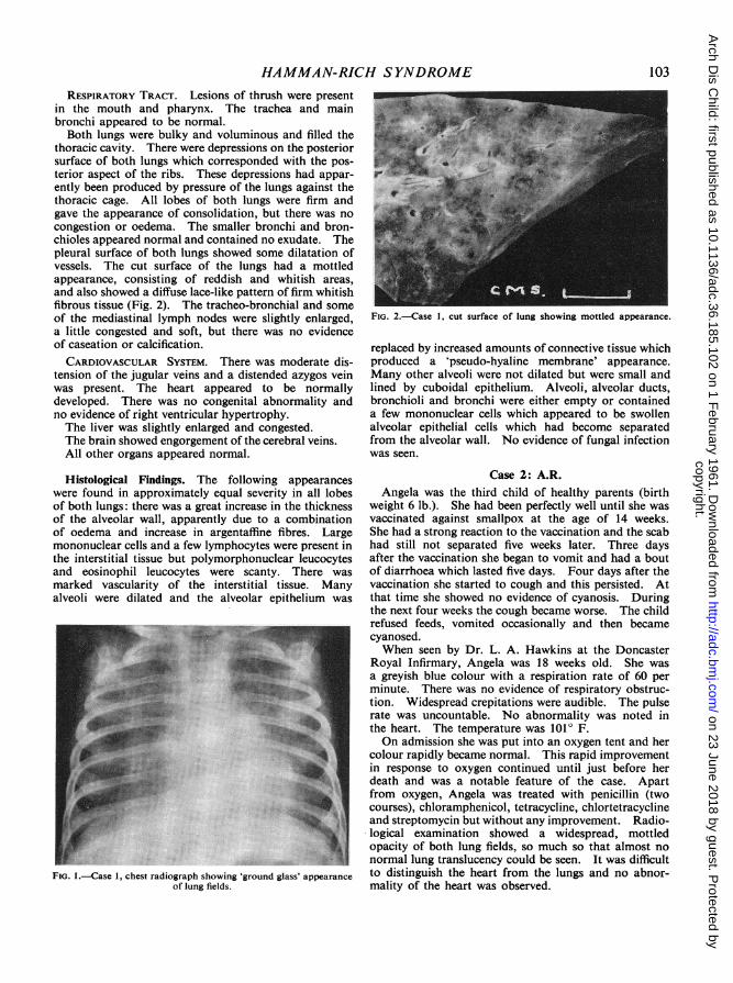
steadily deteriorated. He became a blue-grey colourwhen not receiving oxygen, lost weight and his haemo-globin level rose from 12- 1 g. % on his first admissionto 14-4 g. % just before death. It was thought that heprobably had a congenital heart lesion with cardiacfailure. The electrocardiogram showed evidence ofright ventricular diastolic overloading, but a cine-angio-cardiogram was normal. Repeated blood cultures werenegative. He was treated with tetracycline on fouroccasions, penicillin twice, chloramphenicol once, andwas also given digoxin, triamcinolone and nystatin forpersistent oral thrush. No therapy except oxygenappeared to help him. All investigations remainednegative, but the chest radiograph showed an increasinghaziness of the whole lung field with an almostground-glass appearance (Fig. 1). He died at the age of61 months.
Necropsy. The body was that of an extremely wastedmale infant weighing 3,097g. Scattered petechialhaemorrhages were present over the face, abdomen andankles, and fading petechial haemorrhages were presentin both groins.
102
copyright. on 23 June 2018 by guest. P
rotected byhttp://adc.bm
j.com/
Arch D
is Child: first published as 10.1136/adc.36.185.102 on 1 F
ebruary 1961. Dow
nloaded from
HAMMAN-RICH SYNDROMERESPIRATORY TRACT. Lesions of thrush were present
in the mouth and pharynx. The trachea and mainbronchi appeared to be normal.Both lungs were bulky and voluminous and filled the
thoracic cavity. There were depressions on the posteriorsurface of both lungs which corresponded with the pos-terior aspect of the ribs. These depressions had appar-ently been produced by pressure of the lungs against thethoracic cage. All lobes of both lungs were firm andgave the appearance of consolidation, but there was nocongestion or oedema. The smaller bronchi and bron-chioles appeared normal and contained no exudate. Thepleural surface of both lungs showed some dilatation ofvessels. The cut surface of the lungs had a mottledappearance, consisting of reddish and whitish areas,and also showed a diffuse lace-like pattern of firm whitishfibrous tissue (Fig. 2). The tracheo-bronchial and someof the mediastinal lymph nodes were slightly enlarged,a little congested and soft, but there was no evidenceof caseation or calcification.CARDIOVASCULAR SYSTEM. There was moderate dis-
tension of the jugular veins and a distended azygos veinwas present. The heart appeared to be normallydeveloped. There was no congenital abnormality andno evidence of right ventricular hypertrophy.The liver was slightly enlarged and congested.The brain showed engorgement of the cerebral veins.All other organs appeared normal.
Histological Findings. The following appearanceswere found in approximately equal severity in all lobesof both lungs: there was a great increase in the thicknessof the alveolar wall, apparently due to a combinationof oedema and increase in argentaffine fibres. Largemononuclear cells and a few lymphocytes were present inthe interstitial tissue but polymorphonuclear leucocytesand eosinophil leucocytes were scanty. There wasmarked vascularity of the interstitial tissue. Manyalveoli were dilated and the alveolar epithelium was
FIG. I-Case 1, chest radiograph showing 'ground glass' appearanceof lung fields.
FIG. 2.-Case 1, cut surtace o0f lung showing mottled appearance.
replaced by increased amounts of connective tissue whichproduced a 'pseudo-hyaline membrane' appearance.Many other alveoli were not dilated but were small andlined by cuboidal epithelium. Alveoli, alveolar ducts,bronchioli and bronchi were either empty or containeda few mononuclear cells which appeared to be swollenalveolar epithelial cells which had become separatedfrom the alveolar wall. No evidence of fungal infectionwas seen.
Case 2: A.R.Angela was the third child of healthy parents (birth
weight 6 lb.). She had been perfectly well until she wasvaccinated against smallpox at the age of 14 weeks.She had a strong reaction to the vaccination and the scabhad still not separated five weeks later. Three daysafter the vaccination she began to vomit and had a boutof diarrhoea which lasted five days. Four days after thevaccination she started to cough and this persisted. Atthat time she showed no evidence of cyanosis. Duringthe next four weeks the cough became worse. The childrefused feeds, vomited occasionally and then becamecyanosed.When seen by Dr. L. A. Hawkins at the Doncaster
Royal Infirmary, Angela was 18 weeks old. She wasa greyish blue colour with a respiration rate of 60 perminute. There was no evidence of respiratory obstruc-tion. Widespread crepitations were audible. The pulserate was uncountable. No abnormality was noted inthe heart. The temperature was 1010 F.On admission she was put into an oxygen tent and her
colour rapidly became normal. This rapid improvementin response to oxygen continued until just before herdeath and was a notable feature of the case. Apartfrom oxygen, Angela was treated with penicillin (twocourses), chloramphenicol, tetracycline, chlortetracyclineand streptomycin but without any improvement. Radio-logical examination showed a widespread, mottledopacity of both lung fields, so much so that almost nonormal lung translucency could be seen. It was difficultto distinguish the heart from the lungs and no abnor-mality of the heart was observed.
103
copyright. on 23 June 2018 by guest. P
rotected byhttp://adc.bm
j.com/
Arch D
is Child: first published as 10.1136/adc.36.185.102 on 1 F
ebruary 1961. Dow
nloaded from

ARCHIVES OF DISEASE IN CHILDHOOD
Angela's condition deteriorated and she died at theage of 7 months, 10 weeks after the onset of her illness.
Necropsy. The lungs were voluminous and on theposterior aspects, slight markings were present whichcorresponded to the posterior part of the ribs. Thelungs were firm and on cut section showed a fleshyappearance with a variegated pattern of pale and reddishareas. No definite consolidation was present and nofrank pus was seen.
Histological Findings. All lobes of both lungs showeda similar appearance. There was gross thickening of thealveolar walls which appeared to be due to a combina-tion of increased connective tissue, oedema and cellularinfiltrate of mononuclear cells and occasional eosinophilleucocytes. Dilated capillaries were present in the inter-stitial tissue. The majority of alveoli were lined bycuboidal epithelium; other alveoli were distended. Thesmaller bronchi, bronchioles and alveoli were eitherempty or contained swollen mononuclear cells whichappeared to have been derived from the alveolar epi-thelium. The heart showed no gross abnormality andthe other organs were normal.
Case 3: L.S.Linda was well until the age of 13 months when she
developed a respiratory tract infection with otitis mediaand a cough. The otitis improved after two weeks,but the cough became progressively worse and therespiratory rate became rapid. She was admitted to theSheffield Children's Hospital under the care of ProfessorIllingworth.On examination she was a pale, coughing baby with
a tinge of cyanosis. The temperature was 100° F., andthe respiratory rate about 80 per minute. On auscul-tation of her chest, crepitations were heard all over bothlung fields. A radiograph showed emphysematouslung fields with patchy consolidation and one possiblecavity in the right mid-zone. The pulmonary arterywas prominent.
Linda was put into an oxygen tent and treated withtetracycline. Her haemoglobin was 12-9 g. % and hertotal leucocyte count 21,000, 68% of which were neutro-phils. Gastric washings on admission showed no patho-genic organisms.There was a slight initial improvement, but after the
first two weeks she began to deteriorate. She becamevery cyanosed when out of oxygen, but was comparativelycheerful in the oxygen tent. The pulmonary crepitationswere always audible, although her cough became lesstroublesome. Serial radiographs showed increasingemphysema with areas of consolidation uniformly dis-tributed throughout both lung fields (Fig. 3). Noabscess cavities were seen. Her heart enlarged pro-gressively, especially the right ventricle, and the pul-monary artery became very prominent. Her haemo-globin rose, until just before death it was 16-7 g. %.Staphylococcus aureus coagulase positive was grown fromcough swabs on eight occasions in specimens taken ataproxpimately monthly intervals.
Linda was treated with courses of penicillin, chlor-tetracycline, oxytetracycline, sulphadimidine, chloram-phenicol and erythromycin. She was also given corti-sone for two months and, when she was in heart failure,digoxin. No treatment, apart from oxygen, made anydifference to her general condition or altered the per-sistent growth of Staphylococcus aureus in her sputum,even though in vitro studies showed that the organismsremained fully sensitive to chloramphenicol and ery-thromycin throughout.Linda developed progressive cor pulmonale as shown
by chest radiographs and electrocardiograms, and shedied in heart failure eight months after the initialappearance of her symptoms.
Necropsy. The body was that of a wasted femalechild, the sternum was prominent and the antero-posterior diameter of the chest was increased.
RESPIRATORY TRACT. The larynx, pharynx, tracheaand larger bronchi appeared normal.The lungs were slightly more bulky than normal and
the pleural surface showed irregularity due to the presenceof small sub-pleural cysts which varied in size from 1 mm.to 1 cm. in diameter. The pleural surface showedmottled areas of red congested tissue and white depressedareas due to fibrous tissue. On cut section the mottledappearance was seen to be present throughout all lobesof both lungs.CARDIOVASCULAR SYSTEM. The heart was increased
in size due to right ventricular hypertrophy. There wasno evidence of congenital heart disease. The pulmonaryartery appeared slightly increased in thickness. Therewas distension of the jugular veins.The liver was enlarged and congested. The thymus
showed marked involution. All other organs werenormal.
Histological Findings. Throughout all lobes of bothlungs, there was an increase in thickness of the alveolar
FIG. 3.-Case 3, chest radiograph showing areas of consolidation,enlargement of the heart and prominent pulmonary artery.
104
copyright. on 23 June 2018 by guest. P
rotected byhttp://adc.bm
j.com/
Arch D
is Child: first published as 10.1136/adc.36.185.102 on 1 F
ebruary 1961. Dow
nloaded from

HAMMAN-RICH SYNDROME
walls. This was due to increased amounts of connectivetissue and collections of cells which included macro-
phages, fibroblasts, plasma cells and some eosinophils.Many alveoli appeared small or normal in size and were
lined by cuboidal epithelium (Fig. 4). Other alveoliwere dilated and cysts were present which were linedin some areas by a thin layer of fibrous tissue, and inother areas by ciliated columnar epithelium.The heart showed slight hypertrophy of muscle fibres
in the right ventricle. There was no evidence of myo-
carditis.The pulmonary artery showed some sub-intimal
thickening. The thymus showed gross involution. Theliver showed congestion and fatty degeneration. Thespleen was congested but was otherwise normal.
DiscussionAn analysis of the six previously recorded child-
hood cases (excluding the five premature babies)in conjunction with our own three cases, enables a
composite picture of the syndrome to be given.The sex incidence is approximately equal (fourboys and five girls). The ages at onset were:
3 months (two cases), 4 months (two cases), 5 months(two cases), 13 months, 2 years 5 months and 6 years9 months. Five cases survived six months or less,while three cases survived between eight monthsand three and a half years. One 8-year-old boywas still alive at the time his case history was pub-lished one year after the onset of the disease.
Five cases began with an upper respiratory tractinfection. Two cases began with fever and res-
piratory tract symptoms. One case apparentlyfollowed whooping cough and one case followedvaccination against smallpox. The usual story was
FIG. 4. -Case 3, lung section showing broadening of interalveolarinterstitial tissue and cuboidal epithelium lining the alveoli.
(H. and E. x 295.)
of cough, dyspnoea and cyanosis persisting afteran infection had apparently abated. The cyanosiswas relieved by oxygen. Several authors remarkedon the remarkable absence of signs in the chest,but some of the children were seen months after thecommencement of the disease. On the other handa few cases had persistent crepitations throughoutthe illness.
Six out of seven cases died in heart failure. Inthe child who was still alive, the cardiac secondsound was reported as being very loud and split.In one case report the heart was not mentioned.
In our first child the presence of a systolic murmurwhich was shown to be presystolic on a phono-cardiogram, coupled with dilatation of the pul-monary artery on a radiograph, right ventricularoverloading on the electrocardiogram and clinicalevidence of right-sided heart failure, suggested thediagnosis of a congenital heart lesion. An angio-cardiogram, however, was normal. A similar casewas reported by Bradley (1956); the child had a loudsystolic and later diastolic murmur and cardiaccatheterization was performed. A general increaseof pressures in the right side of the heart was found(figures not given), but no other abnormality wasdiscovered. At autopsy no congenital heart lesionwas found.The radiological appearance of the nine cases
varied. Two cases showed patchy consolidationand emphysema, five cases showed diffuse infiltra-tion, and two cases showed a ground glass hazinessof the lung fields. The abnormal appearance wasoften more marked on one side than the other.
In our third case the diagnosis of staphylo-coccal pneumonia either with or without fibrocysticdisease of the pancreas was seriously considered.This was because of the radiological appearance ofpatchy consolidation and possible pneumatoceles,and also because of the constant finding of haemo-lytic staphylococci in the sputum.
Diffuse progressive interstitial fibrosis of the lungsis a clinical syndrome which should be consideredin any child with persistent dyspnoea, tachypnoea,cyanosis improved by oxygen, and heart failure.The most important differential diagnosis is con-genital heart disease.
Aetiology. Various possibilities have been sug-gested as aetiological factors in cases of 'idiopathic'diffuse interstitial pulmonary fibrosis. These in-clude a reaction to some form of chemical irritant,the result of a viral infection or infection withpleuropneumonia-like organisms or an incompletelyresolved pneumonia. A relationship with thecollagen disease has also been suggested, and cases
105
copyright. on 23 June 2018 by guest. P
rotected byhttp://adc.bm
j.com/
Arch D
is Child: first published as 10.1136/adc.36.185.102 on 1 F
ebruary 1961. Dow
nloaded from

106 ARCHIVES OF DISEASE IN CHILDHOOD
of rheumatoid arthritis with progressive pulmonaryfibrosis have been recorded in adults (Rubin andLubliner, 1957).The concept that auto immunity may be concerned
has received some support from Read (1958a and1958b) who prepared, in rabbits, an anti-rat lungserum which, when administered to rats, resulted ininterstitial fibrosis of the rat lung.
Pathology. The naked eye appearances of thelung in all our three cases showed a similar appear-ance. The lungs were voluminous and increasedin bulk. They were uniformly firm in consistenceand the cut surface showed a variegated pattern ofgreyish white and pale reddish areas. In two casesthere were prominent dilated blood vessels presenton the pleural surface. In one case, the presenceof small cysts was noted.
Histological Findings. The histopathological find-ings in our three cases fulfilled the criteria describedby Hamman and Rich. The main pathologicalfeature was broadening of the interstitial tissue ofinteralveolar septa. This pathological change in-volved all lobes of both lungs. The broadening ofinterstitial tissue was due to oedema, increasednumbers of argentaffine and collagenous fibres andcellular infiltration. The cellular infiltrate com-prised large mononuclear cells, lymphocytes, plasmacells and eosinophil leucocytes in varying propor-tions in the three cases.
In Case 3 of our series frequent isolation ofhaemolytic Staphylococcus aureus from the sputum,together with the cystic changes in the lungs, wassuggestive of staphylococcal pneumonia. In organ-izing pneumonia, however, changes are usuallylobar or lobular and not diffuse and even if bothlungs were involved uniformly, the alveoli themselvesare filled with organized exudate. In this casethere was no organized alveolar exudate, no evidenceof suppuration and the walls of the cyst were linedby a thin layer of fibrous tissue and in some partsrespiratory epithelium. The cysts were probablyproduced by dilatation of bronchioles and therewas no evidence of a surrounding inflammatoryreaction.
SummaryThree cases of the Hamman-Rich syndrome in
childhood are described. The ages of the childrenat death were 6A, 7 and 23 months.
These cases were manifested clinically by dys-pnoea, tachypnoea and cyanosis. The respiratorysymptoms became steadily more marked and two ofthe children died with right-sided cardiac failure.The pathological findings in the lungs were similar
in all three cases, the main macroscopic appearancesconsisting of increase in bulk of the lungs withevidence of fibrosis, and the main histologicalfeature being broadening of the interalveolarinterstitial tissue.No treatment was of any avail apart from oxygen.The Hamman-Rich syndrome should be con-
sidered in any child who has persistent cough,dyspnoea, tachypnoea, cyanosis improved byoxygen, and heart failure.We would like to thank Professor R. S. Illingworth,
under whom two of these cases were admitted, andDr. J. L. Emery for kindly criticizing this paper. Dr.L. A. Hawkins kindly supplied us with the clinicaldetails of Case 2.
REFERENCESBaar, H. S. and Braid, F. (1957). Diffuse progressive interstitial
fibrosis of the lungs in childhood. Arch. Dis. Childh., 32, 199.Beams, A. J. and Harmos, 0. (1949). Diffuse progressive inter-
stitial fibrosis of the lungs. Amer. J. Med., 7, 425.Bradley, C. A. (1956). Diffuse interstitial fibrosis of the lungs in
children. J. Pediat., 48, 442.Diamond, I. (1958). The Hamman-Rich syndrome in childhood.
Report of a case with unilateral pulmonary arterial and venousstenosis and atriovsnous occlusion. Pediatrics, 22, 279.
Feinerman, B. and Harris, L. E. (1957). Unusual interstitial pneu-monitis. Report of two cases occurring in children. Proc.Mayo Clin., 32, 637.
Grant, I. W. B., Hillis, B. R. and Davidson, J. (1956). Diffuse inter-stitial fibrosis of the lungs (Hamman-Rich syndrome). Amer.Rev. Tuberc., 74, 485.
Hamman, L. and Rich, A. R. (1935). Fulminating diffuse inter-stitial fibrosis of the lungs. Trans. Amer. clin. Climatol. Ass.,51, 154.
(1944). Acute diffuse interstitial fibrosis of the lungs.Bull. Johns Hopk. Hosp., 74, 177.
Mann, T. P. (1959). Diffuse progressive interstitial fibrosis of lungsin infancy. Proc. roy. Soc. Med., 52, 638.
Read, J. (1958a). The pathogenesis of the Hamman-Rich syndrome.A review from the standpoint of possible allergic etiology.Amer. Rev. Tuberc., 78, 353.(1958b). The pathological changes produced by anti-lung
serum. J. Path. Bact., 76, 403.Rubin, E. H. and Lubliner, R. (1957). The Hamman-Rich syndrome:
Review of the literature and analysis of 15 cases. Medicine(Baltimore), 36, 397.
Scadding, J. G. (1960). Chronic diffuse interstitial fibrosis of thelungs. Brit. med. J., 1, 443.
Wilson, M. G. and Mikity, V. G. (1960). A new form of respiratorydisease in premature infants. A.M.A. J. Dis. Child., 99, 489.
copyright. on 23 June 2018 by guest. P
rotected byhttp://adc.bm
j.com/
Arch D
is Child: first published as 10.1136/adc.36.185.102 on 1 F
ebruary 1961. Dow
nloaded from