PROGRESS IN HEMATOLOGY New and old wonders of …PROGRESS IN HEMATOLOGY New and old wonders of...
8
PROGRESS IN HEMATOLOGY New and old wonders of erythropoiesis A regulatory network governing Gata1 and Gata2 gene transcription orchestrates erythroid lineage differentiation Takashi Moriguchi • Masayuki Yamamoto Received: 12 February 2014 / Revised: 4 March 2014 / Accepted: 4 March 2014 / Published online: 18 March 2014 Ó The Japanese Society of Hematology 2014 Abstract GATA transcription factor family members GATA1 and GATA2 play crucial roles in the regulation of lineage-restricted genes during erythroid differentiation. GATA1 is indispensable for survival and terminal differ- entiation of erythroid, megakaryocytic and eosinophilic progenitors, whereas GATA2 regulates proliferation and maintenance of hematopoietic stem and progenitor cells. Expression levels of GATA1 and GATA2 are primarily regulated at the transcriptional level through auto- and reciprocal regulatory networks formed by these GATA factors. The dynamic and strictly controlled change of expression from GATA2 to GATA1 during erythropoiesis has been referred to as GATA factor switching, which plays a crucial role in erythropoiesis. The regulatory net- work comprising GATA1 and GATA2 gives rise to the stage-specific changes in Gata1 and Gata2 gene expression during erythroid differentiation, which ensures specific expression of early and late erythroid genes at each stage. Recent studies have also shed light on the genome-wide binding profiles of GATA1 and GATA2, and the signifi- cance of epigenetic modification of Gata1 gene during erythroid differentiation. This review summarizes the cur- rent understanding of network regulation underlying stage- dependent Gata1 and Gata2 gene expressions and the functional contribution of these GATA factors in erythroid differentiation. Keywords GATA1 GATA2 Transgenic mouse Differentiation Erythroid BAC (bacterial artificial chromosome) Introduction The mechanisms underlying lineage-specific hematopoietic differentiation have been studied extensively to elucidate how particular transcription factor networks influence each differentiation process. Upon hematopoietic differentiation, lineage-specific gene expression programs are directed through such transcription factor networks to generate the diversity of cellular function in each hematopoietic lineage. The GATA family of transcription factors, which com- prises six members (GATA1 through GATA6) in mam- mals, is one of the key regulators orchestrating such transcription factor networks [1–3]. GATA proteins bind most avidly to the consensus motif (T/A)GATA(A/G) through two characteristic zinc-finger motifs, which are conserved among the six GATA family members [4, 5]. Of these, GATA1–3 constitute the ‘‘hematopoietic GATA’’ subfamily due to their prominent expression in hemato- poietic cells [5–7]. GATA1 is a prototypical transcription factor that promotes hematopoietic differentiation in ery- throid, eosinophilic and megakaryocytic lineages. A series of gene-targeting studies in mice revealed that GATA1 is essential for the differentiation of erythroid cells [8–10]. In contrast, GATA2, which is predominantly expressed in hematopoietic stem and progenitor cells, regulates their proliferation and maintenance [11, 12]. Regulatory interactions between these two GATA fac- tors during erythropoiesis have long been examined exploiting a variety of experimental approaches. A growing body of data has provided substantial insight into the T. Moriguchi M. Yamamoto (&) Department of Medical Biochemistry, Graduate School of Medicine, Tohoku University, 2-1 Seiryo-machi, Aoba-ku, Sendai 980-8575, Japan e-mail: [email protected] 123 Int J Hematol (2014) 100:417–424 DOI 10.1007/s12185-014-1568-0
Transcript of PROGRESS IN HEMATOLOGY New and old wonders of …PROGRESS IN HEMATOLOGY New and old wonders of...
PROGRESS IN HEMATOLOGY New and old wonders of erythropoiesis
A regulatory network governing Gata1 and Gata2 genetranscription orchestrates erythroid lineage differentiation
Takashi Moriguchi • Masayuki Yamamoto
Received: 12 February 2014 / Revised: 4 March 2014 / Accepted: 4 March 2014 / Published online: 18 March 2014
� The Japanese Society of Hematology 2014
Abstract GATA transcription factor family members
GATA1 and GATA2 play crucial roles in the regulation of
lineage-restricted genes during erythroid differentiation.
GATA1 is indispensable for survival and terminal differ-
entiation of erythroid, megakaryocytic and eosinophilic
progenitors, whereas GATA2 regulates proliferation and
maintenance of hematopoietic stem and progenitor cells.
Expression levels of GATA1 and GATA2 are primarily
regulated at the transcriptional level through auto- and
reciprocal regulatory networks formed by these GATA
factors. The dynamic and strictly controlled change of
expression from GATA2 to GATA1 during erythropoiesis
has been referred to as GATA factor switching, which
plays a crucial role in erythropoiesis. The regulatory net-
work comprising GATA1 and GATA2 gives rise to the
stage-specific changes in Gata1 and Gata2 gene expression
during erythroid differentiation, which ensures specific
expression of early and late erythroid genes at each stage.
Recent studies have also shed light on the genome-wide
binding profiles of GATA1 and GATA2, and the signifi-
cance of epigenetic modification of Gata1 gene during
erythroid differentiation. This review summarizes the cur-
rent understanding of network regulation underlying stage-
dependent Gata1 and Gata2 gene expressions and the
functional contribution of these GATA factors in erythroid
differentiation.
Keywords GATA1 � GATA2 � Transgenic mouse �Differentiation � Erythroid � BAC (bacterial artificial
chromosome)
Introduction
The mechanisms underlying lineage-specific hematopoietic
differentiation have been studied extensively to elucidate
how particular transcription factor networks influence each
differentiation process. Upon hematopoietic differentiation,
lineage-specific gene expression programs are directed
through such transcription factor networks to generate the
diversity of cellular function in each hematopoietic lineage.
The GATA family of transcription factors, which com-
prises six members (GATA1 through GATA6) in mam-
mals, is one of the key regulators orchestrating such
transcription factor networks [1–3]. GATA proteins bind
most avidly to the consensus motif (T/A)GATA(A/G)
through two characteristic zinc-finger motifs, which are
conserved among the six GATA family members [4, 5]. Of
these, GATA1–3 constitute the ‘‘hematopoietic GATA’’
subfamily due to their prominent expression in hemato-
poietic cells [5–7]. GATA1 is a prototypical transcription
factor that promotes hematopoietic differentiation in ery-
throid, eosinophilic and megakaryocytic lineages. A series
of gene-targeting studies in mice revealed that GATA1 is
essential for the differentiation of erythroid cells [8–10]. In
contrast, GATA2, which is predominantly expressed in
hematopoietic stem and progenitor cells, regulates their
proliferation and maintenance [11, 12].
Regulatory interactions between these two GATA fac-
tors during erythropoiesis have long been examined
exploiting a variety of experimental approaches. A growing
body of data has provided substantial insight into the
T. Moriguchi � M. Yamamoto (&)
Department of Medical Biochemistry, Graduate School of
Medicine, Tohoku University, 2-1 Seiryo-machi, Aoba-ku,
Sendai 980-8575, Japan
e-mail: [email protected]
123
Int J Hematol (2014) 100:417–424
DOI 10.1007/s12185-014-1568-0
regulatory functions of each cis-element in the Gata1 and
Gata2 genes. In the present review, we summarize recent
topics that address the molecular mechanisms of the reg-
ulatory network underlying lineage-specific Gata1 and
Gata2 gene expression and the intimate cooperation of
these transcription factors during erythropoiesis.
Gata1 gene structure and Gata1 hematopoietic
regulatory domain
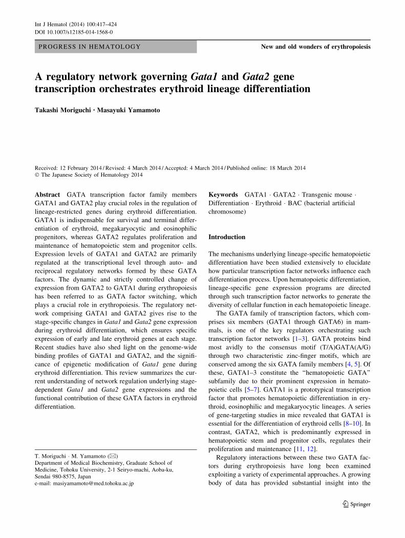
The murine Gata1 gene contains two non-coding first
exons, termed IT and IE, and five coding exons (Fig. 1a)
[6, 13, 14]. The proximal IE exon directs Gata1 expression
in hematopoietic cells [13, 15], while the distal IT exon
primarily directs Gata1 expression in testicular Sertoli cells
[16, 17]. The regulation of Gata1 gene was initially studied
using erythroid cell lines, MEL and K562 [18, 19], and was
subsequently studied in greater detail through in vivo
transgenic reporter mouse approaches [14, 20]. We found
that an 8.5-kb Gata1 genomic region covering 3.9-kb
upstream of exon IE to the second exon harbors sufficient
regulatory information to induce hematopoietic lineage-
specific expression of b-galactosidase reporter in both yolk
sac-derived primitive and fetal liver-derived definitive
hematopoietic cells in transgenic mouse assays [14, 20].
This region is referred to as the Gata1 hematopoietic reg-
ulatory domain (G1HRD; Fig. 1b) and utilized as an
extremely useful genetic tool that directs erythroid- and
megakaryocyte-specific expression of various transgenes
[21–23].
We have generated a mutant mouse line bearing a Gata1
knockdown allele, Gata1.05, in which Gata1 mRNA
expression is suppressed to approximately 5 % of the wild-
type level [9]. Hemizygous (Gata1G1.05/Y) male embryos
harboring Gata1.05 knockdown allele succumb around
embryonic day 10.5 (E10.5) due to severe anemia, indi-
cating that 5 % of GATA1 production is insufficient to
support embryonic erythropoiesis [9]. Importantly, when
GATA1 is expressed under the direction of G1HRD,
transgenic GATA1 restores hematopoiesis in Gata1.05
knockdown mice, thereby rescuing knockdown mice from
embryonic lethality [10], which indicates that G1HRD
harbors sufficient regulatory information to support Gata1
gene expression for physiological hematopoiesis.
Sequences and mechanisms that regulate Gata1
transcription
The G1HRD-based transgenic rescue system has been
extensively used for the dissection of functional domains of
GATA1 protein and the functional evaluation of hemato-
poietic disease-related mutants of GATA1 [24, 25]. A
number of studies have addressed the regulatory mecha-
nisms by which Gata1 gene expression is regulated through
the use of a G1HRD-based b-galactosidase reporter trans-
genic mouse system. These analyses have revealed the
existence of multiple cis-regulatory elements that confer
lineage- and stage-specific Gata1 gene expression [14, 20,
26, 27].
Of the cis-acting regulatory elements, Gata1 hemato-
poietic enhancer (G1HE), which is located at the 50 end of
G1HRD (Fig. 1a), is one such regulatory region. In trans-
genic reporter mouse assays, a deletion of 1.3-kb region
from the 50 end of G1HRD (including G1HE) completely
abolished LacZ reporter gene expression in erythroid and
megakaryocytic lineage cells in the fetal liver hematopoi-
etic cells [14, 20]. Further dissection of G1HE revealed that
a 235-bp region at the most 50 end of G1HE, which is
designated as the G1HE core region, is essential for the
activity of G1HE element (Fig. 1a) [20, 26]. The G1HE
core region contains an evolutionarily conserved GATA-
binding motif. A substitution mutation of the GATA motif
in the context of G1HRD-based vector significantly
decreased the expression of LacZ reporter in fetal liver
hematopoietic cells, underscoring the importance of the
GATA site for hematopoietic GATA1 expression [20].
Also of interest, two adjacent CACCC boxes are located
at around -201 to -186 bp in close proximity to the IE
promoter (Fig. 1a). This region was originally reported as a
DNase I hypersensitive (HS) site [18]. While the contri-
bution of these CACCC boxes to the Gata1 gene expres-
sion was shown by transient reporter transfection assays in
erythroid cell lines in the initial stage of Gata1 study [18],
the physiological significance of these elements in vivo
a
b
c
Fig. 1 a Mouse Gata1 gene harbors two first exons (IT, testis exon 1;
IE, hematopoietic exon 1) and four regulatory modules (G1HE core,
dbGATA/CP2, CACCC and first intron element). b G1HRD (Gata1
Hematopoietic Regulatory Domain) carries a 3.9-kb 50-flanking
sequence, IE exon, a 4.2-kb first intron, and second exon non-coding
sequences. c A 659-bp GdC minigene containing G1HE, double
GATA and CACCC motifs in the G1HRD. Note that this minigene
elicits sufficient regulatory activity to direct erythrocyte-specific
Gata1 expression upon equipping the first intron element
418 T. Moriguchi, M. Yamamoto
123
remains unclear. One transgenic reporter mouse experi-
ment has shown that mutation in one of the two CACCC
boxes hardly affects the G1HRD-directed LacZ reporter
expression, suggesting that one of the two CACCC ele-
ments may suffice for hematopoietic Gata1 expression [26,
27]. The most plausible trans-activating factor that binds to
these elements would appear to be Erythroid Kruppel-like
Factor (EKLF or KLF1) [28]; however, no direct evidence
for this assertion has been reported.
Thirdly, a palindromic GATA-binding site (dbGATA) is
located 700-bp 50 to the IE exon (Fig. 1a), which has eight-
fold higher binding affinity than a single GATA site [29, 30].
Targeted deletion of the dbGATA site (DdbGATA) leads to
selective loss of the eosinophil lineage [31], while the sig-
nificance of the dbGATA site for Gata1 gene expression in
erythroid and megakaryocytic progenitor cells has largely
been unexplored. In this regard, recent studies using the
G1HRD-based transgenic reporter mouse assay have
revealed that the dbGATA site is essential for GATA1
expression in fetal liver hematopoietic cells [26, 27].
CP2 is a ubiquitously expressed transcription factor
belonging to the Drosophila grainyhead-like gene family
[32]. CP2 participates in the regulation of a- and b-globin
gene expression [32, 33]. CP2-binding sites are located in
close proximity to GATA-binding sites in the promoter and
enhancer region of erythroid genes (e.g., GATA1, EKLF,
and p45 NF-E2). Indeed, an evolutionally conserved CP2-
binding site lies adjacent to the dbGATA site in the Gata1
regulatory region (Fig. 1a) [34]. In the G1HRD-directed
reporter assay system, both the dbGATA- and CP2-binding
sites participate cooperatively in Gata1 gene regulation in
erythroid cells [27]. Direct interactions between CP2 and
GATA1 may be responsible for erythroid-specific regula-
tion of the Gata1 gene [34].
Finally, an element localized in the first intron appears
to be essential for definitive hematopoietic cell-specific
G1HRD transgene expression in mouse fetal liver, whereas
this region is dispensable for Gata1 gene expression in
yolk sac primitive erythroid cells [14]. In the transgenic
reporter analysis, Gata1 expression in definitive erythroid
cells and megakaryocytes requires a 320-bp region in the
Gata1 first intron [26], which contains GATA motifs and
AP1 repeats (Fig. 1a). Seven highly conserved GATA
motifs, and two alternative transcription start sites (IEb and
IEc), are also identified in this region [15].
GdC minigene: minimal sufficient regulatory element
for hematopoietic Gata1 expression
Efforts to isolate minimal cis-acting element sufficient for the
hematopoietic Gata1 gene expression led to the identification
of a 659-bp small DNA fragment containing the
aforementioned three core elements [i.e., G1HE, double
GATA and CACCC (?CP2) motifs in the G1HRD]. This
fragment elicits regulatory activity sufficient to direct Gata1
expression in yolk sac erythroid cells [26], and is referred to
as Gata1 GdC minigene (Fig. 1c). The GdC-minigene frag-
ment is indeed capable of functionally replacing the 3.7-kb 50-upstream regulatory region of Gata1 gene by directing the
hematopoietic lineage-specific gene expression in the context
of a Gata1 bacterial artificial chromosome (BAC)-based
transgenic mouse assay [35]. These three regulatory elements
are thus crucial for hematopoietic Gata1 gene expression.
Dynamic expression profiles of GATA1 and GATA2
during erythropoiesis
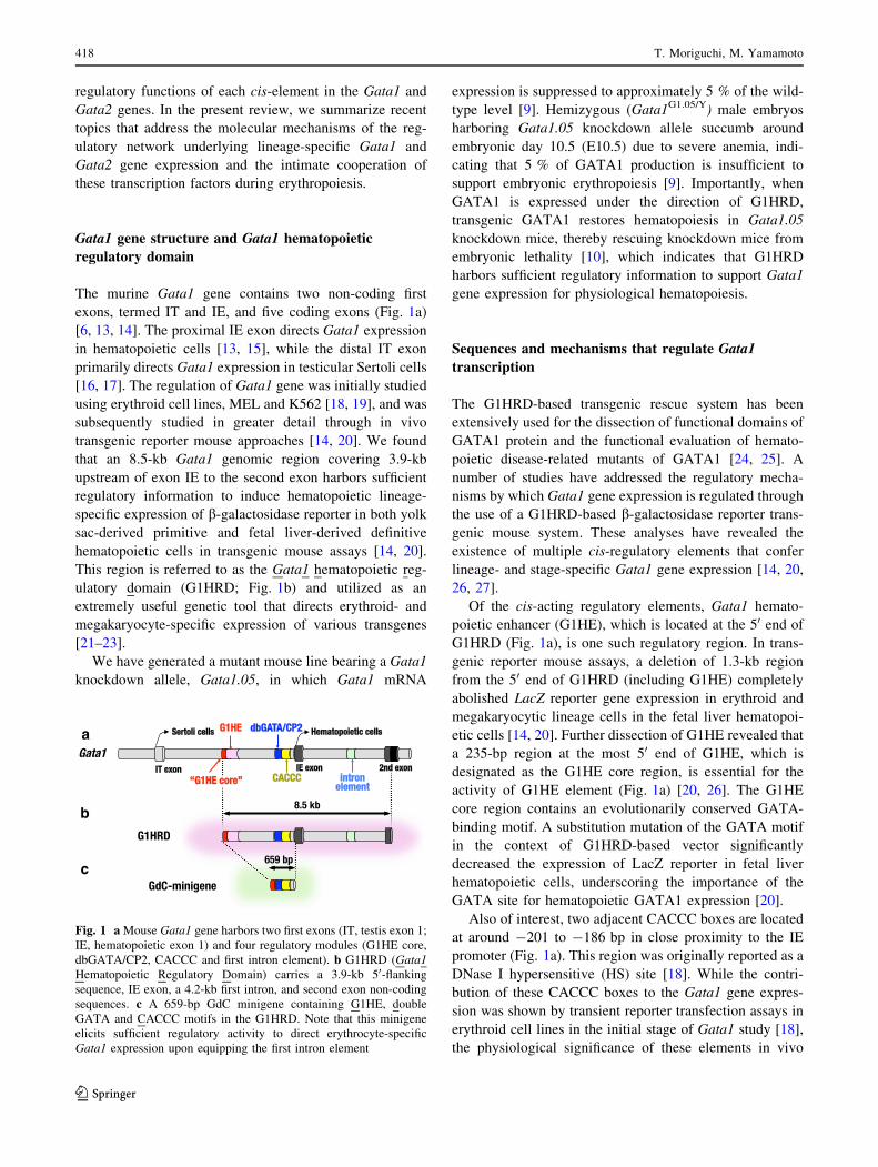
Throughout the erythroid differentiation process, GATA1
orchestrates dramatic changes in the expression of a series of
genes, which promote essential steps required for the pro-
liferation and differentiation of erythroid progenitors. In the
early stage of erythroid commitment, GATA1 expression is
first initiated at the common myeloid progenitor (CMP) stage
(Fig. 2). Subsequently, GATA1 expression increases and
reaches a peak when erythroid-committed progenitors give
rise to proerythroblasts [36, 37]. When proerythroblasts
enter terminal erythroid differentiation, GATA1 directly
activates a number of erythroid-affiliated genes, i.e. b-glo-
bin, Alas2 and Gata1 itself, and represses a number of genes
essential for progenitor proliferation in the early stage of
hematopoiesis, including Gata2, c-Kit, c-Myb and c-Myc [7].
From the late erythroblast stage onward, GATA1 expression
levels decrease toward maturation of red blood cells [36, 37].
Fig. 2 Reciprocal expression profiles of GATA1 and GATA2 during
erythropoiesis. GATA1 expression is initiated at common myeloid
progenitors (CMP) stage and reaches a peak at the proerythroblast
(ProEB) stage. From the late erythroblast stage onward, GATA1
expression levels decrease toward maturation of red blood cell (RBC).
GATA2 is preferentially expressed in hematopoietic stem cell (HSC)
and hematopoietic progenitor cells, including CMP, megakaryo-
erythroid progenitor (MEP), and burst forming unit-erythroid (BFU-
E). GATA2 expression is suppressed by the increase of GATA1
activity from CFU-E (colony forming unit-erythroid) stage onward
A regulatory network governing Gata1 and Gata2 gene transcription 419
123
This series of changes in GATA1 expression level is essen-
tial for erythropoiesis. Indeed, forced GATA1 expression
driven by human b-globin gene promoter in the terminal
erythroid differentiation stage leads to defective erythroid
cell maturation, indicating that aberrant GATA1 activity
inhibits terminal erythroid differentiation [38].
In contrast to GATA1, GATA2 is preferentially expressed
in hematopoietic stem cells (HSC) and early hematopoietic
progenitor cells (HPC; Fig. 2). GATA2 is essential for the
development and maintenance of these fractions [11, 39–41].
GATA2 haploinsufficiency impairs the quality of both
embryonic and adult HSC, and eventually leads to a reduction
of early HSC population [12]. Two independent studies dem-
onstrated that Gata2 4th intron regulatory elements (Gata2 VE;
vascular enhancer/?9.5-kb GATA motifs) [42] are crucial for
GATA2 expression in HSCs [43, 44]. Of note, a heterozygous
28-bp deletion within this element was identified in a patient
with MonoMAC syndrome, an autosomal dominant condition
that features recurrent mycobacterial infection associated with
deficiencies of monocytes, B cells and NK cells, and myelo-
dysplasia [43]. Gata2 expression from the mutated allele was
reduced in the peripheral blood monocyte of the patient, sug-
gesting a causal contribution of the ?9.5-kb deletion to the
diminished GATA2 level and the subsequent pathogenesis of
MonoMAC syndrome.
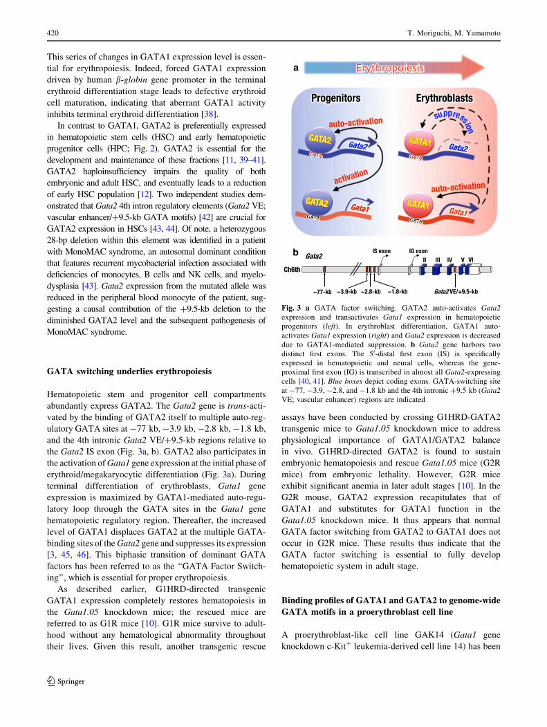
GATA switching underlies erythropoiesis
Hematopoietic stem and progenitor cell compartments
abundantly express GATA2. The Gata2 gene is trans-acti-
vated by the binding of GATA2 itself to multiple auto-reg-
ulatory GATA sites at -77 kb, -3.9 kb, -2.8 kb, -1.8 kb,
and the 4th intronic Gata2 VE/?9.5-kb regions relative to
the Gata2 IS exon (Fig. 3a, b). GATA2 also participates in
the activation of Gata1 gene expression at the initial phase of
erythroid/megakaryocytic differentiation (Fig. 3a). During
terminal differentiation of erythroblasts, Gata1 gene
expression is maximized by GATA1-mediated auto-regu-
latory loop through the GATA sites in the Gata1 gene
hematopoietic regulatory region. Thereafter, the increased
level of GATA1 displaces GATA2 at the multiple GATA-
binding sites of the Gata2 gene and suppresses its expression
[3, 45, 46]. This biphasic transition of dominant GATA
factors has been referred to as the ‘‘GATA Factor Switch-
ing’’, which is essential for proper erythropoiesis.
As described earlier, G1HRD-directed transgenic
GATA1 expression completely restores hematopoiesis in
the Gata1.05 knockdown mice; the rescued mice are
referred to as G1R mice [10]. G1R mice survive to adult-
hood without any hematological abnormality throughout
their lives. Given this result, another transgenic rescue
assays have been conducted by crossing G1HRD-GATA2
transgenic mice to Gata1.05 knockdown mice to address
physiological importance of GATA1/GATA2 balance
in vivo. G1HRD-directed GATA2 is found to sustain
embryonic hematopoiesis and rescue Gata1.05 mice (G2R
mice) from embryonic lethality. However, G2R mice
exhibit significant anemia in later adult stages [10]. In the
G2R mouse, GATA2 expression recapitulates that of
GATA1 and substitutes for GATA1 function in the
Gata1.05 knockdown mice. It thus appears that normal
GATA factor switching from GATA2 to GATA1 does not
occur in G2R mice. These results thus indicate that the
GATA factor switching is essential to fully develop
hematopoietic system in adult stage.
Binding profiles of GATA1 and GATA2 to genome-wide
GATA motifs in a proerythroblast cell line
A proerythroblast-like cell line GAK14 (Gata1 gene
knockdown c-Kit? leukemia-derived cell line 14) has been
a
b
Fig. 3 a GATA factor switching. GATA2 auto-activates Gata2
expression and transactivates Gata1 expression in hematopoietic
progenitors (left). In erythroblast differentiation, GATA1 auto-
activates Gata1 expression (right) and Gata2 expression is decreased
due to GATA1-mediated suppression. b Gata2 gene harbors two
distinct first exons. The 50-distal first exon (IS) is specifically
expressed in hematopoietic and neural cells, whereas the gene-
proximal first exon (IG) is transcribed in almost all Gata2-expressing
cells [40, 41]. Blue boxes depict coding exons. GATA-switching site
at -77, -3.9, -2.8, and -1.8 kb and the 4th intronic ?9.5 kb (Gata2
VE; vascular enhancer) regions are indicated
420 T. Moriguchi, M. Yamamoto
123
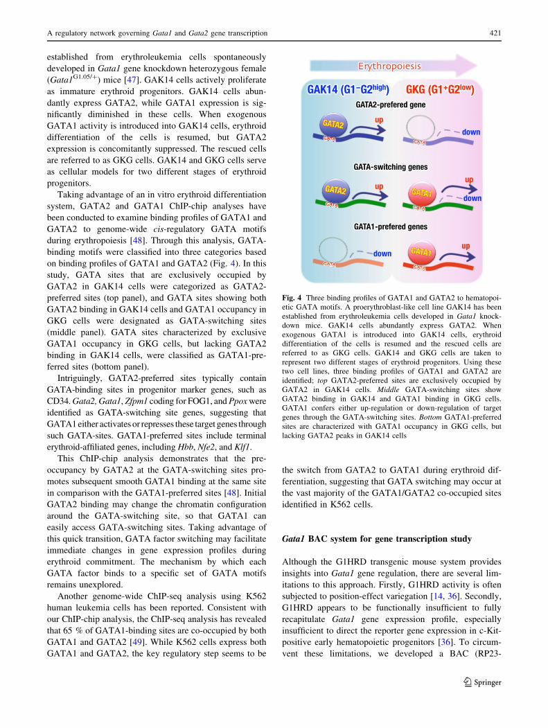
established from erythroleukemia cells spontaneously
developed in Gata1 gene knockdown heterozygous female
(Gata1G1.05/?) mice [47]. GAK14 cells actively proliferate
as immature erythroid progenitors. GAK14 cells abun-
dantly express GATA2, while GATA1 expression is sig-
nificantly diminished in these cells. When exogenous
GATA1 activity is introduced into GAK14 cells, erythroid
differentiation of the cells is resumed, but GATA2
expression is concomitantly suppressed. The rescued cells
are referred to as GKG cells. GAK14 and GKG cells serve
as cellular models for two different stages of erythroid
progenitors.
Taking advantage of an in vitro erythroid differentiation
system, GATA2 and GATA1 ChIP-chip analyses have
been conducted to examine binding profiles of GATA1 and
GATA2 to genome-wide cis-regulatory GATA motifs
during erythropoiesis [48]. Through this analysis, GATA-
binding motifs were classified into three categories based
on binding profiles of GATA1 and GATA2 (Fig. 4). In this
study, GATA sites that are exclusively occupied by
GATA2 in GAK14 cells were categorized as GATA2-
preferred sites (top panel), and GATA sites showing both
GATA2 binding in GAK14 cells and GATA1 occupancy in
GKG cells were designated as GATA-switching sites
(middle panel). GATA sites characterized by exclusive
GATA1 occupancy in GKG cells, but lacking GATA2
binding in GAK14 cells, were classified as GATA1-pre-
ferred sites (bottom panel).
Intriguingly, GATA2-preferred sites typically contain
GATA-binding sites in progenitor marker genes, such as
CD34. Gata2, Gata1, Zfpm1 coding for FOG1, and Ppox were
identified as GATA-switching site genes, suggesting that
GATA1 either activates or represses these target genes through
such GATA-sites. GATA1-preferred sites include terminal
erythroid-affiliated genes, including Hbb, Nfe2, and Klf1.
This ChIP-chip analysis demonstrates that the pre-
occupancy by GATA2 at the GATA-switching sites pro-
motes subsequent smooth GATA1 binding at the same site
in comparison with the GATA1-preferred sites [48]. Initial
GATA2 binding may change the chromatin configuration
around the GATA-switching site, so that GATA1 can
easily access GATA-switching sites. Taking advantage of
this quick transition, GATA factor switching may facilitate
immediate changes in gene expression profiles during
erythroid commitment. The mechanism by which each
GATA factor binds to a specific set of GATA motifs
remains unexplored.
Another genome-wide ChIP-seq analysis using K562
human leukemia cells has been reported. Consistent with
our ChIP-chip analysis, the ChIP-seq analysis has revealed
that 65 % of GATA1-binding sites are co-occupied by both
GATA1 and GATA2 [49]. While K562 cells express both
GATA1 and GATA2, the key regulatory step seems to be
the switch from GATA2 to GATA1 during erythroid dif-
ferentiation, suggesting that GATA switching may occur at
the vast majority of the GATA1/GATA2 co-occupied sites
identified in K562 cells.
Gata1 BAC system for gene transcription study
Although the G1HRD transgenic mouse system provides
insights into Gata1 gene regulation, there are several lim-
itations to this approach. Firstly, G1HRD activity is often
subjected to position-effect variegation [14, 36]. Secondly,
G1HRD appears to be functionally insufficient to fully
recapitulate Gata1 gene expression profile, especially
insufficient to direct the reporter gene expression in c-Kit-
positive early hematopoietic progenitors [36]. To circum-
vent these limitations, we developed a BAC (RP23-
Fig. 4 Three binding profiles of GATA1 and GATA2 to hematopoi-
etic GATA motifs. A proerythroblast-like cell line GAK14 has been
established from erythroleukemia cells developed in Gata1 knock-
down mice. GAK14 cells abundantly express GATA2. When
exogenous GATA1 is introduced into GAK14 cells, erythroid
differentiation of the cells is resumed and the rescued cells are
referred to as GKG cells. GAK14 and GKG cells are taken to
represent two different stages of erythroid progenitors. Using these
two cell lines, three binding profiles of GATA1 and GATA2 are
identified; top GATA2-preferred sites are exclusively occupied by
GATA2 in GAK14 cells. Middle GATA-switching sites show
GATA2 binding in GAK14 and GATA1 binding in GKG cells.
GATA1 confers either up-regulation or down-regulation of target
genes through the GATA-switching sites. Bottom GATA1-preferred
sites are characterized with GATA1 occupancy in GKG cells, but
lacking GATA2 peaks in GAK14 cells
A regulatory network governing Gata1 and Gata2 gene transcription 421
123
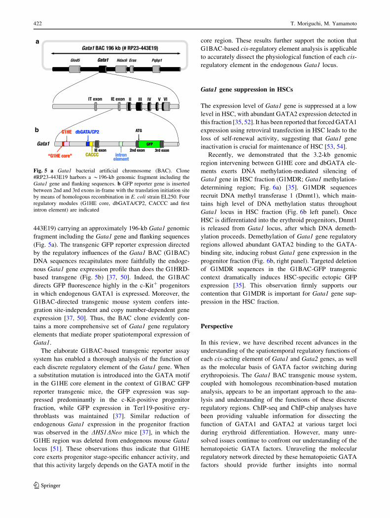
443E19) carrying an approximately 196-kb Gata1 genomic
fragment including the Gata1 gene and flanking sequences
(Fig. 5a). The transgenic GFP reporter expression directed
by the regulatory influences of the Gata1 BAC (G1BAC)
DNA sequences recapitulates more faithfully the endoge-
nous Gata1 gene expression profile than does the G1HRD-
based transgene (Fig. 5b) [37, 50]. Indeed, the G1BAC
directs GFP fluorescence highly in the c-Kit? progenitors
in which endogenous GATA1 is expressed. Moreover, the
G1BAC-directed transgenic mouse system confers inte-
gration site-independent and copy number-dependent gene
expression [37, 50]. Thus, the BAC clone evidently con-
tains a more comprehensive set of Gata1 gene regulatory
elements that mediate proper spatiotemporal expression of
Gata1.
The elaborate G1BAC-based transgenic reporter assay
system has enabled a thorough analysis of the function of
each discrete regulatory element of the Gata1 gene. When
a substitution mutation is introduced into the GATA motif
in the G1HE core element in the context of G1BAC GFP
reporter transgenic mice, the GFP expression was sup-
pressed predominantly in the c-Kit-positive progenitor
fraction, while GFP expression in Ter119-positive ery-
throblasts was maintained [37]. Similar reduction of
endogenous Gata1 expression in the progenitor fraction
was observed in the DHS1DNeo mice [37], in which the
G1HE region was deleted from endogenous mouse Gata1
locus [51]. These observations thus indicate that G1HE
core exerts progenitor stage-specific enhancer activity, and
that this activity largely depends on the GATA motif in the
core region. These results further support the notion that
G1BAC-based cis-regulatory element analysis is applicable
to accurately dissect the physiological function of each cis-
regulatory element in the endogenous Gata1 locus.
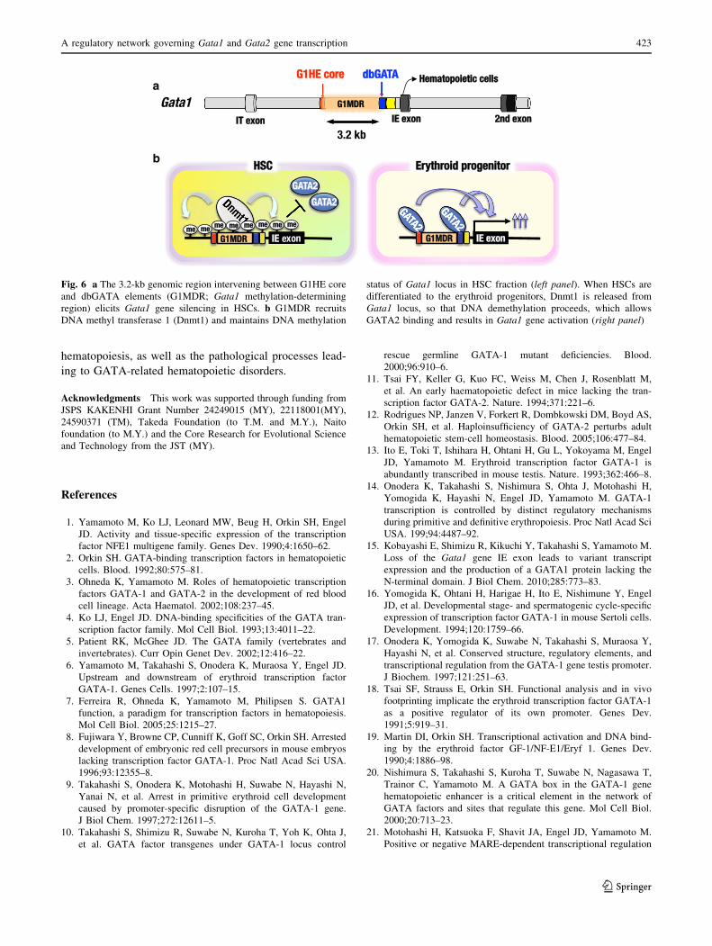
Gata1 gene suppression in HSCs
The expression level of Gata1 gene is suppressed at a low
level in HSC, with abundant GATA2 expression detected in
this fraction [35, 52]. It has been reported that forced GATA1
expression using retroviral transfection in HSC leads to the
loss of self-renewal activity, suggesting that Gata1 gene
inactivation is crucial for maintenance of HSC [53, 54].
Recently, we demonstrated that the 3.2-kb genomic
region intervening between G1HE core and dbGATA ele-
ments exerts DNA methylation-mediated silencing of
Gata1 gene in HSC fraction (G1MDR; Gata1 methylation-
determining region; Fig. 6a) [35]. G1MDR sequences
recruit DNA methyl transferase 1 (Dnmt1), which main-
tains high level of DNA methylation status throughout
Gata1 locus in HSC fraction (Fig. 6b left panel). Once
HSC is differentiated into the erythroid progenitors, Dnmt1
is released from Gata1 locus, after which DNA demeth-
ylation proceeds. Demethylation of Gata1 gene regulatory
regions allowed abundant GATA2 binding to the GATA-
binding site, inducing robust Gata1 gene expression in the
progenitor fraction (Fig. 6b, right panel). Targeted deletion
of G1MDR sequences in the G1BAC-GFP transgenic
context dramatically induces HSC-specific ectopic GFP
expression [35]. This observation firmly supports our
contention that G1MDR is important for Gata1 gene sup-
pression in the HSC fraction.
Perspective
In this review, we have described recent advances in the
understanding of the spatiotemporal regulatory functions of
each cis-acting element of Gata1 and Gata2 genes, as well
as the molecular basis of GATA factor switching during
erythropoiesis. The Gata1 BAC transgenic mouse system,
coupled with homologous recombination-based mutation
analysis, appears to be an important approach to the ana-
lysis and understanding of the functions of these discrete
regulatory regions. ChIP-seq and ChIP-chip analyses have
been providing valuable information for dissecting the
function of GATA1 and GATA2 at various target loci
during erythroid differentiation. However, many unre-
solved issues continue to confront our understanding of the
hematopoietic GATA factors. Unraveling the molecular
regulatory network directed by these hematopoietic GATA
factors should provide further insights into normal
a
b
Fig. 5 a Gata1 bacterial artificial chromosome (BAC). Clone
#RP23-443E19 harbors a *196-kb genomic fragment including the
Gata1 gene and flanking sequences. b GFP reporter gene is inserted
between 2nd and 3rd exons in-frame with the translation initiation site
by means of homologous recombination in E. coli strain EL250. Four
regulatory modules (G1HE core, dbGATA/CP2, CACCC and first
intron element) are indicated
422 T. Moriguchi, M. Yamamoto
123
hematopoiesis, as well as the pathological processes lead-
ing to GATA-related hematopoietic disorders.
Acknowledgments This work was supported through funding from
JSPS KAKENHI Grant Number 24249015 (MY), 22118001(MY),
24590371 (TM), Takeda Foundation (to T.M. and M.Y.), Naito
foundation (to M.Y.) and the Core Research for Evolutional Science
and Technology from the JST (MY).
References
1. Yamamoto M, Ko LJ, Leonard MW, Beug H, Orkin SH, Engel
JD. Activity and tissue-specific expression of the transcription
factor NFE1 multigene family. Genes Dev. 1990;4:1650–62.
2. Orkin SH. GATA-binding transcription factors in hematopoietic
cells. Blood. 1992;80:575–81.
3. Ohneda K, Yamamoto M. Roles of hematopoietic transcription
factors GATA-1 and GATA-2 in the development of red blood
cell lineage. Acta Haematol. 2002;108:237–45.
4. Ko LJ, Engel JD. DNA-binding specificities of the GATA tran-
scription factor family. Mol Cell Biol. 1993;13:4011–22.
5. Patient RK, McGhee JD. The GATA family (vertebrates and
invertebrates). Curr Opin Genet Dev. 2002;12:416–22.
6. Yamamoto M, Takahashi S, Onodera K, Muraosa Y, Engel JD.
Upstream and downstream of erythroid transcription factor
GATA-1. Genes Cells. 1997;2:107–15.
7. Ferreira R, Ohneda K, Yamamoto M, Philipsen S. GATA1
function, a paradigm for transcription factors in hematopoiesis.
Mol Cell Biol. 2005;25:1215–27.
8. Fujiwara Y, Browne CP, Cunniff K, Goff SC, Orkin SH. Arrested
development of embryonic red cell precursors in mouse embryos
lacking transcription factor GATA-1. Proc Natl Acad Sci USA.
1996;93:12355–8.
9. Takahashi S, Onodera K, Motohashi H, Suwabe N, Hayashi N,
Yanai N, et al. Arrest in primitive erythroid cell development
caused by promoter-specific disruption of the GATA-1 gene.
J Biol Chem. 1997;272:12611–5.
10. Takahashi S, Shimizu R, Suwabe N, Kuroha T, Yoh K, Ohta J,
et al. GATA factor transgenes under GATA-1 locus control
rescue germline GATA-1 mutant deficiencies. Blood.
2000;96:910–6.
11. Tsai FY, Keller G, Kuo FC, Weiss M, Chen J, Rosenblatt M,
et al. An early haematopoietic defect in mice lacking the tran-
scription factor GATA-2. Nature. 1994;371:221–6.
12. Rodrigues NP, Janzen V, Forkert R, Dombkowski DM, Boyd AS,
Orkin SH, et al. Haploinsufficiency of GATA-2 perturbs adult
hematopoietic stem-cell homeostasis. Blood. 2005;106:477–84.
13. Ito E, Toki T, Ishihara H, Ohtani H, Gu L, Yokoyama M, Engel
JD, Yamamoto M. Erythroid transcription factor GATA-1 is
abundantly transcribed in mouse testis. Nature. 1993;362:466–8.
14. Onodera K, Takahashi S, Nishimura S, Ohta J, Motohashi H,
Yomogida K, Hayashi N, Engel JD, Yamamoto M. GATA-1
transcription is controlled by distinct regulatory mechanisms
during primitive and definitive erythropoiesis. Proc Natl Acad Sci
USA. 199;94:4487–92.
15. Kobayashi E, Shimizu R, Kikuchi Y, Takahashi S, Yamamoto M.
Loss of the Gata1 gene IE exon leads to variant transcript
expression and the production of a GATA1 protein lacking the
N-terminal domain. J Biol Chem. 2010;285:773–83.
16. Yomogida K, Ohtani H, Harigae H, Ito E, Nishimune Y, Engel
JD, et al. Developmental stage- and spermatogenic cycle-specific
expression of transcription factor GATA-1 in mouse Sertoli cells.
Development. 1994;120:1759–66.
17. Onodera K, Yomogida K, Suwabe N, Takahashi S, Muraosa Y,
Hayashi N, et al. Conserved structure, regulatory elements, and
transcriptional regulation from the GATA-1 gene testis promoter.
J Biochem. 1997;121:251–63.
18. Tsai SF, Strauss E, Orkin SH. Functional analysis and in vivo
footprinting implicate the erythroid transcription factor GATA-1
as a positive regulator of its own promoter. Genes Dev.
1991;5:919–31.
19. Martin DI, Orkin SH. Transcriptional activation and DNA bind-
ing by the erythroid factor GF-1/NF-E1/Eryf 1. Genes Dev.
1990;4:1886–98.
20. Nishimura S, Takahashi S, Kuroha T, Suwabe N, Nagasawa T,
Trainor C, Yamamoto M. A GATA box in the GATA-1 gene
hematopoietic enhancer is a critical element in the network of
GATA factors and sites that regulate this gene. Mol Cell Biol.
2000;20:713–23.
21. Motohashi H, Katsuoka F, Shavit JA, Engel JD, Yamamoto M.
Positive or negative MARE-dependent transcriptional regulation
a
b
Fig. 6 a The 3.2-kb genomic region intervening between G1HE core
and dbGATA elements (G1MDR; Gata1 methylation-determining
region) elicits Gata1 gene silencing in HSCs. b G1MDR recruits
DNA methyl transferase 1 (Dnmt1) and maintains DNA methylation
status of Gata1 locus in HSC fraction (left panel). When HSCs are
differentiated to the erythroid progenitors, Dnmt1 is released from
Gata1 locus, so that DNA demethylation proceeds, which allows
GATA2 binding and results in Gata1 gene activation (right panel)
A regulatory network governing Gata1 and Gata2 gene transcription 423
123
is determined by the abundance of small Maf proteins. Cell.
2000;103:865–75.
22. Suzuki N, Ohneda O, Takahashi S, Higuchi M, Mukai HY, Na-
kahata T, Imagawa S, Yamamoto M. Erythroid-specific expres-
sion of the erythropoietin receptor rescued its null mutant mice
from lethality. Blood. 2002;100:2279–88.
23. Tatsumi K, Yamamoto-Mukai H, Shimizu R, Waguri S, Sou YS,
Sakamoto A, et al. The Ufm1-activating enzyme Uba5 is indis-
pensable for erythroid differentiation in mice. Nat Commun.
2011;2:181.
24. Shimizu R, Ohneda K, Engel JD, Trainor CD, Yamamoto M.
Transgenic rescue of GATA-1-deficient mice with GATA-1
lacking a FOG-1 association site phenocopies patients with
X-linked thrombocytopenia. Blood. 2004;103:2560–7.
25. Shimizu R, Takahashi S, Ohneda K, Engel JD, Yamamoto M.
In vivo requirements for GATA-1 functional domains during
primitive and definitive erythropoiesis. EMBO J. 2001;20:5250–60.
26. Ohneda K, Shimizu R, Nishimura S, Muraosa Y, Takahashi S,
Engel JD, et al. A minigene containing four discrete cis-elements
recapitulates GATA-1 gene expression in vivo. Genes Cells.
2002;7:1243–54.
27. Shimizu R, Hasegawa A, Ottolenghi S, Ronchi A, Yamamoto M.
Verification of the in vivo activity of three distinct cis-acting
elements within the Gata1 gene promoter-proximal enhancer in
mice. Genes Cells. 2013;18:1032–41.
28. Yien YY, Bieker JJ. EKLF/KLF1, a tissue-restricted integrator of
transcriptional control, chromatin remodeling, and lineage
determination. Mol Cell Biol. 2013;33:4–13.
29. Trainor CD, Omichinski JG, Vandergon TL, Gronenborn AM,
Clore GM, Felsenfeld G. A palindromic regulatory site within
vertebrate GATA-1 promoters requires both zinc fingers of the
GATA-1 DNA-binding domain for high-affinity interaction. Mol
Cell Biol. 1996;16:2238–47.
30. Trainor CD, Ghirlando R, Simpson MA. GATA zinc finger
interactions modulate DNA binding and transactivation. J Biol
Chem. 2000;275:28157–66.
31. Yu C, Cantor AB, Yang H, Browne C, Wells RA, Fujiwara Y,
et al. Targeted deletion of a high-affinity GATA-binding site in
the GATA-1 promoter leads to selective loss of the eosinophil
lineage in vivo. J Exp Med. 2002;195:1387–95.
32. Lim LC, Swendeman SL, Sheffery M. Molecular cloning of the
a-globin transcription factor CP2. Mol Cell Biol. 1992;12:
828–35.
33. Chae JH, Kim CG. CP2 binding to the promoter is essential for
the enhanced transcription of globin genes in erythroid cells. Mol
Cells. 2003;15:40–7.
34. Bose F, Fugazza C, Casalgrandi M, Capelli A, Cunningham JM,
Zhao Q, et al. Functional interaction of CP2 with GATA-1 in the
regulation of erythroid promoters. Mol Cell Biol.
2006;26:3942–54.
35. Takai J, Moriguchi T, Suzuki M, Yu L, Ohneda K, Yamamoto M.
The Gata1 50 region harbors distinct cis-regulatory modules that
direct gene activation in erythroid cells and gene inactivation in
HSCs. Blood. 2013;122:3450–60.
36. Suzuki N, Suwabe N, Ohneda O, Obara N, Imagawa S, Pan X,
Motohashi H, Yamamoto M. Identification and characterization
of 2 types of erythroid progenitors that express GATA-1 at dis-
tinct levels. Blood. 2003;102:3575–83.
37. Suzuki M, Moriguchi T, Ohneda K, Yamamoto M. Differential
contribution of the Gata1 gene hematopoietic enhancer to ery-
throid differentiation. Mol Cell Biol. 2009;29:1163–75.
38. Whyatt D, Lindeboom F, Karis A, Ferreira R, Milot E, Hendriks
R, et al. An intrinsic but cell-nonautonomous defect in GATA-1-
overexpressing mouse erythroid cells. Nature. 2000;406:519–24.
39. Tsai FY, Orkin SH. Transcription factor GATA-2 is required for
proliferation/survival of early hematopoietic cells and mast cell
formation, but not for erythroid and myeloid terminal differen-
tiation. Blood. 1997;89:3636–43.
40. Minegishi N, Ohta J, Suwabe N, Nakauchi H, Ishihara H, Hayashi
N, et al. Alternative promoters regulate transcription of the mouse
GATA-2 gene. J Biol Chem. 1998;273:3625–34.
41. Minegishi N, Suzuki N, Yokomizo T, Pan X, Fujimoto T, Ta-
kahashi S et al. Expression and domain-specific function of
GATA-2 during differentiation of the hematopoietic precursor
cells in midgestation mouse embryos. Blood. 2003;102:896–5.
42. Khandekar M, Brandt W, Zhou Y, Dagenais S, Glover TW, Su-
zuki N, Shimizu R, Yamamoto M, Lim KC, Engel JD. A Gata2
intronic enhancer confers its pan-endothelia-specific regulation.
Development. 2007;134:1703–12.
43. Johnson KD, Hsu AP, Ryu MJ, Wang J, Gao X, Boyer ME, et al.
Cis-element mutated in GATA2-dependent immunodeficiency
governs hematopoiesis and vascular integrity. J Clin Invest.
2012;122:3692–704.
44. Lim KC, Hosoya T, Brandt W, Ku CJ, Hosoya-Ohmura S,
Camper SA, et al. Conditional Gata2 inactivation results in HSC
loss and lymphatic mispatterning. J Clin Invest.
2012;122:3705–17.
45. Kaneko H, Shimizu R, Yamamoto M. GATA factor switching
during erythroid differentiation. Curr Opin Hematol.
2010;17:163–8.
46. Suzuki M, Shimizu R, Yamamoto M. Transcriptional regulation
by GATA1 and GATA2 during erythropoiesis. Int J Hematol.
2011;93:150–5.
47. Mukai HY, Suzuki M, Nagano M, Ohmori S, Otsuki A, Tsuchida
K, et al. Establishment of erythroleukemic GAK14 cells and
characterization of GATA1 N-terminal domain. Genes Cells.
2013;18:886–98.
48. Suzuki M, Kobayashi-Osaki M, Tsutsumi S, Pan X, Ohmori S,
Takai J, et al. GATA factor switching from GATA2 to GATA1
contributes to erythroid differentiation. Genes Cells.
2013;18:921–33.
49. Fujiwara T, O’Geen H, Keles S, Blahnik K, Linnemann AK,
Kang YA, et al. Discovering hematopoietic mechanisms through
genome-wide analysis of GATA factor chromatin occupancy.
Mol Cell. 2009;36:667–81.
50. Moriguchi T, Suzuki M, Engel JD, Yamamoto M. GATA1 and
GATA2 function in hematopoietic differentiation. In: Kondo M,
editors. Hematopoietic stem cell biology. New York: Humana
Press Inc; 2010. Pp 172–42.
51. McDevitt MA, Shivdasani RA, Fujiwara Y, Yang H, Orkin SH. A
‘‘knockdown’’ mutation created by cis-element gene targeting
reveals the dependence of erythroid cell maturation on the level
of transcription factor GATA-1. Proc Natl Acad Sci USA.
1997;94:6781–5.
52. Akashi K, Traver D, Miyamoto T, Weissman IL. A clonogenic
common myeloid progenitor that gives rise to all myeloid lin-
eages. Nature. 2000;404:193–7.
53. Iwasaki H, Mizuno S, Wells RA, Cantor AB, Watanabe S, Akashi
K. GATA-1 converts lymphoid and myelomonocytic progenitors
into the megakaryocyte/erythrocyte lineages. Immunity.
2003;19:451–2.
54. Togarrati PP, Suknuntha K. Generation of mature hematopoietic
cells from human pluripotent stem cells. Int J Hematol.
2012;95:617–23.
424 T. Moriguchi, M. Yamamoto
123