PROGRAM - Japan Prize
19
2012 2012
Transcript of PROGRAM - Japan Prize

2012
DIC 196 背幅1mm
2012

16
6:30 PM
6:35 PM
7:55 PM
8:30PM
PROGRAM April 26 (Thursday) Yurakucho Asahi Hall
Opening Remarks
Panel Discussion “Successful Approach Targeting Leukemic Cells” Dr. Janet D. Rowley Dr. Brian J. Druker Dr. Nicholas B. Lydon
Coordinator: Prof. Hiroaki Mitsuya (Professor, Faculty of Life Sciences, Kumamoto University)
Lecture “ How the World’s Strongest ‘Neodymium Magnet’ Came
to Exist” Dr. Masato Sagawa
Closing

17
2012 (28th) Japan Prize Laureate
Research Summary
After about 1965, I began to study chromosomes in bone marrow cells from patients with different kinds of leukemias and preleukemias. Dr. Leon Jacobson who had recruited me was Head of the Section of Hematology at The University of Chicago; he often asked me to analyze the karyotype of his patients with these disorders. It had been discovered in 1960 by Nowell and Hungerford that patients with chronic myeloid leukemia (CML) had an unusually small chromosome, which was called the Philadelphia or Ph chromsome. It was assumed that the piece missing from this chromosome was lost from the cell and that the missing piece contained genes that prevented abnormal cell growth. The general view of chromosomes abnormalities in cancer was that these changes were a consequence of genomic instability associated with cancer rather than a cause.
At the time I started studying chromosome in leukemia, I was working only three days a week because I had four sons (ages 13 to 2 years) and I wanted to spend time with them. Chromosomes were stained with regular Giemsa and they showed fairly uniform staining along the length of the chromosome. Thus one could count the number of chromosomes in a cell; if the number was abnormal, i.e. greater or lesser than 46, one could usually tell what morphological group was involved but not what specific chromosome was gained or lost. I would photograph the metaphase cells and then cut out the chromosomes and arrange them in groups according to the conventional format agreed to by human cytogeneticists. The frustration w a s t h a t w h e n s e v e r a l p a t i e n t ’ s malignant cells were missing or gaining a chromosome in a particular group, one had no way of knowing whether the same chromosome was involved in each patient.
Field : Healthcare and Medical Technology
Dr. Janet D. Rowley (USA)Blum-Riese Distinguished Service Professor of Medicine, Molecular Genetics & Cell Biology and Human Genetics, The University of Chicago Born in 1925
Achievement Development of a new therapeutic drug targeting cancer-specific molecules

18
Fortunately this changed in 1970 when three techniques were developed that led to a unique pattern of dark and light staining bands along the chromosome. The pattern was consistent between normal patients and a conference in Paris in 1971 established the nomenclature for the human karyotype . I could immediately see how banding would relieve my frustration because I would be able to determine precisely what chromosomes were gained or lost from the samples I had studied earlier. First I studied cells from two patients with acute myeloblastic leukemia (AML) whose cells had the same abnormal pattern on unbanded chromosomes. I discovered that both patients had a translocation between chromosomes 8 and 21. This was in the summer of 1972 and it was the first consistent translocation identified in any human malignancy.
I then studied cells from CML patients who were in the terminal acute blast phase of their disease. Their cells had several extra chromosomes and I wanted to identify exactly which chromosomes were abnormal. For this work (as well as for analysis of the previous translocation) I stained cells with regular Giemsa, examined the slide to find well spread cells with non-overlapping chromosomes. I would photograph these cells noting their position on the slide, then I would destain the slide, stain it with quinacrine mustard and find and photograph the same cells with ultraviolet fluorescence. At home, on my dining room table, I could cut out the paired photographs of the
chromosomes stained with Giemsa which gave me good morphology and those stained with quinacrine (Q-banded) which I used to identify each chromosome. I could see that the extra chromosomes were usually number 8. I identified the Ph chromosome as being a deleted chromosome 22. To my surprise, one chromosome 9 had an extra band of pale material which looked just like that missing from the Ph chromosome. Because I was studying ce l ls f rom the late acute phase that already had several new abnormalities, maybe the material at the end of chromosome 9 was just another late abnormality. I could check that by analyzing cells from the same patient’s chronic phase which had only the Ph chromosome. These cells also had the abnormal chromosome 9 which then lent more credence to the idea that the Ph chromosome was the result of a 9;22 translocation. But maybe it was just a normal but rare polymorphism? I obtained peripheral blood from some of these patients and found that the metaphase cells were normal. Thus it seemed almost certain that the Ph chromosome was the result of a translocation. This discovery was published in Nature in the middle of 1973.
Drs. Kaneko and Sakurai at Saitama Cancer Center and I published back-to-back papers in Lancet in 1977 reporting the third recurrent translocation which involved chromosomes 15 and 17 in acute promyelocytic leukemia. Two years later Dr. Fukuhara and I discovered the 14;18

19
translocation in follicular lymphoma, so translocations were not limited to leukemia.
By now it was clear that translocations were important in cancer and that they were often restricted to just one subtype of cancer. Molecular biologists became interested in identifying the genes at translocation breakpoints and in 1982, the first translocation in Burkitt lymphoma was cloned and the involved genes were
the immunoglobulin heavy chain and an oncogene, MYC. In 1984, the breakpoint of the t (9;22) was cloned and it involved the BCR gene and the Abelson (ABL ) gene, also an oncogene. These discoveries showed that translocations were a critical initiating event in cancer. Moreover these discoveries made it very clear that chromosome changes were associated with the earliest stages of cancer, and were likely causative in some cancers; this remains the view today.

20
2012 (28th) Japan Prize Laureate
Research Summary
The work in my laboratory has focused on activated tyrosine kinases with an emphasis on their role in signal transduction and cellular transformation and the application of this knowledge to cancer therapies. While working in Thomas Roberts’ laboratory, I developed an anti-phosphotyrosine antibody, 4G10, to assist in the identification of substrates of activated tyrosine kinases. This antibody was provided to Nick Lydon’s group to assist in their profiling of tyrosine kinase inhibitors. Looking for a human cancer in which an activated tyrosine kinase had a central role, I set up model systems, in collaboration with Dr. Jim Griffin, to study the BCR-ABL oncogene and chronic myeloid leukemia (CML). At the time, 1990, this was the most well-validated kinase target in human cancer and was a particularly good fit for combining my emerging expertise in the field of tyrosine kinase signaling with my background as a medical oncologist.
In 1993, I was recruited by Dr. Grover Bagby at the Oregon Health & Science University to continue my CML studies. My main goal was to find a company with an ABL kinase inhibitor and to move this compound into the clinic. I was quite fortunate that I only had to make one call to Nick Lydon, whose group had synthesized several compounds with PDGFR and ABL kinase inhibitory activity that he was willing to send to me for testing in our model systems. Our lab showed that one of the compounds was superior at specifically killing BCR-ABL-expressing cells in vitro and in vivo and was capable of selecting for the growth of normal hematopoietic cells from patients with CML. This compound, then known as CGP57148, was selected for clinical trials and is now known as imatinib or Gleevec.
Although Novartis was initially somewhat reluctant to begin clinical trials, we ultimately began Phase I clinical trials
Field : Healthcare and Medical Technology
Dr. Brian J. Druker (USA)Professor Director, OHSU Knight Cancer Institute Oregon Health & Science University Born in 1955
Achievement Development of a new therapeutic drug targeting cancer-specific molecules

21
in 1998. Once therapeutic doses were reached, 53/54 patients achieved a complete hematologic response with 55% of patients having a cytogenetic response, a significant decrease in the number of marrow metaphases positive for the Philadelphia chromosome. Therapy with this oral agent was extremely well tolerated with only minimal side effects. These results were confirmed in Phase II clinical trials and in a Phase III study, the five year survival for newly diagnosed patients with CML was 90%. Due to our laboratory’s finding that imatinib also inhibited the KIT tyrosine kinase the studies of imatinib were extended to gastrointestinal stromal tumor (GIST) as Dr. Kitamura at Osaka University, along with my friend and colleague Dr. Yuzuru Kanakura had shown that KIT activating mutations were present in patients with GIST. The clinical results with imatinib in GIST were similarly striking with a 60% response rate in this previously treatment refractory malignancy.
Despite the enormous success of imatinib for treating CML, approximately 17% of patients show some signs of resistance with five years of follow-up. During the early profiling of imatinib, we were puzzled by the finding that it inhibits ABL but not its most closely related kinase family member, SRC. John Kuriyan’s structure of imatinib co-crystallized with ABL showed that imatinib bound to ABL with the activation loop in a unique, inactive conformation, thus explaining this finding. Based on this structure, we mutated all the direct contact points between imatinib and ABL. Most of the mutants were kinase inactive, but
a substitution at threonine 315 retained kinase activity and exhibited an increased IC50 for imatinib of over 10-fold. From these data, we predicted that mutations in the ABL kinase would be a potential mechanism for imatinib resistance, that mutations that favored the activation loop being in an active conformation would preclude imatinib binding and cause resistance, and given that the active conformation of SRC and ABL are similar, we reasoned that a dual SRC/ABL inhibitor might inhibit imatinib-resistant mutants. Each of these predictions has been validated, with approximately 65% of patients with imatinib resistance having kinase mutations scattered throughout the kinase domain of ABL. After showing that a dual SRC/ABL inhibitor could inhibit most imatinib-resistant mutants except T315I, similar compounds dasatinib and bosutinib were advanced to clinical trials, with dasatinib now approved for patients with imatinib resistance. More recently, we have worked closely with ARIAD Pharmaceuticals to develop an inhibitor of T315I that is performing extremely well in early clinical trials.
Virtually all patients with CML on imatinib therapy have BCR-ABL transcripts detectable by RT-PCR and if imatinib is discontinued, relapse has been inevitable. As disease persistence is the major issue faced by patients on imatinib, we have directed effort at understanding the mechanism by which a small number of BCR-ABL-positive cells survive imatinib treatment so that a successful strategy for disease eradication can be devised. Our studies have shown that imatinib treatment of BCR-ABL-expressing

22
stem cells essentially reverts these cells to normal and suggests that therapies that biochemically target BCR-ABL will not eliminate CML stem cells. Thus, we are exploring other strategies to eliminate CML stem cells.
Having established the concept that
targeted molecular approaches could be used for treating human cancer, we extended our studies from cancers that had a well-established genetic basis to other leukemias whose pathogenetic underpinnings are largely unknown. Using functional and genomic approaches, we have identified
numerous kinases that are required for the growth and survival of leukemic cells from patients with acute myeloid leukemia (AML), acute lymphoblastic leukemia (ALL), and a variety of myeloproliferative neoplasms (MPNs). What is striking about our data is that as opposed to the homogeneity of CML, the other leukemias show marked heterogeneity in terms of kinases that are required for their growth and survival. Nonetheless, we are using this information to gain a greater understanding of the molecular pathogenetic events in leukemia and to improve the outcome for patients with these diseases.

23
2012 (28th) Japan Prize Laureate
Research Summary
When the program that yielded Imatinib began in 1985, our understanding of the protein kinase gene family, their functions and the availability of inhibitors to modulate their activity were in its infancy. Research from a number of different avenues had converged to suggest that oncogenic signal transduction would be a fruitful area for pharmacology research. Importantly, the work of Nowell, Hungerford and Janet Rowley had identified a specific chromosomal abnormality in CML. This subsequently led to the elucidation of the central role played by the Bcr-Abl oncogene in Philadelphia chromosome leukemia’s (Fig 1).
On my arrival at Ciba–Geigy (now Novartis), I was convinced that protein kinase pharmacology could be a novel, targeted approach in the oncology field. At that time, the protein kinase gene family had a relatively small number of members (Fig 2), and the task of identifying selective inhibitors did not seem overly challenging.
Based on sequence differences within the kinase domain and knowledge that they bind and phosphorylate different target substrates, we hypothesized that inhibitor selectivity would be possible. However, within a short period of time, the protein kinase gene family expanded
Field : Healthcare and Medical Technology
Dr. Nicholas B. Lydon (USA)Founder and Director Blueprint Medicines Born in 1957
Achievement Development of a new therapeutic drug targeting cancer-specific molecules
ABL
ABL
BCR
BCR
Ph22q-
9 22
9q+
p60 srcsrc
yesgag
gag
gag
gag
gag
gag
gag
gag
gag actin fgr
fps
abl
fes
ros
erb-B
fms
mil
raf
mos
p90 gag-yes
P72 gag-actin-fgr
P16 gag-abl
P130 gag-fps
P85 gag-fes
p60 src
gp72erb-B
gP180 gag-fms
P100 gag-mil
P78 gag-raf
p40 mos
Cyclic AMP-dependentprotein kinase
Class 1-related
Class 1
Figure�1.�Diagram� of� the� translocation� that�creates� the� Philadelphia� chromosome.�The�ABL�and�BCR�genes�reside�on�the�long� arms� of� chromosomes� 9� and� 22,�respectively.� As� a� result� of� the� (9;22)�translocation,� a� BCR-ABL� fusion� gene�is�formed�on�the�derivative�chromosome�22�(Philadelphia�chromosome).

24
to a final complement of ~500 members. This dramatically increased the selectivity challenge and led to skepticism that this approach would not lead to useful molecules.
The initial challenge we faced was the development of assays that were robust enough for compound screening. Based on the need to obtain enzymatically pure protein kinases, we embarked on the systematic expression of protein kinases. Fortunately, I began collaborating with the laboratory of Tom Roberts at the
DFCI, who had pioneered the use of Baculovirus expression of tyrosine kinases. Importantly, the collaboration with Tom led to my first meeting with Brian Druker, who was working in the Roberts lab.
The major medicinal chemistry challenge we faced at the start of the program was the limited number of known chemical starting points. Under the medicinal chemistry leadership of Peter Traxler, we started work on inhibitor synthesis based on early reference compounds and screening
ABL
ABL
BCR
BCR
Ph22q-
9 22
9q+
p60 srcsrc
yesgag
gag
gag
gag
gag
gag
gag
gag
gag actin fgr
fps
abl
fes
ros
erb-B
fms
mil
raf
mos
p90 gag-yes
P72 gag-actin-fgr
P16 gag-abl
P130 gag-fps
P85 gag-fes
p60 src
gp72erb-B
gP180 gag-fms
P100 gag-mil
P78 gag-raf
p40 mos
Cyclic AMP-dependentprotein kinase
Class 1-related
Class 1
Figure�2.�Status�of�the�protein�kinase�family�in�the�mid-1980s,�when�the�kinase�program�at�Ciba-Geigy�began.�“Structures�of�two�groups�of�viral�oncogene�products�and�a�normal�serine-specific�protein� kinase� (bottom)�are�given�by� the�bars,�whose� length� is� proportionate�to� the�number�of�amino�acids� in�each�protein:� the�NH2�end�of�each�chain� is�on� the�left.� The� class� 1� products� display� clear� tyrosine–kinase� activity:� the� class� 1–related�products�do�not.�Analysis�of�the�amino�acid�sequences�of�the�proteins�shows�they�have�a�common�250�amino�acid�region�related�to�the�protein-kinase�domain�of�p60src�(color).�In�most�cases�part�of� the�protein�encoded�by� the�viral�gene�gag� is� synthesized�with�the�oncogene�protein�as�single�product.�Two�proteins�extend�outside�the�cell�and�have�identifiable�transmembrane�domains�(black).”
25
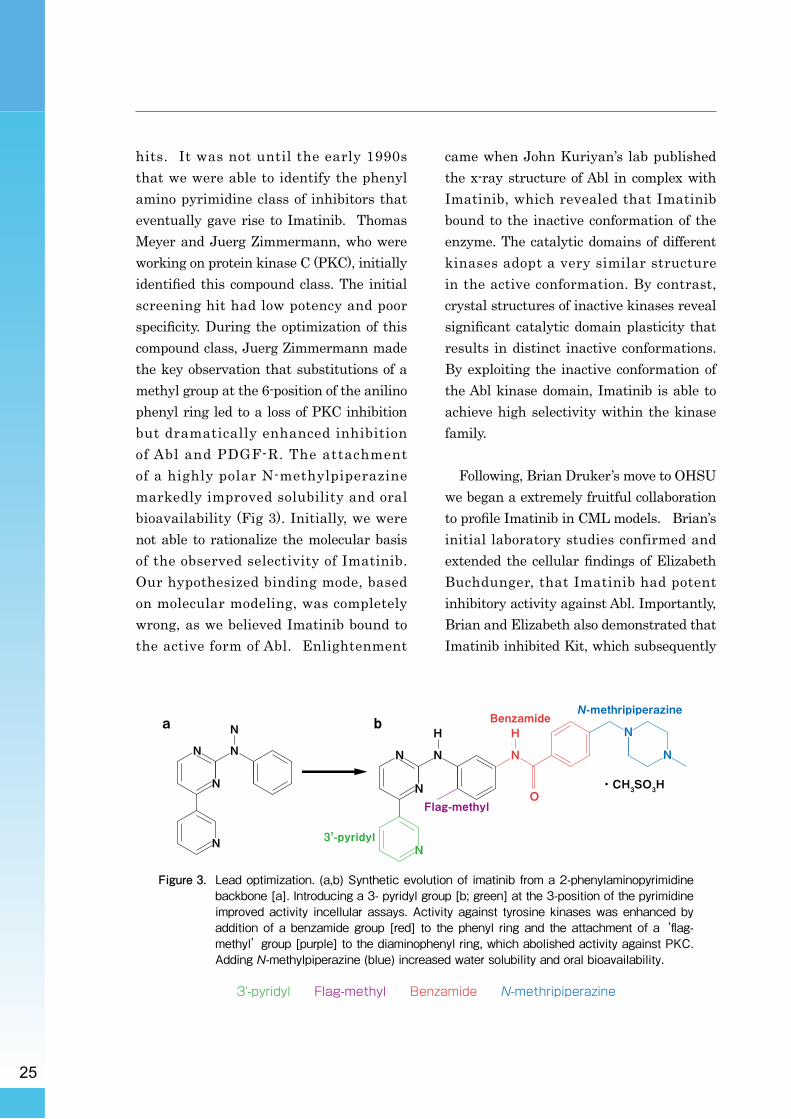
hits. It was not until the early 1990s that we were able to identify the phenyl amino pyrimidine class of inhibitors that eventually gave rise to Imatinib. Thomas Meyer and Juerg Zimmermann, who were working on protein kinase C (PKC), initially identified this compound class. The initial screening hit had low potency and poor specificity. During the optimization of this compound class, Juerg Zimmermann made the key observation that substitutions of a methyl group at the 6-position of the anilino phenyl ring led to a loss of PKC inhibition but dramatically enhanced inhibition of Abl and PDGF-R. The attachment of a highly polar N-methylpiperazine markedly improved solubility and oral bioavailability (Fig 3). Initially, we were not able to rationalize the molecular basis of the observed selectivity of Imatinib. Our hypothesized binding mode, based on molecular modeling, was completely wrong, as we believed Imatinib bound to the active form of Abl. Enlightenment
came when John Kuriyan’s lab published the x-ray structure of Abl in complex with Imatinib, which revealed that Imatinib bound to the inactive conformation of the enzyme. The catalytic domains of different kinases adopt a very similar structure in the active conformation. By contrast, crystal structures of inactive kinases reveal significant catalytic domain plasticity that results in distinct inactive conformations. By exploiting the inactive conformation of the Abl kinase domain, Imatinib is able to achieve high selectivity within the kinase family.
Following, Brian Druker’s move to OHSU we began a extremely fruitful collaboration to profile Imatinib in CML models. Brian’s initial laboratory studies confirmed and extended the cellular findings of Elizabeth Buchdunger, that Imatinib had potent inhibitory activity against Abl. Importantly, Brian and Elizabeth also demonstrated that Imatinib inhibited Kit, which subsequently
・CH3SO3H
a b
N
N
N
N
N 3'-pyridyl
Flag-methyl
BenzamideN-methripiperazine
N
H
N
N
N
N
NH
O
N
Figure�3.�Lead�optimization.�(a,b)�Synthetic�evolution�of�imatinib�from�a�2-phenylaminopyrimidine�backbone�[a].�Introducing�a�3-�pyridyl�group�[b;�green]�at�the�3-position�of�the�pyrimidine�improved�activity� incellular�assays.�Activity�against� tyrosine�kinases�was�enhanced�by�addition�of�a�benzamide�group�[red]� to� the�phenyl� ring�and� the�attachment�of�a�‘flag-methyl’�group�[purple]�to�the�diaminophenyl�ring,�which�abolished�activity�against�PKC.�Adding�N-methylpiperazine�(blue)�increased�water�solubility�and�oral�bioavailability.
3'-pyridyl Flag-methyl Benzamide N-methripiperazine

26
led to clinical testing of Imatinib in GIST. At the time, this finding cause significant concern as we did not know if this activity would be an advantage (i.e. enhance the killing of CML myeloid cells) or result in myelosuppression by blocking bone marrow recovery). A pivotal experiment from Brian’s laboratory, using ex-vivo CML patient cells, showed that Imatinib caused a dramatic reduction in the number of Bcr-Abl–positive colonies formed, with minimal inhibition of normal colony formation. This revealed that Imatinib was selective for killing Bcr-Abl transformed cells, and allowed the growth of normal cells. These exciting results had a major influence on the selection of CML as the first initial clinical indication for developing Imatinib. In further experiments, Imatinib was shown to selectively suppress the proliferation of Bcr-Abl–expressing cells in vitro and in vivo.
Based on its biological profile and its attractive drug like properties, we began development of Imatinib in 1994. However, after relatively smooth progress during the discovery phase, we soon ran into a major problem. Our initial strategy was to develop Imatinib as an i.v. infusion. However, the i.v. formulation failed in toxicology studies due to drug precipitation during infusion.
In retrospect this setback was extremely fortunate, as it forced a switch to an oral dosage form of Imatinib. This approach allowed the subsequent clinical testing of Imatinib in chronic phase CML in the outpatient setting. In June 1998 Imatinib entered clinical trials under Brian’s leadership, and rapidly demonstrated clinical activity, and gained FDA approval.
The c l in ica l act iv i ty o f Imat inib demonstrated that by blocking the “driver” oncogenic event in CML, one could kill cancer cells without major side effects on their normal counterparts. More recent clinical findings using EGF-R and Alk inhibitors have further reinforced the importance of targeting drugs to such “driver” mutations. Despite the success of Imatinib in CML, the challenge for the future will likely be how to develop rational combination therapies based on our evolving understanding of cancer pathogenetics. This will be most challenging in solid tumors, which may have accumulated multiple oncogenic mutations and have greater heterogeneity.
In conclusion, the identification of key drivers in different malignancies, combined with the use of molecular diagnostics and biomarkers, will be essential steps in developing future targeted drugs and their rational use in combination therapies.

27
2012 (28th) Japan Prize Laureate
How the World’s Strongest “Neodymium Magnet” Came to Exist
In finding a new magnetThe first step in finding a new magnet
is to find a new compound, and based on the compound, the second step is to find a new alloy structure suitable for a magnet and the method in which to create it. As a compound, it is important that three criteria are met, namely: (1) a high Curie temperature (temperature at which magnetization becomes virtually zero), (2) high magnetization and (3) high magnetic anisotropy (phenomena in which the magnetic energy changes according to the direction of magnetization). For the alloy structure, cellular constitution is most suitable. A cellular constitution is a structure similar to that of cells in a living organism. A single division corresponding to a cell is called a crystal grain. It is imperative that each one of the crystal grains is magnetically insulated.
When I was work ing for Fu j i t su Laboratories, I was given ‘magnets’ as a
research theme. At the time, the strongest magnet in existence was samarium-cobalt (Sm-Co) magnet. In particular, the Sm2Co17 magnet invented by a Japanese researcher Yoshio Tawara was the strongest. Each year its record strength was renewed, inciting magnet researchers to become enthusiastic about Sm2Co17 studies. Sm2Co17 magnet is based on the Sm2Co17 compound, from which Dr. Tawara discovered an alloy composition and method for creating an ideal cellular alloy structure.
How neodymium magnet was discoveredMy research theme was the development
of magnets for use in specialized electric components. Commercially available S m - C o m a g n e t s w e r e t o o w e a k i n mechanical strength and could not be used in such components. Hence, my task was to develop a Sm-Co magnet of high mechanical strength. Until then, I had studied neither magnets nor magnetic materials. Throughout university and
Field : Environment, Energy and Infrastructure
Dr. Masato Sagawa (Japan)President, Intermetallics Co., Ltd.Born in 1943
Achievement Developing the world's highest performing Nd-Fe-B type permanent magnet and contributing to energy conservation
28
graduate school, I did fundamental research on the structure and properties of solid surfaces and wrote a dissertation on the subject. After joining Fujitsu Laboratories, I wanted to continue my studies on solid surfaces, but I had no choice but to comply with corporate orders. I began studying magnetic materials from scratch and studied Sm-Co magnets by actively participating in conferences and symposiums.
During my graduate school years, my frequent visits to and studies at the laboratories of famous researchers paid off, and my research on magnetic materials made tremendous headway. I gradually became absorbed in my research. I gave myself the research theme of improving the mechanical strength of Sm2Co17 magnets, and I refined the prototype-making equipment and the magnetic property evaluation equipment for Sm2Co17 magnets. No special budget was allotted, which forced me to seek idle facilities within the company to conduct a series of experiments. Owing to Fujitsu Laboratories being a first-tier company, sophisticated analysis equipment and physical properties evaluation equipment were available. I had neither supervisor nor research partner. Happily on my own, I continued my studies and pursued my research.
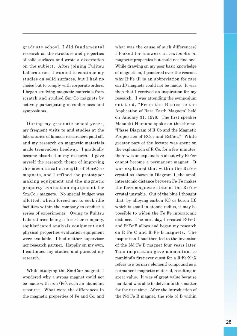
While studying the Sm2Co17 magnet, I wondered why a strong magnet could not be made with iron (Fe), such an abundant resource. What were the differences in the magnetic properties of Fe and Co, and
what was the cause of such differences? I looked for answers in textbooks on magnetic properties but could not find one. While drawing on my poor basic knowledge of magnetism, I pondered over the reasons why R-Fe (R is an abbreviation for rare earth) magnets could not be made. It was then that I received an inspiration for my research. I was attending the symposium ent i t l ed , “From the Bas i c s t o the Application of Rare Earth Magnets” held on January 31, 1978. The first speaker Masaaki Hamano spoke on the theme, “Phase Diagram of R-Co and the Magnetic Properties of RCo5 and R2Co17.” While greater part of the lecture was spent on the explanation of R-Co, for a few minutes, there was an explanation about why R2Fe17 cannot become a permanent magnet. It was explained that within the R2Fe17 crystal as shown in Diagram 1, the small interatomic distance between Fe-Fe makes the ferromagnetic state of the R2Fe17 crystal unstable. Out of the blue I thought that, by alloying carbon (C) or boron (B) which is small in atomic radius, it may be possible to widen the Fe-Fe interatomic distance. The next day, I created R-Fe-C and R-Fe-B alloys and began my research on R-Fe-C and R-Fe-B magnets. The inspiration I had then led to the invention of the Nd-Fe-B magnet four years later. This inspiration gave momentum to mankind’s first-ever quest for a R-Fe-X (X refers to a ternary element) compound as a permanent magnetic material, resulting in great value. It was of great value because mankind was able to delve into this matter for the first time. After the introduction of the Nd-Fe-B magnet, the role of B within

29
the magnet was clarified, as shown in Diagram 2.
At a relatively early stage in my research, I was able to attain the Nd-Fe-B compound which met the three criteria as a permanent magnetic compound described before. I could not, however, create a magnet based on it. I tried various alloy compositions and manufacturing conditions with a cellular structure in mind, but could not create a prototype with magnetic properties. On the other hand, I had reached my goal in the development of a mechanically strong Sm2Co17 magnet
which was the official research subject, and therefore was requested to move on to the next subject. As the next research subject, I suggested the Nd-Fe-B magnet but my request was declined. I felt I was on the verge of a great new discovery, but no one believed me. While carrying on with the next research subject given to me by the company, I continued the research of Nd-Fe-B in between my official duties even though it was a struggle to keep up the momentum of the research. Then one day, I was called by my superior, and for some unknown reason, I was yelled at with a window-shattering voice. I left the
R2Fe17
R Fe NdBFe
Nd2Fe14B
Diagram�2���The�crystal�structure�of�the�Nd2Fe14B�compound� constituting� the�Nd-Fe-B�magnet
��In�this�compound,�B�plays�an�important�role.��Dr.�Junjiro�Kanamori�revealed�that�B�plays�the�role�of�changing�the�electron�state�of�the�neighboring�Fe�into�the�electron�state�similar�to�that�of�Co.
Diagram�1���The� crystal� structure� of� the� R2Fe17�compound��
� �Dr.�Masaki�Hamano�explained�that�the�reason�in�which�the�ferromagnetic�stage�of�R2Fe17�compounds�is� unstable� is� due� to� the� small� Fe-Fe� interatomic�distance� seen� in� this� structure,� in� particular� the�Fe-Fe� interatomic�distance�at� the�atomic�position�called�the�dumbbell�site�shown�in�red.

30
room and promised to come back the next day with my resignation. In the three months from that day until the day of my resignation, I made extensive progress in my research of the Nd-Fe-B magnet because my superior permitted me to engage in experiments during that time.
After submitting my resignation and wondering where to work next, I decided to join Sumitomo Special Metals which has its headquarters in Osaka. When I expounded on my concept regarding the new type of magnet, the then-president Mr. Norishige Okada welcomed the idea with open arms. With several assistant researchers, a Nd-Fe-B magnet research team was set up. Within the first few months, the research proved successful. The Nd-Fe-B magnet, which surpassed the record held by Sm2Co17 as the world’s strongest magnet, was developed. In add i t i on , the prob lem o f l ow heat resistance which was discovered to be the weak point of this magnet immediately after its development, was overcome by means of a heat resistance improvement method which called for substituting a part of Nd with Dy (dysprosium). Two years thereafter, the industrialization of the new magnet was achieved, and three years after my joining the company, its mass production was begun. The production output of the Nd-Fe-B sintered magnet increased exponentially, and in 2000 it surpassed 10,000 tons/year. The main usage for the magnet is in electronic equipment such as hard disk drives.
Supporting further development I worked for Sumitomo Special Metals
for five and a half years, then resigned and founded Intermetallics Company Limited at the beginning of 1988. For 15 years since its founding, I worked as a corporate consultant and travelled around the world about 100 times. Thereafter, I joined a venture-fostering institution called Kyodai Katsura Venture Plaza. Since then, I have resigned from all consultancy work to concentrate on my research. My research agenda was to improve the manufacturing process of Nd-Fe-B magnets, a subject which I had been nurturing for many years.
Presently, the Nd-Fe-B sintered magnet is at its second stage of development. The main usage in this stage is for relatively large motors and generators used in hybrid cars, electric cars, air-conditioners, wind power generators, and elevators. By using the strongest magnet on any of the above, energy conservation and the prevention of global warming can be effected. Nd-Fe-B sintered magnets used for the above purposes must have heat resistance and therefore requires large amount of Dy. The research theme I had been nurturing for many years was the development of a manufacturing process for highly heat resistant Nd-Fe-B sintered magnets without the use of Dy. Intermetallics proposed this research theme, and with the acquisition of research funds from venture capital, bank investors and such large corporations as Mitsubishi Corporation and Daido Steel Company, as well as research budget from the Ministry

31
of Economy, Trade and Industry (METI) and NEDO, I carried out the development of this new process. The study of this new process was successful, and considerable Dy-saving, heat resistant Nd-Fe-B sintered magnets have been achieved. Mass production of the prototype has also been successfully completed in the pilot plant. Presently, a mass production plant using this new process is underway, and from the beginning of 2013, the production of Dy-saving highly heat resistant Nd-Fe-B sintered magnets is scheduled to begin.
Glad to have become a researcher!A researcher studies through brain-
engaging exercises, and if the research is successful, he or she can contribute to society. A researcher is a wonderful occupation, for one is able to contribute to society by means of brain-engaging exercises. I think mankind’s greatest joy comes from being able to contribute to
society. Through research, researchers can find solutions to various challenging social problems such as the prevention of global warming. Also by unveiling such mysteries as the mystery of the universe, the mystery of physical matter and the mystery of biological evolution, mankind’s horizons may be broadened. There is no other occupation quite as fulfilling as that of a researcher or a scientist.
Being awarded the Japan Prize is tangible evidence of how our research results are contributing to society. The prize also conveys this to many people. Scientific development is made possible t h r o u g h t h e c o o p e r a t i o n o f m a n y researchers. I would like to accept this prize on behalf of the researchers working in the same field as myself. I would also like to share the privilege and honor of this prize with my contemporary researchers in the same field.

32
The Japan Prize Is…~ A prestigious international award in the
fields of science and technology ~
The Japan Prize Foundation honors those whose original and outstanding achievements in science and technology are recognized as having advanced the frontiers of knowledge and served the cause of peace and prosperity for mankind. A Japan Prize laureate receives a certificate of merit and a commemorative medal. A cash prize of 50 million Japanese yen is also awarded in each prize category.
The Japan Prize Foundation
The Foundation was established in 1982, aiming to contribute to further development of science and technology. In addition to recognizing outstanding achievements with the Japan Prize, the Foundation has been promoting knowledge and information on science and technology by hosting the "Easy-to-understand Science and Technology" seminars and awarding Research Grants to help nurture young scientists.
“Easy-to-understand Science and Technology” Seminars
The Foundation holds a series of seminars on advanced technologies used widely in everyday-life. In the seminars designed for students and general public, experts in the related fields explain in plain terms the technologies that are also the focus of interest at that time.
Stockholm International Youth Science Seminar (SIYSS)
Under the auspices of the Swedish Federation of Young Scientists and with the support of the Nobel Foundation, The Science and Technology Foundation of Japan sends two Japanese students to the annual Stockholm International Youth Science Seminar.
Research GrantsThe Foundation provides research grants to scientists
and researchers under 35 years of age. Every year, the Foundation selects projects in the same fields as the Japan Prize of that year and gives one million Japanese yen for a project.

2012
DIC 196 背幅1mm
2012


![Donguri shimai - Japan Foundation...Prize for Literature in 1988 for “Mūnraito shadō” [Moonlight Shadow]. Won the Minister of Education Encouragement Prize for New Artists in](https://static.fdocuments.in/doc/165x107/5e94bc0f20b8ee3bee0aa454/donguri-shimai-japan-foundation-prize-for-literature-in-1988-for-aoemnraito.jpg)