Professional Use Information - Food and Drug Administration · Professional Use Information VISX...
80
Professional Use Information VISX STAR S4 IR TM Excimer Laser System and WaveScan WaveFront® System CustomVue T M Treatments for High Myopia and Myopic Astigmatism For the reduction or elimination of myopia and myopic astigmatism from -6.00 to -11.00 D MRSE, with cylinder between 0.00 and -3.00 D RESTRICTED DEVICE: U.S. Federal Law restricts this device to sale, distribution, and use by or on the order of a physician or other licensed eye care practitioner. U.S. Federal Law restricts the use of this device to practitioners who have been trained in its calibration and operation and who have experience in the surgical management and treatment of refractive errors. This document provides information concerning the intended clinical use of the STAR S4 IR Excimer Laser System. For complete information concerning system components, safety instructions, installation, maintenance, and troubleshooting, refer to the STAR S4 IR Excimer Laser System Operator's Manual. Carefully read all instructions prior to use. Observe al! contraindications, warnings, and precautions noted in these instructions. Failure to do so may result in patient and/or user complications. VISX, Incorporated 3400 Central Expressway Santa Clara, CA 95051-0703 U.S.A. Tel: 408.733.2020 CustomVue High Myopia, Rev A 4te
Transcript of Professional Use Information - Food and Drug Administration · Professional Use Information VISX...

Professional Use Information
VISX STAR S4 IRTM Excimer Laser Systemand WaveScan WaveFront® System
CustomVue TM Treatments for High Myopia and Myopic Astigmatism
For the reduction or elimination of myopia and myopic astigmatism from -6.00 to -11.00 DMRSE, with cylinder between 0.00 and -3.00 D
RESTRICTED DEVICE: U.S. Federal Law restricts this device to sale, distribution, and use byor on the order of a physician or other licensed eye care practitioner. U.S. Federal Law restrictsthe use of this device to practitioners who have been trained in its calibration and operation andwho have experience in the surgical management and treatment of refractive errors.
This document provides information concerning the intended clinical use of the STAR S4 IRExcimer Laser System. For complete information concerning system components, safetyinstructions, installation, maintenance, and troubleshooting, refer to the STAR S4 IR ExcimerLaser System Operator's Manual.
Carefully read all instructions prior to use. Observe al! contraindications, warnings, andprecautions noted in these instructions. Failure to do so may result in patient and/or usercomplications.
VISX, Incorporated3400 Central ExpresswaySanta Clara, CA 95051-0703U.S.A.Tel: 408.733.2020
CustomVue High Myopia, Rev A
4te

© Copyright 2005 by VISX®, Incorporated
All Rights Reserved
VISX®, VisionKey®, ActiveTrak®, WaveScan®, WaveScan WaveFront®D, and WavePrint® are registered trademarksof VISX, Incorporated.CustomVuer~t and STAR 34 IR" 4 are trademarks of VISX, Incorporated.The VISX Treatment Card utilizes software owned by Bull Worldwide Information Systems.Accutane® is a registered trademark of Hoffmann-La Roche Inc. Gordarone® is a registered trademark of Sanofi-Synthelabo, Inc. Imitrex® is a registered trademark of GlaxoSmithKline.No part of this publication may be reproduced or transmitted in any form or by any means, electronic or mechanical,including photocopying, recording, or any information storage and retrieval system, without permission in writing fromVISX, Incorporated.
CustomVue High Myopia, Rev A

This Page intentionally Left Blank
CustomVue High Myopia, Rev A
'~,
Table of Contents
General Warnings............................................ 1
1.1 Device D escription .... ..................... ... ... 3
1.1.1 STAR 54 iR" Excimer Laser System . ... ............... ....... _
1.1.2 W aveScan W aveFront® System ................... ............. 4
2.1 Indications, Contraindications, Warnings, Precautions, andA d v e rse E v e n ts......._ _............ ........... _ .......... ...... . .. 7
2.1.1 Indications for Use ...... ......... _ _ . ............... 7
2.1.2 C ontraindications ....... ..... ... _.........................72 .1 .3 W a rn in g s .. ... _ . . ... .. ... .. .. .. .. .. ...... .. ... . ... . _. ........ ... .. .... .8
2.1.4 P recautions . ................................................. .... .........
A . G n r l ... .. .. .. .. ..Gen eral.. .. ........ ... ....... .. .... .. . _ .. .. ... . . .8.....
B . P atient S election .......................................... ..........10C . P ro ce d u re ... .. ... .. ... ... ... . .......... .... 1.. .... .. ... .. .. .. .. . .. . .. .
D . P ost-P roced ure ........................................................ 1 12.1.5 A dverse E vents ................................................. .......... 12
3.1 C lin ical R e s u lts.. ..... ..... _....................-.. ... .......... ... 143.1.1 High Myopia W ith or W ithout Astigmatism ........................ ... __......_14
A . A bout the Study ........... ... ... ......... _ . ................. _ . .. 1 4B. Patient A ccountability 1............................................... _.._ 4C. D ata A nalysis and Results ...... ... _ ............. ............. ............. 1 61) Pre-O perative Characteristics ......................... ... .. _ . ........... 1 62) Uncorrected Visual Acuity (UCVA)........_ ........................ 1 83) Accuracy of MRSE Over Time . .......... .... ... ... ......... 214) Stability of O utcom e .................................... ......... ....... 235) Efficacy of Correction of Astigmatism ................... .. __............. ... 25
6) Higher Order Aberrations ............ __..........267) W aveScan W avefront Diameter ......................................... 288) Best Spectacle-Corrected Visual Acuity (BSCVA) ........... _... _299) C ontrast Sensitivity A nalysis .............................. ............ .... 2910) R etreatm ents ............... ........................... ,31
1 1) Patient Sym ptom s . .................. ..... .. 31
CustomVue High Myopia, Rev A

12) Summary of Key Safety and Effectiveness Variables ... ............................................36
4.1 Surgical Planning and Procedures 43
4.1.1 Introduction 434.1.2 Pre-Operative (Examination of the Patient) 434.1.3 Peri-Operative (Anesthesia and Analgesia) 44
4.1.4 Post-Operative 44A. Medications 44B . F ollow -up C are ............. .......... ............ ......... ......................44
5.1 STAR S4 IRT M Surgical Procedure .................... ............ .................. 4...............455.1.1 Step-by-Step Procedure .................................................. ............................................... 455.1.2 U sing the Iris R egistration System ............................................................................................. 495.1.3 U sing the A ctiveT rak® System ...................................................................... 5.............................51
CustomVue High Myopia, Rev A
C3

List of Tables
Table 2-1 - Summary of Adverse Events N=184. .............................................. 12Table 2-2 - Summary of Complications N=184 ... ..........................................13Table 3-1 - Dem ographics N=184 ... ................................................ 1...................15Table 3-2 - Patient Accountability N=184 .......... .................................................. 15
Table 3-3 - Pre-Operative Refractive Error Stratified by Sphere and Cylinder N=184 ............ 16
Table 3-4 - Pre-Operative Refractive Error Stratified by Spherical Equivalent andCylinder N=t 184 .................................................................................................. 17
Table 3-5 - UCVA Over Time: All Eyes N=184 ....................................................................... 18
Table 3-6 - UCVA Over Time: Spherical Myopia N=83 .......................................................... 19
Table 3-7 - UCVA Over Time: Myopic Astigmatism N=101 ................................... 19
Table 3-8 - Post-Operative Uncorrected Visual Acuity Compared to Pre-Operative BestSpectacle-Corrected Visual Acuity N=184 ............................. 2......................20
Table 3-9 -- Accuracy of Manifest Refraction Attempted vs. Achieved: All Eyes N=1 84 .......... 21Table 3-10 - Accuracy of Manifest Refraction: Attempted vs. Achieved, Eyes with
Spherical Myopia N=83 ..................................................................................... 22Table 3-11 - Accuracy of Manifest Refraction: Attempted vs. Achieved: Eyes with
Myopic Astigmatism N=101 ............................................................................... 22Table 3-12 - Stability of MRSE for Eyes that Underwent the 1, 3, 6, and 9 Month
Exams N=164 ................................. ...................................................................23Table 3-13 - Stability of MRSE for Eyes that Underwent Two Consecutive Visits .................. 24Table 3-14 - Reduction of Absolute (Non-Vector) Cylinder at 6 Months: Myopic
Astigmatism N=95 ............................................................................................. 25Table 3-15 - Vector Analysis at Stability (6 Months): Eyes with Myopic Astigmatism
N=95 ................................................................................................................. 26
Table 3-16- RMS Over Time N=184 ...................................................................................... 27
Table 3-17 - Summary of 6 Month Key Safety and Effectiveness Variables For All Eyes
Stratified by W aveScan Diameter ............................................ 2........................28
Table 3-18 - Change in BSCVA Over Time: All Eyes N=184 ................................................. 29
Table 3-19 - Contrast Sensitivity: All Eyes N=1 84 .... ......................................................... 30
Table 3-20 - Change in Contrast Sensitivity: All Eyes N=184 ................................................. 31
Table 3-21 - Summary of Subject Symptoms: All Eyes N=182 ......................................... 32
CustomrVue High Myopia, Rev A

Table 3-22 - Change in Patient Symptoms: Comparison of Pre-Operative Best-
Corrected Vision to Post-Operative Uncorrected Vision All Eyes N=182 ............ 33
Table 3-23 - Summary of Patient Satisfaction for All Eyes N=182 ............................ 3............4
Table 3-24 - Change in Patient Satisfaction: Comparison of Pre-Operative Best-
Corrected Vision to Post-Operative Uncorrected Vision: All Eyes N=182 ......... 35
Table 3-25 - Summary of Key Safety and Effectiveness Variables: All Eyes N=184 ............. 7
Table 3-26 - Summary of Key Safety and Effectiveness Variables: Spherical MyopiaN =83 ...... ................................................................. .................................... 38
Table 3-27 - Summary of Key Safety and Effectiveness Variables: Myopic AstigmatismN=101 ................................................................................................................ 39
Table 3-28 - Summary of Key Safety and Effectiveness Variables at Stability Endpointof 6 Months Stratified by Pre-Operative MRSE, All Eyes N=178 ........................ 40
Table 3-29 - Summary of Key Safety and Effectiveness Variables at Stability Endpointof 6 Months Stratified by Pre-Operative MRSE, Spherical Myopia N=83 ............ 41
Table 3-30 - Summary of Key Safety and Effectiveness Variables at Stability Endpointof 6 Months Stratified by Pre-Operative MRSE, Myopic Astigmatism N=95 ....... 42
CustomVue High Myopia, Rev A
S£
General Warnings
STAR S4 IR" EXCIMER LASER SYSTEM
RESTRICTED DEVICE: U.S. Federal Law restricts this device to sale, distribution, and use byor on the order of a physician or other licensed eye care practitioner. U.S. Federal Law restrictsthe use of this device to practitioners who have been trained in its calibration and operation andwho have experience in the surgical treatment and management of refractive errors.
Performance of procedures, use of controls, or any other adjustments other than thosespecified herein may result in a hazardous condition.
Never operate the laser in the presence of flammable anesthetics or other volatilesubstances, such as alcohol.
GAS HANDLING: High-pressure gas cylinders are contained in a protected compartment withinthe STAR S4 IR~h Excimer Laser System. Storage of additional cylinders and the replacement ofused cylinders must be done in accordance with "Gas Safety" (Section 4.5) and "GasMaintenance" (Section 14. 1) and must comply with all applicable Occupational Safety andHealth Administration (OSHA), local, and national requirements for gas safety.
The premix (argon/fluorine) gas mixture used in this laser system is highly toxic. VISX,Incorporated, recommends that anyone working with the gas cylinders: 1) be trained in theproper handling of toxic and compressed gases, 2) know the location of the emergency exhaustfan/room purifier switch, 3) have easy access to all required protective equipment, and 4) befamiliar with safety procedures and Materials Safety Data Sheets (MSDS) provided by the site'ssafety officer. Gas discharge into the atmosphere may be evidenced by a sharp, penetratingodor and by eye, nose, and throat irritation.
SKIN AND EYE EXPOSURE: The STAR S4 IR System contains a Class IV laser with an outputat 193 nm, which is potentially hazardous to the skin and the surface layers of the cornea. Thislaser radiation will not enter the eye and poses no threat to retinal structures or the crystallinelens. The fixed optical system restricts the beam path, which is bounded by the operating tableor the floor. Reflectivity from objects in operating rooms, including surgical instruments, isextremely low for 193 nm radiation.
The area of potential hazard (Nominal Hazard Zone) for production of a photochemical keratitishas been determined to be less than 40 cm from the primary beam. All healthcare personnelshould avoid direct exposure to the skin or eye by the primary beam. While no hazard mayexist farther than 40 cm from the beam, the use of protective eyewear is recommended if thepossibility exists that healthcare personnel will approach closer than this distance from theprimary beam.
PRECAUTIONS: Carefully read all instructions prior to use. The laser beam is invisible. Theuser cannot tell if the laser is emitting radiation by looking for the beam. Observe allcontraindications, warnings, and precautions noted in this manual. Failure to do so may result inpatient and/or user complications.
CustomVue High Myopia, Rev A - 2-

ELECTROMAGNETIC FIELD (EMF): The thyratron emits an electromagnetic pulse which isshielded by the metal coverings of the STAR S4 IRTM Excimer Laser System. This metalcovering reduces the EMF below the limits set by applicable standards for electromagneticcompliance.
WARNING: The effects of electromagnetic emissions from the excimer laser system on otherdevices, such as cardiac pacemakers or implanted defibrillators, is unknown. Operation of thelaser in proximity to such devices is not recommended.
AIRBORNE CONTAMINANTS: Airborne contaminants which are produced by the ablationprocess are captured in proximity to the cornea near the point of production and fed into anaspirator with a filter. This aspirator is designed to prevent any of the products of ablationfrom contaminating the surgical suite.
WAVESCAN WAVEFRONT® SYSTEM
PRECAUTIONS: The WaveScan WaveFront System is a Class Ill accessory device. It containsa Class IIIB laser with a 780 nm output. The light levels accessible with the covers off and theinterlocks defeated are potentially hazardous to skin and eyes. Avoid direct exposure to theselight levels. The covers should be removed only by trained service personnel. To avoidinadvertent exposure to laser radiation, never operate the system with the covers opened orremoved. Doing so may expose the user or others to stray laser radiation.
Any service requiring access to the interior of the system should be performed only by VISX®service personnel or by qualified service technicians who have received specific systemtraining. Never try to defeat safety interlocks after removing covers. The safety interlocks arethere for user protection. All power cords must be connected to the medical grade isolationtransformer in the system.
Carefully read all instructions prior to use. Retain all safety and operating instructions for futureuse. Observe all contraindications, warnings, and precautions noted in the WaveScanWaveFront Operator's Manual.
CustomVue High Myopia, Rev A - 3 -
S7

1.1 Device Description
1.1.1 STAR S4 IRTM Excimer Laser System
The STAR S4 IR System is designed to create a superficial lamellar keratectomy on exposedcorneal tissue. Corneal tissue is removed by a process known as AblativePhotodecomposition. Ablative Photodecomposition occurs when far-ultraviolet radiation reactswith organic molecules, resulting in the photochemical breakdown of the molecular bondswithout a significant local thermal effect. The source of the far-ultraviolet photons is a high-efficiency, gas-discharge excimer laser that electronically excites a combination of argon andfluorine, producing an ultraviolet wavelength of 193 nm. The STAR S4 IR Excimer LaserSystem combines submicron precision of tissue removal by an excimer laser with asophisticated computer controlled delivery system.
Features and components of the STAR S4 IR System include:
Excimer LaserAn argon-fluoride excimer laser module, with an output wavelength of 193 nm.
Gas Management SystemA gas cabinet containing a working gas cylinder for laser operation; a gas cleaning system; agas leak audio alarm with a sensor to detect fluorine (one part-per-million); a gas dischargesystem, using an activated charcoal filter to absorb fluorine; an emergency safety system usinga positive-action solenoid safety valve, which automatically seals the premix cylinder in theevent of a power failure; and a second charcoal scrubber to neutralize fluorine in case of a leak.The STAR S4 IR laser software also contains a refinement to the method of STAR laser beamenergy control by inclusion of an ozone compensation system.
Laser Beam Delivery SystemThe STAR S4 IR laser system delivers spatially scanning ultraviolet pulses of variable diametersand slits on to the cornea. The range of diameters and slits available during treatments are 0.65mm to 6 mm. Beam shaping and homogenizing optics designed to produce a uniform, coaxialbeam profile; a spatial integrator and beam rotator for temporal integration; and an irisdiaphragm and rotating slit blades used to shape the beam. Conventional STAR treatmentsutilize sphere, cylinder and axis components which are entered manually into the laser by theoperator to generate the ablation treatment. CustomVueTM treatment information is generatedon the WaveScan® system and transferred to the STAR S4 IR Excimer Laser System. Thetransferred information includes patient information, eye and refraction information, image of theeye, eye alignment information, and ablation instructions to the laser for beam diameters andthe exact locations of the beam on the cornea. The variable spot scanning (VSSTM) feature ofthe laser, used for CustomVueTM treatments delivers variable diameter ultraviolet pulses toprecise locations by the scanning delivery system. The VSS algorithm optimizes the ablationpattern by choosing the best combination of beam diameters and locations to achieve a targetshape. VSS expands the laser capability to achieve a broader spectrum of ablation shapesthan conventional treatments because the conventional algorithm optimizes only the diameterfor myopic treatments and slits for hyperopic treatments.
Patient Management SystemThe ActiveTrak® System, which enables the laser beam to track the patient's eye movementsduring the treatment, an operating microscope with reticle, used to observe a patient procedureand to facilitate accurate focus and laser beam alignment; a debris-removal system designed toevacuate the debris plume that occurs during ablation; a patient operating chair used to align
CustomVue High Myopia, Rev A - 4 -

the patient for treatment; a video camera and monitor used to record and monitor patienttreatment; an illumination device used to illuminate the patient's eye for observation andtreatment, and a fixation LED used by the patient to maintain proper alignment during treatment.Wavefront-guided treatments using the STAR S4 IRTM and WaveScan Systems utilize anautomated iris registration system. The angle of rotation of the patient's eye under the laser isdetermined by comparing features of the iris on the WaveScan image to the same featureslocated in the image of the iris taken using the STAR S4 IR camera. The treatment is rotated toalign precisely with the rotation of the patient's eye under the laser.
Computer ControlA PC-compatible computer, video monitor, keyboard with touchpad for user interface(Windows ® 1standard), printer, a floppy drive to store patient information on floppy disks, a USBport, a VISX® treatment card driver, and system software.
VISX® Treatment CardThe VISX Treatment Card system comprises a card drive and treatment cards. The VISXtreatment card defines the number and the types of treatments available.
1.1.2 WaveScan WaveFront® System
The WaveScan WaveFront System is a diagnostic instrument indicated for the automatedmeasurement, analysis, and recording of refractive errors of the eye: including myopia,hyperopia, astigmatism, coma, spherical aberration, trefoil, and other higher order aberrationsthrough sixth order, and for displaying refractive data of the eye to assist in prescribingrefractive correction..
The WaveScan WaveFront System measures the refractive error and wavefront aberrations ofthe human eye using a Hartmann-Shack wavefront sensor. The measurements can be used todetermine regular (sphero-cylindrical) refractive errors and irregularities (aberrations) thatcause decreased or blurry vision in the human eye.The function of the Hartmann-Shack sensor is to measure the refractive error of the eye byevaluating the deflection of rays emanating from a small beam of light projected onto the retina.To control the natural accommodation of the eye during WaveScan imaging, the systemincorporates a fogged fixation target.The WaveScan® System optical head projects a beam of light onto the retina. The light reflectsback through the optical path of the eye and into the wavefront device. The reflected beam isimaged by a lenslet array onto the charge-coupled device (CCD). Each lens of the array gatherslight information (deflection information) from a different region of the pupil to form an image ofthe light that passes through that region of the pupil. An array of spots are imaged on the CCDsensor. The system compares the locations of the array of spots gathered from the CCD to thetheoretical ideal (the ideal plane wave).The WaveScan System software uses these data to compute the eye's refractive errors andwavefront aberrations using a polynomial expansion. The system displays therefractive errorsand wavefront aberrations as the optical path difference (OPD) between the measured outgoingwavefront and the ideal plane wave. The WaveScan system software subtracts the refractiveerrors from the wavefront errors map and displays the higher order aberrations as OPD errors.Regions of the pupil with positive OPD are in front of the ideal plane wave and areas withnegative OPD are behind the ideal plane wave.
1 Windows® is a registered trademark of Microsoft Corporation.
CustomVue High Myopia, Rev A - 5 -

Features and components of the WaveScan WaveFront® System include:Computer ControlThe WaveScan WaveFront® System includes software to calculate the desired laser visioncorrection treatment (CustomVue T treatment) from the WavePrint® measurement. The softwaregenerates two sets of laser instructions, one for PreVue® plastic and the other for the patientprocedure. Both sets of instructions are loaded on to the STAR S4 IR" System and are used todefine the patient treatment.
PC and MonitorThe computer is PC-compatible. The monitor is a flat-panel LCD display. Keyboard andmouse (or glidepad) are Windows standard.
Isolation TransformerThe medical-grade isolation transformer complies with IEC 601-1 regulations. All power cordsconnect to the isolation transformer.
Power SupplyThe power supply provides DC power to the video cameras (CCDs), and thesuperluminescent diode (SLD).
LEDYellow (D3): Indicates SLD over-power fault. Located on back panel of power supply box.Optical HeadThe optical head includes two optical units for the precompensation of sphere andastigmatism, adjusted by three stepper motors, two CCD cameras, and a light source (theSLD). A circuit continuously measures the incident power of the light source and switchesthe SLD off if the incident power exceeds a defined threshold.PrinterA high resolution color printer is included with the system.Motorized tableThe motorized table supports the WaveScan WaveFront System. Electrical ratings: 120 V -50/60 Hz, 6 A. Vertical position is controlled by a rocker control switch (vertical height can rangefrom 630 mm to 1030 mm). Table top supports the PC monitor, keyboard, mouse (or glidepad),and optical head. Shelves hold PC, printer, isolation transformer, and power supply.
CustomVue High Myopia, Rev A - 6 -
6::¢

This Page intentionallY Left Blank
CustomVue High Myopia, Rev A -7-

2.1 Indications, Contraindications, Warnings,Precautions, and Adverse Events
2.1.1 Indications for Use
The STAR S4 IRTm Excimer Laser System with Variable Spot Scanning (VSSTM) and theWaveScan WaveFront® System is indicated for wavefront-guided laser assisted in situkeratomileusis (LASIK):
* for the reduction or elimination of myopia and myopic astigmatism from -6.00 to -1 1.00 0MRSE, with cylinder between 0.00 and -3.00 0;
* in patients 21 years of age or older; and* in patients with documented evidence of a change in manifest refraction of no more than
1.0 0 (in both cylinder and sphere components) for at least one year prior to the date ofpre-operative examination.
Refer to the preceding General Warnings section of this Professional UseIn formation Manual, in addition to the warnings and precautions found inthis section.
2.1.2 ContraindicationsLaser refractive surgery is contraindicated:
* in patients with collagen vascular, autoimmune or immunodeficiency diseases.* in pregnant or nursing women.* in patients with signs of keratoconus or abnormal corneal topography.* in patients who are taking one or both of the following medications: Isotretinoin
(Accutane%) Amiodarone hydrochloride (Cordarone )
2Accutane is a registered trademark of Hoffmann-La Roche Inc.3Cordarone® is a registered trademark of Sanofi-Synthelabo, Inc
CustomVue High Myopia, Rev A -8-

2.1.3 Warnings
LASIK is not recommended in patients who have:
diabetes.a history of Herpes simplex or Herpes zoster keratitis.significant dry eye that is unresponsive to treatment.severe allergies.
2.1.4 Precautions
A. General
To avoid corneal ectasia, the posterior 250 microns (prm) of corneal stroma should notbe violated.
The treatment of highly myopic eyes necessitates the removal of significant amounts ofcorneal tissue. The WaveScan calculates the estimated residual bed depth using thepachymetry and intended flap thickness entered by the user. Actual flap thicknessesmay vary. All users should be aware that during the FDA clinical trial for highly myopiceyes, an "in the bed" pachymetric measurement was performed to assure a minimumresidual stromal bed of 250 microns. If the corneal flap was thicker than intended andthe 250 micron minimum would have been violated by a CustomVue treatment, userswere instructed not to perform a CustomVue treatment on that eye.
The safety and effectiveness of this laser for LASIK correction have NOT been established inpatients:
with progressive myopia, hyperopia, myopic or hyperopic astigmatism; ocular disease;corneal abnormality; previous corneal or intraocular surgery; or trauma in the ablationzone.with a residual corneal thickness less than 250 microns at the completion of ablation.with a history of glaucoma.
· who are taking the medication Sumatriptan (Imitrex®4).
The effects of laser refractive surgery on visual performance under poor lighting conditions havenot been determined. It is possible, following LASIK treatment, that patients will find it moredifficult than usual to see in conditions such as very dim light, rain, snow, fog, or glare frombright lights at night. Visual performance possibly could be worsened by large pupil sizes ordecentered pupils.
Pupil size should be evaluated under mesopic illumination conditions. Patients with largemesopic pupil size (>6.6 mm) should be advised of the potential for negative effects on visionafter surgery, such as increased frequency of glare and halos, and decreased satisfaction atnight under conditions with glare, such as night driving.
Pre-operative evaluation for dry eye should be performed. Patients should be advised of thepotential for dry eye post-LASIK surgery.
4Imitrex®is a registered trademark of GlaxoSmithKline.
CustomVue High Myopia, Rev A - 9 -
(53

Pre-operative ultrasonic pachymetry measurement must be performed.The physician's adjustment of defocus was used in the clinical study to implement standardnomogram adjustment to the wavefront-guided treatments. No other uses of the physician'sadjustment of defocus have been studied, and their effects on the safety and effectivenessoutcomes for wavefront-guided LASIK are unknown.The safety and effectiveness of wavefront-guided LASIK surgery has ONLY been establishedwith an optical zone of 6 mm and an ablation zone of 8 mm.The WaveScan® sensor measures the higher order aberrations only over the diameter of thepatient's pupil, to a maximum of 7.0 mm. No optical zone diameters other than 6 mm werestudied in the U.S. wavefront-guided clinical trial for high myopia.No higher order aberrations can be measured or treated outside the wavefront measurementregion. If the surgeon extends the optical zone beyond the measured wavefront diameter, thenonuniform wavefront transition zone will overlie the attempted spherocylindrical treatment.Some treatments based on WaveScan measurement diameters of less than 6.0 mm wereused to establish the safety and effectiveness of wavefront-guided high myopia treatmentswith 6.0 mm optical zones. A comparison of results from these treatments and treatmentsbased on measurements that were 6.0 mm or larger showed that outcomes were similar.It is important to maintain a carefully controlled surgical environment. VISX recommends that allCustomVue" treatments be performed in surgical environments where the humidity is between40-45% and the temperature is between 68-720 F for best results.
The safety and effectiveness of the STAR S4 IRTM System have NOT been established forwavefront-guided LASIK surgery in patients:
* with corneal neovascularization within 1.0 mm of the ablation zone.• under 21 years of age.* over the long term (more than 1 year after surgery).* with prior intraocular or corneal surgery of any kind.* For eyes with high myopia or myopic astigmatism:
whose difference between WaveScan and manifest sphere or cylinder powers ismore than ±0.75 diopters, or whose difference between WaveScan and manifestcylinder axes is >15 degrees (for eyes with manifest cylinder power greater than0.50 D).whose difference between manifest and cycloplegic sphere powers is more than+0.75 diopters, or whose difference between manifest and cycloplegic cylinder axesis >15 degrees (for eyes with manifest cylinder power greater than 0.50 D).whose difference between WaveScan and cycloplegic sphere or cylinder powers ismore than ±0.75 diopters, or whose difference between WaveScan and cycloplegiccylinder axes is >15 degrees (for eyes with manifest cylinder power greater than0.50 D).whose BSCVA is worse than 20/20.whose WaveScan® wavefront measurement diameter is < 5 mm.for treatments greater than -11 diopters of MRSE or greater than -3 diopters ofastigmatism.for retreatment with CustomVue"' LAS IK.who were wearing contact lenses unless they had evidence of stability.
CustomVue High Myopia, Rev A - 10-
6¥f

*with anticipated postoperative keratomnetry reading <33 diopters. Anticipated post-operative keratometry values can be calculated by multiplying the magnitude of theMRSE by 0.8, and subtracting that value from the average pre-operativekeratometry value. In other words, [((Kl + K2) x 0.5) - (i MRSEI x 0.6)] > 33 0.
B. Patient Selection
Consideration should be given to the following in determining the appropriate patients forCustomnVue" treatment:
* All patients must be given the opportunity to read and understand the Patient InformationBooklet and to have all their questions answered to their satisfaction before givingconsent for Laser Assisted In Situ Keratomileusis (LASIK).
* Complete examination, including but not limited to, cycloplegic evaluation, must beperformed. The lens must be evaluated, especially in the older patient, to assure thatnuclear sclerosis or any other lens opacity is not present prior to laser surgery. Myopicpatients will have a higher incidence of retinal pathology, and indirect ophthalmoscopythrough a dilated pupil is essential.
* To obtain accurate refractive information, contact lens wearers must be examined afterabstaining from contact lens use for at least 2 weeks for soft lenses and at least 3 weeksfor hard lenses. Prior to treatment and after at least 3 weeks of contact lens abstinence,patients who wear rigid gas permeable or hard (PMVMA) lenses must have 3 centralkeratometry readings and manifest refractions taken at I week intervals, the last 2 ofwhich must not differ by more than 0.50 diopter in either meridian. All mires must beregular. Any patient with keratometry or a clinical picture that is suggestive ofkeratoconus is specifically contraindlicated as described above.
* Glaucoma is more common in myopic patients than in the genera? population. Evaluationof the optic nerve and measurement of the intraocular pressure are necessary. If thereare any concerns regarding the appearance of the optic nerve, a Humphrey 24-2Fastpac or equivalent threshold test of the visual field should be performed. If elevatedintraocular pressure and/or evidence of glaucomatous damage are found, topicalsteroids should be used only with careful medical supervision or the patient should notundergo laser refractive surgery.
* Pre-operative corneal mapping is essential on all patients to exclude topographicalabnormalities. This is especially important when astigmatism or steep keratometryreadings are present, which may indicate the presence of keratoconus or otherirregularities.
* Baseline evaluation of patients requesting CustomVue treatments should be performedwithin 30 days of the laser refractive surgery. This evaluation should address agreementbetween the manifest, cycloplegic, and the WaveScan® refraction and the limits for peakto valley higher order aberrations, BSCVA, and pupil size, as outlined in the previoussection of these Precautions.
CustomnVue High Myopia, Rev A - 1 1 -
The minimum size of the wavefront measurement must be >5 mm to calculate aCustomVue treatment.
If a PreVue® lens is used in the baseline evaluation of patients requesting CustomVue TM
treatments, the vision obtained by the patient through the PreVue lens is not meant to bepredictive of the end result that a patient might achieve. In situations where there is aclinical question regarding the applicability of the computer-generated treatment, aPreVue lens can be ablated to assist both the practitioner and the patient in evaluatingthe appropriateness of this generated treatment.
The patient should have the ability to tolerate local or topical anesthesia.
The patient should have the ability to lie flat without difficulty.
The patient should be able to fixate steadily and accurately for the duration of the laserrefractive procedure.
The patient must be able to understand and give an informed consent.
Patients must be clearly informed of all alternatives for the correction of myopia,hyperopia, and myopic and hyperopic astigmatism. These alternative corrections includebut are not limited to spectacles, contact lenses, and other refractive surgeries.
C. Procedure
The output of the laser is potentially hazardous only to the skin and the surface layers of thecornea. This radiation has not been shown to pose a threat to retinal structures or the crystallinelens. The area of potential hazard (Nominal Hazard Zone) for production of a photochemicalkeratitis has been determined to be less than 40 cm from the primary beam.
All healthcare personnel should avoid direct exposure to the skin or eye by the primary beam.While no hazard may exist farther than 40 cm from the beam, the use of protective eyewear isrecommended if the possibility exists that healthcare personnel will approach closer than thisdistance to the primary beam.
D. Post-Procedure
The following post-operative examinations are recommended on day 1, and at 1, 3, and 6months:
* WaveScan® measurement at 1, 3, and 6 months.* Uncorrected Visual Acuity (UCVA or VA-sc).* Best Spectacle-Corrected Visual Acuity (BSCVA or VA-cc).* Manifest refraction.• Intraocular pressure (Goldmann applanation) at 1, 3, and 6 months.• Slit-lamp examination.* Keratometry and videokeratography at 1, 3, and 6 months.
CustomVue High Myopia, Rev A - 12 -
166

2.1.5 Adverse Events
One hundred eighty-four (1 84) eyes were used for safety analyses. A summary of adverseevents at 1 month and later are provided in Table 2-1 . Complications are presented in Table2-2.
Table 2-1: Summary of Adverse Events (N=184)
<1 1 3 6 9 12Month Month Months Months Months Months
(n=184) (n=184) (n=180) (n=178) (n=170) (n=107)
n % n % n % n % n % n %
Corneal Infiltrate/Ulcer 0 0.0 0 0.0 0 0.0 0 0.0 0 0.0 0 0.0
Corneal epithelial defect involving 0 0.0 0 0.0 0 0.0 0 0.0 0 0.0 0 0.0the keratectomy at I month or later
Corneal edema at 1 month or later 0 0.0 0 0.0 0 0.0 0 0.0 0 0.0 0 0.0
Epithelium in the interface with loss 0 0.0 0 0.0 0 0.0 0 0.0 0 0.000.of 2 or more lines of BSCVA 000.
Miscreated Flap 0 0.0 0 0.0 0 0.0 0 0.0 0 0.0 0 0.0
Melting of the flap 0 0.0 0 0.0 0 0.0 0 0.0 0 0.0 0 0.0
Uncontrolled lOP >1 0mm Hg or any 0 0.0 0 0.0 0 0.0 0 0.0 0 0.0 0 0.0reading > 25 mm Hg
Late onset of haze beyond 6 monthswith loss of 2 lines (1 0 letters) or 0 0.0 0 0.0 0 0.0 0 0.0 0 0.0 0 0.0more BSCVA
Decrease in BSCVA of > 10 letternot due to irregular astigmatism as 0 0.0 0 0.0 0 0.0 0 0.0 0 0.0 0 0.0shown by hard contact lensrefraction, at 6 months or later
Retinal Detachment 0 0.0 0 0.0 0 0.0 0 0.0 0 0.0 0 0.0
Retinal Vascular Accidents 0 0.0 0 0.0- 0 0.0 0 0.0 0 0.0 0 0.0
Other: Two eyes of two separate subjects developed trace diffuse lamellar keratitis (DLK)prior to the 1 -month visit. One eye of one subject experienced a metallic foreign body withsubsequent rust ring. This event occurred at an interim 1 -month visit and resolved by the 3-month visit. At the 9-month visit, one subject reported having undergone excjsion of a benignparotid gland tumor.
CustomnVue High Myopia, Rev A - 13 -

Table 2-2: Summary of Complications (N=184)
<1 Month 1 Month 3 Months 6 Months 9 Months 12 Months(n=184) (n=184) (n=180) (n=178) (n=170) (n=107)
n % n % n % n % n % n %
Misaligned flap 0 0.0 0 0 0 0.0 0 0 0. 0 0.0Corneal edema 5 2.7 1 0.5 0.0 0 0.0 0 0. 0 0.
between 1 week and1 month after theprocedure
Peripheral corneal 4 2.2 0 0.0 0 0.0 0 0.0 0 0.0 0 0.0
epithelial defect at 1month or later
Epithelium in the 1 0.5 2 1.1 0 0.0 0 0.0 0 0.0 0 0.0interface 5
Foreign bodysensation at 1 month 0 0.0 1 0.5 0 0.0 0 0.0 0 0.0 0 0.0or later
Pain ati1 month or 0 0.0 0 0.0 0 0.0 0 0.0 0 0.0 0 0.0later
Diplopia (ghost 0 0.0 11 60 9 5.0 6 3.4 5 2.9 4 3.7Diplopia (ghost 0 0.0 1 1 9
images) in theoperative eye 6
5 Overall, 3 eyes (31184, 1.6%) experienced epithelium in the interface.6 Overall, 16 eyes (16/184, 8.7%) experienced diplopia.
CustomVue High Myopia, Rev A -14 -

3.1 Clinical Results
3.1.1 High Myopia With and Without Astigmatism
A prospective, non-randomized, unmasked, multicenter clinical study was conducted. Therefractive inclusion criteria specified that the patient have myopic astigmatism with cylinderbetween -3.0 and -6.0 D, or myopia from -6.0 D to -14.0 D MRSE with or without astigmatism upto -6.0 D. To qualify for the study, patients also had to demonstrate agreement between themanifest and WaveScan® refraction, and a wavefront measurement size ->5 mm. All studytreatments were conducted using a 6 mm optical zone and an 8 mm ablation zone with intentionof full correction to emmetropia. In the trial for high myopia, a standardized physician adjustmentof -0.5D sphere offset and +4% nomogram boost were used by all sites to increase the ablationefficiency by approximately 10% for all eyes. One hundred and eighty-four (184) eyescomprised the cohort used for both safety and effectiveness evaluations. Results from eyes withspherical myopia (n=83) and myopic astigmatism (n=101) are presented separately. Sphericalmyopia is defined as _< 0.5 D of astigmatism by manifest refraction. Patients who exhibited anyof the following conditions were excluded: anterior segment pathology; residual, recurrent, oractive ocular disease; previous intraocular or comeal surgery in the operative eye; history ofherpes keratitis; or autoimmune disease, systemic connective tissue diseases, or atopy.
A. About the Study
Analyses of results were performed at 1, 3, 6, 9, and 12 months post-treatment. Effectivenessanalyses included uncorrected visual acuity, accuracy of manifest refraction, and stability.Safety analyses included change in best spectacle-corrected visual acuity (BSCVA), change inintraocular pressure, adverse events, and complications. The post-operative spectacle/contactlens wear frequency was not assessed.
B. Patient Accountability
One hundred and eighty-four (184) eyes of 94 subjects treated at seven centers in the UnitedStates were evaluated for safety and effectiveness. The mean age of the 94 subjectsparticipating in this trial was 35.8 ± 7.4 years (range 23 to 55). There were 51 women and 43men. Table 3-1 presents the demographic characteristics of the patient population. Table 3-2presents the percent accountability for all eyes treated in the study. Over 93% accountabilitywas achieved at the 1, 3, 6, 9, and 12-month visits.
CustomVue High Myopia, Rev A - 15 -

Table 3-1: Demographics (N=184)
Average + Standard Deviation 35.8 ± 7.4Age (in Years) Minimum to Maximum 23 to 55
Number % of Eyes
Gender Male 86 46.7
Female 98 53.3
Race Caucasian 135 73.4
Asian/ Pacific Islander 18 9.8
African American 4 2.2
Native American/ Alaskan Native 0 0.0
Hispanic 20 10.9
Other* 7 3.8
Eyes Right 92 50.0
Left 92 50.0
Contact Lens History None 25 13.6
Soft 137 74.5
RGP/PMMA 22 12.0
-'Other" classifications of race include: Hispanic/American Indian, Persian, and Indian
Table 3-2: Patient Accountability (N=184)
1 Month 3 Months 6 Months 9 Months 12 Months
n % n % n % n % n %
Available for Analysis 184 100 180 97.8 178 96.7 170 92.4 107 58.2
Discontinued 0 0.0 0 0.0 0 0.0 0 0.0 2 1.1
Missed Visit 0 0.0 2 1.1 4 2.2 4 2.2 1 0.5
Not yet eligible 0 0.0 0 0.0 0 0.0 6 3.3 68 37.0
Lost to Follow-Up 0 0.0 2 1.1 2 1.1 4 2.2 6 I 3.3
% Accountability* 10096.7% 96 9379%
*Percent accountability = [available for analysis/(enrolled - discontinued - not yet eligible)] x 100.
CustomVue High Myopia, Rev A - 16 -
70

C. Data Analysis and Results
1) Pre-Operative Characteristics
All refractions were tested at four meters and converted to optical infinity for data analysis andpresentation. Table 3-3 presents pre-operative refractive error stratified by manifest sphere andcylinder, while Table 3-4 presents pre-operative refractive error stratified by manifest sphericalequivalent and cylinder, expressed in minus cylinder notation.
Table 3-3: Pre-Operative Refractive Error Stratified by Sphere and Cylinder: (N=184)
Manifest Cylinder (D)
Manifest 0 to -0.5 <-0.5 to -1 <-1 to -2 <-2 to -3 <-3 to -4 <-4 to -5.25 Total
Sphere (D) n % n % n % n % n % n % n %
<-5 to -6 0 0.0 0 0.0 3 1.6 1 0.5 2 1.1 2 1.1 8 4.3
<-6 to -7 13 7.1 11 6.0 7 3.8 10 5.4 3 1.6 0 0.0 44 23.9
<-7 to -8 30 16.3 6 3.3 14 7.6 4 2.2 2 1.1 0 0.0 56 30.4
<-8 to -9 23 12.5 8 4.3 7 3.8 2 1.1 0 0.0 0 0.0 40 21.7
<-,9to-10 8 14.3 4 2.2 7 3.8 1 05 0 0.0 0 0.0 20 10.9
<-_loto-11.25 9 4.9 4 2.2 3 1.6 0 0.0 0 0o0 0 0.0 16 8.7
Total 83 45.1 33 179 41 22.3 18 98 7 3.8 2 1.1 184 100
CustomVue High Myopia, Rev A - 17 -
7!

Table 3-4: Pre-Operative Refractive Error Stratified by Spherical Equivalent and Cylinder (N=184)
Manifest Cylinder (D)
Manifest 0 to -0.5 <-0.5 to -I <-1 to -2 <-2 to -3 <-3 to -4 <-4 to -5.25 TotalSpherical __ __ __ __ _ ____
Equivalent n % n % n % n % n % n % n(D)~~~
<-6 to -7 9 4.9 8 4.3 3 1.6 1 0.5 0 0.0 0 0.0 21 11.4
<-7 to -8 27 14.7 7 3.8 8 4.3 9 4.9 2 1.1 1 0.5 54 29.3
<-8 to -9 26 14.1 5 2.7 16 8.7 2 1.1 3 1.6 1 0.5 53 28.8
<-9toa-10 1 2 6.5 7 3.8 5 2.7 5 2.7 2 1.1 0 0.0 31 16.8
<40 to -I1 7 3.8 3 1.6 6 3.3 1 0.5 0 0.0 0 0.0 1 7 9.2
<-llto -12 2 1.1 3 1.6 3 1.6 0 0.0 0 0.0 0 00 8 4.3
Total 83 45.1 33 17.9 41 22.3 18 9.8 7 3.8 2 1.1 184 100.0
CustomVue High Myopia, Rev A -18 -

2) Uncorrected Visual Acuity (UCVA)
All eyes were targeted for emmetropia. At the 6 month visit, 84.3% (150/178) of all eyesachieved UCVA of 20/20 or better, and 98.3% achieved 20/40 or better. Tables 3-5 to 3-7present UCVA over time for the cohorts of all eyes, eyes with spherical myopia, and eyes withastigmatic myopia, respectively.
Table 3-5: UCVA Over Time: All Eyes (N=184)
Pre-Op I Month 3 Months 6 Months 9 Months 12 Months(n=184) (n=184) (n=180) (n=178) (n=170) (n =107)
n % n % n % n %
20/12.5 or better 0 0.0 7 3.8 12 6.7 26 14.6 18 10.6 10 9.3
20/16 or better 0 0.0 95 51.6 101 56.1 116 65.2 103 60.6 63 58.9
20/20 or better 0 0.0 157 85.3 147 81.7 150 84.3 145 85.3 92 86.0
20/25 or better 0 0.0 170 92.4 168 93.3 166 93.3 162 95.3 103 96.3
20/32 or better 0 0.0 175 95.1 176 97.8 173 97.2 165 97.1 107 100.0
20/40 or better 0 0.0 182 98.9 177 98.3 175 98.3 169 99.4 107 100.0
20/80 or better 0 0.0 184 100.0 180 100.0 178 100.0 170 100.0 107 100.0
20/100 or better 0 0.0 184 100.0 180 100.0 178 100.0 170 100.0 107 100.0
Worse than 20/1 00 184 100.0 0 0.0 0 0.0 0 0.0 0 0.0 0 0.0
CustomVue High Myopia, Rev A - 19 -
'?3

Table 3-6: UCVA Over Time: Spherical Myopia (N=83)
Pre-Op I Month 3 Months 6 Months 9 Months 12 Months(n=83) n=83) (n=82) (n=83) (n=73) (n=46)
n % n % n n % n % n %
20/12.5 or better 0 0.0 4 4.8 8 9.8 16 19.3 6 8.2 7 15.2
20/16 or better 0 0.0 55 66.3 62 75.6 70 84.3 59 80.8 35 76.1
20/20 or better 0 0.0 79 95.2 77 939 82 98.8 72 98.6 45 97.8
20/25 or better 0 0.0 82 98.8 80 97.6 82 98.8 72 98.6 46 100.0
20/32 or better 0 0.0 82 98.8 80 97.6 82 98.8 72 98.6 46 100.0
20/40 or better 0 0,0 82 98.8 81 98.8 82 98.8 72 98.6 46 100.0
20/80 or better 0 0.0 83 100.0 82 100.0 83 100.0 73 100.0 46 100.0
20/1 00 or better 0 0.0 83 100.0 82 100.0 83 100.0 73 100.0 46 100.0
Worse than 20/1 00 83 100.0 0 0.0 0 0.0 0 0.0 0 0.0 0 0.0
Table 3-7: UCVA Over Time: Myopic Astigmatism (N=101)
Pre-Op 1 Month 3 Months 6 Months 9 Months 12 Months
(n=101) (n=101) (n=98) (n=95) (n=97) (n=61)
n % n % n % n % n % n %
20/12.5 or better 0 0.0 3 3.0 4 4.1 10 10.5 12 12.4 3 4.9
20/16 or better 0 0.0 40 39.6 39 39.8 46 48.4 44 45.4 28 45.9
20/20 or better 0 0.0 78 77.2 70 71.4 68 71.6 73 75.3 47 77.0
20/25 or better 0 0.0 88 87.1 88 89.8 84 88.4 90 92.8 57 93.4
20/32 or better 0 0.0 93 92.1 96 98.0 91 95.8 93 95.9 61 100.0
20/40 or better 0 0.0 100 99.0 96 98.0 93 97.9 97. 100.0 61 100.0
20/80 or better 0 0.0 101 100.0 98 100.0 95 100.0 97 100.0 61 100.0
20/100 or better 0 0.0 101 100.0 98 100.0 95 100.0 97 100.0 61 100.0
Worse than 20/100 101 100 0 0.0 0 0.0 0 0.0 0 0.0 0 0.0
CustomVue High Myopia, Rev A -20 -

Table 3-8 presents post-operative uncorrected visual acuity compared to pre-operative bestspectacle-corrected visual acuity. At six months, 78% of eyes were able to achieve a post-operative uncorrected vision that was either the same or better than their pre-operative best-corrected vision.
Table 3-8: Post-Operative Uncorrected Visual Acuity Compared to Pre-Operative Best Spectacle-Corrected Visual Acuity: All Eyes (N=184)
1 Month 3 Months 6 Months 9 Months 12 Months(n=184) (n=180) (n=178) (n=170) (n=107)
n % n % n % n % n %
> 2 lines better 0 0.0% 0 0.0% 0 0.0% 0 0.0% 0 0.0%
2 lines better 2 1.1% 3 2.2% 7 3.9% 5 2.9% 2 1.9%
1 line better 38 20.7% 42 23.3% 60 33.7% 49 28.8% 28 26.2%
<1 line change 91 49.5% 83 46.1% 72 40.4% 74 43.5% 52 48.6%
1 line worse 34 18.5% 32 17.8% 18 10.1% 29 17.1% 18 16.8%
2 lines worse 10 5.4% 13 7.2% 15 8.4% 7 4.1% 5 4.7%
> 2 lines worse 9 4.9% 6 3.3% 6 3.4% 6 3.5% 2 1.9%
Note: "< 1 line change" means "less than 1 line better or worse"; "1 line better" means "equal toor greater than 1 line better, but less than 2 lines better"; "2 lines better" means "equal to 2 linesbetter"; "> 2 lines better" means "greater than 2 lines better"; and so on.
CustomVue High Myopia, Rev A - 21 -
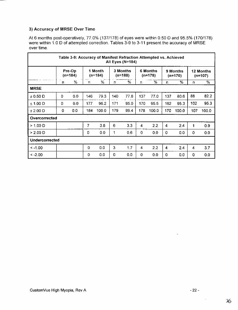
3) Accuracy of MRSE Over Time
At 6 months post-operatively, 77.0% (137/178) of eyes were within 0.50 D and 95.5% (170/178)were within 1.0 D of attempted correction. Tables 3-9 to 3-11 present the accuracy of MRSEover time.
Table 3-9: Accuracy of Manifest Refraction Attempted vs. AchievedAll Eyes (N=184)
Pre-Op I Month 3 Months 6 Months 9 Months 12 Months(n=184) (n=184) (n=180) (n=178) (n=170) (n=107)
n % n % n % n % n % nMRSE
± 0.50 D 0 0.0 146 79.3 140 77.8 137 77.0 137 80.6 88 82.2
± 1.00 D 0 0.0 177 96.2 171 95.0 170 95.5 162 95.3 102 95.3
± 2.00 D 0 0.0 184 100.0 179 99.4 178 100.0 170 100.0 107 100.0
Overcorrected
> 1.00 D 7 3.8 6 3.3 4 2.2 4 2.411 0.9
> 2.00 D 0 0.0 1 0.6 0 0.0 0 0.o o 0.0
Undercorrected
< -1.00 0 0.0 3 1.7 4 2.2 4 2.4 4 3.7
< -2.00 0 0.010 0.0 0 0.0 0 0.0 0 0.0
CustomVue High Myopia, Rev A - 22 -
76
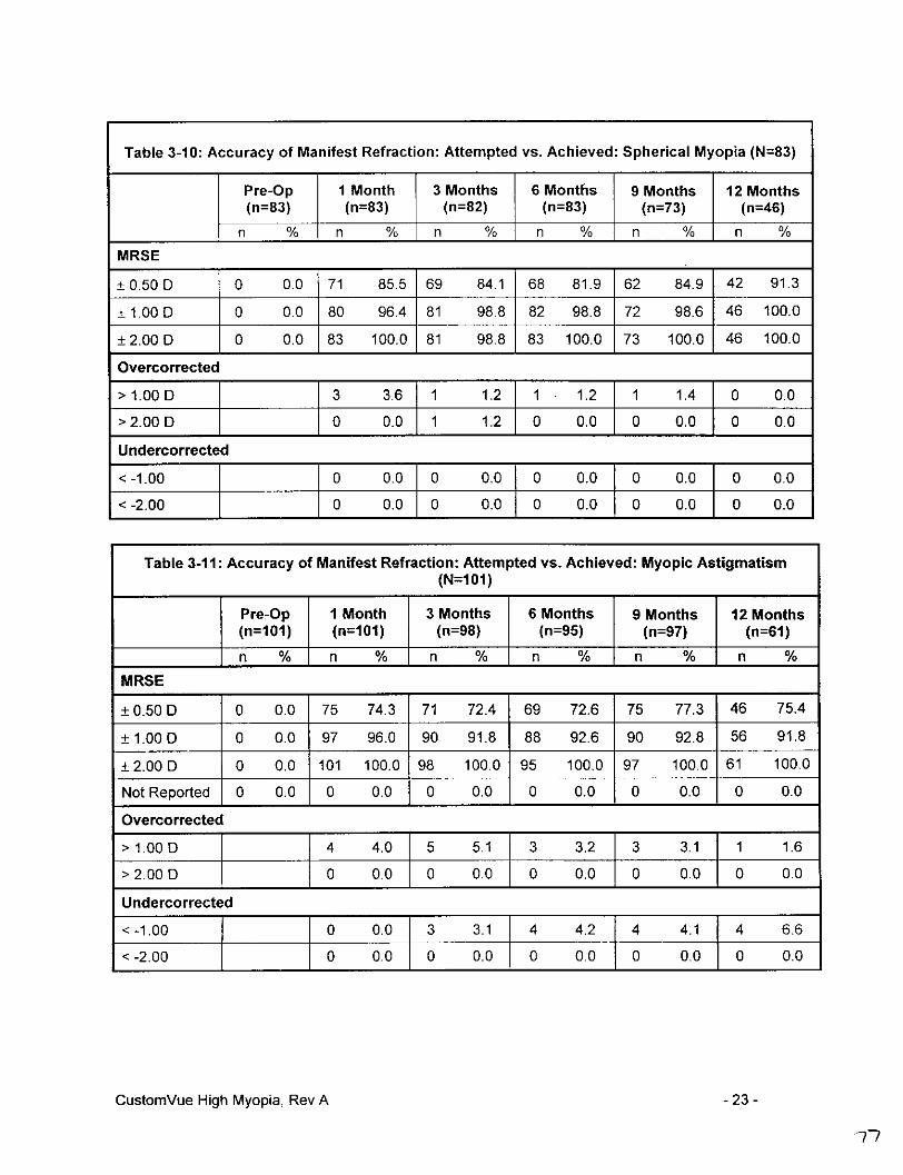
Table 3-10: Accuracy of Manifest Refraction: Attempted vs. Achieved: Spherical Myopia (N=83)
Pre-Op I Month 3 Months 6 Months 9 Months 12 2Months
(n=83) (n=83) (n=82) (n=83) (n=73) (n=46)_ _ _ _ _ _~~~ % n % n _ _n_ _ n _ _ _ _ n_ _ _
MRS E
± 0.50 D 0 0.0 71 85.5 69 84.1 68 81.9 62 84.9 42 91.3
± 1.000D 0 0.0 80 96.4 81 98.8 82 98.8 72 98.6 46 100.0
± 2.000D 0 0.0 83 100.0 81 98.8 83 100.0 73 100.0 46 100.0
Overcorrected
> 1.000D 3 3.6 1 1.2 1 1.2 1 1.4 0 0.0
> 2.000D 0 0.0 1 1.2_j_0 0.0 0 0.0 0 0.0
Undercorrected
< -1.00 0 0.0 0 0.0 0 0.0 0 0.0_J_0 0.0
< -2.00 0 0.0 0 0.0 0 0.0 0 0.0_J_0 0.0
Table 3-1 1: Accuracy of Manifest Refraction: Attempted vs. Achieved: Myopic Astigmatism(N=101)
1Pre-Op I Month 3 Months 6 Months 9 Months 12 Months
____________j 101) .n=101) (n=98) (n=95) (n=97) (n=61)
MRSE
± 0.500D 0 0.0 75 74.3 71 72.4 69 72.6 75 77.3 46 75.4
± 1.000D 0 0.0 97 96.0 90 91.8 88 92.6 90 92.8 56 91.8
± 2.00 D 0 0.0 101 100.0 98 100.0 95 100.0 97 100.0 61 100.0
Not Reported 0 0.0 0 0.0 0 0.0 0 0.0 0 0.0 0 0.0
Overcorrected
*>1.000D 4 4.0 5 5.1 3 3.2 3 3.1_ 1 1.6
*>2.000D 0 0.0 0 0.0 0 0.0 0 0.0 10 0.0
Undercorrected
< -1.00 0 .0 3 3.1 4 4.2 4 4.1__J4 6.6
< -2.00 0 0.0 0 0.0 0 0.0 0 0.0 Jo 0oo
CustomVue High Myopia, Rev A - 23 -

4) Stability of Outcome
Stability of outcome is evaluated both by the cohort of eyes with a refraction at each visit(n=164), as well as the cohort of eyes who were available for two consecutive visits, but not forall visits. Refractive stability is reached at 6 months and confirmed at the 9-month visit. Tables3-12 and 3-13 present refractive stability over time.
Table 3-12: Stability of MRSE for Eyes that Underwent 1, 3, 6, and 9-Month Exams
Between I and 3 Between 3 and 6 Between 6 and 9Months Months Months
All Eyes (n=164)
Change in MRSE by-< 1.0 D, n 161 163 163
% 98.2 99.4 99.4
Mean Change in MRSE ± SD -0.08 ± 0.39 -0.04 ± 0.31 -0.02 ± 0.28
95% CI (-0.14, -0.02) (-0.09, 0.01) (-0.07, 0.02)
Spherical Myopia (n=72)
Change in MRSE by -< 1.0 D, n 70 71 72
% 97.2 98.6 100.0
Mean Change in MRSE ± SD -0.08 ± 0.39 -0.07 ± 0.34 -004 ± 0.29
95% C1 (-0.17, 0.01) (-0.15, 0.01) (-011, 0.03)
Myopic Astigmatism (n=92)
Change in MRSE by < 1.0 D, n 91 92 91
% 98.9 100.0 98.9
Mean Change in MRSE ± SD -0.08 ± 0.40 -0.02 ± 0.29 -0.01 ± 0.28
95% CI (-0.17, 0.00) (-0.08, 0.04) (-007, 0.05)
CustomnVue High Myopia, Rev A - 24 -

Table 3-13: Stability of MRSE for Eyes that Underwent Two Consecutive Visits
Between 1 Between 3 Between 6 Between 9and 3 Months and 6 Months and 9 Months and 12 Months
All Eyes n=180 n=176 n=166 n=107
Change in MRSE by-< 1.0 D, n 177 174 165 107
% 98.3 98.9 99.4 100.0
Mean Change in MRSE ± SD -0.09 ± 0.38 -0.04 ± 0.32 -0.02 ± 0.28 0.00 ± 0.28
95% CI (-0.14, -0.03) (-0.09, 0.01) (-0.07, 0.02) (-0.05, 0.05)
Spherical Myopia n=82 n=82 n=73 n=46
Change in MRSE by - 1.0 D, n 80 80 73 46
% 97.6 97.6 100.0 100.0
Mean Change in MRSE ± SD -0.09 ± 0.37 -0.05 ± 0.35 -0.04 ± 0.28 -0.02 ± 0.30
95% CI (-0.17, -0.01) (-0.13, 0.02) (-0.11, 0.03) (-0.10, 0.07)
Myopic Astigmatism n98 n=94 n93 n61
Change in MRSE by - 1.0 D, n 97 94 92 61I% 99.0 100.0 98.9 100.0
Mean Change in MRSE ± SD -0.08 ± 0.40 -0.03 ± 0.29 -0.01 ± 0.28 0.01 ± 0.26
95% GI (-0.16, 0.00) (-0.08, 0.03) (-0.07, 0.05) (-0.05, 0.08)
CustomVue High Myopia, Rev A -25 -
?<}

When plotted over time, the mean manifest spherical equivalents illustrate that stability isachieved by the 6-month visit.
Figure 3-i - MRSE Over Time (All Eyes, N=184)
Mean MIRSE Over Time
Pre-op i M 3 M 6 M 9 M 12 M1.0 --
-1.0 -0.02 -010 -0¶ f 5 O1 -0.22-2.0 _____ _
CL-5.0
.2~~~~~~~~
a upper95%CI'--9.0 8-- 49 __ __ -- --
5) Efficacy of Correction of Astigmatism
Efficacy of correction of astigmatism was evaluated at the point of stability (6 months) for eyeswith myopic astigmatism. Table 3-14 displays the mean percent reduction of cylinder for eyeswith myopic astigmatism, stratified by pre-operative cylinder.
Table 3-14: Reduction of Absolute (Non-Vector) Cylinder at 6 Months:Myopic Astigmatism (N=95)
Preoperative Cylinder Mean % Reduction RangeAll (n=95) 70.0% -33.0% to 100%< -0.50 to -1.00D(n=33) 60.1% -33.3% to 100%*<-1.0 to -2.00D (n39) 72.2% 12.5% to 1 00%< -2.0 to -3.0 D (n=16) 79.6% 49.8% to 1 00%< -3.0 to -4.0 D (n=7) 81.9% 53.1 % to 1 00%
CustomVue High Myopia, Rev A -26 -

Table 3-15 presents a summary of the vector analysis which includes mean Intended RefractiveChange (IRC), Surgically Induced Refractive Change (SIRC), correction Ratio (CR), and ErrorRatio (ER), at the point of stability (6 months).
Table 3-15: Vector Analysis at Stability (6 Months): Eyes with Myopic Astigmatism (N=95)
IRC* SIRCA CRt ER'Preoperative Cylinder Mean ± SD Mean ± SD Mean ± SD Mean ± SD
All (n=95) -1.34 ± 0.71 -1.19 ± 0.71 0.91 ± 0.32 -0.37 ± 0.43< -0.50 to -1.0 D (n=33) -0.69 ± 0.11 -0.69 ± 0.31 0.99 ± 0.37 -0.48 ± 0.57< -1.0 to -2.0 D (n=39) -1.27 ± 0.24 -1.08 ± 0.41 0.86 ± 0.32 -0.34 ± 0.34< -2.0 to -3.0 D (n=16) -2.13 ± 0.26 -1.81 ± 0.52 0.85 ± 0.24 -0.25 ± 0.24< -3.0 to -4.0 D (n=7) -3.01 ± 0.17 -2.79 ± 0.37 0.93 ± 0.14 -0.22 ± 0.23
*IRC-tntended Refractive Change - vector difference between target refraction and preoperative refractionsASIRC-Surgically Induced Refractive Error- difference between postoperative and preoperative vectorstCR-Correction Ratio - ratio of achieved versus intended vector magnitudeCR mathematical definition: ISIRCI divided by IIRCI0ER-Error Ratio - ratio of Error Vector and IRC magnitudes
Error Vector mathematical definition: vector difference between ISIRCI and IIRCIER mathematical definition: IError Vector] divided by IIRCI
6) Higher Order Aberrations
Although the WaveScan WaveFront® System measures the refractive error and wavefrontaberrations of the human eyes, including myopia, hyperopia, astigmatism, coma, sphericalaberration, trefoil, and other higher order aberrations through sixth order, in the clinical studyfor this PMA, the average higher order aberrations increased after CustomVueTM treatment.The most noticeable increases were in coma and spherical aberration. The clinical data over9 months showed that the changes in all higher order aberrations were stable post-operatively.
CustomVue High Myopia, Rev A -27 -
gl
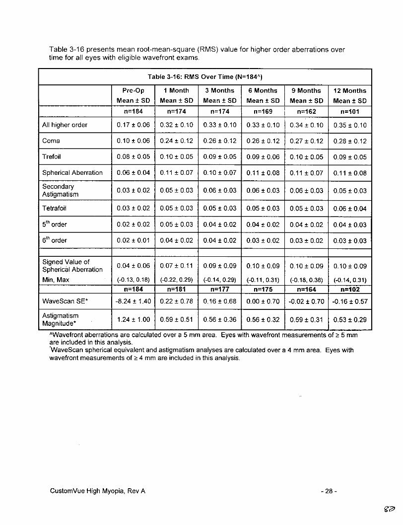
Table 3-16 presents mean root-mean-square (RIMS) value for higher order aberrations overtime for all eyes with eligible wavefront exams.
Table 3-16: RMS Over Time (N=184 A)
Pro-Op I Month 3 Months 6 Months 9 Months 12 MonthsMean ± SD Mean ± SD Mean ± SD Mean ± SD Mean ± SD Mean ± SD
n=184 n=174 n=174 n=169 n=162 n=1O1
All higher order 0.17 ± 0.06 0.32 ± 0.10 0.33 ± 0.10 0.33 ± 0.10 0.34 ± 0.10 0.35 ± 0.10
Coma 0.10 ± 0.06 0.24 ± 0.12 0.26 ± 0.12 0.26 ± 0.12 0.27 ± 0.12 0.28 ± 0.12
Trefoil 0.08 ± 0.05 0.10 ± 0.05 0.09 ± 0.05 0.09 ± 0.06 0.10 ± 0.05 0.09 ± 0.05
Spherical Aberration 0.06 ±0.04 0.11 ± 0.07 0.10 ±0.07 0.11 ± 0.08 0.11 ± 0.07 0.11 ± 0.08
Secondary 0.03 ± 0.02 0.05 ± 0.03 0.06 ± 0.03 0.06 ± 0.03 0.06 ± 0.03 0.05 ± 0.03Astigmatism
Tetrafoil 0.03 ± 0.02 0.05 ± 0.03 0.05 ± 0.03 0.05 ± 0.03 0.05 ± 0.03 0.06 ± 0.04
5 th order 0.02 ± 0.02 0.05 ± 0.03 0.04 ± 0.02 0.04 ± 0.02 0.04 ± 0.02 0.04 ± 0.03
6tIh order 0.02 ±0.01 0.04 ±0.02 0.04 ±0.02 0.03 ± 002 0.03 ± 002 0.03 ± 003
Signed Value ofSpheri~l Aberation 0.04 ± 0.06 0.07 ± 0.11 0.09 ± 0.09 0.10 ± 0.09 0.10 ± 0.09 0.10 ± 0.09
Min, Max (-0.13, 0.18) (-0.22, 0.29) (-0.14, 0.29) (-0.11, 0.31) (-0.18, 0.38) (-0.14, 0.31)n=184 n=181 n=177 n=175 n=164 n=102
WaveScan SE* -8.24±1.40 0.22±0.78 0.16 ±0.68 0.00 ± 0.70 -0.02±0.70 -0.16 ±0.57
Asimatnitudm 1.24 ±1.00 0.59 ± 0.51 0.56 ± 0.36 0.56 ± 0.32 0.59 ±0.31 0.53 ±0.29
AWavefront aberrations are calculated over a 5 mm area. Eyes with wavefront measurements of > 5 mmare included in this analysis.'WaveScan spherical equivalent and astigmatism analyses are calculated over a 4 mm area. Eyes withwavefront measurements of >Ž4 mm are included in this analysis.
CustornVue High Myopia, Rev A -28 -
gr0

7) WaveScan Wavefrant Diameter
A minimum wavefront diameter of 5 mm is required for treatment with CustomVue TILASIK forhigh myopia. In the clinical trial, all treatments used an optical zone of 6 mm and ablation zoneof 8 mm. Some of the eyes in the study (47/1 84, 25.5%) had 6 mm treatments based onwavefront diameters that were smaller than 6 mm. The results of these eyes were compared toresults of eyes with treatments based on wavefront measurements of 6 mm or greater. Bothgroups of eyes had similar results. Table 3-17 presents key safety and effectiveness results forthe two groups at 6 months postoperatively.
Table 3-17: Summary of 6 Month Key Safety and EffectivenessVariables For All Eyes Stratified by WaveScan Diameter
Wavefront Diameter <6 mm Ž!6 mmn %n
EFFECTIVENESS VARIABLES n=45 n=133
UCVA 20/16 or belier 31 68.9 85 63.9
UCVA 20/20 or belier 39 86.7 111 83.5
UCVA 20/25 or belier 43 95.6 123 92.5
UCVA 20/32 or better 43 95.6 130 97.7
UCVA 20/40 or better 44 97.8 131 98.5
MRSE ± 0.500D 30 66.7 107 80.5
MRSE ± 1.00 D 41 91.1 110 97.0
MRSE±+2.00 D 45 100.0 133 100.0
Cylinder ± 0.50 D 33 73.3 102 76.7
Cylinder± 1. 00 D 41 91.1 128 96.2
SAFETY VARIABLES n=45 n=133
BSCVA worse than 20/25 0 0.0 0 0.0
BSCVA worse than 20/40 0 0.0 0 0.0
Loss Ž 2 lines BSCVA 0 0.0 0 0.0
Loss up to 1 line BSCVA 1 2.2 8 6.0
No Change in BSCVA 113 128.9 134 25.6
Gain up to 1 line BSCVA 21 46.7 73 54.9
Gain Ž 2 lines BSGVA 10 22.2 18 13.6
CustomVue High Myopia, Rev A - 29 -
83

8) Best Spectacle-Corrected Visual Acuity (BSCVA)
No eye lost 2 or more lines of BSCVA at any visit. Table 3-18 presents the change in lines ofBSCVA over time.
Table 3-18: Change in BSCVA Over Time: All Eyes (N=184)
I Month 3 Months 6 Months 9 Months 12 Monthsn=184 n=180 n=178 n=170 n=107
n % n % n % n % n %Decrease > 2 Lines 0 0.0 0 0.0 0 0.0 0 0.0 0 0.0
Decrease > 1 to < 2 Lines 1 0.5 3 1.7 0 0.0 0 0.0 0 0.0
Decrease > 0 to_< 1 Line 18 9.8 14 7.8 9 5.1 11 6.5 16 15.0
No Change 69 37.5 55 30.6 46 26.4 47 27.6 27 25.2
Increase >0 to < 1 Line 89 48.4 94 52.2 94 52.8 96 56.5 59 55.1
Increase > 1 to _ 2 Lines 7 3.8 13 7.2 27 15.2 14 8.2 5 4.7
Increase > 2 Lines 0 0.0 1 0.6 1 0.6 2 1.2 0 0.0
9) Contrast Sensitivity Analysis
Table 3-19 presents the results of the contrast sensitivity analysis include mean change,standard error, and p-value from paired t-test. Patient responses to the four spatial frequencies(3, 6, 12 and 18 cycles per degree (CPD)) were measured with the patient's best correctedvision using the VectorVision CSV-1000 and converted from contrast levels to log units. Apositive mean change reflects an improvement in contrast sensitivity, while a negative meanchange reflects a decrease.
CustomVue High Myopia, Rev A -30 -

Table 3-19: Contrast Sensitivity: All Eyes (N=184)
Pre-Op Change from Pre-Op to Change from Pre-Op to(n=184) I Month (n=178) 3 Months (n178)
CPD 3 6 12 18 3 6 12 18 3 6 12 18
Dim wI Glare
Mean 1.52 1 1.00 0.54 -0.01 -0.05 -0.08 -0.01 0.00 -0.02 -0.07 000SE 0.017 0.023 0.030 0.032 0.018 0.028 0.033 00.033 10.0190.029 0.037 0.040
P Value* 0.622 0.065 0.023 0.789 0.923 0.441 0.084 0.973
Dim wlo Glare
Mean 1.59 163 112 0.65 0.00 -0.03 -0.08 -0.03 -0.02 -0.06 -0.08 -0.04
SE 0.020 0.023 0.033 0.035 0.018 0.024 0.033 0.036 0.020 0.028 0.037L0.040
P Value* < 0.972 0.165 0.012 0.471 0.362 0.033 0.038 0.338
Bright wlo Glare
Mean 1.76 1.95 1.61 1.14 -0.02 -0.06 -0.09 -0.05 -0.04 -0.06 -0.10 40.08
SE 0.013 0.014 0.019 0.019 0.015 0.019 0.029 0.026 0.015 0.023 0.029 0.027
P Value*_< 0.256 0.001 0.004 0.041 0.016 0.005 0.001 0.003'Two tailed paired t test for the means.
Table 3-19: Contrast Sensitivity: All Eyes (N=184) (continued)
Change from Pre-Op to Change from Pre-Op to6 Months (n=178) 12 Months (n=107)
CPD 3 6 12 18 3 6 12 18
Dim w/ Glare
Mean 0.01 0.01 0.04 0.06 0.01 0.05[0.12 0.14
SE 0.020 0.026 0.035 0.037 0.026 0.036 0.041_j 0.04
P Value * 0.497 0.610 0.263 0.131 0.720 0.155 0.004 0.001Dim w/o Glare
Mean -0.01 0.02 0.02 0.05 -0.01 0.02 0.08 0.08
SE 0.018 0.023 0.033 0.034 0.028 0.035 0.044 0.048
P Value* < 0.716 0.390 0.602 0.111 0.696 0.604 0.085 0.091
Bright w/o Glare
mean -0.02 -0.01 -0.02 0.00 -0.03 -0.06 -0.06 -0.01
SE 0.017 0.017 0.024 0.023 0.023 0.025 0.036 0.032
P Value* < 0.255 0.480 0.448 0.894 0.230 0.018 0.110 0.830'Two tailed paired t test for the means
CustomVue High Myopia, Rev A - 31 -
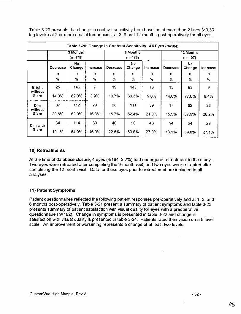
Table 3-20 presents the change in contrast sensitivity from baseline of more than 2 lines (>0.30log levels) at 2 or more spatial frequencies, at 3, 6 and 12-months post-operatively for all eyes.
Table 3-20: Change in Contrast Sensitivity: All Eyes (N=184)3 Months 6 Months 12 Months(n=178) (n=178) (n=107)
No No NoDecrease Change Increase Decrease Change Increase Decrease Change Increase
n n n n n n n n n
% % % % % % % % %
Bright 25 146 7 19 143 16 15 83 9without
Glare 14.0% 82.0% 3.9% 10.7% 80.3% 9.0% 14.0% 77.6% 8.4%
Dim 37 112 29 28 111 39 17 62 28without
Glare 20.8% 62.9% 16.3% 15.7% 62.4% 21.9% 15.9% 57.9% 26.2%
Dimwith 34 114 30 40 90 48 14 64 29
Glare 19.1% 64.0% 16.9% 22.5% 50.6% 27.0% 13.1% 59.8% 27.1%
10) Retreatments
At the time of database closure, 4 eyes (4/184, 2.2%) had undergone retreatment in the study.Two eyes were retreated after completing the 9-month visit, and two eyes were retreated aftercompleting the 12-month visit. Data for these eyes prior to retreatment are included in allanalyses.
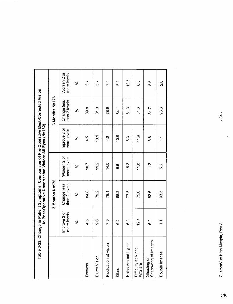
I1) Patient Symptoms
Patient questionnaires reflected the following patient responses pre-operatively and at 1, 3, and6 months post-operatively. Table 3-21 present a summary of patient symptoms and table 3-23presents summary of patient satisfaction with visual quality for eyes with a preoperativequestionnaire (n=182). Change in symptoms is presented in table 3-22 and change insatisfaction with visual quality is presented in table 3-24. Patients rated their vision on a 5 levelscale. An improvement or worsening represents a change of at least two levels.
CustomVue High Myopia, Rev A - 32 -
96

CO2; i~ o 0 0 0 0 0 0 0 O-0 ~ 6 a 6 6 o o a a;0 o a'
O co u ~ 6 0 6 6 a a a
0~~~~'
z c
2o o~ o o o o oo
a. I C d N {5 r o3 d 0 C
Co O Cj c6 6 6 6 6~o~~~~~~~~~~~U
(/) ~ ~ ~ ~ ~ ~ ~~(
-,~ ~. & u ,P W-,i 66 cK 6 u5 ,c5
P r,~ C·- ( or_ o(00 ) (N t- t cc
0>1 ~ ~ ~ ~ ~ C ad C) (c5V N t
E ~ C' C) CO N C)
Ca
Ea o co) - (NJ (NJo 0
.,o 2V 03 N o 0E~o U~ co ~ U? co ~
.- 04
E , .e n cN c-
IS ui oi cd 6 c'
c C) (N (NJ C) (NJ~ -OC) C
2~~~~~~~~~~~~~~~~~~~~~- co_
2 to ~~~ ~~~~Co 0 i co ) cc2 5 t- U t O C')~~0 C co
U) '0 II
(N* 0 CD 03 co V V0c { 5 c ,.i C) ( 0 <
C C C5 ( CCl "N
O(-Q
co cm~~
'0 0 03 0 LA) (0 (NJ 03~7 0 ) 0)
________0 L2 ~~~ C cu03 c ta Z ' C 0, a)z a (N C) (Na),- to 03 a
0 ,0 'co E -, (DU) (m
(NJ~~~~~~~~~~~~~~~~~~~~~~~~~~~~~~L
a a~00 mo 0 :
co > LL o,.:o'0 00 -='-0) - Cf-j ~ ~ ' .-- ,' -.- 'O -0) 0m> ~~ D_- Wv~ E~0D _~ ~ O0~o n°-E o~~~~~
a)~ C t C) N-D 02 C-I~~~~~~~~~~~~C0 0
o~~~~
- z _~~~~0 D -
C) C co CC C) CV
C.) 0 cCo co
Z1 E C)
aU) co ~ ~ ~ ~ ~ c c
C) =
co~~~~~~~~~~~~~~~~~~~~~~~tk- ~ 2- --
-- U -t- 0)U
0 0 00 0 '- 01 t 2 D Ca 04 (D~~~~~~~~~~o .a> - ~C 02 r N1 N C2 NR 02 C
0 c0 0
0Z~0
0 ) 0-0 0CC L )o FE~ wr c
0A
4) ~ ~ ~ ~ ~ ~ ~ ~ ~ ~ ~ iN C
0 ~ ~ ~ ~ ~~0-a. a..cCC: a)
.0 a) ' CA)
0 c -~ 00o (1
M ~ ~ ~ ~ ~ ~ ) 0 0C FU) .C)z 0)
> CD< wC) ~~' .2 a) ZC' 05 0 a D
o m Ii- CD I ~~~~~0 C.)

0~~~~(
4)~ ~ ~ ~ ~ ~~~~~~~~~~~~0
.0 2 C CD 0c0 0
00~~~~~~~(
#A a)~~~ IT Lo 6 c*1=
3:0 _______
0w
to 2 0 0C)C
0) 2 IT6 6R 6 6OC~~~~~~~~~~~~~~~~~~C
(L z~~~~~~(
4) 4) 0)1 IT (Ni CO
* 1- -C I L
EI (0
Z W 2 N to l C
>1 U tN 00 (C) ~ ~ N C C
= ~~~ 2 tr 0)~~U W O to
4 CD~~~~~~~~0 r- r- 65-~ ~ Oo _ _
w 10 0) ~~~~~~~~~~~~~0ao ~ ~ ~ ~ ~ C'~~L CD 6 N C
C)~~~~~~~~~~~~~~~~~~~~~~~~~~~~~~~~~~~~~~~~~~~C
0~~~~04- CD a) m a T~~~~~~~~~~~~~~~~~~~~~~~~~~C
'0 2 tZ CO~~~~~~~~~~~
4- (0 0 (v) (NJ (N CD~~~~~~~ 00 C~~~~~~~~~~
~~~ 4)2 2 ~ ~ ~ ~ ~ o ~ .-
o - .~~~~~~ C) CO (N N~~Z - La. CO~~) u e0 > N- : .
(U~~~~~~~~~~~~~~~~~~

- ~~00I ao) O 0 V ) 0
Z~~~0~~~~~~~~~~~~~~~~~~~~~m0
W1 N
cc >a) ~ 0) a)iC (D.Ifl CO.2 -5 M0 N- N ) C0C C
5 0 r03o
0) COco
Cj (D D NI -0 > LO (D (14 CCD t
QL 0~~~~~~~~~~0
ao
4!> ~EE
It-
CuO toO C O (0 Cfl 0) 0)
00
0)0 CO~~~~~~ C -4r
.> > 0 I
NO~~~~~~~0d - CNN N )0 C2
Ci 030~~~~0~~00
0
a- O~~~~~~~~~~~~~~~0 C)~~~~~~~~~0-E >E
0.
* a.~~~~~~~~~~~~~~a- 0~~~~~~~~~~~~~~~C

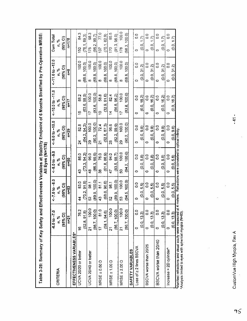
12) Summary of Key Safety and Effectiveness Variables
The key safety and effectiveness variables over time for all eyes, eyes with spherical myopia,and eyes with myopic astigmatism are presented in Tables 3-25 to 3-27. The key safety andeffectiveness variables at the point of stability, stratified by pre-operative manifest refractionspherical equivalent, are presented in Tables 3-28 to 3-30.
CustomVue High Myopia, Rev A -37 -

Table 3-25: Summary of Key Safety and Effectiveness Variables: All Eyes (N=184)
I Month 3 Months 6 Months 9 Months 12 MonthsI% n % n % n %(95% C1) (95% CI) (95% Cl) (95% Cl) (95% Cl)
EFFECTIVENESS VARIABLES*n=184 n=180 n=178 n=170 n=107
UCVA20/20 or better 157 85.3 147 81.7 150 84.3 145 85.3 92 86.0(79.4, 90.1) (75.2, 87.0) (78.1, 89.3) (79.1, 90.3) (77.9, 91.9)
UCVA20/40 or better 182 98.9 177 98.3 175 98.3 169 99.4 107 100.0(96.1, 99.9) (95.2, 99.7) (95.2, 99.7) (96.8, 100.0) (97.2, 100.0)
Sphere ± 0.50 D 144 78.3 145 80.6 141 79.2 140 82.4 93 86.9(71.6, 84.0) (74.0, 86.1) (72.5, 84.9) (75.8, 87.8) (79.0, 92.7)
Sphere+ 1.00D 173 94.0 171 95.0 170 95.5 162 95.3 103 96.3(89.6, 97.0) (90.7, 97.7) (91.3, 98.0) (90.9, 97.9) (90.7, 99.0)
MRSE ± 0.50 D 146 79.3 140 77.8 137 77.0 137 80.6 88 82.2(72.8, 85.0) (71.0, 83.6) (70.1, 82.9) (73.8, 86.2) (73.7, 89.0)
MRSE + 1.00 D 177 96.2 171 95.0 170 95.5 162 95.3 102 95.3(92.3, 98.5) (90.7, 97.7) (91.3, 98.0) (90.9, 97.9) (89.4, 98.5)
MRSE + 2.00D 184 100.0 179 99.4 178 100.0 170 100.0 107 100.0(98.4, 100.0) (96.9, 100.0) (98.3, 100.0) (98.3, 100.0) (97.2, 100.0)
n = 101 n = 98 n = 95 n = 97 n=61CylinderA +_ 0.50 D 74 73.3 65 66.3 62 65.3 68 70.1 41 67.2
(63.5, 81.6) (56.1, 75.6) (54.8, 74.7) (60.0, 79.0) (54.0, 78.7)CylinderA + 1.00 D 96 95.0 94 95.9 87 91.6 90 92.8 59 96.7
(88.8, 98.4) (89.9, 98.9) (84.1, 96.3) (85.7, 97.0) (88.7, 99.6)STABILITY OF MRSE** n=180 n=176 n=166 n=107Change< 1.00D MRSE 177 98.3 174 98.9 165 99.4 107 100.0
(95.2, 99.7) (96.0, 99.9) (96.7, 100.0) (97.2, 100.0)Mean Change in MRSE ± SD -0.09 ± 0.38 -0.04 ± 0.32 -0.02 ± 0.28 0.00 ± 0.28
(-0.14, -0.03) (-0409, 0.01) (-0407, 0.02) (-005, 0.06)STABILITY OF CYLINDERA n=98 n=94 n=93 n=61Change •1.00 D Cylinder 98 100.0 94 100.0 93 100.0 61 100.0
(97.0, 100.0) (96.9, 100.0) (96.8, 100.0) (95.2, 100.0)Mean Change in Cylinder + SD -0.02 ± 0.28 -0.05 ± 0.32 0.01 ± 0.24 -0.06 ± 0.24
(-0.08, 0.04) (-0.12, 0.02) (-0.04, 0.06) (-0.12, 0.00)
SAFETY VARIABLES n=184 n=180 n=178 n=170 n=107Loss of -> 2 lines BSCVA 0 0.0 0 0.0 0 0.0 0 0.0 0 0.0
(0.0, 1.6) (0.0, 1.7 _ (0.0, 1.7) (0.0, 1.7) __0.0, 2.8)BSCVA worse than 20/25 0 0.0 0 0.0 0 0.0 0 0.0 0 0.0
(0.0, 1.6) (0.0, 1.7) 0.0, 1.7 (0.0, 1.7) (00, 2.8)BSCVA worse than 20/40 0 0.0 0 0.0 0 0.0 0 0.0 0 0.0
(0.0, 1.6) (0(0, 1 7) (00, 1.7) (0.0, 2.8)'Manifest refractions and visual acuity were measured at 4 meters Refractions were converted to optical infinity."Analysis of stability is limited to eyes with two consecutive visits.AAnalysis of cylinder was limited to those eyes with a preoperative manifest cylinder <-0.5D.
CustomVue High Myopia, Rev A - 38 -
"3
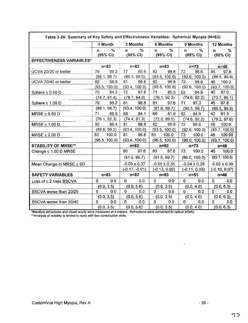
Table 3-26: Summary of Key Safety and Effectiveness Variables: Spherical Myopia (N=83)
I-T lMonth '3 Months 6 Months '9 Months 112 Monthsn n n %% % v
(90C)A5/! 9%C(9595%CCI) 95% Cl)
EFFECTIVENESS VARIABLES*_________________________ n=83 n=82 n=83 n=73 n=46
UCVA 20120 or better 79 95.2 77 93.9 82 98.8 72 98.6 45 97.8(88.1, 98.7) 86.3, 98.0) 93.5, 100.0) 92.6, 100.0) 88.5, 99.9)
UCVA 20/40 or better 82 98.8 81 98.8 82 98.8 72 98.6 46 100.0_93.5, 100Q9}__j93.4, ~100.0 (93.5, 100.0) 92.6, 100.0) _93.7, 100.0)
Sphere± .5 070 84.3 72 87.8 71 85.5 62 84.9 40 87.0(74.7, 91.4) 78.7, 94.0) 76.1, 92.3) 74.6,92.2 (73.7, 95.1)
Sphere ±1.00 0 79 95.2 81 98.8 81 97.6 71 97.3 45 97.8(88.1, 98.7) 93.4, 100.0) (91.6, 99.7) (90.5, 99.7) (88.5, 99.9)
MRSE±0.50 D 71 85.5 69 84.1 68 81.9 62 84.9 42 91.3________________________ 76.1, 92.3) (74.4, 91.3) (72.0, 89.5) 74.6, 92.2) 79.2, 97.6)
MRSE ± 1.000D 80 96.4 81 98.8 82 98.8 72 98.6 46 100.0__________________________ 89.8, 99.2) 93.4, 100.0) 935100 92.6, 100.0) 93.7, 100.0)
MRSE ± 2.00 0 83 100.0 81 98.8 83 100.0 73 100.0 46 100.00(96.5, 100.0) (93.4, 100.0) (96.5, 100.0) (96.0, 100.0) (93.7, 100.0)
STABILITY OF MRSE** n=82 n=82 n=73 n=46Change! •1.000D MRSE 80 97.6 80 97.6 73 100.0 46 100.0
__________________________ ~(91.5, 99.7) (91.5, 99.7) (96.0, 100.0) (93.7,100.0)
Mean Change in MRSE ± SD -0.09 ± 0.37 -0.05 ± 0.35 -0.04 ± 0.28 -0.02 ± 0.30(-0.7, -. 01) (-0.13, 0.02) (-0.11, 0.03) -0.10, 0.07)
SAFETY VARIABLES n=83 n=82 n=83 n=51 n=46Loss of Ž 2 lines BSCVA 0 0.0 0 0.0 0 0.0 0 0.0 0 0.0
(0.0, 3.5) (0.0, 5.6) (0.0, 3.5) 0O.0, 4.0) (0.0, 6.3)BSCVA worse than 20/25 0 0.0 0 0.0 0 0.0 0 0.0 0 0.0
_________________________ 0.0, 3.5) (0.0, 5.6) (0.0, 3.5) 0.0, 4.0) 0.0, 6.3)BSCVA worse than 20/40 0 0.0 0 0.0 0 0.0 0 0.0 0 0.0
_________________________ (0.0, 3.5) (0.0, 5.6) (0.0, 3.5) (0.0, 4.0) (0.0,. 3)'Manifest retractions and visual acuity were measured at 4 meters. Refractions were converted to optical infinity.-Analysis of stability is limited to eyes with two consecutive visits.
CustomVue High Myopia, Rev A -39 -

Table 3-27: Summary of Key Safety and Effectiveness Variables: Myopic Astigmatism (N=101)
__ _I tMonth 3 Months 6 Months 9 Months 12 Monthsn % n % n % n
_ __ (95% Cl) (95% Cl) (95% Cl) (95% Cl) (95% Cl)
EFFECTIVENESS VARIABLES*
n=101 n=98 n=95 n=97 n=61UCVA 20/20 or better 78 77.2 70 71.4 68 71.6 73 75.3 47 77.0
(__67.8, 850) (61.4, 80.1) (61.4, 80.4) (65.5, 83.5) (64.5, 86.8)UCVA20/40 or better 100 99.0 96 98.0 93 97.9 97 100.0 61 100.0
(_94.6, 100.0L (92.8, 99.8) (92.6, 99.7) (97.0,100.0) (95.2, 100.0)Sphere ± 0.50 D 74 73.3 73 74.5 70 73.7 78 80.4 53 86.9
(63.5, 81.6) (64.7, 82.8) (63.6, 82.2) (71.1, 87.8) (75.8, 94.2)
Sphere_± 1.00 D 94 93.1 90 91.8 89 93.7 91 93.8 58 95.1(86.2, 97.2) (84.5, 96.4) (86.8, 97.6) (87.0, 97.7) (86.3, 99.0)
CylinderA ± 0.50 D 74 73.3 65 66.3 62 65.3 68 70.1 41 67.2(63.5, 81.6) (56.1, 75.6) (54.8, 74.7) (60.0, 79.0) (54.0, 78.7)
Cylinder^ ± 1.00 D 96 95.0 94 95.9 87 91.6 90 92.8 59 96.7(88.8, 98.4) (89.9, 98.9) (84.1, 96.3) (85.7, 97.0) (88.7, 99.6)
MRSE ± 0.50 D 75 74.3 71 72.4 69 72.6 75 77.3 46 75.4__(64.6, 82.4) (62.5,81.0) (62.5, 81.3) (67.7, 85.2) (62.7, 85.5)
MRSE + 1.00 D 97 96.0 90 91.8 88 92.6 90 92.8 56 91.8(90.2, 98.9) (84.5, 96.4) (85.4, 97.0) (85.7, 97.0) (81.9, 97.3)
MRSE + 2.00 D 101 100.0 98 100.0 95 100.0 97 100.0 61 100.0(97.1, 100.0) (97.0, 100.0) (96.9, 100.0) (97.0,100.0) (95.2, 100.0)
STABILITY** n= 98 n= 94 n= 93 n=61
Change_< 1.00 D MRSE 97 99.0 94 100.0 92 98.9 61 100.0
(94.4, 100.0) (96.9, 100.0) (94.2,100.0) (95.2, 100.0)
Mean Change in MRSE + SD -0.08 ± 0.40 -0.03 ± 0.29 -0.01 ± 0.28 0.01 ± 0.26(-0.16, 0.00) (-0.09, 0.04) (-0.07, 0.05) (-0.05, 0.08)
Change •1.00 D CylinderA 98 100.0 94 100 93 100.0 61 100.0(97.0, 100.0) (96.9, 100.0) (96.8,100.0) (95.2, 100.0)
Mean Change in Cylinder ± SD -0.02 ± 0.28 -0.05 ± 0.32 0.01 ± 0.24 -0.06 - 0.24(-0.08, 0.04) (-0.12, 0.02) (-0.04, 0.06) (-0.12, 0.00)
SAFETY VARIABLES n= 101 n =98 n= 95 n= 97 n= 61
Loss of > 2 lines BSCVA 0 0.0 0 0.0 0 0.0 0 0.0 0 0.0
(0.0, 2.9) (0.0, 3.0) (0.0, 3.1) (0.0, 3.0) (0.0, 4.8)
BSCVA worse than 20/25 0 0.0 0 0.0 0 0.0 0 0.0 0 0.0(0.0, 2.9) (0.0, 3.0) (0.0, 3.1) (0.0, 3.0) (0.0, 4.8)
BSCVA worse than 20/40 0 0.0 0 0.0 0 0.0 0 0.0 0 0.0(0.0, 2.9) (0.0, 3.0) (0.0, 3.1) (0.0, 3.0) (0.0, 4.8)
'Manifest refractions and visual acuity were measured at 4 meters. Refractions were converted to optical infinity."Analysis of stability is limited to eyes with two consecutive visits.^Analysis of cylinder was limited to those eyes with a preoperative manifest cylinder <-0.5D.
CustomVue High Myopia, Rev A -40-
1'f
W>~ - o 9 9 0 0
Iz 0 oa o0O : .rc co,-o c 0 0 o o a o
o0 0 0 0 0C- 0.-0r00 -00 00 -o o
>' ~~6 C6 6 C6 6 6 6 0 C0.0 ,~~ 0) o5 ~~ 6 6Z 6
V
m ~~~60I - 0.4z ~ ~ ~ ~ ~ 9N.~o9
6 6 6 ,) C (' ~ -o0 CO t; 0' a o 6 6
(0 V ,- 0r 0 0- 0-
°6 ~
o '1*~~~~~-L 0C FC 6 W 0 6 N o 0 6 0 0; 0 0 T
044 vo o 0 00r q0
~II o0w . cONOtO0)O R 0 0 0 0
(- (v(6 4 6=I ()-0 0~ (0 0 ) 0)c>N(00) =)Ofl I I41 IO>
-0Ii -- - o - o o
~ ~ ~ e · . · - · 0 o~'A- f, CO I 0 0 0 0
CD~~~~~~~~~~~~~~~~~~~
'~ c4 oC~ o4 o0 o ? o o o·
' o 8 0 0~. r r P 0 . . .
2Ing 000 a--- I C 0 1 *o
o I~"u
> OCc ci = c 6 6 6 65
o I - flNCJ o oC0o o~ 0 0 6'c N~~~.~ - ~, 0o, 0o 0 0 0 o~
0) V )l tm > :>,n IC)atr
U, ~ ~ ~ ~ f .S. :, >
U;O0C P ~ o)~0.9co ED CD 9 c0 o,, E(U I- - ,20 -l j0 0I0 0 - -
o Ctoc in r - 0 6 n=)
zo M >0 C~~~~~~~N
e ~ ~ ~ ~~~~~~~~ a oo ~
E - >'N L) A >~ (I)
-o o c ,
cn~ ~ ~ ~ ~~~~~~n
a, 'C'N I, I 0.- IN +1 +1
a) w L . > j(/ F- 0 0
0~~~~~~~~~~1

1-0 00 0) 0 0)
E c' c 6 .i 16 9 0 9 9= m 0000000
0 0 0 0P e~~ 6 o~~o o00o0 o 0 0 0: 0
0 ~~ N '~0'0 0' '
- 0) -
04 0 04 0 040 0 0 0
Ci) 8 0 0 9 0In - ~~~6o o 00 000 9 0 0
0 6 N N 9 & N 6 65 oc; Lo Ln 0) 6U'6 c
CD CC (0~~CCD N 0 N N
C~~~~~CC~~~~~~~~~~~~~~~~~~~~~~~~~~~~~~~~N
0u M 00~
C) N C0 0 4;4 4 4-l 0o4 - - N ) 04 04 0
d 04 0 N00N 4
0 C) a~~~m nOt 0i >a 0C 0 9 ta
U)~~~~~~~~~~~a
c q (6 0)~ a 6C? 04) m04 ) 04 4 4a 00
> 000 0 0 0000 0c z-~~ L 61q d 6uo CT r
000000 -0 0 0) 0 0'4- c~~ - - ,- -6 -
U otto c ~~~ 9 N- 9 & 6 ~6 65 65o n0~ o oOa oo '
1 0) 0 0 0 0-
C-4~~~~~4EUC) C N ) 9
CD - ~ 00000 6o6 >co~~~~~~~~~~~~~~~~~~~~i
U] 0
a N 0 A 0)aB a
C"~~~~~~~~~~~~~C
C;
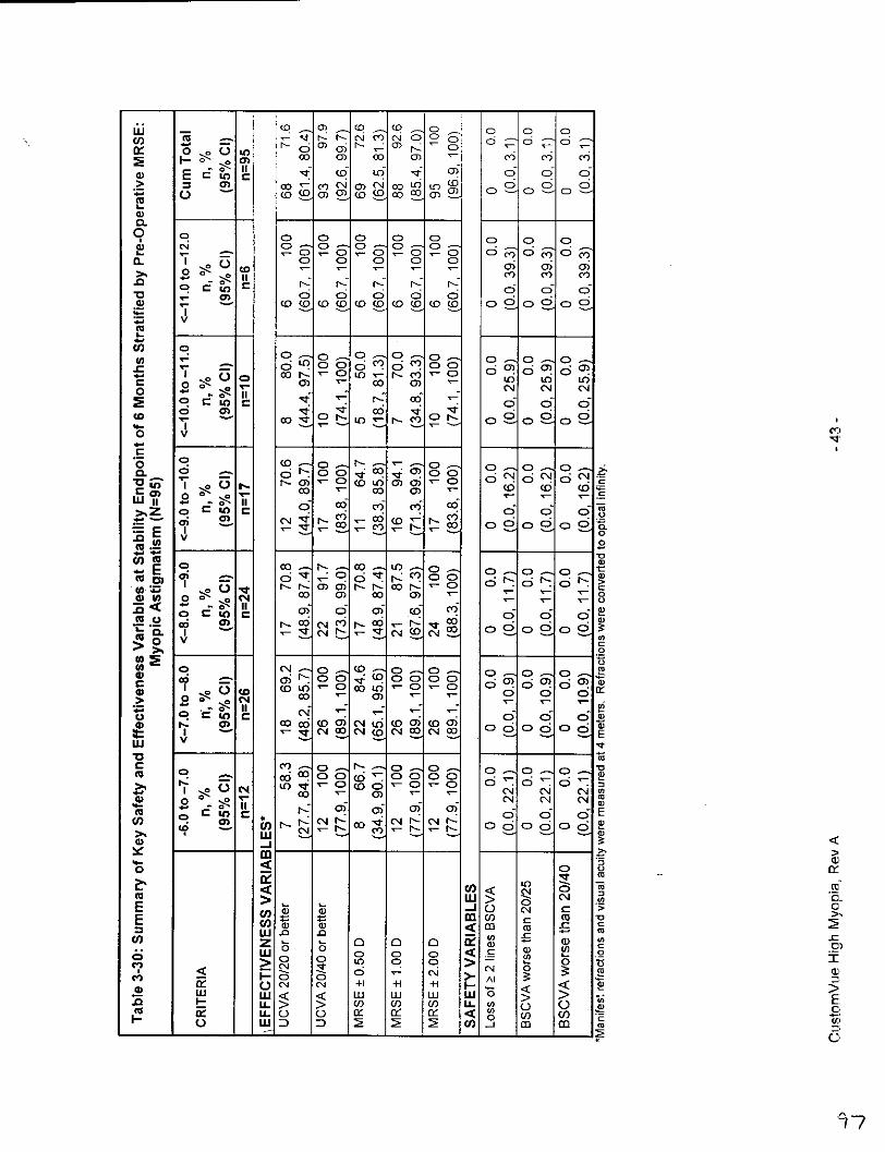
d' -0 5 0 a a
Z~ 0~ o~u o05
5~ 50
c Ecmc cc to v 0Ž ci 6ooX 0ara)'~ N~ otl
= C) -o( (C0 fLoWadV 00 C0 0
0. -
oL 0 0 0 0 0 -- 0Ci CN 0) 0. 0I 0 0 0 0ED 0 6
7 - a IaI CEL o, oC '
1
= V~~~~~~~~~OC(U~~~~~~~~~~c4-~ ~~~~~~~~c
.~ ' - 0 o o 0o 0o 0 0 0 .
. 7 -C'Joo6C6o 0Ž6 Cq
0 VII 03~~0
~~~~~. o oc~~co.N~- 60
o ..-. alr, 0II 0) (Nj01C) C)O
~~~~~~~~~~~~~~~~~~~~~~~~~~~~,. wtc ~- r ,z- & c
:a~ 0 0[ V 6.t- tO tr-Na0N0 09 CO
W) ~ ~ ~ ~ ~ ~ ~ ~ C 0 00
~,. o~O~~~r ' . C0 o, N rnr 0 03 ', -. 2
cu._ 0o - ~- ~ -
0)0 -n~~~C ( ri C
, C 0 0) 1CT; '3 N- CO 1)- 0 9 p 0 0
Eu o
'0~~~~~~~~~~~~~~~~~~
OW cc oN ~~~ 0) 0030 0)0-
- .. cNI , - - c d "- rC- d-)oc c Ž "T r-PCD 6 -6 - I>
4) U) 0 CI 00 0) 00 0) 8~~~~~~~-4
>- o · · 0
a0 CD v -C, ¢I '
> 0(A 0 CD 0: 0 C-) C
( o 0o0 0
-~~~~~~~~~~~~~ r(D 0)~~~~~~~~~~~~~~~~~~~~~~~~~~~~~~~~~~D
o ROLmc &-7 - r 6t I nVN0:w C'JflCNC''4 03 060606
W: S
C CO p ~~ ~~~N p" 0 0 0 0> c
r- `0 -00 - ,-
(;CO r9 (D - -m0 6 c'I C) Ci 0) 0)0
,~,~C co CD ~ ~ --
co (U o "0) 0*CCN :CcJ-c C 6 C0 . .
CF t~~~~~~~ ~~~~~o a'1-O4 o
t W "-j <0JC CME a, _ 'S~~~~~~~~~~~~C:
m nn <Wo E C >)0 0 o p 5 0<C 0
>~ 0 in o =-~~ - P Lq> '- 0
FE L) CM +1 41 11 J >a, W< < W LU LU WO ->m U.LL > co (I co ug U a) F
U.LJC~) C) la• l• C~ U) a, U)C) Wf D n W co In

Facts You Need to Know About CustomVue TMLaser Assisted In-Situ Keratomileusis (LASIK)Laser Treatment
Patient Information Booklet
For High Myopia (Nearsightedness) from -6 to -11 0 MRSE withup to -3 D Astigmatism
Please read this entire booklet. Discuss its contents with yourdoctor so that all your questions are answered to yoursatisfaction. Ask any questions you may have before you agree-to the surgery.
VISX®9, Incorporated3400 Central ExpresswaySanta Clara, CA 95051-0703U.S.A.
Tel: 408.733.2020
CustomnVue TM High Myopia Patient Inforrmation, Rev A
© Copyright 2005 by VISX®, Incorporated
All Rights Reserved
VISX ®, VisionKey, ActiveTrak®, WaveScan®, WaveScan WaveFront®, and WavePrint® are registeredtrademarks of VISX, Incorporated.
CustomVue- and STAR S4" are trademarks of VISX, Incorporated.
Accutane ® is a registered trademark of Hoffmann-La Roche Inc. Cordarone ® is a registered trademark ofSanofi-Synthelabo, Inc. Imitrex® is a registered trademark of GlaxoSmithKline.
No part of this publication may be reproduced or transmitted in any form or by any means, electronic ormechanical, including photocopying, recording, or any information storage and retrieval system, withoutpermission in writing from VISX, Incorporated.
CustomVue TM High Myopia Patient Information, Rev A

Table of Contents
Introduction .................................... 4.......... ........ .How Refractive (Wavefront) Errors Affect Your Vision ................................5The VISX STAR S4 IR' Excimer Laser ............................................................. 7How the CustomVue Tm LASIK Procedure Works ................................................. 8The VISX WaveScan WaveFront® System .......................................................... 8BENEFITS.9Clinical Study to Evaluate Benefits ....................................................................... 9Study Patient Demographicsto ..... ....................................................................... gVisual Acuity Without Glasses After Treatment .................................................. 10Visual Acuity Without Glasses After Treatment and With Glasses BeforeTreatm ent ........................................................................................................... 10Patient Quality of Vision Comparison ................................................................. 11RISKS ................................................................................................................ 12CONTRAINDICATIONS -When Can't You Have LASIK? ............................... 12What Warnings and Other Information Do You Need to Know About? .............. 13
Warnings.~~~~~~~13W a rn in g s ............................................................................................................ 3Precautions ........................................................................................................ 14Clinical Study to Evaluate Risks ......................................................................... 15Visual Acuity With Glasses After Treatment ....................................................... 15Change in Visual Acuity With Glasses After Treatment ...................................... 16Adverse Events and Com plications ................................................................... 7Patient Symptoms After CustomVuew Treatment .............................................. 18The First W eek Following Surgery ..................................................................... 19The First Two To Six Months Following Surgery ................................................ 19Are You A Good Candidate For CustomVueTM LASIK? ..................................... 20Before Surgery ................................................................................................... 20The Day of Surgery ............................................................................................ 21After Surgery ..................................................................................................... 22Questions to Ask Your Doctor ............................................................................ 23Self-Test ............................................................................................................. 24Sum m ary of Im portant Information ..................................................................... 25Answers to Self-Test Questions ......................................................................... 25Glossary ............................................................................................................. 26Patient Assistance Information ........................................................................... 31
CustomVueT M High Myopia Patient Information, Rev A
rto

Introduction
Your doctor and VISX, Incorporated provide the information in this booklet to helpyou decide whether to have a CustomVue TmLASIK treatment. OustomVue LASIK(laser assisted in situ keratomileusis) may be used to correct, or partly correct,high levels of myopia (nearsightedness) with and without astigmatism.
Some other ways to correct your vision are by wearing glasses or contact lenses,or by undergoing other kinds of laser refractive surgery such as non-customLASIK or PRK (photorefractive keratectomy). Other surgical procedures that donot use a laser such as RK (radial keratotomy) and ALK (automated lamellarkeratectomy) may also be an option.
Please read this booklet completely. Discuss any questions with your doctorbefore you decide if CustomVue LASIK is right for you. Only an eye careprofessional trained in laser vision correction can determine whether you are asuitable candidate. Some people, such as military pilots, have job-related visionrequirements that cannot be met by having LASIK.
CustomVuem High Myopia Patient Information, Rev A -4-
IC)
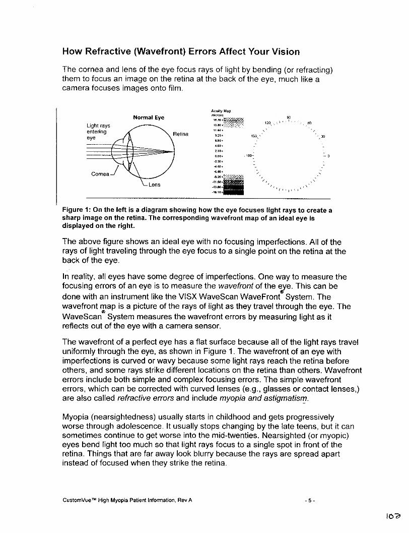
How Refractive (Wavefront) Errors Affect Your Vision
The cornea and lens of the eye focus rays of light by bending (or refracting)them to focus an image on the retina at the back of the eye, much like acamera focuses images onto film.
Acuity M.,
Normal EyeLight rays ~~~~ ~ ~~~~~~~~~~~~~~~~1201
enteringReia.2'0eye retina M~~~~~~~~~~~~~~~~6.0
4.60
Lens~~~~~~~20
Figure 1: On theleft is a diagra showing how0th eye -0ue ih ay ocet
Figue 1:bonvte leftr ihos andiagram showinghwthe eye focusesg ighteraysctin.Alofrethea
rays of light traveling through the eye focus to a single point on the retina at theback of the eye.
In reality, all eyes have some degree of imperfections. One way to measure thefocusing errors of an eye is to measure the wave front of the eye. This can bedone with an instrument like the VISX WaveScan WavelFront System. Thewavefront map is a picture of the rays of light as they travel through the eye. TheWaveScan® System measures the wavefront errors by measuring light as itreflects out of the eye with a camera sensor.
The wavefront of a perfect eye has a flat surface because all of the light rays traveluniformly through the eye, as shown in Figure 1. The wavefront of an eye withimperfections is curved or wavy because some light rays reach the retina beforeothers, and some rays strike different locations on the retina than others. Wavefronterrors include both simple and complex focusing errors. The simple wavefronterrors, which can be corrected with curved lenses (e.g., glasses or contact lenses,)are also called refractive enrors and include myopia and astigmatism.
Myopia (nearsightedness) usually starts in childhood and gets progressivelyworse through adolescence. It usually stops changing by the late teens, but it cansometimes continue to get worse into the mid-twenties. Nearsighted (or myopic)eyes bend light too much so that light rays focus to a single spot in front of theretina. Things that are far away look blurry because the rays are spread apartinstead of focused when they strike the retina.
CustomnVueN~ High Myopia Patient Information, Rev A -5 -
lOY

Acuty M.,
Nearsighted EyeLight rays~~~~~~~~~~~~100
entering~~~~~~~~~~~~920 5
eye etina~ ~ ~~~~~~to
Figure 2: On the left is a diagram of a nearsighted eye showing the light rays focusing infront of the retina. The corresponding wavefront of a myopic eye shows a curvedwavefront surface. The height difference between center and edge is indicated by thechange in grayscale.
Astigmatism causes the rays of light entering through different parts of the eye tofocus unequally so that they do not ever form a single spot. Some rays mayfocus on the retina, but other rays focus in front of the retina. Things look blurrybecause images are not ever focused clearly on the retina.
Muity Map
Nearsighted and Astigmatic Eye to
ight ray.~~~~~~~~~~~~0
!ntering Retina .......0-'~ <S!ye~~~~~~~~~~~~~~~~~IS
Lens~~~~~~0
Figre : O th let i a iagam f a asigmticeyeshoingligt rys hatdo ot0ve
come toa focu at onepoint.The corespondng waveront mp for tis eyeshows0
surface tat is cured more i one diretion.tha the oter
inFigure3 Ontelf is a diagram allwa ann arostigatic eye erih s a showing lihjayuhtson t ee
the complex errors. The combination of simple and complex wavefront errors inany eye is unique. The CustomVue~ treatment is "custom" because it includesinformation from the WaveScan System that is more individualized than what adoctor uses to program a non-custom treatment-
CustomnVuem- High Myopia Patient Information, Rev A -6-
(0)3

Acuity Map Wayefron ih Order A be.ra ions
90 9300
/6
Figure 4: On the left is a wayefront map of all wavefront errors and on the right is awavefront map showing only the complex errors.
The VlSX STAR S4 IRT MExcirner Laser
The excimer laser produces a beam of cool ultraviolet light. The doctor programsyour information into a computer that controls the laser. The laser produces aseries of rapid pulses that remove small and precise amounts of corneal tissue.Excimer laser light does not penetrate into the eye and leaves other eye
structures (iris, lens, retina) undisturbed.
The laser also contains an auto-centering eye tracking system which will alignthe treatment and automatically compensate for many of your eye movementsduring the CustomVue treatment.
CustomnVue o High Myopia Patient Infomation, Rev A 7 -
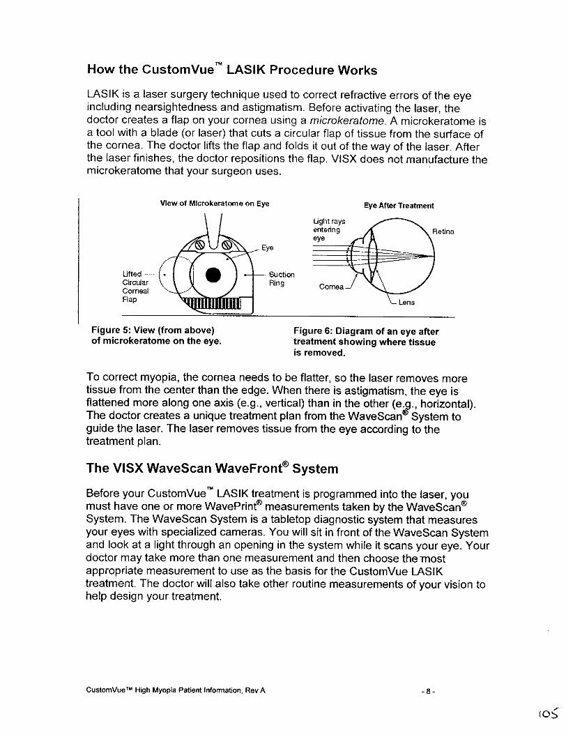
How the CustomVue LASIK Procedure Works
LASIK is a laser surgery technique used to correct refractive errors of the eyeincluding nearsightedness and astigmatism. Before activating the laser, thedoctor creates a flap on your cornea using a microkeratome. A microkeratome isa tool with a blade (or laser) that cuts a circular flap of tissue from the surface ofthe cornea. The doctor lifts the flap and folds it out of the way of the laser. Afterthe laser finishes, the doctor repositions the flap. VISX does not manufacture themicrokeratome that your surgeon uses.
View of MIcrokeratome on Eye Eye After Treatment
Ug'ht raysentering C Retinaeye
Lifted -asuction
Circular RinCorneal creFlap Ln
Figure 5: View (from above) Figure 6: Diagram of an eye afterof microkeratome on the eye. treatment showing where tissue
is removed.
To correct myopia, the cornea needs to be flatter, so the laser removes moretissue from the center than the edge. When there is astigmatism, the eye isflattened more along one axis (e.g., vertical) than in the other (e.g., horizontal).The doctor creates a unique treatment plan from the WaveScan® System toguide the laser. The laser removes tissue from the eye according to thetreatment plan.
The VISX WaveScan WaveFront® System
Before your CustomVue~ LASIK treatment is programmed into the laser, youmust have one or more WavePrint® measurements taken by the WaveScan®System. The WaveScan System is a tabletop diagnostic system that measuresyour eyes with specialized cameras. You will sit in front of the WaveScan Systemand look at a light through an opening in the system while it scans your eye. Yourdoctor may take more than one measurement and then choose the -mostappropriate measurement to use as the basis for the CustomVue LASIKtreatment. The doctor will also take other routine measurements of your vision tohelp design your treatment.
CustomVuew High Myopia Patient Information, Rev A -8-
"~R~~~~~0
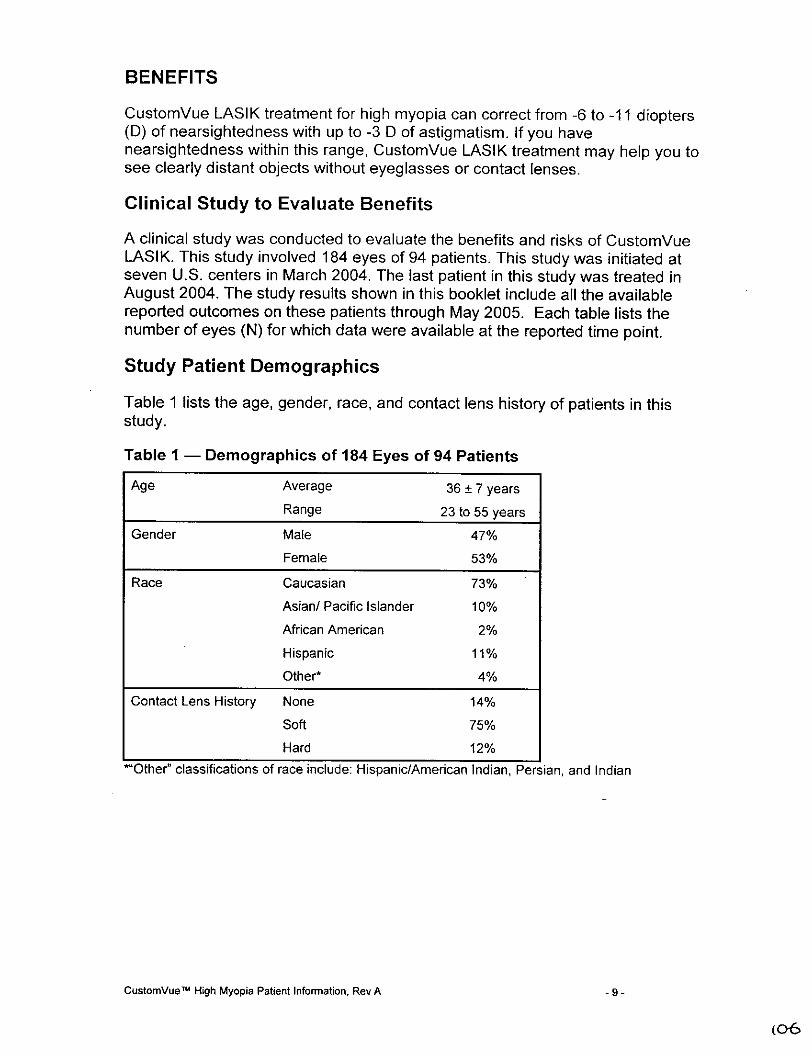
BENEFITS
CustomVue LASIK treatment for high myopia can correct from -6 to -1 1 diopters(D) of nearsightedness with up to -3 D of astigmatism. If you havenearsightedness within this range, CustomVue LASIK treatment may help you tosee clearly distant objects without eyeglasses or contact lenses.
Clinical Study to Evaluate Benefits
A clinical study was conducted to evaluate the benefits and risks of CtistomVueLASIK. This study involved 184 eyes of 94 patients. This study was initiated atseven U.S. centers in March 2004. The last patient in this study was treated inAugust 2004. The study results shown in this booklet include all the availablereported outcomes on these patients through May 2005. Each table lists thenumber of eyes (N) for which data were available at the reported time point.
Study Patient Demographics
Table 1 lists the age, gender, race, and contact lens history of patients in thisstudy.
Table I - Demographics of 184 Eyes of 94 Patients
Age Average 36 ± 7 yearsRange 23 to 55 years
Gender Male 47%
Female 53%
Race Caucasian 73%
Asian/ Pacific Islander 10%
African American 2%
Hispanic 11%
Other* 4%
Contact Lens History None 14%
Soft 75%
Hard 12%
*~Other` classifications of race include: Hispanic/American Indian, Persian, and Indian
CustomnVuew High Myopia Patient Information, Rev A -9-
(0,6

Visual Acuity Without Glasses After Treatment
Visual acuity measures the sharpness of vision using a letter chart. Table 2shows that six months after the treatment, 84% of study patients saw 20/20 orbetter without glasses while 98% of study patients saw 20/40 or better. A visualacuity of 20/40 or better is the standard requirement in most states for you todrive without any glasses or contact lenses.
Table 2 - Visual Acuity Without Glasses After Treatment (N=184)
Time After Treatment 1 Month 3 Months 6 Months 9 Months 12 Months(n=184) (n=180) (n=178) (n=170) (n=107)
20/16 or better 52% 56% 65% 61% 59%
20120 or better 85% 82% 84% 85% 86%
20125 or better 92% 93% 93% 95% 96%
20140 or better 99% 98% 98% 99% 100%
Visual Acuity Without Glasses After Treatment andWith Glasses Before Treatment
Table 3 shows the relative number of lines (rows of letters) on the eye chart thatpatients could see before and after surgery. 78% of the patients saw as well orbetter without glasses 6 months after CustomVue TM treatment as with glassesbefore treatment.
Table 3 - Visual Acuity Without Glasses After TreatmentCompared to With Glasses Before Treatment
Time After Treatment I Month 3 Months 6 Months 9 Months 12 Months(n=184) (n=180) (n=1 78) (n=170) (n=107)
More than 2 lines better 0% 0% 0% 0% 0%
2 lines better 1% 2% 4% 3% 2%
1 line better 21% 23% 34% 29% 26%
Less than 1 line change 49% 46% 40% 44% 49%
I line worse 18% 18% 10% 17% 17%
2 lines worse 5% 7% 8% 4% 5%
More than 2 lines worse 5% 3% 3% 4% 2%
CustomVueM High Myopia Patient Information, Rev A - 10-
(o-7
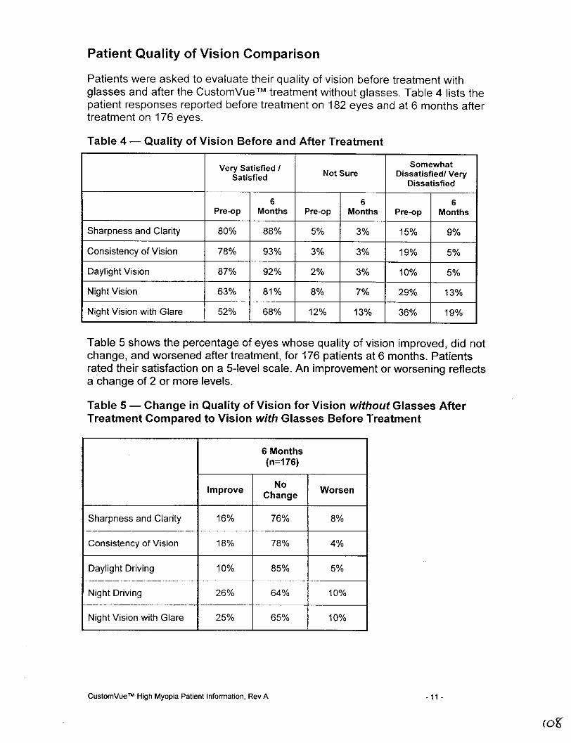
Patient Quality of Vision Comparison
Patients were asked to evaluate their quality of vision before treatment withglasses and after the CustomVue TM treatment without glasses. Table 4 lists thepatient responses reported before treatment on 182 eyes and at 6 months aftertreatment on 176 eyes.
Table 4 - Quality of Vision Before and After Treatment
Very Satisfied ISomewhatSatisfied Not Sure Dissatisfied/ Very
Dissatisfied
6 6 6Pre-op Months Pre-op Months Pre-op Months
Sharpness and Clarity 80% 88% 5% 3% 15% 9%
Consistency of Vision 78% 93% 3% 3% 19% 5%
Daylight Vision 87% 92% 2% 3% 10% 5%
Night Vision 63% 81% 8% 7% 29% 13%
Night Vision with Glare 52% 68% 12% 13% 36% 19%
Table 5 shows the percentage of eyes whose quality of vision improved, did notchange, and worsened after treatment, for 176 patients at 6 months. Patientsrated their satisfaction on a 5-level scale. An improvement or worsening reflectsa change of 2 or more levels.
Table 5 - Change in Quality of Vision for Vision without Glasses AfterTreatment Compared to Vision with Glasses Before Treatment
6 Months(n=176)
Improve No WorsenChange
Sharpness and Clarity 16% 76% 8%
Consistency of Vision 18% 78% 4%
Daylight Driving 10% 85% 5%
Night Driving 26% 64% 10%
Night Vision with Glare 25% 165% 10%
CustomnVueN High Myopia Patient inormation, Rev A -11 -
(O&
RISKS
As with any surgical procedure there are risks associated with OustomVueTMtreatments. It is important to discuss these risks with your doctor before youmake the decision to have the surgery. If the results of the surgery are notsatisfactory, you may need to have additional laser treatment in the same eye.Your doctor may perform CustomVue LASIK for both eyes. However, sometimesit is better to have this procedure done on only one eye. Talk with your doctorabout whether it would be better to treat one or both of your eyes.
Some risks are related to the creation of the corneal flap. Corneal flapcomplications include but are not limited to: cutting an incomplete, irregular flapor free flap; misalignment of the flap; and perforation of the cornea. Corneal flapcomplications range in severity from those that simply require the treatment to bepostponed for several months, to those which create corneal irregularitiesresulting in permanently blurred vision.
IMPORTANT:
You may need reading glasses after laser surgery even if youdid not wear them before. Your vision may not be perfect, andyou may need to wear glasses or contact lenses for someactivities even after laser vision correction.
CONTRAINDICATIONS - When Can't You Have LASIK?
If you have any of the following situations or conditions you should not haveLASIK because the risk is greater than the benefit:
*You are pregnant or nursing, because these conditions may cause temporaryand unpredictable changes in your cornea and a LASIK treatment mayimproperly change the shape of your cornea.
*You have collagen vascular (e.g., rheumatoid arthritis), autoimmune (e.g.,lupus), or immunodeficiency diseases (e.g., AIDS), because these conditionsaffect the body's ability to heal.
*You show signs of keratoconus or any other condition that causes a thinningof your cornea. This condition can lead to serious corneal problems duringand after LASIK surgery. It may result in need for additional surgery and mayresult in poor vision after LASIK.
CustomnVuem High Myopia Patient Information, Rev A -12 -
(093

*You are taking medications with ocular side effects, e.g., Isotretinoin(Accutane®*) for acne treatment or Amiodarone hydrochloride (Cordarone"tfor normalizing heart rhythm, because they may affect the accuracy of theLASIK treatment or the way your cornea heals after LASIK. This may result inpoor vision after LASI K.
*Your corneas are thin. If your corneas are too thin to allow your doctor to cuta proper flap in the LASIK procedure, you can't have LASIK because it isnecessary to have a flap.
What Warnings and Other Information DoYou Need to Know About?
Warnings
If you have any of the following conditions, you may have LASIK if your doctorevaluates the seriousness of your condition and believes the benefit of havingLASIK is greater than the risk.
*Diabetes. If you have diabetes, LASIK may be risky for you because yourdiabetes may interfere with the healing of your eyes.
*History of Herpes simplex or Herpes zoster infection that has affected youreyes. If you have had a Herpes simplex or a Herpes zoster infection thataffected your eyes, and have an infection now, LASIK is more risky for you.
*Symptoms of significant dry eye. If you have severely dry eyes, LASIK mayincrease the dryness. This may or may not go away. This dryness may delayhealing of the flap or interfere with the surface of the eye after surgery.
*Severe allergies. If you have severe allergies and take medicines for them,LASIK is more risky for you.
*Accutane® is a registered trademark of Hoffmann-La Roche, Inc.
Cordarone® is a registered trademark of Sanofi-Synthelabo, Inc
CustomnVueN~ High Myopia Patient Information, Rev A - 13 -
(co
Precautions
The safety and effectiveness of wavefront-guided LASIK with the STAR S4 IR TM
Laser for high myopia with or without astigmatism has not been established inpatients:
*With unstable eyes that have changed in their prescription more than 1.0diopters in nearsightedness or astigmatism in the last 12 months, and yournearsightedness or astigmatism is getting worse. If your eyes are unstable,the right amount of treatment cannot be determined. This may res-ult in poorvision after LASIK.
*With corneal disease or abnormality (e.g., scar, infection, etc.). If you have anabnormal corneal condition, such as corneal scars, it may affect the accuracyof the LASIK treatment or the way your cornea heals after LASIK. This mayresult in poor vision after LASIK. If your eyes have an active disease, it isunknown whether LASIK is safe and effective under this condition.
*With history of injury or surgery to the center of the cornea (for example,surgery to correct vision such as RK, PRK, LASIK), or other surgery on theeye. If your eyes are injured or you have had surgery, it is unknown whetherLASIK will weaken the cornea too much. This may result in poor vision afterLASIK.
*With large pupils. Before surgery, your doctor should measure your pupil sizeunder dim lighting conditions and review how your pupil size may affect yourvision after surgery. If you have large pupils in dim light, there may be anincreased possibility for negative effects on your vision after surgery, such asseeing glare and halos around lights, and being less satisfied with your visionwhen driving at night with oncoming headlights.
*With history of glaucoma or have had pressure greater than 21 mmHg insideyour eyes, because it is unknown whether LASIK is safe and effective for you.
*Who use medications that might make it harder for wounds to heal, such assumnatriptan (Imitrex®) used for migraine headaches, because it is unknownwhether LASIK is safe and effective for this condition.
*Who take other medications. Let your doctor know if you taking anyprescription medicines or any medicines you bought without a prescription.
*Who are younger than 21 years of age, because it is unknown whether LASIKis safe and effective for you.
*Who have nearsightedness worse than 1 1 diopters or astigmatism worse than3 diopters, because it is unknown whether LASIK is safe and effective for you.
*Over the long term (more than 1 year), because it is unknown whether LASIKis safe and effective for periods longer than 1 year.
*Who are considering retreatment with this laser for LASIK, because it isunknown whether LASIK is safe and effective for repeating the LASIK
Imitrex®D is a registered trademark of GlaxoSmithKline.
CustomnVue~ High Myopia Patient Information, Rev A - 14 -

procedure on the same eye.
With undiagnosed dry eyes. Your doctor should also evaluate you for dryeyes before surgery. You may have dry eyes after LASIK surgery even if youdid not have dry eyes before surgery.
In the following conditions: dim lighting, rain, snow, fog, or bright glare. Youmight have difficulty seeing in dim lighting, rain, snow, fog, or bright glare.Whether you may have poor vision under these conditions is hard to predictbecause it has been studied so little.
Clinical Study to Evaluate Risks
In the clinical study on CustomVue T LASIK for high myopia and myopicastigmatism, visual acuity without glasses improved for all eyes treated.
Visual Acuity With Glasses After Treatment
Table 6 shows that all patients in the clinical study saw 20/25 or better withglasses at all time points after treatment.
Table 6 - Visual Acuity With Glasses (Best Vision) After Treatment
I Month 3 Months 6 Months 9 Months 12 Months(n=184) (n=180) (n=178) (n=170) (n=107)
20/16 or better 80% 84% 91% 83% 80%
20(20 or better 98% 97% 100% 100% 99%
20/25 or better 100% 100% 100% 100% 100%
CustomVueTM High Myopia Patient Information, Rev A -15 -

Change in Visual Acuity With Glasses After Treatment
Table 7 shows the change in visual acuity with glasses at 1, 3, 6, 9 and 12months after treatment for the patients in the clinical study.
Table 7 - Change in Visual Acuity With Glasses After TreatmentCompared to Before Treatment
I Month 3 Months 6 Months 9 Months 12 Months(n=184) (n=180) (n=178) (n=170) (n=107)
Eyes with loss > 2 Lines 0% 0% 0% 0% 0%
Eyes with loss > I to < 2 Lines 1% 2% 0% 0% 0%
Eyes with loss > 0 to 1 Line 10% 8% 5% 6% 15%
Eyes with no change 38% 31% 26% 28% 25%
Eyes with gain of >0 to 1 line 48% 52% 53% 56% 55%
Eyes with gain > 1 to 2 Lines 4% 7% 15% 8% 5%
Eyes with gain > 2 Lines 0% 1% 1% 1% 0%
Contrast Sensitivity
Unlike normal vision tests that measure the ability to see a black and white eyechart, contrast sensitivity measures how well one sees in low contrast conditionssuch as driving in rain or fog. While most eyes in the study achieved the same orimproved contrast sensitivity post-operatively as they did pre-operatively, at 6months post-operatively, 11% experienced a significant loss in bright conditions.In dim conditions, 16% experienced a similar loss, and with the addition of glarein dim conditions, 23% experienced such a loss. Table 8 shows the change incontrast sensitivity 6 months after treatment in 178 patients.
CustomVueM High Myopia Patient Information, Rev A - 16 -
1t3
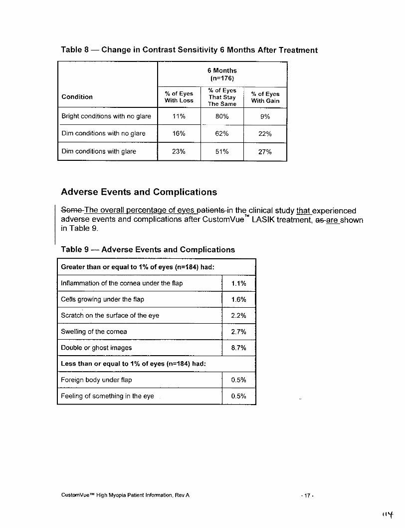
Table 8 - Change in Contrast Sensitivity 6 Months After Treatment
6 Months(n=176)
Codtin fEys %of Eyes % of EyesoftEyes That Stay With Gain
The Same
Bright conditions with no glare 11% 80% 9%
Dim conditions with no glare 16% 62% 22%
Dim conditions with glare 23% 51% 27%
Adverse Events and Complications
Seine-The overall percentage of eyes patients-in the clinical study that experiencedadverse events and complications after CustomVue' LASIK treatment, as-are shownin Table 9.
Table 9 - Adverse Events and Complications
Greater than or equal to 1% of eyes (n=184) had:
Inflammation of the cornea under the flap 1.1%
Cells growing under the flap 1.6%
Scratch on the surface of the eye 2.2%
Swelling of the cornea 2.7%
Double or ghost images 8.7%
Less than or equal to 1% of eyes (n=184) had:
Foreign body under flap 0.5%
Feeling of something in the eye 0.5%
CustomVueM High Myopia Patient Information, Rev A - 17-
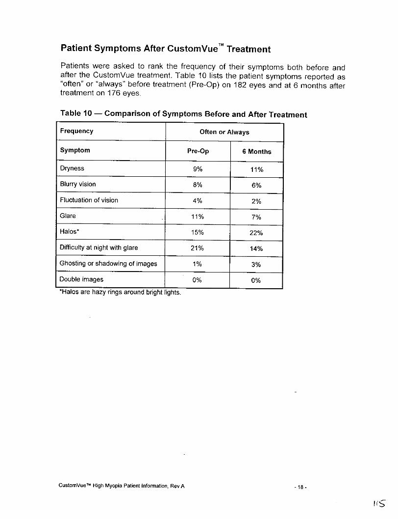
Patient Symptoms After CustomVue TM Treatment
Patients were asked to rank the frequency of their symptoms both before andafter the CustomVue treatment. Table 10 lists the patient symptoms reported as"often" or "always" before treatment (Pre-Op) on 182 eyes and at 6 months aftertreatment on 176 eyes.
Table 10 - Comparison of Symptoms Before and After Treatment
Frequency Often or Always
Symptom Pre-Op 6 Months
Dryness 9% 11%
Blurry vision 8% 6%
Fluctuation of vision 4% 2%
Glare 11% 7%
Halos* 15% 22%
Difficulty at night with glare 21% 14%
Ghosting or shadowing of images 1% 3%
Double images 0% 0%
*Halos are hazy rings around bright lights.
CustomVue"M High Myopia Patient Information, Rev A - 18 -

Table 11 presents the percentage of eyes whose symptoms improved, did notchange, or worsened 6 months after treatment, when vision without glasses aftertreatment was compared to vision with glasses before surgery. Patients ratedtheir symptoms on a 5-level scale. An improvement or worsening reflects achange of 2 or more levels.
Table 11 - Change in Patient Symptoms for Vision Without GlassesAfter Treatment Compared to Vision With Glasses Before Treatment
6 Months(n=176)
NoSymptom Improv Change Worsen
Dryness 5% 90% 6%
Blurry vision 13% 81% 6%
Fluctuation of vision 4% 89% 7%
Glare 11% 84% 5%
Halos* 66% 81% 13%
Difficulty at night with 12% 81% 7%glare
Ghosting or shadowing 7% 85% 9%of images
Double images 1% 96% 3%
*Halos are hazy rings around bright lights.
The First Week Following Surgery
Moderate pain and discomfort may last for up to 3 days after surgery.Blurred vision and tearing will occur as the cornea heals.
You will be sensitive to bright lights.
The First Two To Six Months Following Surgery
Your vision may fluctuate during this time period. You may also experiencesome dryness.
CustomVue T MHigh Myopia Patient Information, Rev A - 19 -

Are You A Good Candidate For CustomVue T M LASIK?
If you are considering CustomVue LASIK, you must:
Be at least 21 years of age and have myopia with or without astigmatism.
Have healthy eyes that are free from eye disease or corneal abnormality(e.g., scar, infection, etc.).
Have documented evidence that your refraction did not change by more than1.0 diopters during the year before your pre-operative examination.
Be informed of LASIK risks and benefits as compared to other availabletreatments for nearsightedness with or without astigmatism.
Be able to lie flat without difficulty.
Be able to tolerate local or topical anesthesia.* Be willing to sign an informed consent form as provided by your eye care
professional.
Be able to keep your eye accurately on the fixation light for the entire lasersurgical procedure.
Before Surgery
If you are interested in having laser vision correction, you will need to have a pre-surgical examination to determine if your eye is healthy and suitable for surgery.This will include a complete medical and eye history, and thorough examinationof both eyes, including WavePrint refractions and computerized mapping of yourcornea.
WARNING:
If you wear contact lenses, it is very important to stop wearing them 2 - 4weeks before examination and treatment for the doctor to obtain a stableeye measurement. Failure to do this might produce suboptimal surgicalresults.
Before the surgery, please tell your doctor whether you take any medications orhave any allergies. Also, talk with your doctor about eating or drinkingimmediately before the surgery. You should also arrange for transportation,since you must not drive immediately after the surgery. You may resumedriving only after receiving permission from your doctor.
CustomVue TM High Myopia Patient Information, Rev A - 20 -
1/7
The Day of Surgery
Before the surgery, local anesthetic (numbing) drops will be placed into the eyeto be treated and you will be escorted into the room with the laser. You will lie onyour back in a reclining chair and look up. An instrument will be placed betweenyour eyelids to hold them open during the surgery. There will also be a temporaryshield covering the eye not having surgery. You will be asked to listen to thesounds of the treatment so that you will be prepared for the noise the lasermakes during the surgery.
The surgery begins with the placement of a suction ring that elevates thepressure in the eye. The vision in the eye will go black as the suction increasesthe pressure in the eye. The microkeratome creates a circular corneal flap. Thedoctor will lift this flap of tissue after the suction is released. Vision will return tothe eye after the suction is released.
The doctor will then reposition your head in the chair and refocus themicroscope. You will be asked to look directly at a blinking light while the laser isrunning. It is important to fix your gaze on the light for the entire laser procedure.Try to keep both eyes open without squinting, as this makes it easier to keeplooking at the light. Small amounts of tissue will then be removed from yourcornea using the VISX STAR S4 IRT Excimer Laser system.
PRECAUTION:
It is very important that you keep looking at the light during the procedure,even if the light fades or becomes dim. You need to concentrate on lookingat this light throughout the treatment to ensure the best results possible.
Typically, the laser beam will be applied to your eye less than 2 minutes and,overall, the surgery may last about 10 minutes.
After the laser surgery is complete, some eye drops may be placed on your eye.The surgery is painless because of the anesthetic drop. When the anestheticdrops wear off (about 30 to 60 minutes), your eye may hurt moderately for 1 to 2days. The discomfort is typically described as "a sandy sensation." Your doctorcan prescribe pain medication to make you more comfortable during this timeafter the surgery. To promote healing and lessen the risk of infection, do NOTrub your eye after surgery.
CustomVue' M High Myopia Patient Information, Rev A -21 -
After Surgery
You will be mildly sensitive to light and have the feeling that something is in youreye for 1 to 2 days. Sunglasses may make you more comfortable during thistime.
IMPORTANT:Use the lubricants and eye medications as directed by your doctor. Yourresults depend upon you following your doctor's instructions.
WARNING:
Your doctor will monitor you for any side effects if you need to use atopical steroid medication. Possible side effects of prolonged topicalsteroid use are:
* ocular hypertension (an increase in the eye pressure).
• glaucoma (a condition usually associated with high eye pressure thatresults in damage to the nerve in the eye and possible loss of vision).
• cataract formation (an opacity or clouding of the lens inside the eye thatcan cause a loss of vision).
CustomVue TM High Myopia Patient Information, Rev A -22 -

Questions to Ask Your Doctor
You may want to ask the following questions to help you decide ifCustomVue TM LASIK is right for you:
What other options are available for correcting my vision?
Will I have to limit my activities after surgery, and for how long?
What are the benefits of CustomVue LASIK for my amount of refractive error?
What vision can I expect in the first few months after surgery?
If CustomVue LASIK does not correct my vision, what is the possibility thatmy glasses will need to be stronger than before? Could my need for glassesincrease over time?
Will I be able to wear contact lenses after laser surgery if I need them?
How is CustomVue LASIK likely to affect my need to wear glasses or contactlenses as I get older?
Will my cornea heal differently if injured after having laser surgery?
Should I have CustomVue LASIK in my other eye?
How long will I have to wait before I can have surgery on my other eye?
What vision problems might I experience if I have CustomVueTM LASIK onlyon one eye?
Discuss the cost of surgery and follow-up care requirements with your doctor, asCustomVue LASIK is not covered by most health insurance policies.
CustomVue T MHigh Myopia Patient Information, Rev A -23 -
(.;>O
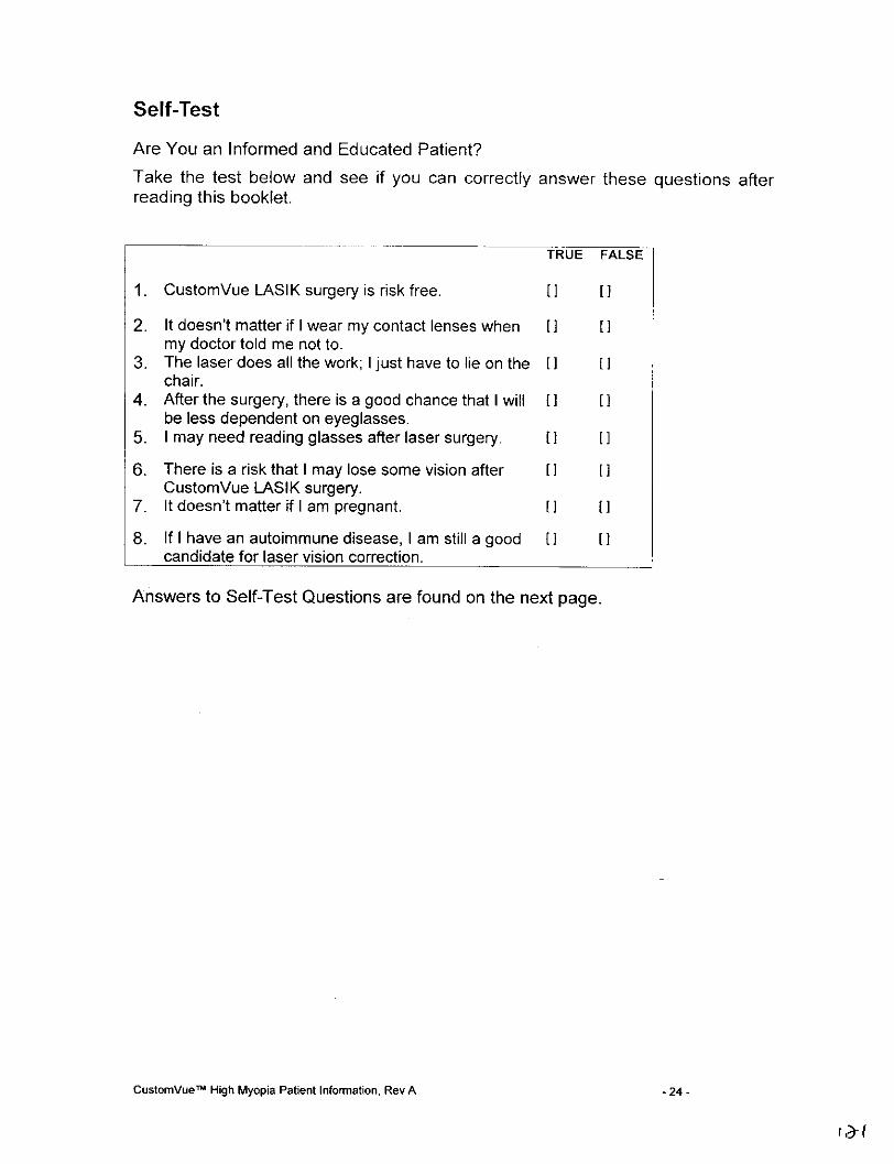
Self-Test
Are You an Informed and Educated Patient?
Take the test below and see if you can correctly answer these questions afterreading this booklet.
TRUE _FALSE
1 . CustomVue LASIK surgery is risk free. [1 II
2. It doesn't matter if I wear my contact lenses when [] H1my doctor told me not to.
3. The laser does all the work; I just have to lie on the [I I]chair.
4. After the surgery, there is a good chance that I will [ I ]be less dependent on eyeglasses.
5. I may need reading glasses after laser surgery. (1 [I
6. There is a risk that I may lose some vision after [ I 1]Custom Vue LASIK surgery.
7. It doesn't matter if I am pregnant.I] [
8. If I have an autoimrmune disease, I am still a good [1 1candidate for laser vision correction.
Answers to Self-Test Questions are found on the next page.
CustomnVuetm High Myopia Patient Information, Rev A -24 -

Summary of Important Information
CustomVue LASIK is a permanent operation to the cornea and isirreversible.
CustomVue LASIK may not eliminate the need for reading glasses, even ifyou never have worn them before.
Your vision must be stable for at least one year before CustomVue LASIK.You will need written evidence that your nearsightedness with or withoutastigmatism has changed less than 1.0 diopters.
Pregnant and nursing women should wait until they are not nursing and notpregnant to have the surgery.
You are not a good candidate if you have degenerative or autoimmunediseases, or have a condition that makes wound healing difficult.
CustomVue LASIK may result in some discomfort. The surgery is not risk-free. Please read this entire booklet, especially the sections on Benefits andRisks before you agree to the surgery.
Alternatives to CustomVue LASIK include, but are not limited to, glasses,contact lenses, non-custom LASIK and PRK, RK, and ALK.
Some people, such as military pilots, have job-related vision requirementsthat cannot be met by having CustomVue LASIK.
* Before considering laser vision correction you should:
a. Have a complete eye examination.
b. Talk with one or more eye care professionals about the potential benefits oflaser refractive surgery, and the complications, risks, and time required forhealing.
Answers to Self-Test Questions
1. False (see Risks on page 13); 2. False (see Before Surgery on page 21); 3.False (see The Day of Surgery on page 22); 4. True (see Benefits on page 10);5. True (see Risks on page 13); 6. True (see Risks on page 13); 7. False (seeContraindications on page 13); 8. False (see Contraindications on page 13).
CustomVue- High Myopia Patient Information, Rev A - 25 -
(aa-

Glossary
This section contains definitions of terms used in this information booklet.Please discuss with your doctor any questions you may have about theseterms.
Antibiotic Medication: a drug used to treat or prevent infection.
Anti-inflammatory Medication: a drug that reduces redness and swellingassociated with inflammation. May be a corticosteroid, or a nonsteroidal anti-inflammatory drug.
Astigmatism: The cornea and lens focus light rays from horizontal and verticallines at different distances from the retina. The multiple focal distances result inblurred vision. Astigmatism may occur alone or along with nearsightedness andother refractive errors.
Automated Lamellar Keratectomy (ALK): a type of surgery used to correctvision by removing a cap of cornea using a microkeratome (an automatedinstrument), reshaping or flattening the cap of cornea, and then replacing thecap on the corneal bed.
Cataract: an opacity or clouding of the lens inside the eye that can cause aloss of vision.
Collagen Vascular Disease: a condition that may result in inflammation orswelling of parts of the body, such as muscles, joints, and blood vessels.Examples of this type of disease are lupus and rheumatoid arthritis.
Contraindications: any special condition that results in the treatment beinginadvisable.
Cornea: the clear front surface of the eye. Surgery such as PRK and LASIKreshape or flatten this surface to correct vision.
Corneal Epithelium: the top layer of the cornea. The doctor removes this layerduring PRK surgery. The epithelium then grows back a few days after PRKsurgery.
Corneal Haze: a cloudiness of the cornea that may occur after PRK andrarely after LASK.
Corneal Ulcer: an infection of the cornea that may result in a loss of vision.
Diopter (0): a unit used to measure the amount of myopia, hyperopia, orastigmatism of any eye.
Glaucoma: a condition usually associated with high eye pressure. This conditionresults in damage to the nerve at the back of the eye and possible loss of vision.
CustomVue~h High Myopia Patient Information, Rev A - 26 -

Halos: circular flares or rings of light that may appear around a headlightor other lighted object.
Herpes Simplex: a type of infection caused by a virus that can recur. This virustypically causes cold sores and/or vesicles to appear on the face or other partsof the body.
Herpes Zoster: a type of infection caused by a virus that can recur. Vesiclestypically appear on only one side of the body.
Immunodeficiency Disease: a condition that alters the body's ability to fightinfection. An example is AIDS.
Intraocular Pressure (lOP): fluid pressure inside the eye. Your doctormeasures the pressure inside the eye with a tonometer.
Keratoconus: a condition of the cornea that results in a thinning of the cornea. Achange in corneal shape like a cone typically occurs.
LASIK: a type of surgery used to correct vision by raising a flap of corneausing a microkeratome (an automated instrument), then reshaping thecornea underneath using an excimer laser, and then replacing the flap on thecorneal bed.
Lens: a structure inside the eye that helps to focus light onto the back of theeye, or an optical instrument for forming an image by focusing rays of light.
Microkeratome: an automated surgical tool that cuts a flap of tissue from thefront surface of the eye with a blade or laser.
Myopia: a refractive error in which the cornea and lens focus light rays fromdistant objects in front of the retina, causing images of distant objects to appearblurry. Nearsightedness is another term for myopia.
Nearsightedness: a refractive error in which the cornea and lens focus light raysfrom distant objects in front of the retina, causing images of distant objects toappear blurry. Myopia is another term for nearsightedness.
Ocular Hypertension: an increase in the pressure inside the eye.
Photorefractive Keratectomy (PRK): a type of surgery used to correctvision by reshaping the top surface of the cornea using an excimer laser.
Radial Keratotomy (RK): a type of surgery used to correct vision by flatteningthe cornea with a scalpel.
Refract: to bend or focus rays of light.
Refraction: the focusing power of a lens or eye.
CustomVueTM High Myopia Patient Information, Rev A - 27 -
la- f

Refractive Error: a focusing error of the eye, in which parallel light rays are notbrought to a sharp focus precisely on the retina, producing a blurred image.Refractive errors can be myopic, astigmatic, or hyperopic.
Regression: a decrease in the amount of vision correction after LASIKsurgery.
Retina: the back surface of the eye. The retina senses focused light andtransfers signals to the brain.
Wavefront: a surface representing the cross-section of the paths that light raysfollow as they travel through the eye.
Wavefront error: simple and complex focusing errors in the eye that arerevealed by differences in th6 paths of light rays as they are bent by the eye.
WavePrint : a color map that displays wavefront errors measured by the VISXWaveScan WavelFront System.
WaveScan : the VISX WaveScan WavelFront System is a diagnosticinstrument to objectively measure the refractive errors of the eye.
CustomnVuew High Myopia Patient Information, Rev A -28 -

NOTES
CustomVue T High Myopia Patient Information, Rev A - 29 -

NOTES
CustomVueTh High Myopia Patient Information, Rev A - 30 -

Patient Assistance Information
Primary Eye Care Professional
Name:
Address:
Phone:
Laser Vision Correction Doctor
Name:
Address:
Phone:
Treatment Location
Name:
Address:
Phone:
Laser Manufacturer:
VISX, Incorporated3400 Central ExpresswaySanta Clara, CA 95051U.S.A.Tel: 408.733.2020
CustomVue' M High Myopia Patient Information, Rev A - 31 -