Professional Development Guide - mind.ilstu.edu · Professional Development Guide ... Step-by-Step...
61
A state of the art cocaine addiction science lab Science Curriculum for High School Teachers Developed by The MIND Project http://www.mind.ilstu.edu Professional Development Guide for The Virtual Neuroscience Lab #1: Cocaine Study Illinois State University
Transcript of Professional Development Guide - mind.ilstu.edu · Professional Development Guide ... Step-by-Step...

A state of the art cocaine addiction science lab Science Curriculum for High School Teachers Developed by The MIND Project http://www.mind.ilstu.edu
Professional Development Guide for The Virtual Neuroscience Lab #1: Cocaine Study
I l l i n o i s S t a t e U n i v e r s i t y
2
Table of Contents MIND Project Philosophy............................................................................................... 4 Cocaine Lab Overview .................................................................................................. 5
The Student’s Role ................................................................................................................. 5 Experience the Lab Offers ..................................................................................................... 6
Options for Implementing.............................................................................................. 7
Using the Entire Lab .............................................................................................................. 7 Using Parts of the Lab............................................................................................................ 7
Concepts Covered ......................................................................................................... 9 Standards...................................................................................................................... 11
Illinois State Standards ........................................................................................................ 11 National Standards ............................................................................................................... 12
Cocaine Lab Objectives............................................................................................... 13
Teacher’s Lab Guide .............................................................................................. 14
Tips for Getting Started........................................................................................................ 14 Desk Area Guide ................................................................................................................... 16
Student Lab Notebook Questions ....................................................................................... 17 Desk Area Implementation Ideas........................................................................................ 17
Prep Area Guide.................................................................................................................... 18
Overview ............................................................................................................................. 18 Prerequisite Skills .............................................................................................................. 18 Specific Tasks..................................................................................................................... 19 Special Notes...................................................................................................................... 19 Student Lab Notebook Questions ....................................................................................... 19 Prep Area Implementation Ideas ........................................................................................ 20
Surgery Area Guide .............................................................................................................. 21
Overview ............................................................................................................................. 21 Teaching Method Tip .......................................................................................................... 21 Step-by-Step Surgery Discussion Questions...................................................................... 22 Student Lab Notebook Questions ....................................................................................... 23 Surgery Area Implementation Ideas ................................................................................... 23
3
Experiment Area Guide ........................................................................................................ 24 Overview ............................................................................................................................. 24 Student Tasks ..................................................................................................................... 24 Teaching Method Tips ........................................................................................................ 25 Student Lab Notebook Questions ....................................................................................... 25 Additional Questions ........................................................................................................... 25 Experiment Area Implementation Ideas.............................................................................. 26
Final Activity: The Publication............................................................................................. 26
Appendix Lab Notebook- Student Reproducible ...................................................................... 28 Lab Notebook- Teacher Answer Key.......................................................................... 31 Mathematical Conversion Worksheet: Student Reproducible ....................................................................................................................................... 34 Mathematical Conversion Worksheet: Teacher Answer Sheet ....................................................................................................................................... 37 Answers to the Final Publication Questions ............................................................. 40

4
MIND Project Philosophy This project has two main foci that addresses a national problem and offers a national solution. The first focus is that not enough cutting edge science, math, and technology makes it into K-12 classrooms, especially content in medical science. And the consequences are especially troubling in the area of health science. Students are entering adulthood in the 21st century without the background required to make wise decisions about their own health and to vote wisely on health policy. The second focus is that students have little awareness of their career options in the many new and exciting fields of medical science and technology. Those fields about which they have heard are typically thought of as boring, tedious affairs. It is vital to incorporate exciting, inquiry-based curriculum modules into the K-12 curriculum so that students learn the practical science that will enhance their lives and so that they are inspired to consider careers in health science fields – especially research careers. The curriculum modules developed by The MIND Project will provide teachers with the opportunity to have a closer connection to working scientists. The modules bring the excitement of genuine scientific discovery into the classroom for both teachers and students by utilizing the science the students are exploring that is torn from the headlines rather than from the history books. In addition, teachers and students are part of an intellectual community, with each member engaged in doing science. With The MIND Project, aims for teachers to identify themselves as contributing members of a teaching
and learning community where the terms, "teacher" and "scientist" do not distinguish entirely different professions but merely reflect different hats worn by each member of the community at different parts in the process. If the community functions properly, the line between doing and teaching will begin to fade – not only for the teacher but for the students as well. This professional development manual and The MIND Project webpage provides teachers with specialized resources to that are a direct connection to cutting edge science, not yet available in text books. Teachers can teach the material with confidence, knowing that they need to be the facilitator, not the expert. The MIND Project also provides a dynamic scientific community that generously welcomes them into their laboratories, bringing them right to the dramatic center of scientific discovery. The MIND Project has created interactive, inquiry-based modules to be used in K-12 classrooms. The focus is in two main areas. 1) To expose students to cutting edge science, math, and technology that applies content they learn in the classroom. 2) To introduce students to exciting career options in science—particularly research.

5

Cocaine Lab Overview The Student’s Role
In this lab students are introduced to the role that neurotransmitters play in the brain, with special emphasis on dopamine. It has been well know for some time that when cocaine reaches the relevant centers in the brain it is accompanied by a large spike of dopamine. What was not known until recent studies, is the role that dopamine plays in the cocaine-seeking phase, before the drug is taken into the brain.
In this virtual laboratory, students become neurobiologists and conduct a study on the effects of cocaine on the behavior of rats. In particular, they gather data on the correlation between dopamine levels and rat behavior. Students begin the task by acquainting themselves with the lab and then (at the virtual desk) learn about brain structure and neurotransmitters. The first activity is to weigh the rat, measure the right amount of anesthesia, and inject the rat – preparing it for surgery.
Next the student observes the surgical procedures that implant electrodes and sensors in the rat's skull. This recording equipment permits the monitoring of dopamine levels in the rat's brain while the rat’s behavior is observed.
With careful observation students will recognize that there is a dopamine spike right before the rat pushes the lever that self-administers the cocaine. This confirms the hypothesis that dopamine is involved in cocaine-seeking, not merely in cocaine consumption. And
when a light and tone appear, that have been associated with the presence of the drug, the cocaine-seeking dopamine spike occurs even when no cocaine is received. There is reason to believe that this may give crucial insight into our understanding of addiction, even in humans. For example, recovering addicts may receive a dopamine spike simply by being around old friends and old environments associated with past cocaine-seeking activities, even if they never actually take the drug.
This module has a creative ending. Instead of leaving the student only with what the student is capable of remembering, the program produces a journal article; not a previously published article, but a Virtual Publication that includes all of the descriptions, experimental designs, observations, hypotheses, data collection, and final conclusions arrived at by the student in the course of conducting the experiment. When the student leaves, she will have a record of what she has done, packaged in the form of a publication with her own name on it that will, we hope, be the first of many that she will produce as she pursues a career in medical research.
Experience That This Lab Offers
This particular study was picked for several reasons. First, students learn about brain physiology and the biochemistry of neurotransmitters in a fun, engaging environment. Second, it doesn’t merely convey “facts” about which there is a settled consensus, but it

7
takes the student into an exciting controversy. For years it was believed that dopamine functioned as a simple “reward.” It does do that, but the situation is much more complicated than that. By bringing students into the middle of this drama, they learn something of the excitement involved in doing breakthrough research. Third, the research methods used are “state-of-the-art.” In fact, they are more than state-of-the-art. The design involves two cutting edge elements. It is only recently that a special type of chemical microsensor technique called fast-scan cyclic voltammetry was developed that can measure dopamine 10 times per second, which is faster than the animal bar presses in intracranial self’ stimulation.
At the moment, that technique requires long cables stretching from implants in the rat’s skull to the computers and the source of cocaine. But Dr. Paul Garris, who is consultant on this project and whose work is featured in our labs, is part of a team that is developing a patented system to reduce all of the equipment to tiny transistors and processors that can fit in a pack on the rat’s back – like the one featured in this lab So while this technology is technically “science fiction” this system is projected to be in use in 12-24 months. In fact, within that time span it may even be approved for use with humans. Getting ahead of the medical / technological curve is part of the spirit of The Mind Project approach to K-12 curriculum.

8
Options for Implementing Using the Entire Lab This neurobiology virtual lab is organized into four (4) main areas; the Desk Area, the Prep Area, Surgery Area, and the Experimental area. There are multiple ways to implement this virtual lab as a part of your curriculum depending on the content you want to cover and the ability level of students in your courses. The experience could be
for students to move through the entire lab themselves online, or you may choose to do parts of the lab corporately working with students through the content using discussion. In the “Teacher’s Lab Guide” you will find tips on different ways you could implement each of the 4 areas with your students.
Using Parts of the Lab Even if you don’t think you have time to use the entire lab there are many parts that could be used individually to supplement lesson plans you already have in place. Below is a table of the features you may chose to use as stand-alone supplements in your curriculum. Content Description Where in the lab it is
located Action Potential Animated graphic with eight screens. A
graph of electrical potential is paired with animated ions crossing the plasma membrane to demonstrate what happens inside a neuron during an electrical impulse. Graphic includes text explaining each phase of an impulse.
Virtual Text Book #1 (red) Chapter 2
Neuron Synapse Animated graphic of neurotransmission: Shows how neurotransmitters move between an axon and dendrite to produce an electrical impulse—includes vesicle and mitochondria’s role in the process.
Virtual Text Book #1 (red) Chapter 2
Mathematical Conversions
Interactive Activity: Student must calculate the amount of anesthesia appropriate for the weight of the rat. Cannot administer shot if dose is not correct.
Prep Area Worksheet in Appendix

9
Connection between hypothesis and predictions
Use this experiment as an example to help students see how predictions are what will be observed if the hypothesis is supported/verified.
Book 3 (green) Chapter 1 Experiment Area
Making scientific observations: making inferences
Interactive Activity: Have students watch rat and the levels of dopamine—have them look for a pattern and to figure out what that may mean.
Experiment Area
Behavioral Research
Interactive Activity: Focus on how the rat behaves for the 10 minutes of the experiment. Have students watch rat and the levels of dopamine—have them look for a pattern and to figure out what that may mean.
Experiment Area

10
Concepts Covered Based on the Concepts Covered in this Neuroscience Virtual Cocaine Lab, it could fit into any one of the following units commonly taught in a Biology, Health, Anatomy & Physiology, Advanced Biology, or AP Biology high school curriculum. Consider using this lab when discussing: • Brain anatomy • Nervous System • Drug Addiction’s affect on the body • Nerves and how they function • The Scientific Method • Scientific Research (Experimental Design) • Behavioral Research • Dopamine neurons
Here is a list of the concepts covered in the virtual textbooks.
Book 1: Nervous System Anatomy and Function
Nervous System anatomy
Neuronal signaling: structure of neurons
Conduction:
• Includes an animated graphic showing how ions move across a plasma membrane during an impulse.
Neurotransmission:
• Includes an animated graphic of how neurotransmitters flow between pre and postsynaptic nerves.
Rats vs. Humans
Book 2: Dopamine and Cocaine
The dopamine neuron
Dopamine neuron system
Approaches for assessing dopamine function
Dopamine neurons and intracranial self-stimulation
Dopamine neurons and drugs of abuse

11
Book 3: The Experiment and Lab Procedures
Lab Procedures
Hypothesis
Introduction
Objective, hypothesis, and predications
Technical issues
Experimental design
Prep area lab procedures
Experiment area lab procedures
12
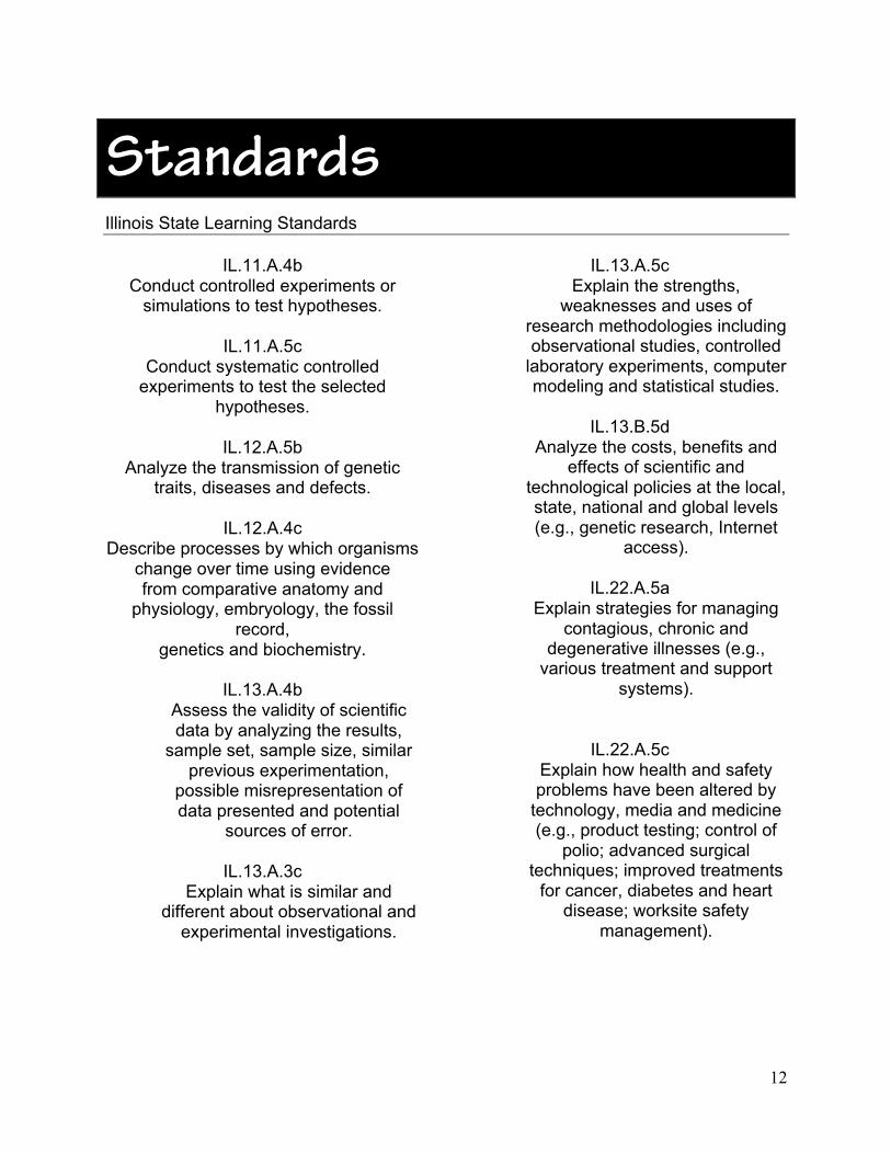
Standards Illinois State Learning Standards
IL.11.A.4b Conduct controlled experiments or
simulations to test hypotheses.
IL.11.A.5c Conduct systematic controlled
experiments to test the selected hypotheses.
IL.12.A.5b
Analyze the transmission of genetic traits, diseases and defects.
IL.12.A.4c
Describe processes by which organisms change over time using evidence from comparative anatomy and
physiology, embryology, the fossil record,
genetics and biochemistry.
IL.13.A.4b Assess the validity of scientific data by analyzing the results,
sample set, sample size, similar previous experimentation,
possible misrepresentation of data presented and potential
sources of error.
IL.13.A.3c Explain what is similar and
different about observational and experimental investigations.
IL.13.A.5c Explain the strengths,
weaknesses and uses of research methodologies including observational studies, controlled
laboratory experiments, computer modeling and statistical studies.
IL.13.B.5d
Analyze the costs, benefits and effects of scientific and
technological policies at the local, state, national and global levels (e.g., genetic research, Internet
access).
IL.22.A.5a Explain strategies for managing
contagious, chronic and degenerative illnesses (e.g.,
various treatment and support systems).
IL.22.A.5c Explain how health and safety problems have been altered by
technology, media and medicine (e.g., product testing; control of
polio; advanced surgical techniques; improved treatments
for cancer, diabetes and heart disease; worksite safety
management).

13
IL.23.B.2 Differentiate between positive and negative effects of health-
related actions on body systems (e.g., drug use, exercise, diet).
IL.23.B.3 Explain the effects of health-
related actions upon body systems (e.g., fad diets,
orthodontics, avoiding smoking, alcohol use and other drug use).
National Standards
NSES-S.9-12.A.1 STANDARD: Science As Inquiry -- As a
result of activities in grades 9-12, all students should develop abilities necessary to do scientific inquiry
NSES-S.9-12.A.2 STANDARD: Science As Inquiry -- As a
result of activities in grades 9-12, all students should develop understandings
about scientific inquiry

14
Cocaine Lab Objectives The students will identify parts of the brain and what each is responsible for.
The students will explain how neuronal signaling works using the following terms: action potentials, synapses, axon, dendrite, cell body, axon terminal, ions, resting potential, neurotransmitters, presynaptic neuron, and postsynaptic neuron.
The students will keep a virtual notebook of readings they have done within the lab by answering questions prompted within the notebook.
The students will read the lab procedures and be able to explain why each step of the procedure is completed.
The students will calculate the amount of anesthesia a rat should be given based on its weight in grams.
The students will observe a rat undergo surgery to implant a wireless telemetry system in its brain so that dopamine levels can be measured while it is freely moving.
The students will monitor a rat’s behavior and dopamine levels during cocaine self-stimulation in order to collect data for the experiment.
The student will determine whether the data collected supports or rejects the hypothesis that dopamine is involved in cocaine seeking.
The students will describe in their own words how cocaine becomes addictive.
The students will interact with fields of the cognitive and learning sciences. The students will “become” researchers as they immerse themselves in a virtual environment that teaches them recent breakthroughs in medical science, and the scientific methods by which those discoveries have been made. The student will participate in practical lessons about how to save lives, be introduced to a wide range of medical careers, and learn about recent breakthroughs in medical science. The student will identify and analyze what the life is like as a medical researcher. The student will utilize scientific inquiry methods to:
• make observations • pose questions • investigate prior knowledge • plan investigations • use tools to gather, analyze, and
interpret data • propose answers, explanations
and predictions • communicate results

15
Teacher’s Lab GuideDo the lab yourself before using it with your students. Then before allowing individual students to log on to begin the virtual lab, you may want to consider introducing the lab via projector.
Also introduce students to the navigation buttons in the lower right hand corner of the screen. The on/off button shows/hides text labels within an area. The Objective and Task buttons
are what students will use throughout the lab to know what specifically they should be doing in each area. When students hit the Help button, they have several options. Student should NOT use the back button. Instead they should click on the Lab button. Clicking the Lab button gets them to the overall lab view and is used to move between the different lab areas.
1st Button (Orange button) On/Off Button: shows/hides text labels within an area
2nd Button—Lab Button takes you to the overall lab view
3rd Button (Starred button) Objective Button
4th Button (Checkmark button) Task Button
5th Button (Question mark) Help Button

16
Overall Lab View
After students see how the navigation bar works, introduce them to the set up of the lab, and to the four areas; Desk area, Prep area, Surgery area, and Experiment area—in that order. Remind students that anytime they want to get back to this screen, they should use the Lab Button in the navigation bar.
17
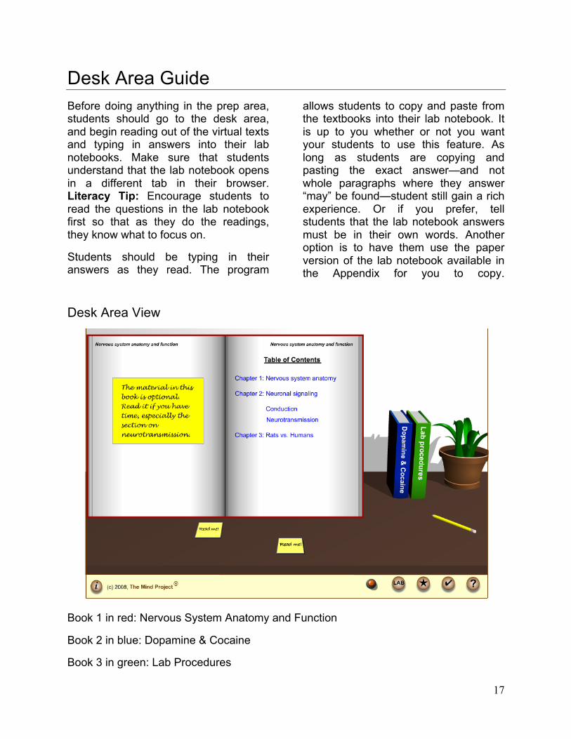
Desk Area Guide Before doing anything in the prep area, students should go to the desk area, and begin reading out of the virtual texts and typing in answers into their lab notebooks. Make sure that students understand that the lab notebook opens in a different tab in their browser. Literacy Tip: Encourage students to read the questions in the lab notebook first so that as they do the readings, they know what to focus on.
Students should be typing in their answers as they read. The program
allows students to copy and paste from the textbooks into their lab notebook. It is up to you whether or not you want your students to use this feature. As long as students are copying and pasting the exact answer—and not whole paragraphs where they answer “may” be found—student still gain a rich experience. Or if you prefer, tell students that the lab notebook answers must be in their own words. Another option is to have them use the paper version of the lab notebook available in the Appendix for you to copy.
Desk Area View
Book 1 in red: Nervous System Anatomy and Function
Book 2 in blue: Dopamine & Cocaine
Book 3 in green: Lab Procedures

18
Student Lab Notebook Questions
Here are the questions students are asked to answer in their Lab Book. (See Appendix for Teacher Answer Key.)
1. What are the two phases of motivated behavior?
2. What is known about the effects of cocaine on dopamine levels in the brain?
3. What is the hypothesis being tested in this experiment?
4. If the hypothesis is true, then what should you observe?
5. What is the name of the method used to measure dopamine levels?
6. What is the formula for computing how much anesthesia to administer?
7. Where will you inject the rat with the anesthesia?
Desk Area Implementation Ideas 1) Students read the virtual text online using class time and fill out the virtual Lab Notebook as they read. 2) Students read the virtual text online outside of class and fill out the virtual Lab Notebook as they read. 3) Students read the paper versions of the virtual text & fill out a paper version of the Lab Notebook either as homework or class work. Note: Use the “Concepts Covered” section in this manual to help you decide how you might fit this lab into your curriculum. Once you familiarize
yourself with the content within these texts, you can direct students to read the sections that best suit your needs.
Lab Notebook: The purpose of the lab notebook is for students to have a place to type in content information from the reading. The lab notebook was not intended to be used as an assessment tool, but students could print it off for a grade if you wished. Students will use the lab notebook as their reference in order to complete the questions at the end of the lab, for the published article.
Once students have completed the readings at the desk area and completed the questions in their lab notebook, they are ready to move on to the Preparation area.

19
Preparation Area Guide Overview: Students will select a rat, determine how much anesthesia to administer based on the rat's weight, administer the anesthesia via syringe, and clean up the lab area. The prep desk area is interactive. Students will be picking up the rat by clicking and dragging, filling the syringe by clicking on the correct graduated number, and clicking and dragging the syringe to indicate where on the rat the injection should be given.
Prerequisite Skills: Students will need to know how to calculate how
much anesthesia to give the rat. (1 ml/kg) The weight of the rat will be given to student in grams. So students must be able to convert grams to kilograms. Example calculation: 1 mL/kg x 200g (rat)/1 x 1 kg/1000g = 0.2 mL You may want to do some sample conversions if students have not done these before. You could also suggest that they show their work in their lab notebook to make sure they understand the concept.
In the lab, the Female rat (in the pink box) weighs 300 grams and the male rat (in the blue box) weighs 350 grams.
Preparation Area View

20
Specific Tasks students will be asked to complete at the prep area:
1) Read sticky note from advisor (students will refer to this throughout this section)
2) Put on gloves
3) Select rat (they pick either a female or male rat), weigh it, calculate amount of anesthesia, and then place the rat in the prep tray.
4) Place syringe and anesthesia bottle in prep tray.
5) Extract the correct amount of anesthesia (0.3 ml for female rat, and 0.35 ml for male rat)
6) Inject anesthesia in the correct location on the rat.
7) Dispose of the syringe & put bottle away.
Special Notes
While the advisor's note tell students to calculate the weight of the rat in the third task, students don't actually need to do anything with that number until they fill the syringe a few tasks later.
Reminder: Remind students that their lab notebook answers are important because they will be using those later in the lab to answer questions that will help them get "published." They will have full access to their notes as they answer a series of questions after they have completed the experiment and collected data.
Student Lab Notebook Questions
Here is the question students are asked to answer in their Lab Book. (See Appendix for Teacher Answer Key.)
8. Based on the weight of the rat you selected, how much anesthesia should you extract?
21
Prep Area Implementation Ideas
1) Prerequisite: Conversions Worksheet: If your students have not learned about mathematical conversions, or need reminding, use the worksheet found in the Appendix.
2) Individually: Have students log on individually or in pairs, start at the prep area and complete the tasks.
3) Whole Class: With one person logged on to a computer hooked up to a projector, have the class work through the prep area allowing students from the class help complete the tasks.
Once students have completed the tasks in the prep area, they are ready to move to the surgery area.

22
Surgery Area Guide Overview:
During this part of the virtual lab, the student will observe electrodes being inserted into the rat’s brain in order to monitor dopamine levels during cocaine self-administration. The surgery area is not interactive. Students instead read about how the surgery is completed, clicking the arrows to move ahead a step or go back a step.
Teaching Method Tip:
Depending on how you decide to have students complete the lab, this part could be done as a class so that you could add your own comments as you go and ask questions to ensure students understand the process of the surgery and why.
Surgery Area View

23
Step-by-Step Surgery Discussion Questions
Full Gear
The students must wear full gear (mask, gloves, and lab coat) in order to prevent infection in the rat. Betadine is also applied to the surgery area for this reason.
Why the Jugular?
Intrajugular cocaine injection is the most efficient way to deliver cocaine to the rat.
The Sterotaxic Apparatus
The stereotaxic apparatus does not harm the rat and ensures that all electrodes are inserted in the proper places. The rat is secured in the apparatus by both a nose/upper jaw clip and ear bars.
Getting to the Skull
Cutting the muscle and skin is a little more tedious than displayed. These tissue layers are held together by connective tissue that needs to be scraped away from the surgery site.
Why use a reference electrode?
The reference electrode acts as a chemical “control”, stabilizing the brain’s
environment so the other electrodes can work properly.
Why use the sensing electrode?
The sensing electrode is placed in the striatum of the rat where the dopamine neurons from the substantia nigra terminate. This is where the best measure of dopamine release can be found.
What’s the stimulating electrode for?
The stimulating electrode delivers voltage to the axons of the dopamine neurons. This stimulates the neurons to activate dopamine release.
Rat needs a heating pad
The rat is on a heating pad during the surgery and recuperation because, while under anesthesia, it cannot maintain its own body heat.
We use cement on the rat’s head?
The cap and cement hold all the electrodes in place, allowing for the measure of dopamine release and stimulation of the dopamine neurons while the rat moves around.

24
Additional Questions: You could use these as you circulate around the room while students are moving through the surgery area in the virtual lab. Or you may want to have an “exit-slip” question that students answer before they move onto the next part of the lab.
• Why is the sensing electrode placed in the nucleus accumbens?
• Why is the stimulating electrode placed in the ventral tegmental?
• Explain why the rat needs to be on a heating pad during the surgery and recovery times.
Student Lab Notebook Questions
Here are the questions students are asked to answer in their Lab Book. (See Appendix for Teacher Answer Key.)
9. What is the function of the canula?
10. What is the name of the device that allows us to implant electrodes in precise locations?
11. Which electrode measures dopamine levels?
Surgery Area Implementation Ideas
1) Individually: Have students log on individually or in pairs, start at the surgery area and complete move through this area and answer questions in their lab book.
2) Whole Class: With one person logged on to a computer hooked up to a projector, have the class work through the surgery area leading the class in discussion of what is happening during the surgery.
Understanding what electrodes are placed where is crucial for understanding this surgery. The other main idea student’s should grasp, is that the purpose of this surgery is to place electrodes into the skull, but then to allow the animal to recuperate so levels of dopamine can be measured while the animal is freely moving. The details of the surgery may not be necessary as long as students understand the purpose of the surgery.

25
Experiment Guide Overview
In the experiment portion of the lab, students will observe the rat’s dopamine levels as it self-administers cocaine. Since cocaine produces a release of dopamine in the brain, one would expect to find an increase in dopamine only after the rat presses the bar. According to this experiment, however, the dopamine spikes as the rat approaches the bar to press it. This would indicate that dopamine if involved in the
anticipation of actions and may give further insight into drug addiction.
Student Tasks
Read the advisor’s sticky notes.
Begin the experiment.
Observe the animal and the data for at least 10 minutes.
Experiment Area View

26
Teaching Method Tips
This is the crux of the virtual lab experience. You will know the students who are having difficulty because they will hurry to get the experiment going (which will run for 10 minutes) and they won’t know what they are supposed to be doing during that time. To prevent this, have a discussion with students about how to make careful-scientific observations. To help them focus on what they should be observing, make sure they have visited Book 3 in the desk area. That will help them identify rat behaviors with what those behaviors mean.
Students should get a good feel for the tediousness of collecting data—even if it is only for 10 minutes. If students know what they are supposed to be doing during data collection, they often enjoy the process. However, if they don’t, they find it difficult and not enjoyable. Spend some time before the experiment area to discuss this with students.
Students will also need to know how to read graphs for this section of the lab. They should be looking for patterns of the level of dopamine and what the rat is doing during that level—specifically what the rat is doing right before it self-administers cocaine.
Encourage Students to Enjoy Data Collection: Help prepare students for the experiment. Remind students to read the questions they are to answer in their Lab Book. Be sure that they know that they are looking to link patterns between the rat’s behavior and the dopamine level.
Student Lab Notebook Questions
Here are the questions students are asked to answer in their Lab Book. (See Appendix for Teacher Answer Key.)
12. What is the function of the telemetry pack?
13. What behavior are you looking for when the rat is seeking cocaine?
14. What behavior(s) do you observe when the rat is not seeking cocaine?
15. Do you observe any change in dopamine levels that is consistent with what is known about cocaine being consumed? If so, describe those changes.
16. Do you observe any changes in dopamine levels while the rat is engaged in cocaine seeking behavior? If so, describe those changes.
17. Do the results of this experiment support the hypothesis? Explain why or why not.
27
Additional Questions: You could use these as you circulate around the room while students are moving through the experimental area in the virtual lab. Or you may want to have an “exit-slip” question that students answer before they move onto the next part of the lab or as an “entrance-slip” the class session after they have completed the lab.
1. Describe the dopamine activity in the rat’s brain during the experiment. What was occurring when it went up? What was occurring when it went down?
2. Interpret these results in light of what you read about how animals behave. What stage(s) of behavior does dopamine appear to be involved with? Explain your answer.
3. Explain how these results might help researchers in the area of drug addiction and other behaviors.
Final Activity: The Publication After completing the experiment and answering the questions in their Lab Book, students will be asked to move the final part of the virtual lab where they use their Lab Book notes to answer questions regarding the information they learned at each area of the lab. Students will be have to get the questions correct before moving on to the next question. These answers will then be used to become a “published” article.
As their teacher, you could have them print a copy of this and turn it in. Or if you prefer to make it more of a completion grade, and to make your grading load a bit easier, have students call you over when they’ve completed the lab to show you their article.
See Appendix for an answer key to the questions students will be asked to complete.

28
Appendix
Lab Notebook- Student Reproduceable..................................................................... 28 Lab Notebook- Teacher Answer Key.......................................................................... 31 Mathematical Science Conversions- Student Reproducible ................................... 34 Mathematical Science Conversions- Teacher Answer Key ..................................... 37 Answers to the Final Publication Questions ............................................................. 40

29
Name_________________________________ Class __________ Date ____________
Neurobiology Lab NotebookDesk Area Questions
1. What are the two phases of motivated behavior?
a)
b)
2. What is known about the effects of cocaine on dopamine levels in the brain?
3. What is the hypothesis being tested in this experiment?
4. If the hypothesis is true, then what should you observe?
5. What is the name of the method used to measure dopamine levels?

30
6. What is the formula for computing how much anesthesia to administer?
7. Where will you inject the rat with the anesthesia?
Prep Area Questions
8. Based on the weight of the rat you selected, how much anesthesia should you extract?
Surgery Area
9. What is the function of the canula?
10. What is the name of the device that allows us to implant electrodes in precise locations?
11. Which electrode measures dopamine levels?
31
Experiment Area
12. What is the function of the telemetry pack?
13. What behavior are you looking for when the rat is seeking cocaine?
14. What behavior(s) do you observe when the rat is not seeking cocaine?
15. Do you observe any change in dopamine levels that is consistent with what is known about cocaine being consumed? If so, describe those changes.
16. Do you observe any changes in dopamine levels while the rat is engaged in cocaine seeking behavior? If so, describe those changes.
17. Do the results of this experiment support the hypothesis? Explain why or why not.

32
Teacher’s Answer Key
Neurobiology Lab NotebookDesk Area Questions
1. What are the two phases of motivated behavior?
Answer is located in Book 2, Chapter 4. The appetitive phase consists of those behaviors related to “approaching” the goal. In sexual behavior, for instance, the appetitive phase consists of behaviors that establish, maintain, or promote sexual interaction. Generally speaking, appetitive behaviors allow an animal to come into contact with its goal. The consummatory phase represents the actual “consuming” of the goal. In the case of sexual behavior, the consummatory phase is sexual intercourse. Collectively, appetitive and consummatory aspects characterize a sexual encounter, which is a motivated behavior. Addicted behavior is motivated behavior too.
2. What is known about the effects of cocaine on dopamine levels in the brain?
Answer is located in Book 2, Chapter 5. The pharmacological effect of cocaine on dopamine neurons is that cocaine blocks the dopamine transporter. This prevents dopamine from being transported back into the presynaptic neuron from the synaptic cleft. Norepinephrine neurons and serotonin neurons have their own specific transporters too. Cocaine binds to and blocks the action of the norepinephrine and serotonin transporters as well. Thus, by preventing the removal of released neurotransmitter, cocaine increases the extracellular levels of dopamine, norepinephrine, and serotonin in the brain.
3. What is the hypothesis being tested in this experiment?
Answer is located in Book 3, Chapter 1. Hypothesis: Dopamine is involved in cocaine seeking.
4. If the hypothesis is true, then what should you observe?
Student Inference: A dopamine spike right before the rat pushes the lever that self-administers the cocaine.
5. What is the name of the method used to measure dopamine levels?
Answer is located in Book 3, Chapter 1. The chemical microsensor technique that will be used in our experiment, fast-scan cyclic voltammetry at a carbon-fiber microelectrode,

33
6. What is the formula for computing how much anesthesia to administer?
Answer is located in Book 3, Chapter 2. 1 ml/kg: one milliliter per one kilogram. (Example: inject 0.4 ml into a 400 g rat)
7. Where will you inject the rat with the anesthesia?
Answer is located in Book 3, Chapter 2. The peritoneal cavity; If too high (in thoracic cavity), causes pneumothorax, deflating lungs and killing animal. If too low (in bladder), causes complications and perhaps death
Prep Area Questions
8. Based on the weight of the rat you selected, how much anesthesia should you extract?
Female Rat (pink box): 300g x 1kg/1000g x 1ml/kg = .3 ml
Male Rat (blue box): 350g x 1 kg/1000g x 1ml/kg = .35 ml
Surgery Area
9. What is the function of the canula?
Answer is located in Book 2, Chapter 5 (and in text box during the surgery) The cannula is a small hollow tube inserted into the jugular vein that will allow cocaine to be injected. This allows cocaine self-administration by the rat because each time the rat hits the lever, cocaine will be administered.
10. What is the name of the device that allows us to implant electrodes in precise locations?
Answer is located in text box during the surgery The stereotaxic apparatus.
11. Which electrode measures dopamine levels?
Answer is located in text box during the surgery The sensing electrode (Figure 1 in Book 3 calls it a microsensor, but sensing electrode is the more precise answer)

34
Experiment Area
12. What is the function of the telemetry pack?
Answer is located in Book 3, Chapter 1 (and in text box during the surgery) The electrically evoked dopamine signal will be transmitted to the computer by a wireless piece of equipment in a backpack using radio waves or telemetry. In this way, dopamine can be monitored in the brain of the animal without being connected or “hardwired” to the equipment. The computer will register a lever press and send a signal to the wireless backpack to administer cocaine.
13. What behavior(s) are you looking for when the rat is seeking cocaine?
Student Inference (with info from Book 2—Dopamine and Cocaine): Appetitive behavior—the phase when the rat is approaching or seeking the goal
14. What behavior(s) do you observe when the rat is not seeking cocaine?
Student Inference (with info from Book 2—Dopamine and Cocaine): The consummatory phase in which the goal is being achieved.
15. Do you observe any change in dopamine levels that is consistent with what is known about cocaine being consumed? If so, describe those changes.
Student Observation & Inference (during the experiment): Dopamine begins to spike while the rat is seeking cocaine and then goes up further when cocaine is received.
16. Do you observe any changes in dopamine levels while the rat is engaged in cocaine seeking behavior? If so, describe those changes.
Student Observation: The level of dopamine spikes up during the process of seeking cocaine (the appetitive phase).
17. Do the results of this experiment support the hypothesis? Explain why or why not.
Student Inference: Yes. Dopamine spikes during cocaine seeking (the appetitive phase) and not just during the ingestion of cocaine (the consummatory phase).
The whole idea here is that dopamine is involved with the appetitive phase of motivation and not just the consummatory phase as originally thought. This may give some insight into the reoccurrence of drug addiction as many addicts go back to their same neighborhoods, friends, patterns of behavior, etc. and are therefore more susceptible to fall back into addiction.

35
Name_________________________________ Class __________ Date ____________
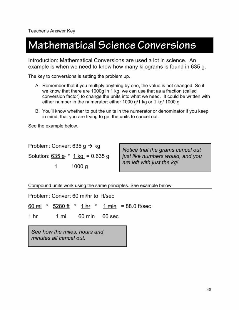
Mathematical Science ConversionsIntroduction: Mathematical Conversions are used a lot in science. An example is when we need to know how many kilograms is found in 635 g.
The key to conversions is setting the problem up.
A. Remember that if you multiply anything by one, the value is not changed. So if we know that there are 1000g in 1 kg, we can use that as a fraction (called conversion factor) to change the units into what we need. It could be written with either number in the numerator: either 1000 g/1 kg or 1 kg/ 1000 g
B. You’ll know whether to put the units in the numerator or denominator if you keep in mind, that you are trying to get the units to cancel out.
See the example below.
Problem: Convert 635 g kg
Solution: 635 g * 1 kg = 0.635 g
1 1000 g
Compound units work using the same principles. See example below:
Problem: Convert 60 mi/hr to ft/sec
60 mi * 5280 ft * 1 hr * 1 min = 88.0 ft/sec
1 hr 1 mi 60 min 60 sec
Notice that the grams cancel out just like numbers would, and you are left with just the kg!
See how the miles, hours and minutes all cancel out.

36
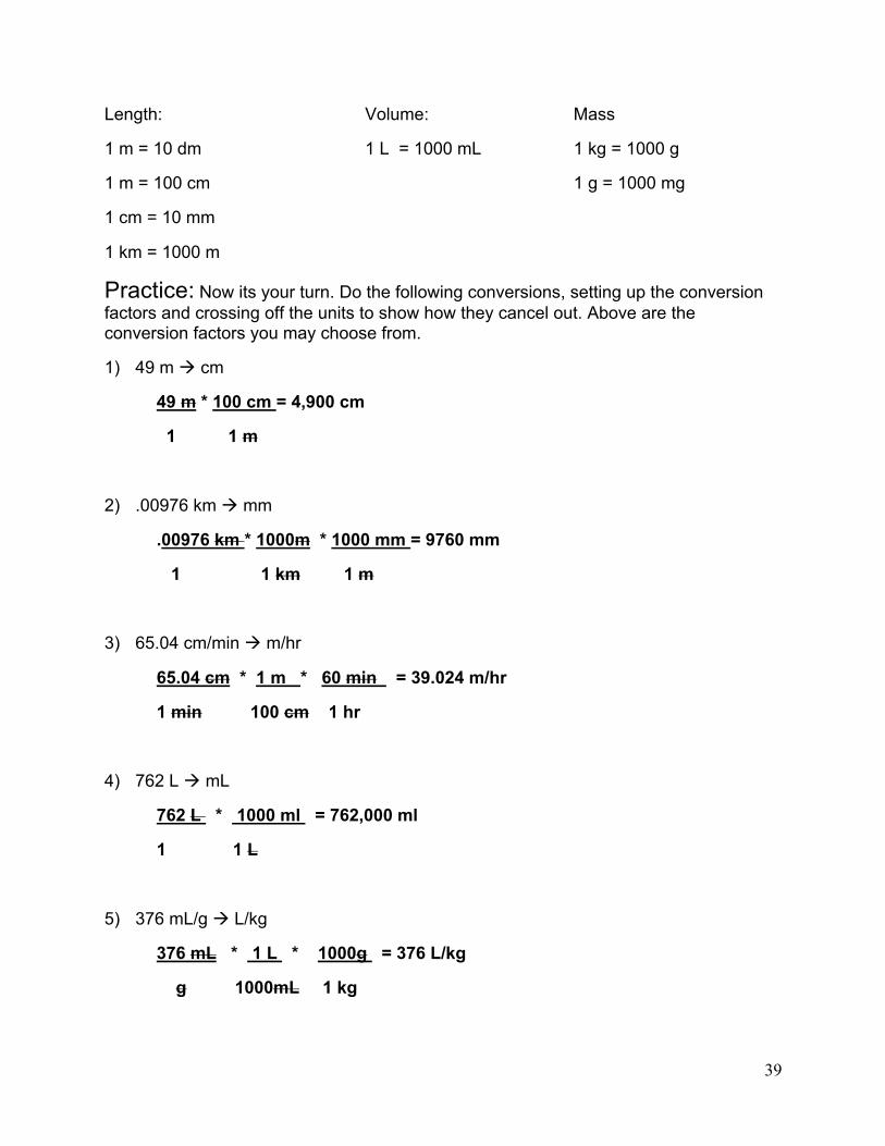
Length: Volume: Mass
1 m = 10 dm 1 l = 1000 ml 1 kg = 1000 g
1 m = 100 cm 1 g = 1000 mg
1 cm = 10 mm
1 km = 1000 m
Practice: Now its your turn. Do the following conversions, setting up the conversion factors and crossing off the units to show how they cancel out. Above are the conversion factors you may choose from.
1) 49 m cm
2) .00976 km mm
3) 65.04 cm/min m/hr
4) 762 l ml
5) 376 ml/g l/kg

37
More Practice: Ok, now for our Virtual Neuroscience Application. When anesthetizing a rat to prepare for surgery, the amount of anesthesia given must be carefully calculated based on the weight of the rat. The rat should receive 1 ml of drug for each kilogram of the weight. The conversion factor is: 1ml/kg. However, rats don’t usually weigh a kilogram, so you’ll need to convert kg to grams. Do the following problems, showing how the units cancel out.
The conversion factor is: 1ml of anesthesia 1 kg of rat weight 6) How much anesthesia should you give a 225 g rat? 7) How much anesthesia should you give a 860 g rat? 8) How much anesthesia should you give a 475 g rat? 9) How much anesthesia should you give a 579 g rat? 10) How much anesthesia should you give a 480 g rat?
38
Teacher’s Answer Key
Mathematical Science ConversionsIntroduction: Mathematical Conversions are used a lot in science. An example is when we need to know how many kilograms is found in 635 g.
The key to conversions is setting the problem up.
A. Remember that if you multiply anything by one, the value is not changed. So if we know that there are 1000g in 1 kg, we can use that as a fraction (called conversion factor) to change the units into what we need. It could be written with either number in the numerator: either 1000 g/1 kg or 1 kg/ 1000 g
B. You’ll know whether to put the units in the numerator or denominator if you keep in mind, that you are trying to get the units to cancel out.
See the example below.
Problem: Convert 635 g kg
Solution: 635 g * 1 kg = 0.635 g
1 1000 g
Compound units work using the same principles. See example below:
Problem: Convert 60 mi/hr to ft/sec
60 mi * 5280 ft * 1 hr * 1 min = 88.0 ft/sec
1 hr 1 mi 60 min 60 sec
Notice that the grams cancel out just like numbers would, and you are left with just the kg!
See how the miles, hours and minutes all cancel out.
39
Length: Volume: Mass
1 m = 10 dm 1 L = 1000 mL 1 kg = 1000 g
1 m = 100 cm 1 g = 1000 mg
1 cm = 10 mm
1 km = 1000 m
Practice: Now its your turn. Do the following conversions, setting up the conversion factors and crossing off the units to show how they cancel out. Above are the conversion factors you may choose from.
1) 49 m cm
49 m * 100 cm = 4,900 cm
1 1 m
2) .00976 km mm
.00976 km * 1000m * 1000 mm = 9760 mm
1 1 km 1 m
3) 65.04 cm/min m/hr
65.04 cm * 1 m * 60 min = 39.024 m/hr
1 min 100 cm 1 hr
4) 762 L mL
762 L * 1000 ml = 762,000 ml
1 1 L
5) 376 mL/g L/kg
376 mL * 1 L * 1000g = 376 L/kg
g 1000mL 1 kg

40
More Practice: Ok, now for our Virtual Neuroscience Application. When anesthetizing a rat to prepare for surgery, the amount of anesthesia given must be carefully calculated based on the weight of the rat. The rat should receive 1 ml of drug for each kilogram of the weight. The conversion factor is: 1mL/kg. However, rats don’t usually weigh a kilogram, so you’ll need to convert kg to grams. Do the following problems, showing how the units cancel out.
The conversion factor is: 1mL of anesthesia 1 kg of rat weight 6) How much anesthesia should you give a 225 g rat?
225 g * 1 kg * 1 mL = 0.225 mL 1 1000 g 1 kg 7) How much anesthesia should you give a 860 g rat?
860 g * 1 kg * 1 mL = 0.860 mL 1 1000 g 1 kg 8) How much anesthesia should you give a 475 g rat?
475 g * 1 kg * 1 mL = 0.475 mL 1 1000 g 1 kg 9) How much anesthesia should you give a 579 g rat?
579 g * 1 kg * 1 mL = 0.579 mL 1 1000 g 1 kg 10) How much anesthesia should you give a 480 g rat?
480 g * 1 kg * 1 mL = 0.480 mL
1 1000 g 1 kg

41

42
BOOK 1: Nervous System Anatomy and Function Ch. 1: Nervous System Anatomy
The nervous system performs many tasks: it coordinates the activity of the muscles, it monitors the organs, it processes input from the senses, and it initiates action. The brain and spinal cord comprise the central nervous system. The central nervous system is protected by bone (skull and vertebrae). The peripheral nervous system is not because its function is to relay information to and from the organs and the limbs. The peripheral nervous system consists of all the other structures that do not lie within the
central nervous system. These other structures include motor neurons, which stimulate muscle tissue, and sensory neurons, which include those connected to pain- or temperature-sensitive receptors in the skin.
Anatomically, the brain is divided into three parts: forebrain, midbrain, and hindbrain (see Figure 1). These subdivisions control different functions. Not unexpectedly, these parts come in varying sizes in different animals, depending upon specific needs or features of the animal.
Fig1
Fig. 1: Major divisions of the brain.

Collectively, the cerebellum, medulla, and pons are called the hindbrain and perform “lower-level functions.” The cerebellum is the ball-like structure resting on the back of the brain. It controls fine motor movement, coordination, and posture. Closest to the spinal cord, the medulla controls breathing and heart beat. Above the medulla is the pons (or “bridge”). It relays sensory information between the cerebellum and the cerebrum. The midbrain (which lies between the hindbrain and the forebrain) is involved in movement and audio-visual processing. The medulla, pons, and midbrain comprise the brainstem.
The forebrain consists of the diencephalon and the two cerebral hemispheres (or the cerebrum). The diencephalon (“in between” brain) is comprised of two prominent structures: the hypothalamus and thalamus. The hypothalamus is located at the very
bottom of the brain, directly on top of the roof of the mouth. It controls a variety of involuntary functions, including: blood pressure, temperature regulation, feeding, sexual behavior, and the pituitary gland – the body's master gland. The thalamus resides on top of the hypothalamus and is the major relay system in the brain. All sensory information except smell first comes to the thalamus before being sent to other regions for processing.
In humans, the two cerebral hemispheres (or cerebrum) are by far the largest structure of the brain. They are involved in cognition, memory, language processing, motor processing, and sensory information processing. They also contain the basal ganglia — the group of nuclei that is central to the control of movement. Parkinson’s disease and Huntington’s disease are diseases that result from damage to the basal ganglia.
Fig 2
Fig. 2: Four lobes and basal ganglia.

44
Each cerebral hemisphere is composed of four lobes: frontal, parietal, temporal, and occipital (see Figure 2). The frontal lobes are considered the site of executive decisions, planning, and personality. They are also the primary seat of motor information processing. The parietal lobes are involved with
attention and somatosensory (i.e., “body senses”) information processing. The temporal lobes are involved in recognition and auditory information processing. The occipital lobes, which have no cognitive function, contain the primary visual information processing centers.
Ch. 2: Neuronal signaling
Neurons are the basic information processing structures in the brain. A "typical" neuron has four distinct parts (or regions). The first part is the cell body (or soma). This is not only the metabolic "control center" of the neuron, it is also its "manufacturing and recycling plant." (For instance, it is within the cell body that neuronal proteins are synthesized.) The second and third parts are processes — structures that extend away from the cell body. Generally speaking, the function of a process is to be a conduit through which signals flow to or away from the
cell body. Incoming signals from other neurons are (typically) received through its dendrites. The outgoing signal to other neurons flows along its axon. A neuron may have many thousands of dendrites, but it will have only one axon. The fourth distinct part of a neuron lies at the end of the axon, the axon terminals. These are the structures that contain neurotransmitters. Neurotransmitters are the chemical medium through which signals flow from one neuron to the next at chemical synapses.
Fig. 3: Structure of a neuron.
To support the general function of the nervous system, neurons have evolved unique capabilities for intra-cellular signaling (communication within the cell) and inter-cellular signaling (communication between cells). To achieve long distance, rapid communication, neurons have evolved special abilities for sending electrical signals (action potentials) along axons. This mechanism, called conduction, is how the cell body of a neuron communicates with its own terminals via the axon. Communication between neurons is achieved at synapses by the process of neurotransmission.

45
Conduction
To begin conduction, an action potential is generated near the cell body portion of the axon. An action potential is an electrical signal very much like the electrical signals in electronic devices. But whereas an electrical signal in an electronic device occurs because electrons move along a wire, an electrical signal in a neuron occurs because ions move across the neuronal membrane. How this occurs is illustrated in the following animation.
Animation 1: Generation of the neural impulse.
An action potential propagates along the axon quickly, moving at rates up to 150 meters (or roughly 500 feet) per second. Conduction ends at the axon terminals. Axon terminals are where neurotransmission begins.
Neurotransmission
Neurotransmission is communication of information between neurons as accomplished by the movement of chemicals or electrical signals across a synapse. There are two kinds of synapses (see Figure 4).
Fig. 4: Two types of synapses.
At electrical synapses, two neurons are physically connected to one another through gap junctions. Gap junctions permit changes in the electrical properties of one neuron to effect the other, and vice versa, so the two neurons essentially behave as one.

46
At chemical synapses, two neurons are not physically connected to one another. As such, the arrival of an action potential in the presynaptic neuron triggers the release of chemical neurotransmitters. It is through chemical neurotransmitters that the presynaptic neuron communicates with the postsynaptic neuron. In mammals, most neurons communicate through chemical means. There are two models for how this occurs; namely, “classic” chemical neurotransmission and volume neurotransmission. Here only the first will be discussed.
In “classic” chemical neurotransmission, the presynaptic neuron and the postsynaptic neuron are separated by a small gap — the synaptic cleft. The synaptic cleft is filled with extracellular fluid (the fluid bathing all the cells in the brain). Although very small, typically on the order of a few nanometers (a billionth of a meter), the synaptic cleft creates a physical barrier for the electrical signal carried by one neuron to be transferred to another neuron. In electrical terms, the synaptic cleft would be considered a “short” in an electrical circuit. The function of neurotransmitter is to overcome this electrical short. How this occurs is illustrated in the following animation.
Animation 2: "Classic" chemical neurotransmission.
The entry of ions through ion channels produces a local partial depolarization (change in voltage) of the membrane. The postsynaptic neuron can have thousands of chemical synapses producing local partial depolarizations of its membrane (see Figure 5 ).
Fig. 5
When the sum of all its partial depolarizations exceeds the threshold, the postsynaptic neuron will fire an action potential, thus completing neuronal signaling. It then becomes the presynaptic neuron to all the neurons it is connected to via synapses at its axon terminals (see Conduction above).
Chemical neurotransmission is terminated by removal of neurotransmitter from the cleft. Besides through diffusion, this can occur in different ways. Some types of neurotransmitter are removed by degradative enzymes found in the synaptic cleft. The
47
function of these enzymes is to break down or deactivate a neurotransmitter so that it can no longer bind to a receptor. However, for most types of neurotransmitter, removal from the cleft is accomplished through a special protein on the presynaptic neuron called a transporter. A transporter protein acts as a pump. It binds neurotransmitter in a way that is similar to the way a receptor does, but then it moves the neurotransmitter back into the neuron, a process called reuptake. Thus, for most neurons, the same neuron that initiates neurotransmission by releasing neurotransmitter also terminates neurotransmission by removing neurotransmitter. Once inside the neuron, neurotransmitter molecules are either re-packaged into vesicles for use again or deactivated and broken down via degradative enzymes found in the cell.
Ch. 3: Rats vs. Humans
Rats are often used in research because of the similarities they have with humans. In rats, as in humans, the nervous system is divided into the central nervous system and peripheral nervous system. Rats and humans share all of the major subdivisions of the brain and their general functions. And rats and humans also share similar dopamine neuron systems. Dopamine is an important neurotransmitter involved in movement and motivated behavior (see Book 2).
However, important differences exist between rats and humans. For example,
humans have a considerably larger cerebral cortex, while rats have a more prominent olfactory bulb. This latter region of the forebrain is important for smell, a sense that is extremely keen in rats, but less so in humans. Another important difference is the physical relationship between the brain and the spinal cord. As an animal that stands upright, the brain and spinal cord in a human form a right angle, with the spinal cord extending down from the base of the brain. Because rats “stand” on all four paws, the relationship is more linear.

48
BOOK 2: Dopamine and Cocaine Chapter 1: The Dopamine Neuron
A dopamine neuron is a neuron that uses the neurotransmitter dopamine for chemical neurotransmission. As will be discussed, dopamine neurons are important for motor movements, motivated behavior, and in mediating the effects of drugs of abuse such as cocaine.
By binding to receptors, neurotransmitters act as a chemical messenger or link connecting the action potential from one neuron with a membrane potential in another. But unlike receptors found in “classic” chemical neurotransmission, dopamine receptors function a bit differently. In “classic” chemical neurotransmission, neurotransmitter binds to receptors that open ion channels. This allows ions to pass through the neuronal membrane. In this way, the chemical signal, the neurotransmitter, is transduced into an electrical signal, because ion flow will generate a change in voltage at the postsynaptic membrane. If this voltage change reaches threshold, the target neuron will fire an action potential. In contrast, the dopamine receptor is called a G-protein coupled receptor. Instead of directly opening an ion channel, dopamine binding to its receptor activates a G-protein that in turn activates a second messenger inside the target neuron. The second message can cause several changes in the postsynaptic neuron. These changes include opening and closing ion channels, but they also include gene transcription (the synthesis of RNA from
DNA) and protein synthesis (the translation of RNA into amino-acids sequences to form proteins).
Chemical neurotransmission is terminated by removal of neurotransmitter from the cleft. For dopamine, like most neurotransmitters, this is done through transporter proteins on the presynaptic neuron. Once inside the neuron, dopamine is either re-packaged into vesicles for use again or degraded via degradative enzymes found only in the cell should the concentration of dopamine be too high. Interestingly, high concentrations of dopamine appear to be toxic, so only by degrading dopamine or repackaging it into vesicles is the dopamine neuron protected. Also, similar to the receptors found in the postsynaptic neuron, the presynaptic dopamine neuron contains dopamine receptors itself. These autoreceptors function as a “thermostat,” either shutting down the dopamine neuron when it is too active or speeding it up when too lethargic.
It is worth noting that the synapse between a dopamine neuron and its target appears to function rather differently than in the “classic” view. In the “classic” view of chemical neurotransmission, because transporters or degradative enzymes prevent neurotransmitter from escaping the synaptic cleft, the action of neurotransmitter is confined to the synaptic cleft. But at a dopamine chemical synapse however, dopamine released into the cleft readily diffuses

49
out. For this reason, dopamine is often called an extrasynaptic messenger. Unlike “classic” chemical neurotransmission, the target of dopamine release is not restricted to the postsynaptic neuron. Instead, the target is any neuron with a dopamine receptor close enough to the dopamine synapse to be exposed to a high dopamine concentration. Hence, in addition to “classic” chemical synapses at their terminals, dopamine neurons also form
en passant (“in passing”) synapses. These synapses allow a dopamine neuron to affect a target neuron without terminating the axon. The ability of dopamine to escape the synapse readily is why this neurotransmitter can be measured chemically in the brain without a sensor or probe that is small enough to be placed inside the synaptic cleft. To date, no such probe or sensor exists.
Ch. 2: Dopamine neuron systems
In addition to extensive overlap in brain anatomy, organization, and function, rats and humans also share similar dopamine neuron systems. A neuronal system describes the origins, projections, and terminations of a collection of like neurons. Thus, a dopamine neuron system is defined by the incoming or afferent neurons, the locations of dendrites, cell bodies, axons and terminals, and finally the outgoing or efferent neurons.
The brain contains several dopamine neuron systems. One important group originates in the hypothalamus. Consistent with the function of the hypothalamus, these dopamine neurons are involved in sexual behavior and the
regulation of the pituitary gland. Although many of these dopamine neurons both begin and terminate within the hypothalamus, others project to and terminate in the spinal cord. This later subset of hypothalamic dopamine neurons would be said to be descending in anatomical terms, going from the forebrain to the spinal cord.
Another important group of dopamine neurons originates in the midbrain. These dopamine neurons are ascending because they project to and terminate in the forebrain. The ascending dopamine neurons originate in two regions of the midbrain, the substantia nigra and ventral tegmental area (see Figure 1).
fig1
Fig. 1

The brain is symmetrical in rats and humans, so each hemisphere contains a substantia nigra and striatum. The dopamine neurons originating in the substantia nigra terminate in the striatum. The striatum is a part of the brain located "under" the cerebral cortex. It receives projections from most,
if not all, cortical areas. Because the striatum is an important region of the basal ganglia of the cerebrum, these dopamine neurons play an important role in movement. Motor diseases such as Parkinson's and Huntington's seriously affect the striatum.
Ch. 3: Approaches for Assessing Dopamine Function
Several approaches have been used to assess the role of dopamine in behavior (e.g., lesioning of dopamine neurons, use of dopamine receptor drugs, etc.). This chapter will consider only certain monitoring techniques.
One type of monitoring approach is to use a microelectrode (a microscopic probe typically made of glass or metal) to record the firing rate. The firing rate is the frequency of action potentials a dopamine neuron generates over a period of time. This technique is called electrophysiology. Because the cell body is physically the largest portion of the neuron, it generates the largest electrical signals. Hence, electrophysiological recordings are usually performed in a region of the brain containing neuron cell bodies. For midbrain dopamine neurons, this region would be either the substantia nigra or ventral tegmental area. On the downside, to use electrophysiology one must assume that an action potential occurring at the cell body always causes dopamine release at the axon terminal. This may not always be true. For instance, dopamine autoreceptors can regulate dopamine release at the terminal independent of control by the cell body.
Other monitoring techniques have been developed to measure dopamine directly in terminal fields, which are those regions of the brain where dopamine neurons make synapses (or “terminate”). For instance, the striatum is the terminal field of midbrain dopamine neurons originating in the substantia nigra. One widely used monitoring technique is microdialysis. In general, dialysis is a procedure in which some but not all molecules move across a membrane. Molecules are typically excluded based on size. Such a membrane is said to be semi-permeable. Based on this principle, a dialysis machine is used to filter the blood of patients with kidney problems.
In microdialysis of the brain, a probe is implanted in a terminal field so dopamine can pass from extracellular fluid across a dialysis membrane and into the center of the probe. By pumping artificial extracellular fluid through the inside of the probe, dopamine is collected and measured outside of the animal using very sensitive and selective instrumentation.
Although microdialysis is used in animal experiments of many kinds, even to deliver drugs to specific brain regions via a procedure called reverse dialysis, it has two main disadvantages. First,

51
samples are usually collected every few minutes. This is slow relative to many behaviors. Second, microdialysis probes are relatively large, about 300 microns (1000 microns = 1 millimeter) in diameter. Thus, a probe can cause damage to the region where dopamine is monitored.
Another technique for directly monitoring dopamine in terminal fields uses a chemical microsensor. A common type is made from a carbon fiber. Carbon is a biologically inert chemical, so it causes a minimal reaction when implanted in the brain. And carbon fibers can be made very small, 5 microns, which is 60 times smaller than the diameter of a microdialysis probe. Hence, a carbon-fiber microelectrode causes less damage than a microdialysis probe. The carbon fiber also provides an excellent surface for electrochemistry (the transfer of electrons between molecules), which is how chemical microsensors measure dopamine. Two events must occur before dopamine is monitored by a chemical microsensor. First, dopamine must come in contact with the carbon
fiber, or at least within a few nanometers. Second, to pull off electrons from dopamine, the carbon fiber must be made positive electrically, just like the positive end of a battery. Electrons are small charged particles found in all molecules. The removal of electrons from a chemical is called oxidation. The rate of electrons flowing to the carbon fiber during oxidation is related to the concentration of dopamine near the microsensor. Consequently, monitoring dopamine using a chemical microsensor is called electrochemical measurement.
In addition to small size, electrochemical microsensors also have the important advantage of making dopamine measurements very quickly, even several times a second. Thus, chemical microsensors are a powerful tool for measuring dopamine changes during behavior. The downside is that many chemicals besides dopamine can be oxidized. This means that knowing what is measured by the chemical microsensor is an important consideration.
Ch. 4: Dopamine Neurons and Motivated Behavior
Motivated behavior
Motivated behavior is behavior directed toward receiving a reward or goal. The reward may be natural (e.g., food, sex, etc.) or artificial (e.g., drugs of abuse). There are two components or phases of motivated behavior. The appetitive phase consists of those behaviors related to “approaching” the goal. In sexual behavior, for instance, the appetitive phase consists of behaviors that establish, maintain, or promote sexual interaction. Generally speaking,
appetitive behaviors allow an animal to come into contact with its goal. The consummatory phase represents the actual “consuming” of the goal. In the case of sexual behavior, the consummatory phase is sexual intercourse. Collectively, appetitive and consummatory aspects characterize a sexual encounter, which is a motivated behavior. Addicted behavior is motivated behavior too.
The neurobiology of motivation is a field that seeks to identify the neural

52
substrates (the brain regions, neuronal systems, neurotransmitters, receptors, etc.) that mediate motivated behavior. As described below, the classic experiment of intracranial self-stimulation demonstrated the existence of a brain reward system. The nucleus accumbens plays a central role in this system. Through dopamine neurons, it links motivational information processed in the cortex with emotional information processed in the limbic system, and then sends this combined information to regions of the brain controlling motor output, hormone release, and the fight-or-flight response. Thus, dopamine neurons terminating in the nucleus accumbens play an important role in motivated behavior. Not unexpectedly, the activity of these dopamine neurons changes during motivated behavior. And such changes can be monitored while an animal engages in motivated behavior.
Intracranial self-stimulation
One of the earliest experiments identifying a relationship between nucleus accumbens dopamine neurons and motivated behavior was intracranial self-stimulation. During this experiment, a stimulating electrode is implanted in the ventral tegmental area to activate dopamine neurons artificially using electrical pulses. The stimulating electrode and the instrument generating the electrical pulses are connected to the lever of a bar press machine. When the animal presses the lever, electrical pulses are delivered to the stimulating electrode. Thus, the animal controls stimulation of its dopamine neurons. (This type of control is called contingent. When a scientist controls the stimulation, the control is called non-contingent.) To obtain the “rewarding”
electrical stimulation, rats lever press at astonishing rates, sometimes as fast a five times per second. They will also lever press continuously for hours.
Early studies with intracranial self-stimulation were very informative. Indeed, the highest rates of lever pressing during intracranial self-stimulation occurred with the stimulating electrode activated dopamine neurons directly. Collectively, such experiments led neuroscientists studying the neurobiology of motivated behavior to conclude that dopamine was the neural substrate of reward. In this view, dopamine is released when the animal consumes the reward, and the amplitude of dopamine release reflects the magnitude of the reward (or how good does this reward make it feel). Thus, dopamine is said to act as the neural substrate of reward during the consummatory phase of motivated behavior. Moreover, all rewards, whether natural (e.g., food, sex, etc) or artificial (e.g., electrical stimulation or drugs of abuse), were thought to be mediated by dopamine release.
Recent results challenge the traditional view that dopamine is the neural substrate of reward. One of the key considerations with this new evidence is this: To understand fully the role of dopamine in motivated behavior, one must be able to monitor dopamine very quickly because behavior can be very fast as well. For example, microdialysis clearly shows that dopamine release increases when animals lever press for a rewarding electrical stimulation. But rats will bar press at rates upwards of 5 per second during intracranial self-stimulation. Microdialysis can in no way tell us what happens to dopamine release with each bar press. Nor can it

53
tell us what happens to dopamine levels just before the animal bar presses. Both the bar press and the time leading up to it constitute the appetitive phase of this motivated behavior. Because of faster sampling rates, chemical microsensors can do both these things. One type of chemical microsensor technique, fast-scan cyclic voltammetry (or voltammetry), can measure dopamine 10 times per second.
When voltammetry was used to monitor dopamine release, some very unusual findings were obtained. For example, when the electrical stimulus was applied by the experimenter, the same stimulus that animals will lever press for, dopamine levels increase in the nucleus accumbens. This result suggested that each lever press during intracranial self-stimulation appeared to be rewarding in the same way as other rewarding stimuli. During training of lever press behavior, when the animal learns to associate the lever with the rewarding electrical stimulation, voltammetry showed that dopamine is also released
during intracranial self-stimulation. However, in well trained animals, intracranial self-stimulation did not release dopamine; that is, animals lever pressed and received electrical stimulation but dopamine release did not increase.
Moreover, the same record of lever press activity, when replayed to the same and other animals, caused dopamine release. Remarkably, these animals received the same number and timing of the electrical stimulation as during intracranial self-stimulation, but in this case, dopamine release was observed. Remarkably, non-contingent but not contingent electrical stimulation caused dopamine release. What this interesting experiment demonstrates is that dopamine is not absolutely necessary for the consumption of an award. Instead, it appears to play a role perhaps related to learning of the cues associated with reward. In intracranial self-stimulation, the cue would be the lever press. This type of learning is called associative learning.
Ch. 5: Dopamine Neurons and Drugs of Abuse
In the United States, one of the more prominent drugs of abuse is cocaine. Cocaine has multiple effects on the body, both peripherally outside the brain and centrally within the brain. Cocaine was originally used in medicine as a local anesthetic during eye surgery. The effect as a local anesthetic is independent of any action cocaine has on the brain. Cocaine also has potent effects directly on the cardiovascular system, which consists of the heart and all of the blood vessels. In general, cocaine increases blood pressure by stimulating the beating of the heart and
by causing blood vessels to constrict. Large doses of cocaine can result in cardiac failure. The actions of cocaine on the brain are generally thought to be mediated by three neurotransmitters: dopamine, norepinephrine, and serotonin.
Two classic experiments are used to demonstrate that cocaine and other drugs of abuse are rewarding: conditioned place preference and drug self-administration. In conditioned place preference, an animal is released into a chamber that is demarcated into

54
different quadrants. Over a period of time, the animal, when venturing into a specific quadrant, is injected with the test substance. After sufficient time, the animal learns the association between a quadrant and the drug injection. The animal is then allowed to enter the chamber but without any drug injection. If a drug is considered rewarding, the animal will tend to localize in the quadrant causing the drug injection. If a drug is considered aversive, the animal will tend to localize in the other quadrants. If a drug is considered neutral, no pattern of localization will occur.
Drug self-administration is analogous to intracranial self-stimulation, except that instead of a lever press delivering a rewarding electrical stimulation, an injection of a drug is administered. The drug is typically delivered intravenously. The goal of cocaine self-administration training is to teach a laboratory rat to press a lever in order to obtain an injection of cocaine. Cocaine will be injected into the jugular vein by a cannula, a small hollow tube inserted into the blood vessel. The jugular vein will carry the cocaine to the heart, which will then pump it to the brain in a matter of 30 seconds. Lever pressing rates for drug self-administration are considerably lower than for intracranial self-stimulation or even lever pressing
for food reward. The reason for this is that drugs have a long period when they are active in the brain, so they do not need to be administered as often.
Two regimens are used to train animals to self-administer drugs: manual shaping and autoshaping. In manual shaping, the experimenter guides the animal to lever press for the drug using the technique of successive approximation. In other words, the researcher reinforces behavior increasingly similar to the wanted behavior. In contrast, autoshaping lets chance do most of the work. Both manual shaping and autoshaping are time-consuming processes, often taking several days to train an animal to self-administer a drug.
The pharmacological effect of cocaine on dopamine neurons is that cocaine blocks the dopamine transporter. This prevents dopamine from being transported back into the presynaptic neuron from the synaptic cleft. Norepinephrine neurons and serotonin neurons have their own specific transporters too. Cocaine binds to and blocks the action of the norepinephrine and serotonin transporters as well. Thus, by preventing the removal of released neurotransmitter, cocaine increases the extracellular levels of dopamine, norepinephrine, and serotonin in the brain.

55
BOOK 3: The Experiment and Lab Procedures Ch. 1: The Hypothesis and Overview
Introduction
This experiment will investigate how cocaine acts on dopamine neurons in the brain. Cocaine is a drug of abuse that increases extracellular dopamine levels in the nucleus accumbens by blocking the dopamine transporter. The nucleus accumbens links the limbic system (the brain network controlling motivation and emotion) to behavior. Addictive behavior is a kind of motivated behavior. Motivated behavior is divided into two components. The appetitive component consists of those behaviors related to “approaching” the goal (e.g., pressing a bar). The consummatory component represents the actual “consuming” of the goal (e.g., cocaine reaches the brain).
An important question in the neurobiology of cocaine concerns the role of dopamine: If cocaine increases brain dopamine when consumed, does dopamine only play a role during the consummatory phase of cocaine use? If so, is dopamine solely related to the reward or pleasurable aspects of cocaine consumption? Or, similar to other motivated behaviors like sex and intracranial self-stimulation, is dopamine involved in the appetitive phase when the rat is seeking cocaine?
Objective, hypothesis, and prediction
The overall objective of this experiment is to investigate the role of dopamine neurons in cocaine use. Three important questions will be asked:
(1) What happens to brain dopamine levels when cocaine is used?
(2) When do changes in brain dopamine levels occur when cocaine is used?
(3) How do changes in brain dopamine levels affect behavior?
With most research, it is often best to focus on one or two questions at a time by simplifying the experiment. Therefore, in this experiment, we shall investigate what cocaine does to dopamine neurons during the appetitive phase of motivated behavior and what this action of dopamine neurons means for behavior. Thus, we will be examining how cocaine affects the behaviors of the animal directed towards obtaining cocaine. These behaviors have collectively been called 'cocaine seeking'. Since we shall be monitoring cocaine levels continuously, we should be able to observe what happens to dopamine levels when cocaine reaches the brain.

56
The hypothesis to be tested by our experiment is the following:
Hypothesis: Dopamine is involved in cocaine seeking.
To test our hypothesis, we will make a prediction regarding the outcome of our experiments. If the outcome of the experiment agrees with our prediction, then our hypothesis is supported. The prediction we shall make is this:
Prediction: Dopamine release will increase during cocaine seeking.
If our prediction is verified, then there are two other questions our experiment can answer:
(1) When during cocaine seeking is dopamine release increased?
(2) Does the increase in dopamine release cause a subsequent change in behavior?
Technical issues
Determining whether dopamine is involved in cocaine seeking requires us to consider several technical issues. The first is that we need a laboratory animal to engage in cocaine seeking. This issue is not too difficult, because cocaine-addicted laboratory rats will self-administer for cocaine.
Perhaps the most challenging technical issue is the actual monitoring of dopamine during cocaine seeking. Several measurement requirements must be considered: temporal resolution (How fast is the measurement?), selectivity (What is being measured?), and spatial resolution (Over what area of the brain is the measurement
collected?). As described below, the chemical microsensor technique that will be used in our experiment, fast-scan cyclic voltammetry at a carbon-fiber microelectrode, has suitable measurement characteristics for monitoring dopamine during cocaine seeking. .
First, the monitoring of dopamine must be sufficiently fast to capture changes during the cocaine seeking portion of cocaine self-administration. Rats self-administer cocaine at a rate of about once every 10 min on average. (NOTE: Our virtual rats will lever press more frequently.) At this rate, even slow monitoring techniques could assess dopamine changes before and after a lever press for cocaine. However, the goal of this experiment is to monitor dopamine during the behavior(s) just prior to a lever press for cocaine self-administration. During this time, dopamine neurons may be responding to external and internal cues to direct the rat to move towards the lever and to press it. Thus, a fast measurement technique will be necessary to monitor dopamine levels during the time just before the lever press. Fortunately, fast-scan cyclic voltammetry, which monitors dopamine several times a second, is well suited for these measurements. Therefore, voltammetry has the appropriate temporal resolution to capture changes in brain dopamine levels during cocaine seeking.
Second, there are many chemicals in the brain that, like dopamine, are electroactive (which means they can be measured electrochemically). These electroactive substances could potentially interfere with the measurement of dopamine using voltammetry and the chemical
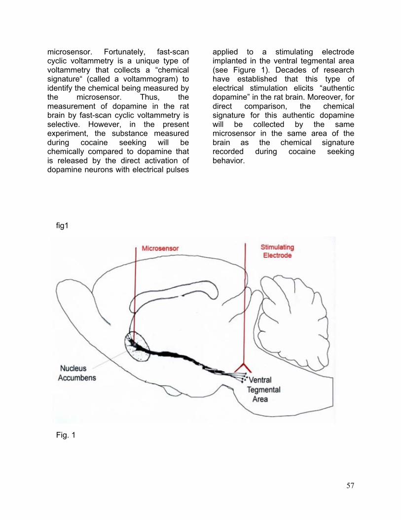
57
microsensor. Fortunately, fast-scan cyclic voltammetry is a unique type of voltammetry that collects a “chemical signature“ (called a voltammogram) to identify the chemical being measured by the microsensor. Thus, the measurement of dopamine in the rat brain by fast-scan cyclic voltammetry is selective. However, in the present experiment, the substance measured during cocaine seeking will be chemically compared to dopamine that is released by the direct activation of dopamine neurons with electrical pulses
applied to a stimulating electrode implanted in the ventral tegmental area (see Figure 1). Decades of research have established that this type of electrical stimulation elicits “authentic dopamine” in the rat brain. Moreover, for direct comparison, the chemical signature for this authentic dopamine will be collected by the same microsensor in the same area of the brain as the chemical signature recorded during cocaine seeking behavior.
fig1
Fig. 1

Finally, it is necessary to consider where in the brain dopamine will be monitored and the extent of damage the implanted microsensor will cause. While several regions could potentially be sampled, for three main reasons, the best place to record dopamine is the nucleus accumbens. First, this region is part of one of the major midbrain dopamine systems, the mesolimbic dopamine system, which originates in the ventral tegmental area and terminates in the nucleus accumbens. Second, this region links reward-related information processed in the limbic system and cortex with motor output. Thus, the nucleus accumbens occupies a key role in the brain circuitry supporting motivated behavior. Third, it is well established that cocaine self-administration will increase dopamine release in the nucleus accumbens as measured by microdialysis. However, the probe associated with this technique is rather large compared to the size of the nucleus accumbens. And the microdialysis probe is known to cause damage to the adjacent tissue. Fortunately, the carbon-fiber microelectrode used with fast-scan cyclic voltammetry is considerably smaller. Hence, its tip can be readily implanted within the nucleus accumbens to measure dopamine only in this region. Also, because of smaller size, the carbon-fiber microelectrode causes considerably less tissue damage when implanted.
Experimental design
The experimental design will be divided into three parts:
PART 1: Training a rat to self-administer cocaine.
In this experiment, any animal you will work with has already been trained to self-administer for cocaine.
PART 2: Preparation and surgery for voltammetry measurements of dopamine.
In preparation for brain surgery, a rat must be selected, weighed, and anesthetized properly. You will complete all the steps leading up to surgery. Professor Neuro will complete the surgery procedure while you observe. When surgery is completed, the rat will have the hardware implanted that will allow the recording of dopamine levels and the administration of cocaine.
PART 3: Monitoring dopamine during cocaine seeking.
Two weeks after surgery, the rat is ready to take part in the experiment. You will perform the experiment on a rat that has already recovered. Just before the rat is allowed to engage in cocaine seeking, a microdrive armed with a microsensor will be loaded into the surgically implanted hub. The microsensor will then be lowered into the nucleus accumbens. The electrically evoked dopamine signal will be transmitted to the computer by a wireless piece of equipment in a backpack using radio waves or telemetry. In this way, dopamine can be monitored in the brain of the animal without being connected or “hardwired” to the equipment. The computer will register a lever press and send a signal to the wireless backpack to administer cocaine.
The wireless telemetry and drug delivery systems used in this “virtual” lab were not used in the actual experiments on which this one was based. However,
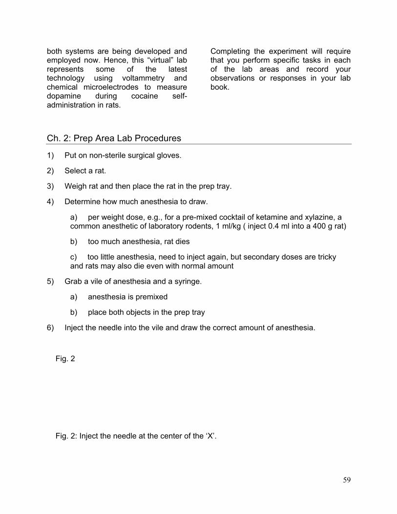
59
both systems are being developed and employed now. Hence, this “virtual” lab represents some of the latest technology using voltammetry and chemical microelectrodes to measure dopamine during cocaine self-administration in rats.
Completing the experiment will require that you perform specific tasks in each of the lab areas and record your observations or responses in your lab book.
Ch. 2: Prep Area Lab Procedures
1) Put on non-sterile surgical gloves.
2) Select a rat.
3) Weigh rat and then place the rat in the prep tray.
4) Determine how much anesthesia to draw.
a) per weight dose, e.g., for a pre-mixed cocktail of ketamine and xylazine, a common anesthetic of laboratory rodents, 1 ml/kg ( inject 0.4 ml into a 400 g rat)
b) too much anesthesia, rat dies
c) too little anesthesia, need to inject again, but secondary doses are tricky and rats may also die even with normal amount
5) Grab a vile of anesthesia and a syringe.
a) anesthesia is premixed
b) place both objects in the prep tray
6) Inject the needle into the vile and draw the correct amount of anesthesia.
Fig. 2
Fig. 2: Inject the needle at the center of the ‘X’.
60
7) Properly inject the anesthesia.
a) inject into peritoneal cavity
(1) if too high (in thoracic cavity), causes pneumothorax, deflating lungs and killing animal
(2) if too low (in bladder), causes complications and perhaps death
b) properly dispose of needle in sharps container
8) Place the rat in the prep tray.
a) rat needs to be on a heating pad as anesthesia prevents thermoregulation
(1) rat can die from being too hot or too cold
Ch. 3: Experiment Area Lab Procedures
If this experiment were to be completed in real time instead of through the virtual world, one would have to perform certain tasks before beginning the actual experiment. As the experimenter, you would be responsible for verifying that the hardware is attached properly, secure, and that the animal is healthy. If this is not done, hardware failure will prevent dopamine measurement or cocaine injection. If a rat is unhealthy, it could die from cocaine injection, and the data would be skewed.
A recovered rat will be placed in the experiment area and the telemetry unit will be attached to the animal. The jugular injection line is flushed with saline to verify that the line is not clogged. The microdrive is then loaded with the dopamine microsensor to verify the microsensor functions correctly. The protective cap covering the brain is removed from the cemented hub and the microdrive is then attached to the
hub, which was implanted during surgery into the rat’s brain. The connections are then attached between the telemetry unit and the reference electrode, the stimulating electrode, the microsensor, and the jugular cannula. The microsensor is lowered to the desired depth at the appropriate speed and the correct depth so as not be break the microsensor or provide appropriate measurement of the dopamine in the nucleus accumbens.
The rat must now be allowed to habituate to the experiment area in order to feel comfortable with its surroundings and not be stressed. A stressed animal may behave erratically and have an adverse reaction to cocaine. The microsensors must then be allowed to come to equilibrium in the brain so the microsensors will not perform erratically. A test stimulation will be performed to check the functionality of the microsensor and have it replaced
61
if necessary, and verify the microsensor has been placed in a dopamine-rich area so that appropriate readings will take place.
Next, the stimulating electrodes must be checked to make sure there is not a bad connection, a bad stimulation unit, or the stimulating electrode has been moved. Wireless communication must also be checked to verify there is no problem
with the battery, the telemetry pack, or the computer.
The next step is to turn on the video camera in order to record the rat’s behavior. It is now time to begin the experiment and to monitor dopamine during cocaine self-stimulation. The following steps will take place during the lab experiment.
a) start session by pressing the on button
b) session starts with extension of lever and display of data on monitor
c) every bar press results in
i) six second injection of cocaine
ii) illuminating the light above lever for 20 seconds
iii) auditory tone for 20 seconds
iv) during this 20 seconds, additional lever presses will not result in additional injections of cocaine
d) session ends
i) lever is retracted
ii) typical session duration is 120 min (NOTE: Our’s will be much shorter.)
e) repeat test stimulation
i) check for functioning microsensor
Once you have completed the experiment, it is time for you to write up your analysis in your lab report