
Product Training - Raj Sarkar, PM OsteoFlap ® System Overview.
15
Product Training - Raj Sarkar, PM OsteoFlap ® System Overview
-
Upload
marcel-matherson -
Category
Documents
-
view
216 -
download
3
Transcript of Product Training - Raj Sarkar, PM OsteoFlap ® System Overview.
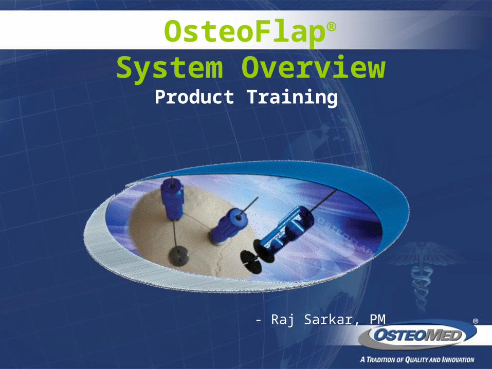
Product Training
- Raj Sarkar, PM
OsteoFlap®
System Overview
Market OverviewSeveral methods are usually utilized to achieve re-attachment of the cranial flap after a craniotomy:
Plates and screws
Steel wire sutures
Clamping rachet designs
OsteoFlap®

Product OverviewIndications: OsteoFlap™ is indicated for the re-attachment of the cranial flap after a craniotomy. Device Specification:
Made of titanium alloy (6Al-4V)MRI compatible Available in 11mm, 17mm and 22mm disk diametersTwo tools available for disk removal
OsteoFlap®

OsteoFlap®
Features & Benefits
Feature Benefit
Titanium Alloy Strength & Biocompatibility
Concave shape Conforms to shape of cranial surfaceReduces soft tissue irritationStrength due to formed shapeRecess formed to reduce potential fixation rod protruding into soft tissue
Small, Fine threads Infinite adjustability for adapting to flap thicknessNarrow diameter chosen to allow for intra-kerf placementThreaded & welded into inferior plate for additional strength

Outward radiating slots on plate
Flanged for flexibility in adapting to cranium interiorFlanges also can conform independently and add rotation resistance of implant
Plate thickness, 0.83mm Minimal invasion & protrudence into dura
3/4” Outer Diameter of Tool
Large diameter for ease of use, minimal effort required
Grooves on Outer Diameter of Tool
Easy grip with gloved fingers
Instrument-free design No instrumentation to be sterilized
OsteoFlap®
Features & Benefits

Surgical TechniqueImplant Placement
After the surgical intervention, position the OsteoFlap Cranial Flap Fixation Disks equally spaced on the dura. Position the inferior disk so that the anti-rotation tab on the inferior disk falls within the kerf width. Replace the bone flap, allowing it to rest between the inferior and superior disks.
The superior disks are threaded down on each device with the use of the attached driver tool. Tighten the superior disk by pulling the rod in a superior direction with one hand, and turning the driver tool with the other hand (Fig. A). DO NOT secure any single device until all of the devices are loosely tightened and the flap is correctly oriented. Final tightening should be firm and represent the maximum force of the fingers. Avoid capturing soft tissue or debris between the disks and the bone.
NOTE: If the tab on the superior plate is not in the kerf width when the device is secured, continue to turn the driver tool until the tab is in the kerf width.
ANTI-ROTATION TAB
DRIVER TOOL
OsteoFlap®

Surgical TechniqueFig. B
Once all the implants have been secured, rock the disposable driver tool in the direction of the arrows on the driver tool (Fig. B). The center rod should break clean. Dispose of the driver tool and
broken center rod according to standard hospital procedure. Repeat for all implants.
OsteoFlap®
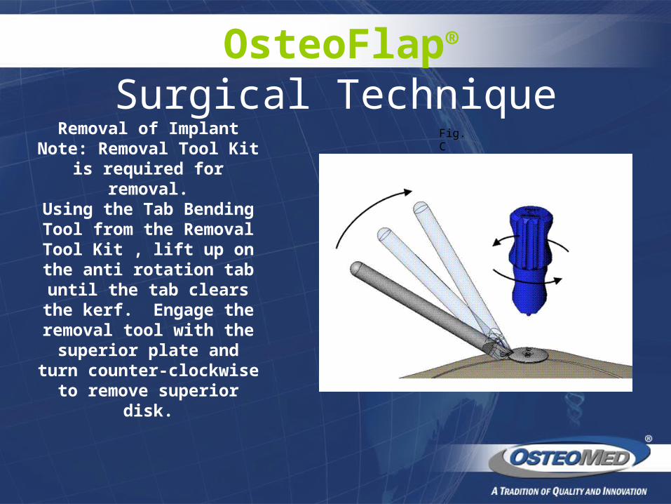
Surgical Technique
Fig. C
Removal of ImplantNote: Removal Tool Kit is
required for removal.Using the Tab Bending Tool from the Removal Tool Kit , lift up on the anti rotation tab until the tab clears the kerf. Engage the removal
tool with the superior plate and turn counter-clockwise
to remove superior disk.
OsteoFlap®

CLINICAL PHOTOGRAPHS
Ordering Information
Part Number Description
219-0011 11mm Cranial Flap Fixation Device
219-0017 17mm Cranial Flap Fixation Device
219-0022 22mm Cranial Flap Fixation Device
219-0004 Cranial Flap Fixation Removal Kit
OsteoFlap®

Clinical ArticlesReliability of Cranial Flap Fixation Techniques: Comparative Experimental Evaluation of Suturing, Titanium Mini-plates, and a New Rivet-like Titanium Clamp (CranioFix) Neurosurgery, Vol. 44, No. 4, April 1999
Bone Flap Fixation With Titanium Clamps: A New Technique Surgical Neurology, Volume 53, Number 4, April 2000
Cranial Bone Flap Fixation Clamps: Compatibility at MR Imaging Radiology, June 1998
OsteoFlap®

Competitive ProductAesculap
CranioFix™ Titanium Clamp System
Advantages: First to market Sterile packaged
Disadvantages: Requires instrumentation Holding Forceps Applying Forceps Pin Cutter Removal Forceps 4 steps to fixation Sterilizing, organizing, storing of instruments. Finite adjustability
Other Information: Available in 11mm, 16mm & 20mm Made of Titanium Alloy Biocompatible MR-compatible
OsteoFlap®

Competitive ProductSynthes
Flap-Fix™ Cranial Clamp
Advantages: Low profile reduces palpability Clover leaf design helps provide for adaptation to the curvature of the cranium.
Disadvantages: Requires instrument to cut implant
Other Information: Available in 13mm & 18mm
OsteoFlap®

Competitive ProductW. Lorenz
RapidFLAP™
Advantages: Sterile packaged
Disadvantages: Requires instrumentation Applier Post Cutter Post Holder Removal Forceps 4 steps to fixation Sterilizing, organizing, storing of instruments
Other Information: Available in 12mm, 16mm & 20mm Made of Titanium AlloyAlso available in Spin Down design
OsteoFlap®

Competitive ProductLeibinger
QuikDisk Cranial Fixation System
Advantages: Unique Grip-n-Zip application Packaged sterile Easy removal
Disadvantages: Requires instrumentation Sterilizing, organizing, storing of instruments
Other information: Available in 14mm & 20mm
OsteoFlap®

FOR FURTHER PRODUCT RELATED QUESTIONS, PLEASE CONTACT RAJ SARKAR ( Product Manager-
Neuro) AT [email protected] or CALL (972)677-4735.
For all sales related questions please contact your regional sales support
representative.
OsteoFlap®