Product Manual Human beta actin RT-PCRmer™
23
Product Manual Human beta actin RT-PCRmer™ Human beta actin RT-PCR primer sets Catalog No.: 40-1001-10, 40-101X-10 Store at –20 o C For research use only. Not for use in diagnostic procedures for clinical purposes.
Transcript of Product Manual Human beta actin RT-PCRmer™

Product Manual
Human beta actin RT-PCRmer™ Human beta actin RT-PCR primer sets Catalog No.: 40-1001-10, 40-101X-10 Store at –20oC
For research use only. Not for use in diagnostic procedures for clinical purposes.

Human beta actin RT-PCRmer™ Amplification of human beta actin cDNA specific fragments For research use only. Not for use in diagnostic procedures for clinical purposes.
Material Supplied
Human beta actin RT-PCRmer™ Human cytoplasmic beta actin RT-PCR primer sets
Storage Instructions: 1. Shipped lyophilized at room temperature. 2. Store at –20 oC upon receipt. 3. Store at –20 oC after reconstitution.
Catalog No. Product Size 40-1001-10 Human beta actin A RT-PCRmer™ (F1/R1); Exon 3 10 nmols 40-1021-10 Human beta actin B RT-PCRmer™ (F4/R3B); Exon 2-5 10 nmols 40-1022-10 Human beta actin C RT-PCRmer™ (F6/R1); 5’UTR-Exon 3 10 nmols 40-1023-10 Human beta actin D RT-PCRmer™ (F2/R2); Exon 4-3’ UTR 10 nmols 40-1024-10 Human beta actin F RT-PCRmer™ (F6/R2); 5’ UTR- 3’ UTR 10 nmols 40-1025-10 Human beta actin G RT-PCRmer™ (F4/R1); Exon 2-3 10 nmols 40-1026-10 Human beta actin TM RT-PCRmer™ for Taqman assay (F2/R3); Exon 4-5 10 nmols 40-1027-02 Human beta actin TaqMan Probe (F2/R3); Exon 5 2 nmols 40-1028-10 Human beta actin MB RT-PCRmer™ for Molecular Beacon assay
(F2/R3B); Exon 4-5 10 nmols
40-1029-02 Human beta actin Molecular Beacon Probe (F2/R3B); Exon 5 2 nmols
Each tube supplied contains the lyophilized primer RT-PCRmer™ pair. Please refer to label on the specific tube. Each tube contains 10 nmols. The quantity supplied is sufficient for 400 regular 50 μl PCR reactions.
*The polymerase chain reaction (PCR) process is covered by patents owned by Hoffmann-La Roche. A license to perform is automatically granted by the use of authorized reagents.
M40-1001-10 Ver2.1 Page 2 of 12

Human beta actin RT-PCRmer™ Amplification of human beta actin cDNA specific fragments For research use only. Not for use in diagnostic procedures for clinical purposes.
Human beta actin RT-PCRmer™ Human cytoplasmic beta actin RT-PCR primer sets
Description RT-PCRmerTM are primer pairs for specific amplification of cDNA. ß-actin is ubiquitously expressed and serves as a positive control for northern and other expression studies. These are generally used as controls for measuring cDNA synthesis efficiency by reverse transcription and as controls for mRNA (cDNA) quantitative expression studies. The RT-PCRmerTM are supplied as a lyophilized powder in aliquots of 10 nmols. The 10 nmols of primer when dissolved in 500 µl sterile water or TE will give a solution of 20 µMolar i.e. 20 pmols/µl. The quantity supplied is sufficient for at least 400 regular 25 µl PCR reaction* for ethidium bromide stained visualization. The amplified product may also be detected by hybridization using Gene Link OligoProberTM that are specific probes directed to the amplified fragment. These are available with either a free 5' OH for 32P labeling or 5’ biotinylated probe for non-radioactive detection. The product is supplied as a lyophilized powder, after reconstitution store at -20 oC. Oligo purity is greater than 98% as determined by denaturing polyacrylamide gel electrophoresis.
Human beta actin RT-PCRmer™ Specifications
Catalog No. Primer Set Primer Position cDNA Fragment Size Genomic Fragment Size
40-1001-10 Human beta actin A (F1/R1)* Exon 3 289 bp 289 bp 40-1021-10 Human beta actin B (F4/R3B) Exon 2/ Exon 5 860 bp 1507 bp 40-1022-10 Human beta actin C (F6/R1) 5’UTR/ Exon 3 816 bp 2156 bp 40-1023-10 Human beta actin D (F2/R2) Exon 4/ 3’ UTR 759 bp 870 bp 40-1024-10 Human beta actin F (F6/R2) 5’UTR/ 3’UTR 1769 bp 3312 bp 40-1025-10 Human beta actin G (F4/R1) Exon 2/ Exon 3 388 bp 940 bp 40-1026-10 Human beta actin TM (F2/R3) for
Taqman assay Exon 4/ Exon 5 130 bp 244 bp
40-1027-02 Human beta actin TaqMan Probe (F2/R3)
Exon 5
40-1028-10 Human beta actin MB (F2/R3B) for Molecular Beacon assay
Exon 4/ Exon 5 167 bp 281 bp
40-1029-02 Human beta actin Molecular Beacon Probe (F2/R3B)
Exon 5
Human Beta actin ref mRNA: NM_001101; Gene: NC_000007 *All primers span introns except F1/R1 (Fragment A).
M40-1001-10 Ver2.1 Page 3 of 12

Human beta actin RT-PCRmer™ Amplification of human beta actin cDNA specific fragments For research use only. Not for use in diagnostic procedures for clinical purposes.
Procedure
Genemer™ Reconstitution Stock Primer Mix: Dissolve the supplied lyophilized RT-PCRmer™ in 100 μl sterile TE. The 10 nmols of primers when dissolved in 100 μl will give a solution of 100 μM i.e. 100 pmols/μl. Primer Mix: Prepare a 10 pmols/μl Primer Mix solution by a ten fold dilution of the stock primer mix. Example: Add 180 μl sterile TE to a new tube, to this tube add 20 μl of primer stock solution. Label this tube as Primer Mix 10 pmols/μl.
TE Buffer pH 7.5 Composition
1 X TE Buffer pH 7.5 10 mM Tris-HCl pH 7.5 1 mM EDTA
cDNA Amplification and Detection
Set up the following amplification files on a thermal cycler. Please refer to the instrument manufacturer’s manual for setting up of the program.
Hot Start
Step Time & Temperature Cycles Initial Denaturation 95 oC for 5 minutes 1
Annealing 50 oC Hold Infinity Hold Comments: Add Taq premix while on hold.
Amplification Profile
Step Temperature & Time Cycles Denaturation 94°C for 30 seconds Annealing 55oC for 30 seconds Extension 72oC for 60 seconds
30
Fill up 72 °C for 7 minutes 1 Hold 4 °C hold for infinity Hold
PCR*
1. PCR Premix Preparation (PP). Label tube “PP”
PCR Premix Preparation (PP) Component 1 X 50 µl Rxn. 10 X 50 µl Rxns.
Sterile Water 32 µl 320 µl 10 X PCR Buffer 4.5 µl 45 µl
2.0 mM dNTP 5 µl 50 µl 10 pmol/µl Primer Mix 2.5 µl 25 µl
Template cDNA (~50 ng) 1-2 µl Add cDNA to each tube Total Volume 45 µl
After adding template start hot start PCR File Dispense 44 µl of the above PCR premix to individual PCR tubes for each amplification reaction and then add the template DNA. Start “Hot Start” thermal cycler file. While holding at 50oC add 5 µl of the Taq Enzyme Mix (EM). Start amplification file.
Program thermal cycler instrument
with an amplification profile prior to starting the amplification protocol. Consult appropriate instrument manufacturer’s manual.
M40-1001-10 Ver2.1 Page 4 of 12

Human beta actin RT-PCRmer™ Amplification of human beta actin cDNA specific fragments For research use only. Not for use in diagnostic procedures for clinical purposes.
2. Taq Polymerase mix Preparation (EM). Label tube “EM”
Taq Enzyme Mix Preparation (EM) Component 1 X 50 µl Rxn. 10 X 50 µl Rxns.
Sterile Water 5 µl 50 µl 10 X PCR Buffer 0.5 µl 5 µl Taq Polymerase 0.5 µl 5 µl
Add 5 µl to each reaction when holding after hot start
D. Agarose Electrophoresis Load 5 to 10 µl samples to 0.8% agarose gel. Run at 60 mAmps. Confirm correct amplification fragment size.
Ethidium bromide is a carcinogen. Follow Health and Safety Procedures established by your institution. Follow proper Hazardous Material Disposal procedures established by your institution.
M40-1001-10 Ver2.1 Page 5 of 12

Human beta actin RT-PCRmer™ Amplification of human beta actin cDNA specific fragments For research use only. Not for use in diagnostic procedures for clinical purposes.
Results and Interpretation
RT-PCR™ primer sets are specific for amplification of different size fragments of cDNA. Each RT-PCRmer™ primer pair spans varying number of intron segments and amplifies a specific fragment.
Human beta actin RT-PCRmer™ Specifications
Primer Set Primer Position cDNA Fragment Size Genomic Fragment Size
Human beta actin A (F1/R1)* Exon 3 289 bp 289 bp Human beta actin B (F4/R3B) Exon 2/ Exon 5 860 bp 1507 bp Human beta actin C (F6/R1) 5’UTR/ Exon 3 816 bp 2156 bp Human beta actin D (F2/R2) Exon 4/ 3’ UTR 759 bp 870 bp Human beta actin F (F6/R2) 5’UTR/ 3’UTR 1769 bp 3312 bp Human beta actin G (F4/R1) Exon 2/ Exon 3 388 bp 940 bp Human beta actin TM (F2/R3) for Taqman assay Exon 4/ Exon 5 130 bp 244 bp Human beta actin TM Probe (F2/R3) Exon 5 Human beta actin MB (F2/R3B) for Molecular Beacon assay
Exon 4/ Exon 5 167 bp 281 bp
Human beta actin MB Probe (F2/R3B) Exon 5 Human Beta actin ref mRNA: NM_001101; Gene: NC_000007
*All primers span introns except F1/R1 (Fragment A).
Human pooled cDNA (10-0100-05; Omni-cDNA™ Human Pooled First Strand cDNA) 50 ng was used as the
template in a 100 µl PCR reaction volume.
M40-1001-10 Ver2.1 Page 6 of 12

Human beta actin RT-PCRmer™ Amplification of human beta actin cDNA specific fragments For research use only. Not for use in diagnostic procedures for clinical purposes.
Appendix
PCR Components and Analysis PCR buffer conditions vary and it is imperative to optimize buffer conditions for each amplification reaction. At Gene Link most amplification reactions have been optimized to work with the following standard buffer condition, unless otherwise indicated.
Standard Gene Link PCR Buffer Composition 10 X PCR buffer 1 X PCR buffer
100 mM Tris-HCl pH 8.3 10 mM 500 mM KCl 50 mM 15 mM MgCl2 1.5 mM 0.01% Gelatin 0.001%
dNTP Concentration Standard dNTP concentration of 0.2 mM of each base is used. See section on PCR additives when dNTP concentration is changed. 2.0 mM dNTP Stock Solution Preparation*
Component Volume 100 mM dGTP 100 μl 100 mM dATP 100 μl 100 mM dTTP 100 μl 100 mM dCTP 100 μl Water 4.6 ml Total Volume 5 ml *Aliquot and freeze
Buffer Condition
MgCl2 Concentration The concentration of Mg++ will vary from 1-5 mM, depending upon primers and substrate. Since Mg2+ ions form complexes with dNTPs, primers and DNA templates, the optimal concentration of MgCl2 has to be selected for each experiment. Low Mg2+ ions result in a low yield of PCR product, and high concentrations increase the yield of non-specific products and promote mis-incorporation. Lower Mg2+ concentrations are desirable when fidelity of DNA synthesis is critical. The recommended range of MgCl2 concentration is 1-4 mM, under the standard reaction conditions specified. At Gene Link, using the standard PCR buffer with KCl, a final dNTP concentration of 0.2 mM, a MgCl2 concentration of 1.5 is used in most cases. If the DNA samples contain EDTA or other chelators, the MgCl2 concentration in the reaction mixture should be raised proportionally. Given below is an MgCl2 concentration calculation and addition table using a stock solution of 25 mM MgCl2.
MgCl2 Concentration & Addition Table
Final concentration of MgCl2 in 50 µl reaction mix, (mM) 1.0 1.25 1.5 1.75 2.0 2.5 3.0 4.0
Volume of 25 mM MgCl2, µl 2 2.5 3 3.5 4 5 6 8
Primer Concentration
The final concentration of primers in a PCR reaction is usually 0.5 to 1 μM (micromolar). This is equivalent to 0.5 to 1 pmol/μl. For a 100 μl reaction you would add 50 to 100 pmols. At Gene Link we use 0.5 pmol/μl in the final PCR.
Always use filter barrier pipette tips to prevent cross contamination
Genemer™ Reconstitution Stock Primer Mix: Dissolve the supplied 10 nmols of lyophilized Genemer™ in 100 µl sterile TE. The 10 nmols of primers when dissolved in 100 µl will give a solution of 100 µM i.e. 100 pmols/µl.
TE Buffer pH 7.5 Composition
1 X TE Buffer pH 7.5 10 mM Tris-HCl pH 7.5 1 mM EDTA
Primer Mix: Prepare a 10 pmols/µl Primer Mix solution by a ten fold dilution of the stock primer mix. Example: Add 180 µl sterile TE to a new tube, to this tube add 20 µl of primer stock solution. Label this tube as Primer Mix 10 pmols/µl.
Amplification Thermal Cycling • Program your thermal cycler instrument
with an amplification profile prior to beginning the amplification protocol.
Hot Start: It is essential to have a ‘Hot Start’ profile for amplification of any fragment from a complex template like human genomic DNA. Taq polymerase has low activity at room temperature and it is essential to minimize any mis-priming in the first cycle of amplification. A typical hot start profile is given below. Various enzyme preparations are available which are activated by heat in the first cycle. A simple hot start protocol is given below that can be used with regular Taq polymerase. See the section on PCR additives for amplification of products from high GC content templates.
Consult your appropriate instrument manufacturer’s manual.
M40-1001-10 Ver2.1 Page 7 of 12
Human beta actin RT-PCRmer™ Amplification of human beta actin cDNA specific fragments For research use only. Not for use in diagnostic procedures for clinical purposes.
Typical PCR Premix (/50μl) Component Volume 10 x PCR Buffer 5 μl 2.0 mM dNTP mix (each) 5 μl Primer Mix (10 pmol/μl each) or 2.5μl of 10 pmol/μl of individual primer (final 25 pmol of each primer/50μl)
2.5 μl
H2O 37.5 μl Total Volume 50 μl
Hot Start Step Time & Temperature Cycles Initial
Denaturation 95 oC for 5 minutes 1
60 oC Hold Infinity Annealing Hold Comments: Add Taq premix while on hold.
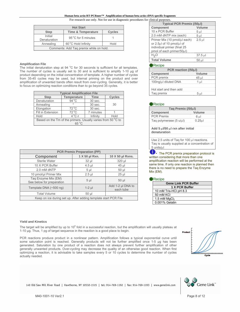
Amplification File The initial denaturation step at 94 oC for 30 seconds is sufficient for all templates.
The number of cycles is usually set to 30 and is sufficient to amplify 1-10 µg of product depending on the initial concentration of template. A higher number of cycles from 35-45 cycles may be used, but internal priming on the product and over amplification of unwanted bands often result from over-cycling. Generally, it is better to focus on optimizing reaction conditions than to go beyond 35 cycles.
PCR reaction (/50μl)
Typical Amplification File
Step
M40-1001-10 Ver2.1 Page 8 of 12
Temperature Time Cycles Denaturation 94 oC 30 sec. Annealing * 30 sec. Elongation 72 oC
30
30 sec. Fill in Extension 72 oC 7 minutes 1 Hold 4 oC r Infinity Hold Based on the Tm of the primers. Usually varies from 50 oC to
65 oC
Component Volume PCR premix 45 μl 100ng/μl diluted DNA 1 μl Hot start and then add Taq premix 5 μl
Taq Premix (/50μl) Component Volume PCR Premix 6 μl Taq polymerase (5 u/μl) 0.25μl Add 5 μl/50 μl rxn after initial denaturation Use 2.5 units of Taq for 100 μl reactions. Taq is usually supplied at a concentration of 5 units/μl
PCR Premix Preparation (PP) Component 1 X 50 µl Rxn. 10 X 50 µl Rxns.
Sterile Water 32 µl 320 µl 10 X PCR Buffer 4.5 µl 45 µl
2.0 mM dNTP 5 µl 50 µl 10 pmol/µl Primer Mix 2.5 µl 25 µl Taq Enzyme Mix (EM)
See below for preparation 5 µl 50 µl
Template DNA (~500 ng) 1-2 µl Add 1-2 µl DNA to each tube
Total Volume 50 µl Keep on ice during set up. After adding template start PCR File
• The PCR premix preparation protocol is written considering that more than one amplification reaction will be performed at the same time. If only one reaction is planned then there is no need to prepare the Taq Enzyme Mix (EM).
Gene Link PCR Buffer
1 X PCR Buffer 10 mM Tris-HCl pH 8.3 50 mM KCl 1.5 mM MgCl20.001% Gelatin
Yield and Kinetics
The target will be amplified by up to 106 fold in a successful reaction, but the amplification will usually plateau at 1-10 µg. Thus, 1 pg of target sequence in the reaction is a good place to begin. PCR reactions produce product in a nonlinear pattern. Amplification follows a typical exponential curve until some saturation point is reached. Generally products will not be further amplified once 1-5 µg has been generated. Saturation by one product of a reaction does not always prevent further amplification of other generally unwanted products. Over-cycling may decrease the quality of an otherwise good reaction. When first optimizing a reaction, it is advisable to take samples every 5 or 10 cycles to determine the number of cycles actually needed.
Human beta actin RT-PCRmer™ Amplification of human beta actin cDNA specific fragments For research use only. Not for use in diagnostic procedures for clinical purposes.
M40-1001-10 Ver2.1 Page 9 of 12
Gel Electrophoresis of PCR Products
Gel electrophoresis of PCR products is the standard method for analyzing reaction quality and yield. PCR products can range up to 10 kb in length, but the majority of amplifications are at 1 kb and below. Agarose electrophoresis is the classical method to analyze amplification products from 150 bp to greater than 10 kb. Polyacrylamide gel electrophoresis should be used for resolution of short fragments in the range of 100 bp to 500 bp when discrimination of as small as a 10 bp difference is required.
PAGE gels for PCR products formulated with the amount of cross-linker chosen to give pore sizes optimal for the size of DNA fragment desired. Gels are most often stained in ethidium bromide, even though the fluorescence of this stain is quenched by polyacrylamide, which decreases sensitivity 2-5 fold. This decrease in sensitivity generally does not present a problem, because most PCR reactions yield product levels in the microgram range, and ethidium will detect as little as 1/10 of this amount. Polyacrylamide gels can be stained by silver staining for more sensitive detection.

Human beta actin RT-PCRmer™ Amplification of human beta actin cDNA specific fragments For research use only. Not for use in diagnostic procedures for clinical purposes.
M40-1001-10 Ver2.1 Page 10 of 12
Purification of PCR Product Various purification methods are available for the purification of PCR products. The selection of a particular method over another is based on the downstream application and the initial robustness of the amplification. Usually no further purification is required for most cloning experiments if a single fragment is amplified, whereas for sequencing applications the amplified product should be purified from the primers and any other minor amplification products. The preferred method of purification of an amplified fragment is the excision of the fragment band after agarose gel electrophoresis. This method yields the purification of a single fragment; as such care should be taken to excise a gel piece containing a single electrophoretically resolved fragment. The Omni-Clean™ Purification System available from Gene Link can be used for this purpose. Catalog No. 40-4110-10 for bead based system; 40-4120-10 for spin column based system and 40-4130-10 for DNA concentration. Please refer to product insert for detailed protocol or visit www.genelink.com.
A. Purification of DNA from gel slices using glass beads. Provides purified single fragment. [Omni-Clean™ Gel DNA Beads Purification System; Catalog No. 40-4110-10]
Protocol 1. By weight, determine the volume of the excised DNA fragment. 2. Add 3 volumes of NaI solution and heat to 55 °C. Visually determine the dissolution of gel pieces. 3. Add 1 μl of glass bead suspension per μg of DNA and vortex. 4. Centrifuge at 2K rpm for 20 seconds to pellet glass bead/DNA complex. Discard supernatant. 5. Re-suspend pellet in 400 μl Omni-Clean™ wash buffer. Centrifuge at 2K rpm for 20 seconds and discard wash buffer. 6. Pipet out any remaining buffer in the tube. 7. Add 25 μl water or TE; re-suspend pellet and centrifuge at 2K rpm for 20 seconds. 8. The supernatant contains the purified DNA. Using a pipet, collect the supernatant and transfer to a new appropriately labeled tube.
B. Purification of DNA from gel slices using spin column. Provides purified single fragment.
[Omni-Clean™ Gel DNA Spin Column Purification System; Catalog No. 40-4120-50] Protocol
1. By weight, determine the volume of the excised DNA fragment. 2. Add 3 volumes of NaI solution and heat to 55 °C. Visually determine the dissolution of gel pieces. 3. Add the above solution to the spin column assembled on a collection tube. 4. Let the solution flow by gravity or centrifuge at 2K rpm for 20 seconds. Discard flow through collected in the collection tube. 5. Add 400 μl Omni-Clean™ wash buffer to the spin column. Centrifuge at 2K rpm for 2 minutes and discard wash buffer collected in the
collection tube. 6. Replace the collection tube with a new appropriately labeled eppendorf tube. 7. Add 25 μl water or TE to the spin column. Let sit for 3 minutes. 8. Centrifuge at 2K rpm for 2 minutes. 9. The collection tube contains the purified DNA. C. Purification of DNA from solution using glass beads. Provides removal of salts, primers and dNTP.
[Omni-Clean™ DNA Beads Concentration System; Catalog No. 40-4130-10] Protocol
1. Determine volume of DNA solution and add 3 volumes of NaI solution. 2. Add 1 μl of glass bead suspension per μg of DNA. 3. Centrifuge at 2K rpm for 20 seconds to pellet glass bead/DNA complex. Discard supernatant. 4. Re-suspend pellet in 400 μl Omni-Clean™ wash buffer. 5. Centrifuge at 2K rpm for 20 seconds and discard wash buffer. 6. Pipet out any remaining buffer in the tube. 7. Add 25 μl water or TE; re-suspend pellet and centrifuge at 2K rpm for 20 seconds. 8. The supernatant contains the purified DNA. Using a pipet, collect the supernatant and transfer to a new appropriately labeled tube.
D. Purification of DNA from solution using spin column. Provides removal of salts, primers and dNTP.
[Omni-Clean™ DNA Spin Column Concentration System; Catalog No. 40-4140-10] Protocol
1. Determine volume of DNA solution and add 3 volumes of NaI solution. 2. Add the above solution to the spin column assembled on a collection tube. 3. Let the solution flow by gravity or centrifuge at 2K rpm for 20 seconds. Discard flow through collected in the collection tube. 4. Add 400 μl Omni-Clean™ wash buffer to the spin column. Centrifuge at 2K rpm for 2 minutes and discard wash buffer collected in the
collection tube. 5. Replace the collection tube with a new appropriately labeled eppendorf tube. 6. Add 25 μl water or TE to the spin column. Let sit for 3 minutes. 7. Centrifuge at 2K rpm for 2 minutes. 8. The collection tube contains the purified DNA.

Human beta actin RT-PCRmer™ Amplification of human beta actin cDNA specific fragments For research use only. Not for use in diagnostic procedures for clinical purposes.
PCR Additives DNA polymerases need to elongate rapidly and accurately to function effectively in vivo and in vitro, yet certain DNA regions appear to interfere with their progress. One common problem is pause sites, at which DNA polymerase molecules cease elongation for varying lengths of time. Many strong DNA polymerase pauses are at the beginnings of regions of strong secondary structure such as template hairpins (1). Taq polymerase used in PCR suffers the same fate and GC-rich DNA sequences often require laborious work to optimize the amplification assay. The GC-rich sequences possess high thermal and structural stability, presumably because the high duplex melting temperature that permits stable secondary structures to form, thus preventing completion of a faithful replication (2). Nucleotide analog 7-deaza dGTP is effective in reducing the secondary structure associated with GC rich region by reducing the duplex stability (4). Betaine, DMSO and formamide reduces the Tm and the complex secondary structure, thus the duplex stability (1-5). Tetramethyl ammonium chloride (TMAC) actually increases the specificity of hybridization and increases the Tm. The use of TMAC is recommended in PCR conditions using degenerate primers. These PCR additives and enhancing agents have been used to increase the yield, specificity and consistency of PCR reactions. These additives may have beneficial effects on some amplification and it is impossible to predict which agents will be useful in a particular context and therefore they must be empirically tested for each combination of template and primers.
PCR Additives Additive Purpose & Function Concentration 7-deaza-2'-deoxyguanosine; 7-deaza dGTP
GC rich region amplification. Reduce the stability of duplex DNA
Totally replace dGTP with 7-deaza dGTP; or use 7-deaza dGTP: dGTP at 3:1
Betaine (N,N,N-trimethylglycine = [carboxymethyl]trimethylammonium)
Reduces Tm facilitating GC rich region amplification. Reduces duplex stability
Use 3.5M to 0.1M betaine. Be sure to use Betaine or Betaine (mono)hydrate and not Betaine HCl.
BSA (bovine serum albumin)
BSA has proven particularly useful when attempting to amplify ancient DNA or templates, which contain PCR inhibitors such as melanin.
BSA concentration of 0.01 µg/µl to 0.1 µg/ µl can be used.
DMSO (dimethyl sulfoxide)
DMSO is thought to reduce secondary structure and is particularly useful for GC rich templates.
DMSO at 2-10% may be necessary for amplification of some templates, however 10% DMSO can reduce Taq polymerase activity by up to 50% so it should not be used routinely.
Formamide Reduces secondary structure and is particularly useful for GC rich templates.
Formamide is generally used at 1-5%. Do not exceed 10%.
Non-ionic detergents e.g. Triton X-100, Tween 20 or Nonidet P-40 (NP-40)
Non-ionic detergents stabilise Taq polymerase and may also supress the formation of secondary structure.
0.1-1% Triton X-100, Tween 20 or NP-40 may increase yield but may also increase non-specific amplification. As little as 0.01% SDS contamination of the template DNA (left-over from the extraction procedure) can inhibit PCR by reducing Taq polymerase activity to as low as 10%, however, inclusion of 0.5% Tween-20 or -40 will effectively neutralize this effect.
TMAC (tetramethylammonium chloride)
TMAC is used to reduce potential DNA-RNA mismatch and improve the stringency of hybridization reactions. It increases Tm and minimizes mis-pairing.
TMAC is generally used at a final concentration of 15-100 mM to eliminate non-specific priming.
References
1. Kovarova, M; and Draber, P; (2000) New Specificity and yield enhancer for polymerase chain reactions (2000) Nucl. Acids. Res. 28: e70. 2. Henke, W., Herdel, K., Jung, K., Schnorr, D. and Stefan A. Loening, S. (1997) Betaine improves the PCR amplification of GC-rich DNA sequences. Nucl. Acids Res. 25: 3957-3958. 3. Daniel S. Mytelka, D.S., and Chamberlin, M.J.,(1996) Analysis and suppression of DNA polymerasepauses associated with a trinucleotide consensus. Nuc. Acids Res.,. 24:2774–278. 4. Keith, J. M., Cochran, D.A.E., Lala, G.H., Adams, P., Bryant, D.and Mitchelson, K.R. (2004) Unlocking hidden genomic sequence. Nucl. Acids Res. 32: e35. 5. Owczarzy, R., Dunietz, I., Behlke, M.A., Klotz, I.M. and Joseph A. Walder. (2003) Thermodynamic treatment of oligonucleotide duplex–simplex equilibria. PNAS, 100:14840-14845.
M40-1001-10 Ver2.1 Page 11 of 12

Human beta actin RT-PCRmer™ Amplification of human beta actin cDNA specific fragments For research use only. Not for use in diagnostic procedures for clinical purposes.
Ordering Information
Human beta actin RT-PCRmer™ Primer pair for specific amplification of cDNA. Special optimized conditions may be required for certain amplifications Product Size Catalog No. Price, $ Human beta actin A RT-PCRmer™ (F1/R1); Exon 3 10 nmols 40-1001-10 100.00 Human beta actin B RT-PCRmer™ (F4/R3B); Exon 2-5 10 nmols 40-1021-10 100.00 Human beta actin C RT-PCRmer™ (F6/R1); 5’UTR-Exon 3 10 nmols 40-1022-10 100.00 Human beta actin D RT-PCRmer™ (F2/R2); Exon 4-3’ UTR 10 nmols 40-1023-10 100.00 Human beta actin F RT-PCRmer™ (F6/R2); 5’ UTR- 3’ UTR 10 nmols 40-1024-10 100.00 Human beta actin G RT-PCRmer™ (F4/R1); Exon 2-3 10 nmols 40-1025-10 100.00 Human beta actin TM RT-PCRmer™ for Taqman assay (F2/R3); Exon 4-5 10 nmols 40-1026-10 100.00 Human beta actin TaqMan Probe (F2/R3); Exon 5 2 nmols 40-1027-02 100.00 Human beta actin MB RT-PCRmer™ for Molecular Beacon assay (F2/R3B); Exon 4-5 10 nmols 40-1028-10 100.00 Human beta actin Molecular Beacon Probe (F2/R3B); Exon 5 2 nmols 40-1029-02 100.00
*Please visit www.genelink.com for other RT-PCRmer™ not listed here
Mouse beta actin RT-PCRmer™ Primer pair for specific amplification of cDNA. Special optimized conditions may be required for certain amplifications Product Size Catalog No. Price, $ Mouse beta actin A RT-PCRmer™ (F1/R1); Exon 2-3 10 nmols 40-1003-10 100.00 Mouse beta actin B RT-PCRmer™ (F2/R2); Exon 6-7 10 nmols 40-1014-10 100.00 Mouse beta actin C RT-PCRmer™ (F3/R3); Exon 2-7 10 nmols 40-1015-10 100.00 Mouse beta actin D RT-PCRmer™ (F4/R4); Exon 2-9 10 nmols 40-1016-10 100.00
*Please visit www.genelink.com for other RT-PCRmer™ not listed here
GAPDH RT-PCRmer™ Primer pair for specific amplification of cDNA. Special optimized conditions may be required for certain amplifications Product Size Catalog No. Price, $ GAPDH A RT-PCRmer™ (F1/R1); Exon 2-3 10 nmols 40-1005-10 100.00 GAPDH B RT-PCRmer™ (F2/R2); Exon 6-7 10 nmols 40-1006-10 100.00 GAPDH C RT-PCRmer™ (F1/R2); Exon 2-7 10 nmols 40-1007-10 100.00 GAPDH D RT-PCRmer™ (F1/R3); Exon 2-9 10 nmols 40-1008-10 100.00
*Please visit www.genelink.com for other RT-PCRmer™ not listed here
RT-PCRmer™ Primer pair for specific amplification of cDNA. Special optimized conditions may be required for certain amplifications Product Size Catalog No. Price, $ human beta actin RT-PCRmer; 10 nmols 40-1001-10 100.00 rat beta actin RT-PCRmer; 10 nmols 40-1002-10 100.00 mouse beta actin RT-PCRmer; 10 nmols 40-1003-10 100.00 beta2 microglobulin RT-PCRmer; 10 nmols 40-1004-10 100.00 Beta actin control PCR mix (human & rat) 200 ul 40-1002-00 110.00
*Please visit www.genelink.com for other RT-PCRmer™ not listed here Genemer™ Primer pair for gene or mutation specific amplification. Special optimized conditions may be required for certain amplification Product Size Catalog No. Price, $ Fragile X (spanning CGG triple repeat region) 10 nmols 40-2004-10 100.00 Huntington Disease (spanning CAG triple repeat region) 10 nmols 40-2025-10 100.00 Myotonic Dystrophy (spanning CTG triple repeat region) 10 nmols 40-2026-10 100.00 Friedreich’s Ataxia (spanning GAA triple repeat region) 10 nmols 40-2027-10 100.00 Factor V 10 nmols 40-2035-10 100.00 Factor VIII (Hemophilia) Genemer™ Pack 10 nmols 40-2036-10 750.00 STS (Steroid Sulfatase) 10 nmols 40-2023-10 100.00 HGH (Human Growth Hormone) 10 nmols 40-2024-10 100.00 Sickle Cell 10 nmols 40-2001-10 100.00 RhD (Rh D gene exon 10 specific) 10 nmols 40-2002-10 100.00 Rh EeCc (Rh Ee and Cc exon 7 specific) 10 nmols 40-2003-10 100.00 Gaucher (various mutations) 10 nmols 40-2047-XX 100.00 Cystic Fibrosis (various mutations) 10 nmols 40-2029-XX 100.00 SRY (sex determining region on Y) 10 nmols 40-2020-10 100.00 X alphoid repeat 10 nmols 40-2021-10 100.00 Y alphoid repeat 10 nmols 40-2022-10 100.00
*Please visit www.genelink.com for other Genemer™ not listed here *Please visit www.genelink.com for other GeneProber™ not listed here
**The polymerase chain reaction (PCR) process is covered by patents owned by Hoffmann-La Roche. A license to perform is automatically granted by the use of authorized reagents. Prices subject to change without notice All Gene Link products are for research use only.
M40-1001-10 Ver2.1 Page 12 of 12

P r o d u c t M a n u a l
Custom Oligo Synthesis, antisense oligos, RNA oligos, chimeric oligos, Fluorescent dye labeled oligos, Molecular Beacons, TaqMan Probes
siRNA, Aptamers
Control Fluorescent Molecular Probes For research use only. Not for use in diagnostic procedures for clinical purposes.
Applications
Real Time Quantitative PCR Analysis (QPCR) Probes Fluorescent Genotyping
siRNA Gene Knockout Validation Allelic Discrimination
Antisense Targeting SNP Detection
Aptamers Detection Probes

Control_RT_Probes_Manual_V2.3.doc Page 2 of 11
Endogenous Control Fluorescent Molecular Probes
IMPORTANT NOTE
1. Consult product specification sheet and material supplied for specifications of product received.
2. This product guide is not specific to any particular endogenous control fluorescent probe.
3. This product guide should be used in conjunction with the particular instrument manual and specifications and is not intended to replace those specifications.
Storage Instructions: 1. Shipped lyophilized at room temperature. 2. Store at –20
oC upon receipt.
3. Store at –20oC after reconstitution.

Control_RT_Probes_Manual_V2.3.doc Page 3 of 11
Endogenous Control Fluorescent Molecular Probes
Reconstitution, Use & Stability of Fluorescent Probes
All Gene Link custom oligo products including, molecular probes, RNA and siRNA includes a datasheet that contains the exact nmols, µg, or A260 units (OD Units) and other physical data. This data is important for reconstituting the product. All fluorescent probes are shipped in amber tubes to prevent exposure to light and minimize photo-bleaching. Gene Link guarantees the stability of oligos for 1 year and fluorescent molecular probes for 6 months if reconstituted and stored appropriately as detailed below. In our experience unmodified oligos are stable for numerous years if reconstituted and stored properly. Avoid multiple freeze thaws; do not exceed 6-10 freeze thaw cycles. If the same oligo is intended to be used repeatedly then it is prudent to make a numerous aliquots of the stock solution and frozen.
Reconstitution & Storage Gene Link fluorescent probes are supplied lyophilized in amber tubes to protect from light and to reduce photo bleaching. These are stable at room temperature for an extended period of time but should preferably be frozen upon receipt. TE buffer is recommended for dissolving the probes and oligonucleotides; EDTA inhibits the activity of the nucleases.
Preferred TE Buffer Reconstitution & Storage pH for Fluorescent Probes
6-FAM, HEX, TET, ROX, and TAMRA TE Buffer pH 7.5 or 8.0
Cy3, Cy3.5, Cy5, and Cy5.5 TE Buffer pH 7.0 or 7.5
Alexa dyes TE Buffer pH 7.5 or 8.0
Cy dyes rapidly degrade in acidic pH
Further dilution can be made in TE buffer. After reconstitution store the stock solution at -80°C or -20°C. Fluorescently labeled oligos should be stored protected from light.
Preparation of Probe Stock Solution of 20 pmols/μl [20 μM] Gene Link provides the exact amount of nmols of each probe supplied on the tube and on the Product Specifications Sheet included with the product. Example: 2 nmols lyophilized product. Dissolve the 2 nmol probe in 100 μl of TE pH7.5 (10 mM Tris pH7.5, 1 mM EDTA) to get 20 pmols/μl stock solution [20 μM]. Working Probe Stock Solution 2 pmols/μl [2 μM] Dilute 10 fold to prepare a 2 pmols/μl [2 μM]
Preparation of Primer Mix Stock Solution of 30 pmols/μl [30 μM] Gene Link provides the exact amount of nmols of each probe supplied on the tube and on the Product Specifications Sheet included with the product. Most Gene Link Genemer™ primer mix are supplied as 10 nmols or 3 nmols lyophilized. Example: 10 nmols lyophilized product.

Control_RT_Probes_Manual_V2.3.doc Page 4 of 11
Dissolve the 10 nmols of primer mix in 334 μl of TE pH7.5 (10 mM Tris pH7.5, 1 mM EDTA) to get 30 pmols/μl stock solution [30 μM]. Example: 3 nmols lyophilized product. Dissolve the 3 nmols of primer mix in 100 μl of TE pH7.5 (10 mM Tris pH7.5, 1 mM EDTA) to get 30 pmols/μl stock solution [30 μM]. Working Probe Stock Solution 3 pmols/μl [3 μM] Dilute 10 fold to prepare a3 pmols/μl [3 μM].
Working Stock solution in TE pH 7.5
Primer 3 µMolar [3 pmol/µL]
Probe 2 µMolar [2 pmol/µL] Storage For optimal long-term storage, it is recommended that the oligonucleotides should be stored dry at -20°C in the dark. If numerous experiments are planned using the same oligonucleotide, prepare aliquots, dry them and store the aliquots at -20°C. Stability Gene Link guarantees the stability of oligos for 1 year and fluorescent molecular probes for 6 months if reconstituted and stored appropriately as recommended by Gene Link. The stability can be increased several fold by instituting proper handling conditions, avoiding exposure to light and multiple freeze thaws.

Control_RT_Probes_Manual_V2.3.doc Page 5 of 11
Typical TaqMan Reaction Conditions
Component Stock Solution Final Concentration per 25 µL rxn 5x 25 µL reaction mix
Water Water 10 µL 50 µL
10X PCR buffer 10X 1X 2.5 µL 12.5 µL
dNTP 2 mM 200 μM 2.5 µL 12.5 µL
MgCl2 25 mM 3 mM 3 µL 15 µL
Primer Mix 3 μM 300 nM (0.3 pmol/ul) 3 µL 15 µL
Probe 2 μM 200 nM (0.2 pmol/ul) 3 µL 15 µL
Template ~10ng/μL 1 µL
Taq polymerase 5u/µL 0.025 unit/µL 0.5 µL
Total Volume 25 µL
QPCR Reaction Dispense 25 µL of above QPCR reaction mix to each tube then add 1 µL template and start cycling. Typical TaqMan Cycling Conditions
GL TaqMan PCR cycling Time Temperature Cycles
Stage 1 GL Initial denaturation 2 min 95 °C 1
Stage 2 Denature 15 sec 95 °C 40
Anneal/Extend-OPTICS ON 1 min 60 °C
Instrument Run & Results Consult instrument manufacturer protocol and manual

Control_RT_Probes_Manual_V2.3.doc Page 6 of 11
Typical Molecular Beacon Reaction Conditions
Component Stock Solution Final Concentration per 25 µL rxn 5x 25 µL reaction mix
Water Water 10 µL 50 µL
10X PCR buffer 10X 1X 2.5 µL 12.5 µL
dNTP 2 mM 200 μM 2.5 µL 12.5 µL
MgCl2 25 mM 3 mM 3 µL 15 µL
Primer Mix 3 μM 300 nM (0.3 pmol/ul) 3 µL 15 µL
Probe 2 μM 200 nM (0.2 pmol/ul) 3 µL 15 µL
Template ~10ng/μL 1 µL
Taq polymerase 5u/µL 0.025 unit/µL 0.5 µL
Total Volume 25 µL
QPCR Reaction Dispense 25 µL of above QPCR reaction mix to each tube then add 1 µL template and start cycling.
Stage Molecular Beacon QPCR cycling Time Temperature Cycles
Stage 1 Initial denaturation 2 min 95 °C 1
Stage 2
Denature 15 sec 95 °C
40
Anneal-OPTICS ON 30 sec 60 °C
Extend-OPTICS OFF 30 sec 72 °C
Instrument Run & Results Consult instrument manufacturer protocol and manual

Control_RT_Probes_Manual_V2.3.doc Page 7 of 11
Gene Link Molecular Beacon Melt Curve Protocol
1. Prepare Molecular Beacon stock solution at 100 pmols/l [ 100 M (micromolar) ] in 1 X PCR Buffer. Gene Link provides the exact amount of nmoles of each oligo supplied on the tube and on the Oligo Report. Multiply the 'nmol' amount by 10 to arrive at the volume of solvent to be added.
2. Prepare Molecular Beacon working solution at 5 pmols/l [ 5 M (micromolar) ] in 1 X PCR Buffer.
3. Set up two 25 l reactions, one with probe alone, one with target + probe as follows.
Molecular Beacon Probe Alone Molecular Beacon Probe plus Target
2.5 l 10X PCR buffer 2.5 l 10x PCR buffer
3 l 25 mM MgCl2 3 l 25 mM MgCl2
1 l 5 pmol/ul probe [0.2 pmol/l final or 200 nM] 1 l 5 pmol/ul probe [0.2 pmol/l final or 200 nM]
3 l 5 pmol/ul target [0.6 pmol/l final or 600 nM]
18.5 l H20 15.5 l H2O
The protocol for Molecular Beacon Melt Curve ramps from 40 to 95 degrees C at 0.2 degrees/second
Screen capture of graphs from a Cepheid SmartCycler
Multiple Molecular Beacon Probe Alone Melt Curve Molecular Beacon Probe plus Target and Probe Alone Melt Curve
Notes: -MgCl2 needs to be higher for MB reactions than for regular PCR as it helps to stabilize the stem structure of the probe during the high ramp rate. Final concentration of MgCl2 should be between 2.5 and 4 mM. Here we use 3 mM final. This is the same range of concentration used in an actual amplification reaction. -Final concentration of probe should be 200-600 nM. Here we use 200 nM. This is also the same concentration range used for the real time reaction. -For a melt curve it is important to saturate the probe with target. Use 2-3 X the Molar amount of target. Here we use 3X target for a final concentration of 600 nM. For real time monitoring, 500 ng genomic DNA, diluted 10X to various concentrations can be used as a starting point. QPCR Once you have your melt curve you want to select an annealing temperature for your real time PCR where the probe alone is completely closed (shows no fluorescence), and the probe+target is completely open (shows maximal fluorescence). This temperature should be about 5-8 degrees below the Tm of the probe/target hybrid (red vertical line on melt curve of probe+target). It is important to test your primers at the annealing T to ensure that you will have strong, clean amplification at this temperature. Signal-to-noise (S:N) ratios is calculated by dividing the fluorescence signal of a 25-mer in the presence of a two to five-fold excess of an exactly complementary target sequence by the fluorescence intensity of the probe alone.
Control_RT_Probes_Manual_V2.3.doc Page 8 of 11
Fluorophore Spectral Data
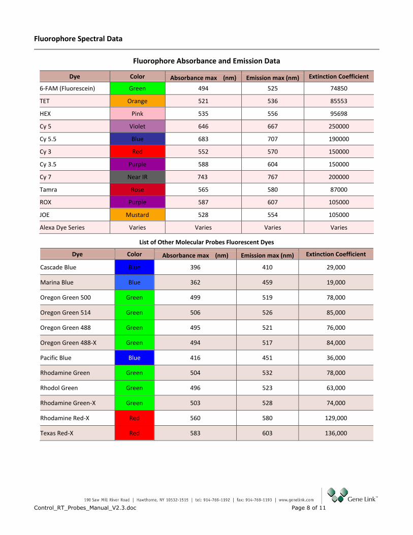
Fluorophore Absorbance and Emission Data
Dye Color Absorbance max (nm) Emission max (nm) Extinction Coefficient
6-FAM (Fluorescein) Green 494 525 74850
TET Orange 521 536 85553
HEX Pink 535 556 95698
Cy 5 Violet 646 667 250000
Cy 5.5 Blue 683 707 190000
Cy 3 Red 552 570 150000
Cy 3.5 Purple 588 604 150000
Cy 7 Near IR 743 767 200000
Tamra Rose 565 580 87000
ROX Purple 587 607 105000
JOE Mustard 528 554 105000
Alexa Dye Series Varies Varies Varies Varies
List of Other Molecular Probes Fluorescent Dyes
Dye Color Absorbance max (nm) Emission max (nm) Extinction Coefficient
Cascade Blue Blue 396 410 29,000
Marina Blue Blue 362 459 19,000
Oregon Green 500 Green 499 519 78,000
Oregon Green 514 Green 506 526 85,000
Oregon Green 488 Green 495 521 76,000
Oregon Green 488-X Green 494 517 84,000
Pacific Blue Blue 416 451 36,000
Rhodamine Green Green 504 532 78,000
Rhodol Green Green 496 523 63,000
Rhodamine Green-X Green 503 528 74,000
Rhodamine Red-X Red 560 580 129,000
Texas Red-X Red 583 603 136,000

Control_RT_Probes_Manual_V2.3.doc Page 9 of 11
Applied Biosystems Proprietary Dyes & Possible Substitutions
Dye Absorbance max (nm) Emission max (nm)
VIC 538 554
HEX 535 556
NED 546 575
Cy3 550 570
PET 558 595
Cy3.5 588 604
ROX 575 602
Texas Red 583 603
LIZ 630 652
Cy5 646 667
Alexa Fluor® Dyes
Dye Color Absorbance max (nm) Emission max (nm) Extinction Coefficient
Alexa Fluor 350 Blue 346 442 19,000
Alexa Fluor 405 Green 401 421 34,000
Alexa Fluor 430 Green 433 541 16,000
Alexa Fluor 488 Green 495 519 71,000
Alexa Fluor 532 Yellow 532 553 81,000
Alexa Fluor 546 Red 556 573 104,000
Alexa Fluor 555 Red 555 565 150,000
Alexa Fluor 568 Red 578 603 91,000
Alexa Fluor 594 Red 590 617 73,000
Alexa Fluor 633 Violet 632 647* 100,000
Alexa Fluor 647 Violet 650 665* 239,000
Alexa Fluor 660 Purple 663 690* 123,000
Alexa Fluor 680 Blue 679 702* 184,000
Alexa Fluor 700 Near IR 702 723* 192,000
Alexa Fluor 750 Near IR 749 775* 240,000

Control_RT_Probes_Manual_V2.3.doc Page 10 of 11
Spectral characteristics of the Alexa Fluor dyes. Extinction coefficient at l max in cm -1m-1. *Human vision is insensitive to light beyond~650nm, and therefore it is not possible to view the far -red- fluorescent dyes by looking through the eyepiece of a conventional fluorescent microscope. Alexa Fluor® Dyes is sold under license from Invitrogen Corporation. These may only be used for R&D and may not be used for clinical or diagnostic use.
*Disclaimer of License Statement for Molecular Beacons Products - *PHRI Molecular Beacon License Agreement -"This product is sold under license from the Public Health Research Institute. It may be used under PHRI Patent Rights only for the purchaser's research and development activities". **Black Hole Quencher License Agreement "Black Hole Quencher", "BHQ-1", "BHQ-2" and "BHQ-3" are registered trademarks of Biosearch Technologies, Inc., Novato, CA. The BHQ technology is licensed and sold under agreement with Biosearch and these products are sold exclusively for R&D use by the purchaser. They may not be used for clinical or diagnostic purposes and they may not be resold, distributed or re-packaged. *Alexa Fluor® Dyes are sold under license from Invitrogen Corporation. These may only be used for R&D and may not be used for clinical or diagnostic use.

Control_RT_Probes_Manual_V2.3.doc Page 11 of 11
T o o l s : A n A r r a y o f G e n e t i c T o o l s Tools application from Gene Link for iPhone/iPod/iPad
The Gene Link Tools app also has advanced modules for setup of Polymerase Chain Reaction (PCR) and Quantitative Real Time PCR (Q-PCR).
The main focus of this app is to have a handy source of calculation modules and quick reference sections for designing and executing experiments involving PCR and Q-PCR.
Tools Modules 1. Oligo Tm: A robust oligo melting temperature calculation module using three methods; it also calculates other physical attributes. 2. Oligo Resuspension 5. Oligo Dilution 6. PCR & QPCR: Convenient calculator for multiple reaction setup for PCR, TaqMan QPCR and Molecular Beacon QPCR setup. Includes stock solution information and cycling profiles 7. Molarity Calculator 8. Reagent Dilution 9. Measurement Convertor: A convenient selection of calculators to convert length, area, mass, temperature and volume units. 10. Genetic Code Translator: Enter DNA sequence to see coding pattern.
Reference Modules
A selection of topics, relevant to life scientists for quick access to basic information. This section includes the following sections and sub sections. 1. Nucleic Acid Basics 2. Modifications Table: A list of common modifications with molecular structure and basic properties. 3. Dye & Quencher Table: A convenient list of fluorophores and quencher matching the emission max. 4. Duplex Stability & Nuclease Resistance 5. Gene Information: Simply enter the accession number and retrieve detailed gene information from NCBI database, 6. Amino Acid Table: Molecular structure and detailed physical properties of all amino acids. 7. Translation Table: Genetic code for all amino acids. 8. Codon Picker: Select codon sequence and see the corresponding amino acid and detailed information.