PRODUCT INFORMATION & MANUAL - … · skeletal and cardiac muscle (5, 6 ... Any variation in...
23
PRODUCT INFORMATION & MANUAL Human VEGF Valukine TM ELISA VAL106 For the detection of human VEGF For research use only. Not for diagnostic or therapeutic procedures. Bio-Techne China Co. Ltd P: +86 (21) 52380373 P: 8009881270 F: +86 (21) 52381001 [email protected] Novus kits are guaranteed for 3 months from date of receipt
Transcript of PRODUCT INFORMATION & MANUAL - … · skeletal and cardiac muscle (5, 6 ... Any variation in...
PRODUCT INFORMATION & MANUAL
Human VEGF ValukineTM ELISA
VAL106
For the detection of human VEGF
For research use only. Not for diagnostic or therapeutic procedures.
Bio-Techne China Co. Ltd P: +86 (21) 52380373 P: 8009881270 F: +86 (21) 52381001
Novus kits are guaranteed for 3 months from date of receipt
Tel: 800-988-1270 Email: [email protected] 1 Web Site: www.novusbio.com/china
TABLE OF CONTENTS I. BACKGROUND ...................................................................................................... 2
II. OVERVIEW ............................................................................................................ 3
III. ADVANTAGES ........................................................................................................ 3
IV. EXPERIMENT ........................................................................................................ 5
V. KIT COMPONENTS AND STORAGE ..................................................................... 5
VI. PREPARATION ...................................................................................................... 6
VII. ASSAY PROCEDURE ............................................................................................ 8
VIII. RERERENCES ...................................................................................................... 9
IX. TROUBLESHOOTING ......................................................................................... 10
Tel: 800-988-1270 Email: [email protected] 2 Web Site: www.novusbio.com/china
I. BACKGROUND
Vascular endothelial growth factor (VEGF or VEGF-A), also known as vascular permeability factor (VPF), is a potent mediator of both angiogenesis and vasculogenesis in the fetus and adult (1-3). It is a member of the PDGF family that is characterized by the presence of eight conserved cysteine residues in a cystine knot structure and the formation of antiparallel disulfide-linked dimers (4). Humans express alternately spliced isoforms of 121, 145, 165, 183, 189, and 206 amino acids (aa) in length (4). VEGF165 appears to be the most abundant and potent isoform, followed by VEGF121 and VEGF189 (3, 4). Isoforms other than VEGF121 contain basic heparin-binding regions and are not freely diffusible (4). Human VEGF165 shares 88% aa sequence identity with corresponding regions of mouse and rat VEGF. VEGF is expressed in multiple cells and tissues including skeletal and cardiac muscle (5, 6), hepatocytes (7), osteoblasts (8), neutrophils (9), macrophages (10), keratinocytes (11), brown adipose tissue (12), CD34+ stem cells (13), endothelial cells (14), fibroblasts, and vascular smooth muscle cells (15). VEGF expression is induced by hypoxia and cytokines such as IL-1, IL-6, IL-8, oncostatin M and TNF-α (3, 4, 9, 16). VEGF isoforms are differentially expressed during development and in the adult (3).
VEGF dimers bind to two related receptor tyrosine kinases, VEGF R1 (also called Flt-1) and VEGF R2 (Flk-1/KDR), and induce their homodimerization and autophosphorylation (3, 4, 7, 17, 18). These receptors have seven extracellular immunoglobulin-like domains and an intracellular split tyrosine kinase domain. They are expressed on vascular endothelial cells and a range of non-endothelial cells. Although VEGF affinity is highest for binding to VEGF R1, VEGF R2 appears to be the primary mediator of VEGF angiogenic activity (3, 4). VEGF165 also binds the semaphorin receptor, neuropilin-1, which promotes complex formation with VEGF R2 (19).
VEGF is best known for its role in vasculogenesis. During embryogenesis, VEGF regulates the proliferation, migration, and survival of endothelial cells (3, 4), thus regulating blood vessel density and size, but playing no role in determining vascular patterns. VEGF promotes bone formation through osteoblast and chondroblast recruitment and is also a monocyte chemoattractant (20-22). After birth, VEGF maintains endothelial cell integrity and is a potent mitogen for micro- and macro-vascular endothelial cells. In adults, VEGF functions mainly in wound healing and the female reproductive cycle (3). In diseased tissues, VEGF promotes vascular permeability. It is thus thought to contribute to tumor metastasis by promoting both extravasation and tumor angiogenesis (23, 24). Various strategies have been employed therapeutically to antagonize VEGF-mediated tumor angiogenesis (25). Circulating VEGF levels correlate with disease activity in autoimmune diseases such as rheumatoid arthritis, multiple sclerosis and systemic lupus erythematosus (26).
Tel: 800-988-1270 Email: [email protected] 3 Web Site: www.novusbio.com/china
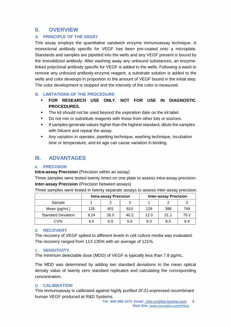
II. OVERVIEW A. PRINCIPLE OF THE ASSAY This assay employs the quantitative sandwich enzyme immunoassay technique. A monoclonal antibody specific for VEGF has been pre-coated onto a microplate. Standards and samples are pipetted into the wells and any VEGF present is bound by the immobilized antibody. After washing away any unbound substances, an enzyme- linked polyclonal antibody specific for VEGF is added to the wells. Following a wash to remove any unbound antibody-enzyme reagent, a substrate solution is added to the wells and color develops in proportion to the amount of VEGF bound in the initial step. The color development is stopped and the intensity of the color is measured.
B. LIMITATIONS OF THE PROCEDURE FOR RESEARCH USE ONLY. NOT FOR USE IN DIAGNOSTIC
PROCEDURES. The kit should not be used beyond the expiration date on the kit label. Do not mix or substitute reagents with those from other lots or sources. If samples generate values higher than the highest standard, dilute the samples
with Diluent and repeat the assay. Any variation in operator, pipetting technique, washing technique, incubation
time or temperature, and kit age can cause variation in binding.
III. ADVANTAGES A. PRECISION Intra-assay Precision (Precision within an assay) Three samples were tested twenty times on one plate to assess intra-assay precision. Inter-assay Precision (Precision between assays) Three samples were tested in twenty separate assays to assess inter-assay precision. Intra-assay Precision Inter-assay Precision
Sample 1 2 3 1 2 3 Mean (pg/mL) 128 401 810 129 388 749
Standard Deviation 8.24 26.0 40.2 12.0 31.1 70.2 CV% 6.4 6.5 5.0 9.3 8.0 9.4
B. RECOVERY The recovery of VEGF spiked to different levels in cell culture media was evaluated. The recovery ranged from 113-135% with an average of 121%.
C. SENSITIVITY The minimum detectable dose (MDD) of VEGF is typically less than 7.8 pg/mL.
The MDD was determined by adding two standard deviations to the mean optical density value of twenty zero standard replicates and calculating the corresponding concentration.
D. CALIBRATION This immunoassay is calibrated against highly purified Sf-21-expressed recombinant human VEGF produced at R&D Systems.
Tel: 800-988-1270 Email: [email protected] 4 Web Site: www.novusbio.com/china
E. LINEARITY To assess the linearity of the assay, four cell culture media samples were spiked with high concentrations of VEGF and diluted with Diluent 1× to produce samples with values within the dynamic range of the assay.
Dilution Average % of Expected Range (%) 1:2 100 96 – 104 1:4 101 95 – 108 1:8 102 94 – 111 1:16 101 88 – 110
F. SAMPLE VALUES Human peripheral blood mononuclear cells (1×10⁶ cells/mL) were cultured in RPMI supplemented with 5% fetal calf serum, 50 μM β-mercaptoethanol, 2 mM L-glutamine, 100 U/mL penicillin, and 100 μg/mL streptomycin sulfate. Cells were cultured unstimulated or stimulated 10 μg/mL PHA. Aliquots of the cell culture supernates was removed and assayed for levels of natural VEGF.
Condition Day 1 (pg/mL) Day 5 (pg/mL) Unstimulated 356 332 Stimulated 14 1440
G. SPECIFICITY This assay recognizes both natural and recombinant human VEGF. The following factors were prepared at 50 ng/mL and assayed for cross-reactivity. Preparations of the following factors at 50 ng/mL in a mid-range rhVEGF control were assayed for interference. No significant cross-reactivity or interference was observed.
Recombinant human: Recombinant mouse: ANG sgp130 IL-8 PDGF-AA IL-1α IL-13 AR GROα IL-10 PDGF-AB IL-1β MIP-1α CNTF GROβ IL-11 PDGF-BB IL-3 MIP-1β
β-ECGF EGF
GROγ IL-12 P/GF IL-4 P/GF-2
Epo HB-EGF IL-13 TGF-β1 IL-5 TNF-α FGF acidic HGF LAP (TGF-β1) TNF-α IL-6 VEGF120 FGF basic IL-1α M-CSF TNF R IL-9 VEGF164 FGF-4 IL-1β MCP-1 sTNF RII IL-10 VEGF-R3 FGF-5 IL-2 MIP-1α VEGF165/P/GF
FGF-6 IL-3 MIP-1β VEGF-B167
FGF-7 IL-4 β-NGF VEGF-C Recombinant rat: G-CSF IL-5 OSM VEGF-D VEGF
GM-CSF IL-6 PD-ECGF VEGF-R3
VEGF-related factors showing cross-reactivity or interference: • rhVEGF R1/Fc Chimera interfered at levels >500 pg/mL • rhVEGF R2/Fc Chimera interfered at levels >2000 pg/mL • rmVEGF R1 interfered at levels >500 pg/mL • rmVEGF R2 interfered at levels >4000 pg/mL • Canine VEGF showed 67% cross-reactivity
Tel: 800-988-1270 Email: [email protected] 5 Web Site: www.novusbio.com/china
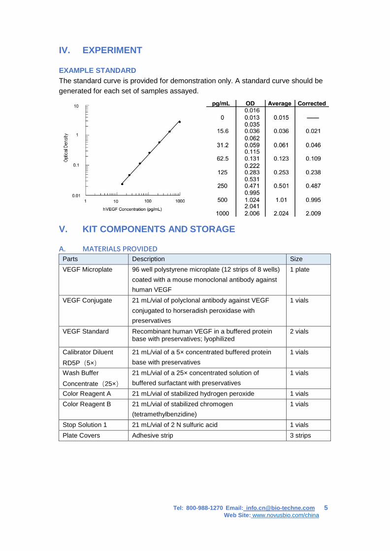
IV. EXPERIMENT EXAMPLE STANDARD The standard curve is provided for demonstration only. A standard curve should be generated for each set of samples assayed.
V. KIT COMPONENTS AND STORAGE
A. MATERIALS PROVIDED
Parts Description Size VEGF Microplate 96 well polystyrene microplate (12 strips of 8 wells)
coated with a mouse monoclonal antibody against human VEGF
1 plate
VEGF Conjugate 21 mL/vial of polyclonal antibody against VEGF conjugated to horseradish peroxidase with preservatives
1 vials
VEGF Standard Recombinant human VEGF in a buffered protein base with preservatives; lyophilized
2 vials
Calibrator Diluent RD5P(5×)
21 mL/vial of a 5× concentrated buffered protein base with preservatives
1 vials
Wash Buffer Concentrate(25×)
21 mL/vial of a 25× concentrated solution of buffered surfactant with preservatives
1 vials
Color Reagent A 21 mL/vial of stabilized hydrogen peroxide 1 vials Color Reagent B 21 mL/vial of stabilized chromogen
(tetramethylbenzidine) 1 vials
Stop Solution 1 21 mL/vial of 2 N sulfuric acid 1 vials Plate Covers Adhesive strip 3 strips
Tel: 800-988-1270 Email: [email protected] 6 Web Site: www.novusbio.com/china
B. STORAGE Unopened Kit
Store at 2-8℃. Do not use past kit expiration date.
Opened/ Reconstituted Reagents
Diluted Wash Solution
May be stored for up to 1 month at 2-8℃.*
Stop Solution 1 Dilution 1× Conjugate Unmixed Substrate A Unmixed Substrate B
Standard Aliquot and store for up to 1 month at <-20℃ in a manual defrost freezer.* Avoid repeated freezed-thaw cycles.
Microplate Wells
Return unused wells to the foil pouch containing the desiccant pack, reseal along entire edge of zip-seal. May be stored for up to 1 month at 2-
8℃.*
* Provided this is within the expiration date of the kit. C. OTHER SUPPLIES REQUIRED • Microplate reader capable of measuring absorbance at 450 nm, with the correction
wavelength set at 540 nm or 570 nm. • Pipettes and pipette tips. • Deionized or distilled water. • Squirt bottle, manifold dispenser, or automated microplate washer. • 500 mL graduated cylinder.
D. PRECAUTION • The Stop solution 1 provided with this kit is an acid solution. Wear eye, hand, face,
and clothing protection when using this material. • VEGF is detectable in saliva. Take the necessary precautions to prevent
contamination of the kit reagents.
VI. PREPARATION
A. SAMPLE COLLECTION AND STORAGE Cell Culture Supernates -Remove particulates by centrifugation and assay immediately or aliquot and store samples at ≤-20 ℃. Avoid repeated freeze-thaw cycles. Samples may require dilution with Diluent 1×.
B. REAGENT PREPARATION Note: Bring all reagents to room temperature before use.
Wash Solution - If crystals have formed in the concentrate, warm to room temperatureand mix gently until the crystals have completely dissolved. Dilute 20 mL of
Tel: 800-988-1270 Email: [email protected] 7 Web Site: www.novusbio.com/china
Wash Solution Concentrate into deionized or distilled water to prepare 500 mL of Wash Buffer.
Substrate Solution - Substrates A and B should be mixed together in equal volumes within 15 minutes of use. Protect from light. 200 μL of the resultant mixture is required per well.
Diluent 1× - Add 20 mL of Diluent Concentrate 5× into 80 mL of deionized or distilled water to prepare 100 mL of Diluent 1×.
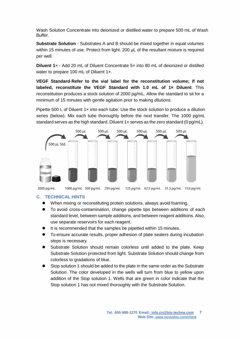
VEGF Standard-Refer to the vial label for the reconstitution volume; if not labeled, reconstitute the VEGF Standard with 1.0 mL of 1× Diluent. This reconstitution produces a stock solution of 2000 pg/mL. Allow the standard to sit for a minimum of 15 minutes with gentle agitation prior to making dilutions.
Pipette 500 L of Diluent 1× into each tube. Use the stock solution to produce a dilution series (below). Mix each tube thoroughly before the next transfer. The 1000 pg/mL standard serves as the high standard. Diluent 1× serves as the zero standard (0 pg/mL).
C. TECHNICAL HINTS When mixing or reconstituting protein solutions, always avoid foaming. To avoid cross-contamination, change pipette tips between additions of each
standard level, between sample additions, and between reagent additions. Also, use separate reservoirs for each reagent.
It is recommended that the samples be pipetted within 15 minutes. To ensure accurate results, proper adhesion of plate sealers during incubation
steps is necessary. Substrate Solution should remain colorless until added to the plate. Keep
Substrate Solution protected from light. Substrate Solution should change from colorless to gradations of blue.
Stop solution 1 should be added to the plate in the same order as the Substrate Solution. The color developed in the wells will turn from blue to yellow upon addition of the Stop solution 1. Wells that are green in color indicate that the Stop solution 1 has not mixed thoroughly with the Substrate Solution.
Tel: 800-988-1270 Email: [email protected] 8 Web Site: www.novusbio.com/china
VII. ASSAY PROCEDURE Note: Bring all reagents and samples to room temperature before use. It is recommended that all samples and standards be assayed in duplicate.
1. Prepare all reagents and working standards as directed in the previous sections.
2. Remove excess microplate strips from the plate frame, return them to the foil pouch containing the desiccant pack, and reseal.
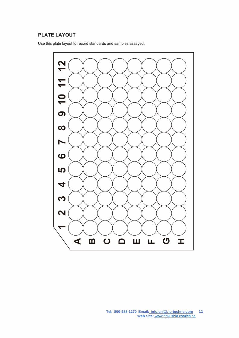
3. Add 100 μL of Standard, sample, or control per well. Cover with the adhesive strip provided. Incubate for 2 hours at room temperature. A plate layout is provided for a record of standards and samples assayed.
4. Aspirate each well and wash, repeating the process three times for a total of four washes. Wash by filling each well with Wash Solution (400 μL) using a squirt bottle, manifold dispenser, or autowasher. Complete removal of liquid at each step is essential to good performance. After the last wash, remove any remaining Wash Buffer by aspirating or decanting. Invert the plate and blot it against clean paper towels.
5. Add 200 μL of VEGF Conjugate to each well. Cover with a new adhesive strip. Incubate for 2 hours at room temperature.
6. Repeat the aspiration/wash as in step 4.
7. Add 200 μL of Substrate Solution to each well. Incubate for 30 minutes at room temperature. Protect from light.
8. Add 50 μL of Stop Solution 1 to each well. The color in the wells should change from blue to yellow. If the color in the wells is green or if the color change does not appear uniform, gently tap the plate to ensure thorough mixing.
9. Determine the optical density of each well within 30 minutes, using a microplate reader set to 450 nm. If wavelength correction is available, set to 540 nm or 570 nm. If wavelength correction is not available, subtract readings at 540 nm or 570 nm from the readings at 450 nm. This subtraction will correct for optical imperfections in the plate. Readings made directly at 450 nm without correction may be higher and less accurate.
10. CALCULATION OF RESULTS:Average the duplicate readings for each standard, control, and sample and subtract the average zero standard optical density. Create a standard curve by reducing the data using computer software capable of generating a four parameter logistic (4-PL) curve-fit. As an alternative, construct a standard curve by plotting the mean absorbance for each standard on the y-axis against the concentration on the x-axis and draw a best fit curve through the points on the graph. The data may be linearized by plotting the log of the VEGF concentrations versus the log of the O.D. and the best fit line can be determined by regression analysis. This procedure will produce an adequate but less precise fit of the data. If samples have been diluted, the concentration read from the standard curve must be multiplied by the dilution factor.
Tel: 800-988-1270 Email: [email protected] 9 Web Site: www.novusbio.com/china
VIII. RERERENCES
1. Leung, D.W. et al. (1989) Science 246:1306. 2. Keck, P.J. et al. (1989) Science 246:1309. 3. Byrne, A.M. et al. (2005) J. Cell. Mol. Med. 9:777. 4. Robinson, C.J. and Stringer, S.E. (2001) J. Cell. Sci. 114:853. 5. Richardson, R.S. et al. (1999) Am. J. Physiol. 277:H2247. 6. Sugishita, Y. et al. (2000) Biochem. Biophys. Res. Commun. 268:657. 7. Yamane, A. et al. (1994) Oncogene 9:2683. 8. Goad, D.L. et al. (1996) Endocrinology137:2262. 9. Gaudry, M. et al. (1997) Blood 90:4153. 10. Mclaren, J. et al. (1996) J. Clin. Invest. 98:482. 11. Diaz, B.V. et al. (2000) J. Biol. Chem. 275:642. 12. Asano, A. et al. (1997) Biochem. J. 328:179. 13. Bautz, F. et al. (2000) Exp. Hematol. 28:700. 14. Namiki, A. et al. (1995) J. Biol. Chem. 270:31189. 15. Nauck, M. et al. (1997) Am. J. Respir. Cell. Mol. Biol. 16:398. 16. Angelo, L.S. and R. Kurzrock (2007) Clin. Cancer Res. 13:2825. 17. Neufeld, G. et al. (1999) FASEB. J. 13:9. 18. Kowalewski, M.P. et al. (2005) Accession #ABB82619. 19. Pan, Q. et al. (2007) J. Biol. Chem. 282:24049. 20. Weis, S.M. and D.A. Cheresh (2005) Nature 437:497. 21. Breier, G. (2000) Semin. Thromb. Hemost. 26:553. 22. Barleon, B. et al. (1996) Blood 87:3336. 23. Weis, S.M. and D.A. Cheresh (2005) Nature 437:497. 24. Thurston, G. (2002) J. Anat. 200:575. 25. Grothey, A. and E. Galanis (2009) Nat. Rev. Clin. Oncol. 6:507. 26. Carvalho, J.F. et al. (2007) J. Clin. Immunol. 27:246.
Tel: 800-988-1270 Email: [email protected] 10 Web Site: www.novusbio.com/china
IX. TROUBLESHOOTING
Problem Probable Cause Suggestion No signal Failure to add all
components Prepare a check-list and add the components in the correct order
Low signal Not enough supernatant per well
Check the protein concentration. Add more supernatant.
High background Wells are not washed enough.
Wash plates thoroughly after incubation with detecting.
Tel: 800-988-1270 Email: [email protected] 11 Web Site: www.novusbio.com/china
产品信息及操作手册
人 VEGF ValukineTM ELISA 试剂盒
目录号:VAL106
适用于定量测定细胞培养上清液中人血管内皮生长因子(VEGF)的含量
科研专用,不可用于临床诊断
Bio-Techne China Co. Ltd
P: +86 (21) 52380373 P: 8009881270 F: +86 (21) 52381001 [email protected]
Novus 试剂盒确保在你收货日期 3 个月内有效
Tel: 800-988-1270 Email: [email protected] 13 Web Site: www.novusbio.com/china
目录
I. 背景 ................................................................ 14
II. 概述 ................................................................ 14
III. 优势 ................................................................ 15
IV. 实验 ................................................................ 16
V. 试剂盒组成及储存 .................................................... 17
VI. 实验前准备 .......................................................... 18
VII. 操作步骤 ............................................................ 19
VIII.参考文献 ............................................................ 20
IX. 疑难解答 ............................................................ 21
Tel: 800-988-1270 Email: [email protected] 14 Web Site: www.novusbio.com/china
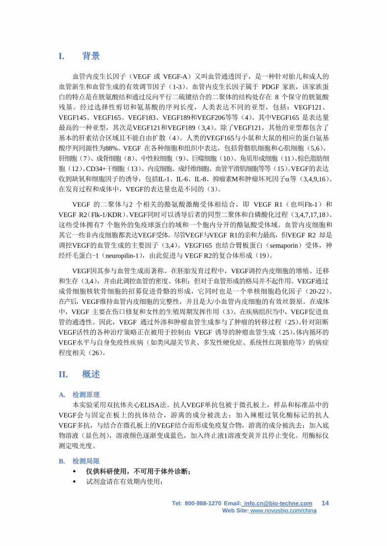
I. 背景
血管内皮生长因子(VEGF 或 VEGF-A)又叫血管通透因子,是一种针对胎儿和成人的
血管新生和血管生成的有效调节因子(1-3)。血管内皮生长因子属于 PDGF 家族,该家族蛋
白的特点是在胱氨酸结和通过反向平行二硫键结合的二聚体的结构处存在 8 个保守的胱氨酸
残基。经过选择性剪切和氨基酸的序列长度,人类表达不同的亚型,包括:VEGF121、VEGF145、VEGF165、VEGF183、VEGF189和VEGF206等等(4)。其中VEGF165 是表达量
最高的一种亚型,其次是VEGF121和VEGF189(3,4)。除了VEGF121,其他的亚型都包含了
基本的肝素结合区域且不能自由扩散(4)。人类的VEGF165与小鼠和大鼠的相应的蛋白氨基
酸序列同源性为88%。VEGF 在各种细胞和组织中表达,包括骨骼肌细胞和心肌细胞(5,6)、肝细胞(7)、成骨细胞(8)、中性粒细胞(9)、巨噬细胞(10)、角质形成细胞(11)、棕色脂肪细
胞(12)、CD34+干细胞(13)、内皮细胞、成纤维细胞、血管平滑肌细胞等等(15)。VEGF的表达
收到缺氧和细胞因子的诱导,包括IL-1、IL-6、IL-8、抑瘤素M和肿瘤坏死因子α等(3,4,9,16)。在发育过程和成体中,VEGF的表达量也是不同的(3)。
VEGF 的二聚体与2 个相关的酪氨酸激酶受体相结合,即 VEGF R1(也叫Flt-1)和
VEGF R2(Flk-1/KDR)。VEGF同时可以诱导后者的同型二聚体和自磷酸化过程(3,4,7,17,18)。这些受体拥有7 个胞外的免疫球蛋白的域和一个胞内分开的酪氨酸受体域。血管内皮细胞和
其它一些非内皮细胞都表达VEGF受体。尽管VEGF与VEGF R1的亲和力最高,但VEGF R2 却是
调控VEGF的血管生成的主要因子(3,4)。VEGF165 也结合臂板蛋白(semaporin)受体,神
经纤毛蛋白-1(neuropilin-1),由此促进与 VEGF R2的复合体形成(19)。
VEGF因其参与血管生成而著称。在胚胎发育过程中,VEGF调控内皮细胞的增殖、迁移
和生存(3,4),并由此调控血管的密度、体积;但对于血管形成的格局并不起作用。VEGF通过
成骨细胞核软骨细胞的招募促进骨骼的形成,它同时也是一个单核细胞趋化因子(20-22)。在产后,VEGF维持血管内皮细胞的完整性,并且是大/小血管内皮细胞的有效丝裂原。在成体
中,VEGF 主要在伤口修复和女性的生殖周期发挥作用(3)。在疾病组织当中,VEGF促进血
管的通透性。因此,VEGF 通过外渗和肿瘤血管生成参与了肿瘤的转移过程(25)。针对阻断
VEGF活性的各种治疗策略正在被用于控制由 VEGF 诱导的肿瘤血管生成(25)。体内循环的
VEGF水平与自身免疫性疾病(如类风湿关节炎、多发性硬化症、系统性红斑狼疮等)的病症
程度相关(26)。
II. 概述
A. 检测原理 本实验采用双抗体夹心ELISA法。抗人VEGF单抗包被于微孔板上,样品和标准品中的
VEGF会与固定在板上的抗体结合,游离的成分被洗去;加入辣根过氧化酶标记的抗人
VEGF多抗,与结合在微孔板上的VEGF结合而形成免疫复合物,游离的成分被洗去;加入底
物溶液(显色剂),溶液颜色逐渐变成蓝色,加入终止液1溶液变黄并且停止变化。用酶标仪
测定吸光度。
B. 检测局限 仅供科研使用,不可用于体外诊断;
试剂盒请在有效期内使用;
Tel: 800-988-1270 Email: [email protected] 15 Web Site: www.novusbio.com/china
不同试剂盒及不同批号试剂盒的组分不能混用;
样本值若大于标准曲线的最高值,应将样本用稀释剂(1×)稀释后重新检测;若细
胞培养上清液样本需分布稀释,除最后一步用稀释剂稀释外,其它中间稀释可采用细
胞培养基;
检测结果的不同可由多种因素引起,包括实验人员的操作、移液器的使用方式、洗板
技术、反应时间或温度、试剂盒的储存等。
III. 优势
A. 精确度
板内精确度(同一板内不同孔间的精确度)
已知浓度的三个样本,在同一板内分别检测20次,以确定板内精确度。
板间精确度(不同板之间的精确度)
已知浓度的三个样本,在不同板间分别检测20次,以确定板内精确度。
板内精确度 板间精确度
样本 1 2 3 1 2 3
平均值 (pg/mL) 128 401 810 129 388 749
标准差 8.24 26.0 40.2 12.0 31.1 70.2
CV% 6.4 6.5 5.0 9.3 8.0 9.4
B. 回收率
在细胞培养基样本中掺入不同水平的人VEGF,测定其回收率。回收率范围在113-135%,平
均回收率在121%。
C. 灵敏度
人VEGF 的最低可测值一般小于 7.8 pg/mL。
最低可测值是根据20个标准曲线零点吸光值的平均值加两倍标准差计算得到的相对应浓度。
D. 校正
此ELISA试剂盒经由R&D Systems 生产的大肠杆菌表达的高纯重组人VEGF 蛋白所校正。
E. 线性
4个不同的样本中掺入高浓度的人VEGF,然后用稀释剂(1×)将样本稀释到检测范围内,
测定其线性。
稀释倍数 平均值/期待值(%) 范围 (%)
1:2 100 96 – 104
1:4 101 95 – 108
1:8 102 94 – 111
1:16 101 88 – 110
F. 样本预值
人的外周血单核细胞(1×10⁶ 细胞/mL)培养于含有5%胎牛血清的RPMI1640 培养基中,细
胞培养基还含有2 mM L-谷氨酰胺、50 μM β-巯基乙醇、100 U/mL青霉素, 100 μg/mL 链霉素;不
刺激细胞,或加10 μg/mL PHA 刺激细胞,取细胞上清液测定 VEGF 含量,结果见下表。
Tel: 800-988-1270 Email: [email protected] 16 Web Site: www.novusbio.com/china
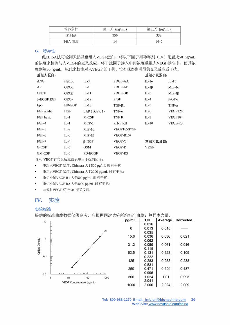
培养条件 第一天 (pg/mL) 第五天 (pg/mL)
未刺激 356 332
PHA 刺激 14 1440
G. 特异性
此ELISA法可检测天然及重组人VEGF蛋白。将以下因子用稀释剂(1×)配置成50 ng/mL 的浓度来检测与人VEGF的交叉反应。将干扰因子掺入中间浓度重组人VEGF标准中,使其浓
度到达50 ng/mL,以此来检测对人VEGF 的干扰。没有观察到明显的交叉反应或干扰。
重组人蛋白: 重组小鼠蛋白:
ANG sgp130 IL-8 PDGF-AA IL-1α IL-13
AR GROα IL-10 PDGF-AB IL-1β MIP-1α
CNTF GROβ IL-11 PDGF-BB IL-3 MIP-1β
β-ECGF EGF GROγ IL-12 P/GF IL-4 P/GF-2
Epo HB-EGF IL-13 TGF-β1 IL-5 TNF-α
FGF acidic HGF LAP (TGF-β1) TNF-α IL-6 VEGF120
FGF basic IL-1 M-CSF TNF R IL-9 VEGF164
FGF-4 IL-1 MCP-1 sTNF RII IL-10 VEGF-R3
FGF-5 IL-2 MIP-1α VEGF165/P/GF
FGF-6 IL-3 MIP-1β VEGF-B167
FGF-7 IL-4 β-NGF VEGF-C 重组大鼠蛋白:
G-CSF IL-5 OSM VEGF-D VEGF
GM-CSF IL-6 PD-ECGF VEGF-R3
与人 VEGF 有交叉反应或表现出干扰的因子:
• 重组人VEGF R1/Fc Chimera 大于500 pg/mL 时有干扰;
• 重组人VEGF R2/Fc Chimera 大于2000 pg/mL 时有干扰;
• 重组小鼠VEGF R1 大于500 pg/mL 时有干扰;
• 重组小鼠VEGF R2 大于4000 pg/mL 时有干扰;
• 与犬科VEGF 有67%的交叉反应.
IV. 实验 实验标准
提供的标准曲线数据仅供参考,应根据同次试验所绘标准曲线计算样本含量。
Tel: 800-988-1270 Email: [email protected] 17 Web Site: www.novusbio.com/china
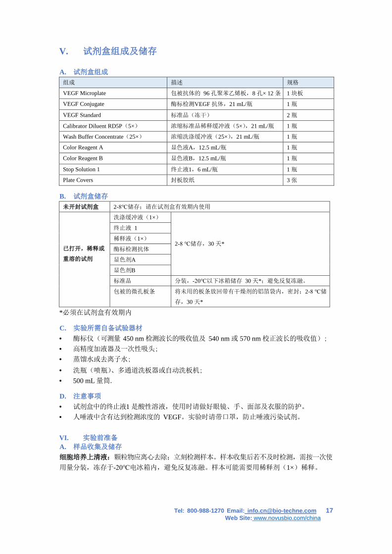
V. 试剂盒组成及储存
A. 试剂盒组成
组成 描述 规格
VEGF Microplate 包被抗体的 96 孔聚苯乙烯板,8 孔× 12 条 1 块板
VEGF Conjugate 酶标检测VEGF 抗体,21 mL/瓶 1 瓶
VEGF Standard 标准品(冻干) 2 瓶
Calibrator Diluent RD5P(5×) 浓缩标准品稀释缓冲液(5×),21 mL/瓶 1 瓶
Wash Buffer Concentrate(25×) 浓缩洗涤缓冲液(25×),21 mL/瓶 1 瓶
Color Reagent A 显色液A,12.5 mL/瓶 1 瓶
Color Reagent B 显色液B,12.5 mL/瓶 1 瓶
Stop Solution 1 终止液1,6 mL/瓶 1 瓶
Plate Covers 封板胶纸 3 张
B. 试剂盒储存
未开封试剂盒 2-8℃储存;请在试剂盒有效期内使用
已打开,稀释或
重溶的试剂
洗涤缓冲液(1×)
2-8 ℃储存,30 天*
终止液 1
稀释液(1×)
酶标检测抗体
显色剂A
显色剂B
标准品 分装,-20℃以下冰箱储存 30 天*;避免反复冻融。
包被的微孔板条 将未用的板条放回带有干燥剂的铝箔袋内,密封;2-8 ℃储
存,30 天*
*必须在试剂盒有效期内
C. 实验所需自备试验器材
• 酶标仪(可测量 450 nm 检测波长的吸收值及 540 nm 或 570 nm 校正波长的吸收值);
• 高精度加液器及一次性吸头;
• 蒸馏水或去离子水;
• 洗瓶(喷瓶)、多通道洗板器或自动洗板机;
• 500 mL 量筒.
D. 注意事项
• 试剂盒中的终止液1 是酸性溶液,使用时请做好眼镜、手、面部及衣服的防护。
• 人唾液中含有达到检测浓度的 VEGF。实验时请带口罩,防止唾液污染试剂。
VI. 实验前准备 A. 样品收集及储存
细胞培养上清液:颗粒物应离心去除;立刻检测样本。样本收集后若不及时检测,需按一次使
用量分装,冻存于-20℃电冰箱内,避免反复冻融。样本可能需要用稀释剂(1×)稀释。
Tel: 800-988-1270 Email: [email protected] 18 Web Site: www.novusbio.com/china
B. 检测前准备工作
使用前请将所有试剂放置于室温
洗涤液:从冰箱中取出的浓缩洗涤液可能有结晶,属于正常现象;放置室温,轻摇混匀,待结
晶完成溶解后再配制洗涤液。可将20 mL 浓缩洗涤液用蒸馏水或去离子水稀释配置成
500mL 工作浓缩的洗涤液。未用完的放回4℃。
显色剂:按当次试验所需要用量将显色剂A和显色剂B 等体积混合,避光;在使用前15 分钟准
备,仅供当日使用;每孔需100 μL。
稀释剂(1×):可将20 mL浓缩稀释剂用 80mL 蒸馏水或去离子水稀释配置成100 mL工作浓
度的稀释剂。
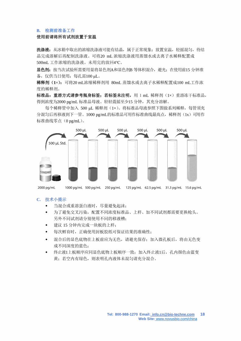
标准品:重溶方式请参考瓶身标签;若标签未注明,用 1 mL 稀释剂(1×)重溶冻干标准品,
得到浓度为2000 pg/mL 标准品母液。轻轻震摇至少15 分钟,其充分溶解。
每个稀释管中加入 500 μL 稀释剂(1×)。将标准品母液参照下图做系列稀释,每管须充
分混匀后再移液到下一管。1000 pg/mL的标准品可用作标准曲线最高点,稀释剂(1x)可用作
标准曲线零点(0 pg/mL)。
C. 技术小提示
当混合或重溶蛋白液时,尽量避免起沫;
为了避免交叉污染,配置不同浓度标准品、上样、加不同试剂都需要更换枪头。
另外不同试剂请分别使用不同的移液槽;
建议 15 分钟内完成一块板的上样;
每次孵育时,正确使用封板胶纸可保证结果的准确性;
混合后的显色底物在上板前应为无色,请避光保存;加入微孔板后,将由无色变
成不同深度的蓝色;
终止液1上板顺序应同显色底物上板顺序一致;加入终止液1后,孔内颜色由蓝变
黄;若空内有绿色,则表明孔内液体未混匀请充分混合。
Tel: 800-988-1270 Email: [email protected] 19 Web Site: www.novusbio.com/china
VII. 操作步骤 使用前请将所有试剂和样本放置于室温,建议所有的实验样本和标准品做复孔检测。
1. 按照上一节的说明,准备好所有需要的试剂和标准品;
2. 从已平衡至室温的密封袋中取出微孔板,未用的板条请放回铝箔袋内,重新封口;
3. 分别将不同浓度标准品,实验样本或者质控品加入相应孔中,每孔100 μL。用封板胶纸封
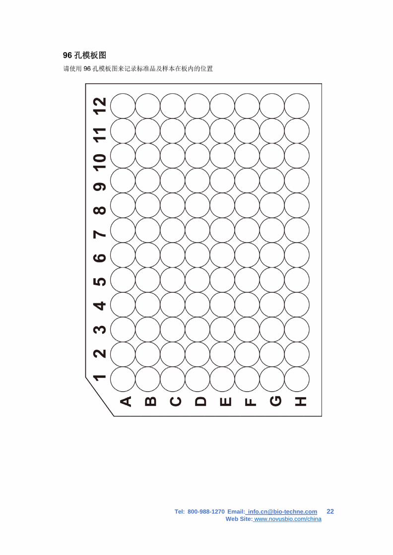
住反应孔,室温孵育2小时。说明书提供了一张 96 孔模板图,可用于记录标准品和试验
样本的板内位置;
4. 将板内液体吸去,使用洗瓶、多通道洗板器或自动洗板机洗板。每孔加洗涤液400 μL,然
后将板内洗涤液吸去。重复操作4次。每次洗板尽量吸去残留液体会有助于得到好的实验结
果。最后一次洗板结束,请将板内所有液体吸干或将板倒置,在吸水纸拍干所有残留液体;
5. 在每个微孔内加入200 μL 酶标检测抗体。用封板胶纸封住反应孔,室温孵育2 小时;
6. 重复第4 步洗板操作;
7. 在每个微孔内加入200 μL 显色底物,室温孵育30 分钟。注意避光;
8. 在每个微孔内加入50 μL 终止液1,孔内溶液颜色会从蓝色变为黄色。如果溶液颜色变为
绿色或者颜色变化不一致,请轻拍微孔板,使溶液混合均匀;
9. 加入终止液1后 30分钟内,使用酶标仪测量450 nm 的吸光度值,设定540 nm 或 570 nm 作为校正波长。如果没有使用双波长校正,结果准确度可能会受影响;
10.计算结果:每一个标准品及样本的复孔校正吸收光值(OD450-OD540/OD570)取平均
值,再减去标准曲线零点的OD值。利用酶标仪携带的软件,绘制一个4参数(4-PL)线性
标准曲线,曲线横坐标为标准曲线点的人VEGF浓度值,纵坐标为标准曲线点的OD平均值。
通过样本的OD值,可从标准曲线上得到样本中人VEGF的浓度。若样本经过稀释,计算浓
度时应乘以稀释倍数。
Tel: 800-988-1270 Email: [email protected] 20 Web Site: www.novusbio.com/china
VIII. 参考文献 1. Leung, D.W. et al. (1989) Science 246:1306. 2. Keck, P.J. et al. (1989) Science 246:1309. 3. Byrne, A.M. et al. (2005) J. Cell. Mol. Med. 9:777. 4. Robinson, C.J. and Stringer, S.E. (2001) J. Cell. Sci. 114:853. 5. Richardson, R.S. et al. (1999) Am. J. Physiol. 277:H2247. 6. Sugishita, Y. et al. (2000) Biochem. Biophys. Res. Commun. 268:657. 7. Yamane, A. et al. (1994) Oncogene 9:2683. 8. Goad, D.L. et al. (1996) Endocrinology137:2262. 9. Gaudry, M. et al. (1997) Blood 90:4153. 10. Mclaren, J. et al. (1996) J. Clin. Invest. 98:482. 11. Diaz, B.V. et al. (2000) J. Biol. Chem. 275:642. 12. Asano, A. et al. (1997) Biochem. J. 328:179. 13. Bautz, F. et al. (2000) Exp. Hematol. 28:700. 14. Namiki, A. et al. (1995) J. Biol. Chem. 270:31189. 15. Nauck, M. et al. (1997) Am. J. Respir. Cell. Mol. Biol. 16:398. 16. Angelo, L.S. and R. Kurzrock (2007) Clin. Cancer Res. 13:2825. 17. Neufeld, G. et al. (1999) FASEB. J. 13:9. 18. Kowalewski, M.P. et al. (2005) Accession #ABB82619. 19. Pan, Q. et al. (2007) J. Biol. Chem. 282:24049. 20. Weis, S.M. and D.A. Cheresh (2005) Nature 437:497. 21. Breier, G. (2000) Semin. Thromb. Hemost. 26:553. 22. Barleon, B. et al. (1996) Blood 87:3336. 23. Weis, S.M. and D.A. Cheresh (2005) Nature 437:497. 24. Thurston, G. (2002) J. Anat. 200:575. 25. Grothey, A. and E. Galanis (2009) Nat. Rev. Clin. Oncol. 6:507. 26. Carvalho, J.F. et al. (2007) J. Clin. Immunol. 27:246. 32. Engelmann, H. et al. (1990) J. Biol. Chem. 265:1531.
Tel: 800-988-1270 Email: [email protected] 21 Web Site: www.novusbio.com/china
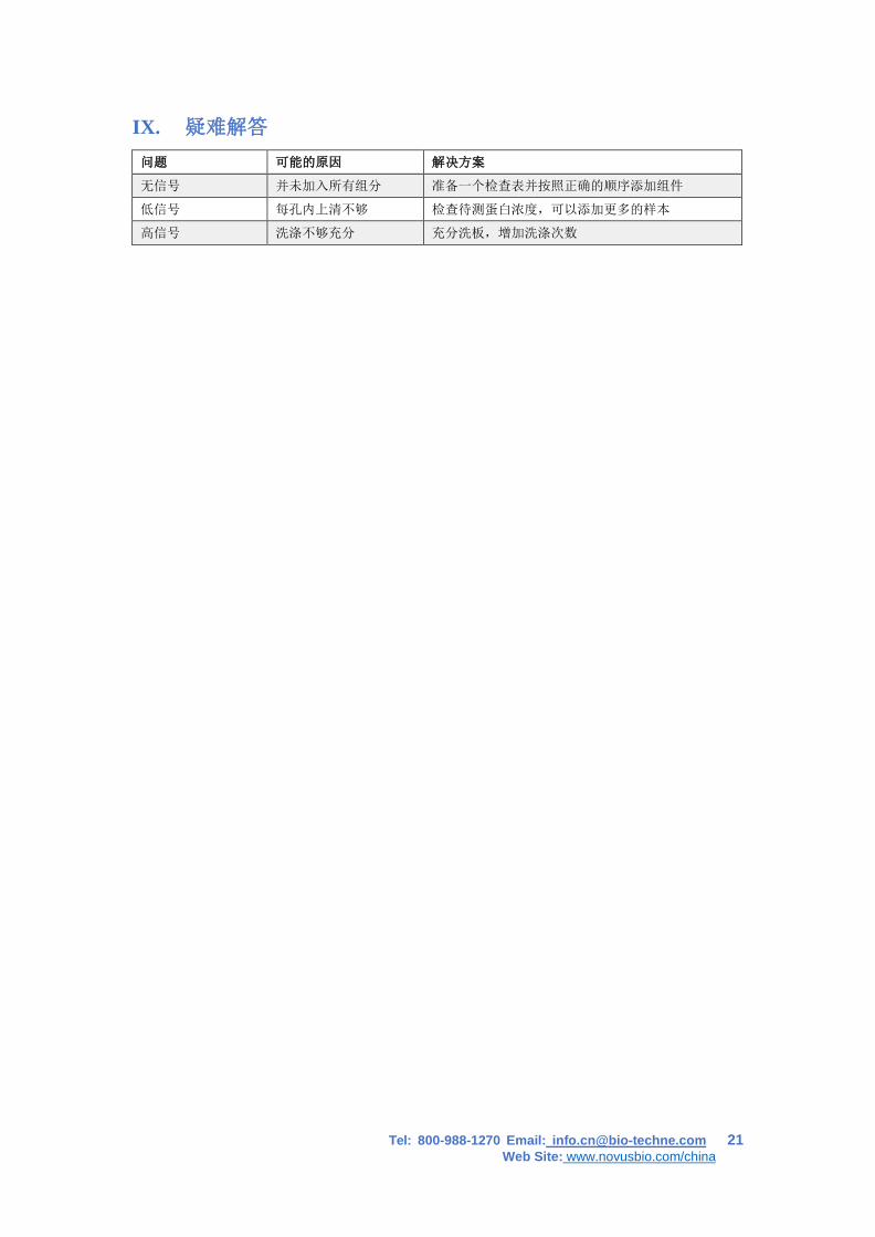
IX. 疑难解答
问题 可能的原因 解决方案
无信号 并未加入所有组分 准备一个检查表并按照正确的顺序添加组件
低信号 每孔内上清不够 检查待测蛋白浓度,可以添加更多的样本
高信号 洗涤不够充分 充分洗板,增加洗涤次数
Tel: 800-988-1270 Email: [email protected] 22 Web Site: www.novusbio.com/china