Produced by the - Science | AAAS in cell culture techniques have allowed for the more skillful...
36
Produced by the Science/AAAS Custom Publishing Office Sponsored by
Transcript of Produced by the - Science | AAAS in cell culture techniques have allowed for the more skillful...

Produced by the Science/AAAS Custom
Publishing Office
Sponsored by

Introductions 2 The Future is Now Sean Sanders
3 Inspiration for Life Science Cosmo Bio Co., Ltd.
Articles 4 Isolation of Single Human Hematopoietic Stem Cells Capable of Long-Term Multilineage Engraftment Faiyaz Notta, Sergei Doulatov, Elisa Laurenti et al. Science 8 July 2011, p. 218
10 Interconversion Between Intestinal Stem Cell Populations in Distinct Niches Norifumi Takeda, Rajan Jain, Matthew R. LeBoeuf et al. Science 9 December 2011, p. 1420
17 Pluripotent Stem Cell–Based Cancer Therapy: Promise and Challenges Saul J. Sharkis, Richard J. Jones, Curt Civin et al. Science Translational Medicine 28 March 2012, ps. 9
22 Direct Conversion of C. elegans Germ Cells intoSpecificNeuronTypes Baris Tursun, Tulsi Patel, Paschalis Kratsios et al. Science 21 January 2011, p. 304
32 Technical Note
Contents
Editor: Sean Sanders, Ph.D.; Proofing: Yuse Lajiminmuhip; Design: Amy Hardcastle
© 2012 by The American Association for the Advancement of Science. All rights reserved.22 June 2012
Cover Image: From Science 26 June 2009, Oded Kopper and Nissim Benvenisty,
The Hebrew University of Jerusalem
1

The advances in our knowledge and understanding of stem cells have fundamentally changed
the way we see human biology and physiology, not to mention disease. It has become clear in recent years that not only are our bodies more plastic than previously thought, but the num-ber and type of stem cells that play a role in the generation and regenera-tion of human tissue far exceed origi-nal expectations.
In order to characterize stem cells, they must first be identified. Markers for many different stem cell popula-tions have been elucidated, but chal-lenges still remain. The papers pre-sented in this booklet by Notta et al. (p. 4) and Takeda et al. (p. 10) high-light these hurdles. For instance, in Takeda’s words, “The location and identity of ISCs [intestinal stem cells] have been a subject of much research and debate…” The identification of the precise location of these stem cells, which support the rapid replacement of cells in the intestine, is integral to understanding both normal gut ho-meostasis, as well as what occurs dur-ing disease, particularly cancer. Notta and colleagues take on a similar chal-lenge with hematopoietic stem cells (HSCs) as they demonstrate that a specific adhesion protein, CD49f, can be used to separate true HSCs from their multipotent progeny.
Embryonic stem cells (ESCs), as well as multipotent and unipotent stem and stem-like cells, all hold promise for potential disease treatments. In-duced pluripotent stem cells (iPSCs) that have been generated in the lab by genetic reprogramming of a somatic
cell have the advantage that they are near identical to ESCs, but can be artificially created and therefore cir-cumvent many of the ethical concerns with the extraction and use of ESCs. In a recent paper published in Science Translational Medicine, Sharkis et al. (p. 17) outline potential applications for iPSCs in disease therapy and regen-erative medicine, while also remaining realistic about residual impediments to their broad clinical application, particu-larly safety concerns.
Advances in cell culture techniques have allowed for the more skillful manipulation of stem cells in vitro to generate almost any differentiated cell type required. The balance is delicate, but once achieved, stem cells can be bent to the will of the researcher to create any number of lineages for fur-ther study or even transplantation. On page 22, Tursen et al. describe one such example, in which they use just a single transcription factor to gener-ate a number of differentiated neuro-nal cell types from mitotic germ cells in C. elegans.
The field of regenerative medicine is moving forward at an increasingly rapid pace. As in vitro cultured cells for human treatment become a real-ity, and the techniques for recreating even complex organs becomes pos-sible, the opportunity to apply stem cells to the treatment of many different diseases and conditions seems more attainable than ever.
Sean Sanders, Ph.D.Editor, Custom PublishingScience
2
The Future is Now

INTRO
3
5

4
Mature blood cell lineages are gener-ated from a network of hierarchi-cally distinct progenitors that arise
from self-renewing hematopoietic stem cells (HSCs). The extensive regenerative poten-tial of HSCs makes them attractive targets for cellular and genetic therapies. The mo-lecular regulation of specific HSC properties such as long-term self-renewal is beginning to be elucidated for murine HSCs (1). How-ever the biology of human HSCs remains poorly understood because of their rarity and the lack of methods to segregate HSCs from multipotent progenitors (MPPs) to obtain pure populations for biological and molecu-lar analysis.
The bulk of HSCs are CD34+, as evidenced by human transplantation and xenograft re-population assays; however, most CD34+
cells are lineage-restricted progenitors and HSCs remain rare. HSCs can be enriched fur-ther on the basis of CD45RA (2), Thy1 (3–5), and CD38 (6, 7) expression. Loss of Thy1 expression in the CD34+CD38−CD45RA− compartment of lineage-depleted cord blood (CB) was recently proposed to be sufficient to separate HSCs from MPPs (5). However, more than a third of Thy1− primary recipi-ents gave rise to engraftment in secondary animals, raising uncertainty about whether Thy1 can absolutely segregate HSCs from MPPs. To resolve the relationship between these two subsets, the number of cells in each subset that are capable of short-term and long-term engraftment must be quantified at clonal resolution. We recently optimized the HSC xenograft assay by using intrafemoral injection into female NOD-scid-IL2Rgc−/− (NSG) mice (8–10). Flow-sorted CB HSCs (CD34+CD38−CD45RA−Thy1+; Thy1+) (Fig. 1, P5) and MPPs (CD34+CD38−CD45RA−
Thy1−; Thy1−) (Fig. 1, P2) fractions were functionally characterized with our HSC assay. A priori, HSCs were operationally defined by lymphomyeloid engraftment that persisted for at least 20 weeks after trans-plant. This duration represents a stringent test of long-term repopulation and encom-passes the total engraftment time of primary and secondary transplants historically used to assess the self-renewal capacity of human
Isolation of Single Human Hematopoietic Stem Cells Capable of Long-Term Multilineage EngraftmentFaiyaz Notta,1,2* Sergei Doulatov,1,2* Elisa Laurenti,1,2 Armando Poeppl,1Igor Jurisica,3,4 John E. Dick1,2†
1Division of Stem Cell and Developmental Biology, Campbell Family Institute for Cancer Research/Ontario Cancer Institute, Toronto, Ontario, Canada. 2Department of Molecular Genetics, University of Toronto, Toronto, Ontario, Canada. 3Ontario Cancer Institute and Campbell Family Institute for Cancer Research, Toronto, Ontario, Canada. 4Departments of Computer Science and Medical Biophysics, University of Toronto, Toronto, Ontario, Canada.
*These authors contributed equally to this work.†To whom correspondence should be addressed. Toronto Medical Discovery Tower, Room 8-301, 101 College Street, Toronto, Canada M5G 1L7. E-mail: [email protected]
Lifelong blood cell production is dependent on rare hematopoietic stem cells (HSCs) to perpetually replenish mature cells via a series of lineage-restricted intermediates. Investi-gating the molecular state of HSCs is contingent on the ability to purify HSCs away from transiently engrafting cells. We demonstrated that human HSCs remain infrequent, using current purification strategies based on Thy1 (CD90) expression. By tracking the expres-sion of several adhesion molecules in HSC-enriched subsets, we revealed CD49f as a specific HSC marker. Single CD49f+ cells were highly efficient in generating long-term multilineage grafts, and the loss of CD49f expression identified transiently engrafting multipotent progenitors (MPPs). The demarcation of human HSCs and MPPs will enable the investigation of the molecular determinants of HSCs, with a goal of developing stem cell–based therapeutics.

5
HSCs in xenograft models. At nonlimiting cell doses, recipients of Thy1+ and Thy1− cells had similar levels of human chimerism and lineage distribution (injected femur: P = 0.17; Fig. 2, A and B; fig. S1; and table S1). To assess whether Thy1− cells would persist beyond the allotted 20-week primary trans-plant period, we performed secondary trans-plants for an additional 12 to 14 weeks. This revealed that Thy1− cells could be serially transplanted, albeit with lower efficiency than Thy1+ cells (table S2), which is consis-tent with previous work (5). These data sug-
gest that cells with extensive self-renewal potential exist in both Thy1+ and Thy1− sub-sets, although the basis for the disparity in secondary transfer efficiency between these subsets remained unknown.
We next performed limiting dilution analy-sis (LDA) to measure the frequency of HSCs within Thy1+ and Thy1− fractions. One in 20 Thy1+ cells (5%) clonally initiated long-term hematopoiesis in NSG mice as compared to 1 in 100 (1%) Thy1− cells (P = 0.0003, Fig. 2C). Double sorting and high-stringency sort modes used in our experimental design
Figure 2. Functional char-acterization of the Thy1+ and Thy1− subsets. (A and B) Engraftment and lineage potential of Thy1+ (P5) and Thy1− (P2) cells assessed in NSG mice 20 weeks after trans-plant (Thy1+: n = 65 mice; Thy1−: n = 30 mice, 6 ex-periments). IF, injected femur; BM, left femur, tibiae; SP, spleen; TH, thymus. (C) Frequency of long-term repopulat-ing cells within Thy1+ and Thy1− populations mea-sured by LDA. (D) Phe-notype of Thy1+ (left) and Thy1− (right) cells cultured on OP9 stroma. Plots are gated on CD34+CD38− cells (figs. S4 and S14). (E) Phenotype of Thy1+ and Thy1− cells engrafted in NSG mice. (F) Popula-tions labeled + and – from (D) were isolated from stroma and transplanted into NSG mice (200 to 400 cells, two experiments, n = 10 mice per group). Their engraftment po-tential (as percent of CD45+ cells) is shown in the bottom panel. All data are presented as means ± SEM. PB, peripheral blood.
Figure 1. Cell sorting scheme used to isolate human HSCs and MPPs. Lin− CB cells were stained with monoclonal anti-bodies against CD34, CD38, CD45RA, Thy1 (CD90), CD49f antigens, and the mitochondrial membrane dye Rho. The fre-quency of each subpopulation (P1 to P10) is based on the par-ent gate shown on the top right of each plot.

6
ruled out the possibility that HSC activity from Thy1− cells was due to residual con-tamination from Thy1lo/+ cells (figs. S2 and S3). The inability of prior studies to detect engraftment from Thy1− cells was probably due to the less sensitive xenograft models employed (3). Thus, although Thy1+ enrich-es for HSCs, long-term repopulating activity persists in the Thy1− fraction previously be-lieved to represent MPPs (5).
To examine the hierarchical relationship between the Thy1 subsets, we cultured sort-ed Thy1+ and Thy1− cells with stroma cells known to express HSC-supportive ligands (11). Both Thy1+ and Thy1− cells (>70%) remained CD34+CD38− on stromal cultures (fig. S4, column 3). Unexpectedly, Thy1− cells consistently generated Thy1+ cells on stroma (Fig. 2D, right panel) and also in vivo within the bone marrow microenvironment of NSG mice that received transplants (Fig. 2E). Thy1+ cells arising from Thy1− cells after culture, as well as Thy1+ cells that re-tained their Thy1 expression, had robust re-populating activity in NSG mice 20 weeks after transplantation. Engraftment and lin-eage potentials were identical for Thy1+ cells derived from either Thy1 subfraction (Fig. 2F and fig. S5). Cells that remained Thy1− after being cultured on OP9 stroma did not sustain a long-term graft (Fig. 2F, right pan-el); however, they transiently repopulated (fig. S6). These results demonstrate that the Thy1− compartment is heterogeneous and contains a small fraction with repopulating activity and a larger fraction with MPP-like activity.
To further purify HSCs in both Thy1+ and Thy1− subsets, we searched for a different cell surface marker. We hypothesized that integrins would mark human HSCs, because they mediate niche interactions and have been used to isolate murine HSCs and other somatic stem cells (12, 13). We compared the surface expression of several adhesion molecules between HSC-enriched (Thy1+) and -depleted (Thy1−) fractions. Among our candidates, only ITGA6 (integrin α6, termed CD49f) was differentially expressed (Fig. 3A and fig. S7), with 50 to 70% of Thy1+ cells expressing CD49f versus 10 to 20% of Thy1− cells.
To determine whether human HSCs could
be delineated using CD49f expression, we partitioned Thy1+ cells into CD49f+/hi (here called Thy1+CD49f+; Fig. 1, P9) and CD-49flo/− (here called Thy1+CD49f−; Fig. 1, P8) subfractions and evaluated their capacity for long-term multilineage chimerism in NSG recipients. Mean chimerism in the injected femur was 6.7-fold higher for Thy1+CD49f+ than for Thy1+CD49f− cells (22.7% versus 3.4%, P < 0.0001, Fig. 3B, left panel), and only Thy1+CD49f+ cells could be serially transplanted (table S3). LDA revealed that 9.5% (1 in 10.5) of Thy1+CD49f+ cells had long-term repopulating activity as compared with 0.9% (1 in 111.3) Thy1+CD49f− cells (P = 9.9 × 10⁻⁹, Fig. 3C, and tables S1 and S4). Because we had found that the Thy1− frac-tion was heterogeneous, we tested whether CD49f expression also marked Thy1− HSCs. Indeed, only Thy1−CD49f+ cells reconstitut-ed NSG mice 20 weeks after transplant (Fig. 3B, right panel). LDA indicated that approxi-mately 4.5% (1 in 22.1) of cells in this frac-tion had long-term multilineage engraftment potential as compared to 0.13% (1 in 735.2) of Thy1−CD49f− cells (Fig. 3C and table S4). No difference in lineage potential was observed between Thy1+CD49f+ and Thy1−
CD49f+ cells (fig. S8), although recipients of Thy1+CD49f+ cells trended toward higher levels of chimerism at similar cell doses (ta-ble S1). We estimate that although most hu-man HSCs are Thy1+, consistent with prior work, 1 in 5.5 CB HSCs lack Thy1 expres-sion. These data indicate that human HSCs are marked by CD49f, and they establish the existence of Thy1− HSCs.
The absence of long-term grafts in Thy1−
CD49f− recipients raised the possibility that the loss of CD49f demarcated human MPPs in the Thy1− subset (5). To test this idea, we temporally monitored the peripheral blood and marrow of NSG recipients transplanted with all four Thy1 and CD49f subsets for 30 weeks. Although levels of chimerism gradually increased in the peripheral blood of mice transplanted with CD49f+ HSC subsets, engraftment of Thy1−CD49f− cells peaked between 2 and 4 weeks and then declined (Fig. 3D). The bone marrow of Thy1−CD49f− recipients displayed signifi-cantly higher levels of chimerism at 2 weeks than did CD49f+ HSCs in both the injected

7
femur and noninjected bones, indicating that Thy1−CD49f− cells have a higher en-graftment and differentiation potential than HSCs immediately after transplant (Fig. 3E). These results also rule out the idea that Thy1−CD49f− cells have impaired capacity to home and proliferate in the marrow. B cells, monocytes, granulocytes, and eryth-rocytes were detected in the bone marrow of Thy1−CD49f− mice (Fig. 3E and fig. S9). HSC-enriched fractions displayed a delay
in engraftment until 4 weeks (Fig. 3F). The engraftment kinetics of Thy1+CD49f− cells were intermediate to HSC and Thy1−CD49f− subsets (Fig. 3, D to F). These data demon-strate that Thy1−CD49f− cells can give rise to all major hematopoietic lineages but fail to engraft long-term, indicating that these are bona fide MPPs. To remain consistent with previous work (5), we defined transiently en-grafting Thy1−CD49f− cells as MPPs. How-ever, considering that Thy1−CD49f− cells
Figure 3. Human HSCs and MPPs are demarcated by CD49f expression. (A) Cell surface expression of adhesion molecules (CD49b, CD49d, CD49e, CD49f, CD44, and CD184) measured as the ratio of mean fluorescence inten-sity (MFI) between HSC-enriched (Thy1+) and -depleted (Thy1−) populations. (B) Mean engraftment levels of cells fractionated by Thy1 and CD49f expression in NSG mice (n = 4 experiments). Cell doses and number of mice are shown in table S1. (C) Long-term repopulating cell frequency in Thy1 and CD49f subsets mea-sured by LDA. (D) Kinetic analysis of peripheral blood engraftment (measured as percent of CD45+ cells) by cells fractionated on Thy1 and CD49f after transplanta-tion into NSG mice (100 to 200 cells per recipient). The legend is consistent with (C). (E and F) Short-term engraftment by cells fractionated on Thy1 and CD49f at 2 (E) and 4 (F) weeks after transplant. The human graft consisted of GlyA+ erythroid (white bars) and CD45+ myelolymphoid cells (black bars). All data are presented as means ± SEM. *P < 0.05; **P < 0.001. BM, non-injected bones.

8
differ from CD49f+ HSCs solely in the ability to engraft durably, an alternate interpretation is that Thy1−CD49f− cells are short-term HSCs.
To provide an independent line of evidence for distinguish-ing our functionally defined HSC and MPP populations, we carried out global gene expression analysis of sorted CD49f+ and Thy1−CD49f− subsets. Unsupervised hier-archical clustering revealed that the CD49f+ HSCs clustered together irrespective of Thy1 status (fig. S10A). No significant differ-ences in gene expression were detected be-tween these subsets, although Thy1−CD49f+ HSCs displayed an intermediate pattern to Thy1+CD49f+ HSCs and MPPs (fig. S10B), consistent with the lower frequency of HSCs within this population. In contrast, the Thy1−
CD49f− MPPs clustered independently from both CD49f+ HSC subsets (fig. S10A). Sev-enty differentially expressed genes segre-gated Thy1−CD49f− MPP versus HSC sub-sets (fig. S10B and table S5), consistent with previous murine studies (14). Stem and com-mitted progenitor cells (15) differed more dramatically (500 to 3000 genes), under-scoring the close relationship between im-mature cell types. These findings support our functional delineation of Thy1+CD49f+ and Thy1−CD49f+ HSCs as distinct from Thy1−
CD49f− MPPs and identify gene expression
changes associated with the earliest steps of human HSC differentiation.
Although sorting based on CD49f enables the highest reported purity of human HSCs, this test still falls short of the most defini-tive assessment of HSC potential: single-cell transplantation. A single long-term mouse HSC provides lifelong blood production (16). Because only 9.5% of Thy1+CD49f+ cells were HSCs by LDA, additional strate-gies were needed to efficiently assess single human HSCs. High efflux of the mitochon-drial dye rhodamine-123 (Rho) could enrich for HSCs within the Lin−CD34+CD38− frac-tion (17). To test whether Rho efflux marked HSCs in conjunction with Thy1, we trans-planted limiting numbers of Thy1+ cells sort-ed based on high (Thy1+Rholo; Fig. 1, P6) and low (Thy1+Rhohi; Fig. 1, P7) Rho efflux. Recipients of Thy1+Rholo cells exhibited 40-fold higher chimerism in the injected femur (fig. S11) and displayed twofold enrichment for HSCs as compared to Thy1+ alone (table S1).
We next questioned whether the addi-tion of Rho to Thy1+CD49f+ would permit
Figure 4. Engraftment of single human HSCs. (A) Experimental design used to sort and transplant single P10 (Thy1+RholoCD49f+) cells into NSG mice. (1) Single-cell fluorescence-activated cell sorting deposition and microscopic visual-ization to verify the presence of a single cell. (2) Uptake into syringe. (3) Verification of cell aspiration. (4) Transplant into NSG mice. (B) Cloning efficiency, (C) B-lymphoid (CD19+) and myeloid (CD33+) lin-eage distribution, and (D) mean lev-els of human chimerism in the inject-ed femur and non-injected bones of single human Thy1+RholoCD49f+ HSCs from two independent CB samples engrafting NSG mice. (E) Flow cytometric analysis of three representative mice engrafted with single Thy1+RholoCD49f+ cells. Auto, auto-fluorescence.

9
robust engraftment of single human HSCs. We flow-sorted single Thy1+RholoCD49f+ cells and transplanted them into NSG recipi-ents (Fig. 4A). In our first experiment, 5 of 18 recipients (28%, Fig. 4B) transplanted with single cells displayed multilineage chi-merism 20 weeks after transplant (Fig. 4C and fig. S12). Serial transfer was successful in two of four secondary recipients despite the fact that only 20% of total marrow was used for transplantation (fig. S13), indicat-ing that individual Thy1+RholoCD49f+ cells extensively self-renew. In a second ex-periment, we observed a lower frequency (14%) of single-cell transfer, perhaps reflect-ing the genetic heterogeneity of CB donors (Fig. 4B). Human cell engraftment at mar-row sites distant from the injected femur in-dicated that single Thy1+RholoCD49f+ cells could give rise to systemic grafts (Fig. 4, D to E), fulfilling a key criterion of HSCs. Based on historical data showing xenotrans-plantation inefficiency (18), we are probably underestimating HSC frequency. Engraft-ment of single Lin−CD34+CD38−CD45RA−
Thy1+RholoCD49f+ cells provides evidence that human HSCs express CD49f.
In this study, we purified human HSCs at single-cell resolution, separated HSCs from MPPs, and identified CD34+Thy1− HSCs. The ability to investigate two high-ly enriched and related multipotent cell populations that differ in their capacity for self-renewal opens the way for studies to elucidate developmental programs specific to human HSCs. Because the cell number required for chromatin immunoprecipitation coupled with high-throughput sequencing (ChIP-seq) and DNA methylation profiling is constantly decreasing (19–21), it will be critical to subject HSCs and MPPs to these technologies. Such analyses will aid in identifying gene regulatory networks that govern human HSC function and in turn facilitate manipulating and expanding hu-man HSCs ex vivo to overcome barriers to successful transplantation.
References and Notes 1. M. Sauvageau, G. Sauvageau, Cell Stem Cell 7, 299 (2010). 2. H. Mayani, W. Dragowska, P. M. Lansdorp, Blood 82, 2664 (1993). 3. C. M. Baum, I. L. Weissman, A. S. Tsukamoto,
A. M. Buckle, B. Peault, Proc. Natl. Acad. Sci. U.S.A. 89, 2804 (1992). 4. W. Craig, R. Kay, R. L. Cutler, P. M. Lansdorp, J. Exp. Med. 177, 1331 (1993). 5. R. Majeti, C. Y. Park, I. L. Weissman, Cell Stem Cell 1, 635 (2007). 6. Q. L. Hao, A. J. Shah, F. T. Thiemann, E. M. Smogorzewska, G. M. Crooks, Blood 86, 3745 (1995). 7. M. Bhatia, J. C. Wang, U. Kapp, D. Bonnet, J. E. Dick, Proc. Natl. Acad. Sci. U.S.A. 94, 5320 (1997). 8. L. D. Shultz et al., J. Immunol. 174, 6477 (2005). 9. J. L. McKenzie, O. I. Gan, M. Doedens, J. E. Dick, Blood 106, 1259 (2005).10. F. Notta, S. Doulatov, J. E. Dick, Blood 115, 3704 (2010).11. H. Ueno et al., Nat. Immunol. 4, 457 (2003).12. J. Stingl et al., Nature 439, 993 (2006).13. P. Benveniste et al., Cell Stem Cell 6, 48 (2010).14. M. J. Kiel et al., Cell 121, 1109 (2005).15. S. Doulatov et al., Nat. Immunol. 11, 585 (2010).16. M. Osawa, K. Hanada, H. Hamada, H. Nakauchi, Science 273, 242 (1996).17. J. L. McKenzie, K. Takenaka, O. I. Gan, M. Doedens, J. E. Dick, Blood 109, 543 (2007).18. L. D. Shultz, F. Ishikawa, D. L. Greiner, Nat. Rev. Immunol. 7, 118 (2007).19. M. Adli, J. Zhu, B. E. Bernstein, Nat. Methods 7, 615 (2010).20. A. Goren et al., Nat. Methods 7, 47 (2010).21. H. Ji et al., Nature 467, 338 (2010).
Acknowledgments: We thank K. Moore and the obstetrics unit of Trillium Hospital (Mississauga, Ontario) for providing CB, the Dick Lab and M. Cooper for critical manuscript review, C. Nostro from the Keller lab for assistance with immunofluorescence, and P. A. Penttilä and the University Health Network/Sickkids flow cytometry facility. This work was supported by Canadian Institutes for Health Research (CIHR) studentships (F.N. and S.D.) and Swiss National Science Foundation and Roche fellowships (E.L.). Grants were received from the CIHR, Canadian Cancer Society, Terry Fox Foundation, Genome Canada through the Ontario Genomics Institute, Ontario Institute for Cancer Research with funds from the province of Ontario, and a Canada Research Chair. This research was funded in part by the Ontario Ministry of Health and Long Term Care (OMOHLTC). The views expressed do not necessarily reflect those of the OMOHLTC. Microarray data are available online at the Gene Expression Omnibus Web page (accession code GSE29105). F.N., S.D., and J.E.D. designed the study; F.N., S.D., and A.P. performed experiments; E.L., S.D., and I.J. analyzed gene expression data; F.N. prepared the manuscript; S.D. and J.E.D. edited the manuscript; and J.E.D supervised the study.
Supporting Online Materialwww.sciencemag.org/cgi/content/full/333/6039/218/DC1Materials and MethodsFigs. S1 to S14Tables S1 to S5References
3 December 2010; accepted 2 June 201110.1126/science.1201219

10
The multicellular epithelium of the intestine is replaced every few days, and this renewal process is main-
tained by multipotent intestinal stem cells (ISCs) (1, 2). The location and identity of ISCs have been a subject of much research and debate, with implications for under-standing gastrointestinal cancer, repair after intestinal injury, and normal physiology. Numerous reports have suggested that ISCs are located at the +4 position relative to the crypt base (3, 4), while a separate body of work has identified a distinct stem cell niche at the crypt base where crypt base columnar (CBC) cells are interspersed between Pan-eth cells (5–7). The +4 cells correspond to the location of slow-cycling, label-retain-ing cells (LRCs) (3, 8) and colocalize with Bmi1-expressing cells (4), as well as those expressing an mTert transgene (9, 10). CBC stem cells, by contrast, are marked by Lgr5
(5). Although +4 cells and CBCs are clearly distinct, lineage-tracing studies have shown that both can give rise to all the various cell types comprising the small intestine epi-thelium: goblet cells, neuroendocrine cells, Paneth cells, and epithelial absorptive cells. However, the relationship between these two distinct stem cell populations remains incompletely understood. A recent report suggests that +4 cells can compensate for the loss of CBCs to maintain homeostasis after experimental ablation of Lgr5-expressing cells (11). However, a bidirectional lineage relationship between active and quiescent populations of stem cells in multiple tissues has also been postulated (2), though experi-mental evidence to support this proposal has been lacking. Here, we show that quiescent +4 ISCs express the atypical homeobox gene Hopx and give rise to Lgr5-expressing CBCs. Conversely, rapidly cycling CBCs ex-pressing Lgr5 give rise to +4 cells expressing Hopx. These findings reconcile controversies regarding the location and identity of ISCs and demonstrate interconversion between organ-specific stem cell niches.
Hopx encodes an atypical homeodomain-containing protein that has previously been studied in the heart and neural stem cells (12–14). Analysis of the intestines of Hopx lacZ knock-in (HopxLacZ/+) mice revealed ro-bust expression of β-galactosidase (β-Gal) in intestinal crypts along the entire length of the intestine (Fig. 1A and fig. S1A). Expression
Interconversion Between Intestinal Stem Cell Populations in Distinct NichesNorifumi Takeda,1,2,3* Rajan Jain,1,2,3* Matthew R. LeBoeuf,1,4
Qiaohong Wang,1,2,3 Min Min Lu,2 Jonathan A. Epstein,1,2,3†
1Department of Cell and Developmental Biology, Perelman School of Medicine at the University of Pennsylvania, Philadelphia, PA 19104, USA. 2Penn Cardiovascular Institute, Perelman School of Medicine at the University of Pennsylvania, Philadelphia, PA 19104, USA. 3Institute of Regenerative Medicine, Perelman School of Medicine at the University of Pennsylvania, Philadelphia, PA 19104, USA. 4Department of Dermatology, Perelman School of Medicine at the University of Pennsylvania, Philadelphia, PA 19104, USA.
*These authors contributed equally to this work.†To whom correspondence should be addressed. E-mail: [email protected]
Intestinal epithelial stem cell identity and location have been the subject of substantial research. Cells in the +4 niche are slow-cycling and label-retaining, whereas a different stem cell niche located at the crypt base is occupied by crypt base columnar (CBC) cells. CBCs are distinct from +4 cells, and the relationship between them is unknown, though both give rise to all intestinal epithelial lineages. We demonstrate that Hopx, an atypical homeobox protein, is a specific marker of +4 cells. Hopx-expressing cells give rise to CBCs and all mature intestinal epithelial lineages. Conversely, CBCs can give rise to +4 Hopx-positive cells. These findings demonstrate a bidirectional lineage relationship between active and quiescent stem cells in their niches.

11
was strongest in the +4 region and included label-retaining cells identified after irradia-tion and pulse labeling with 5-bromodeoxy-uridine (BrdU) (9, 15, 16) (Fig. 1B and fig. S1B). Eighty-six percent (68/79) of non-Pa-neth BrdU-retaining cells expressed β-Gal, and nearly all were at or near the +4 posi-tion (Fig. 1C). Similar results were found in unirradiated animals; 92% (35/38) of non-Paneth BrdU-retaining cells expressed β-Gal (fig. S1C). To track the fate of Hopx cells, we generated a tamoxifen-inducible Cre
(ERCre) knock-in targeted to the 3′ untrans-lated region of the Hopx locus following an internal ribosomal entry sequence (IRES) (HopxERCre/+) (fig. S2) and crossed them with R26RstoplacZ (R26LacZ/+) indicator mice (fig. S3) (17, 18). Two months after tamoxi-fen induction, localized staining indica-tive of β-Gal activity in cells derived from Hopx-expressing precursors was evident in intestinal crypts with a proximal-to-distal gradient (Fig. 1D). Under the conditions used, tamoxifen induction was only partially
Figure 1. Hopx la-bels ISCs at the +4 position in intestinal epithelial crypts. (A) Whole-mount X-gal–stained small intes-tine from a Hopx-LacZ/+ mouse. (B) Double staining of HopxLacZ/+ intestine for LacZ and BrdU using a postirradia-tion regeneration la-beling protocol. (C) Quantification of BrdU-positive cells at each crypt posi-tion. (D) Composite image (nine images combined into one) of X-gal–stained HopxERCre/+;R26LacZ/+ small intestine 2 months after a 5-day tamoxifen pulse. (E and F) X-gal stain-ing 13 months after tamoxifen induction; whole-mount image (E) and eosin coun-terstained section (F). (G) Immuno-histochemical GFP staining in crypts of HopxERCre/+;R26mT-
mG/+ mice 18 hours after tamoxifen induction. GFP-positive cells are at the +4 position. (H) Time course of Hopx lineage trac-
ing (HopxERCre/+;R26LacZ/+) after a single tamoxifen pulse. (I) X-gal staining of Lgr5EGFP-ERCre/+;R26LacZ/+ intestine 18 hours after tamoxifen induction. Lgr5-positive cells are found between Paneth cells at the crypt base. (J) Comparison of the location of Lgr5- and Hopx-positive cells in the small intestine. Lgr5-expressing cells are predominantly at the 1′ or 2′ position, whereas most Hopx-expressing cells are at the +4 or +5 position (5). (Analysis was performed 18 hours after a single pulse of tamoxifen.) (K and L) The pattern of X-gal staining was analyzed in HopxERCre/+;R26LacZ/+ (K) and Lgr5EGFP-ERCre/+;R26LacZ/+ (L) mice after a 5-day tamoxifen induction (chase periods as indicated; d, days; M, months). Staining patterns were scored according to the key below the graphs. Scale bars: 10 μm (B, H, I), 25 μm (G), 100 μm (E and F), 250 μm (A), and 1000 μm (D).

12
efficient; nevertheless, entire crypt-villus structures were labeled, suggesting clonal origins. Hopx descendants were present at least 13 months after induction (Fig. 1, E and F), the latest time point we examined, even though the entire intestinal epithelium replenishes every ~5 days, suggesting that Hopx cells self-renew and/or give rise to multipotent ISCs. All differentiated intes-tinal epithelial cell types can derive from Hopx cells, including Paneth, goblet, neu-roendocrine, and absorptive cells (fig. S4). Eighteen hours after a single pulse of tamox-ifen to HopxERCre/+;R26mT-mG/+ mice, distinct single cells at the +4 position expressed green fluorescent protein (GFP) (Fig. 1G). Serial analysis of Hopx descendants after ini-tial pulse labeling of Hopx clones indicated that Hopx cells at the +4 position gave rise to progeny that populated the crypt base and the villus epithelium (Fig. 1H). The entire crypt base, including regions occupied by
CBCs expressing Lgr5, expressed β-Gal. The location of Hopx-expressing cells in
the intestine was distinct from that of Lgr5-positive cells (compare Fig. 1, G and I). The position of β-Gal–positive cells in 334 crypts was recorded 18 hours after tamoxifen induc-tion, confirming a propensity for the +4 posi-tion (Fig. 1J). In contrast, a similar analysis performed with Lgr5EGFP-IRES-ERCre/+;R26LacZ/+ mice (Lgr5EGFP-ERCre/+;R26LacZ/+) (5) placed Lgr5-positive cells predominantly between Paneth cells in the +1/+2 position (Fig. 1, I and J). Serial analysis over 13 months indi-cated that increasing numbers of crypt-villus structures were entirely labeled over time (Fig. 1K, purple). We observed a gradual decrease in the number of crypts that had two or more LacZ-positive cells without ubiquitous labeling (Fig. 1K, green), and an increase in crypts with a single labeled pe-riodic acid–Schiff–positive Paneth cell (Fig. 1K, red), consistent with the relatively long
Figure 2. Organoid cultures of Hopx-labeled cells. (A) X-gal–stained organoids from HopxLacZ/+ mice after 9 days of crypt culture. (B) Time course of the percentage of LacZ-positive organoids derived from HopxLacZ/+ mice. Two hundred organoids from three different HopxLacZ/+ mice were analyzed (error bars: ±1 SD). (C) Examples of crypt organoids from HopxLacZ/+ mice stained with X-gal. (D) Examples of crypt organoids from HopxERCre/+;R26LacZ/+ mice (tamoxifen pulse for 12 hours on day 0.5 of culture). X-gal–stained images are shown with a 14-day eosin counterstained image. Scale bars: 50 μm (A) and 20 μm (C and D).

13
Figure 3. FACS and gene expression of Hopx descendants. (A and B) FACS-sorted Hopx descendants after a pulse of tamoxifen (HopxERCre/+;R26mT-mG/+). Representative plots from one of three experiments are shown. Analysis of Hopx descendants at the indicated time periods after a single pulse of tamoxifen to HopxERCre/+;R26mT-mG/+ mice demonstrates a gradual increase in the number of cells expressing GFP (orange), representing Hopx derivatives, and a concomitant loss of the tdTomato signal (blue, ubiquitously expressed until inactivated by HopxERCre/+ expression). Percentages of cells expressing GFP are shown in the gated population in (B). (C) Gene expression of GFP-positive cells was determined by quantitative reverse transcription–polymerase chain reaction (normalized to glyceraldehyde-3-phosphate dehydrogenase). Results are expressed relative to the level of gene expression observed 18 hours after tamoxifen induction, n = 3 experiments. (D) Rate of growth per organoid, n = 5 organoids. Hopx derivatives 18 hours (blue) or 4 days (brown) after tamoxifen induction of HopxERCre/+;R26mT-mG/+ mice, and Lgr5hi (green) cells from HopxLacZ/+;Lgr5EGFP-ERCre/+ mice were used (error bars: ±1 SD).

14
survival of Paneth cells compared with other epithelial cell types. Parallel experiments with Lgr5EGFP-ERCre/+;R26LacZ/+ mice showed more rapid acquisition of ubiquitous crypt-villus labeling (Fig. 1L), consistent with the interpretation that Lgr5-positive cells are more rapidly cycling stem cells (n = 3 mice for each genotype; >190 crypts were scored for each time point). Taken together, these data indicate that Hopx-positive cells at the +4 position are multipotent, slow-cycling, label-retaining ISCs, which are distinct from Lgr5-positive cells at the crypt base.
CBCs have been shown to form crypt-villus structures and generate all intestinal epithelial cell types in organoid culture (19). Cultures of crypt epithelial cells from HopxLacZ/+ mice, which constitutively ex-press β-Gal from the Hopx locus (18), pro-duced two types of organoids: those that express β-Gal and those that do not (Fig. 2A). Over time in culture, the percentage of organoids expressing β-Gal increased from ~70% at day 2 to >95% by day 14 (Fig. 2B). β-Galactosidase produced from the Hopx lo-cus was relatively stable and perdured, as has been reported in other cases [for example, see (20)]. Thus, all cells expressing Hopx and their early descendants express β-Gal. The presence of unlabeled organoids in these experiments indicated the existence of ISCs that do not express, and did not recently ex-press, Hopx. Examination of organoids from HopxLacZ/+;Lgr5EGFP-ERCre/+ mice indicated that β-Gal–negative organoids expressed GFP from the Lgr5 locus (fig. S5, A and B).
In cultures in which β-Gal expression was identified, labeled cells were initially located immediately above Paneth cells (identified by their dense granules). Labeled cells ex-panded to generate crypt-villus “buds” in organoid culture (Fig. 2C). Similar experi-ments in which HopxERCre/+;R26LacZ/+ cultures were exposed to a single pulse of tamoxifen confirmed the clonal origin of developing crypt-villus structures and epithelial de-rivatives (Fig. 2D). Cultures derived from HopxLacZ/+;Lgr5EGFP-ERCre/+ mice also pro-duced organoids in which β-Gal (perduring in Hopx-derived cells) overlapped with ex-pression of GFP derived from the Lgr5 locus (fig. S5, C and D).
Hopx cells at the +4 position and Hopx
descendants located between Paneth cells at the crypt base were isolated by la-ser capture microdissection (LCM) from HopxERCre/+;R26LacZ/+ mice 18 and 48 hours after tamoxifen induction, respectively, and expression levels of stem cell markers were compared. Lgr5 was robustly expressed in Hopx descendant CBCs, whereas Hopx and Bmi1 were more strongly expressed by +4 cells (figs. S6 and S7). In separate experi-ments, Hopx-positive cells and their descen-dants were isolated by fluorescence-activated cell sorting (FACS) from HopxERCre/+;R26mT-
mG/+ mice 18 hours, 2 days, and 4 days after tamoxifen treatment (Fig. 3, A and B), and the expression level of stem cell and differ-entiation markers was analyzed. Expression of Hopx, Bmi1, Msi1, and Tert, markers of +4 cells (4, 9, 10, 21), decreased over time, whereas that of Lgr5, Olfm4, and Ascl2, ex-pressed by CBCs (5, 22, 23), increased (Fig. 3C). Genes expressed by differentiated epi-thelial derivatives including Alpi, Lyz1, and Muc2, also increased (Fig. 3C). Notably, single Hopx-expressing cells isolated 18 hours after tamoxifen induction in these ex-periments remained quiescent in culture. For example, in one experiment only 1 of 7500 cells isolated by FACS expanded substantial-ly during 4 days of culture in the presence of Wnt3A (100 ng/ml). By contrast, cells isolat-ed 4 days after tamoxifen treatment prolifer-ated at a rate equivalent to that of Lgr5hi cells [those with the most robust GFP expression (19)] derived from Lgr5EGFP-ERCre/+ mice (Fig. 3D). Taken together, these findings are con-sistent with the interpretation that Hopx la-bels a quiescent population of ISCs that can give rise to more rapidly proliferating Lgr5-expressing ISCs. This conversion may take place more prominently in vivo than in vitro, perhaps due to signals present in the niche.
Lgr5-positive cells can also give rise to Hopx-expressing cells. Single Lgr5hi cells derived from HopxLacZ/+;Lgr5EGFP-ERCre/+ mice produced organoids with an efficiency of 13.0 ± 3.0% and 3.08 ± 0.57% with and without Wnt3A (100 ng/ml), respectively (n = 2000 cells in each group from three dif-ferent mice) (Fig. 4A), similar to results re-ported previously by others using Lgr5EGFP-
ERCre/+ mice (19, 24). After 21 days in culture, robust β-Gal expression was evident in a

15
Figure 4. Lgr5-positive cells can give rise to Hopx-positive cells. (A and B) Organoid cultures from single Lgr5hi cells (HopxLacZ/+;Lgr5EGFP-ERCre/+). Days of growth are shown above each panel. (B) Day 21 GFP and β-Gal ex-pression. (C) Day 7 single-Lgr5hi cell organoid cultures. There are both LacZ-negative (top) and -positive or-ganoids (bottom). (D) Percentage of organoids derived from single Lgr5hi cells (HopxLacZ/+;Lgr5EGFP-ERCre/+) that are LacZ-positive. (Number of organ-oids analyzed is listed above each time point.) (E) (Left) Confocal im-age of LacZ and GFP double staining 5 months after a 5-day tamoxifen pulse to HopxLacZ/+;Lgr5EGFP-ERCre/+;R26mT-
mG/+ mice (black arrowheads point to double-positive cells). (Right) Light microscope image of the same crypt demonstrating LacZ expression; arrow-heads point to LacZ-positive, +4 cells. GFP expression (shown as a red mem-brane-bound signal) demarcates Lgr5 derivatives, and LacZ (blue) indicates Hopx expression. (F) Representative images of double-positive cells iso-lated after a single pulse of tamoxifen
to HopxLacZ/+;Lgr5EGFP-ERCre/+;R26Tom/+ mice. The percentage of double-positive cells as compared to tdTomato cells is shown. At 18 hours after a single pulse, there are zero double-positive cells. White arrowhead points to double-positive cell in representative images. tdTomato (red) indicates Lgr5 deriva-tives; LacZ (blue) indicates Hopx expression. Scale bars: 25 μm (E and F) and 50 μm (A, B, C).

16
pattern distinct from that of GFP (Fig. 4B). At 3 days of culture, all organoids were LacZ negative, but by 7 days >20% of the organ-oids expressed β-Gal (Fig. 4, C and D), and by 21 days 100% (47/47) expressed β-Gal (Fig. 4D), demonstrating that Lgr5-positive cells can give rise to Hopx-expressing cells in vitro. In vivo, fate mapping of Lgr5 cells with HopxLacZ/+;Lgr5EGFP-ERCre/+;R26mT-mG/+ mice, 5 months after a 5-day tamoxifen pulse, revealed entire crypt-villus struc-tures that express membrane-bound GFP, including cells at the +4 position that si-multaneously expressed β-Gal, indicating that they were derived from Lgr5-positive precursors (Fig. 4E). We also prepared near single-cell suspensions of crypts from HopxLacZ/+;Lgr5EGFP-ERCre/+;R26 tdTomato/+ (HopxLacZ/+;Lgr5EGFP-ERCre/+;R26Tom/+) mice either 18 hours, 5 days, or 10 days after a single pulse of tamoxifen and analyzed the cells for LacZ and tdTomato expression. Eighteen hours after induction, we found no LacZ and tdTomato double-positive cells, consistent with Hopx-expressing cells being distinct from Lgr5-positive cells. However, over the ensuing 10 days, double-positive cells emerged, confirming that Lgr5-positive cells can give rise to Hopx-expressing, +4 cells (Fig. 4F).
Our results provide experimental evi-dence to support a proposed model (2) in which slowly cycling ISCs at the +4 position dynamically interconvert with more rap-idly cycling ISCs at the crypt base (CBCs). Both populations display properties of self-renewal and are multipotent, consistent with stem cell identity. These findings help to reconcile prior controversy in the field and suggest that adult organ-specific stem cells in distinct niches can regenerate one another. Further elucidation of the unique properties of each stem cell population and the signals that regulate interconversion will be likely to inform gastrointestinal pathophysiology and stem cell biology in the future.
References and Notes 1. R. G. Vries, M. Huch, H. Clevers, Mol. Oncol. 4, 373 (2010). 2. L. Li, H. Clevers, Science 327, 542 (2010). 3. C. S. Potten, L. Kovacs, E. Hamilton, Cell Tissue Kinet. 7, 271 (1974). 4. E. Sangiorgi, M. R. Capecchi, Nat. Genet. 40, 915 (2008). 5. N. Barker et al., Nature 449, 1003 (2007). 6. M. Bjerknes, H. Cheng, Gastroenterology 116, 7 (1999). 7. H. Cheng, C. P. Leblond, Am. J. Anat. 141, 537 (1974). 8. C. S. Potten, S. E. Al-Barwari, W. J. Hume, J. Searle, Cell Tissue Kinet. 10, 557 (1977). 9. D. T. Breault et al., Proc. Natl. Acad. Sci. U.S.A. 105, 10420 (2008).10. R. K. Montgomery et al., Proc. Natl. Acad. Sci. U.S.A. 108, 179 (2011).11. H. Tian et al., Nature 478, 255 (2011).12. F. Chen et al., Cell 110, 713 (2002).13. C. H. Shin et al., Cell 110, 725 (2002).14. A. De Toni et al., Neural Dev. 3, 13 (2008).15. C. S. Potten, G. Owen, D. Booth, J. Cell Sci. 115, 2381 (2002).16. X. C. He et al., Nat. Genet. 36, 1117 (2004).17. P. Soriano, Nat. Genet. 21, 70 (1999).18. Materials and methods are available as supporting material on Science Online.19. T. Sato et al., Nature 459, 262 (2009).20. F. Relaix, D. Rocancourt, A. Mansouri, M. Buckingham, Nature 435, 948 (2005).21. C. S. Potten et al., Differentiation 71, 28 (2003).22. L. G. van der Flier, A. Haegebarth, D. E. Stange, M. van de Wetering, H. Clevers, Gastroenterology 137, 15 (2009).23. L. G. van der Flier et al., Cell 136, 903 (2009).24. T. Sato et al., Nature 469, 415 (2011).
Acknowledgments: We thank the Epstein laboratory for helpful discussions; C. J. Lengner, A. Padmanabhan, N. Singh, and K. S. Zaret for critical reading of the manuscript; and the Penn Flow Cytometry core and C. Pletcher for assistance with FACS experiments. This work was supported by an American Heart Association Physician-Scientist/Postdoctoral fellowship to R.J. (AHA 0825548D) and funds from the NIH (R01 HL071546, U01 HL100405), the Spain fund for Regenerative Medicine, and W. W. Smith Endowed Chair to J.A.E.
Supporting Online Materialwww.sciencemag.org/cgi/content/full/science.1213214/DC1Materials and MethodsFigs. S1 to S7References (25–29)
26 August 2011; accepted 28 October 2011Published online 10 November 2011;10.1126/science.1213214

17
Induced pluripotent stem cells (iPSCs) are a new type of stem cell that is gen-erated by reprogramming the genome of
an adult somatic cell, such as a skin fibro-blast, to a pluripotent state. Such iPSCs share many similarities with embryonic stem cells (ESCs). Reprogramming of adult somatic cells to iPSCs requires certain pluripotency factors, including the transcription factors Oct4, Sox2, and Klf4 (1). These iPSCs are able to renew themselves indefinitely and to differentiate into many different cell types, including pancreatic-β cells, liver hepato-cytes, cardiomyocytes, hematopoietic cells, and dopaminergic neurons. Consequently, there has been much enthusiasm about using iPSCs to generate tissues to treat a variety of diseases, including diabetes, liver cirrho-sis, leukemia, and Parkinson’s disease, but ultimately how useful these cells will be for regenerative medicine is still not clear (2).
The first clinical trials to use human ESCs, which are closely related to human iPSCs, have just begun in patients with retinal dis-ease and severe spinal cord injury (2). The goal of these early trials is to establish safety, hopefully with a hint of efficacy. If ESCs are established as a safe cellular therapy, then this will provide a foundation for clinical tri-als using human iPSCs. Unlike human ESCs, which must be prepared from human embry-os, iPSCs can be reprogrammed from many (perhaps most) types of adult cells (3), thus avoiding the real and perceived ethical and pragmatic issues that arise with human ESCs. In addition, because iPSCs can be derived di-rectly from the patient, iPSC-derived tissues would be genetically identical to tissues of the patient enabling autologous transplanta-tion. In contrast, it would not be feasible to make ESCs for every patient, and so the use
of ESCs in regenerative medicine would be limited to allogeneic transplantation.
Given that iPSCs do not have the ethical or immunogenic limitations associated with ESCs, they represent a stem cell technology that is more likely to be translatable to the clinic. One of the advantages of iPSCs is that they can be generated from diverse tissues of the human body and methods to improve human iPSC technology now allow the gen-eration of iPSCs from various adult human tissues, including skin, hair, blood, and liver (3). Building on the knowledge obtained from human ESC studies, direct differen-tiation methods have been implemented to obtain highly specialized cell types from hu-man iPSCs (3). The enthusiasm of scientists for using human iPSCs is due in part to the fact that nuclear reprogramming of adult hu-man cells generates iPSCs endowed with the unlimited potential to reconstruct genetically identical tissues. This biomedical tool offers unprecedented opportunities to develop scal-able, yet personalized cell-based therapy for patients with a variety of different diseases. Not only could a patient’s iPSCs be used to generate cells for transplantation to repair damaged tissue (for example, transplanting iPSC-derived liver cells to repair or regener-ate injured liver), but also the differentiated progeny of such cells could be used to screen candidate drugs to treat the disease (Fig. 1). In addition, by focusing on diseases of
1Department of Oncology, The Sidney Kimmel Comprehensive Cancer Center, Johns Hopkins University School of Medicine, Baltimore, MD 21231, USA. 2The Center for Stem Cell Biology and Regenerative Medicine, University of Maryland School of Medicine, Baltimore, MD 21201, USA. *To whom correspondence should be addressed. E-mail: [email protected]
Pluripotent Stem Cell–Based Cancer Therapy: Promise and ChallengesSaul J. Sharkis,1 Richard J. Jones,1 Curt Civin,2 Yoon-Young Jang,1*
The development of induced pluripotent stem cell (iPSC) technology has generated enthusi-asm about the therapeutic potential of these cells for treating a variety of diseases. However, the evidence that they actually will be clinically useful is limited. Here, we discuss the potential therapeutic applications of iPSCs for treating cancer and other diseases and highlight the cur-rent barriers restricting their use.

18
unknown or unclear etiology, iPSC technol-ogy offers a robust platform to efficiently dissect molecular and cellular pathophysiol-ogy for the purposes of developing diagnos-tic, therapeutic, and preventive applications (Fig. 1).
POTENTIAL APPLICATION OF IPSCS FOR CANCER TREATMENTHuman iPSCs have been touted as the route to produce a variety of tissues that could be used in regenerative medicine. For ex-ample, iPSCs generated from the fibroblasts of type 1 diabetes patients have been in-duced to differentiate into insulin-producing pancreatic-β cells in vitro (4). In a mouse model of sickle cell anemia, transplantation of hematopoietic progenitor cells derived from autologous mouse iPSCs that had been genetically corrected resulted in rescue of the disease phenotype (5).
But how can the regenerative potential of iPSCs be commandeered to help treat cancer patients? There are two possible scenarios where iPSCs might be of value. First, one could envisage using iPSC-derived tissue to replace or repair tissues of cancer patients that have been injured by radiation, chemo-therapy, or the surgical treatment necessary to eliminate the tumors. Because most can-cers involve acquired genetic mutations in a specific tissue, iPSCs derived from other healthy tissues of the same patient theoreti-cally could be used to regenerate those tis-sues damaged by the tumors themselves or subsequent treatments. However, human iPSC-mediated regenerative therapy requires that the iPSC-derived tissue shows robust engraftment in vivo. Unfortunately, only a few human ESC- or iPSC-derived cell types such as ESC-derived dopaminergic neurons or ESC- and iPSC-derived hepatocytes have been shown to engraft successfully in animal models of Parkinson’s disease and liver cir-rhosis, respectively (6, 7). So far, many other cell types—such as hematopoietic cells de-rived from ESCs or iPSCs—that would have been predicted to engraft efficiently in vivo have proved surprisingly recalcitrant to en-graftment. Recently, we have demonstrated that hepatic cells derived from iPSCs gen-erated from healthy human donors or from patients with inherited liver disease, liver
cirrhosis, or liver cancer were able to engraft the livers of mice treated with a chemical to induce liver injury (7, 8). Up to 30% of the mouse liver was composed of human hepa-tocytes after transplantation, and the engraft-ment levels were comparable with those observed after transplant of primary human hepatocytes (7, 8). Although this study is preliminary, it suggests that it may be pos-sible to generate human hepatocytes from iPSCs derived from the nonliver tissue of pa-tients with liver cirrhosis that could then be transplanted into these patients to promote liver regeneration and repair. Such a strategy might also have the potential to prevent pa-tients with liver cirrhosis from progressing to hepatocellular carcinoma.
A second area in which patient-specific iP-SCs may offer a distinct advantage for can-cer treatment is immune therapy (9). It has been shown that human iPSCs derived from T lymphocytes retain the pre-rearranged T cell receptor (TCR) gene (10–13), suggest-ing that these iPSCs could be induced to differentiate into functionally active T cells. It will be interesting to see whether a large quantity of functional T lymphocytes carry-ing specificity against certain tumor antigens could be generated in vitro by reprogram-ming selected T cell clones into iPSCs and then subsequently differentiating them back into T lymphocytes that could then be in-fused into the patient. One of the concerns related to this approach, however, is safety. A mouse study has shown that mice derived from a mature T cell reprogrammed by so-matic cell nuclear transfer (a procedure in use before the advent of iPSC technology) developed spontaneous T cell lymphomas at high frequency (14). It is currently unclear if this observed phenomenon is intrinsic to T cells bearing pre-rearranged TCRs. Future studies are needed to demonstrate the safety of human iPSCs derived from T cells.
One area where human iPSCs could be very useful for cancer treatment is for screening new drugs. Human iPSCs can be obtained from the cancer cells of patients, and these iPSCs and their progeny would provide cellular targets containing all of the mutations of the patient’s tumor on the genetic background of the patient. The cell types obtained by differentiating patient

19
iPSCs derived from cancer tissues may be more biologically relevant to human tumors than the traditional cancer cell lines, mouse tumors, or mouse xenograft models that are currently used in drug screening. In addition, hepatocytes generated from human iPSCs of different genetic backgrounds will also aid efforts to test the toxicity of candidate cancer drugs—hepatotoxicity is the most common reason that many promising cancer drugs never make it into the clinic (15).
CURRENT HURDLES TO iPSC-BASED CANCER THERAPY There are many roadblocks that may hinder or delay clinical applications of human iP-SCs for treating a variety of diseases, includ-ing cancer. Such roadblocks include (i) safe-ty issues related to the generation of iPSCs, (ii) a lack of effective protocols for differen-tiating human iPSCs into different functional
cell types, and (iii) a lack of GMP-compliant protocols for deriving, expanding, and dif-ferentiating human iPSCs.
A principal safety concern is insertional mutagenesis associated with the traditional derivation of iPSCs from adult human cells by use of a transcription factor transgene cocktail delivered by retrovirus or lentivirus vectors. This roadblock has been partly ad-dressed by using virus-free and integration-free methods such as episomal vectors or mRNA- or protein-based strategies to re-program adult human cells; these strategies work with varying efficiencies to generate iPSCs (16–21). Human iPSCs derived by these protocols are free of exogenous DNA sequences, thereby eliminating the potential for insertional mutagenesis and undesired cell behaviors caused by reactivation of re-programming transgenes. However, just like other processes involving cell division and
Figure 1. Roadblocks to translating human iPSC technology to the clinic. Human iPSC technol-ogy potentially can be used for screening new cancer drugs (blue box) and ultimately for providing cells for transplant to treat a variety of diseases, including cancer (yellow box). Genetic mutations can be corrected in patient-derived iPSCs by gene targeting approaches. The main hurdles to using patient-specific iPSCs for disease modeling, drug screening, and transplantation purposes are (i) a lack of effective differentiation protocols, (ii) little or no engraftment capability for the majority of human iPSC-derived specialized cells, (iii) difficulties in modeling multifactorial diseases, (iv) the need for GMP-com-pliant conditions at each step, and (v) safety concerns regarding the potential tumorigenicity of iPSCs associated with their pluripotent state or with insertional- or culture-driven mutagenesis. Dotted arrow, not yet tested; solid arrow, only a few studies available; blue arrow, feasible but requires further study.
CR
EDIT
: B. S
TRAU
CH
/SC
IEN
CE
TRAN
SLAT
ION
AL M
EDIC
INE

20
expansion, genomic mutations are inevitable during reprogramming and the subsequent expansion steps, as has been demonstrated in a recent whole-exome sequencing study (22). This study showed that human iPSC lines, regardless of the reprogramming methods used, contain approximately six protein-coding point mutations per exome. As whole-genome sequencing technology continues to develop and the costs for such studies continue to drop, it will become both feasible and necessary to examine in-dividual human iPSC lines for their genomic integrity to ensure their safety for clinical applications.
Many cell types have been generated from human iPSCs; however, the functional-ity of most of the human iPSC-derivatives remains unknown and requires extensive testing. Although differentiation protocols have been established for obtaining hemato-poietic progenitor-like cells as well as more specialized blood cell types, it has proved a daunting task to generate transplantable he-matopoietic stem cells from human ESCs or iPSCs that can home to bone marrow after transplant and engraft successfully (23, 24). In contrast, human ESC- and iPSC-derived hepatic progenitors and mature hepatocytes have been shown to engraft mouse liver tis-sue after transplant and to be functional (7, 8, 25–28). The functionality of these iPSC-derived liver cells still needs to be improved for them to be comparable with their primary human hepatocyte counterparts. To achieve this goal will require more advanced knowl-edge about normal liver development. Stud-ies using mouse iPSCs and more recently human iPSCs have suggested that there is epigenetic memory retained in the iPSCs reflecting their derivation from adult so-matic (parental) cells and that such molecu-lar memory may influence their potential to differentiate into certain tissues, including hematopoietic cells and pancreatic cells (7, 29–31). Understanding the mechanisms un-derlying the epigenetic memory (7, 29–31) retained by iPSCs will also benefit efforts to derive functional cell types that are safer and more suitable for therapy.
Currently, most of the reprogramming and differentiation experiments have been con-ducted as proof-of-principle studies. Trans-
lating this research into clinical therapies will require significant efforts to develop GMP-compliant conditions. Clinical grade iPSCs and differentiation reagents need to be developed. Given that it takes several months to generate, expand, and select hu-man iPSCs even before the differentiation process begins, for clinical purposes it may be more practical to establish human iPSC banks that contain a wide range of iPSC lines derived from diverse human lymphocyte antigen (HLA) haplotypes. These banked iPSCs could then be used to produce HLA-compatible tissues and organs for allogeneic rather than autologous transplantation.
TREATING HEMATOLOGICAL MALIGNANCIES WITH ADULT STEM CELLS Allogeneic blood or bone marrow transplan-tation (BMT) is the standard-of-care treat-ment for many hematological malignancies, such as leukemias, that cannot be cured with conventional-dose therapies. Histocompat-ibility barriers have historically limited the applicability of BMT to those patients who have HLA-matched donors. Transplants of HLA-mismatched bone marrow produce mortality rates in excess of 50%, mostly as a result of graft-versus-host disease (GVHD) (32, 33). Accordingly, upwards of 50% of patients, and the majority of some ethnic groups such as African-Americans (34), in need of allogeneic BMT lack appropriate donor options. The ability to generate he-matopoietic stem cells from patient-derived iPSCs could theoretically provide a graft for all in need of BMT. However, the potent im-munological allogeneic graft-versus-tumor effect, which is generally the most impor-tant antitumor activity of allogeneic BMT, would be lacking if the patient’s own iPSC-derived hematopoietic stem cells were used unless similar anticancer activity could be engineered into these cells. HLA-matched allogeneic iPSCs would be another alterna-tive. On the other hand, recent data indicate that HLA barriers may no longer be a major impediment to the more widespread use of allogeneic BMT. Recent data show that al-logeneic BMT from mismatched (haploi-dentical)–related BMT donors can be just as effective as that from matched siblings, with
21
similar rates of GVHD and survival (35, 36). Importantly, such an approach maintains the allogeneic graft-versus-tumor effect.
WHAT DOES THE FUTURE HOLD? It is currently difficult to predict whether human iPSCs have real potential in regen-erative medicine because in vivo studies reporting engraftment of cells derived from iPSCs are sparse. The two main uses of hu-man iPSC technology would be for drug discovery and cell transplantation (Fig. 1). Using iPSCs for drug development would require establishing patient iPSC-based disease models for testing potential drugs. Pathogenesis research using patient-relevant models of complex diseases such as cancer would help in the discovery of better cellular and molecular targets for drug development. Regenerative therapy using iPSCs will re-quire correction of the genetic defect (when necessary) and in vivo functional testing of these cells. Perhaps a more immediate use of human iPSC-derived cells will be for in vitro screening of candidate antitumor drugs given that improving and evaluating in vivo function for most iPSC-derived cell types is still in the future. Patient iPSC-based drug screening is likely to be particularly benefi-cial for cancer patients with late-stage inop-erable tumors because conventional cancer cell lines and animal models have not been effective for developing drugs that are pow-erful enough to combat the intra- and intertu-mor heterogeneity of advanced cancers (37). Although there are many technical hurdles to overcome before establishing iPSC-based tailored therapy for cancer patients, the po-tential of pluripotent stem cell therapy is great. Together with continued improve-ments in current cancer therapies, iPSC tech-nology provides an additional therapy to add to the antitumor armamentarium, but much more work needs to be done to demonstrate their true value in the clinic.
References and Notes 1. K. Takahashi et al., Cell 131, 861 (2007). 2. S. M. Wu, K. Hochedlinger, Nat. Cell Biol. 13, 497 (2011).
3. Y. S. Chun, P. Chaudhari, Y. Y. Jang, Int. J. Biol. Sci. 6, 796 (2010). 4. R. Maehr et al., Proc. Natl. Acad. Sci. U.S.A. 106, 15768 (2009). 5. J. Hanna et al., Science 318, 1920 (2007). 6. S. Kriks et al., Nature 480, 547 (2011). 7. H. Liu, Y. Kim, S. Sharkis, L. Marchionni, Y. Y. Jang, Sci. Transl. Med. 3, 82ra39 (2011). 8. S. M. Choi et al., Cell Cycle 10, 2423 (2011). 9. L. Yang, D. Baltimore, Proc. Natl. Acad. Sci. U.S.A. 102, 4518 (2005).10. M. E. Brown et al., PLoS ONE 5, e11373 (2010).11. Y. H. Loh et al., Cell Stem Cell 7, 15 (2010).12. T. Seki et al., Cell Stem Cell 7, 11 (2010).13. J. Staerk et al., Cell Stem Cell 7, 20 (2010).14. T. Serwold et al., Proc. Natl. Acad. Sci. U.S.A. 107, 18939 (2010).15. R. A. Wilke et al., Nat. Rev. Drug Discov. 6, 904 (2007).16. D. Kim et al., Cell Stem Cell 4, 472 (2009).17. J. Yu, K. Hu et al., Science 324, 797 (2009).18. L. Warren et al., Cell Stem Cell 7, 618 (2010).19. S. M. Choi et al., Blood 118, 1801 (2011).20. B. K. Chou et al., Cell Res. 21, 518 (2011).21. K. Hu et al., Blood 117, e109 (2011).22. A. Gore et al., Nature 471, 63 (2011).23. D. S. Kaufman, Blood 114, 3513 (2009).24. Z. Ye, L. Cheng, Regen. Med. 5, 521 (2010).25. Z. Song et al., Cell Res. 19, 1233 (2009).26. H. Liu, Z. Ye, Y. Kim, S. Sharkis, Y. Y. Jang, Hepatology 51, 1810 (2010).27. S. T. Rashid et al., J. Clin. Invest. 120, 3127 (2010).28. G. J. Sullivan et al., Hepatology 51, 329 (2010).29. K. Kim et al., G. Q. Daley, Nature 467, 285 (2010).30. J. M. Polo et al., Nature Biotechnol. 28, 848 (2010).31. O. Bar-Nur, H. A. Russ, S. Efrat, N. Benvenisty, Cell Stem Cell 9, 17 (2011).32. R. Szydlo et al., J. Clin. Oncol. 15, 1767 (1997).33. P. J. Shaw et al., Blood 116, 4007 (2010).34. A. Dew et al., Biol. Blood Marrow Transplant. 14, 938 (2008).35. L. Luznik et al., Biol. Blood Marrow Transplant. 14, 641 (2008).36. C. G. Brunstein et al., Blood 118, 282 (2011).37. T. A. Yap, et al., Sci Transl. Med. 4, 127ps10 (2012).
Funding: Supported by grants CA15396, R21AA020020, and RO1 DK 070971 and by MSCRF grants 2010-MSCRFII-0101-00 and 2011-MSCRE-00-51-00.
Citation: S. J. Sharkis, R. J. Jones, C. Civin, Y.-Y. Jang, Pluripotent stem cell–based cancer therapy: Promise and challenges. Sci.Transl. Med. 4, 127ps9 (2012).10.1126/scitranslmed.3003920

22
There is much interest in developing methods to direct cell differentia-tion to a specific cell fate. However,
the transcription factors required to induce the identity of specific cell types in a mul-ticellular organism are remarkably ineffec-tive in imposing their fate-inducing activity when ectopically expressed, resulting in the generally accepted paradigm that transcrip-tion factors can exert their activities only in specific cellular contexts (1, 2). Classic ex-amples for such context dependency are the cell type–restricted ability of human MyoD to induce muscle cell features (3), the region-restricted ability of ectopically expressed Drosophila Eyeless to induce ectopic eyes (4), or, as a more recent example, the re-stricted ability of a cocktail of transcription factors to directly reprogram the identity of mouse pancreatic cell types (5). Our un-derstanding of the mechanistic basis of the context dependency of transcription factor activity is limited. Overcoming such context dependency would have major implications for a variety of different applications. For example, the generation of specific cell types through transcription factor–mediated repro-gramming strategies may allow the estab-lishment of in vitro disease models for basic
research or the provision of source material for cellular replacement therapies.
CONTEXT DEPENDENCY OF CHE-1 ACTIVITYWe sought to establish a system in which we could study the mechanistic basis of the con-text dependency of transcription factor activ-ity. To this end, we used a genetic approach in the nematode C. elegans, using the zinc finger transcription factor CHE-1, which is required to induce the identity of a specific class of gustatory neurons called ASE neurons (6, 7). CHE-1 exerts this activity through binding directly to a cis-regulatory motif (termed the “ASE motif”) present in many ASE-specific terminal differentiation genes, such as those encoding chemoreceptors, neurotransmitter receptors, signaling proteins, and neurotrans-mitter transporters (6). Like CHE-1, several other invertebrate and vertebrate transcrip-tion factors are also known to co-regulate many terminal features of differentiated neu-rons in such a manner and have been termed “terminal selectors” (8).
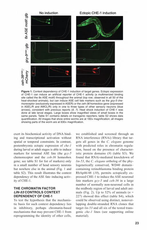
To test whether CHE-1 is not only re-quired but also sufficient to induce ASE fate, we ectopically expressed CHE-1 throughout the entire animal in either larval or adult stages, using an inducible heat shock pro-moter. Such misexpression results in broad ectopic expression of an artificial reporter of CHE-1 transcription factor activity, which is composed of a multimerized ASE mo-tif (“8x ASE motif reporter”) (6) (Fig. 1). This indicates that, in principle, CHE-1 can
Howard Hughes Medical Institute, Department of Biochemistry and Molecular Biophysics, Columbia University Medical Center, New York, NY 10032, USA. *To whom correspondence should be addressed. E-mail: [email protected] (B.T.); or [email protected] (O.H.)
Direct Conversion of C. elegans Germ Cells into Specific Neuron Types Baris Tursun,* Tulsi Patel, Paschalis Kratsios, Oliver Hobert*
The ability of transcription factors to directly reprogram the identity of cell types is usually restricted and is defined by cellular context. Through the ectopic expression of single Cae-norhabditis elegans transcription factors, we found that the identity of mitotic germ cells can be directly converted into that of specific neuron types: glutamatergic, cholinergic, or GABAergic. This reprogramming event requires the removal of the histone chaperone LIN-53 (RbAp46/48 in humans), a component of several histone remodeling and modifying complexes, and this removal can be mimicked by chemical inhibition of histone deacetylases. Our findings illustrate the ability of germ cells to be directly converted into individual, terminally differentiated neuron types and demonstrate that a specific chromatin factor provides a barrier for cellular reprogramming.
23
exert its biochemical activity of DNA bind-ing and transcriptional activation without spatial or temporal constraints. In contrast, postembryonic ectopic expression of che-1 during larval or adult stages is able to induce markers for terminal ASE fate (the gcy-5 chemoreceptor and the ceh-36 homeobox gene; see table S1 for list of markers) only in a small number of head sensory neurons but nowhere else in the animal (Fig. 1 and table S2). This result illustrates the context dependency of the ASE fate–inducing activ-ity of CHE-1.
THE CHROMATIN FACTOR LIN-53 CONTROLS CONTEXT DEPENDENCY OF CHE-1 To test the hypothesis that the mechanis-tic basis for such context dependency lies in inhibitory, perhaps chromatin-based mechanisms that may prevent CHE-1 from reprogramming the identity of other cells,
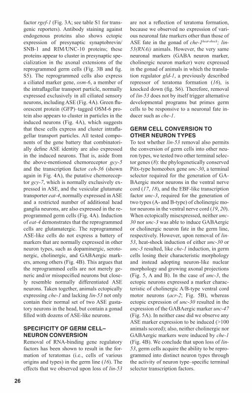
we established and screened through an RNA interference (RNAi) library that tar-gets all genes in the C. elegans genome with predicted roles in chromatin regula-tion, based on the presence of character-istic protein domains (9) (table S3). We found that RNAi-mediated knockdown of lin-53, the C. elegans ortholog of the phy-logenetically conserved, WD40 domain–containing retinoblastoma binding protein RbAp46/48 (10), permits ectopically ex-pressed CHE-1 to induce the ASE neuronal fate markers gcy-5 and ceh-36 in a large number of normally non-neuronal cells in the midbody region of larval and adult ani-mals (Fig. 2). Up to 52% of animals (n = 227) showed this effect (table S4), which could be observed using distinct, nonover-lapping double-stranded RNA clones that target lin-53 and all six of the tested trans-genic che-1 lines (see supporting online material).
Figure 1. Context dependency of CHE-1 induction of target genes. Ectopic expression of CHE-1 can induce an artificial reporter of CHE-1 activity (a multimerized binding site called the 8x ASE motif) throughout the animal (top row; observed in all 50 of the heat-shocked animals), but can induce ASE cell fate markers such as the gcy-5 che-moreceptor (exclusively expressed in ASER) or the ceh-36 homeobox gene (expressed in ASEL/R and AWCL/R) only in one to three types of other sensory neurons (blue arrows), consistent with previous reports (6, 7). Heat shock induction of CHE-1 was done at late larval stages. Large boxes show magnified views of small boxes in the same panels. Table S1 contains details on transgenic reporters; table S2 shows data quantification. All images that show entire worms are at 160× magnification; all images showing parts of the worm are at 630× magnification.

24
GERM CELLS ARE THE TARGET OF REPROGRAMMINGCloser examination of the animals revealed that the cells showing gcy-5 and ceh-36 in-duction are located within the gonad (Fig. 2). Differential interference contrast (DIC) microscopy showed that germ cells of che-1heat-shock; lin-53(RNAi) animals lose their characteristic, fried egg–shaped nuclear and nucleolar morphology and adopt a speck-led neuronal nuclear morphology (Fig. 2). Moreover, these cells grow cellular exten-
sions resembling axo-dendritic projections (Fig. 2 and movies S1 to S3). These neuron-like cells indeed originate from the germ line, as they were not observed after ectopic che-1 expression and lin-53 removal in a glp-4 mutant background (fig. S1), in which no germ line is formed (11). Genetic removal of sperm [fem-3lf mutants (12)] or oocyte [fem-3gf mutants (13)] or prevention of en-try into meiosis [glp-1gf mutants (14)] does not affect neuron induction, whereas severe reduction of the mitotic pool [glp-1lf mutants
Figure 2. Removal of lin-53 permits che-1 to induce ASE fate markers in the germ line. (A) ASE cell fate markers (gcy-5 and ceh-36), a transformation of nuclear mor-phology (from germ line with large nucleolus to neuronal with characteristic intra-nuclear speckles), and outgrowth of cellular extensions [blue arrows; also see (B)] are induced in the gonad of lin-53(–) animals upon ubiquitous induction of che-1 expres-sion. Noninduced lin-53(+) controls are shown in Fig. 1. This phenotype is observed in the germ line of up to 52% of heat-shocked animals (see tables S4 and S5 and fig. S3 for quantification) and in all six transgenic che-1heat-shock lines; the line shown here is otIs284. Heat shock of lin-53(–) animals that do not carry the che-1heat-shock array do not show any ectopic marker induction. (B) Higher-magnification image of axonal ex-tensions induced by ectopic che-1 expression in lin-53(RNAi) animals. The protruding vulva (Pvul) phenotype is a characteristic pleiotropy of loss of lin-53. Movies S1 to S3 show additional examples of axonal projections.

25
(14)] does, which suggests that it is the mitotic germ cells that be-come neuron-like (fig. S1). This is further supported by the position of the converted cells relative to mitotic and meiotic markers (fig. S2) and the observation that ani-mals in the second and third lar-val stages, which contain mostly mitotic but no meiotic cells, show germ cell–neuron conversion upon ectopic che-1 expression and lin-53 knockdown (table S4). The con-version to neuron-like cells is efficient and fast; as many as 60 germ cells (of a total of about 200 mitotic germ cells) undergo neu-ronal induction (fig. S3), and morphological changes and marker induction first occur 6 hours after che-1 induction (table S5). For comparison, the induction of the 8x ASE motif, an indicator of CHE-1 transcriptional activity occurs 4 hours after che-1 induction in the gonad. The induction of the 8x ASE motif in the gonad of wild-type animals, as well as antibody staining conducted in both wild-type and lin-53(RNAi) animals, rules out the possibility that lin-53(RNAi) merely results in germline derepression of the che-1 transgene. [We also note that lin-53(RNAi) does not result in germline derepression of a previously described transgenic ar-ray, let-858::gfp, known to be derepressed
after loss of several different chromatin factors (15).]
EXTENT OF GERM CELL–NEURON CONVERSIONWe assessed the nature of these che-1–in-duced, neuron-like cells with a number of fate markers. Through antibody staining of a marker that labels specific germ cell struc-tures, the P-granules, we confirmed that this germ cell feature is indeed lost upon ecto-pic che-1 expression and lin-53 knockdown (fig. S4). Moreover, the reprogrammed cells express all five tested pan-neuronal reporter genes: the rab-3/Rab3 and snb-1/synap-tobrevin genes, which encode presynaptic proteins normally exclusively expressed in all cells of the nervous system; the pan-neuronal axonal regulators unc-33/CRMP and unc-119; and the pan-neuronal signaling
Figure 3. Reprogrammed germ cells express pan-neuronal markers and cluster presynaptic proteins. (A) In-duction of pan-neuronal reporter genes in che-1heat-shock; lin-53(RNAi) animals. (B) Induction of the presyn-aptic SNB-1 protein, as assessed by antibody staining. An individual ASE-like neuron (labeled with gcy-5::gfp) is shown in higher magnification to show punctate staining of SNB-1 in red marker along the extension of the process (white arrowheads). che-1 expression was induced in late (L3/L4) larval stages of animals exposed to lin-53(dsRNA) starting in the pa-rental generation. Adult midbody re-gions are shown. White stippled lines indicate outline of the animal; yellow stippled lines indicate outline of the gonad. The penetrance of induction of the markers shown here is the same as for the gcy-5::gfp marker shown in Fig. 2 and table S4. See table S1 for information about transgenes and their wild-type expression pattern.
26
factor rgef-1 (Fig. 3A; see table S1 for trans-genic reporters). Antibody staining against endogenous proteins also shows ectopic expression of presynaptic synaptobrevin/SNB-1 and RIM/UNC-10 proteins; these proteins appear to cluster in presynaptic spe-cialization in the axonal extensions of the reprogrammed germ cells (Fig. 3B and fig. S5). The reprogrammed cells also express a ciliated marker gene, osm-6, a member of the intraflagellar transport particle, normally expressed exclusively in all ciliated sensory neurons, including ASE (Fig. 4A). Green flu-orescent protein (GFP)–tagged OSM-6 pro-tein also appears to cluster in particles in the induced neurons (Fig. 4A), which suggests that these cells express and cluster intrafla-gellar transport particles. All tested compo-nents of the gene battery that combinatori-ally define ASE identity are also expressed in the induced neurons. That is, aside from the above-mentioned chemoreceptor gcy-5 and the transcription factor ceh-36 (shown again in Fig. 4A), the putative chemorecep-tor gcy-7, which is normally exclusively ex-pressed in ASE, and the vesicular glutamate transporter eat-4, normally expressed in ASE and a restricted number of additional head ganglia neurons, are also expressed in the re-programmed germ cells (Fig. 4A). Induction of eat-4 demonstrates that the reprogrammed cells are glutamatergic. The reprogrammed ASE-like cells do not express a battery of markers that are normally expressed in other neuron types, such as dopaminergic, seroto-nergic, cholinergic, and GABAergic mark-ers, among others (Fig. 4B). This argues that the reprogrammed cells are not merely ge-neric and/or misspecified neurons but close-ly resemble normally differentiated ASE neurons. Taken together, animals ectopically expressing che-1 and lacking lin-53 not only contain their normal set of two ASE gusta-tory neurons in the head, but contain a gonad filled with dozens of ASE-like neurons.
SPECIFICITY OF GERM CELL– NEURON CONVERSIONRemoval of RNA-binding gene regulatory factors has been shown to result in the for-mation of teratomas (i.e., cells of various origins and types) in the germ line (16). The effects that we observed upon loss of lin-53
are not a reflection of teratoma formation, because we observed no expression of vari-ous neuronal fate markers other than those of ASE fate in the gonad of che-1heat-shock; lin-53(RNAi) animals. However, the very same neuronal markers (GABA neuron marker, cholinergic neuron marker) were expressed in the gonad of animals in which the transla-tion regulator gld-1, a previously described repressor of teratoma formation (16), is knocked down (fig. S6). Therefore, removal of lin-53 does not by itself trigger alternative developmental programs but primes germ cells to be responsive to a neuronal fate in-ducer such as che-1.
GERM CELL CONVERSION TO OTHER NEURON TYPESTo test whether lin-53 removal also permits the conversion of germ cells into other neu-ron types, we tested two other terminal selec-tor genes (8): the phylogenetically conserved Pitx-type homeobox gene unc-30, a terminal selector required for the generation of GA-BAergic motor neurons in the ventral nerve cord (17, 18), and the EBF-like transcription factor unc-3, required for the generation of two types (A- and B-type) of cholinergic mo-tor neurons in the ventral nerve cord (19, 20). When ectopically misexpressed, neither unc-30 nor unc-3 was able to induce GABAergic or cholinergic neuron fate in the germ line, respectively. However, upon removal of lin-53, heat-shock induction of either unc-30 or unc-3 resulted, like che-1 induction, in germ cells losing their characteristic morphology and instead adopting neuron-like nuclear morphology and growing axonal projections (Fig. 5, A and B). In the case of unc-3, the ectopic neurons expressed a marker charac-teristic of cholinergic A/B-type ventral cord motor neurons (acr-2; Fig. 5B), whereas ectopic expression of unc-30 resulted in the expression of the GABAergic marker unc-47 (Fig. 5A). In neither case did we observe any ASE marker expression to be induced (>100 animals scored); also, neither cholinergic nor GABAergic markers were induced by che-1 (Fig. 4B). We conclude that upon loss of lin-53, germ cells acquire the ability to be repro-grammed into distinct neuron types through the activity of neuron type–specific terminal selector transcription factors.

27
Figure 4. Reprogrammed germ cells express markers for ASE, but not other neuronal fates. Experimental conditions and image labeling are as described in Fig. 3. (A) ASE-specific markers expressed in che-1heat-shock; lin-53(RNAi) animals. osm-6::gfp is a full-length protein fusion. (B) Other neuronal fates are not expressed in che-1heat-shock; lin-53(RNAi) animals. The left column shows an overall view of the entire animal to illustrate the expression of non-ASE markers (all mCherry-based reporters) in other neuron types, and the right column shows an enlarged view (white box) of the gonad region, illustrating that these markers do not become induced in the che-1 reprogrammed germ cells, labeled with gcy-5::gfp.

28
Does lin-53 removal prime germ cells to respond only to factors that induce neuronal fates, or can they now respond to other fac-tors as well? To address this question, we ectopically expressed a selector gene, the C. elegans MyoD homolog hlh-1 that was previously shown to be able to ectopically induce muscle fate in early embryos (21). We found that hlh-1 is unable to convert the germ cells of lin-53(RNAi) animals into muscle cells (fig. S7). These negative results need to be cautiously interpreted, but may represent a first hint toward a target selectiv-ity of lin-53. That is, lin-53 may only restrict the developmental potential specifically to-ward a neuronal developmental program, whereas other factors may serve to prevent the induction of other, non-neuronal differ-entiation programs.
LIN-53, which is ubiquitously expressed (10), is one of several phylogenetically con-served histone chaperones that are thought to assist in the recruitment of various distinct
types of histone modifiers or remodelers (including histone methyltransferases, his-tone acetylases, and histone deacetylases) to histone H3 and histone H4 (22, 23). LIN-53 orthologs in various species have been found to be integral components of at least six dif-ferent protein complexes, each displaying diverse biochemical and biological roles: the NURD and NURF nucleosome remod-eling complexes, the CAF-1 chromatin as-sembly factor complex, the Sin3A transcrip-tional repressor complex, the PRC2 histone methyltransferase complex, and the HAT1 histone acetyltransferase complex (22, 23) (table S6). RNAi of representative members of each of these complexes either did not phenocopy the lin-53(RNAi)–induced repro-gramming ability of ectopic CHE-1 expres-sion or could not be interpreted because of early lethality induced by RNAi (table S6). However, two of the LIN-53–associated complexes (NURD and Sin3a) each contain at least two histone deacetylase (HDAC)
Figure 5. Reprogramming activity of other selector genes in lin-53(–) animals. (A) Heat-shock induction of the unc-30 terminal selector induces GABAergic, D-type motor neuron fate (as assessed by neuronal morphology and unc-47 vesicular GABA transporter expression) in the germ line of lin-53(RNAi) animals. White asterisks indicate ventral cord neurons that normally express the marker and are located in proximity to the gonad. Six of 32 unc-30heat-shock; lin-53(RNAi) animals (line 1, otEx4442), 4 of 21 animals (line 2, otEx4443), and 5 of 23 animals (line 3, otEx4444) show germline unc-47::gfp (oxIs12) expression. Without heat shock and RNAi, none of >100 animals show the effect. (B) Heat-shock induction of the unc-3 terminal selector induces A/B-type motor neuron fate in the germ line of lin-53(RNAi) animals. White asterisks indicate ventral cord neurons that normally express the marker and are located in proximity to the gonad. The white arrows indicate axonal-like extensions. Five of 24 unc-3heat-shock; lin-53(RNAi) animals show expression of a gfp reporter for the autoreceptor acr-2 expression; none of 20 unc-3heat-
shock animals [no lin-53(RNAi)] show this effect. Without heat shock and RNAi, none of >100 animals show the effect.
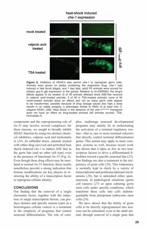
29
components and the reprogramming role of lin-53 may involve several complexes; for these reasons, we sought to broadly inhibit HDAC function by using two distinct chemi-cal inhibitors, valproic acid and trichostatin A (24). At sublethal doses, animals treated with either drug survived and permitted heat shock–induced che-1 to induce ASE fate in the germ line (and no other cell type) even in the presence of functional lin-53 (Fig. 6). Even though these drug effects may be unre-lated to normal lin-53 function, these results nonetheless provide a strong indication that histone modifications are key players in re-stricting the ability of a transcription factor to reprogram cellular identity.
CONCLUSIONSOur finding that the removal of a single chromatin factor, together with the induc-tion of single transcription factors, can pro-duce distinct and specific neuron types in a heterologous cellular context is a testament to the simplicity of programs that control neuronal differentiation. The role of com-
plex, multistage neuronal developmental programs may mainly lie in orchestrating the activation of a terminal regulatory rou-tine—that is, one or more terminal selectors that directly control terminal differentiation genes. This notion may apply to more com-plex systems as well, because recent work has shown that it takes as few as two tran-scription factors to drive a differentiated fi-broblast toward a specific neuronal fate (25). Our findings are also a testament to the toti-potency of germ cells (26). This totipotency is normally kept in check by a variety of transcriptional and posttranscriptional mech-anisms (26), but is unleashed either spon-taneously in pathological situations (germ cell tumors) (27) or upon culturing germ stem cells under specific conditions, which transform these cells into cells indistin-guishable from pluripotent embryonic stem cells (28).
We have shown that the ability of germ cells to be directly reprogrammed into neu-rons can be unleashed, even in the adult ani-mal, through removal of a single gene that
Figure 6. Inhibitors of HDACs also permit che-1 to reprogram germ cells. Animals were grown on plates containing the respective drug, che-1 was induced in late larval stages, and 1 day later, adult P0 animals were scored for ectopic gcy-5::gfp expression in the gonad. Relative to lin-53(RNAi), the drug’s effects appear to be weaker (5 of 40 animals affected show ASE-like neurons in valproic acid–treated animals, 3 of 40 in TSA-treated animals; none of 50 mock-treated animals show an effect) and not as many germ cells appear to be transformed, possibly because of drug dosage issues [too high a dose results in no viable progeny, a phenotype similar to RNAi of at least one C. elegans HDAC (29)]. Heat shock in the absence of the che-1heat-shock transgene does not have an effect on drug-treated animals (40 animals scored). TSA, trichostatin A.

30
we speculate to be involved in rendering neuronal differentiation genes inaccessible to transcriptional induction by, for example, contributing to the formation of facultative (i.e., conditional and developmentally regu-lated) heterochromatin. Seen in a broader context, our results indicate that the repro-gramming of cellular identity may critically depend not just on providing the correct transcription factor(s) that induce a specific fate, but also on the removal of inhibitory mechanisms that restrict transcription fac-tor activity. We anticipate that the disable-ment of such inhibitory mechanisms may provide an efficient strategy to ectopically generate neuron types in other organisms, or perhaps even in cell culture, using isolated germ cells.
References and Notes 1. T. Graf, T. Enver, Nature 462, 587 (2009). 2. Q. Zhou, D. A. Melton, Cell Stem Cell 3, 382 (2008). 3. H. Weintraub et al., Proc. Natl. Acad. Sci. U.S.A. 86, 5434 (1989). 4. G. Halder, P. Callaerts, W. J. Gehring, Science 267, 1788 (1995). 5. Q. Zhou, J. Brown, A. Kanarek, J. Rajagopal, D. A. Melton, Nature 455, 627 (2008). 6. J. F. Etchberger et al., Genes Dev. 21, 1653 (2007). 7. O. Uchida, H. Nakano, M. Koga, Y. Ohshima, C. The, Development 130, 1215 (2003). 8. O. Hobert, Proc. Natl. Acad. Sci. U.S.A. 105, 20067 (2008). 9. M. Cui, M. Han, WormBook 10.1895/ wormbook.1.139.1 (2007).10. X. Lu, H. R. Horvitz, Cell 95, 981 (1998).11. M. J. Beanan, S. Strome, Development 116, 755 (1992).12. J. Hodgkin, Genetics 114, 15 (1986).13. M. K. Barton, T. B. Schedl, J. Kimble, Genetics 115, 107 (1987).14. J. Austin, J. Kimble, Cell 51, 589 (1987).
15. W. G. Kelly, A. Fire, Development 125, 2451 (1998).16. R. Ciosk, M. DePalma, J. R. Priess, Science 311, 851 (2006).17. H. Cinar, S. Keles, Y. Jin, Curr. Biol. 15, 340 (2005).18. Y. Jin, R. Hoskins, H. R. Horvitz, Nature 372, 780 (1994).19. B. C. Prasad et al., Development 125, 1561 (1998).20. B. Prasad, O. Karakuzu, R. R. Reed, S. Cameron, Dev. Biol. 323, 207 (2008).21. T. Fukushige, M. Krause, Development 132, 1795 (2005).22. M. Eitoku, L. Sato, T. Senda, M. Horikoshi, Cell. Mol. Life Sci. 65, 414 (2008).23. A. Loyola, G. Almouzni, Biochim. Biophys. Acta 1677, 3 (2004).24. R. W. Johnstone, Nat. Rev. Drug Discov. 1, 287 (2002).25. T. Vierbuchen et al., Nature 463, 1035 (2010).26. G. Seydoux, R. E. Braun, Cell 127, 891 (2006).27. B. Blum, N. Benvenisty, Adv. Cancer Res. 100, 133 (2008).28. K. Ko et al., Cell Stem Cell 5, 87 (2009).29. Y. Shi, C. Mello, Genes Dev. 12, 943 (1998).30. Supported by NIH grants R01NS039996-05 and R01NS050266-03. O.H. is an Investigator of the HHMI and B.T. is a Francis Goelet Research Scientist. We thank T. Schedl, M. Krause, J. McGhee, P. Sengupta, Y. B. Qi, Y. Jin, M. Nonet, S. Strome, and members of the Hobert lab for reagents; I. Greenwald and members of the Hobert lab for comments on the manuscript; and Q. Chen for expert assistance in strain generation.
Supporting Online Materialwww.sciencemag.org/cgi/content/full/science.1199082/ DC1Materials and MethodsFigs. S1 to S7Tables S1 to S6 Movies S1 to S3References
14 October 2010; accepted 23 November 2010Published online 9 December 2010;10.1126/science.1199082

Career advice I Job postings I Job Alerts I Career Forum Crafting resumes/CVs I Preparing for interviews
There’s only one Galileo Galilei
orn in 1564, Galileo Galilei once contemplated a career in the priesthood. It’s perhaps fortunate for science that upon the urging of his father, he instead decided to enroll at the University of Pisa. His career in science began with medicine and from there he subsequently went on to become a philosopher, physicist, mathematician, and astronomer, for which he is perhaps best known. His astronomical observations and subsequent improvements to telescopes built his reputation as a leading scientist of his time, but also led him to probe subject matter counter to prevailing dogma. His expressed views on the Earth’s movement around the sun caused him to be declared suspect of heresy, which for some time led to a ban on the reprinting of his works. Galileo’s career changed science for all of us and he was without doubt a leading light in the scientific revolution, which is perhaps why Albert Einstein called him the father of modern science. Want to challenge the status quo and make the Earth move? At Science we are here to help you in your own scientific career with expert career advice, forums, job postings, and more—all for free. Visit Science today at ScienceCareers.org.
B
Careers
ScienceCareers.org
For your career in science, there’s only one

32