Processing Streptococcal CellWalls … · Afew studies have described the processing ... toneal...
8
INFECTION AND IMMUNITY, Sept. 1977, p. 591-598 Copyright X 1977 American Society for Microbiology Vol. 17, No. 3 Printed in U.S.A. Processing of Streptococcal Cell Walls by Rat Macrophages and Human Monocytes In Vitro RALPH J. SMIALOWICZt AND JOHN H. SCHWAB* Department of Bacteriology and Immunology, School of Medicine, University of North Carolina, Chapel Hill, North Carolina 27514 Received for publication 15 March 1977 Phagocytosis and degradation of cell walls by peritoneal macrophages obtained from Fischer 344 or Buffalo rats was measured in tissue culture. Group A cell wall antigen, detected by immunofluorescence, persisted in cultured rat macrophages for at least 40 days, whereas group D cell wall material was eliminated by 6 to 8 days. This same pattern of persistence of group A cell walls and elimination of group D cell walls was observed in cultures of human monocytes followed for 24 days in culture. Group A streptococcal cell walls labeled with either [14C]alanine or ['4C]glucose were handled in a similar manner by macrophages from either Fischer 344 or Buffalo rats. In contrast, ['4C]glucose-labeled group D cell walls were degraded at a much faster rate. Buffalo macrophages were more efficient than Fischer 344 macrophages in degrading group D cell walls. The inability of macrophages to degrade group A cell walls was not due to a failure of lysosomes to fuse with phagosomes. Neither serum lysozyme in the culture medium nor cell wall-associated autolysin contributed to the degradation of group D cell walls by macrophages. Neither immune serum nor macrophages obtained from specifi- cally immunized rats influenced phagocytosis or persistence of group A cell walls. Several experimental models of chronic gran- ulomatous inflammation can be produced by the injection of group A streptococcal cell wall fragments. Chronic remittent multinodular le- sions develop in rabbit skin after a single intra- dermal injection of cell walls (10, 15). Rheu- matic-like heart lesions are produced in mice (4, 11) and chronic polyarthritis is produced in rats by a single intraperitoneal injection (14; W. J. Cromartie, J. G. Craddock, and J. H. Schwab, Fed. Proc., p. 477, 1968). In each of these models, chronic inflammation is associated with the localization and persistence of cell wall anti- gens. The material responsible for the induction and perpetuation of chronic lesions is the muco- peptide-C polysaccharide complex of the cell wall (14). The persistence of cell wall fragments from group A streptococci and certain other bacteria is postulated to play a role in the path- ogenesis of chronic diseases that develop subse- quent to bacterial infection (14). Cell walls from group D streptococci are rapidly eliminated from tissue and do not induce chronic or recur- rent lesions (16). For this reason group D cell walls have been used as a control material in animals and are used in the present study as an example of a biodegradable bacterial cell wall. In experimental models a consistent finding is t Present address: Environmental Protection Agency, Re- search Triangle Park, NC 27711. the localization of cell wall material within mac- rophages in the lesions. This indicates that the macrophage has an essential role in the distri- bution and persistence of bacterial components. It is important, therefore, to examine in detail the interaction of streptococcal cell walls and macrophages; i.e., to determine (i) what the macrophage does to the cell wall structures and (ii) how the ingested cell wall affects macro- phage functions. As an experimental approach to these problems we have studied rat perito- neal macrophages in tissue culture. This is part of a study designed to establish the mechanism by which poorly degradable group A streptococ- cal cell walls induce chronic inflammatory dis- ease in the periodontal tissue and joints of rats and to define the nature of genetic control of susceptibility in these experimental diseases. A few studies have described the processing of radiolabeled bacteria after phagocytosis in vi- tro (1, 3, 20) or incubation with leukocyte ex- tracts (6-8). However, only Gallis et al. (6) have followed the fate of an identifiable cell struc- ture, and most studies were limited to a few hours of culture. The present work follows the fate of phagocytized radiolabeled streptococcal cell walls in macrophages cultured in vitro from 1 to 6 weeks. The phagocytized cell wall struc- tures are identified by immunofluorescent tech- niques as well as radioisotopes. In this paper the 591 on September 6, 2018 by guest http://iai.asm.org/ Downloaded from
Transcript of Processing Streptococcal CellWalls … · Afew studies have described the processing ... toneal...
INFECTION AND IMMUNITY, Sept. 1977, p. 591-598Copyright X 1977 American Society for Microbiology
Vol. 17, No. 3Printed in U.S.A.
Processing of Streptococcal Cell Walls by Rat Macrophagesand Human Monocytes In VitroRALPH J. SMIALOWICZt AND JOHN H. SCHWAB*
Department ofBacteriology and Immunology, School ofMedicine, University ofNorth Carolina, Chapel Hill,North Carolina 27514
Received for publication 15 March 1977
Phagocytosis and degradation of cell walls by peritoneal macrophages obtainedfrom Fischer 344 or Buffalo rats was measured in tissue culture. Group A cell wallantigen, detected by immunofluorescence, persisted in cultured rat macrophagesfor at least 40 days, whereas group D cell wall material was eliminated by 6 to 8days. This same pattern of persistence of group A cell walls and elimination ofgroup D cell walls was observed in cultures of human monocytes followed for 24days in culture. Group A streptococcal cell walls labeled with either [14C]alanineor ['4C]glucose were handled in a similar manner by macrophages from eitherFischer 344 or Buffalo rats. In contrast, ['4C]glucose-labeled group D cell wallswere degraded at a much faster rate. Buffalo macrophages were more efficientthan Fischer 344 macrophages in degrading group D cell walls. The inability ofmacrophages to degrade group A cell walls was not due to a failure of lysosomesto fuse with phagosomes. Neither serum lysozyme in the culture medium nor cellwall-associated autolysin contributed to the degradation of group D cell walls bymacrophages. Neither immune serum nor macrophages obtained from specifi-cally immunized rats influenced phagocytosis or persistence of group A cell walls.
Several experimental models of chronic gran-ulomatous inflammation can be produced bythe injection of group A streptococcal cell wallfragments. Chronic remittent multinodular le-sions develop in rabbit skin after a single intra-dermal injection of cell walls (10, 15). Rheu-matic-like heart lesions are produced in mice (4,11) and chronic polyarthritis is produced in ratsby a single intraperitoneal injection (14; W. J.Cromartie, J. G. Craddock, and J. H. Schwab,Fed. Proc., p. 477, 1968). In each of thesemodels, chronic inflammation is associated withthe localization and persistence of cell wall anti-gens. The material responsible for the inductionand perpetuation of chronic lesions is the muco-peptide-C polysaccharide complex of the cellwall (14). The persistence of cell wall fragmentsfrom group A streptococci and certain otherbacteria is postulated to play a role in the path-ogenesis of chronic diseases that develop subse-quent to bacterial infection (14). Cell walls fromgroup D streptococci are rapidly eliminatedfrom tissue and do not induce chronic or recur-rent lesions (16). For this reason group D cellwalls have been used as a control material inanimals and are used in the present study as anexample of a biodegradable bacterial cell wall.
In experimental models a consistent finding ist Present address: Environmental Protection Agency, Re-
search Triangle Park, NC 27711.
the localization of cell wall material within mac-rophages in the lesions. This indicates that themacrophage has an essential role in the distri-bution and persistence of bacterial components.It is important, therefore, to examine in detailthe interaction of streptococcal cell walls andmacrophages; i.e., to determine (i) what themacrophage does to the cell wall structures and(ii) how the ingested cell wall affects macro-phage functions. As an experimental approachto these problems we have studied rat perito-neal macrophages in tissue culture. This is partof a study designed to establish the mechanismby which poorly degradable group A streptococ-cal cell walls induce chronic inflammatory dis-ease in the periodontal tissue and joints of ratsand to define the nature of genetic control ofsusceptibility in these experimental diseases.A few studies have described the processing
of radiolabeled bacteria after phagocytosis in vi-tro (1, 3, 20) or incubation with leukocyte ex-tracts (6-8). However, only Gallis et al. (6) havefollowed the fate of an identifiable cell struc-ture, and most studies were limited to a fewhours of culture. The present work follows thefate of phagocytized radiolabeled streptococcalcell walls in macrophages cultured in vitro from1 to 6 weeks. The phagocytized cell wall struc-tures are identified by immunofluorescent tech-niques as well as radioisotopes. In this paper the
591
on Septem
ber 6, 2018 by guesthttp://iai.asm
.org/D
ownloaded from

592 SMIALOWICZ AND SCHWAB
ingestion and processing of cell wall fragmentsby cultured macrophages is compared in perito-neal celLs obtained from Fischer 344 rats, whichare susceptible to streptococcal-induced polyar-thritis, and Buffalo rats, which do not developthese joint lesions (S. K. Anderle and W. J. Cro-martie, Abstr. Annu. Meet. Am. Soc. Microbiol.1976, B49, p. 19). An accompanying paper com-
pares the activation of macrophages from theserat strains to become cytotoxic for target cells(19).
MATERIALS AND METHODSMacrophages. We define macrophages as adher-
ent, mononuclear cells that actively phagocytize parti-cles. Peritoneal macrophages were obtained from nor-
mal or thioglycolate-stimulated female Buffalo or
Fischer 344 rats (Simonsen Laboratories, Inc., Gilroy,Calif.) weighing 100 to 200 g. Five days after intraperi-toneal injection of 7 ml of fluid thioglycolate broth(Baltimore Biological Laboratory, Cockeysville, Md.),the peritoneum was washed three times with coldHanks balanced salt solution containing 10 U of pre-
servative-free heparin sodium (The Upjohn Co., Ka-lamazoo, Mich.), 100 mg of streptomycin sulfate, and100 U of penicillin G per ml. After washing the perito-neal cells twice in medium 199 (Grand Island Biologi-cal Co., Grand Island, N.Y.) supplemented with 10%heat-inactivated horse serum and antibiotics, 2 x 1OBto 4 x 10⁶ cells were added to tissue culture dishes (10by 35 mm; Falcon Plastics, Oxnard, Calif.) or sterileglass cover slips (22 by 22 mm). After 2 h of incuba-tion at 37°C in 5% CO2 the medium was changed, andcultures were incubated for 18 h. Before addition ofstreptococcal cell walls, macrophage cultures were
washed three times with medium 199 to remove non-
adherent and nonviable cells. Cultures containedgreater than 95% adherent macrophages, based on
morphology of Giemsa-stained slides and phagocyto-sis of polystyrene latex particles (The Dow ChemicalCo., Midland, Mich.). Continued adherence of cellscontaining cell wall material was considered evidencethat the macrophages remained viable during the cul-ture period. Trypan blue staining was also done on
representative cultures at intervals up to 40 days.Human peripheral blood monocytes, band 1 cells
from Ficoll-Hypaque gradients, were supplied byJohn Spitznagel. These were washed twice in TC 199containing 10% heat-inactivated fetal calf serum andantibiotics. Monocytes (106) were cultured on glasscover slips at 37°C in 5% CO2 and 100% humidity.After 24 h, cultures were washed three times with TC199, and fresh medium with serum and antibiotics wasadded.
Radiolabeled streptococci. Group A streptococ-cal cels (type 3, strain D58) were labeled with either[14C]alanine or [14C]glucose. Cells were cultured for 24h in a modified medium consisting of 1 volume of"depleted broth" (filtered, spent medium of an 18-hTodd-Hewitt broth culture of group A streptococci)and 1 volume of "glucose-salts" medium modifiedfrom that of Shockman et al. (17). To this mediumwas added 1 MCi of DL-[1-14C]alanine per ml (specificactivity, 23.8 mCi/mmol; Amersham/Searle, Arling-ton Heights, Ill.).
Labeled glucose was incorporated into cells by cul-turing them for 6 h in freshly prepared beef heartinfusion broth, in which the only glucose added wasD-[U-14C]glucose, 1.5 ,LCi/ml (specific activity, 180mCi/mmol; ICN Canada Ltd., Montreal, Quebec,Canada). After 6 h the culture was supplemented withglucose-salts medium (17) and incubated for an addi-tional 12 h. Group D (strain F-24) streptococcal cellswere labeled with ['4C]glucose in a similar procedureexcept that the total incubation was 12 h, to precludeautolysis of these organisms.
Preparation of streptococcal cell wail frag-ments. The preparation of unlabeled streptococcalcell wall fragments from group A (strain D58) andgroup D (strain F-24) streptococci has been described(10). Cell wall fragments from radiolabeled group Aand group D streptococci were prepared according tothe procedure described by Salton and Home (13),using a Mickle cell disintegrator with glass beads.After disruption of cells, cell wall fragments were col-lected by differential centrifugation followed by treat-ment with trypsin and ribonuclease (10). Cell wallpreparations were exhaustively washed with phos-phate-buffered saline to remove unincorporated ra-dioactivity and subsequently stored at -20°C. Rham-nose content of cell wall fragments was determined bythe method of Dische and Shettles (5).
Distribution of radiolabel in cell wall frag-ments. The distribution and identification of radiola-bel incorporated into the cell walls of group A strepto-cocci was determined by paper chromatography (9).Acid-hydrolyzed labeled cell wall fragments werechromatographed on Whatman no. 1 filter paper(Whatman, Inc., Clifton, N.J.) by ascending chroma-tography, with n-butanol-acetic acid-water (60:20:10)as the first solvent and a water-saturated phenol solu-tion with an ammonium atmosphere as the secondsolvent. Amino acids and amino sugars were devel-oped with ninhydrin spray (E. Merck AG, Darmstadt,Germany), and sugars were developed with anilinephthalate spray (E. Merck). Reference amino acidsand sugars were chromatographed individually and inmixture to determine their distribution. Two chro-matograms for each labeled cell wall preparation wererun simultaneously. One was developed, whereascoincident areas from the undeveloped chromatogramwere cut, placed in scintillation fluid, and counted in aliquid scintillation counter. The amount of radioactiv-ity in a given area of a chromatogram was comparedto the amount of radioactivity added initially and ex-pressed as the percent distribution of radiolabel in cellwalls.
Phagocytosis and fate of cell walls. The fate ofphagocytized streptococcal cell walls was followed byimmunofluorescent and radioisotope techniques. Aqualitative estimate of the persistence of phagocytizedcell walls in macrophages was accomplished by deter-mining the number of macrophages in a microscopefield that displayed fluorescence, and the intensity ofthis fluorescence, after direct staining with fluores-cein-conjugated rabbit antistreptococcal antibodies(10). Quantitative measurement of the phagocytosisand degradation of streptococcal cell walls by culturedmacrophages was done with radiolabeled cell walls. Atvarious times after the addition of labeled cell walls tomacrophage cultures, replicate cultures were washed
INFECT. IMMUN.
on Septem
ber 6, 2018 by guesthttp://iai.asm
.org/D
ownloaded from
BACTERIAL CELL WALLS
three times with phosphate-buffered saline and di-gested with 0.1 N NaOH for 30 min. Portions werethen taken for protein determination according to themethod of Oyama and Eagle (12) to determine therelative number of macrophages remaining in culture.The remaining cell digest was placed on GF-82 glassfiber paper (W. and R. Balston Ltd., London, Eng-land), air dried, placed in Omnifluor-toluene (NewEngland Nuclear, Boston, Mass.), and counted in aPackard liquid scintillation counter (Packard Instru-ment Co., Inc., Rockville, Md.). Degradation of strep-tococcal cell walls by macrophages was determined bymeasuring the amount of radioactivity that passesthrough a 0.22-pm membrane filter (Millipore Corp.,Bedford, Mass.). Portions of medium, washes, andNaOH digests of macrophage cultures that had phag-ocytized labeled cell walls were filtered, and the radio-activity that passed through the filter was degradedcell wall material, whereas retained material was con-sidered nondegraded. Evidence that "C that passesthrough the filter represents degraded cell wall isprovided by the control cell wall suspensions, not fedto macrophages, in which the filterable "C was alwaysless than 4%. A positive control was provided bygroup D cell wall label, which does pass through thefilter as it is degraded.
Immunofluorescence was used in addition to thequantitative radioisotope technique, because merelyfollowing 14C gives no indication of the nature of thelabeled material. Conjugated antibody at least dem-onstrates that the ingested cell wall retains structuresthat can bind antibody.
linmunization of rats. Buffalo and Fischer 344rats were immunized with heat-killed vaccines ofgroup A (strain D58) streptococci by injection intothe tail vein twice weekly for 3 weeks. Rats were "sen-sitized" by subcutaneous injection into the hind foot-pads and back with a viable group A streptococcalvaccine in complete Freund adjuvant as described byBtltmann et al. (2).
Fluorescein-conjugated rabbit antiserum.Rabbits were hyperimmunized with group A strepto-coccal vaccine or group D cell walls to obtain antibod-ies against cell wall antigens. The immunization pro-cedure, absorption of serum, and fluorescein conjuga-tion of the antibodies have been described (10).
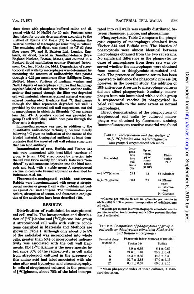
RESULTSDistribution of radiolabel in streptococ-
cal cell walls. The incorporation and distribu-tion of [14C]alanine and ['4C]glucose into groupA streptococcal cell walls with culture condi-tions described in Materials and Methods areshown in Table 1. Although only about 3 to 5%of the radiolabel was incorporated into wholecells, greater than 50% of incorporated radioac-tivity was associated with the cell wall frag-ments. DL-[1-_4C]alanine is the more specific la-bel, since 85% of the radioactivity in cell wallsfrom streptococci cultured in the presence ofthis amino acid had label associated with ala-nine after acid hydrolysis and chromatography.In cells of streptococci cultured in the presenceof ['4C]glucose, about 75% of the label incorpo-
rated into cell walls was equally distributed be-tween rhamnose, glucose, and glucosamnine.Phagocytosis. Table 2 compares the phago-
cytic capacity of macrophages obtained fromFischer 344 and Buffalo rats. The kinetics ofphagocytosis were almost identical betweenmacrophages obtained from the two rat strains.No significant difference in the phagocytic in-dexes of macrophages from these rats was ob-served whether the macrophages were obtainedfrom normal or thioglycolate-stimulated ani-mals. The presence of immune serum has beenreported to influence the phagocytic process (3);however, in the present study, the addition of10% anti-group A serum to macrophage culturesdid not affect phagocytosis. Similarly, macro-phages from rats immunized with a viable groupA streptococcal vaccine (2) phagocytized la-beled cell walls to the same extent as normnalmacrophages.
Direct demonstration of the phagocytosis ofstreptococcal cell walls by cultured macro-phages was obtained by fluorescent staining.Immunofluorescent reactive material was found
TABLE 1. Incorporation and distribution ofDL-[1- '4C]alanine and r[U- '4C]glucoseinto group A streptococcal cell walls
Incor- Sp actpora- Spaction (cpm/ Distri-
Radiolabel into y of butioncell rham- (%)bwalls nse)M1, (X 103)
DL-[1-'4C]alanine 53.8 1.4 85 (Alanine)
D-[U-'4C]glucose 80.3 2.9 25 (Rham-nose)
24 (Glucosa-mine)
24 (Glucose)a (Counts per minute in cell walls/counts per minute in
whole cells) x 100 = percent incorporation of radiolabel intocell walls.
b (Counts per minute in chromatographic area/total countsper minute added to chromatogram) x 100 = percent distribu-tion of radiolabel.
TABLE 2. Comparison ofphagocytosis ofgroup Acell walls by thioglycolate-stimulated Fischer 344
and Buffalo macrophages
Period of phag- Phagocytic index,,(cpm/JLg of protein)ocytosis (h) Fischer 344 Buffalo
1 6.9 + 0.69 6.4 ± 0.693 24.6 ± 1.48 25.2 + 0.416 44.3+2.66 44.3±5.39 52.7 + 2.88 57.0 ± 3.15
12 60.1 ± 0.57 69.6 + 8.15
a Mean phagocytic index of three cultures, ± stan-dard deviation.
VOL. 17, 1977 593
on Septem
ber 6, 2018 by guesthttp://iai.asm
.org/D
ownloaded from
594 SMIALOWICZ AND SCHWAB
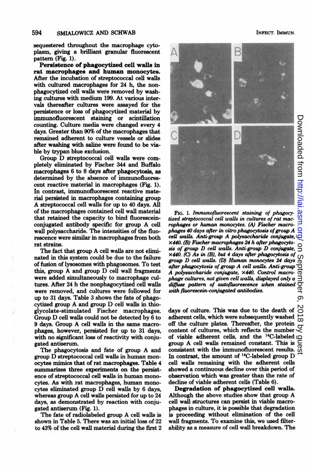
sequestered throughout the macrophage cyto-plasm, giving a brilliant granular fluorescentpattern (Fig. 1).Persistence of phagocytized cell walls in
rat macrophages and human monocytes.After the incubation of streptococcal cell wallswith cultured macrophages for 24 h, the non-phagocytized cell walls were removed by wash-ing cultures with medium 199. At various inter-vals thereafter cultures were assayed for thepersistence or loss of phagocytized material byimmunofluorescent staining or scintillationcounting. Culture media were changed every 4days. Greater than 90% of the macrophages thatremained adherent to culture vessels or slidesafter washing with saline were found to be via-ble by trypan blue exclusion.Group D streptococcal cell walls were com-
pletely eliminated by Fischer 344 and Buffalomacrophages 6 to 8 days after phagocytosis, asdetermined by the absence of immunofluores-cent reactive material in macrophages (Fig. 1).In contrast, immunofluorescent reactive mate-rial persisted in macrophages containing groupA streptococcal cell walls for up to 40 days. Allof the macrophages contained cell wall materialthat retained the capacity to bind fluorescein-conjugated antibody specific for group A cellwall polysaccharide. The intensities of the fluo-rescence were similar in macrophages from bothrat strains.The fact that group A cell walls are not elimi-
nated in this system could be due to the failureof fusion of lysosomes with phagosomes. To testthis, group A and group D cell wall fragmentswere added simultaneously to macrophage cul-tures. After 24 h the nonphagocytized cell wallswere removed, and cultures were followed forup to 31 days. Table 3 shows the fate of phago-cytized group A and group D cell walls in thio-glycolate-stimulated Fischer macrophages.Group D cell walls could not be detected by 6 to9 days. Group A cell walls in the same macro-phages, however, persisted for up to 31 days,with no significant loss of reactivity with conju-gated antiserun.The phagocytosis and fate of group A and
group D streptococcal cell walls in human mon-ocytes mimics that of rat macrophages. Table 4summarizes three experiments on the persist-ence of streptococcal cell walls in human mono-cytes. As with rat macrophages, human mono-cytes eliminated group D cell walls by 6 days,whereas group A cell walls persisted for up to 24days, as demonstrated by reaction with conju-gated antiserum (Fig. 1).The fate of radiolabeled group A cell walls is
shown in Table 5. There was an initial loss of 22to 43% of the cell wall material during the first 2
INFECT. IMMUN.
FIG. 1. Immunofluorescent staining of phagocy-tized streptococcal cell walls in cultures of rat mac-rophages or human monocytes. (A) Fischer macro-phages 40 days after in vitro phagocytosis ofgroupAceU waUs. Anti-group A polysaccharide conjugate,x440. (B) Fischer macrophages 24 h afterphagocyto-sis of group D ceU wais. Anti-group D conjugate,x440. (C) As in (B), but 4 days after phagocytosis ofgroup D cell walls. (D) Human monocytes 24 daysafter phagocytosis ofgroup A ceU waUs. Anti-groupA polysaccharide conjugate, x440. Control macro-phage cultures, not given ceU walls, displayed only adiffuse pattern of autofluorescence when stainedwith fluorescein-conjugated antibodies.
days of culture. This was due to the death ofadherent cells, which were subsequently washedoff the culture plates. Thereafter, the proteincontent of cultures, which reflects the numberof viable adherent cells, and the '4C-labeledgroup A cell walls remained constant. This isconsistent with the immunofluorescent results.In contrast, the amount of '4C-labeled group Dcell walls remaining with the adherent cellsshowed a continuous decline over this period ofobservation which was greater than the rate ofdecline of viable adherent cells (Table 6).Degradation of phagocytized cell walls.
Although the above studies show that group Acell wall structures can persist in viable macro-phages in culture, it is possible that degradationis proceeding without elimination of the cellwall fragments. To examine this, we used filter-ability as a measure of cell wall breakdown. The
l1
on Septem
ber 6, 2018 by guesthttp://iai.asm
.org/D
ownloaded from

BACTERIAL CELL WALLS 595
radioactivity that passed through a 0.22-pinMillipore membrane filter was defined as de-graded cell wall material. Less than 4% of theradioactivity of the original labeled cell wall wasfilterable, and this did not increase after treat-ment with 0.1 N NaOH. In all cultures, 95% or
more of the radioactivity initially phagocytizedcould be accounted for in the medium, washes,and macrophages from a given culture.
Less than 4% of the phagocytized group Astreptococcal cell walls that remained intracel-lular in normal macrophages from either ratstrain was degraded after 8 days in culture (Ta-ble 5). At this time, 10 to 16% of the radioactiv-ity released into the culture medium from mac-
rophages was degraded. Although this mightsuggest that some slow degradation of group Acell walls has occurred, this is of doubtful signif-icance because only a small fraction (0 to 10%)of the 14C label within adherent cells appearsextracellular. Cell walls labeled with ['4C]glu-cose were degraded by macrophages from bothrat strains to a slightly greater extent than[14C]alanine-labeled cell walls.
In contrast to group A cell walls, up to 83% ofthe group D cell walls within macrophages was
degraded after 8 days in culture (Table 6). TheFischer 344 macrophages retained a greateramount of intracellular degraded cell wall mate-rial. The extracellular medium of macrophagecultures from normal and thioglycolate-stimu-lated Buffalo rats contained significantly more
degraded cell wall material, compared withFischer 344 macrophage cultures (Table 6).This was observed in a number of experiments
TABLE 3. Fate of simultaneously phagocytizedgroup A and group D cell walls in thioglycolate-
stimulated Fischer macrophagesDays after re-
Fluorescein-conju- moval of non- Intensity ofgated antiserum phagocytized fluorescence
cell wallsa
Anti-A 1 4+2 4+4 4+6 4+9 4+
11 4+31 4+
Anti-D 1 4+2 4+4 2+69 -
a A niixture of 50 pg of group A cell walls per ml and100 pg of group D cell walls per ml was added tomacrophage cultures simultaneously.
TABLE 4. Fate ofphagocytized group A andgroup DceU walls in human monocytesa
Days after re- InCell walls phagocy- moval of non- ltensty of
tized phagocytized cencecell walls
Group A, 50 iLg/ml 4 4+6 4+12 4+16 4+24 4+
Group D, 100 jig/ml 2 3+4 1+6 ±
10 -
a Monocytes were cultured in TC 199 with 10% heat-inactivated fetal calf serum. Duplicate cover slip cul-tures were stained with fluorescein-conjugated anti-body specific for group A or group D cell wall anti-gens.
and is the only consistent difference betweenthese rat strains in the degradative capacities ofmacrophages. Macrophage protein increasedafter 8 days in culture after ingestion of group Acell walls (Table 5). This is because proteinsynthesis is stimulated in activated macro-phages. Protein did not increase after ingestionof group D cell walls (Table 6), consistent withthe failure to maintain an activated state inmacrophages.
Heat inactivation of the autolysin associatedwith radiolabeled group D cell walls (18) andabsorption of serum lysozyme with bentonitedid not effect degradation.The persistence of radiolabeled group A
streptococcal cell walls in macrophages wasfound to be essentially the same in culturescontaining either normal or immune serum.Furthermore, macrophages obtained from im-munized rats in the presence or absence of non-adherent cells (lymphocytes) exhibited similarpatterns of cell wall persistence.
DISCUSSIONThe degradation of isotopically labeled bacte-
ria after phagocytosis by cells in culture hasbeen studied by several investigators (1, 3, 20).Cohn (3) found that the rate of degradation ofbacterial lipids, nucleic acids, and proteins byphagocytic cells is primarily dependent uponthe composition of the ingested organism and,particularly, the composition of the bacterialsurface or cell wall. Spector et al. (20), usingbacteria iodinated with '25I to study the fate ofgranuloma-inducing bacteria phagocytized bycultured macrophages, found that by 72 h 95%of group A streptococci were degraded to mate-
VOL. 17, 1977
on Septem
ber 6, 2018 by guesthttp://iai.asm
.org/D
ownloaded from

596 SMIALOWICZ AND SCHWAB
TABLE 5. Comparison of the fates ofgroup A cell walls in unstimulated Fischer 344 and Buffalomacrophages
Period % Protein % 14C remain- Degradation (%)dMacrophages Radiolabeled of cul- mminb ingc (mean ±(rat strain) cell walls ture
(days)a (mean±SD) SD) Intracellular Extracellular
Fischer 344 ['4C]alanine 2 78.4 ± 7.02 78.4 ± 5.72 2.2 ± 0.20 3.4 ± 0.154 59.2 ± 3.69 74.1 ± 6.19 2.2 ± 0.20 5.9 ± 0.268 89.9 ± 5.25 72.7 ± 11.8 3.3 ± 0.62 10.7 ± 0.40
Fischer 344 ['4C]glucose 2 62.1 ± 7.56 75.8 ± 14.7 2.7 ± 0.15 7.7 ± 0.204 54.9 ± 2.48 63.3 ± 7.41 3.1 ± 0.25 11.3 ± 0.478 60.5 ± 4.59 63.8 ± 14.2 3.9 ± 0.62 16.2 ± 0.79
Buffalo [I4C]alanine 2 61.4 ± 1.39 71.1 ± 5.97 2.2 ± 0.15 3.8 ± 0.054 63.6 ± 6.51 67.2 ± 5.44 1.9 ± 0.23 6.0 ± 0.268 92.9 5.17 74.5 ± 3.20 3.0 ± 0.11 11.8 ± 0.86
Buffalo ['4C]glucose 2 60.8 ± 3.20 56.9 ± 8.14 3.0 ± 0.32 6.5 ± 0.324 58.7 ± 15.7 55.4 ± 4.38 2.9 ± 0.28 9.1 ± 0.058 71.1 4.48 57.2 ± 8.53 3.7 ± 0.40 14.4 ± 0.32
a Period of culture after a 24-h challenge with 40,ug of ['4C]alanine- or 20 ,ig of ['4C]glucose-labeled group Acell walls per culture. Cultures were washed to remove nonphagocytized cell walls after 24 h.
b Measure of viable adherent cells. Protein content at given time compared to cultures before cell walls wereadded. SD, Standard deviation.
c Calculated as percentage of radioactivity in macrophages at given time as compared with that present afterthe intial 24-h phagocytic period. Three determinations. SD, Standard deviation.
d Percent degradation: intracellular, fraction of radioactivity within macrophages that is filterable; extracellu-lar, fraction of radioactivity in culture medium that is filterable. Mean (± standard deviation) of three determi-nations.
TABLE 6. Comparison of the fates ofgroup D cell walls labeled with [14C]glucose in Fischer 344 and Buffalonornal and stimulated macrophagest
Macrophagesb (rat strain)
Period % Protein re- % 14C remain-of cul- maining (mean ing (mean ±
(duayes) ± SD) SD)
Degradation (%)
Intracellular Extracellular
Fischer 344 (normal)
Fischer 344 (stimulated)
Buffalo (normal)
Buffalo (stimulated)
2 80.6 ± 6.85 79.3 ± 8.70 57.8 ± 4.73 16.2 ± 1.514 53.8 ± 7.27 47.5 ± 4.34 75.4 ± 0.10 33.4 ± 1.158 41.4 ± 7.26 31.2 ± 2.94 83.5 ± 0.32 62.2 ± 1.85
2 81.7 ± 5.14 79.6 ± 2.10 79.2 ± 1.65 11.5 ± 1.044 65.2 ± 6.68 57.4 ± 2.91 81.4 ± 0.20 28.5 ± 1.478 53.5 ± 1.68 30.8 ± 3.24 82.3 ± 0.85 54.5 ± 2.30
2 58.8 ± 10.9 55.8 ± 1.76 63.3 ± 1.05 26.9 ± 2.104 50.6 ± 3.75 40.8 ± 1.50 69.4 ± 0.15 53.1 ± 4.378 54.7 ± 7.60 22.7 ± 3.41 71.4 ± 1.95 97.4 ± 4.88
2 91.2 ± 4.80 92.6 ± 10.7 73.4 ± 4.08 21.3 ± 1.874 57.7 ± 11.5 60.4 ± 3.97 77.4 ± 1.55 38.2 ± 4.208 61.1 ± 7.33 28.0 ± 6.20 78.3 ± 1.72 90.8 ± 2.10
a See footnotes to Table 5.b Adherent cells obtained from normal peritoneal washing or a peritoneal exudate stimulated by injection of
thioglycolate.
rial soluble in acid and able to diffuse from the streptococci following phagocytosis by humanmacrophage into the medium. This, however, leukocytes. After 12 h in culture, approximatelycan only reflect digestion of some unknown pro- 20% of the 32P and 10% of the 14C from labeledtein and is not a measure of degradation of streptococcal cells were released in a dialyzablebacterial cells. Ayoub and Wannamaker (1) form as a result of phagocytic degradation.studied the fate of 32P- and "C-labeled group A Ginsburg and co-workers have compared lysis
INFECT. IMMUN.
on Septem
ber 6, 2018 by guesthttp://iai.asm
.org/D
ownloaded from
BACTERIAL CELL WALLS 597
of several bacterial species by extracts of leuko-cytes (8). They also report that extracts of mac-rophages are less bacteriolytic than polymor-phonuclear leukocytes. Recently Gallis et al. (6)reported that native radiolabeled group A strep-tococci are resistant to degradation by egg lyso-zyme and human lysosomal enzymes. Only afterN-acetylation of the cell walls was there any sig-nificant degradation by these enzymes. Thelimitations of these studies are that the isotopescannot be related to specific bacterial struc-tures, and/or the studies were very short-term.Thus, they provide no direct information on thecapacity of identifiable bacterial components toresist degradation and persist for extended pe-riods within tissue or phagocytic cells. We re-port here that the cell walls of group A strepto-cocci that are preferentially labeled in the pepti-doglycan or polysaccharide moieties with either['4C]alanine or [14C]glucose, respectively, arenot appreciably degraded by cultured macro-phages for at least 8 days after phagocytosis. Infact, in vitro phagocytized group A streptococ-cal cell walls were demonstrated by immunoflu-orescent microscopy to persist undiminished incultured rat macrophages for at least 40 daysand in human monocytes for at least 24 days.Group D streptococcal cell walls, however, areeliminated by 8 days in cultured rat macro-phage and by 6 days in cultured human mono-cytes. These in vitro observations confirm andextend in vivo findings on the fate of streptococ-cal cell walls (10, 11, 14).
After a single intraperitoneal or intravenousinjection of group A streptococcal cell wall frag-ments, rats of several strains, including Fischer344 rats, develop inflammation, bone erosion,and ankylosis of joints similar to that found inrheumatoid arthritis in humans (Anderle andCromartie, Abstr. Annu. Meet. Am. Soc. Micro-biol. 1976, B49, p. 19). However, rats of the Buf-falo strain are much less susceptible. Further-more, whereas group A streptococcal cell wallantigens could be demonstrated in the joints ofFischer 344 rats, no cell wall material could befound in the joints of Buffalo rats (Anderle andCromartie, Abstr. Annu. Meet. Am. Soc. Micro-biol. 1976, B49, p. 19). In vitro culture tech-niques were developed to help determine if thisdifference in susceptibility reflects a differencebetween abilities of macrophages from theserats to phagocytize or degrade streptococcal cellwalls. The phagocytosis of group A and group Dstreptococcal cell walls by macrophages ob-tained from Fischer 344 or Buffalo rats wasfound to be essentially the same. Similarly, noappreciable difference in the ability to degradegroup A streptococcal cell walls by culturedmacrophages from these two rat strains was ob-
served. However, Buffalo macrophages werefound to degrade group D streptococcal cellwalls faster and to a greater extent than Fischer344 macrophages. This may represent a func-tional difference in the degradative capacity ofmacrophages from Buffalo rats not apparentwith more resistant group A cell walls. It is notknown if macrophages from Buffalo rats containhigher levels of lysozyme, which degrades groupD streptococci (7).These observations do not explain the fact
that group A cell walls are not found in thejoints of Buffalo rats. The translocation of mac-rophages that have phagocytized bacterial cellwalls may be the critical factor in determining ifthis material localizes in a target organ or tissuewith subsequent development of chronic inflam-mation. Different species have different sites oflocalization of these cell walls, which subse-quently give rise to chronic inflammation.Whereas intraperitoneal injection of rats givesrise to joint involvement, mice develop rheu-matic-like heart lesions (4, 11, 14). Using an-other approach, we have shown that Fischer 344macrophages containing phagocytized group Acell walls are cytotoxic for L-cells, whereas Buf-falo macrophages and macrophages from eitherstrain containing the degradable group D cellwalls are not cytotoxic (19).
ACKNOWLEDGMENTS
We are grateful to W. C. Schmidt for advice on radiolabel-ing of bacterial cell walls and J. K. Spitznagel for providingthe human peripheral blood monocytes from Ficoll-Hypaquegradients.
This work was supported by Public Health Service re-search grants number DE 03751-91, from the National Insti-tute of Dental Research, and number 1-POl-AI 13464, fromthe National Institute of Allergy and Infectious Diseases.
LITERATURE CITED
1. Ayoub, E. M., and L. W. Wannamaker. 1967. The fateof group A streptococci following phagocytosis. In vitrophagocytosis studies of isotope-labeled streptococci. J.Immunol. 99:1099-1105.
2. Bultmann, B., B. Heymer, W. Schachenmayr, 0.Haferkamp, and W. C. Schmidt. 1973. Inhibition ofmacrophage migration by nucleotide-containing strep-tococcal preparations. Infect. Immun. 8:700-707.
3. Cohn, Z. A. 1963. The fate of bacteria within phagocyticcells. I. The degradation of isotopically labelled bacte-ria by polymorphonuclear leucocytes and macro-phages. J. Exp. Med. 117:27-42.
4. Cromartie, W. J., and J. G. Craddock. 1966. Rheu-matic-like cardiac lesions in mice. Science 154:285-287.
5. Diwche, F., and L B. Shettles. 1948. A specific colorreaction of methylpentoses and a spectrophotometricmicromethod for their determination. J. Biol. Chem.175:595-603.
6. Gallis, H. A., S. E. Miller, and R. W. Wheat. 1976.Degradation of "4C-labeled streptococcal cell wails byegg white lysozyme and lysosomal enzymes. Infect. Im-mun. 13:1459-1466.
VOL. 17, 1977
on Septem
ber 6, 2018 by guesthttp://iai.asm
.org/D
ownloaded from

598 SMLALOWICZ AND SCHWAB
7. Glick, A. D., J. M. Ranhand, and R. KL Cole. 1972.Degradation of group A streptococcal cell walls by egg-white lysozyme and human lysosomal enzymes. Infect.Immun. 6:403413.
8. Lahar, M., N. Ne'eman, J. James, and L. Ginsburg.1975. The effect of leucocytic hydrolases on bacteria.IH. Bacteriolysis induced by extracts of different leuko-cyte populations and the inhibition of lysis by macro-molecular substances. J. Infect. Dis. 131:149-157.
9. Michel, M. F., and H. Gooder. 1962. Amino acids,amino sugars, and sugars present in the cell wall ofsome strains of Streptococcus pyogenes. J. Gen. Micro-biol. 29:199-205.
10. Ohanian, S. H., and J. H. Schwab. 1967. Persistence ofgroup A streptococcal cell wall related to chronic in-flammation of rabbit dermal connective tissue. J. Exp.Med. 125:1137-1148.
11. Ohanian, S. H., J. H. Schwab, and W. J. Cromartie.1969. Relation of rheumatic-like cardiac lesions of themouse to the localization of group A streptococcal cellwall. J. Exp. Med. 129:37-49.
12. Oyama, V. L, and HL. Eagle. 1956. Measurement of cellgrowth in tissue culture with a phenol reagent (Folin-Ciocalteau). Proc. Soc. Exp. Biol. Med. 91:305-307.
13. Salton, KL R. J., and R. W. Horne. 1951. Studies on thebacteri cell wall. H. Methods of preparation and some
INFECT. IMMUN.
properties of cell walls. Biochim. Biophys. Acta7:177-197.
14. Schwab, J. H. 1970. Significance of bacterial compo-nents in the pathogenesis of connective tissue disease.Proc. Int. Congr. Pharmacol. 4:226-232.
15. Schwab, J. H., zpd W. J. Cromartie. 1957. Studies ona toxic cellular component of group A streptococci. J.Bacteriol. 74:673-679.
16. Schwab, J. H., qnd S. H. Ohanian. 1967. Degradationof streptococca} cell wall antigens in vivo. J. Bacteriol.94:1346-1352.
17. Shockman, G. D., M. J. Conover, J. J. Kolb, L S.Riley, and G. Toennies. 1961. Nutritional require-ments for bacterial cell wall synthesis. J. Bacteriol.81:44-50.
18. Shockman, G. D., J. S. Thompson, and K. J. Con-over. 1967. The autolytic enzyme system of Strepto-coccus faecalis. II. Partial characterization of the au-tolysin and its substrate. Biochemistry 6:1054-1065.
19. Smialowicz, R. J., and J. H. Schwab. 1977. Cytotoxic-ity of rat macrophages activated by persistent or biode-gradable bacterial cell walls. Infect. Immun. 17:599-606.
20. Spector, W. G., N. Reichhold, and G. B. Ryan. 1970.Degradation of granuloma-inducing micro-organismsby macrophages. J. Pathol. 101:339-354.
on Septem
ber 6, 2018 by guesthttp://iai.asm
.org/D
ownloaded from