
Processing Faces and Facial Expressionsherve/PosamentierAbdi_Final_May2003-Pretty.pdf · When we...
31
Neuropsychology Review, Vol. 13, No. 3, September 2003 ( C 2003) Processing Faces and Facial Expressions Mette T. Posamentier 1,2 and Herv´ e Abdi 1 This paper reviews processing of facial identity and expressions. The issue of independence of these two systems for these tasks has been addressed from different approaches over the past 25 years. More recently, neuroimaging techniques have provided researchers with new tools to investigate how facial information is processed in the brain. First, findings from “traditional” approaches to identity and expression processing are summarized. The review then covers findings from neuroimaging studies on face perception, recognition, and encoding. Processing of the basic facial expressions is detailed in light of behavioral and neuroimaging data. Whereas data from experimental and neuropsychological studies support the existence of two systems, the neuroimaging literature yields a less clear picture because it shows considerable overlap in activation patterns in response to the different face-processing tasks. Further, activation patterns in response to facial expressions support the notion of involved neural substrates for processing different facial expressions. KEY WORDS: faces; face recognition; facial expressions; emotion; neuroimaging; fMRI; PET. Faces constitute perhaps the most important stimuli in so- cial interactions. When we see a face, we need to infer two main types of information. First, the face has to be identified as belonging to a unique individual, taking into account transformations resulting from changes in view- ing angle and facial expressions, as well as changes in appearance and aging. Second, the facial expression has to be interpreted for emotional context, which sets the tone for the social interaction. The relative ease and speed with which facial identity and facial expression process- ing are accomplished suggest the engagement of a highly specialized system or systems. Face recognition has been described as the acme of human visual perception, as such, we really appreciate the elegance and complexity of the system when it fails: when, for example, patients can- not recognize familiar faces or basic facial expressions. This pattern describes an important double dissociation: in some people, the ability to recognize facial expressions is intact but these people fail to identify the person bear- ing the expression; other people can identify the person 1 Program in Cognition and Neuroscience, The University of Texas at Dallas, Dallas, Texas. 2 To whom correspondence should be addressed at The Univer- sity of Texas Southwestern Medical Center at Dallas, 5323 Harry Hines Blvd., Dallas, Texas 75390-8874. E-mail: Mette.Posamentier@ utsouthwestern.edu but the ability to recognize expressions is impaired. This dissociation between facial identity and facial expression processing has been well documented and together with behavioral observations and experimental evidence led to the development of the well-known cognitive model of face recognition proposed by Bruce and Young in 1986. In the past decade, the study of face processing has bene- fitted from a new tool. Brain imaging has given cognitive psychologists new insights to the brain’s inner workings, thereby changing the “brain black box” approach to cog- nition. Acronyms such as PET, fMRI, rCBF, BOLD, SPM, and ROIs 3 have become part of our “face” vocabulary. In this review we examine what the brain imaging approach has added to our understanding of the processes involved in face perception and recognition and the processing of facial expressions. The paper is organized in the following manner. First, we summarize findings from neuropsychological studies of prosopagnosia which clearly established a dissociation between identity and emotion processing. We then move to briefly review findings from psychological studies which lead into a review of the Bruce and Young model of face 3 PET (Positron Emission Tomography), fMRI (functional Magnetic Res- onance Imaging), rCBF (regional Cerebral Blood Flow), BOLD (Blood Oxygen Level Dependent), SPM (Statistical Parametric Mapping), ROI (Region of Interest). 113 1040-7308/03/0900-0113 C 2003 Plenum Publishing Corporation
Transcript of Processing Faces and Facial Expressionsherve/PosamentierAbdi_Final_May2003-Pretty.pdf · When we...
P1: ZBU
Neuropsychology Review pp970-nerv-470732 August 19, 2003 3:12 Style file version March 18, 1999
Neuropsychology Review, Vol. 13, No. 3, September 2003 (C© 2003)
Processing Faces and Facial Expressions
Mette T. Posamentier1,2 and Herve Abdi1
This paper reviews processing of facial identity and expressions. The issue of independence of thesetwo systems for these tasks has been addressed from different approaches over the past 25 years. Morerecently, neuroimaging techniques have provided researchers with new tools to investigate how facialinformation is processed in the brain. First, findings from “traditional” approaches to identity andexpression processing are summarized. The review then covers findings from neuroimaging studieson face perception, recognition, and encoding. Processing of the basic facial expressions is detailed inlight of behavioral and neuroimaging data. Whereas data from experimental and neuropsychologicalstudies support the existence of two systems, the neuroimaging literature yields a less clear picturebecause it shows considerable overlap in activation patterns in response to the different face-processingtasks. Further, activation patterns in response to facial expressions support the notion of involved neuralsubstrates for processing different facial expressions.
KEY WORDS: faces; face recognition; facial expressions; emotion; neuroimaging; fMRI; PET.
Faces constitute perhaps the most important stimuli in so-cial interactions. When we see a face, we need to infertwo main types of information. First, the face has to beidentified as belonging to a unique individual, taking intoaccount transformations resulting from changes in view-ing angle and facial expressions, as well as changes inappearance and aging. Second, the facial expression hasto be interpreted for emotional context, which sets thetone for the social interaction. The relative ease and speedwith which facial identity and facial expression process-ing are accomplished suggest the engagement of a highlyspecialized system or systems. Face recognition has beendescribed as the acme of human visual perception, as such,we really appreciate the elegance and complexity of thesystem when it fails: when, for example, patients can-not recognize familiar faces or basic facial expressions.This pattern describes an important double dissociation:in some people, the ability to recognize facial expressionsis intact but these people fail to identify the person bear-ing the expression; other people can identify the person
1Program in Cognition and Neuroscience, The University of Texas atDallas, Dallas, Texas.
2To whom correspondence should be addressed at The Univer-sity of Texas Southwestern Medical Center at Dallas, 5323 HarryHines Blvd., Dallas, Texas 75390-8874. E-mail: [email protected]
but the ability to recognize expressions is impaired. Thisdissociation between facial identity and facial expressionprocessing has been well documented and together withbehavioral observations and experimental evidence led tothe development of the well-known cognitive model offace recognition proposed by Bruce and Young in 1986.In the past decade, the study of face processing has bene-fitted from a new tool. Brain imaging has given cognitivepsychologists new insights to the brain’s inner workings,thereby changing the “brain black box” approach to cog-nition. Acronyms such as PET, fMRI, rCBF, BOLD, SPM,and ROIs3 have become part of our “face” vocabulary. Inthis review we examine what the brain imaging approachhas added to our understanding of the processes involvedin face perception and recognition and the processing offacial expressions.
The paper is organized in the following manner. First,we summarize findings from neuropsychological studiesof prosopagnosia which clearly established a dissociationbetween identity and emotion processing. We then move tobriefly review findings from psychological studies whichlead into a review of the Bruce and Young model of face
3PET (Positron Emission Tomography), fMRI (functional Magnetic Res-onance Imaging), rCBF (regional Cerebral Blood Flow), BOLD (BloodOxygen Level Dependent), SPM (Statistical Parametric Mapping), ROI(Region of Interest).
113
1040-7308/03/0900-0113C© 2003 Plenum Publishing Corporation
P1: ZBU
Neuropsychology Review pp970-nerv-470732 August 19, 2003 3:12 Style file version March 18, 1999
114 Posamentier and Abdi
recognition. This model has served as a general frameworkfor the study of face processing, not only from a psycho-logical approach but also in event-related potential (ERP)studies, which will also be reviewed. In the second partof the paper, following a brief introduction to neuroimag-ing techniques, we will examine findings from imagingstudies of face and facial expression processing.
A CLEAR DISSOCIATION:NEUROPSYCHOLOGICAL STUDIESOF IDENTITY AND EMOTION PROCESSING
The most striking support for different systems forprocessing facial identity and facial emotion comes fromthe double dissociation between recognition of people andidentification of emotion. Losing the ability to recognizepeople results generally from focal damage to selective re-gions of the brain, although this impairment can also occurin the absence of any obvious brain damage. This con-dition is called prosopagnosia (from the Greek prosopon“face,” and agnosia, “ignorance” or “lack of knowledge”).The neuropsychological literature describes a number ofpatients with deficiency in face recognition (for detailedreviews, see Damasio et al., 1982, 1990; De Renzi et al.,1991, 1994; Farah, 1991; Wacholtz, 1996; and Sacks,1985, for a popular case study). The impairment can beso severe that prosopagnosic patients fail to recognize theface of close relatives and even of themselves, and insteadrely on other cues such as voice, gait, and other characteris-tic features to recognize people. Yet, some prosopagnosicpatients can still recognize and read emotional clues fromfaces, as illustrated, for example, by Tranel, Damasio, andDamasio (1988), Etcoff (1984), and Posamentier (2002),who describe prosopagnosic patients with normal perfor-mance in their ability to recognize basic facial expressionsof emotion.
Generally, the literature distinguishes between “as-sociative” and “apperceptive” prosopagnosia (Damasioet al., 1990; see also Farah, 1991). In “associative”prosopagnosia, the perceptual system seems adequate toallow for recognition, yet recognition cannot take place.Prosopagnosia of the “associative” type is generallycaused by bilateral damage in inferior occipital and tem-poral visual cortices [inferior components of Brodmannareas (BA) 18 and 19], as well as damage to areas in theposterior temporal region (BA 37). In “apperceptive” faceagnosia, recognition fails because of an impairment in vi-sual perception. The patient cannot see the face normally,hence cannot recognize it. This type of prosopagnosiais associated with damage to the right visual associationcortices within the occipital and parietal regions. Further-
more, the right visual cortices have been shown to be moreimportant for face processing than the left cortices.
Although anatomical evidence from patients with fo-cal brain lesions suggests that certain regions are indis-pensable for certain behaviors to occur, the interpreta-tion of such data is delicate because the brain becomesreorganized as a result of the damage. Localizations ofspecialized functional areas are also difficult to assessprecisely because brain damage typically affects largeand diffuse areas. However, considering prosopagnosiaas a pure face disorder remains controversial because sev-eral prosopagnosic subjects exhibit also difficulties in ob-ject recognition. Therefore some researchers argue thatprosopagnosia results from deficits in within-category dis-crimination (Damasio et al., 1982; Gauthier et al., 1999a).Nevertheless, evidence from neuropsychological studiesof prosopagnosic patients does strongly point to the ex-istence of dissociable systems involved in the process-ing of facial identity and emotion as signaled by facialexpressions.
PSYCHOLOGICAL STUDIES
Early experiments in face processing were not ex-plicitly designed to investigate differences in processingof facial identity and facial expressions, but they ratherexamined the effects of changes in appearance, viewingangle, and expressions on face recognition. As a classicillustration, two experiments by Patterson and Baddeley(1977) investigated the effects of changes in appearance ofunfamiliar faces from presentation to test on face recogni-tion. They found that a moderate change in both viewingangle and facial expression (i.e., from frontal unsmilingto 3/4 view smiling) between learning and test did not sig-nificantly affect recognition performance. But a change ofpose from frontal to profile view clearly impaired recog-nition. When the target faces displayed changes in appear-ance, such as hairstyle or facial hair, recognition becameseriously impaired. Davies et al. (1978) also showed thatsubjects’ recognition performance was not significantlyaffected by smaller changes in viewing angle (from frontalto 3/4 and vice versa). These early studies suggested thata single view of a face may contain enough invariant in-formation to allow recognition despite moderate changesin pose and expression. However, in another experiment,Baddeley and Woodhead (1981) observed lowered recog-nition when changes in pose occurred between learningand testing. Along the same lines, Ellis et al. (1979) sug-gested that repeated interaction with a face may lead tothe establishment of a structural code that emphasizes theinternal features of the face. Then, because familiar and

P1: ZBU
Neuropsychology Review pp970-nerv-470732 August 19, 2003 3:12 Style file version March 18, 1999
Processing Facial Information 115
unfamiliar faces differ in their representation, Ellis et al.theorized that these two types of faces need to be analyzeddifferently. As such, any face-processing theory must in-tegrate familiarity as a factor.
In a departure from earlier studies in face recogni-tion that involved presenting pictures of unfamiliar faces,Bruce (1982) decided to use both familiar and unfamil-iar faces to examine the effect of familiarity on recog-nition accuracy and response latency when the view offaces changed between learning and testing. In one exper-iment, all the faces were unfamiliar to the subjects. Theseunfamiliar learned faces were presented at test either un-changed, with a change in viewing angle (frontal to 3/4),in expression (smiling to unsmiling), or in both angle andexpression. In agreement with previous findings, Brucefound that unchanged faces were recognized more accu-rately and faster than faces with a change in viewing angleor expression, which in turn were recognized better thanfaces with changes in both viewing angles and expres-sions. In a different experiment, Bruce also examined theeffect of familiarity with faces by having subjects learnboth familiar and unfamiliar faces. At test, all the faceswere tested either unchanged or with a change in bothangle and expression. Unfamiliar faces were recognizedmore slowly and less accurately when a change had oc-curred, whereas familiar faces were recognized at a slowerrate but not less accurately. The results were interpretedas suggesting that subjects’ ability to encode any invariantfeatures of a face is limited by exposure time and also in-fluenced by encoding instructions. If an unfamiliar face istested in a different view, recognition is dependent uponthe similarity between the new picture and the old originalpicture, or upon the extraction of invariant features. Brucesuggested that the critical difference between familiar andunfamiliar faces is that familiar faces are already repre-sented structurally and semantically in long-term memory.
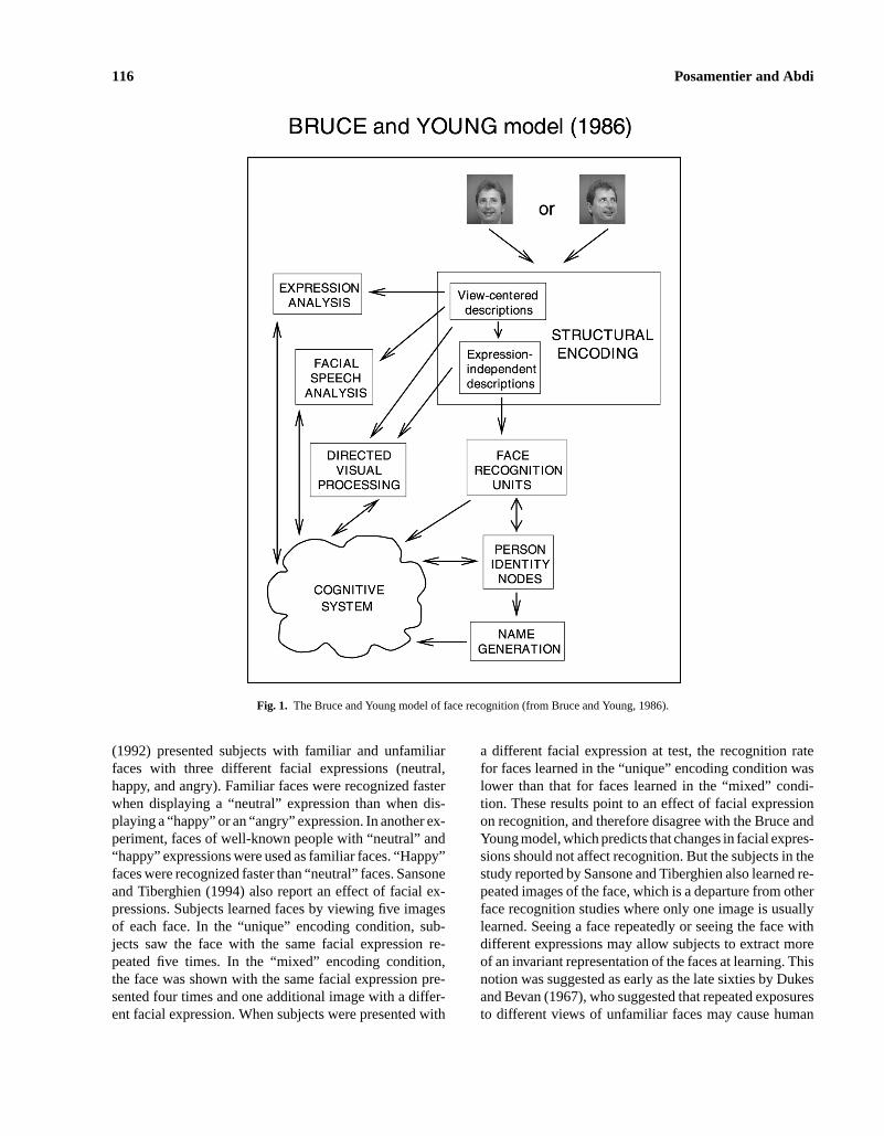
Casting findings from behavioral and neuropsycho-logical observations and the experimental approach into aunified framework, Bruce and Young (1986) developed anow classic model of face recognition expressed in termsof processing pathways and modules for recognition of fa-miliar faces (see Fig. 1). They suggested that seven typesof information can be derived from faces: pictorial, struc-tural, visually derived semantics (age and sex), identity-specific semantics, name, expression, and facial speech(movements of the lips during speech production) codes.
The pictorial code provides information about light-ing, grain, and possible imperfections in a photograph, aswell as information about pose and facial expression. Inother words, the pictorial code corresponds to the 2D im-age of the face. The pictorial code can mediate recognitiontasks in studies of episodic memory for faces because the
same pictures of previously unfamiliar faces are used atlearning and at test. The structural code captures the as-pects of the configuration of a face that distinguishes itfrom other faces. It corresponds to a 3D (view invariant)representation of the face. Bruce and Young distinguishedbetween the structural code for already familiar faces andthe tenuous structural code for unfamiliar faces, which islimited to the information that can be extracted from aninitial exposure, such as whether the face was seen withvarying poses and/or facial expressions. The Bruce andYoung model assumes that facial expressions, except forpossible characteristic expressions, are not important forface recognition. Facial expressions are treated as view-centered descriptions that vary across encounters with theface, and are consequently analyzed separately by the cog-nitive system for their affective content. A more abstract,expression-independent description of the face is the in-put to the face recognition units (FRUs), which containthe structural codes for faces. When a face is presentedfor recognition, the facial expression is parsed out andthe expression-independent description or “normalized”face is forwarded to the FRUs for further identification.The model therefore assumes functional independence be-tween identity and expression processing, and further dis-tinguishes between the processes involved in recognitionof familiar and unfamiliar faces.
The Bruce and Young model provided researcherswith a general framework for face processing. For exam-ple, the Bruce and Young model predicts that processingof familiar faces should be automatic and rapid, whereasprocessing of unfamiliar faces should require more effortand time. Additionally, the model predicts that judgmentsabout facial expressions should not be influenced by thefamiliarity of the face. Such predictions were tested byYoung et al. (1986), who examined the effect of face fa-miliarity in identity and expression matching tasks. In anidentity-matching task, subjects had to decide whether si-multaneously presented photographs of faces were pic-tures of the same or different persons. In agreement withthe Bruce and Young model, subjects were faster to de-cide that the persons were the same when they were fa-miliar than when they were unfamiliar. In an expression-matching task, subjects had to decide whether people inphotographs displayed the same or different facial ex-pressions. Again, the prediction of the model held up:There was no difference in reaction times for expressionjudgment of familiar and unfamiliar faces. These resultssupport independence in processing facial identity andexpressions.
However, some studies have challenged the assump-tion of the Bruce and Young model that facial expres-sions and identity are processed independently. Endo et al.
P1: ZBU
Neuropsychology Review pp970-nerv-470732 August 19, 2003 3:12 Style file version March 18, 1999
116 Posamentier and Abdi
Fig. 1. The Bruce and Young model of face recognition (from Bruce and Young, 1986).
(1992) presented subjects with familiar and unfamiliarfaces with three different facial expressions (neutral,happy, and angry). Familiar faces were recognized fasterwhen displaying a “neutral” expression than when dis-playing a “happy” or an “angry” expression. In another ex-periment, faces of well-known people with “neutral” and“happy” expressions were used as familiar faces. “Happy”faces were recognized faster than “neutral” faces. Sansoneand Tiberghien (1994) also report an effect of facial ex-pressions. Subjects learned faces by viewing five imagesof each face. In the “unique” encoding condition, sub-jects saw the face with the same facial expression re-peated five times. In the “mixed” encoding condition,the face was shown with the same facial expression pre-sented four times and one additional image with a differ-ent facial expression. When subjects were presented with
a different facial expression at test, the recognition ratefor faces learned in the “unique” encoding condition waslower than that for faces learned in the “mixed” condi-tion. These results point to an effect of facial expressionon recognition, and therefore disagree with the Bruce andYoung model, which predicts that changes in facial expres-sions should not affect recognition. But the subjects in thestudy reported by Sansone and Tiberghien also learned re-peated images of the face, which is a departure from otherface recognition studies where only one image is usuallylearned. Seeing a face repeatedly or seeing the face withdifferent expressions may allow subjects to extract moreof an invariant representation of the faces at learning. Thisnotion was suggested as early as the late sixties by Dukesand Bevan (1967), who suggested that repeated exposuresto different views of unfamiliar faces may cause human
P1: ZBU
Neuropsychology Review pp970-nerv-470732 August 19, 2003 3:12 Style file version March 18, 1999
Processing Facial Information 117
observers to extract some invariant information about theface, thereby allowing for better recognition from a newview of the face.
To summarize, the emergent picture from psycho-logical studies indicates that the factors that contribute tosuccessful face recognition are quite complex. Some stud-ies show that moderate changes in viewing angle and fa-cial expressions do not affect recognition accuracy. Otherwork shows that changes in facial expression and angledo affect recognition of unfamiliar faces whereas recog-nition of familiar faces is not affected by such changes.This suggests that different processes are engaged in therecognition of familiar and unfamiliar faces. Identifica-tion/recognition of familiar faces appears to be relativelyrobust against changes in viewing angle, whereas unfamil-iar faces are affected by rotation of the head. But whetheridentity and expressions are processed independently isstill not answered satisfactorily because several of thesestudies manipulated both changes in viewing angle and/orfacial expressions.
NEUROPHYSIOLOGICAL STUDIES
The earliest neurophysiological investigations intoface processing were done using single-cell recordings inthe temporal cortex in monkeys. Such studies have foundthat cells are preferentially tuned to the identity and ex-pression of the face. Although the results from these stud-ies cannot be taken as direct evidence of similar processesin humans, they provided direction for further studies withhuman populations.
Evidence for face-specific processing from single-cell recordings in monkeys has been established in a num-ber of studies, as reviewed by Perrett et al. (1992), andDesimone (1991). Populations of cells in the fundus ofthe superior temporal sulcus were shown to be selectivelyresponsive to human and monkey faces. Most cells re-sponded equally to faces of different individuals acrossdifferent orientations (in particular frontal and profileviews), but some cells also showed of sensitivity to theidentity of specific faces. Cells also responded to facecomponents, but not to other complex stimuli such assnakes, spiders, and food. Further, the cells were moresensitive to natural-looking stimuli than line drawings orschematic representations. The responses to inverted faceswere weaker with longer latency than to upright faces.
Single-cell recordings have also shed further lighton the presumed independence of facial identity and ex-pressions. Hasselmo et al. (1989) investigated the role ofexpression and identity in face-selective responses of neu-rons in the temporal visual cortex of macaque monkeys.
Hasselmo et al. recorded the responses of 45 neurons to astimulus set of pictures of three monkeys displaying threedifferent facial expressions (calm, slight threat, and fullthreat). Fifteen neurons responded to identity indepen-dently of expression, and nine neurons responded to thedifferent expressions independently of identity. Hasselmoet al. further found a differential response to the differ-ent expressions, with a stronger response to expressionsof full threat. The neurons responsive to expressions werefound primarily in the superior temporal sulcus, whereasneurons responding to identity were located in the infe-rior temporal gyrus. Although most single-cell responseshave been recorded in the visual cortex, this is not the onlyarea that has shown specific responses to faces. Leonardet al. (1985) found population of cells in the amygdala ofmacaque monkeys to be also responsive to faces.
From primate studies we then have an emergent pic-ture of neuronal activation patterns in response to faces inthe temporal region of the brain. Would a similar patternbe observable in human populations? Single cell record-ings in humans are usually done on epileptic patients whoundergo craniotomies in an attempt to find the locationof their seizures. In the sixties, Penfield and Perot (1963)used this technique to chart large regions of the cerebralcortex and gave use of the now famous picture of the ho-munculus illustrating how the body surface is representedon the brain surface. The first single-cell recordings of faceprocessing in humans were conducted by Ojemann et al.(1992) on 11 patients. Neuronal activity was measuredin 21 neural populations at 13 sites in the right superiorand middle temporal gyri. Identity matching and expres-sion labeling tasks showed significant neural activationchanges of 62% and 52%, respectively. Further, the facialexpression task showed more localized response patternsin the middle temporal gyrus, suggesting some specificityof neuronal responses to faces. These findings parallelHasselmo et al.’s findings of differential activation pat-terns (Hasselmo et al., 1989) to identity and expressionsin primates.
In the decade since Ojemann et al.’s study, severalgroups have used ERPs to determine both the time courseand localization of face processing. Some of these studieshave been done intercranially on epileptic patients beingevaluated for surgery, whereas other studies have usedscalp recordings on normal subjects. As we will see, elec-trophysiological recordings have shown a consistent pat-tern of results.
In three papers, Allison, McCarthy, Puce, and col-leagues report their findings from a number of electro-physiological studies of face processing in a large group ofepileptic patients. The 98 subjects included in these stud-ies had electrodes placed directly on the cortical surface
P1: ZBU
Neuropsychology Review pp970-nerv-470732 August 19, 2003 3:12 Style file version March 18, 1999
118 Posamentier and Abdi
to monitor epileptic seizures. In the first paper, Allisonet al. (1999) established the existence of differential ERPsgenerated by faces, sinusoidal gratings, objects, and letterstrings. Compared with other categories, the response tofaces is characterized as complex and reflected by at leastfour types of activity in the ventral occipitotemporal re-gion (N200, P290, P350, and N700, of which the N200is further explored in the next two papers of the series).The responses to faces were found bilaterally but with aslightly higher response in the right hemisphere.
In the second paper in the series, McCarthy et al.(1999) examined the face-specific responses in greater de-tail. In summary, they found the previously establishedN200 response to be insensitive to color, size, and spa-tial frequency manipulation. Inverted faces evoked a laterN200 amplitude than upright faces. The right hemisphereresponded faster to upright faces, whereas the invertedfaces evoked a more rapid response in the left hemisphere.Additionally, the N200 amplitude was largest for full facesand decreased when eyes, face contours, lips, and noseswere presented in isolation. Faces with averted eye gaze orclosed eyes evoked a larger potential than straightforward-looking faces. There was no difference in the response tofull and three-quarter views of face, but the amplitude forthese views was significantly larger than the response toprofile views.
The work reported in the third paper in the series(Puce et al., 1999) investigated the influence of top-downprocessing by examining the effect of emotionally chargedstimuli, repetition, priming, familiarity, and face identifi-cation. The N200 amplitude was significantly larger forfaces than affective stimuli. The N200 did not show aneffect of habituation nor semantic priming, and was alsounaffected by familiarity with the presented faces. Takentogether, the N200 response appears to be relatively con-stant for faces, as well as invariant to experimental ma-nipulations. The authors suggest that the N200 is the ear-liest evidence for face-specific processing and may reflectan instantiation of the “structural encoding” stage of theBruce and Young model.
Using scalp recordings, a number of studies have alsoexamined electrophysiological responses to faces in nor-mal populations. A consistent finding across studies isa negative evoked potential around 170 ms in responseto faces. In a series of experiments, Bentin et al. (1996)used scalp recordings on normal subjects to examine theface-specific response properties established in the sub-dural recordings reviewed above. Bentin et al. found thatfaces consistently evoked a negative potential around 170ms (N170), which was absent for other nonface stimuli.The N170 was largest in the posterior temporal scalp inthe right hemisphere. The response to inverted faces was
delayed, but the amplitude was the same as for uprightfaces. Similar response to inverted faces has been reportedby Rossion et al. (1999, 2000), Eimer (2000a), and Itierand Taylor (2002), as well as McCarthy et al. (1999) asreviewed above. Bentin et al. also report that distorted hu-man faces elicited an N170 amplitude similar to that fornormal faces. When presented in isolated, eyes actuallyelicited a larger N170 than whole faces, whereas nosesand lips elicited later and smaller negative ERPs. Bentinet al. also cast the scalp recorded N170 as the “structuralencoder” suggested by Bruce and Young, which is also theinterpretation of the subdurally recorded N200. The rela-tionship between these two responses is uncertain. Onecould speculate that the placement of the electrodes (sub-durally on the fusiform gyrus vs. the T6 location on thescalp) could account for the differences. Another inter-pretation takes into account the sensitivity of the N170to eyes alone. This may reflect the activation of an eye-sensitive region, which possibly serves as an orientationmechanism for the “face structural encoder.”
Bentin and Deouell (2000) also examined a possibleeffect of top-down processing in the encoding of faces. Inone experiment, subjects viewed pictures of famous faces,nonfamous faces, and butterflies. Subjects responded onlyto the butterfly stimuli, which were used as targets. There-fore, no explicit processing was done on the face stimuli.The N170 did not show a familiarity effect as famousand nonfamous faces evoked similar patterns of scalp dis-tribution and amplitude. But an effect of familiarity wasobserved later, at around 350 ms, with familiar faces elic-iting a more negative response, which was termed theN400 response. In a second experiment, subjects wereasked to process explicitly the faces in an identificationtask. The N170 again did not differentiate between fa-miliar and unfamiliar faces and was largest in the lateralparieto-occipital region of the right hemisphere. A differ-ence between familiar and unfamiliar faces was again ob-served with a larger latency, where familiar faces evokeda significantly larger negative response at N400. Bentinand Deouell interpret this N400 to reflect the activationof semantic memory or the “personal identity nodes” inthe Bruce and Young model. Similar results were alsoobserved by Eimer (2000b). Integrating the results fromtheir ERP studies as well as neuroimaging studies (whichwill be reviewed in detail later), Bentin and colleagues(Bentin and Carmel, 2002; Carmel and Bentin, 2002) pro-pose a model for face processing where visual stimuli,here faces, that include physiognomic information are dis-tinctively integrated into a perceptual representation. Thisprocess is performed by a complex neural mechanism thatprocess faces holistically but also is able to detect phys-iognomic features (e.g., eyes). The global processing is
P1: ZBU
Neuropsychology Review pp970-nerv-470732 August 19, 2003 3:12 Style file version March 18, 1999
Processing Facial Information 119
accomplished by a neural network located in the mid-dle fusiform gyrus, whereas componential processing isperformed by networks located in the superior temporalsulcus and the posterior inferior temporal gyrus. The sub-durally recorded N200 reflects the global processing in thefusiform gyrus. The N170 is also associated with globalprocessing as well as component-oriented activity. Bentinthus favors a domain-specific interpretation of the N170activity as it relates to faces. In brief, Bentin builds hisargument for domain specificity on the literature that sup-ports a view of faces as being “special.” For example,infants show preference for face-like visual stimuli overother (cf. Morton and Johnson, 1991), faces are processedas a whole (holistical or configural processing), whereasobjects are processed as a set of features (cf. Farah et al.,1998), recognition of inverted faces, but not objects, is im-paired (Yin, 1969). However, this view of face processingas being special is not unequivocally endorsed. Gauthierand colleagues in a series of publications have strongly ar-gued that faces do not hold a special status (Gauthier andTarr, 1997; Tarr and Gauthier, 2000; Rossion et al., 2002).Instead they view face processing as a case of within-category discrimination of stimuli that subjects have ex-pertise in processing. These diverging opinions of domainspecific vs. subordinate-level expertise account of faceprocessing will be discussed in more detail when review-ing neuroimaging studies that have examined the responseto faces in the fusiform gurus.
Event-related potentials have also been used to studyprocessing of facial expressions. In a study designed toexamine both processing of facial identity and facial ex-pressions in normal subjects, M¨unte et al. (1998) recordedERPs from multiple scalp locations to assess the timingand distribution of effects related to the processing of iden-tity and expressions. The earliest ERP differences in anidentity matching task were found around 200 ms. Theearliest ERP effects in an expression matching task camein later, around 450 ms. The tasks also differed in theirlocalization or scalp distribution: Identity was associatedwith a fronto-central effect, whereas expression was asso-ciated with a centroparietal effect.
Streit et al. (2000) evaluated differences in ERPs inemotional and structural face processing. In the emotionalprocessing task, subjects were asked to identify which oneof six facial expressions was present on the face. In thestructural encoding task, blurred images of faces and fiveother categories were presented and subjects had to iden-tify the category of each stimulus. Pictures of facial ex-pressions and blurred facial images evoked similar eventpotentials around 170 ms, which should be specificallyrelated to processing of facial stimuli and thus replicatingprevious findings. However, the facial expressions decod-
ing task evoked a peak around 240 ms, whereas such aresponse was absent for the blurred facial images. Ac-cording to the authors, this peak latency of 240 ms mightthen represent specific processes underlying the decodingof facial expressions. This 240-ms peak is different fromthe time sequence reported by M¨unte et al. for expressionmatching. The discrepancy may be due to task differences:a matching task is probably more effortful than an encod-ing task and would therefore take more time. Herrmannet al. (2002) replicated these findings.
Taking into account that prosopagnosia is character-ized by a disproportionate deficiency in face recognitioncompared with other stimulus categories, we should ex-pect to observe different patterns of evoked response po-tentials in prosopagnosic subjects. This idea has recentlybeen supported by findings from two studies. First, Bentinet al. (1999) examined face processing in a developmen-tal prosopagnosic subject. In a group of normal controlsubjects, faces exclusively evoked the now familiar N170.But in the prosopagnosic subject, the N170 was elicitedby both faces and objects, thus showing no selectivity.The nonselective N170 suggests that the impairment isat the level of structural encoding or reflects a failure toextract face-specific information as input to FRUs. In addi-tion, Eimer and McCarthy (1999) found a similar patternin a severely prosopagnosic subject who was comparedwith 24 normal controls. The control subjects showed anenhanced N170 in response to faces as compared withhouses. This effect was absent in the prosopagnosic sub-ject whose N170 did not discriminate between faces andhouses. When presented with inverted faces and houses,the N170 effect was present for the control subjects, butabsent in the prosopagnosic subject. Thus, the nonselec-tive N170 and the absent N170 in these two prosopagnosicsubjects indicate a dissociation between face and nonfaceprocessing. Neither of these studies examined ERPs inresponse to facial expression processing which usuallyremains intact in prosopagnosic subjects. This is unfortu-nate because preserved emotion processing still providesthe strongest evidence for a dissociation between the pro-cessing of identity and emotion.
In summary, findings from ERP studies have revealeddissociations between various face-processing tasks thatcan be cast in the framework of the Bruce and Youngmodel. The N170 shows the dissociation between pro-cessing of face and nonface stimuli and is thought to bethe instantiation of the processes involved in the structuralencoding of faces. Further, the Bruce and Young modelpredicts differential processing of familiar and nonfamil-iar faces, which is supported by the modulation of laterresponses. The N400 response, in particular, is thoughtto reflect the activation of the personal identity nodes.
P1: ZBU
Neuropsychology Review pp970-nerv-470732 August 19, 2003 3:12 Style file version March 18, 1999
120 Posamentier and Abdi
Additionally, the different time course for processing theemotional content from a face suggests that different pro-cesses underlie the processing of facial identity and facialexpressions, which is a dissociation clearly evidenced inprosopagnosic subjects.
So far, we have accumulated evidence from neu-ropsychological, behavioral, experimental, and neurophy-siological approaches to face processing that support dis-sociations between the various types of information thatcan be extracted from faces, in particular identity and emo-tion. The next sections of the paper will review findingsfrom the relatively new field of brain imaging that has al-ready made significant contributions to our understandingand knowledge of how faces and facial expressions areprocessed.
BRAIN IMAGING OVERVIEW
The last decade has seen rapid developments in func-tional neuroimaging methods which provide a measure ofthe brain at work. Positron Emission Tomography (PET)and functional Magnetic Resonance Imaging (fMRI) arethe two main techniques used to image face-processingtasks. Any change in behaviors and cognitive demandscauses a change in neural activity. Neural activation can bemeasured indirectly through changes in the hemodynamicresponse or certain properties of the blood, such as bloodflow or oxygen content. PET uses a radioactive tracer tomeasure rCBF, whereas fMRI measures changes in theoxygen content in the blood through the Blood Oxygena-tion Level Dependent contrast mechanism, referred to asthe BOLD signal. There are pros and cons to both imagingmethods. PET requires the injection of a radioactive tracerand this limits the number of scans that can be obtainedfrom a given subject. Being a noninvasive technique, fMRIis considered safe for both children and adults, but the en-vironment in the scanner is quite noisy and restrictive. ThefMRI signal is also extremely sensitive to motion of anykind. With PET, the signal averages approximately 10 mmin spatial resolution and 30 s to 1 min in temporal reso-lution. High-speed fMRI (so-called echo planar imaging)gives both better spatial and temporal resolution, with 3–8 mm and 100 ms respectively. Whereas techniques suchas ERPs can detect neuronal responses as early as 170 msafter stimulus onset, as we saw in the case of faces, thetime course for BOLD signal is actually rather sluggish.Using current fMRI technology, the first detectable changein hemodynamics appears after a delay of 2 s, reaches itspeak around 7–10 s, and remains elevated as long as theactivity continues, and returns to baseline 8–11 s after theactivity ceases. In a recent paper, Logothetis et al. (2001)
report on a study that simultaneously recorded intracor-tical neural signals and fMRI responses in anesthetizedmonkeys’ visual cortex. The BOLD signal actually corre-lated most strongly with the local field potentials (LFPs),not single- or multiunit activity which represents “neuralspiking” (neurons firing or action potentials). The LFPsreflect the aggregate activity from a population of neu-rons located in close vicinity of the recording electrodetip. Logothetis et al. conclude that their results unequiv-ocally show that an increase in the BOLD contrast di-rectly and monotonically reflects an increase in neural ac-tivity, and propose further that the statistical analysis andthresholding methods currently applied to fMRI data prob-ably underestimate the neural activity related to a stimu-lus or task. In a commentary, Bandettini and Ungerleider(2001) acknowledge the findings by Logothetis et al. aslandmark, but caution consideration because the resultswere obtained in anesthetized monkeys, and that the vari-ance explained by the LFP was only 7.6% larger com-pared with multiunit activity. A further discussion of theBOLD signal comes from Attwell and Iadecola (2002),who consider the hemodynamic response to be driven byneurotransmitter-related signaling and not directly by thelocal energy needs of the brain. These new insights intothe interpretation of the BOLD contrast are important, butwhether these results would require a complete reinter-pretation of past fMRI results would probably be takingthese findings too far. Clearly, as will be evidenced in thefollowing review, the brain does show different patternsof activation in response to different types of stimuli andtasks.
During a typical PET or fMRI scanning session, sub-jects attend to repeated blocks of various types of sensory,motor, or cognitive tasks for periods lasting up to severalminutes. However, with increasingly more refined scan-ning technology and better temporal resolution of fMRI,random stimulus presentation or event-related fMRI isused increasingly in experimental designs. The concept ofrandom stimulus presentation is quite significant, becausethe ability to analyze the response to randomly presentedstimuli allows for the import of traditional experimentalparadigms into imaging studies. Additionally, one also canhope to avoid the possible confounds of habituation andpossible strategies adopted while attending to blocks ofrepeated stimuli (Buckner and Logan, 2001; D’Espositoet al., 1999).
Before delving into the brain imaging literature onface processing, we present a brief overview of the sta-tistical techniques currently used in the analysis of imag-ing data. Prior to statistical analyses, the brain images gothrough a series of preprocessing steps that includes coreg-istration, spatial normalization, and smoothing. In general,
P1: ZBU
Neuropsychology Review pp970-nerv-470732 August 19, 2003 3:12 Style file version March 18, 1999
Processing Facial Information 121
the areas of activation are reported by giving the coordi-nates of a standard brain space called the Talairach andTournoux coordinates (Talairach and Tournoux, 1988).Several strategies can be used to perform the statisticalanalyses of brain imagery data. These include a voxel- (a3D pixel) by-voxel-based analysis [e.g., as implementedby SPM, cf. Friston et al., 1995; Friston, Holmes, Poline,Price, and Frith, 1996a], an ROI approach, and subtraction.In a voxel-by-voxel-based analysis, only those voxels thatexceed a set criterion value are kept in the subsequent con-trast analyses. A Bonferroni correction is routinely per-formed to correct for multiple comparisons by dividingtheα level by the number of voxels. When applied to acomplete brain image, this correction drastically reducesthe power of the statistical test (i.e.,α = 0.05/40, 000voxels gives a new significance level of 0.000,001,25).When researchers have a specific hypothesis as to whatregions of the brain should activate, they can increase thepower of the test by defininga priori an ROI, which is asubset of voxels covering a specific brain area. This re-duces the number of voxels subjected to statistical testingand so increases statistical power. Another way to iden-tify patterns of activation is to use the subtraction method.The basic premise of so-called cognitive subtraction isthat a cognitive task is made up of separable, but additivecomponents. By holding all but one component constant,any observed changes in activation should be due onlyto the experimental manipulation of the factor of interest.The signal or pattern of activation is averaged on a voxel-per-voxel basis for each experimental condition and thetwo averages are subtracted. Ideally, the resulting imageshould then reveal neural activation by the factor of in-terest. For example, in assessing activation in response tofearful facial expressions, the activation observed whenpresenting neutral expressions would be subtracted fromthe activation by fearful expressions. The activation pat-tern obtained from the subtraction should then reveal theareas most active in processing fearful expressions. De-spite its wide use, it should be mentioned that the basicpremise of cognitive subtraction has been questioned byresearchers who suggest that the brain is not a simple lin-ear system with response properties consistent with pureinsertion and additivity (Friston et al., 1996b; Price et al.,1997; Sidtis et al., 1999).
To date, a number of brain imaging studies have ex-amined neural activation patterns in response to a varietyof face-processing tasks. So far, we have established thatin particular the middle region of the fusiform gyrus is in-volved in the perception of faces, but we still do not havea complete picture of which structures or functional sys-tems are involved when having to recognize our frowningneighbor with a new hairstyle. The next two sections of
the paper will review findings from brain imaging stud-ies that examine neural activation patterns in response tofaces and facial expressions.
BRAIN IMAGING STUDIES OF FACES
One of the earliest works to use faces as stimuli wasa PET study conducted by Haxby et al. in 1991, whoshowed a dissociation between object (in this case, faces)and spatial visual processing. Face matching activated apathway in the ventral/occipitotemporal cortex, whereaslocation matching activated a dorsal/occipitoparietal path-way. One of the first, and by now, widely cited, neuroimag-ing study to deliberately examine activation by differentface-processing tasks was conducted by Sergent et al.(1992), who identified specific regions activated by faces.Since then, several research groups have used both PETand fMRI to investigate various aspects of face process-ing. In particular, the research groups led respectively byKanwisher and Gauthier have examined the role of thefusiform gyrus in early face perception and these twogroups have ended up with divergent interpretations. Theresults from these studies, as well as other studies thathave examined more complex face-processing tasks, suchas encoding, recognition, and the effect of face familiar-ity, will be evaluated in turn. Finally, a recent model offace processing based on brain imaging data proposed byHaxby and colleagues will be introduced.
Face Perception
Lesion studies of prosopagnosic subjects had earlyimplicated the importance of the occipital and temporallobes in face processing. In 1991, Haxby et al. used PETto show that faces did activate the occipitotemporal region.However, the functional aspects of these areas related toface processing had not yet been properly defined. UsingPET, Sergent, Otha, and MacDonald’s study directly ex-amined the functional neuroanatomy of face and objectprocessing (Sergent et al., 1992). They measured rCBF insix normal adults who performed a number of tasks involv-ing sinusoidal gratings, faces, and objects. Comparisonsof activation between tasks used the subtraction method. Agender categorization task (gender discrimination minusgrating) revealed strongest activation in the right occipitalgyrus (inferior, medial, and lateral) and right lingual gyrus(medial occipitotemporal gyrus), and lesser activation inthe left lateral occipital gyrus. A face identity task (faceidentity minus gender discrimination) revealed additionalactivation in the fusiform gyrus, anterior temporal cortex,
P1: ZBU
Neuropsychology Review pp970-nerv-470732 August 19, 2003 3:12 Style file version March 18, 1999
122 Posamentier and Abdi
and temporal poles of both hemispheres. Object recog-nition (objects minus gratings) showed activation in theleft occipitotemporal cortex and the left fusiform gyrus,and did not involve the right hemisphere regions activatedduring the face identification task. In addition, a high levelof activation was observed in the parahippocampal gyrusduring the face identification task. The parahippocampalformation is part of the limbic system, and is viewed asan area of convergence for perceptual and memory in-formation. Sergent et al. proposed that face identificationrequires the functional integrity of three specific corticalregions: the right lingual and fusiform gyrus, the rightparahippocampal gyrus, and the anterior temporal cortex.
Since the Sergent et al. study, a number of researchgroups have applied both PET and fMRI to explore how,where, and why faces activate the fusiform gyrus in partic-ular. In a series of publications, Kanwisher and colleagueshave examined the role of the fusiform gyrus in face per-ception. Kanwisher argues that this area can be viewedas a specialized module for face perception and thereforetermed this area as the Fusiform Face Area (FFA). In afirst paper, Kanwisher et al. (1997), using fMRI, initiallyidentified an area in the right fusiform gyrus that was sig-nificantly more active (in 12 of 15 subjects) when viewingfaces than common objects. Defining the fusiform gyrus astheir region of interest, Kanwisher et al. then ran a series ofexperiments testing for face-specific activation with a sub-set of five subjects. The right fusiform gyrus or the FFAresponded stronger to passive viewing of intact than toscrambled faces, frontal views of faces than frontal viewsof houses (another subordinate-level category), and three-quarter view of faces than human hands (passive viewingas well as consecutive matching task). Further examiningthe response properties of the fusiform gyrus, Tong et al.(2000) presented various types of face stimuli, includinganimal faces. The FFA response was weakest for nonfaceobjects and houses, but was equally large for cat, cartoon,and human faces. The response was equal for frontal andprofile views of faces, which is of interest in light of theresults from single-cell recordings that also show strongerresponse to frontal and profile views. Activation of theFFA seems also to be dependent on the level of atten-tion paid to the face stimuli: When the face stimuli appearoutside the focus of attention (covert visual attention), theactivity is reduced (Wojciulik et al., 1998). On the basis oftheir findings, Kanwisher and colleagues have concludedthat the FFA is selectively involved in the perception offaces, and that these results refute alternative accounts foractivation such as subordinate-level classification.
Kanwisher et al. (1998) also investigated the well-known face inversion effect. As documented in numerousstudies, inversion of faces disrupts the holistic or con-
figural processing of faces (Bartlett, Searcy, and Abdi,2003; Bartlett and Searcy, 1993; Farah et al., 1995, 1998;Moscovitch et al., 1997; Rhodes et al., 1993; Valentine,1988; Yin, 1969, 1970). In their fMRI study, Kanwisheret al. found that the average percent signal change in re-sponse to inverted grayscale faces was small and was alsonot consistent across subjects. This suggests that the FFAresponse is correlated with the perception of faces, ratherthan low-level visual features present in faces. Similarly,Haxby et al. (1999) also found that inverted faces pro-duced small and nonselective activation changes in theface-selective region. Interestingly, inverted faces actuallyproduced increased activation in a house-selective region,which suggests that inverted faces recruit the object per-ception system. Such a pattern of results is consistent withbehavioral findings in prosopagnosic subjects who oftendo not show an inversion effect and are thought to processfaces in an object fashion. Along the same lines, Aguirreet al. (1999) also report only small effects of face inversion.
Activation of the fusiform gyrus in response to faceshas consistently been replicated. For example, McCarthyet al. (1997) found activation in bilateral regions of theposterior fusiform gyrus when faces were viewed amonga montage of phase-scrambled objects. When faces wereviewed among normal objects, only the right fusiform re-gion was activated. To see whether another set of familiarwithin-category stimuli would activate the same regions,subjects viewed flowers presented in the same conditionsas faces. Flowers presented along with phase-scrambledobjects showed bilateral fusiform activation (greater in lefthemisphere), but no activation was obtained when flowerswere presented among objects. On the basis of these ac-tivation patterns, McCarthy et al. also conclude that facesspecifically activate the right fusiform gyrus. Similar acti-vation patterns have been reported by Clark et al. (1996);Haxby et al. (1994); Hoffman and Haxby (2000); and Puceet al. (1995, 1996).
From all these studies it seems that the designation ofthe fusiform gyrus (primarily the right) as the FFA is war-ranted. However, this view has been met with challenges.Specifically, Gauthier and colleagues argue that the acti-vation patterns in the fusiform gyrus in response to facesis due to the very high similarity between faces and sub-jects’ expertise with such stimuli. Therefore, activation inthis particular area of the fusiform gyrus may reflect pro-cessing of highly similar objects rather than facesper se.Recall from the discussion of ERP studies that Gauthierand colleagues take the same position when interpretingthe N170 response to faces. Going back to Rosch’s workon categorization (Rosch, 1978), objects can be classifiedinto a hierarchical set of categories. The superordinatecategories are the largest, composed of general categories
P1: ZBU
Neuropsychology Review pp970-nerv-470732 August 19, 2003 3:12 Style file version March 18, 1999
Processing Facial Information 123
such as furniture. Within this category are basic-level cat-egories, such as tables and chairs, which in turn containsubordinate categories, such as kitchen table and living-room table. Most object recognition takes place at the ba-sic level, and identifying an object as being a face (asopposed to another object) also takes place at the basiclevel. Face recognition, on the other hand, takes place atthe subordinate level between subsets of the basic-levelcategory “faces.” In other words, Gauthier and colleaguesview the fusiform gyrus as optimal for subordinate-levelcategorization or within-category discrimination with theimportant caveat that the observer has considerable exper-tise in processing the particular category of stimuli underconsideration.
Gauthier and colleagues contend that studies thathave concluded that the fusiform gyrus is a “special-purpose cortical machinery” for face recognition failedto consider experimental designs or stimuli that might re-fute such a conclusion. In their view, no attempts weremade to engage the “face module” by presenting onlynonface objects. Specifically, in an fMRI study, Gauthieret al. (1997) investigated the effects of category level of thestimuli in a matching task of nonface objects. Subjects per-formed two tasks, one visual and one semantic at the basicand subordinate category levels. In the visual task, sub-jects judged whether simultaneously presented picturesand words matched. In the semantic tasks, subjects de-cided if the object could move by its own power. Threeregions of interest were defineda priori: fusiform and in-ferior temporal gyri, lingual gyrus, and occipital cortex.The strongest activation associated with subordinate-levelvisual recognition was found in the fusiform and inferiortemporal gyri for seven out of eight subjects (bilateral forall but one subject who showed left hemisphere activa-tion), with additional activation in the left occipital lobe.The occipital lobe showed left activation in four subjects.Subordinate-level processing for the semantic task showedstrongest activation in the occipital lobe (two subjects bi-laterally, five subjects on the left only). The second mostactivated area was the fusiform and inferior temporal re-gion (two subjects bilaterally, two subjects on the left andright). To test specific activation in the fusiform and infe-rior temporal region shown in other studies to be respond-ing to faces, a double subtraction [visual(subordinate–basic)–semantic(subordinate–basic)] showed activationof the fusiform and inferior temporal gyri for subordinate-level processing.
Gauthier et al. (1999b) in an fMRI study furtherinvestigated whether another subordinate-level class ofobjects would activate the FFA. The stimuli they usedwere “Greebles.” First introduced by Gauthier and Tarr(1997), Greebles constitute a novel class of computer-
generated stimuli specifically designed to be similar tofaces along several dimensions. All Greebles are made upof the same number of parts set in the same configurationand displays individual characteristics. As a face, eachGreeble also belongs to a particular gender and family.Subjects were trained to become experts at recognizingGreebles and reached expertise level after an average of3240 trials. Their recognition patterns then showed ef-fects typically associated with face recognition, such asconfigural and holistic processing. In their imaging study,Gauthier et al. used the Greebles to examine activationin an a priori selected ROI (bilateral middle and ante-rior fusiform gyrus) usually activated by face process-ing. Subjects were scanned across six sessions when theyperformed sequential-matching judgments of unfamiliarfaces and Greebles (upright and inverted). By the end of thesixth session, Greebles activated the right middle fusiformgyrus as much as faces. The same area was also moreactivated by “Greeble experts” during passive viewing ofGreebles. Gauthier et al. (2000) further extended this find-ing to subjects who were experts at recognition of otherhomogeneous categories: birds and cars. The bilateral FFAand the right “occipital face area” were chosen as ROIs.The FFA showed higher activation in response to birdsand cars than to familiar objects. In addition, both regionsof interest showed significant expertise effects. It is there-fore the opinion of Gauthier and colleagues that the levelof categorization (subordinate-level discrimination) andexpertise are the determining factors for activation of theputative FFA rather than facesper se. In other words, theFFA is specialized in processing stimuli that display highwithin-group similarities and further that the subject ishighly specialized in processing such stimuli.
Thus, from the studies reviewed so far, we are leftwith two different interpretations of fusiform gyrus ac-tivation in response to facial stimuli. The positions ofthese two groups are further outlined in a set of commen-taries (Kanwisher, 2000; Tarr and Gauthier, 2000). In brief,on the basis of behavioral, neuropsychological, and neu-roimaging data, Kanwisher argues for a domain-specificview of face perception. Gauthier and Tarr argue that theFFA should be considered a flexible (as opposed to face)fusiform area for subordinate-level visual processing thatis automated by expertise.
However, a third account of the observed activationpatterns in the fusiform gyrus in response to different cate-gories of objects (including faces) has been put forward byHaxby and colleagues. As earlier reported, Haxby et al.(1991) showed differential activation patterns for facesand objects, with faces generally activating a ventral visualpathway and objects activating a dorsal visual pathway.Several studies by this group have also reported activation
P1: ZBU
Neuropsychology Review pp970-nerv-470732 August 19, 2003 3:12 Style file version March 18, 1999
124 Posamentier and Abdi
by faces in the fusiform gyrus (Haxby et al., 1994, 1996,1999). Ishai et al. (1999) propose that the ventral visualpathway is characterized by a distributed representation ofobjects. To support their view, subjects in an fMRI studywere scanned during passive viewing and delayed match-to-sample tasks of faces, houses, and chairs. Patterns ofactivation in response to the different categories variedacross the ventral temporal cortex in a highly consistenttopological arrangement, which was similar in both hemi-spheres. Houses elicited greatest activation in the medialfusiform gyrus and chairs elicited greatest activation inthe inferior temporal gyrus. Faces elicited greatest activa-tion in the lateral fusiform and occipitotemporal gyri. Ishaiet al. acknowledged that indeed these patterns of resultscan be interpreted as evidence for modularity, but each ofthe stimuli categories also showed significant patterns ofactivation in secondary areas that responded maximallyto a different category. Houses also elicited activation inthe area responding maximally to chairs and vice versa.Faces also activated the house and chair regions, but to alesser extent than houses and chair activated other regions,which the authors attribute to faces being processed moreautomatically. Activation in the secondary regions wasalso modulated by additional attention, as was required inthe matching task, which suggests that the secondary re-gions are recruited to enhance perception of highly similarstimuli.
Similar distributed patterns of response to faces,houses, and chairs were also found in bilateral regionsof the ventral occipital cortex (Ishai et al., 2000). The re-sponse to faces again appeared more restricted in spatialextent and the face area was also activated more automati-cally than the areas responsive to houses and chairs. Addi-tionally, a region in the superior temporal sulcus was iden-tified that responded almost exclusively to faces. Thesepatterns of results lead Ishai et al. to propose that the func-tional architecture of the ventral visual pathway, includ-ing both the occipital and temporal regions, is based ona distributed representation of objects. Objects, includ-ing faces, that share attributes or are common in theirform tend to cluster together. This creates a consistenttopographical arrangement and activation pattern. Usinga larger number of object categories, Haxby et al. (2001)confirmed their previous findings of distinct patterns of re-sponse for different stimulus categories. When excludingthe area that responded maximally to each object cate-gory, the category being viewed could still be identifiedon the basis of the pattern of response in the other ar-eas with a mean accuracy of 94%. Taken together, Haxbyand colleagues propose that the representations of facesand objects of the ventral temporal cortex are widely dis-tributed and overlapping. In their view, activation patterns
in the FFA does not solely represent the perception of thespatial arrangement of human faces, but that the FFA ispart of a more extended system of representation for allobjects. Spiridon and Kanwisher (2002) replicated thesefindings and further found that the pattern of activationextended to within-category stimuli that differed in view-point, exemplar, or image format.
The studies reviewed so far have examined activa-tion patterns in normal subjects. However, the study ofsubjects with cognitive impairments have provided valueinsights into cognitive processes, a case in point beingthe amnesic patient HM (Milner, 1966), whose patternof deficits contributed to the study and understanding ofhuman memory. As previously discussed, the study of im-paired face-processing skills in prosopagnosic subjectshas demonstrated a dissociation between face and non-face processing as well as facial identity and facial ex-pression processing. Acquired prosopagnosia occurs afterbilateral damage to the inferior port of the temporal cortex,including the fusiform gyrus, although only right hemi-sphere damage can also produce face recognition deficits.One explanation offered for the observed face recognitiondeficits in prosopagnosic patients are that impaired at pro-cessing faces as a whole, rather they are thought to rely on atime-consuming feature- or parts-based analysis of faces.Holistic faces processing is thought to be a right hemi-sphere function, whereas featural analysis is supported bythe left hemisphere (Rhodes, 1985, 1993). Rossion et al.(2000) found evidence for such differential processing offace stimuli in their PET study. The right middle fusiformwas more activated when subjects matched whole faces,whereas the left fusiform activated more when subjectsmatched facial features. These lateralized differences werenot observed when subjects processed objects as wholesor parts. If face recognition then relies on holistic process-ing of faces involving the right fusiform gyrus, and holisticface processing is impaired in prosopagnosics, one shouldexpect to see increased activation in the left fusiform whichnow appears to support more of a feature-based analy-sis of faces. This is the pattern that Marotta et al. (2001)observed in an fMRI study of two prosopagnosic sub-jects. The prosopagnosic subjects showed activation in thefusiform gyrus, but more voxels activated in a posterior re-gion than what was observed for the control subjects. Onepatient showed surprising evidence of posterior activationin the fusiform gyrus in the left hemisphere, which ledthe authors to suggest that this posterior activation reflectscompensatory processes when more anterior regions of thefusiform are damaged. This compensatory process couldthen be indicative of a feature-based approach to face pro-cessing. The three prosopagnosic subjects in Hadjikhaniand de Gelder’s fMRI study (Hadjikhani and de Gelder,
P1: ZBU
Neuropsychology Review pp970-nerv-470732 August 19, 2003 3:12 Style file version March 18, 1999
Processing Facial Information 125
2002) failed to show the normal pattern of higher activa-tion to faces than houses in the mid-fusiform and the infe-rior occipital gyrus. None of these prosopagnosic subjectshad any difficulty in a face detection task (face presentedin a noise pattern or whether the stimulus is a face or not).This raises the issue of the necessity of these brain regionsfor face recognition but not for face detection.
Another group of subjects that show impaired faceprocessing are individuals with autism spectrum disorders(ASD), including Asperger syndrome. Deficits in socialcognition which includes the inattentiveness to faces ofothers and failure to make appropriate eye contact suggestthat individuals with ADS probably have not developedtypical face expertise (Schultz et al., 2000b). Such indi-viduals generally rely on a feature-based analysis of theface rather than configural processing, which is one ofthe hallmarks of normal face processing. Schultz et al.(2000a,b) used fMRI to study face and object perceptionin such a group of subjects. Regions of interest were firstlocated in two groups of normal subjects to whom the acti-vation patterns in the patients groups were then compared.In the first experiment, the activation patterns in the pa-tient groups were compared with the first control groupon a face discrimination task. The autistic group showedsignificantly more activation in the right inferior temporalgyrus than the right fusiform gyrus. A second experimentcompared the patient group to the second control groupand generally replicated these initial findings. The autisticgroup showed larger activation in the left inferior tempo-ral gyrus compared with the controls. No consistent differ-ences were found between the control groups and the autis-tic group on a subordinate-level matching task of objects.The authors concede that the laterality differences needto be further clarified, but overall, the pattern of resultslends support to the claim that autistic subjects processfaces using an object-based processing strategy, which ac-tivates the inferior temporal gyrus. Further, Grelotti et al.(2002) propose that impaired face processing in ASD in-dividuals can best be cast in the framework of the expertisemodel rather than the FFA model. Cortical face special-ization failed to develop in ASD individual because oftheir reduced social interest. Although processing of fa-cial expressions will be reviewed in detail later, it is atthis point relevant to mention that high-functioning autis-tic adolescent were found to show impaired perception offacial expressions (Teunisse and de Gelder, 2001). Theirdeficits in processing facial expressions is obviously partof the pattern of impaired social cognitive skills, but moredirectly, the deficits can also be due in part to how autis-tic individual process facial expressions. A recent studyfound that autistic subjects focused on the mouth areas ofactors whereas normal controls focused more on the eyes
(Klin et al., in press). Whereas a correlation was found innormal subjects between fusiform and amygdala activa-tion when attending to the gaze direction of faces (Georgeet al., 2001), the reported lack of attention to the eyes inautistic individuals may then explain the low activationpatterns in the fusiform gyrus (Grelotti et al., 2002).
Encoding, Recognition, and Face Familiarity
So far, we have established that faces, when usedin relatively simple visual tasks, consistently activate thefusiform gyrus. However, the complexity of face process-ing in real life and even in the laboratory goes beyond pas-sive viewing or matching tasks. Our knowledge about theprocesses underlying face recognition comes from behav-ioral studies that employ more complex testing paradigmsthan the relatively simple visual tasks that have estab-lished activation patterns in the fusiform gyrus—whetherit should be designated as a special face-processing area ornot. Typically, face recognition has been investigated byhaving subjects first learn a set of faces and then recognizethem in a later testing session. Further, as we have seen,familiarity with faces is an important factor in face recog-nition. How informative have neuroimaging techniquesbeen in uncovering the areas activated with such experi-mental manipulations?
Haxby et al. (1996) used PET and a traditional be-havioral testing paradigm to examine areas of activationin response to encoding and recognition of faces. In thefirst scan, subjects viewed 32 faces presented sequentiallythree times in different orders for a total viewing timeof 8 min. The subjects were informed that they wouldbe tested on a recognition task in a later scan. Face per-ception and a sensorimotor task were included as controltasks. The activation patterns observed during encodingand recognition showed a dissociation between the neu-ral systems involved in these processes. As no ROI wasidentified a priori, the voxel-by-voxel statistical analy-sis revealed a number of regions involved in these tasks.Comparison of the memory tasks with the perceptual tasksshowed little overlap. Encoding of faces, compared withthe perceptual task, indicated increased rCBF in a num-ber of areas: left prefrontal cortex, right medial tempo-ral region, hippocampus, anterior cingulate cortex, andleft inferior temporal gyrus. Face recognition comparedwith the perception control task activated the right pre-frontal cortex, anterior cingulate cortex, bilateral inferiorparietal cortex, bilateral ventral occipital cortex, and thecerebellum. When compared with the sensorimotor con-trol task, encoding and recognition also showed activationin a large bilateral region of the ventral occipitotempo-ral cortex, in addition to the areas already mentioned. Of
P1: ZBU
Neuropsychology Review pp970-nerv-470732 August 19, 2003 3:12 Style file version March 18, 1999
126 Posamentier and Abdi
particular interest is the switch between hemispheres inthe prefrontal cortex depending on the task: left prefrontalcortex for encoding and right prefrontal cortex for recog-nition. The predominance of the right prefrontal cortexin recognition has also recently been reported by Wiseret al. (2000), who, in their PET study, compared memoryfor novel vs. well-learned faces. Subjects were asked torecognize faces that had been learned immediately beforescanning and faces that had been learned to the point offaultless recognition 1 week before scanning. Both tasksdid engage highly similar neural circuits, but memory fornovel faces involved more the frontal lobe areas, whereasmemory for well-learned faces engaged more the posteriorregions. Additionally, recognition of well-learned faces re-sulted in smaller activation nodes, suggesting less effortfulretrieval processes in recognition of familiar faces.
Bernstein et al. (2002) also examined the effect of en-coding strategy using PET. Subjects were scanned duringencoding of faces by using either a “deep” task (pleas-antness judgment), a “shallow” task (face orientation),or intentional learning, and were also scanned during asubsequent face recognition task. Encoding activated pri-marily a ventral system including bilateral temporal andfusiform regions and left prefrontal cortices. Recognitionactivated primarily a dorsal set of regions which includedthe right prefrontal and parietal areas. An effect of encod-ing strategy was observed with deep encoding activatingthe amygdala and left anterior cingulate. Differential ac-tivity was observed in the fusiform gyrus, which suggeststhat this area was also modulated by controlled processes.
The hemispheric switch between activation duringencoding and retrieval processes observed by Haxby et al.,Wiser et al., and Bernstein et al. is consistent with thepredictions from Tulving’s hemisphere encoding-retrievalasymmetry model (Tulving et al., 1994; see also Bartlettet al., 2003), which, roughly, postulates that encoding ac-tivates the left hemisphere and recognition activates theright hemisphere.
A study conducted by Kuskowski and Pardo (1999)also looked at encoding of faces, but manipulated the num-ber of times subjects saw the faces by presenting the facesone time only or repeatedly. Following the scans, subjectswere tested on their memory for the learned faces. Onlyscans with a postscan recognition rate of 70% correct wereincluded in the subsequent analysis. In the “face memory”condition, subjects were asked to learn 21 unfamiliar facesthat were presented individually without repetition. In the“face repeat” condition, subjects learned four unfamiliarfaces presented individually and repeatedly. The designalso included “face watching” and “scrambled face” con-ditions as controls. Again, the results showed activationacross several regions during face encoding, but the areas
of activation also appeared to be dependent on the en-coding condition. For the “face memory minus scrambledface” comparison, the largest magnitude of responses waslocated in the right mid-fusiform gyrus, with additionalactivation in both anterior fusiform gyri, and prefrontalcortex. “Face repeated minus scrambled face” showed ac-tivation in the right precentral gyri, left cingulate gyrus,left lateral cerebellum, prefrontal cortex, and temporal cor-tex (left anterior fusiform foci). “Face watching minusscrambled face” revealed activation in the bilateral tem-poral pole regions, bilateral anterior fusiform regions, leftparietal cortex, left prefrontal, right inferior frontal, andprecuneus. The strongest positive correlations with recog-nition test scores were found in the right mid-fusiform re-gions. No hippocampal or parahippocampal activation wasrecorded, which contrasts with previous findings (Haxbyet al., 1996; Kapur et al., 1995; Sergent et al., 1992).
As previously mentioned, familiarity is a strong me-diating factor in face recognition. According to the Bruceand Young model, recognition of familiar faces is medi-ated by an established structural code in long-term mem-ory. By contrast, recognition of unfamiliar faces is, to alarge degree, dependent upon the strength of a structuralcode that can be extracted from the initial exposure to theface, as well as upon the pictorial code. Dubois et al. (1999)investigated the effect of face familiarity in a PET study.Subjects performed a gender categorization task on bothfamiliar and unfamiliar faces, as well as face recognitionand pattern discrimination tasks, which were included ascontrol tasks. All face tasks showed bilateral activation inthe fusiform gyrus, with activation patterns in the rightanterior area closely matching the mean coordinates re-ported by Kanwisher et al. (1997). Dubois et al. also founddifferential activation patterns for known and unknownfaces, with rCBF increasing in the left amygdala in cate-gorization of unknown faces (subtraction of known fromunknown faces). The amygdala activation in response tothe gender categorization task was unexpected. For exam-ple, Sergent et al. (1992) did not observe any activationin the amygdala in response to this task. On the otherhand, it is well established that the amygdala is activatedby processing of threatening stimuli, as well as a varietyof facial expressions. For example, the facial expressionof fear consistently activates the amygdala (Morris et al.,1996, 1998; Whalen et al., 1998). The amygdala also re-sponds to happy and neutral facial expressions, but showsrapid habituation effects (Breiter et al., 1996). Therefore,unknown faces may initially have been processed as pos-sible threatening stimuli, whereas the known faces hadbeen encountered without negative implications duringthe familiarization process and therefore did not presenta “threat” that would activate the amygdala. Additionally,
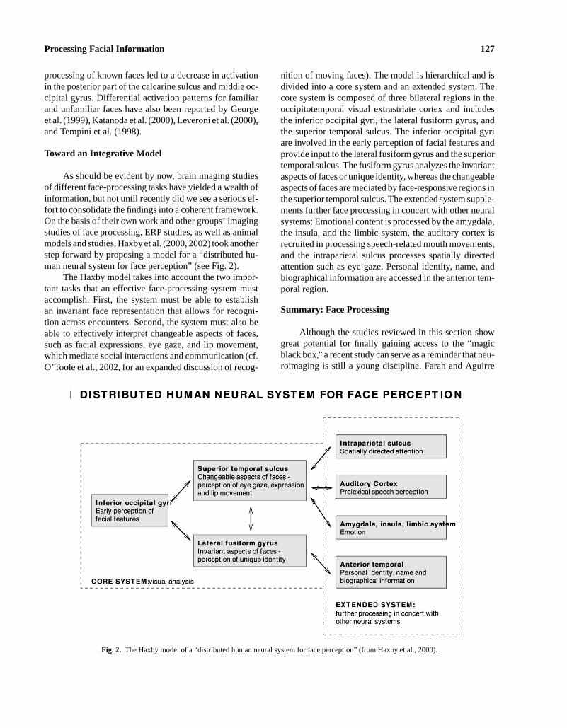
P1: ZBU
Neuropsychology Review pp970-nerv-470732 August 19, 2003 3:12 Style file version March 18, 1999
Processing Facial Information 127
processing of known faces led to a decrease in activationin the posterior part of the calcarine sulcus and middle oc-cipital gyrus. Differential activation patterns for familiarand unfamiliar faces have also been reported by Georgeet al. (1999), Katanoda et al. (2000), Leveroni et al. (2000),and Tempini et al. (1998).
Toward an Integrative Model
As should be evident by now, brain imaging studiesof different face-processing tasks have yielded a wealth ofinformation, but not until recently did we see a serious ef-fort to consolidate the findings into a coherent framework.On the basis of their own work and other groups’ imagingstudies of face processing, ERP studies, as well as animalmodels and studies, Haxby et al. (2000, 2002) took anotherstep forward by proposing a model for a “distributed hu-man neural system for face perception” (see Fig. 2).
The Haxby model takes into account the two impor-tant tasks that an effective face-processing system mustaccomplish. First, the system must be able to establishan invariant face representation that allows for recogni-tion across encounters. Second, the system must also beable to effectively interpret changeable aspects of faces,such as facial expressions, eye gaze, and lip movement,which mediate social interactions and communication (cf.O’Toole et al., 2002, for an expanded discussion of recog-
Fig. 2. The Haxby model of a “distributed human neural system for face perception” (from Haxby et al., 2000).
nition of moving faces). The model is hierarchical and isdivided into a core system and an extended system. Thecore system is composed of three bilateral regions in theoccipitotemporal visual extrastriate cortex and includesthe inferior occipital gyri, the lateral fusiform gyrus, andthe superior temporal sulcus. The inferior occipital gyriare involved in the early perception of facial features andprovide input to the lateral fusiform gyrus and the superiortemporal sulcus. The fusiform gyrus analyzes the invariantaspects of faces or unique identity, whereas the changeableaspects of faces are mediated by face-responsive regions inthe superior temporal sulcus. The extended system supple-ments further face processing in concert with other neuralsystems: Emotional content is processed by the amygdala,the insula, and the limbic system, the auditory cortex isrecruited in processing speech-related mouth movements,and the intraparietal sulcus processes spatially directedattention such as eye gaze. Personal identity, name, andbiographical information are accessed in the anterior tem-poral region.
Summary: Face Processing
Although the studies reviewed in this section showgreat potential for finally gaining access to the “magicblack box,” a recent study can serve as a reminder that neu-roimaging is still a young discipline. Farah and Aguirre
P1: ZBU
Neuropsychology Review pp970-nerv-470732 August 19, 2003 3:12 Style file version March 18, 1999
128 Posamentier and Abdi
(1999) addressed some of the difficulties in interpretingactivation patterns in imaging studies aimed at identifyingneural substrates of visual recognition. They performed ameta-analysis of 20 tasks from 17 different imaging stud-ies of visual recognition of words, objects, and faces. A to-tal of 84 local maxima of activation were plotted using theTalairach and Tournoux coordinates. Failing to observea focal clustering of activation even within each stimu-lus class, the authors reached the conclusion that “visualrecognition is a function of the posterior half of the brain”(p. 181). On the other hand, when isolating the points of ac-tivation for passive viewing of faces only, the coordinatesof activation do fall close to those reported by Kanwisheret al. (1997). The general failure to observe a consensusamong the studies included in the meta-analysis was at-tributed to methodological issues, spatial normalizationtechniques, statistical analysis, and individual differencesin subjects’ brain structure.
As we have seen, the fusiform gyrus has been stronglyimplicated in face processing. Recall that there are di-vergent opinions as to whether the fusiform gyrus (righthemisphere in particular) may be considered as the FFA asillustrated by the exchange between Kanwisher and Gau-thier. Is it then possible to arrive at a reconciliation ofthese two views? Perhaps it is worth considering the typeof stimuli that is under investigation. Few would disagreethat faces constitute the category that the human percep-tual and cognitive systems are most highly specialized toprocess. No other category of visual stimuli comes closeto engaging the cognitive system as faces do, unless theobserver is an apparent specialist in processing such cate-gories at a subordinate level. Thus, the designation of thefusiform gyrus as the FFA can be considered a “defaultoption” which remains in effect until the system operatoroverrides or complements the specialization for faces withanother category of stimuli. Of course, the results from theHaxby group indicate that activation in the fusiform gyrusin response to face stimuli is part of a distributed and over-lapping system that processes both faces and objects.
Other studies have gone beyond the fusiform gyrusand attempted to identify regions and chart patterns of acti-vation in response to more challenging face memory tasks,such as encoding and recognition. As expected, when face-processing tasks become more demanding, the activationpatterns become more complex. Far more areas light upbeyond the now expected activation in the fusiform gyrus.The model proposed by Haxby et al. for a distributed neu-ral system for face processing provides a sound frameworkfor future investigations. The challenges we will face inthe next decade will be to further refine our investigativemethods to answer questions: What is the functionality ofthe areas of activation? How do the various areas interact?
What is the temporal sequence of activation? And how dowe establish an invariant representation of faces that canalso accommodate the processing of changeable aspects,such as facial expressions?
BRAIN IMAGING STUDIES OF FACIALEXPRESSIONS
Facial expressions have been studied for more than100 years: As early as in the 1870s, Charles Darwin wroteof emotion and facial expressions. A century later, thework by Ekman (1972, 1992) and Izard (1971, 1977) ledthe field in the study of perception and categorization of fa-cial expressions. It is generally accepted that there exist atleast six basic categories of emotion that can be conveyedthrough facial expressions: happiness, surprise, fear, sad-ness, anger, and disgust. The perception and assignment offacial expressions to these categories are claimed to be uni-versal, as members of both literate and preliterate culturescategorize facial expressions along the same dimensions.However, this approach has not escaped criticism relatedmainly to the problem of ecological validity. For exam-ple, Russel (1997) argues that the kinds of expressionsseen in Ekman and Friesen’s photographs (Ekman andFriesen, 1976) are rare occurrences in daily interactions.Setting controversies aside, the database of facial expres-sions compiled by Ekman and Friesen has been widelyused by a number of research groups in the area of per-ception and categorization of facial expressions, as wellas face recognition studies.
As we have previously seen, lesion studies suggestthat some specific brain structures or different neural sub-strates are involved in processing different types of face-related information. In fact, we are looking at a doubledissociation: prosopagnosic patients are impaired at facerecognition but have intact facial expression processing,and other subjects are impaired at facial expression pro-cessing but can still recognize faces. Results from stud-ies of facial expression processing suggest that differ-ent emotions are also processed by different regions ofthe brain. For example, Adolphs et al. (1996) investi-gated facial expression recognition in a large number ofsubjects with focal brain damage. The authors hypothe-sized that cortical systems primarily responsible for recog-nition of facial expressions would involve discrete re-gions of higher-order sensory cortices. Recognition ofspecific emotions would depend on the existence of par-tially distinct systems. This predicts that different patternsof expression recognition deficits should depend upon thelesion site. In general, none of the subjects showed impair-ment in processing happy facial expressions, but several
P1: ZBU
Neuropsychology Review pp970-nerv-470732 August 19, 2003 3:12 Style file version March 18, 1999
Processing Facial Information 129
subjects displayed difficulty in recognizing negative emo-tions (especially fear and sadness). The authors proposethat deficits in processing negative emotions can be due tothe fact that the number of negative emotions is largerthan the number of positive emotions. In fact, there isonly one positive emotion, namely happiness. As such, ahappy smile should be easily recognizable. It is also possi-ble that negative emotions show a deficit because they canbe easily confused, such as mistaking fear for surprise, oranger for disgust.
So far, a general consensus has been reached on theanatomical structures involved in the processing of fearand disgust, with findings coming from both lesion andbrain imaging studies. We also have an emerging pictureof the perception of happy facial expressions, because thisexpression has often been used as a basis for comparison instudies of different expressions. Other facial expressionssuch as anger and sadness have received more limitedattention. So far, no brain imaging study has examined theexpression of surprise.
The following sections will review processing of thesix basic emotions in turn: fear, disgust, sadness, anger,happiness, and surprise. Each type of facial expression willbe examined from both human subjects and neuroimagingstudies.
Fear: Behavioral Studies
Close links have been established between the emo-tion of fear and the amygdala. For example, LeDoux(1996) proposes that the fear reaction system involves par-allel transmissions to the amygdala from both the thala-mus and the sensory cortex, which he refers respectively toas the “low” and “high” roads. The direct pathway fromthe thalamus to the amygdala is the shortest and fastesttransmission route. This “low road” provides the amyg-dala with a crude representation of the stimulus and allowsfor immediate response to the present danger. The “highroad” involves elaborated processing in the sensory cortexbefore the input reaches the amygdala and is slower butmore precise. In agreement with LeDoux’s theorization,neurons in the monkey amygdala have been shown to re-spond to the affective significance of sensory stimuli, andlesions of the amygdala render the animal insensitive tostimuli that normally evoke fear. In humans, the amyg-dala has been studied as a result of surgical lesions andelectrical stimulation, in particular in epileptic patients.The role of the amygdala in processing fear and aggres-sion in social behaviors has been firmly established (cf.Aggleton, 1992; Davis and Whalen, 2001, for reviews).Animals studies have shown that the amygdala also re-
ceives highly processed visual input, and contains neuronsthat respond selectively to faces (Rolls, 1992).
The role of the amygdala in processing facial expres-sions has been highlighted in three interesting case studies.In all cases, the perception of fear is particularly compro-mised. The first case of SM was reported by Adolphs et al.(1994, 1995). A female in her early thirties at the time oftesting, SM suffers from Urbach-Wiethe disease, whichhas resulted in bilateral calcification and atrophy of theamygdala. Her performance on facial expression percep-tion was evaluated in a series of experimental tasks andcompared with the performance of several control groups(unilateral amygdala damage, other brain damage, andnormal subjects). When SM judged faces showing fear,her ratings of fear were less intense than any of the controlgroups’ ratings. In addition, SM’s ratings for afraid facescorrelated poorly with normal ratings, and she often de-scribed faces displaying a fearful expression as surprisedor angry. Multidimensional scaling of perceived similar-ity judgments of facial emotions revealed that SM is lessable to interpret similarity or blends between expressionsthan are normal controls. When asked to draw variousfacial expressions, SM was unable to draw the expres-sion of fear, stating that she did not know what an afraidface looks like. Yet, she understands the verbal concept offear. SM showed no difficulty in recognizing people fromtheir faces, providing additional evidence for a dissocia-tion between recognition of emotion and identity in faces(Damasio et al., 1990; Humphreys et al., 1993; Tranelet al., 1988). Adolphs et al. conclude that the amygdalais required for linking visual representation of facial ex-pressions with representations that constitute the conceptof fear.
The second case, DR, was first reported by Younget al. (1995). DR, a female in her early fifties at time oftesting, became epileptic at the age of 28. Brain imagingshowed extensive lesions in left amygdala and smaller le-sions in the right amygdala, resulting from a series of sur-gical procedures to the amygdala. Although DR has someproblems in recognizing familiar people out of context aswell as faces encountered postoperatively, her unfamiliarface matching capability (as evaluated by the Benton test)was unimpaired. By contrast, her performance on facialexpression matching and expression recognition tasks wasseverely impaired, but as SM, DR can describe and defineemotions.
In a follow-up study, Young et al. (1996) furtherevaluated DR’s facial expression processing. In a facialexpression-matching task, DR experienced difficultieswhen the same face was shown with different expres-sions, and when different faces were shown with the sameexpression. In an identity-matching task, DR exhibited
P1: ZBU
Neuropsychology Review pp970-nerv-470732 August 19, 2003 3:12 Style file version March 18, 1999
130 Posamentier and Abdi
problems whenever the same face was presented with dif-ferent expressions. In addition, she was poor at imaginingfacial expressions.
Calder et al. (1996) also tested DR’s recognition offacial expressions, using morphed facial expressions be-tween faces taken from the Ekman and Friesen series offaces. This testing paradigm uses photographic images ofsix prototype emotions that are interpolated or morphedto create a hexagonal continuum (running from happinessto surprise to fear to sadness to disgust to anger back tohappiness). Control subjects identified these morphed im-ages of facial expressions as belonging to distinct regionsof the continuum, corresponding to the nearest prototypeexpression, which was not shown. DR was impaired onthis task, in particular at identifying morphed images offear, and to a lesser extent disgust and anger. By contrast,DR showed no impairment in an identification task basedon morphed faces of famous people.
The third case was reported by Anderson and Phelps(2000). SP, a 54-year-old female at time of testing, had theright amygdala removed, showed a lesion in the left amyg-dala, and had a right temporal lobe resection. SP’s facialrecognition was intact and she could correctly judge sexand age from faces. Compared with normal controls, SPwas severely impaired in her appraisal of fear, as well asdisgust, sadness, and to a lesser extent happiness. Ratingsof surprise and anger were not impaired. As both SM andDR, SR’s lexical affect identification was normal. Whenasked to generate facial expressions (which were video-taped for later ratings), SR’s facial expressions were ratedas accurate as those generated by normal controls. Thus,she cannot perceive a face as fearful but she can herselfproduce the expression of fear. This is in contrast to SM,who was not able to produce a drawing of a fearful face.
Taken together, the above findings (Adolphs et al.,Anderson and Phelps, Calder et al., and Young et al.)strongly support the amygdala’s involvement in recogni-tion of emotion, and fear in particular. Furthermore, thesestudies also support the existence of a dissociation be-tween processes underlying facial identity and facial af-fect. Insofar as these studies indicate that some emotionshave specific pathways, they also militate for the existenceof a more general dissociation between facial identity andfacial affect.
Unfortunately, the picture may not be as clear asthe previous studies indicate. Specifically, Hamann et al.(1996) found intact recognition of emotions in two pa-tients (ages 73 and 59) with bilateral amygdala lesionsresulting from herpes simplex encephalitis after age 50.These patients were tested on the same material as patientSM (refer to case study already described earlier in thissection). The inconsistent results can be attributed to two
possibilities. First, structures outside the amygdala may beimplicated in recognition of facial emotions. SM did showsome damage to areas outside the amygdala (anterior en-torhinal cortex). Second, and more importantly, impairedrecognition of emotions in facial expressions may resultonly if the amygdala lesions occurred early in life, suggest-ing a developmental or learning component in the abilityto process facial expressions as socially relevant stimuli.This would be consistent with SM’s congenital conditioncaused by Urbach-Wiethe disease.
Fear: Neuroimaging Studies
As we have seen, lesions of the amygdala lead to se-lective deficits in processing emotional facial expressions.Several groups of researchers have turned to neuroimagingtechniques to investigate how normal subjects process fa-cial expressions. In general, the testing paradigm adoptedin such studies has been to present faces displaying fear,happy, and/or neutral expression, and examine differentialactivation through subtraction. This section will concen-trate on the activation patterns obtained for the expressionof fear.
Morris et al. (1996), using PET, measured rCBF infive subjects who viewed fearful and happy faces. Eachface within the emotion category showed six levels ofintensity of expression (obtained with morphed images),resulting in a 2× 6 factorial design. As predicted, presen-tation of fearful faces (as assessed by ana priori contrastwith happy faces) showed activation in the left amygdalaand left peri-amygdaloid cortex. No activation was ob-served in the right amygdala. Other areas of activationincluded the left cerebellum, the right superior frontalgyrus, and the left cingulate gyrus. The presentation ofhappy faces (contrasted with fearful) showed activation inthe right medial temporal gyrus, right putamen, left su-perior parietal lobule, and left calcarine sulcus. The con-trast of emotional faces (both happy and fearful) vs. neu-tral faces produced activation in the left pulvinar and theright orbitofrontal cortex. Additionally, orthogonal con-trasts comparing neutral and emotional intensity levelshowed that the pattern of activation was affected by thelevel of emotional intensity (increasing with increasinglevel of fear and decreasing with increasing level of hap-piness). Areas responsive to increasing intensity of fearwere the left anterior insula, left pulvinar, and the rightanterior cingulate, as well as the amygdala. The left amyg-dala was also responsive to decreases in the intensity ofhappy expression. No explicit recognition or classifica-tion of the emotional expressions was required as the sub-jects classified the faces by gender. Therefore, activation
P1: ZBU
Neuropsychology Review pp970-nerv-470732 August 19, 2003 3:12 Style file version March 18, 1999
Processing Facial Information 131
of the amygdala does not appear to require explicit at-tention (e.g., classification of the expression) being paidto the facial expression. Further, regression analysis in-dicates that activation in the amygdala appears to modu-late expression-specific neural activity in the extrastriatecortex.
Breiter et al. (1996) used fMRI and presented stan-dardized facial expressions of fear and happiness to exam-ine activation in the amygdala. Their first experiment ex-amined visual processing of fearful and happy vs. neutralfaces in normal subjects. Presentation of the face stimuliinvolved a fixed order (first fear, then happy, to optimizeactivation by fearful faces) both within and across runs.Fearful expressions activated the amygdala bilaterally, butshowed higher activation for the left (0.68 signal change)than the right (0.52). No significant signal changes wereassociated with happy faces compared with neutral facesin the amygdala. The second experiment counterbalancedthe expression condition within and across runs to con-trol for order effects. An ROI comprising the left amyg-dala again showed significant activation for the fear vs.neutral contrast. Breiter et al. also found posterior amyg-dala activation for each type of facial expression againsta low-level baseline, which suggests that the amygdalamight respond to faces in general. Post hoc analysis ofthe within-run signal changes across runs showed a sig-nificant order or habituation effect in an ROI comprisingthe amygdala for both fearful and happy expressions. Thefusiform gyrus, known to be activated during the process-ing of faces, was chosena priori as a control region. Thisarea showed the same general activation for both fearfulvs. neutral and happy vs. neutral comparisons, and did notshow a within-run signal decrement (habituation) pattern.Wright et al. (2001) found differential habituation patternsin the amygdala with the right amygdala showing greaterhabituation to emotionally valenced stimuli than the leftamygdala. Feinstein et al. (2002) found evidence of habit-uation outside the amygdala, using a test/retest paradigmwith different facial expression (including fear). With theapparent salience of the affective stimuli decreasing at thesecond presentation, habituation was observed in areas ina right hemispheric network (right frontal and precentralgyrus, right insula, right postcentral gyrus, and right infe-rior parietal lobule). However, a left hemispheric network(angular gyrus, posterior superior temporal gyrus, precen-tral gyrus, and insula) showed increased activation at thesecond presentation of the stimuli. This left network is in-volved in object recognition, and it is thought that the faceswere encoded as unfamiliar objects during the first expo-sure and recognized as being familiar during the secondexposure, which was reflected by the increased activationpattern. This study did not observe any significant activa-
tion in the amygdala, but the significant activation in theprefrontal cortex may have attenuated the response of theamygdala.
As we have already seen, the amygdala is activatedin response to fearful facial expressions without any ex-plicit processing instructions (Morris et al., 1996, 1998).Whalen et al. (1998) investigated whether the amygdala isactivated in response to emotional stimuli, even when thesubjects do not explicitly know that such stimuli are be-ing presented. They used a backward masking procedureconsisting of 33-ms presentation of fearful and happy ex-pressions followed by 167-ms presentation of neutral fa-cial expressions. Eight of 10 subjects reported that theyonly saw neutral faces. Activation in the amygdala wassignificantly different when subjects viewed masked fear-ful expressions than happy faces. Fearful faces increasedthe response significantly and happy faces decreased theresponse, a pattern which is consistent with the directionalpattern found by Morris et al. (1996). In addition, signalintensity in response to masked fearful faces showed a sim-ilar habituation pattern as the one reported by Breiter et al.(1996). The experimental manipulation also produced ac-tivation in the substantia innominata in response to bothfearful and happy expressions. The substantia innominataactivation might represent a more generalized response tothe salience or arousal level of the stimuli, whereas theamygdala activation is based on the valence of the stim-uli. A number of studies report activation in the amygdalain response to visual stimuli with strong negative valence,including faces (Hamann, 2001; Irwin et al., 1996; Tayloret al., 1998, 2000).
Iidaka et al. (2001), using fMRI with a 3T magnet,revealed a differential role of the left and right amygdalaand their neural connections. The analysis showed thatthe left amygdala was predominantly involved in the pro-cessing of negative expressions but the right amygdala didnot show any differentiation for the expression conditions.The activation in the left amygdala covaried with activityin the left ventrolateral and right dorsolateral prefrontalcortices, suggesting that processing of negative expres-sion is based on activation of a loop between the amyg-dala and the prefrontal cortex. The activation in the rightamygdala interacted with the hippocampus and the tem-poral cortex in the right hemisphere. Further, Hariri et al.(2000) investigated the existence of a neural network thatcan control and modulate instinctive emotional responses(in this case labeling an emotional experience). Subject ei-ther matched the facial expression of two faces to a target(perceptual task) or identified the expression by choosingone of two presented linguistic labels (intellectual task).The matching task showed increased activation in both theleft and right amygdala. However, the labeling task was
P1: ZBU
Neuropsychology Review pp970-nerv-470732 August 19, 2003 3:12 Style file version March 18, 1999
132 Posamentier and Abdi
associated with decreased activation in the amygdala, butthis decrease was correlated with an increase in activa-tion in the right prefrontal cortex which is implicated inregulating emotional responses.
The neuroimaging studies reviewed so far have usedadult subjects (mean age across four studies was 29.9years). A recent fMRI study conducted by Baird et al.(1999) examined activation of the amygdala in a groupof adolescents (mean age= 13.9 years) when fearful fa-cial expressions were presented. This study found no sig-nificant difference between left and right amygdala acti-vation. The data were therefore combined and showed asignificant activation compared with looking at a fixationpoint. An obvious limitation of this study is that subjectssaw only one type of facial expression. By including sev-eral emotions in the same study, it would be possible tocompare differences in activation patterns involved in pro-cessing the different expressions. Nevertheless, the findingdemonstrates that the amygdala is involved in processingfearful expressions before adulthood. However, becausethe children studied are relatively old, the data reportedcannot eliminate a learning factor in the involvement ofthe amygdala in processing facial expressions. Such learn-ing may occur earlier in development. Using a group ofyounger children as well as young adults, Thomas et al.(2001) report an fMRI study that assessed amygdala ac-tivation in response to a passive viewing task using fear-ful and neutral faces. The mean age of the children was11 years and the mean age of the adults was 24 years. Theadults showed increased left amygdala activation for fear-ful faces compared with neutral faces. This pattern wasnot found in the children who actually showed greater ac-tivation in the amygdala in response to neutral faces whencompared with a fixation point. Behavioral data showedthat the children experienced problems in categorizing fa-cial expressions as fearful and neutral, which suggest thatchildren may not reach adult levels in such discriminationtasks until early adolescence, which is the pattern observedwith regard to face recognition. Thus, these results do pointto a learning component. The higher activation for neutralfaces was attributed to a perceived ambiguity in the stim-uli, which may result in increased vigilance and therebyincreasing the activity of the amygdala. Further, genderdifferences were observed in the children: Boys showed adecrease in response with repeated presentations of fear-ful faces, but the response in girls remained stable. Theseresults are quite intriguing as they point to not only a de-velopmental effect but also a gender effect. Kesler-Westet al. (2001) also found gender differences in their sub-ject pool of adult men and women. Men showed greaterbilateral activation while viewing angry faces than happyfaces; greater right hemisphere activation while viewing
angry faces than sad faces; and greater left hemisphere ac-tivation to sad faces than happy faces. No such differenceswere reported for the female participants.
In summary, results from both human subjects andanimals, lesion, and neuroimaging studies concur on theimportance of the amygdala in the processing of fearfulstimuli, including facial expression. On the other hand, theamygdala is also activated by other facial expressions andeven faces in general, and shows rapid habituation effects.One possible explanation for this generalized response tofaces is that there may be differential responses by subsetsof nuclei within the amygdala structure. The amygdala isa small structure situated deep within the brain and there-fore is hard to image. However, the use of higher fieldstrength magnets (3T vs. 1.5T magnets) and more refinedimage acquisition techniques would increase the abilityto detect more subtle activation changes in deep brainstructures.
Disgust: Behavioral Studies
Evidence for a specific neutral substrate dedicated tothe processing of stimuli involving disgust comes fromnonhuman primate studies. Yaxley et al. (1988) identi-fied the primate anterior insular cortex as the gustatorycortex, because this structure contained neurons that re-spond to pleasant and unpleasant tastes. Can a similararea be identified in the human brain that would respondto “disgusting” stimuli, in particular the perception of dis-gusted facial expressions? In what follows, impaired per-ception of disgust is illustrated different patient groups[Huntington’s, obsessive–compulsive disorder (OCD),and Tourette’s syndrome patients with and without OCD].Then, evidence from neuroimaging studies supporting aspecific neural substrate for disgust is presented.
Among the many symptoms of Huntington’s diseaseare deficits in visual and auditory perception of social stim-uli. Patients suffer generalized intellectual deterioration,including impaired face and facial expression processing(Jacobs et al., 1995). Initial neural degeneration affectsthe basal ganglia and the caudate nucleus. Postmortemstudies have also revealed tissue loss in the amygdala re-gion. Sprengelmeyer et al. (1996) investigated face per-ception and emotion recognition in a group of patients withHuntington’s disease. The patient group performed a se-ries of tasks that explored perception of age, sex, unfa-miliar face identity (the Benton test), and gaze direction.They showed a marked deficit in evaluating gaze direction,and their performance on the Benton test was significantlybelow the controls. Using morphed facial images, sub-jects’ perception of male–female continua, identity (i.e.,
P1: ZBU
Neuropsychology Review pp970-nerv-470732 August 19, 2003 3:12 Style file version March 18, 1999
Processing Facial Information 133
Grant-Bogart) continua, and emotion continua (sadness–happiness and fear–anger) were evaluated. Patients withHuntington’s disease performed the same as controls on alltasks except the fear–anger continua. To further evaluateemotion recognition, subjects identified morphed imagesof facial expressions from an “emotional hexagon.” Thesame task was employed by Calder et al. (1996) in thecase study of DR reviewed previously. The only emotionthat was not impaired was happiness. Subjects had diffi-culty in recognizing anger, fear, and particularly disgust(63%, 57%, and 24% correctly identified, respectively).Identification of disgust was below chance level. Subjectswith Huntington’s disease also committed a significantnumber of so-called “remote prototype errors” (misclas-sifying emotions far apart on the emotional hexagon, suchas mistaking surprise for anger). A study by Gray et al.(1997) further confirms the selective impairment for dis-gust in Huntington’s disease. They studied a group of sub-jects who carried the gene for Huntington’s disease butotherwise were clinically presymptomatic with no cog-nitive deterioration. Identification of famous faces, unfa-miliar face matching, recognition memory for faces werenormal. Facial expression recognition showed a selec-tive deficit in recognition of disgust with some impair-ment for fear and anger. Interestingly, a study involvingschizophrenic patients and their unaffected first-degreerelatives found impaired face recognition in both theschizophrenics and the relatives (Conklin et al., 2002).Impaired-face-processing skills have been consistentlydemonstrated in schizophrenic patients (Archer et al.,1994; Baudouin et al., 2002; Heimberg et al., 1992; Mandalet al., 1998). Further studies are of course warranted, butimpaired processing of facial information may provideinsight into the etiology and symptomatology of differentdisorders. Sprengelmeyer et al. (1997) report that patientssuffering from OCD are also selectively impaired in rec-ognizing the expression of disgust, but that they under-stand the verbal meaning of disgust. Interestingly, groupsof patients with generalized anxiety disorder tested in thesame study showed an enhanced recognition of fear andanger.
Because the study conducted by Sprengelmeyer et al.of patients with Huntington’s disease used the same testprocedure and material as Calder et al. (1996) in assess-ing bilateral amygdala damage, the performance of thesetwo patient groups can be compared. Both groups showedselective impairment in processing fear, which implicatesthe involvement of the amygdala. But the disproportionateimpairment in recognition of fear in patients with amyg-dala damage and the selective deficit in recognition ofdisgust in patients with Huntington’s disease and OCDpoints to a double dissociation, which suggests that dif-
ferent emotions may have dedicated neural substrates. Inaddition to the involvement of the basal ganglia struc-ture, the processing of the facial expression of disgustwould likely take place in a neural structure or networkthat includes the insular cortex and the orbitofrontal cor-tex. This network should be able to integrate visual, audi-tory, as well as olfactory information. Strengthening thesupport for a specific region dedicated to processing ofdisgust is the report a patient who suffered focal dam-age to the left insula and putamen and was impaired atthe recognition of the facial expression of disgust andthe experience of disgust (Calder et al., 2000). Interest-ingly, Harmer et al. (2002) found that a group of euthymicpatients with bipolar disorder actually showed enhancedrecognition of the expression of disgust. This finding raisesthe possibility that the study of facial expression recog-nition may provide insights into the neural networks thatare involved in the regulation of mood as well as otherdisorders.
Disgust: Neuroimaging Studies
Following up the clinical evidence that suggests ded-icated neural substrates for different emotions, Phillipset al. (1997) sought to find distinct neural substrates forthe perception of fear and disgust. An fMRI study was de-signed to replicate amygdala activation in fear perception,and to explore the possibility of a separate substrate forthe perception of disgust. Subjects viewed faces from theEkman and Friesen face set displaying disgusted, fearful,and neutral expressions. The disgusted and fearful expres-sions were morphed to show two levels of intensity (75%and 150%). Four conditions (presented randomly) incor-porated an alternating (neutral/emotional) design for eachemotion (fear/disgust) and intensity of expression (75%and 150%). During scanning, subjects performed a genderidentification task on the faces presented. A generic brainmap showed activation of the left insula and amygdala for75% fearful faces vs. neutral faces. No activation was ob-served at the 150% level of fearful expressions, possiblydue to a type II error or to a rapid habituation of amyg-dala response (cf. Breiter et al., 1996). Adopting an ROIapproach of 18 voxels representing the amygdala regionbilaterally, activation was observed in the right amygdalaand the putamen for the 150% fear intensity level. Themost significant finding for the perception of facial expres-sion of disgust was activation in the right insula, but notin the amygdala. Activation in the anterior insula becamegreater with increased emotional intensity. Additional ar-eas activated by disgust were the medial frontal cortex,and right putamen (part of the basal ganglia structure) and
P1: ZBU
Neuropsychology Review pp970-nerv-470732 August 19, 2003 3:12 Style file version March 18, 1999
134 Posamentier and Abdi
thalamus for the 150% intensity level. The anterior insulais connected to ventro-posterior-medial thalamic nucleus,which has been identified as the gustatory cortex in pri-mates. In humans, this area is active during salt tasting andperception of aversive stimuli. Thus, the neural responseto facial expressions of disgust appears closely related tothe appraisal of distasteful stimuli and may well be servedby the same neural substrate.
Sprengelmeyer et al. (1998) further examined cere-bral activation in response to disgusted as well as fearful,angry, and neutral facial expressions. Six normal subjectstook part in the fMRI study and performed a gender clas-sification task on the face images. A voxel-by-voxel sta-tistical analysis compared each expression (fear, anger,and disgust) against neutral faces as the baseline con-dition. The areas activated by the expression of disgustclosely matched the findings of Phillips et al., with ac-tivation of the left insular cortex and the right putamen(basal ganglia area). Thus, converging evidence from clin-ical populations (Huntington’s disease and OCD) and neu-roimaging studies supports a dedicated neural substratefor processing the facial expression of disgust, which isa neural network incorporating the basal ganglia and theinsular cortex. Contrary to previous findings, this studydid not find activation in the amygdala in response tofear. This was attributed to rapid habituation effects inthe amygdala (cf. Breiter et al., 1996). Fearful expressionactivated the right fusiform gyrus and the left dorsolateralfrontal cortex. Angry expression evoked activation in theleft inferior frontal lobe and the posterior part of the lefttemporal lobe. The results generally showed no overlapin the areas activated by the different emotions with theexception of the inferior frontal cortex, which was equallyactivated to faces showing anger, fear, and disgust as com-pared with neutral faces. Illustrating impaired facial ex-pression in schizophrenics, Phillips et al. (1999) found thatnonparanoid schizophrenics categorized the expression ofdisgust as either anger or fear more frequently than para-noids, and showed activation in the amygdala in responseto disgust. Paranoid schizophrenics were more accuratein recognizing expressions, and showed greater activationpatterns than nonparanoids.
A recent study by Gorno-Tempini et al. (2001) alsoreports activation of in the right putamen and caudate nu-cleus in response to disgust, in addition to activation in theright thalamus and the left amygdala. Recall that activationin the caudate nucleus and the amygdala was not reportedby Phillips et al., which Gorno-Tempini et al. attributeto experimental design issues as they contrasted disgustwith happiness and used an explicit expression recogni-tion task. The contrast for happiness vs. disgust showedbilateral activation in the orbitofrontal region.
The study of the expression of disgust further sup-ports the idea that different structures are involved in theperception of different facial expressions. The link be-tween the amygdala and fear has already been established.It now appears that the basal ganglia structure and the in-sular cortex are involved in the perception of disgust.
Sadness and Anger: Behavioral Studies
The expression and perception of emotions such assadness and anger are closely related to the concept ofempathy—the identification with and understanding ofanother’s situation, feelings, and motives. Sociopathy ischaracterized by disregard for others and aggressive be-haviors. In a first study to investigate the relationship be-tween expression recognition and behavioral problems,Blair and Coles (2000) showed that in their sample ofadolescent children, the ability to recognize sad and fear-ful expressions was inversely related to levels of affective–interpersonal disturbance, impulsiveness, and conductproblems. Further, Blair and Cipolotti (2000) reported ona patient with damage to the right frontal region, includ-ing the orbitofrontal cortex, who showed a case of whatthey termed “acquired sociopathy.” The patient showeddifficulties in the area of social cognition, in particularfacial expression recognition of anger and fear. The pa-tient’s impairment was attributed to a reduced ability togenerate expectation of other’s negative emotional reac-tions and to suppress inappropriate behaviors. The authorspropose that the orbitofrontal cortex is implicated in thegeneration of such expectations and suppression of behav-iors. Psychopathic patients generally show reduced auto-nomic responses to sad and angry expressions, impover-ished aversive conditioning, and reduced startle reflexes,which is a syndrome similar to patients with amygdala le-sions. Patients with lesions to the temporal lobe, includingthe amygdala, consistently fail to recognize fear as well asimpaired recognition of sad facial expressions (Fine andBlair, 2000). Both animal and human lesion studies haveimplicated the orbitofrontal cortex in behavioral extinc-tion and reversal learning (see Rolls, 2000). Therefore, theamygdala and orbitofrontal cortex would be likely struc-tures for processing facial expressions for sadness andanger respectively.
Sadness and Anger: Neuroimaging Studies
A recent study by Blair et al. (1999) reports findingsof dissociable neural responses to the facial expressions ofsadness and anger. Thirteen male subjects took part in the
P1: ZBU
Neuropsychology Review pp970-nerv-470732 August 19, 2003 3:12 Style file version March 18, 1999
Processing Facial Information 135
PET study. During scanning, subjects performed a sex dis-crimination task on morphed facial expressions of sadnessand anger displaying varying degrees of intensity. The sta-tistical analysis identified differential responses to sad andangry faces. Signal intensity also increased as a functionof degree of emotional intensity displayed by the face.Sad expressions showed activation in the left amygdalaand the right middle and inferior temporal gyrus. Angryexpressions showed significant activation in the right or-bitofrontal cortex (as predicted) and bilateral activationin the anterior cingulate cortex. Conjoint activation wasfound in the right temporal pole and anterior cingulatecortex as a function of increased emotional intensity. Theunilateral response in the left amygdala to sad expressionsparallels the response to fearful faces observed in otherimaging studies. Unfortunately, this study did not includethe perception of fear as a control stimuli. As such weare left to speculate whether there would be a differentialresponse to sad and fearful expressions in the amygdala.As suggested before, it is possible that different nucleiof the amygdala may respond to different emotions. Theactivation of the right inferior and middle temporal gyriin response to sad expressions is in line with the findingsof Adolphs et al. (1996) who proposed an extended righthemisphere neural system for processing facial expres-sions. On the basis of their results, Blair et al. suggest theinvolvement of at least two dissociable, but interlockingsystems in the processing of negative facial expressions.One system responds to facial stimuli (sad, fearful) in-volved in (social) aversive conditions; the other systemimplicates regions involved in behavioral extinction by re-sponding to angry facial expressions. The differential ac-tivation patterns obtained by Sprengelmeyer et al. (1998)for fear, anger, and disgust support this position.
Recent studies have examined perception of facial ex-pressions in schizophrenic patients. Schizophrenics haveconsistently performed worse than controls in facial ex-pression discrimination tasks (Heimberg et al., 1992).Phillips et al. (1999) replicated activation in the left amyg-dala in normal controls in response to fearful expres-sions. Schizophrenics showed no activation in the amyg-dala in this implicit gender discrimination task. Further,Schneider et al. (1998) found amygdala activation in nor-mal controls in a sad mood induction task, using sad fa-cial expressions. The schizophrenics in this study did notshow any activation. Kosaka et al. (2002) had schizophren-ics and normal controls perform an emotional intensityjudgment task on positive (happy) and negative (angry/disgusted/sad) faces. Here, in contrast, the schizophrenicsshowed greater activation in the amygdala than did thenormal controls. In a study with another type of patientgroup (manic patients), Lennox et al. (2002) found that
normal controls displayed the expected activation patternof increased activation in the amygdala and the anteriorcingulate cortex to increasing intensities of sadness. Thispattern of response was absent in the manic patients.
Happiness: Behavioral Studies
Smiling appears to be innate. An infant will producethe first smile anywhere from 2 to 12 hr after birth. In fact,even blind and deaf babies smile. Ekman has identifiedseveral different types of smiles; enjoyment, dampened,miserable, qualifier, compliance, coordination, flirtatious,and embarrassed (in McNeill, 1998, pp. 206–208). Thetype of smile most used in facial expression studies is thatof enjoyment or the so-called Duchenne smile. Accordingto the norms published by Ekman and Friesen (1976),mean accuracy for recognition of the facial expression ofhappiness reached 100% for the subset of faces used byYoung et al. (1996), which makes the smile the most easilyrecognized expression.
So far, no patient groups have displayed problems inrecognizing happy facial expression. Both patients withamygdala damage (SM and DR) and patients withHuntington’s disease performed at normal levels in pro-cessing happy facial expressions. Similarly, Adolphs et al.(1996) did not find any patients that displayed problemsin recognizing happy facial expressions. In fact, recogni-tion scores for happy expression did not correlate with therecognition of any other emotion. Taken together, theseresults suggest that a happy facial expression may be pro-cessed differently than all other expressions.
Happiness: Neuroimaging Studies
Neuroimaging studies of the perception of happy fa-cial expressions have usually contrasted this expressionwith the perception of negative facial expressions (i.e.,fear, anger, and disgust). So far, no consistent pattern of ac-tivation has been found in response to smiling. Morris et al.(1996) found no activation in the amygdala for the con-trast of happy–fearful expressions. Statistical parametricmapping of this contrast showed activation in the right me-dial temporal gyrus, right putamen, left superior parietallobe, and left calcarine sulcus. On the other hand, Breiteret al. (1996), in their second experiment, found that theleft anterior amygdala responded preferentially to happyvs. neutral faces. This suggests a possible generalized re-sponse to emotionally valenced stimuli in the amygdala,but rapid habituation effects to both happy and fearful ex-pressions were observed. Support for a broader role for
P1: ZBU
Neuropsychology Review pp970-nerv-470732 August 19, 2003 3:12 Style file version March 18, 1999
136 Posamentier and Abdi
the amygdala in modulating the vigilance level during theperception for both positive and negative comes from arecent study by Yang et al. (2002), who found reliable bi-lateral amygdala activation to happy as well as sad, angry,and fearful faces. Gorno-Tempini et al. (2001) found bi-lateral activation in the orbitofrontal regions (BA 11 and47) in both implicit and explicit processing of happy ex-pressions.
A PET study conducted by Dolan et al. (1996) inves-tigated neural activation during covert processing of happyfacial expressions. Prior to scanning, subjects were pre-sented with a face that they were explicitly asked to hold inmind for 45 s after which they had to match the target faceto one of two faces. Repeated blood flow measures wereobtained whereas the subjects held happy and neutral facesin mind. Presentation of happy faces (compared to neutraland a controlled fixation condition) was associated withsignificant activation in left ventral prefrontal cortex, leftanterior cingulate cortex, thalamus, and the right fusiformgyrus. In contrast to other studies, the amygdala was notactivated.
It thus appears that there is a differential activationpattern for negative and positive facial expressions. Pro-cessing of negative facial expressions involves regions thatspecifically have been damaged in clinical populations,such as the amygdala for fear, and the basal ganglia andthe insula for disgust. No consistent activation patterns inresponse to a smiling face have been identified, nor didAdolphs et al. (1996) find any patients who were deficientin processing happy facial expressions.
Surprise
So far, no major studies have focused on the facialexpression of surprise. The few data available on the per-ception, classification, and identification of surprised fa-cial expressions have been collected in conjunction withassessment of the five other basic emotions (Adolphs et al.,1994, 1995; Calder et al., 1996; Young et al., 1995, 1996).For example, patient SM rated facial expressions of sur-prise, anger, and fear as less intense than controls. Bi-lateral damage to the amygdala has been shown to im-pair judgment of the intensity of fearful expressions, andalso of expressions normally judged to be similar to fear,such as surprise. As the main focus of these studies wasfear and the involvement of the amygdala, any observeddeficit related to the perception of surprise was not fur-ther addressed. The expression of surprise bears strongresemblances with fear: eyes and mouth wide open. Sur-prise is also the briefest expression, lasting less than 1 s.An important point to consider is that emotional states are
dynamic. This is especially important for surprise, becausean expression of surprise can quickly turn into fear or hap-piness, depending upon the nature of the surprise. In fact,surprisecan be defined as a transitory emotion which leadsinto the appropriate reaction for the emotional event fac-ing the person. Given the short-lived nature of a surprisedexpression and given that surprise often transitions into anexpression of fear, it is quite likely that the amygdala isalso involved in processing the perception of surprise.
Context Effects
The areas involved in the processing of facial expres-sions have been identified by the use of relatively simpletasks, such as gender categorization and passive view-ing, which involves mostly implicit processing. A majorpoint to consider when assessing such activation patternsis how well these tasks replicate face processing in reallife. Few would argue that the environment in a scanningroom and even psychology laboratories, for that matter,comes close to replicating real-life situations. In the realworld, a correct interpretation of facial expressions wouldalso be guided by the situational context, which is usu-ally accompanied by a verbal message and a body lan-guage. Thus, we can add factors such as prosody as wellas gaze to aid the interpretation of a facial expression.In addition, the salience and valence of the expressionsmust also be taken into account. Interpretation of facialexpressions in real life would therefore generally appearto benefit from additional top-down or explicit process-ing. Two recent studies have established differences inactivation patterns between explicit and implicit process-ing of facial expression. In an explicit processing task,Critchley et al. (2000) had subjects judge happy, angry,and neutral facial expressions. In an implicit processingtask, subjects performed a gender categorization task onfaces displaying the same expressions as in the explicittask. Overall, processing of facial expressions increasedactivity in the fusiform and middle temporal gyri, the hip-pocampus, the amygdala-hippocampal junction, and thepulvinar nucleus. The neural substrates for explicit andimplicit processing were shown to be dissociable, with ex-plicit processing activating the temporal lobe cortex andimplicit processing activating the amygdala. The latter re-sult thus replicates findings from previous experiments asreviewed above.
The study by Gorno-Tempini et al. (2001), alreadyreferred to in the discussion on processing of disgust,used both explicit and incidental tasks, expression recog-nition and gender decision respectively, in processing ofdisgusted, happy, and neutral expressions. Regions of
P1: ZBU
Neuropsychology Review pp970-nerv-470732 August 19, 2003 3:12 Style file version March 18, 1999
Processing Facial Information 137
activation varied by both task and facial expression: theright neostriatum and the left amygdala showed higher ac-tivation in explicit processing than in incidental process-ing. The bilateral orbitofrontal cortex activated to explicitrecognition of happy expressions. Common activation forall conditions were found in the right temporal occipitaljunctions, which would be indicative of visual and per-ceptual processing, and the left temporal and left inferiorfrontal cortex, which would be involved in semantic pro-cessing. Clearly then, activation is both distributed andtask-modulated.
The observed difference in activation patterns be-tween explicit and implicit processing, or what could alsobe viewed as conscious and unconscious processing, fallsin line with LeDoux’s concept of the “low and high roads”for processing emotional stimuli (LeDoux, 1996). Recallthat the “low road” provides a crude representation of thestimuli to the amygdala, whereas the “high road” involveselaborated processing in the sensory cortex. By addingfactors such as social context, prosody, gaze, and body lan-guage to the relatively simple visual perception of facialexpressions, the conscious processing of facial expressioncan then be also subsumed under the concept of socialcognition (see Adolphs, 1999, 2001, for recent reviewsof the field of social cognition). For example, damage tothe amygdala goes beyond pure impairments in facial ex-pression recognition and appears to play a role in socialdecision making. This is illustrated by a study of threesubjects with bilateral damage to the amygdala who wereimpaired at facial expression recognition and who devi-ated from normal controls in social judgments involvingfacial stimuli. The subjects with amygdala damage judgedfaces that normal subjects had deemed most untrustwor-thy and unapproachable as trustworthy and approachable(Adolphs, 1999; Adolphs et al., 1998). Adolphs et al.(2001) recently extended this finding to a group of high-functioning autistic subjects who were tested on the samematerial as the bilateral-amygdala-damaged patients. Theautistic subjects showed normal social judgments fromlexical stimuli, but, as the amygdala patients, showed ab-normal social judgments regarding the trustworthiness offaces. These findings support the role of the amygdala inlinking visual perception of socially relevant stimuli withthe retrieval of social knowledge and subsequent socialbehaviors.
Summary: Facial Expressions
The goal of imaging studies of the perception of facialexpressions has been to evaluate whether there are distinctneural substrates dedicated to processing emotions as dis-
played by different facial expressions. Evidence from be-havioral and lesion studies do suggest that different struc-tures are activated by different emotions. The role of theamygdala in processing fearful stimuli has been well es-tablished. Recall that the patients who presented lesionsof the amygdala and were impaired at processing negativeemotions with fear being most strongly affected. Patientswith Huntington’s disease display loss of amygdala andbasal ganglia tissue, associated with impaired processingof fear, anger, and disgust in particular. However, no sub-ject groups displayed any difficulties in processing happyfacial expression. This suggests differential processing ofpositive and negative emotions. So far, a number of neu-roimaging studies have shown differential activation pat-terns in response to five of the six basic emotions displayedby facial expression. No studies have examined activationby surprised facial expression.
Activation of the amygdala by fearful expressionsshould come as no surprise as reported by Morris et al.(1996, 1998) and Breiter et al. (1996). But, note that thefacial expressions of sadness and happiness also activatedthe amygdala. Amygdala activation has been also reportedin a categorization task of unknown faces (Dubois et al.,1999). Thus, it is quite likely that the amygdala also re-sponds to faces in general with the purpose of assessingthe possible threatening valence of a stimuli (e.g., “Is thisa friend or foe?”). Further, the results from the imagingstudies of disgust implicate the basal ganglia structure aswell as the insular cortex in the processing of this emo-tion (Gorno-Tempini et al., 2001; Phillips et al., 1997;Sprengelmeyer et al., 1997). Interestingly, the two fa-cial expressions for which consistent patterns of activa-tion have been established are fear and disgust. These areemotions that are evoked in direct threats to the system.
The majority of studies that have examined activationin response to different facial expressions also have foundactivation in other areas (e.g., prefrontal cortex, inferiorfrontal cortex, right medial temporal region, anterior cin-gulate cortex, inferior temporal cortex, and orbitofrontalcortex) in addition to the regions of particular interest.The inferior frontal cortex is commonly activated in re-sponse to different facial expressions, and may serve as anarea for integration or semantic processing of informationcontained in facial expressions. Additionally, activationin the orbitofrontal cortex in response to facial expres-sions is quite interesting because this region is implicatedin numerous cognitive activities, including decision mak-ing, response selection and reward value, behavior con-trol, as well as judgments about olfactory stimuli (Abdi,2002; Blair and Cipolotti, 2000; Rolls, 2000). Attractivefaces produced activation in the medial orbitofrontal cor-tex, and the response was enhanced if the face was smiling
P1: ZBU
Neuropsychology Review pp970-nerv-470732 August 19, 2003 3:12 Style file version March 18, 1999
138 Posamentier and Abdi
in addition to being attractive (O’Doherty et al., 2003).However, the areas mentioned above are also activated byother face-processing tasks, such as encoding and recog-nition. Therefore, the functionality or exact role of theseadditional areas of activation remains unclear.
According to Haxby et al.’s distributed neural modelof face perception (Haxby et al., 2000), some of these ad-ditional areas of activation would be considered to be partof the extended network dedicated to the processing ofchangeable aspects of the face. One approach to furtherexamine the functionality of these extended activation pat-terns would be to include facial expressions as part of anexperimental design that manipulates facial expressionsduring both encoding and recognition of faces.
In summary, we have accumulated considerable ev-idence that facial expression processing is supported byhighly specialized circuits or neural substrates. Accord-ing to Haxby et al.’s model, changeable aspects of facesprocessing that includes the perception of facial expres-sions, as well as gaze and lip movement, are processed inthe temporal sulcus which inputs to the amygdala, insula,and limbic system for processing emotion, the intrapari-etal sulcus for gaze detection, and the auditory cortexfor processing of speech. By specifically addressing thechangeable aspects of face processing (including facialexpressions), the model of a distributed neural system offace perception proposed by Haxby and colleagues com-plements the Bruce and Young cognitive model of facerecognition, which mainly specified the pathways to suc-cessful face recognition. Perhaps the word “distributed”is by far the best descriptor of the neural activity patternsevoked by both faces and facial expressions. Taken to-gether, these two models present a more complete pictureof how facial information is processed in the human brain.
CONCLUSION
We began this review by reporting a dissociationin the processing of facial identity and facial emotionsas evidenced in certain patient populations. For exam-ple, prosopagnosic patients fail to recognize faces, butsome show no impairment in processing facial expres-sions, whereas patients with amygdaloid lesions displayproblems in processing facial expressions but not facialidentity. Although the Bruce and Young model states thatidentity processing follows a different route than facial ex-pression processing, findings from experimental and be-havioral studies alone failed to a certain degree to establishfunctional independence between the two subsystems, be-cause the testing paradigms employed often confoundedfacial expression and facial identification tasks. From the
field of neurophysiology, single-cell recordings in pri-mates have identified differential neuronal responses tofacial identity and expressions, as well as different brainareas. Event-related potential studies have established adifferent time course as well as different foci for identityand expression processing. Taken together, the findingsfrom experimental, neuropsychological, and neurophysi-ological approaches strongly support the existence of dis-sociable systems for processing facial identity and facialexpressions, and thus validate the Bruce and Young model,which postulates independent processing of identity andexpression.
How successful has the newer brain imaging ap-proach been in supporting the existence of separate sys-tems dedicated to the processing of facial identity and fa-cial expression? So far, activation of the fusiform gyrus hasbeen well established in a number of face-processing tasks.The activation patterns in the fusiform gyrus in response toface perception do correlate with lesion sites resulting in aninability to recognize faces, as seen in prosopagnosic pa-tients. However, the fusiform gyrus is also activated whenfacial expressions are processed. But because the major-ity of studies of facial expression processing used an ROIapproach, additional areas of activation were largely notevaluated or at least not reported. The role of the amygdalain processing fearful facial expressions is also well estab-lished, but the amygdala has also been found to show ageneralized response to faces. Thus, it becomes quite dif-ficult to tell whether observed activation patterns are inresponse to facial identity or facial expression process-ing. Because the areas of neural activation in response toboth face-processing tasks are quite extensive and showconsiderable overlap, we are still faced with the task ofinterpreting and assessing the functionality of these areasof activation. Going beyond the fusiform gyrus, the ac-tivation patterns in response to different face-processingtasks reveal to a large extent a task-dependent as well asdistributed network.
Although neuroimaging studies of face perceptionare answering questions related to basic research into faceprocessing, the results from neuroimaging studies of facialexpression processing in patient populations show perhapsgreater promise of having direct applications. As we haveseen, deficits in facial expression processing have beenfound in a number of different disorders, ranging fromHuntington’s disease, schizophrenia, depression, mania,OCD, sociopathy to autism. Gaining an understanding ofthe neural networks involved in the processing of affec-tive stimuli may provide further insights into the spectrumof deficits associated with these disorders. For example,Sheline et al. (2001) reported normalized amygdala activa-tion in a group of depressed patients with antidepressant
P1: ZBU
Neuropsychology Review pp970-nerv-470732 August 19, 2003 3:12 Style file version March 18, 1999
Processing Facial Information 139
treatment. One should, however, keep in mind that thefindings that have been reviewed in this paper come fromstudies that report patterns of activation at a group level,sometimes with a relatively small sample size. Extendingthese findings to clinical applications at an individual levelwill require more work. Whether having patients somehowbeing trained in interpreting or attending to facial expres-sions could become a viable treatment option also remainsopen for future research. However, a recent case study of apatient with Balint’s syndrome showed that cognitive re-habilitation training resulted in improved performance indifferent areas, including the ability to recognize famousfaces (Rosselli et al., 2001).
The Bruce and Young model for face recognitionintegrated findings from experimental, behavioral, andneuropsychological studies and provided a sound frame-work for future experimental probes of face processing.As we have seen, 15 years later the Bruce and Youngmodel still guides researchers using new technologies.The model proposed by Haxby and colleagues also in-tegrates the findings from numerous imaging studies ofvarious face-processing tasks and shows promise of pro-viding a similar framework for future work. It is howeverprudent to remember that the discipline of neuroimagingis still relatively young. Issues pertaining to both imagingtechniques, statistical analysis, as well as experimentaldesign, must be carefully evaluated and refined before wecan ascertain the reliability of this approach in not onlydissociating facial identity and facial expression process-ing, but also assessing the functionality of the differentareas of activation as well as the temporal sequence offace processing.
ACKNOWLEDGMENTS
Thanks are due to Jim Bartlett and Betty Edelmanfor helpful comments.
REFERENCES
Abdi, H. (2002). What can psychology and sensory evaluation learn fromeach other?Food Qual. Pref.13: 445–451.
Adolphs, R. (1999). Social cognition and the human brain.Trends Cogn.Sci.3: 469–479.
Adolphs, R. (2001). The neurobiology of social cognition.Curr. Opin.Neurobiol.11: 231–239.
Adolphs, R., Tranel, D., Damasio, H., and Damasio, A. R. (1994).Impaired recognition of emotion in facial expressions followingbilateral damage to the human amygdala.Nature 372: 613–614.
Adolphs, R., Tranel, D., Damasio, H., and Damasio, A. R. (1995). Fearand the human amygdala.J. Neurosci.15: 5879–5891.
Adolphs, R., Damasio, H., Tranel, D., and Damasio, A. R. (1996). Cor-tical systems for the recognition of emotion in facial expressions.J. Neurosci.16(23): 7678–7687.
Adolphs, R., Tranel, D., and Damasio, A. R. (1998). The human amyg-dala in social judgment.Nature393:470–474.
Adolphs, R., Sears, L., and Piven, J. (2001). Abnormal processing ofsocial information from faces in autism.J. Cogn. Neurosci.13(2):232–240.
Aggleton, J. P. (1992). The functional effects of amygdala lesions inhumans: A comparison with findings from monkeys. In Aggleton,J. P. (ed.),The Amygdala, Wiley-Liss, New York, pp. 485–503.
Aguirre, G. K., Singh, R., and D’Esposito, M. (1999). Stimulus inversionand the response of face object-sensitive cortical areas.Neuroreport10: 194–198.
Allison, T., Puce, A., Spencer, D. D., and McCarthy, G. (1999). Electro-physiological studies of human face perception. I: Potentials gener-ated in occipitotemporal cortex by face and non-face stimuli.Cereb.Cortex9: 415–430.
Anderson, A. K., and Phelps, E. A. (2000). Expression without recogni-tion: Contributions of the human amygdala to emotional commu-nication.Psychol. Sci.11: 106–111.
Archer, J., Hay, D. C., and Young, A. W. (1994). Movement, face pro-cessing, and schizophrenia: Evidence of a differential deficit in ex-pression analysis.Br. J. Clin. Psychol.33: 517–528.
Attwell, D., and Iadecola, C. (2002). The neural basis of functional brainimaging signals.Trends Neurosci.25: 621–625.
Baddeley, A. D., and Woodhead, M. (1981). Techniques for improvingeyewitness identification skills. Paper presented at theSSRC Lawand Psychology Conference, Trinity College, Oxford.
Baird, A. A., Staci, M. A., Gruber, E. M., Fein, D. A., Maas, J. C.,Steingard, R. J., et al. (1999). Functional magnetic resonance imag-ing of facial affect recognition in children and adolescents.J. Am.Acad. Child Adolesc. Psychiatry38: 195–199.
Bandettini, P. A., and Ungerleider, L. G. (2001). From neuron to BOLD:New connections.Nat. Neurosci.4: 864–866.
Bartlett, J. C., and Searcy, J. (1993). Inversion and configuration of faces.Cognit. Psychol.25: 281–316.
Bartlett, J. C., Searcy, J., and Abdi, H. (2003). What are the routes to facerecognition? In Peterson, M., and Rhodes, G. (eds.),Analytic andHolistic Processes in the Perception of Faces, Object, and Scenes,Oxford University Press, Oxford, pp. 21–52.
Baudouin, J.-Y., Martin, F., Tiberghien, G., Verlut, I., and Frank,N. (2002). Selective attention to facial emotion and identity inschizophrenia.Neuropsychologia40: 503–511.
Bentin, S., and Carmel, D. (2002). Accounts for the N170 face-effect:A reply to Rossion, Curran, & Gauthier.Cognition 85: 197–202.
Bentin, S., and Deouell, L. Y. (2000). Structural encoding and identifi-cation in face processing: ERP evidence for separate mechanisms.Cogn. Neuropsychol.17: 35–54.
Bentin, S., Allison, T., Puce, A., Perez, E., and McCarthy, G. (1996).Electrophysiological studies of face perception in humans.J. Cogn.Neurosci.8: 551–565.
Bentin, S., Deouell, L. Y., and Soroker, N. (1999). Selective vi-sual streaming in face recognition: Evidence from developmentalprosopagnosia.Neuroreport10: 823–827.
Bernstein, L. J., Beig, S., Siegenthaler, A. L., and Grady, C. L. (2002).The effect of encoding strategy of the neural correlates of memoryfor faces.Neuropsychologia40: 86–98.
Blair, R. J., and Cipolotti, L. (2000). Impaired social response reversal.A case of “acquired sociopathy.”Brain 123:1122–1141.
Blair, R. J., and Coles, M. (2000). Expression recognition and be-havioural problems in early adolescence.Cogn. Dev.15: 421–434.
Blair, R. J. R., Morris, J. S., Frith, C. D., Perrett, D. I., and Dolan,J. R. (1999). Dissociable neural responses to facial expressions ofsadness and anger.Brain 122:883–893.
Breiter, H. C., Etcoff, N., Whalen, P. J., Kennedy, W. A., Rauch, S. L.,Buckner, R. L., et al. (1996). Response and habituation of the humanamygdala during visual processing of facial expression.Neuron17:875–887.
Bruce, V. (1982). Changing faces: Visual and non-visual coding processin face recognition.Br. J. Psychol.73: 105–116.
P1: ZBU
Neuropsychology Review pp970-nerv-470732 August 19, 2003 3:12 Style file version March 18, 1999
140 Posamentier and Abdi
Bruce, V., and Young, A. (1986). Understanding face recognition.Br. J.Psychol.77: 305–327.
Buckner, R. B., and Logan, J. M. (2001). Functional neuroimaging meth-ods: PET and fMRI. In Cabeza, R., and Kingstone, A. (eds.),Hand-book of Functional Neuroimaging of Cognition, The MIT Press,Cambridge, pp. 27–48.
Calder, A. J., Young, A. W., Rowland, D., Perrett, D. I., Hodges, J. R.,and Etcoff, N. L. (1996). Facial emotion recognition after bilateralamygdala damage: Differentially severe impairment of fear.Cogn.Neuropsychol.13: 699–745.
Calder, A. J., Keane, J., Manes, F., Antoun, N., and Young, A. W. (2000).Impaired recognition and experience of disgust following brain in-jury. Nat. Neurosci.3: 1077–1078.
Carmel, D., and Bentin, S. (2002). Domain specificity versus expertize:Factors influencing distinct processing of faces.Cognition83: 1–29.
Clark, V. P., Keil, K., Maisog, J. M., Courtney, S., Ungerleider, L. G.,and Haxby, J. V. (1996). Functional magnetic resonance imagingof human visual cortex during face matching: A comparison withpositron emission tomography.Neuroimage4: 1–15.
Conklin, H. M., Calkins, M. E., Anderson, C. W., Dinzeo, T. J.,and Iacono, W. G. (2002). Recognition memory for faces inschizophrenia patients and their first-degree relatives.Neuropsy-chologia40(13): 2314–2324.
Critchley, H., Daly, E., Phillips, M., Brammer, M., Bullmore, E.,Williams, S., et al. (2000). Explicit and implicit neural mecha-nisms for processing of social information from facial expressions:A functional magnetic resonance imaging study.Hum. Brain Mapp.9: 93–105.
Damasio, A. R., Damasio, H., and Van Hoesen, G. W. (1982). Prosopag-nosia: Anatomic basis and behavioral mechanisms.Neurology32:331–341.
Damasio, A. R., Tranel, D., and Damasio, H. (1990). Face agnosia andthe neural substrates of memory.Annu. Rev. Neurosci.13: 89–109.
Davies, G. M., Ellis, H. D., and Shepherd, J. W. (1978). Face recognitionaccuracy as a function of mode of representation.J. Appl. Psychol.63: 180–187.
Davis, M., and Whalen, P. J. (2001). The amygdala: Vigilance and emo-tion. Mol. Psychiatry6: 13–34.
De Renzi, E., Faglioni, R., Grossi, D., and Nichelli, P. (1991). Apper-ceptive and associative forms of prosopagnosia.Cortex27: 213–222.
De Renzi, E., Perani, D., Carlesimo, G. A., Silveri, M. C., and Fazio, F.(1994). Prosopagnosia can be associated with damage confined tothe right hemisphere. An MRI and PET study and a review of theliterature.Neuropsychologia32: 893–902.
Desimone, R. (1991). Face selective cells in the temporal cortex of mon-keys.J. Cogn. Neurosci.3: 1–8.
D’Esposito, M., Zarahn, E., and Aguirre, G. K. (1999). Event-relatedfunctional MRI: Implications for cognitive psychology.Psychol.Bull. 125:155–164.
Dolan, R. J., Fletcher, P., Morris, J., Kapur, N., Deakin, F. W., and Frith,C. D. (1996). Neural activation during covert processing of positiveemotional facial expressions.Neuroimage4: 194–200.
Dubois, S., Rossion, B., Schiltz, C., Bodart, J. M., Michel, C., Bruyer,R., et al. (1999). Effect of familiarity on the processing of humanfaces.Neuroimage9: 278–289.
Dukes, W. D., and Bevan, W. (1967). Stimulus variation and repetitionin the acquisition of naming responses.J. Exp. Psychol.74: 178–181.
Eimer, M. (2000a). Event-related brain potentials distinguish processingstages involved in face perception and recognition.Clin. Neuro-physiol.111:694–705.
Eimer, M. (2000b). Effects of face inversion on the structural encodingand recognition of faces: Evidence from event-related brain poten-tials.Cogn. Brain Res.10: 145–158.
Eimer, M., and McCarthy, R. A. (1999). Prosopagnosia and structuralencoding of faces: Evidence from event-related potentials.Neurore-port 10: 255–259.
Ekman, P. (1972). Universals and cultural differences in facial expres-sions of emotion. In Cole, J. (ed.),Nebraska Symposium on Moti-vation, University of Nebraska Press, Lincoln, pp. 207–283.
Ekman, P. (1992). Facial expression and emotion.Am. Psychol.48(4):384–392.
Ekman, P., and Friesen, W. (1976).Pictures of Facial Affect, ConsultingPsychological Press, Palo Alto, CA.
Ellis, H. D., Shepherd, J. W., and Davies, G. M. (1979). Identification offamiliar and unfamiliar faces from internal and external features:Some implications for theories of face recognition.Perception8:431–439.
Endo, N., Endo, M., Kirita, T., and Maruyama, K. (1992). The effects ofexpression on face recognition.Tohoku Psychol. Folia52: 37–44.
Etcoff, N. L. (1984). Selective attention to facial identity and facial emo-tion. Neuropsychologia22: 281–295.
Farah, M. J. (1991). Patterns of co-occurrence among the associativeagnosias: Implications for visual object representation.Cogn. Neu-ropsychol.8: 1–19.
Farah, M. J., and Aguirre, G. K. (1999). Imaging visual recognition:PET and fMRI studies of the functional anatomy of human visualrecognition.Trends Cogn. Sci.3: 179–186.
Farah, M. J., Wilson, K. D., Drain, M., and Tanaka, J. N. (1995). Theinverted face effect in prosopagnosia: Evidence for mandatory, face-specific perceptual mechanisms.Vision Res.35: 2089–2093.
Farah, M. J., Wilson, K. D., Drain, M., and Tanaka, J. N. (1998). Whatis “special” about face perception?Psychol. Rev.105:482–498.
Feinstein, J. S., Goldin, P. R., Stein, M. B., Brown, G. G., and Paulus,M. P. (2002). Habituation of attentional networks during emotionprocessing.Neuroreport13: 1255–1258.
Fine, C., and Blair, R. J. R. (2000). Mini review: The cognitiveand emotional effects of amygdala damage.Neurocase6: 435–450.
Friston, K. J., Holmes, A., Worsley, K. J., Poline, J.-B., Frith, C. D.,and Frackowiak, R. S. J. (1995). Statistical parametric maps infunctional imaging: A general linear approach.Hum. Brain Mapp.2: 189–210.
Friston, K. J., Holmes, A., Poline, J.-B., Price, C. J., and Frith, C. D.(1996a). Detecting activations in PET and fMRI: Levels of inferenceand power.Neuroimage40: 223–235.
Friston, K. J., Price, C. J., Fletcher, R., Moore, C., Frackoviak, R. S. J.,and Dolan, R. J. (1996b). The trouble with cognitive subtraction.Neuroimage40: 97–104.
Gauthier, I., Anderson, A. W., Tarr, M. J., Skudlarski, P., and Gore, J. C.(1997). Levels of categorization in visual recognition studied withfunctional MRI.Curr. Biol. 7: 645–651.
Gauthier, I., Behrmann, M., and Tarr, M. (1999a). Can face recognitionreally be dissociated from object recognition?J. Cogn. Neurosci.11: 349–370.
Gauthier, I., Tarr, M. J., Anderson, A. W., Skudlarski, P., and Gore, J.C. (1999b). Activation of the middle fusiform “face area” increaseswith expertise in recognizing novel objects.Nat. Neurosci.2: 568–573.
Gauthier, I., Skudlarski, P., Gore, J. C., and Anderson, A. W. (2000).Expertize for cars and birds recruits brain areas involved in facerecognition.Nat. Neurosci.3: 191–197.
Gauthier, I., and Tarr, M. J. (1997). Becoming a “greeble” expert: Explor-ing mechanisms for face recognition.Vision Res.37: 1673–1682.
George, N., Dolan, R. J., Fink, G. R., Baylis, G. C., Russell, C., andDriver, J. (1999). Contrast polarity and face recognition in the hu-man fusiform gyrus.Nat. Neurosci.2: 574–580.
George, N., Driver, J., and Dolan, R. (2001). Seen gaze-direction modu-lates fusiform activity and its coupling with other brain areas duringface processing.Neuroimage13: 1102–1112.
Gorno-Tempini, M. L., Pradelli, S., Serafini, M., Pagnoni, G., Baraldi,P., Porro, C., et al. (2001). Explicit and incidental facial expressionprocessing: An fMRI study.Neuroimage14: 465–473.
Gray, J. M., Young, A. W., Barker, W. A., Curtis, A., and Gibson, D.(1997). Impaired recognition of disgust in Huntington’s diseasegene carriers.Brain 120:2029–2038.
P1: ZBU
Neuropsychology Review pp970-nerv-470732 August 19, 2003 3:12 Style file version March 18, 1999
Processing Facial Information 141
Grelotti, D. J., Gauthier, I., and Schultz, R. T. (2002). Social interestand the development of cortical face specialization: What autismteaches us about face processing.Dev. Psychobiol.40: 213–225.
Hadjikhani, N., and de Gelder, B. (2002). Neural basis of prosopagnosia:An fMRI study.Hum. Brain Mapp.16: 176–182.
Hamann, S. (2001). Cognitive and neural mechanisms of emotionalmemory.Trends Cogn. Sci.5: 394–400.
Hamann, S. F., Stefanacci, L., Squire, L., Adolphs, R., Tranel, D.,Damasio, H., et al. (1996). Recognizing facial emotion.Nature379:497.
Hasselmo, M. E., Rolls, E. T., and Baylis, G. C. (1989). The role ofexpression and identity in the face-selective responses of neuronsin the temporal visual cortex of the monkey.Behav. Brain Res.32:203–218.
Hariri, A. R., Bookheimer, S. Y., and Mazziotta, J. C. (2000). Modulatingemotional responses: Effects of neocortical network on the limbicsystem.Neuroreport11: 43–48.
Harmer, C. J., Grayson, L., and Goodwin, G. M. (2002). Enhanced recog-nition of disgust in bipolar illness.Biol. Psychiatry51: 298–304.
Haxby, J. V., Grady, C. L., Horwitz, B., Ungerleider, L. G., Mishkin,M., Carson, R. E., et al. (1991). Dissociation of spatial as visualprocessing pathways in the human extrastriate cortex.Proc. Natl.Acad. Sci. U.S.A.88: 1621–1625.
Haxby, J. V., Horwitz, B., Underleider, L. J., Maisog, J. M., Pietrini,P., and Grady, C. L. (1994). The functional organization of humanextrastriate cortex: A PET-rCBF study of selective attention to facesand locations.J. Neurosci.14: 6336–6353.
Haxby, J. V., Ungerleider, L. G., Horwitz, B., Maisog, J. M., Rapoport,S. I., and Grady, C. L. (1996). Face encoding and recognition in thehuman brain.Proc. Natl. Acad. Sci. U.S.A.93: 922–927.
Haxby, J. V., Ungerleider, L. G., Clark, V. P., Schouten, J. L., Hoffman,E. A., and Martin, A. (1999). The effect of face inversion on activityin human neural systems for face and object perception.Neuron22:189–199.
Haxby, J. V., Hoffman, E. A., and Gobbini, I. M. (2000). The distributedhuman neural system for face perception.Trends Cogn. Sci.4: 223–233.
Haxby, J. V., Gobbini, I. M., Furey, M. L., Ishai, A., Schouten, J. L.,and Pietrini, P. (2001). Distributed and overlapping representationof faces and objects in the ventral temporal cortex.Science293:2425–2430.
Haxby, J. V., Hoffman, E. A., and Gobbini, I. M. (2002). Human neuralsystems for face recognition of social communication.Biol. Psy-chiatry51: 59–67.
Heimberg, C., Gur, R. E., Erwin, R. J., Shtasel, D. L., and Gur, R. C.(1992). Facial emotion discrimination: III. Behavioral findings inschizophrenia.Psychiatry Res.42: 253–265.
Herrmann, M. J., Aranda, D., Ellgring, H., Mueller, T. J., Strik, W. K.,Heidrich, A., et al. (2002). Face-specific event-related potential inhumans is independent from facial expression.Int. J. Psychophysiol.45: 241–244.
Hoffman, E. A., and Haxby, J. V. (2000). Distinct representations of eyegaze and identity in the distributed human neural system for faceperception.Nat. Neurosci.3: 80–84.
Humphreys, G. W., Donnelly, N., and Riddoch, M. J. (1993). Expressionis computed separately from facial identity, and it is computed sep-arately for moving and static faces: Neuropsychological evidence.Neuropsychologia31: 173–181.
Iidaka, T., Omori, M., Murata, T., Kosaka, H., and Yonekura, Y. (2001).Neural Interaction of the Amygdala with the Prefrontal and Tem-poral Cortices in the Processing of Facial Expressions as Revealedby fMRI. J. Cogn. Neurosci.13: 1035–1047.
Irwin, W., Davidson, R. J., Lowe, M. J., Mock, B. J., Sorenson, J. A.,and Turski, P. A. (1996). Human amygdala activation detected withecho-planar functional magnetic resonance imaging.Neuroreport7: 1765–1769.
Ishai, A., Ungerleider, L. G., Martin, A., Schouten, J. L., and Haxby, J. V.(1999). Distributed representation of objects in the human ventralvisual pathway.Proc. Natl. Acad. Sci. U.S.A.96: 9379–9384.
Ishai, A., Ungerleider, L. G., Martin, A., and Haxby, J. V. (2000). Therepresentation of objects in the human occipital and temporal cortex.J. Cogn. Neurosci.12: 35–51.
Itier, R. J., and Taylor, M. J. (2002). Inversion and contrast polarity re-versal affect both encoding and recognition processes of unfamiliarfaces: A repetition study using ERPs.Neuroimage15: 353–372.
Izard, C. E. (1971).The Face of Emotion, Appleton Century Crofts,New York.
Izard, C. E. (1977).Human Emotions, Plenum, New York.Jacobs, D. H., Shuren, J., and Heilman, K. M. (1995). Impaired per-
ception of facial identity and facial affect in Huntington’s disease.Neurology45: 1217–1218.
Kanwisher, N. (2000). Domain specificity in face perception.Nat. Neu-rosci.3: 759–763.
Kanwisher, N., McDermott, J., and Chun, M. M. (1997). The fusiformface area: A module in human extrastriate cortex specialized forface perception.J. Neurosci.17(11): 4302–4311.
Kanwisher, N., Tong, F., and Nakayama, K. (1998). The effect of faceinversion of the human fusiform face area.Cognition 68: B1–B11.
Kapur, N., Friston, K. J., Young, A., Frith, C. D., and Frackowiak, R.S. J. (1995). Activation of human hippocampal formation duringmemory for faces: A PET study.Cortex31: 99–108.
Katanoda, K., Yoshikawa, K., and Sugishita, M. (2000). Neural sub-strates for the recognition of newly learned faces: A functionalMRI study.Neuropsychologia38: 1616–1625.
Kesler-West, M. L., Andersen, A. D., Smith, C. D., Avison, M. L., Davis,C. E., Kryscio, R. J., et al. (2001). Neural substrates of facial emo-tion processing using fMRI.Cogn. Brain Res.11: 213–226.
Klin, A., Jones, W., Schultz, R., Volkmar, F., and Cohen, D. (2002). Vi-sual fixation patterns during viewing of naturalistic social situationsas predictors of social competence in individuals with autism.Arch.Gen. Psychiatry59(9): 809–816.
Kosaka, H., Omori, M., Murata, T., Iidaka, T., Yamada, H., Okada, T.,et al. (2002). Differential amygdala response during facial recogni-tion in patients with schizophrenia: An fMRI study.Schizophr. Res.57: 87–95.
Kuskowski, M. A., and Pardo, J. V. (1999). The role of the fusiform gyrusin the successful encoding of face stimuli.Neuroimage9: 599–610.
LeDoux, J. (1996).The Emotional Brain, Simon & Schuster, New York.Lennox, B., Jacob, R., Calder, A., Lupson, V., and Bullmore, E. (2002).
Behavioural insensitivity to sad facial expressions is associated withattenuated activation in amygdala and cingulate regions in patientswith mania.Bipolar Disord.4: 89.
Leonard, C. M., Rolls, E. T., Wilson, F. A. W., and Baylis, G. C. (1985).Neurons in the amygdala of the monkey with responses selectivefor faces.Behav. Brain Res.15: 159–176.
Leveroni, C. L., Seidenberg, M., Mayer, A. R., Mead, L. A., Binder, J. R.,and Rao, S. M. (2000). Neural systems underlying the recognitionof familiar and newly learned faces.J. Neurosci.20(2): 878–886.
Logothetis, N., Pauls, J., Augath, N., Trinath, T., and Oeltermann, A.(2001). Neurophysiological investigation of the basis of the fMRIsignal.Nature412:150–157.
Mandal, M. K., Pandey, R., and Prasad, A. B. (1998). Facial expressionsof emotion and schizophrenia.Schizophr. Bull.24: 399–412.
Marotta, J. J., Genovese, C. R., and Behrmann, M. (2001). A functionalMRI study of face recognition in patients with prosopagnosia.Neu-roreport12: 1581–1587.
McCarthy, G., Puce, A., Gore, J. C., and Allison, T. (1997). Face-specificprocessing in the human fusiform gyrus.J. Cogn. Neurosci.9: 605–610.
McCarthy, G., Puce, A., Belger, A., and Allison, T. (1999). Electrophys-iological studies of human face perception. II: Response proper-ties of face-specific potentials generated in occipitotemporal cortex.Cereb. Cortex9: 431–444.
McNeill, D. (1998).The Face, Little, Brown, Boston.Milner, B. (1966). Amnesia following operation on the temporal
lobes. In Whitty, C. W. M., and Zangwill, O. L. (eds.),Amnesia,Butterworths, London, pp. 109–133.
P1: ZBU
Neuropsychology Review pp970-nerv-470732 August 19, 2003 3:12 Style file version March 18, 1999
142 Posamentier and Abdi
Morris, J. S., Frith, C. D., Perrett, D. I., Rowland, D., Young, A. W.,Calder, A. J., et al. (1996). A differential neural response in thehuman amygdala to fearful and happy facial expressions.Nature383:812–815.
Morris, J. S., Friston, K. J., Buchel, C., Frith, C. D., Young, A. W., Calder,A. J., et al. (1998). A neuromodulatory role of the human amygdalain processing emotional facial expressions.Brain 121:47–57.
Morton, J., and Johnson, M. H. (1991). CONSPECT and CONLERN:A two-process theory of infant recognition.Psychol. Rev.98: 164–181.
Moscovitch, M., Winocur, G., and Behrmann, M. (1997). What is specialabout face recognition? Nineteen experiments on a person withvisual object agnosia and dyslexia but normal face recognition.J.Cogn. Neurosci.9: 555–604.
Munte, T. F., Brack, M., Grootheer, O., Wieringa, B. N., Matzke,M., and Johannes, S. (1998). Brain potentials reveal the timingof face identity and expression judgments.Neurosci. Res.30:25–34.
O’Doherty, J., Winston, J., Critchley, H., Perrett, D., Burt, D. M., andDolan, R. J. (2003). Beauty in a smile: The role of medial or-bitofrontal cortex in facial attractiveness.Neuropsychologia41:147–155.
Ojemann, J. G., Ojemann, G. A., and Lettich, E. (1992). Neural activityrelated to faces and matching in human right nondominant temporalcortex.Brain 115:1–13.
O’Toole, A. J., Roark, D. A., and Abdi, H. (2002). Recognizing movingfaces: A psychological and neural synthesis.Trends Cogn. Sci.6:261–266.
Patterson, K. E., and Baddeley, A. D. (1977). When face recognitionfails. J. Exp. Psychol. [Hum. Learn.]3: 406–417.
Penfield, W., and Perot, P. (1963). The brain’s record of auditory andvisual experience.Brain 86: 595–696.
Perrett, D. I., Hietanen, J. K., Oram, M. W., and Benson, P. J. (1992). Or-ganization and functions of cells responsive to faces in the temporalcortex.Philos. Trans. R. Soc. Lond. B Biol. Sci.335:23–30.
Phillips, M. L., Young, A. W., Senior, C., Brammer, M., Andrew, C.,Calder, A. J., et al. (1997). A specific neutral substrate for perceivingfacial expressions of disgust.Nature389:495–498.
Phillips, M. L., Williams, L., Senior, C., Bullmore, E. T., Brammer, M.,Andrew, C., et al. (1999). A differential neural response to threat-ening and non-threatening negative facial expressions in paranoidand non-paranoid schizophrenics.Psychiatry Res.: Neuroimag.92:11–33.
Posamentier, M. T. (2002). Developmental Prosopagnosia: A Case Study.PhD dissertation, The University of Texas at Dallas.
Price, C. J., Moore, C. J., and Friston, K. J. (1997). Subtractions, conjunc-tions, and interactions in experimental design of activation studies.Hum. Brain Mapp.5: 264–272.
Puce, A., Allison, T., Gore, C., and McCarthy, G. (1995). Face-sensitiveregions in human extrastriate cortex studied by functional MRI.J.Neurophysiol.74: 1192–1199.
Puce, A., Allison, T., Asgari, M., Gore, J. C., and McCarthy, G. (1996).Differential sensitivity of human visual cortex to faces, letterstrings,and textures: A functional magnetic resonance imaging study.J.Neurosci.16: 5205–5215.
Puce, A., Allison, T., and McCarthy, G. (1999). Electrophysiologicalstudies of human face perception. III: Effects of top-down process-ing on face-specific potentials.Cereb. Cortex9: 445–458.
Rhodes, G. (1985). Lateralized processes in face recognition.Br. J. Psy-chol.76: 249–271.
Rhodes, G. (1993). Configural coding, expertise, and the right hemi-sphere advantage for face recognition.Brain Cogn.22: 19–41.
Rhodes, G., Brake, S., and Atkinson, A. P. (1993). What’s lost in invertedfaces?Cognition47: 25–57.
Rolls, E. T. (1992). Neurophysiological mechanisms underlaying faceprocessing within and beyond the temporal cortical visual areas.Philos. Trans. R. Soc. Lond. B Biol. Sci.335:11–21.
Rolls, E. T. (2000). The brain and emotion.Behav. Brain Sci.23: 177–234.
Rosch, E. (1978). Principles of categorization. In Rosch, R., and Lloyd,B. (eds.),Cognition and Categorization, Erlbaum, Hillsdale, NJ,pp. 27–48.
Rosselli, M., Ardila, A., and Beltran, C. (2001). Rehabilitation of Balint’ssyndrome: A single case report.Appl. Neuropsychol.8: 242–247.
Rossion, B., Campanella, S., Gomez, C. M., Delinte, A., Debatisse, D.,Liard, L., et al. (1999). Task modulation of brain activity relatedto familiar and unfamiliar face processing: An ERP study.Clin.Neurophysiol.110:449–462.
Rossion, B., Gauthier, I., Tarr, M. J., Despland, P., Bruyer, R., Linotte,S., et al. (2000). The N170 occipito-temporal component is delayedand enhanced to inverted faces but not to inverted objects: An elec-trophysiological account of face-specific processes in the humanbrain.Neuroreport11: 69–74.
Rossion, B., Curran, T., and Gauthier, I. (2002). A defense of thesubordinate-level expertise account for the N170 component.Cog-nition 85: 189–196.
Russel, J. A. (1997). Reading emotions from and into faces: Resur-recting a dimensional–contextual perspective. In Russel, J. A., andFernandez-Dols, J. M. (eds.),The Psychology of Facial Expression,Cambridge University Press, New York, pp. 295–320.
Sacks, O. W. (1985).The Man Who Mistook His Wife for a Hat, Harper-Collins, New York.
Sansone, S., and Tiberghien, G. (1994). Traitement de l’expression fa-ciale et reconnaissance des visages: Ind’ependence ou interaction?Psychol. Fr.39(4): 327–344.
Schneider, F., Weiss, U., Kessler, C., Salloum, J. B., Posse, S., Grodd,W., et al. (1998). Differential amygdala activation in schizophreniaduring sadness.Schizophr. Res.34: 133–142.
Schultz, R. T., Gauthier, I., Klin, A., Fulbright, R. K., Anderson, A.W., Volkmar, F., et al. (2000a). Abnormal ventral temporal corticalactivity during face discrimination among individuals with autismand Asperger syndrome.Arch. Gen. Psychiatry57: 331–340.
Schultz, R. T., Romanski, L. M., and Tsatsanis, K. D. (2000b). Neu-rofunctional models of autistic disorder and Asperger syndrome.In Klin, A., Volkmar, F. R., and Sparrow, S. S. (eds.),AspergerSyndrome, Guilford Press, New York, pp. 172–209.
Sergent, J., Otha, S., and MacDonald, B. (1992). Functional neu-roanatomy of face and object processing.Brain 115:15–36.
Sheline, Y. I., Barch, D. M., Donnelly, J. M., Ollinger, J. M., Snyder, A. Z.,and Mintun, M. A. (2001). Increased amygdala response to maskedemotional faces in depressed subjects resolves with antidepressanttreatment: An fMRI study.Biol. Psychiatry50: 651–658.
Sidtis, J. T., Strother, S. C., Anderson, J. R., and Rottenberg, D. A. (1999).Are brain functions really additive?Neuroimage9: 490–496.
Spiridon, M., and Kanwisher, N. (2002). How distributed is visual cat-egory information in human occipito-temporal cortex? An fMRIstudy.Neuron35: 1157–1165.
Sprengelmeyer, R., Young, A. W., Calder, A. J., Karnat, A., Lange, H.,Homberg, V., et al. (1996). Loss of disgust: Perception of faces andemotions in Huntington’s disease.Brain 119:1647–1665.
Sprengelmeyer, R., Young, A. W., Pundt, I., Sprengelmeyer, A., Calder,A. J., Berrios, G., et al. (1997). Disgust implicated in obsessive–compulsive disorder.Proc. R. Soc. Lond. B Biol. Sci.264: 1676–1773.
Sprengelmeyer, R., Rausch, M., Eysel, U. T., and Przuntek, H. (1998).Neural structures associated with recognition of facial expressionsof basic emotions.Proc. R. Soc. Lond. B Biol. Sci.265: 1927–1931.
Streit, M., Wulver, W., Brinkmeyer, J., Ihl, R., and Gaebel, W. (2000).Electrophysiological correlates of emotional and structural faceprocessing in humans.Neurosci. Lett.278:13–16.
Talairach, J., and Tournoux, P. (1988).Co-Planar Stereotaxic Atlas ofthe Human Brain, Thieme Medical, New York.
Tarr, M., and Gauthier, I. (2000). FFA: A flexible fusiform area forsubordinate-level visual processing automatized by expertise.Nat.Neurosci.3: 764–769.
Taylor, S. F., Liberzon, I., Fig, L. M., Decker, L. R., Minoshima, S., andKoeppe, R. A. (1998). The effect of emotional content on visual
P1: ZBU
Neuropsychology Review pp970-nerv-470732 August 19, 2003 3:12 Style file version March 18, 1999
Processing Facial Information 143
recognition memory: A PET activation study.Neuroimage8: 188–197.
Taylor, S. F., Liberzon, I., and Koeppe, R. A. (2000). The effect of gradedaversive stimili on limbic and visual activation.Neuropsychologia38: 1415–1425.
Tempini, M. L. G., Price, C. J., Josephs, O., Vandenberghe, R., Cappa,S. F., Kapur, N., et al. (1998). The neural systems sustaining faceand proper-name processing.Brain 121:2103–2118.
Teunisse, J.-P., and de Gelder, B. (2001). Impaired categorical perceptionof facial expressions in high-functioning adolescents with autism.Child Neuropsychol.7: 1–14.
Thomas, K. M., Drevets, W. C., Whalen, R. J., Eccard, C. H., Dahl, R. E.,Ryan, N. D., et al. (2001). Amygdala response to facial expressionsin children and adults.Biol. Psychiatry49: 309–316.
Tong, F., Nakayama, K., Moscovitch, M., Weinrib, O., and Kanwisher,N. (2000). Response properties of the human fusiform face area.Cogn. Neuropsychol.17: 257–279.
Tranel, D., Damasio, A. R., and Damasio, H. (1988). Intact recogni-tion of facial expression, gender, and age in patients with impairedrecognition of face identity.Neurology38: 690–696.
Tulving, E., Kapur, S., Craik, F. I., Moscovitch, M., and Houle, S. (1994).Hemispheric encoding/retrieval asymmetry in episodic memory:Positron emission tomography findings.Proc. Natl. Acad. Sci.U.S.A.96: 2016–2020.
Valentine, T. (1988). Upside-down faces: A review of the effectof inversion upon face-recognition.Br. J. Psychol.79: 471–491.
Wacholtz, E. (1996). Can we learn from the clinically significant faceprocessing deficits, prosopagnosia and capgras delusion?Neuropsy-chol. Rev.6: 203–258.
Whalen, P. J., Rauch, S. L., Etcoff, N. L., McInerney, S. C., Lee, M. B.,and Jenike, M. A. (1998). Masked presentations of emotional facial
expressions modulate amygdala activity without explicit knowl-edge.J. Neurosci.18: 411–418.
Wiser, A. K., Andreasen, N. C., O’Leary, D. S., Crespo-Facorro, B.,Boles-Ponto, L. L., Watkins, G. L., et al. (2000). Novel vs. well-learned memory for faces: A positron emission tomography study.J. Cogn. Neurosci.12: 255–266.
Wojciulik, E., Kanwisher, N., and Driver, J. (1998). Covert visual atten-tion modulates face-specific activity in the human fusiform gyrus:fMRI study.J. Neurophysiol.79: 1574–1578.
Wright, C. I., Fischer, H., Whalen, P. J., McInerney, S. C., Shin, L. M.,and Rauch, S. L. (2001). Differential prefrontal cortex and amygdalahabituation to repeatedly presented emotional stimuli.Neuroreport12: 379–383.
Yang, T. T., Menon, V., Eliez, S., Blasey, C., White, C. D., Reaid, A.J., et al. (2002). Amygdalar activation associated with positive andnegative facial expressions.Neuroreport13: 1737–1741.
Yaxley, S., Rolls, E. T., and Sienkiewicz, Z. J. (1988). The responsivenessof neurons in the insular gustatory cortex of the macaque money isindependent of hunger.Physiol. Behav.43: 223–229.
Yin, R. K. (1969). Looking at upside-down faces.J. Exp. Psychol.81:141–145.
Yin, R. K. (1970). Face recognition by brain-injured patients: A disso-ciable ability?Neuropsychologia8: 395–402.
Young, A. W., McWeeny, K. H., Hay, D. C., and Ellis, A. W. (1986).Matching familiar and unfamiliar faces on identity and expression.Psychol. Res.48: 63–68.
Young, A. W., Aggleton, J. P., Hellawell, D. J., Johnson, M., Broks,P., and Hanley, J. R. (1995). Face processing impairments afteramygdalotomy.Brain 118:15–24.
Young, A. W., Hellawell, D. J., van de Wal, C., and Johnson, M. (1996).Facial expression processing after amygdalotomy.Neuropsycholo-gia 34: 31–39.