
Procesamiento cortical rápido de estímulos emocionales y...
117
Procesamiento cortical rápido de estímulos emocionales y toma de decisiones en humanos Petroni, Agustín 2012 03 30 Tesis Doctoral Facultad de Ciencias Exactas y Naturales Universidad de Buenos Aires www.digital.bl.fcen.uba.ar Contacto: [email protected] Este documento forma parte de la colección de tesis doctorales y de maestría de la Biblioteca Central Dr. Luis Federico Leloir. Su utilización debe ser acompañada por la cita bibliográfica con reconocimiento de la fuente. This document is part of the doctoral theses collection of the Central Library Dr. Luis Federico Leloir. It should be used accompanied by the corresponding citation acknowledging the source. Fuente / source: Biblioteca Digital de la Facultad de Ciencias Exactas y Naturales - Universidad de Buenos Aires
Transcript of Procesamiento cortical rápido de estímulos emocionales y...
Procesamiento cortical rápido de estímulosemocionales y toma de decisiones en humanos
Petroni, Agustín2012 03 30
Tesis Doctoral
Facultad de Ciencias Exactas y NaturalesUniversidad de Buenos Aires
www.digital.bl.fcen.uba.ar
Contacto: [email protected]
Este documento forma parte de la colección de tesis doctorales y de maestría de la BibliotecaCentral Dr. Luis Federico Leloir. Su utilización debe ser acompañada por la cita bibliográfica conreconocimiento de la fuente.
This document is part of the doctoral theses collection of the Central Library Dr. Luis Federico Leloir.It should be used accompanied by the corresponding citation acknowledging the source.
Fuente / source: Biblioteca Digital de la Facultad de Ciencias Exactas y Naturales - Universidad de Buenos Aires
Universidad de Buenos Aires
Facultad de Ciencias Exactas y Naturales
Departamento de Fisiología, Biología
Molecular y Celular
Procesamiento cortical rápido de estímulos emocionales y toma de decisiones en humanos.
Tesis presentada para optar por el título de Doctor de la Universidad de Buenos Aires en el área de Ciencias Biológicas
Lic. Agustin Petroni
Director de Tesis: Dr. Mariano Sigman Tutor: Dra. Lidia Szczupak
Buenos Aires, 2012

2
Procesamiento cortical rápido de estímulos emocionales y toma de decisiones en humanos.
La percepción de rostros depende de mecanismos complejos que involucran procesamiento paralelo y
masivo, a un costo computacional alto. Existe un área cerebral principalmente implicada en el
procesamiento esctructural de rostros, el área fusiforme, localizada en la región ventral de los lóbulos
occipitales. El área fusiforme está funcionalmente conectada con la amígdala, lo que sugiere la
existencia de un circuito involucrado en la extracción rápida de rasgos emocionales. Bajo el postulado
teórico de que este circuito estaría embebido en una red extensa, sustrato de cognición compleja
necesaria para la interacción social, incluyendo teoría de la mente, testeamos la hipótesis que
establece que potenciales corticales modulados por contenido emocional de caras predicen habilidades
sociales de los individuos. Nuestros resultados sugieren una asociación directa entre potenciales
cerebrales modulados por emociones faciales y cognición social, medida con tres tareas. Testeamos el
mismo paradigma experimental en dos grupos de pacientes psiquiátricos que presentan déficits
emocionales y ejecutivos: Trastorno por déficit de atención con hiperactividad – y síndrome bipolar.
Ambos desórdenes mostraron una ausencia de modulación cortical emocional, y los pacientes
bipolares mostraron que la variabilidad en la modulación de componentes en respuesta a emociones
faciales es explicada por el estado emocional del paciente (para índices de manía y depresión).
Finalmente, mostramos que estos pacientes presentan deficiencias en el procesamiento cortical de
recompensas monetarias en una tarea de toma de decisión. La modulación de los componentes
electroencefalográficos en respuesta a recompensas mostró una asociación con tareas de cognición
compleja
Palabras clave: ERP, cognición social, toma de decisiones, ADHD, desorden bipolar

2
Rapid cortical processing of emotional stimuli and decision making in humans.
Face perception relies on a complex mechanism that involves massive parallel processing with high
computational cost. The brain region implicated in the structural processing of faces in humans is the
fusiform face area, located in the ventral part of the occipital lobes. The fusiform face area is
functionally connected with the amygdala, which suggests a circuit involved in the rapid extraction of
emotional features. Under the theoretical postulate that this circuit is embedded in a more extensive
network that supports complex cognition necessary for social interaction including theory of mind, we
tested the hypothesis that early cortical potentials modulated by emotional facial stimuli will predict
individual social skills. Our results suggest a direct association between brain potentials modulated by
facial emotions and social cognition, measured by three different tasks. We tested the same paradigm
in two psychiatric disorders that present emotional and executive deficits: bipolar disorder and
Attention-Deficit Hyperactivity Disorder (ADHD). Both disorders showed an absence of cortical
emotional modulation, and bipolars showed that the variability in the emotional modulation is
explained by the emotional state of the patient (for both maniac and depression scores). Finally, we
showed that these patients have deficiencies in the processing of monetary reward under a decision
making task. Importantly, the feedback modulation is associated to complex cognition.
Keywords: ERP, social cognition, decision making, ADHD, bipolar disorder
3
Agradezco a las siguientes instituciones por permitir la realización de esta tesis:
Facultad de Ciencias Exactas y Naturales, Universidad de Buenos Aires, CONICET, INECO,
FINECO, ANPCyT, Human Frontiers Science program
Agradezco a las siguientes personas por su colaboración en los trabajos que abarcaron la tesis.
Mariano Sigman, Agustín Ibáñez, Facundo Manes, Hugo Urquina, Esteban Hurtado, Raphael Guex,
Sandra Báez, Micaela do Nascimento, Alejandro Blenkmann, Nicolás von Ellenrieder, Leandro
Beltrachini, Alicia Lischinsky, Teresa Torralba, Fernando Torrente, Marcelo Cetkovich, Juan
Kamienkowski, Sergio Strejilevich y Julia Teitelbaum
Un subconjunto de los resultados que integran esta tesis, que se encuentran en el capítulo 2, forman
parte de la tesis de maestría en preparación del licenciado Hugo Urquina, Facultad de Psicología,
UBA.

4
INDEX List of Abbreviations 1.0 General Introduction 1.1 Aims and Background 1.1 Hypothesis 2.1
General Methods 3.1
Results
Chapter I
Section I . A cortical electrophysiological marker of the processing of facial
emotion (N170) is associated with individual differences in complex social
cognition skills 4.1
Section II . Bipolar Disorder patients present cortical deficits in processing
emotional facial information 5.1
Section III. ADHD patients present cortical deficits in processing emotional facial information 6.1
Chapter II
Error monitoring of monetary reward is affected in bipolar disorder and ADHD 7.1 General Discussion 8.1
References 8.10

2
LIST OF ABBREVIATIONS ACC Anterior cingulate cortex ADHD Attention deficit hyperactivity disorder ARD Automatic relevance determination algorithm ASD Autism spectrum disorders BD Bipolar disorder BDS Backwards digit span BIS Barrat impulsivity scale COWAT Controlled oral word association test dSPM Dinamic statistical parametric maps DTI Diffusion tensor imaging DVT Dual valence task DSM-IV Diagnostic and statistical manual of mental disorders EEG Electroencephalography EOG Electrooculogram ERN Error related negativity ERP Event related potentials FDS Forward digit span fERN Feedback error related negativity FG Fusiform gyrus FPT Faux pas test FTD Frontotemporal dementia IFS INECO frontal screening IGT Iowa gambling task INECO Instituto de neurologia cognitiva MADRS Montgomery-Asberg depression rating scale MCC Medial cingulate cortex MRI Magnetic resonance imaging PCC Posterior cingulate cortex PFC Prefrontal cortex RDGT Rapid decision gambling task RDMUR Rapid decision making under risk RMET Reading the mind in the eyes ROI Region of interest RT Response time RVLT Rey verbal list test SLB Solution of bayesian learning STAI Stait-Trait anxiety inventory TMT Trail making test ToM Theory of mind WAIS Weschler adult intelligence scale YMRS Young mania rating scale
3
General Introduction
Personal background
Before starting this thesis project I worked for more than two years with Dr. Della Maggiore at the
School of Medicine, Universidad de Buenos Aires. During those years my research focused on the
physiology of the human mirror neuron system.
The mirror neuron system is a frontoparietal network that activates motor brain areas when an
individual observes an action passively, that is, without measurable muscular activity. Furthermore,
the primary motor cortex of the observer activates in topographical regions congruent to the observed
action (e.g. the observation of an arm movement activates arm-muscle neurons in the motor cortex).
This process, also called motor resonance, occurs automatically and without awareness of the
observer. The mirror neuron mechanism enables the embodiment of motor acts and complex actions
of conspecifics. It is proposed as a neural mechanism for empathy, imitation, and action
understanding. Several lines of evidence show that human mirror system is only activated by a
fraction of the observed actions. In particular, motor resonance occurs with already learned actions,
those that belong to our “motor repertoire”(Calvo-Merino, Glaser et al. 2005).
My research project explored how humans acquire new representations in motor resonance. Is the
observation of a new action sufficient to retrieve the corresponding motor representation? Or,
alternatively, it is necessary the sensorimotor contingency between the observed action and the
executed action that drive motor resonance?
We tested the hypothesis that motor resonance arises from sensorimotor contingencies by measuring
corticospinal excitability in response to abstract cues previously associated with an action.
Corticospinal excitability was higher during the observation of a colored cue that preceded a
movement involving the recorded muscle than during the observation of a different colored cue that
preceded a movement involving a different muscle. Crucially this facilitation was only observed when
the cue was associated with an executed movement but not when it was associated with an observed
movement (Petroni, Baguear et al. 2010).
My results provided crucial evidence in support of the sensorimotor hypothesis stating that mirror
properties develop from hebbian associations between observed and executed actions (Keysers and
Perrett 2004).
Starting a new project
This Ph.D. thesis started at the Integrative Neuroscience Laboratory, Universidad de Buenos Aires,
4
under the supervision of Professor Sigman. We initiated a collaboration project in social neuroscience
with Dr. Facundo Manes and Dr. Agustin Ibañez, director and researcher at the Instituto de Neurología
Cognitiva (INECO), respectively. They were interested in the physiological basis of emotion
processing of Bipolar and Attention-Deficit Hyperactivity Disorder (ADHD) patients. Dr. Ibañez and
Dr. Manes contributed with access to patients, clinical assessment and my training on
neuropsychology. Our contribution consisted on physiological testing with our EEG equipment,
neuropsychological testing and data analysis. In this way, we started a new interdisciplinary exciting
project about emotion perception and social cognition.
Aims and background
The general aim of this thesis is to understand the associations between low level brain signals and
complex individual social cognition skills.
The particular aims are:
A0) to design and test an experimental paradigm in which a brain electrophysiological signal evoked
by emotional stimuli (faces and words) can be correctly estimated.
A1) to assess individual social cognition skills in healthy subjects and examine its relation to brain
signals evoked by emotional stimuli measured in A0.
A2) to investigate this putative association in two psychiatric disorders that present shared emotional
and executive deficits.
A3) to estimate the brain components evoked by monetary feedback processing in a decision making
task.
A4) to assess clinical and social cognition individual measures in two psychiatric disorders and
healthy subjects to examine its association to the brain components estimated in A3.
Humans, as other primates, live immerse in complex social networks. An effective and dynamic
interaction between conspecifics requires a precise information exchange about their internal state,
mental content and intentions. The body-part that conveys most of this information is the face, one of
the most important visual stimuli for humans (Leopold and Rhodes 2010). Efficient processing of
emotional facial expressions allows humans and other animals to infer the internal states of their
conspecifics (Parr, Waller et al. 2008) [see reviews (Leopold and Rhodes 2010) and (Tate, Fischer et
5
al. 2006)]. The perception of facial emotion plays a major role for social communication and the
regulation of social behavior. Our brain can extract a huge amount of emotional information from
very subtle facial cues, such as the curvature of the mouth. Given a massive parallel processing that
occurs at high visual areas, the perception of a facial emotion results effortless and almost
instantaneously, even in infants. While computers can solve an immense amount of arithmetic
calculations in a fraction of a second (note that a simple arithmetic calculation as 357 x 491 seems
very difficult to a human), they generally fail in facial affection recognition.
Although little is known about the detailed architecture of the face-processing circuit, some recent
studies of advanced magnetic resonance imaging (MRI) techniques combined with lesion studies are
starting to shed some light on the main areas and connections involved in this process.
Facial stimuli are processed in special areas of the cortex: Fusiform face area and occipital face
area.
Face processing relies on a distributed network of cortical regions in the temporal and frontal lobes
together with other cortical regions that are not primarily visual (such as somatosensory cortex), and
subcortical structures such as the amygdala (Atkinson and Adolphs 2011). Lesion studies combined
with functional magnetic imaging (fMRI) shows that there are at least two specialized areas in the
visual cortex that encode the structure of faces, known as the fusiform face area, located in the
fusiform gyrus and the occipital face area, located in the lateral occipital lobe. Structural encoding is
the integration among parts of a face and their spatial relation into a particular salient object, which
naturally emerges as a face in a crowded scene and is perceived as a “pop-up”.
Patients who have bilateral focal lesions in the fusiform face area present serious deficits in face
recognition. This impairment is called prosopagnosia. Prosopagnosic patients cannot perceive faces.
For instance, they usually do not recognize close relatives by their face, and must rely on other cues to
identify them. However, in facial perception the fusiform gyrus may play a nodal role within a much
complex network.
Facial areas are directly linked to the amygdala
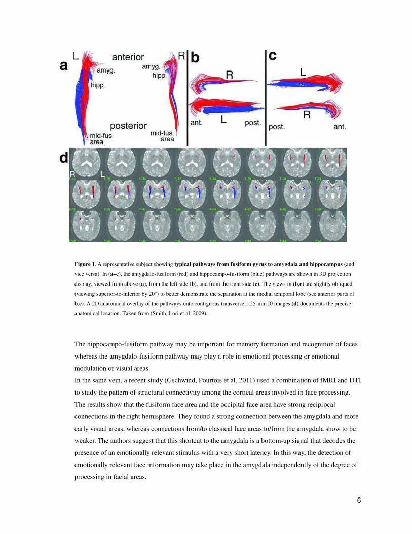
A study that employed tractography techniques (Diffusion Tensor Imaging, DTI) revealed that the mid
fusiform gyrus is directly connected to the amygdala and the hippocampus (Smith, Lori et al. 2009).
Figure 1 shows the density and thickness of these pathways.
6
Figure 1. A representative subject showing typical pathways from fusiform gyrus to amygdala and hippocampus (and
vice versa). In (a–c), the amygdalo-fusiform (red) and hippocampo-fusiform (blue) pathways are shown in 3D projection
display, viewed from above (a), from the left side (b), and from the right side (c). The views in (b,c) are slightly obliqued
(viewing superior-to-inferior by 20°) to better demonstrate the separation at the medial temporal lobe (see anterior parts of
b,c). A 2D anatomical overlay of the pathways onto contiguous transverse 1.25-mm I0 images (d) documents the precise
anatomical location. Taken from (Smith, Lori et al. 2009).
The hippocampo-fusiform pathway may be important for memory formation and recognition of faces
whereas the amygdalo-fusiform pathway may play a role in emotional processing or emotional
modulation of visual areas.
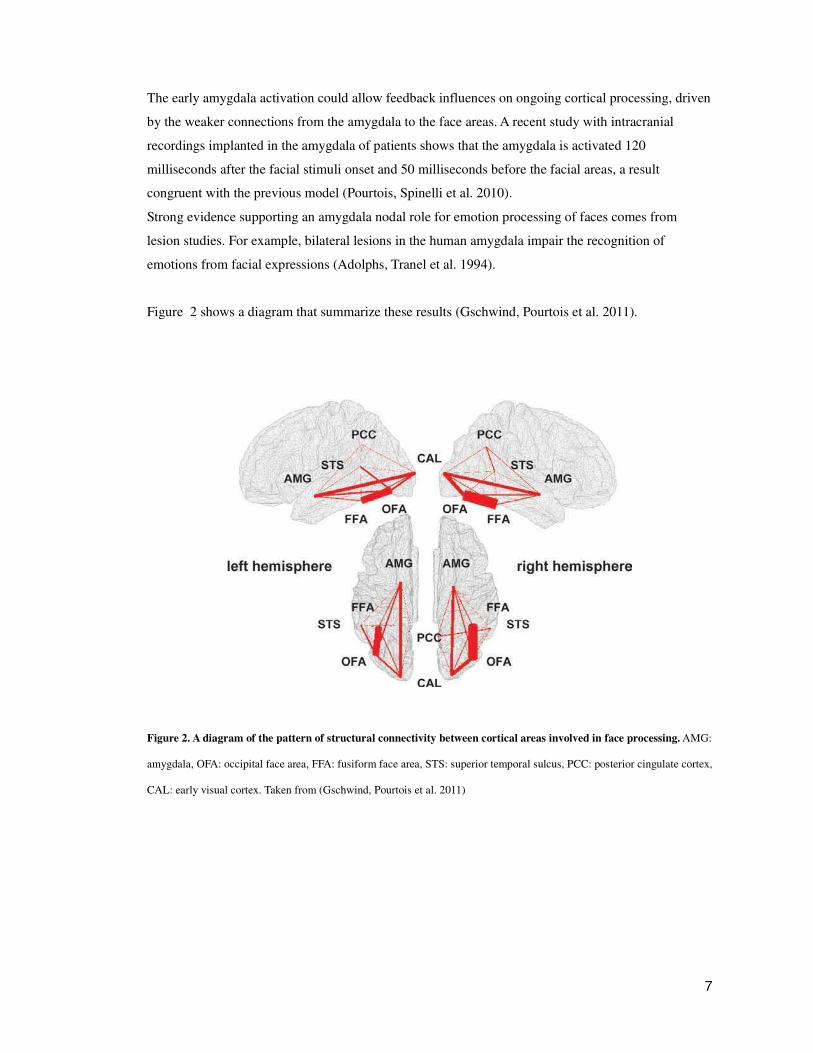
In the same vein, a recent study (Gschwind, Pourtois et al. 2011) used a combination of fMRI and DTI
to study the pattern of structural connectivity among the cortical areas involved in face processing.
The results show that the fusiform face area and the occipital face area have strong reciprocal
connections in the right hemisphere. They found a strong connection between the amygdala and more
early visual areas, whereas connections from/to classical face areas to/from the amygdala show to be
weaker. The authors suggest that this shortcut to the amygdala is a bottom-up signal that decodes the
presence of an emotionally relevant stimulus with a very short latency. In this way, the detection of
emotionally relevant face information may take place in the amygdala independently of the degree of
processing in facial areas.
7
The early amygdala activation could allow feedback influences on ongoing cortical processing, driven
by the weaker connections from the amygdala to the face areas. A recent study with intracranial
recordings implanted in the amygdala of patients shows that the amygdala is activated 120
milliseconds after the facial stimuli onset and 50 milliseconds before the facial areas, a result
congruent with the previous model (Pourtois, Spinelli et al. 2010).
Strong evidence supporting an amygdala nodal role for emotion processing of faces comes from
lesion studies. For example, bilateral lesions in the human amygdala impair the recognition of
emotions from facial expressions (Adolphs, Tranel et al. 1994).
Figure 2 shows a diagram that summarize these results (Gschwind, Pourtois et al. 2011).
Figure 2. A diagram of the pattern of structural connectivity between cortical areas involved in face processing. AMG:
amygdala, OFA: occipital face area, FFA: fusiform face area, STS: superior temporal sulcus, PCC: posterior cingulate cortex,
CAL: early visual cortex. Taken from (Gschwind, Pourtois et al. 2011)
8
Although there were discrepancies among the results from different studies in the relative weight of
the direct connections between the amygdala and facial areas, they all highlighted the importance of
these connections. A direct connection between facial related areas in the fusiform gyrus and the
amygdala strongly suggests that this circuit may play an important role in the rapid access to facial
emotion encoding, bypassing other higher order processes such as the semantic content of the
stimulus.
The amygdala: a central player in emotional regulation
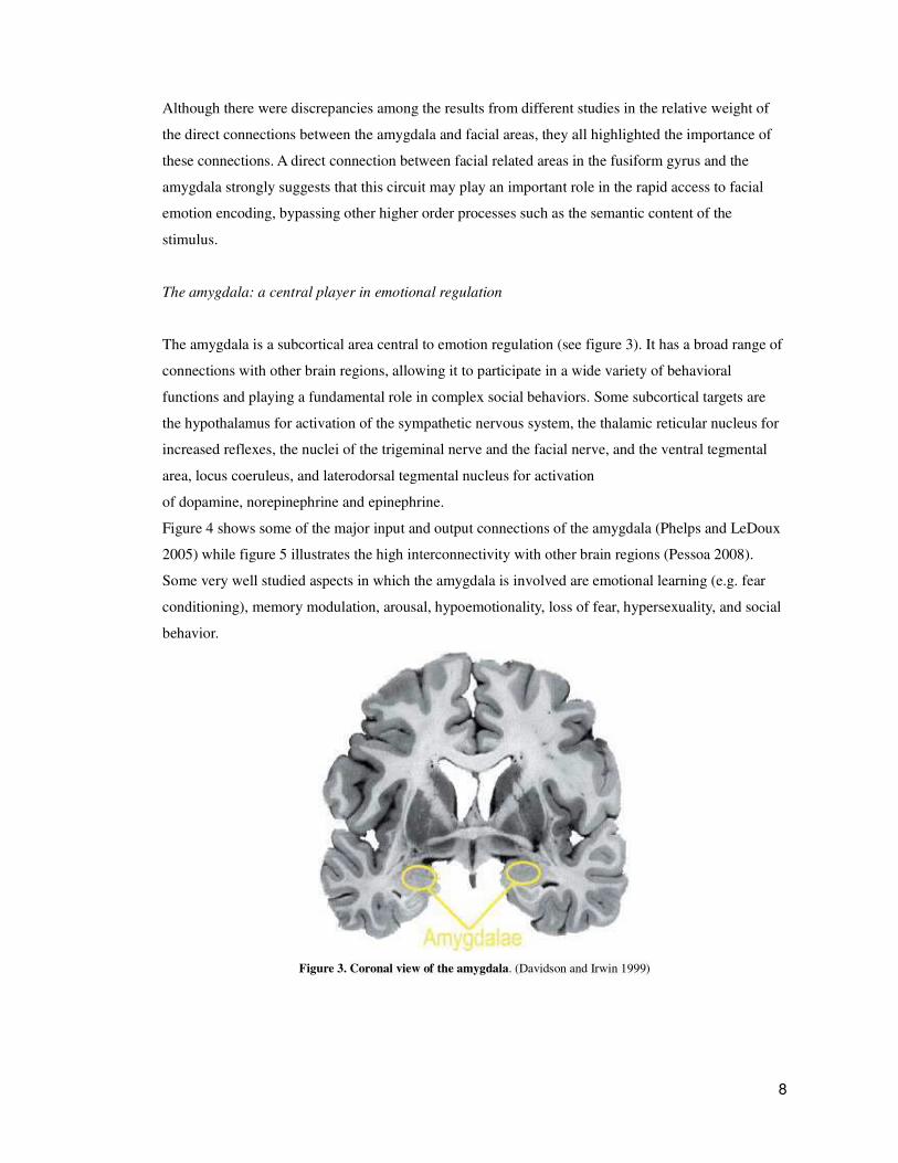
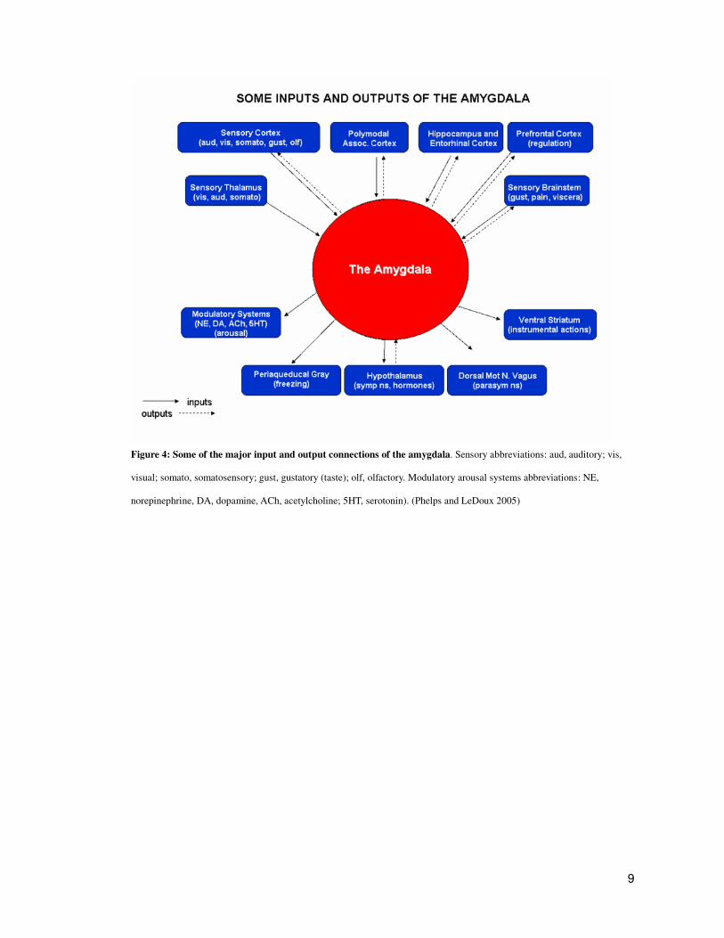
The amygdala is a subcortical area central to emotion regulation (see figure 3). It has a broad range of
connections with other brain regions, allowing it to participate in a wide variety of behavioral
functions and playing a fundamental role in complex social behaviors. Some subcortical targets are
the hypothalamus for activation of the sympathetic nervous system, the thalamic reticular nucleus for
increased reflexes, the nuclei of the trigeminal nerve and the facial nerve, and the ventral tegmental
area, locus coeruleus, and laterodorsal tegmental nucleus for activation
of dopamine, norepinephrine and epinephrine.
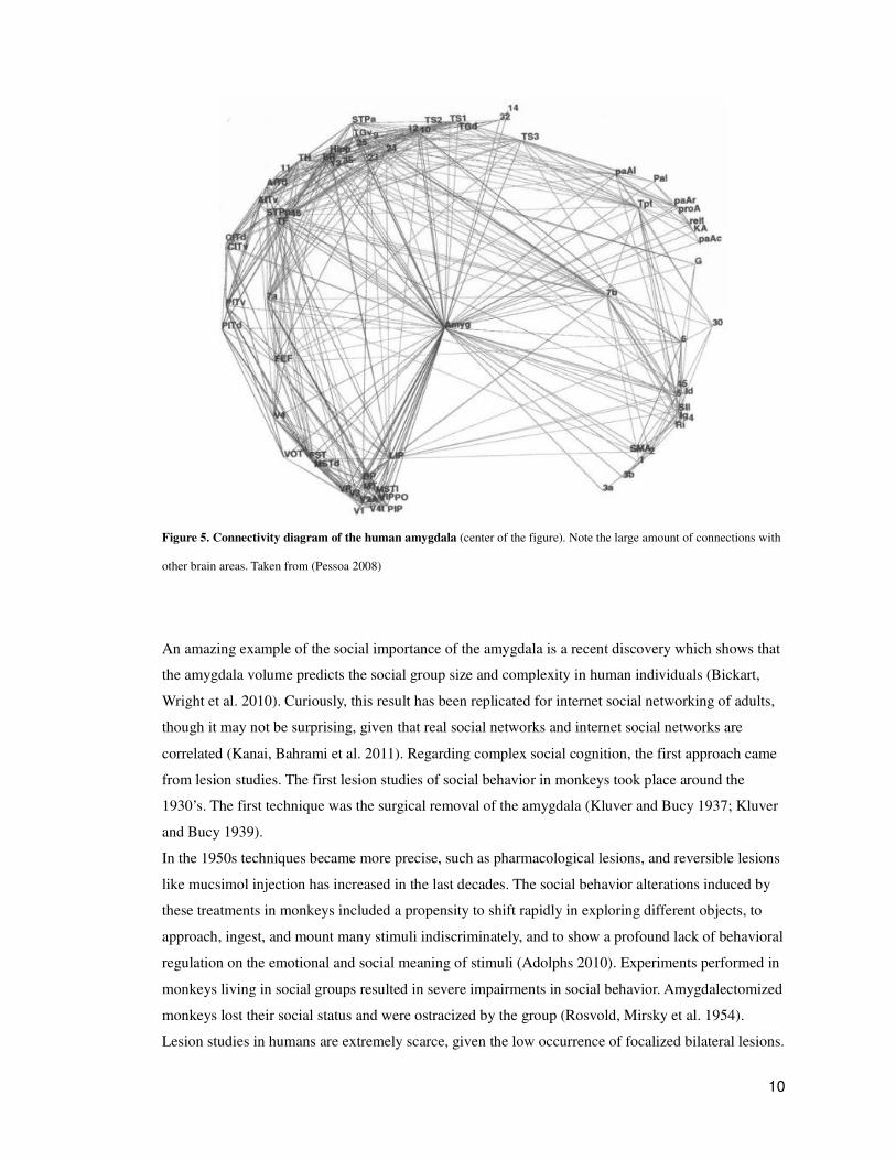
Figure 4 shows some of the major input and output connections of the amygdala (Phelps and LeDoux
2005) while figure 5 illustrates the high interconnectivity with other brain regions (Pessoa 2008).
Some very well studied aspects in which the amygdala is involved are emotional learning (e.g. fear
conditioning), memory modulation, arousal, hypoemotionality, loss of fear, hypersexuality, and social
behavior.
Figure 3. Coronal view of the amygdala. (Davidson and Irwin 1999)
9
Figure 4: Some of the major input and output connections of the amygdala. Sensory abbreviations: aud, auditory; vis,
visual; somato, somatosensory; gust, gustatory (taste); olf, olfactory. Modulatory arousal systems abbreviations: NE,
norepinephrine, DA, dopamine, ACh, acetylcholine; 5HT, serotonin). (Phelps and LeDoux 2005)
10
Figure 5. Connectivity diagram of the human amygdala (center of the figure). Note the large amount of connections with
other brain areas. Taken from (Pessoa 2008)
An amazing example of the social importance of the amygdala is a recent discovery which shows that
the amygdala volume predicts the social group size and complexity in human individuals (Bickart,
Wright et al. 2010). Curiously, this result has been replicated for internet social networking of adults,
though it may not be surprising, given that real social networks and internet social networks are
correlated (Kanai, Bahrami et al. 2011). Regarding complex social cognition, the first approach came
from lesion studies. The first lesion studies of social behavior in monkeys took place around the
1930’s. The first technique was the surgical removal of the amygdala (Kluver and Bucy 1937; Kluver
and Bucy 1939).
In the 1950s techniques became more precise, such as pharmacological lesions, and reversible lesions
like mucsimol injection has increased in the last decades. The social behavior alterations induced by
these treatments in monkeys included a propensity to shift rapidly in exploring different objects, to
approach, ingest, and mount many stimuli indiscriminately, and to show a profound lack of behavioral
regulation on the emotional and social meaning of stimuli (Adolphs 2010). Experiments performed in
monkeys living in social groups resulted in severe impairments in social behavior. Amygdalectomized
monkeys lost their social status and were ostracized by the group (Rosvold, Mirsky et al. 1954).
Lesion studies in humans are extremely scarce, given the low occurrence of focalized bilateral lesions.
11
Nevertheless, there are a few interesting case studies. A famous case is the patient SM. A series of
studies in this patient has documented a remarkably specific impairment in recognizing fear from
facial expressions, together with impairments in a variety of social judgments from faces.
Regarding social behavior, SM was notably dis-inhibited and showed a propensity to approach
and engage with others that occasionally resulted in social difficulties in real life (Buchanan, Tranel et
al. 2009)
The amygdala is also implicated in some aspects of decision making and reward processing, as in risk
based or emotional decision making (Gospic, Mohlin et al. 2011; St Onge, Stopper et al. 2012). It
interacts with prefrontal cortex and frontostriatal loops. A current view denies the strict distinction of
cognitive and emotional areas in two separate categories, proposing an integrative view that
establishes that a certain area can be cognitive or emotional depending on the global state of the
network (Pessoa 2008)
Prefrontal cortex. Emotional perception is regulated by executive control.
Another major player in face perception is the prefrontal cortex (PFC). The PFC is a cortical area
central in executive function, decision making and inhibition and also in emotion processing. Its
activity determines the state of other areas, including those involved in face emotion perception,
allowing the control of behavior by a strong interaction between executive control and emotional
responses. A recent study revealed that prefrontal cortex is necessary to control automatic responses
triggered by social emotional stimuli, overriding those responses and guiding action (Volman, Roelofs
et al. 2011). Prefrontal cortex and amygdala also interact actively when humans make decisions under
risky conditions (e.g. gambling). A recent study in rodents provide a more detailed circuitry of this
interaction (St Onge, Stopper et al. 2012).
A subregion of the prefrontal cortex, the ventromedial prefrontal cortex, ventral and medial regions of
the prefrontal cortex, encompass several interconnected regions that process reward and punishment,
regulate emotion, and maintain homeostasis (Ongur and Price 2000). The ventromedial prefrontal
cortex has been linked to social behavior since the historical case of Phineas Gage, a nineteenth-
century railroad worker who had an iron rod blasted through the front of his head in an accident. Gage
survive with preserved intellectual abilities, but his personality changed from shrewd, persistent, and
respectable to profane, capricious, and unreliable after the accident (Damasio, Grabowski et al. 1994).
Studies involving gambling games show that ventromedial prefrontal cortex patients experience
diminished emotional arousal before making risky choices (Bechara, Damasio et al. 1994), as well as
diminished regret when considering alternate outcomes after making risky choices. In such games,
patients persistently make disadvantageous choices. These results support an influential theory about
12
the role of emotion in decision-making (including social decision-making), the so-called somatic
marker hypothesis (Damasio 1996). The hypothesis states that emotional signals, mediated in part by
regions in the ventromedial prefrontal cortex, can be elicited by the anticipation or consideration of
the future outcomes of one's actions, and that this signal guides the decision that is made.
Another frontal area involved in emotional regulation is the anterior cingulate cortex (ACC). The
ACC is believed to be involved in the executive control of actions, such as in monitoring conflicting
response demands, detecting errors, and evaluating the emotional significance of events. The dorsal
part of the ACC seems to play a key role in reward-based decision-making and learning. The rostral
part of the ACC, on the other hand, is believed to be more involved with affective responses to errors.
Summarizing, face emotional perception relies on areas extracting structural visual information, areas
providing memory, areas processing the emotional meaning of a certain face, and areas that control
the circuit necessary to organize in space and time a line of action.
Cortical facial processing can be measured with electroencephalography. The N170 potential.
A classical approach to study facial processing with high temporal resolution is
electroencephalography (EEG). When a human observes a face, a potential called N170 can be
measured at the scalp. The N170 is a cortical marker specifically linked to facial processing, with
neural generators in the fusiform gyrus and superior temporal sulcus. The N170 changes its amplitude
in response to different facial emotions (see a detailed description in materials and methods section).
Limbic and frontal activity is abnormal in bipolar disorder and ADHD
The interaction between executive functions and emotion regulation is altered in some psychiatric
disorders, such as bipolar disorder, and ADHD. The close interaction between limbic and frontal areas
may explain the co-occurrence of emotional and executive deficits in these patients.
The bipolar disorder (BD) is a neuropsychiatric disease characterized by unpredictable manic,
hypomanic and depressive episodes, related to several neuroanatomical and neuropsychological
deficits (Green, Cahill et al. 2007; Delaloye, de Bilbao et al. 2009). Mania is a state of abnormally
elevated or irritable mood, arousal, and/ or energy levels. In a way, it is the opposite of depression.
Mania varies in intensity, from mild mania (hypomania) to full-blown mania with psychotic features,
13
including hallucinations, delusion of grandeur, suspiciousness, catatonic behavior, aggression, and a
preoccupation with thoughts and schemes that may lead to self neglect. Signs and symptoms of the
depressive phase of bipolar disorder includes persistent feelings
of sadness, anxiety, guilt, anger, isolation, or hopelessness, disturbances in sleep and appetite, fatigue
and loss of interest in usually enjoyable activities, problems concentrating; loneliness, self-loathing,
apathy or indifference, loss of interest in sexual activity, shyness or social anxiety, irritability, lack of
motivation, and morbid suicidal ideation (Semple 2005).
Euthymia is a middle or equilibrium state between mania and depression.
Studies of structural MRI show that BD patients have a significant reduction in amygdala volume
(Rosso, Killgore et al. 2007). Functional and structural connectivity studies, reveal a functional
connectivity decrease between amygdala and ACC in BD, and a significant positive association
between ACC-amygdala functional coupling and ventrofrontal white matter thickness (Wang, Kalmar
et al. 2009).
Recent reports of BD show cognitive and social cognition deficits including attention, memory,
executive functions and social cognition (Inoue, Tonooka et al. 2004; Robinson and Ferrier 2006;
Lahera, Montes et al. 2008; Jamrozinski, Gruber et al. 2009; Martinez-Aran, Scott et al. 2009;
Martino, Strejilevich et al. 2010).
Probably, several social cognition domains affected in BD are related to more basic (facial and
emotional) process. For instance, emotional processing information in BD seems to be impaired
(Malhi, Ivanovski et al. 2007; Hassel, Almeida et al. 2008; M'Bailara, Demotes-Mainard et al. 2009;
Rosen and Rich 2010). Abnormalities in face processing and emotion recognition have been reported
in BD patients (Getz, Shear et al. 2003; Malhi, Ivanovski et al. 2007; Wessa and Linke 2009).
Furthermore, those deficits in facial emotional expressions have been associated to psychosocial
impairments and mania risk in children and adolescents (Brotman, Guyer et al. 2008; Rich, Fromm et
al. 2008). Indeed, brain networks involved in facial encoding and emotional processing overlap with
fronto-striatal circuit affected in BD (Pavuluri, O'Connor et al. 2007; Kalmar, Wang et al. 2009). In a
similar vein, impaired face emotion processing and social cognition would be related to a deficient
connectivity between the amygdala and temporally associated cortical regions (Leppanen 2006; Rich,
Grimley et al. 2008). Therefore, deficits in emotional processing in BD are observed at a behavioral as
well as a neural level (Guyer, McClure et al. 2007). Basic emotional and social impairments would
trigger a vulnerable cognitive profile toward mood regulation and interpersonal behavior deficits
(Scott and Pope 2003).
ADHD is a neuropsychiatric condition with onset in childhood that extends over adolescent and adult
life with a considerable symptomatic burden and functional impairment. Its medical profile includes
problems of self-regulation and self-motivation, distractibility, procrastination, and prioritization. A
14
recent meta-analysis suggested that the prevalence of adult ADHD, even using higher figures (2.5%),
is underestimated (Simon et al., 2009).
ADHD in childhood is more related to hyperactivity and impulsiveness, whereas in adulthood it
presents a different profile, with fewer externalizing symptoms and a higher rate of psychiatric
comorbidity (Klassen et al., 2010). Nevertheless, deficits in executive functioning have been
consistently demonstrated in adults with ADHD (Adler, 2010), as well as impairments on executive
function tasks with high working memory demands (Torralva et al., 2010).
Adults with ADHD exhibit a diminished gray matter volume in the ACC and prefrontal cortex,
measured by voxel based morphometry MRI (Amico, Stauber et al. 2011). They also present a
diminished white matter volume in the ACC measured by DTI (Makris, Buka et al. 2008) as well as
cortical thinning in the ACC (Makris, Biederman et al. 2007). Two high resolution MRI studies
showed a diminished volume of the amygdala in ADHD patients (Frodl, Stauber et al. 2010; Posner,
Nagel et al. 2011)
Although deficits in social cognition are an evident clinical phenomena in ADHD, very little research
has been developed in this area (Uekermann et al., 2010). A few reports suggest various deficits in
domains such as facial affect recognition (Marsh & Blair, 2008; Pelc et al., 2006; Sinzig et al., 2008),
prosody perception (Shapiro et al., 1993), theory of mind (ToM; Buitelaar et al., 1999; Sodian et al.,
2003; but see Charman et al., 2001 for different results), social skills (King et al., 2009; Matthys et al.,
1999) and empathy (Braaten & Rosen, 2000; Dyck et al., 2001). Facial emotion processing seems to
be the social cognition process that is most affected in ADHD (Marsh & Williams, 2006). In general
terms, these social cognition impairments are consistent with fronto-striatal dysfunction in ADHD
(Uekermann et al., 2010a), showing the central nature of social dysfunction in this disorder (Hoza et
al., 2000; Maedgen & Carlson, 2000; Wheeler and Carlson, 1994).
Hypothesis
H1) A cortical electrophysiological marker of the processing of facial emotion (N170) is
associated with individual differences in complex social cognition skills.
I propose that individual social cognition complex skills that are mediated in part by an extense
network including the amygdala will be associated to the modulation of rapid electrophysiological
markers of facial emotion, given the strong interaction of visual and facial areas with the amygdala
and other social cognition areas.
The theoretical framework of Hypothesis 1 is an intense current debate in social neuroscience about
the modularity of some social cognition process. An idea dear to evolutionary psychologists argues
that humans suffered selection pressures to evolve social abilities or “modules” of cognition (e.g. the
cognitive niche) (Pinker 2010). These structures are specialized for processing social information.
The inspiring example of such view is, precisely, facial processing. But these concepts are
continuously under debate thanks to the discovery of a disperse and patchy network that activates
during face perception (Haxby, Hoffman et al. 2000). Hypothesis 1 is oriented to test the extent to
which a rapid cortical signal that indexes face processing is dependent or embedded in a sparse
network that support high level social cognition (e.g. ToM).
Three lines of evidence support an association between facial processing and higher-order social
cognition skills: studies of (1) healthy participants (Herzmann, Kunina et al. 2010; Hileman,
Henderson et al. 2011); (2) participants with autism spectrum disorders (ASD) (Barton, Hefter et al.
2007; Clark, Winkielman et al. 2008; Dziobek, Bahnemann et al. 2010; Kleinhans, Richards et al.
2011; Suzuki, Sugihara et al. 2011); and (3) participants with frontotemporal dementia (FTD)
(Fernandez-Duque and Black 2005). All these studies suggest that facial processing is required and
related to different high level social cognition skills. However, a possible association between cortical
markers of facial processing and neuropsychological social cognition has not been proposed yet.
It is therefore expected that N170 would be associated with neuropsychological performance in social
cognition tests. First, N170 would be associated with social cognition, as measured by a test such as
the ToM, with any emotional modulation of N170 being directly related to the reading the mind in the
eyes test [RMET] (i.e., a theory of mind abilities’ evaluation by making emotional inferences from
faces (Stone, Baron-Cohen et al. 1998; Barton, Hefter et al. 2007; Parr, Waller et al. 2008; Ahmed and
Stephen Miller 2010)) because mental inference is determined by facial emotional content. Second,
2
the Faux pas test [FPT], another ToM task, would be mediated by executive functions. This is
expected because the Faux pas test involves dealing with a high number of cognitive and affective
components, including inferences about others’ mental states and contextual cues (Riveros, Manes et
al. 2010). Consequently, FPT should be related to more complex N170 processing. Third and finally,
decision making assessments, using the Iowa Gambling Task [IGT] (especially the first of five
blocks), would be associated with cortical emotional processing. Because the first block of the IGT
can be consistently associated with ambiguity and influenced by emotion heuristics (Dunn, Dalgleish
et al. 2006), it is expected the existence of an association between the N170 emotional discrimination
level and the emotion heuristics during first stages IGT performance.
The association between facial emotional valence discrimination and social cognition skills can be
explored by testing for correlations between these measures (i.e., emotional modulation index and
stimulus type discrimination). Our prediction is that higher performance on the tasks of social
cognition would be related to greater discrimination of facial emotional valence and stimulus type as
shown by the amplitude of the N170 component. Additionally, we examined the associations between
the processing of facial emotion and other neuropsychological abilities, such as general
neuropsychological functioning (i.e., classical measures of IQ, memory and attention) and executive
functioning. Our second prediction was that higher performance on tasks of executive function would
be associated with greater discrimination of emotional valence as shown by the amplitude of the N170
in response to simultaneous (face/word) stimuli.
H2a) BD patients present cortical deficits in processing emotional facial information.
H2b) These deficiencies are associated with clinical measures (indices of mania and depression).
As I have already mentioned, BD patients present abnormal volume and connectivity of the amygdala.
Given that the amygdala is strongly connected to facial and early visual areas, it is expected that the
cortical processing of facial emotions measured by the N170 will be impaired (Hypothesis 2a).
Because the severity of BD symptoms may be associated with amygdala abnormal volume and
thickness, and given that facial emotional processing relies on this area, it is proposed that individual
facial processing measured by the N170 potential modulation will be associated with clinical
measures of mania and depression.
N170 studies of structural and emotional processing in BD are scarce. An ERP design of emotional
face processing in depressed BD type I has been reported (Degabriele, Lagopoulos et al. 2011). In this
study a paradigm of emotional inhibition on presentation of faces, modifying the classic test
3
emotional go/no-go, is used. Results showed abnormal P100 emotional processing and reduced N170
in BD patients. Nevertheless, no previous studies of structural and emotional face/word processing
and their relation to neurocognitive profile in euthymic BD has been reported yet.
H3a) ADHD patients present cortical deficits in processing emotional facial information.
H3b) These deficiencies are associated with executive functioning scores
It has been previously shown that ADHD patients present deficiencies in frontal functioning and in
some social cognition tasks. Furthermore, ADHD deficit is accompanied by prefrontal and ACC
neuroanatomical deficiencies. As it was mentioned, prefrontal cortex and ACC play a regulatory role
in emotional perception. Thus, these network and function deficiencies may be reflected at the cortical
level in rapid processing of faces. Additionally, neuropsychological testing of executive functions, an
estimate of frontal functioning, may correlate with the degree of deficiency in cortical processing
estimated at the N170 potential.
It has been shown that adults with ADHD have impaired social cognition. However, no studies have
focused on the brain correlates of the adult ADHD deficits in emotion processing.
An approach which combines measures from neuropsychological and neurophysiologic markers
represents a valuable tool to understand abnormal cognitive processing in neuropsychiatry and
individual differences. This section seeks to identify behavioral, neuropsychological and
electrophysiological possible markers of abnormal emotion processing for faces in adult ADHD
compared with controls matched by age, gender, educational level and handedness.
Only a single study has previously assessed facial processing in ADHD indexed by the N170, but
included only adolescents. Williams et al (Williams, Hermens et al. 2008) reported an abnormal
emotion-related N170, suggesting that the structural facial processing stage is affected in adolescents
with ADHD. However, these results must be taken with caution because participants with ADHD also
had comorbid depression and anxiety. For adults with ADHD, even though evidence of deficits in the
processing of emotion have been reported (Herrmann et al., 2009), no N170 valence effects elicited by
facial processing have been previously assessed.
4
H4a) BD and ADHD patients have cortical deficits in the processing of monetary reward.
H4b) The altered modulation of monetary reward processing in both patients correlate with
executive functions performance.
Decision-making is essential in our daily lives. We make many different decisions; some are based on
risk and predictability, whereas others are based on uncertainty or emotional heuristics. Current
neuroscience research examining decision-making has assessed multiple processes engaged in this
complex cognitive ability. Evidence from animals, healthy human volunteers and neuropsychiatric
patients highlight the role of the frontostriatal and limbic loops in this process (Glimcher and
Rustichini 2004; Bechara and Van Der Linden 2005; Brand, Labudda et al. 2006; Rangel 2008;
Rangel, Camerer et al. 2008; Rushworth and Behrens 2008; Kable and Glimcher 2009; Gleichgerrcht,
Ibanez et al. 2010). Despite some discrepancies among different decision-making models, three
systems are thought to be involved in the frontostriatal and limbic loop: stimulus encoding (i.e., the
orbitofrontal cortex), reward-based action selection and monitoring (i.e., the cingulate cortex) and
expected reward (i.e., the basal ganglia and amygdala). Thus, impaired decision-making may be the
result of different deficits in these (or other) brain areas and may be affected differentially by disparate
scenarios. Consequently, the nature of these decision-making deficits is dependent on context and
disease.
In this thesis we explore the role of the decision making network in rapid cortical responses to
monetary reward. First, we postulate that two psychiatric disorders that present an altered frontal
network will show an impaired modulation of early components in response to monetary reward.
Second, we predict that this impairment will correlate with their executive functions, measured by
neuropsychological tests.
Bipolar disorder (BD) and attention-deficit/hyperactivity disorder (ADHD) usually manifest shared
clinical symptoms, present high rates of comorbidity and are challenging to differentiate from each
other (Wingo and Ghaemi 2007; Chang 2010; Klassen, Katzman et al. 2010). These disorders affect
people by presenting problems in common decision scenarios that have social and vocational effects.
Decision-making impairments have been reported in patients with ADHD (Ernst, Kimes et al. 2003;
Luman, Sergeant et al. 2010; Schepman, Weyandt et al. 2010) and those with BD (Christodoulou,
Lewis et al. 2006; Jollant, Guillaume et al. 2007). Nevertheless, previous decision-making studies
using neuropsychology methods have shown inconsistent results for both disorders. In addition, no
previous report has assessed a decision-making task that includes the examination of the neural
correlates of reward and gambling in adults with ADHD and those with BD. Finally, no study has
compared these disorders regarding decision-making domains yet.
5
General Methods
A main challenge of the present project is the use of techniques coming from clearly different
historical scientific traditions. A broad classification of these methodologies may be reduced to three
sets: psychophysics, electroencephalography and neuropsychology. In the next paragraphs, the reader
will find a brief introduction to each method.
Psychophysics
What and when: Response type and response time.
Psychophysics is one of the most ancient techniques of experimental psychology. A classical
definition state that psychophysics is the analysis of perceptual processes by studying the effect on a
subject's experience or behavior of systematically varying the properties of a stimulus along one or
more physical dimensions (Bruce, Green et al. 1996)
It is, in fact, a discipline in which the observable is behavior, that is, the measurements are limited to
what an animal or a person does in response to a presented stimulus (in some settings, however, the
responses might not be necessarily linked to an external stimuli).
The stimuli are part of a certain task that takes place in a specially designed experimental setting. The
stimuli are manipulated by the researcher, who expects to find variations in the responses as a function
of stimulus changes. Responses are voluntary motor acts, such as a button press or a vocal sound.
Variations in responses given to continuous or discrete stimulus manipulation are described by
mathematical functions known as psychometric functions or psychometric curves. Two variables are
of interest to psychophysics: Response type and response time. Both give interesting information
about stimulus processing. An example will help to describe the methodology and the type of
information obtained. Fiorentini and Viviani (Fiorentini and Viviani 2011) showed subjects pictures of
an actor, and asked them to respond with a button press if the emotion of the actor was anger or fear
(two alternative forced choice). An interesting manipulation was that they created morphing images
that represented intermediate states between anger and fear. Figure 6 shows a typical psychometric
sigmoid response curve, where a dimension of the stimulus (morphing degree, 1=anger, 50 = fear) is
manipulated in a continuous way.

6
Figure 6. An example of a psychometric curve in response to morphed images between
two emotional states. The probability of responding fear as a function of degree of
morphing between the 2 images (Anger and Fear) is plotted. Note that only morphed
images from 15 to 35 are used, given that more extreme morphs saturated the response (P
= 0 or P = 1). (Fiorentini and Viviani 2011).
The obvious limitation of psychophysics is that it does not allow a direct access to the system (e.g.
muscle, brain or neuron) and no hypothesis can be directly tested about the mechanisms that are
taking place in the substrate. Nevertheless it is of great value to make a first approach or to combine it
with physiological methods.
Dual valence task. Our experimental paradigm to estimate the N170 potential in response to faces.
The dual valence task (DVT) was designed to measure behavioral and electroencephalographical
responses to facial emotions. It included blocks with emotional faces, blocks with emotional words,
and blocks with a simultaneous presentation of emotional faces and words.
Participants were instructed to categorize single (words or faces) or simultaneous (face/word) stimuli
that were displayed for 100 ms on a computer screen according to the stimuli’s valence, responding as
to whether the stimuli were ‘positive’ or ‘negative’ as quickly as possible. Incorrect responses were
indicated with an ’X’ in the center of the screen immediately after the response had been given (see
figure 7).
7
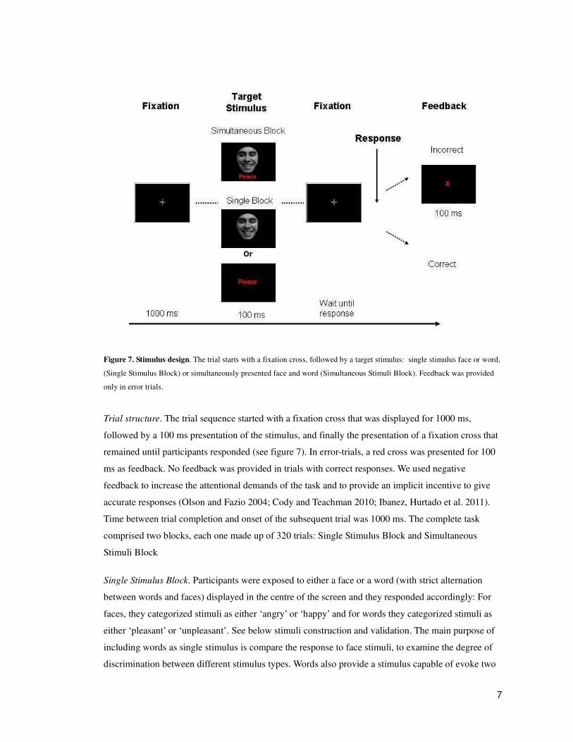
Figure 7. Stimulus design. The trial starts with a fixation cross, followed by a target stimulus: single stimulus face or word,
(Single Stimulus Block) or simultaneously presented face and word (Simultaneous Stimuli Block). Feedback was provided
only in error trials.
Trial structure. The trial sequence started with a fixation cross that was displayed for 1000 ms,
followed by a 100 ms presentation of the stimulus, and finally the presentation of a fixation cross that
remained until participants responded (see figure 7). In error-trials, a red cross was presented for 100
ms as feedback. No feedback was provided in trials with correct responses. We used negative
feedback to increase the attentional demands of the task and to provide an implicit incentive to give
accurate responses (Olson and Fazio 2004; Cody and Teachman 2010; Ibanez, Hurtado et al. 2011).
Time between trial completion and onset of the subsequent trial was 1000 ms. The complete task
comprised two blocks, each one made up of 320 trials: Single Stimulus Block and Simultaneous
Stimuli Block
Single Stimulus Block. Participants were exposed to either a face or a word (with strict alternation
between words and faces) displayed in the centre of the screen and they responded accordingly: For
faces, they categorized stimuli as either ‘angry’ or ‘happy’ and for words they categorized stimuli as
either ‘pleasant’ or ‘unpleasant’. See below stimuli construction and validation. The main purpose of
including words as single stimulus is compare the response to face stimuli, to examine the degree of
discrimination between different stimulus types. Words also provide a stimulus capable of evoke two

8
emotional states, for design purposes (factorial design). Words also prove to evoke the N170, but no
emotional modulation is expected at so early times.
Simultaneous Stimuli Block. Participants were exposed to a face displayed in the centre of the screen
with a word displayed 4 degrees beneath it in the lower hemifield. These stimuli were presented
simultaneously for 100 ms. Participants were asked to indicate the emotion shown by the face and to
ignore the word. In compatible trials the face and a word shared the same valence (e.g. an angry face
with the word ‘angry’), whereas in incompatible trials they represented opposite valences (e.g. an
angry face with a pleasant word). The aim of including words in facial images is to test the
interferences of context (in these case words) on stimuli (faces in the center of the screen).
The presentation order of blocks (Single and Simultaneous Stimuli) was counterbalanced across
participants. Each block was separated into two sub-blocks of 160 trials. Throughout the experiment,
the same two keys were used to indicate responses, but the assignation of key to response type was
inverted between sub-blocks. Each sub-block included a brief explanation at the start of which
category was assigned to which response key, followed by six practice trials. This procedure follows
the designs used in previously published dual-choice association tasks (Hurtado, Haye et al. 2009;
Ibanez, Hurtado et al. 2011).
Stimulus construction and validation.
20 pictures of actors’ faces were selected from a dataset used in previous studies (Hurtado, Haye et al.
2009). A set of 10 happy and 10 angry pictures, controlled for intensity, brightness, color and contrast,
was included. Each actor appeared in two pictures, one of each valence. Pleasant and unpleasant
words, controlled for arousal, content, length and frequency, were selected from another previous
study (Ibanez, Lopez et al. 2006). 33 pleasant and 32 unpleasant words were randomly selected from
the original sets. A greater number of word stimuli were selected relative to the number of faces to
reduce the repetition effect of words (Bentin and Peled 1990), a robust modulator of ERPs (Rugg,
Mark et al. 1997; Doyle and Rugg 1998). On the contrary, facial ERP modulation can be found with a
small number of faces (Maurer, Rossion et al. 2008; Astikainen and Hietanen 2009).
To validate word content, a questionnaire was used to assess pleasantness or unpleasantness
of a set of 150 words with a moderate use frequency (Lifcach frequency software). 50
university students, 33 female, mean age 19.62+ 3.33), participated in the validation (Ibanez,
Lopez et al. 2006). Participants rated the set of words using a Likert scale where 1
represented a very positive valence and 7 represented a very negative valence. Repeated
measures Analysis of Variance (ANOVA) was used to contrast categorizations for the list of
9
pleasant and unpleasant words. Significant differences were obtained for the categorization of
both lists [F(1, 73) = 25161, p < 0.0001]. Pleasant words that were ranked between 1 and 3
where selected (72 out of 75 positive words were included). Unpleasant words rated between
5 and 7 were selected (71 from 75 negative words were included).
Decision-making tasks combined with ERPs
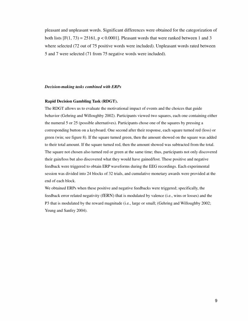
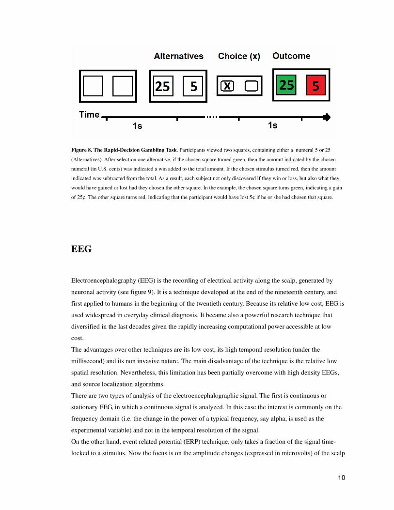
Rapid Decision Gambling Task (RDGT).
The RDGT allows us to evaluate the motivational impact of events and the choices that guide
behavior (Gehring and Willoughby 2002). Participants viewed two squares, each one containing either
the numeral 5 or 25 (possible alternatives). Participants chose one of the squares by pressing a
corresponding button on a keyboard. One second after their response, each square turned red (loss) or
green (win; see figure 8). If the square turned green, then the amount showed on the square was added
to their total amount. If the square turned red, then the amount showed was subtracted from the total.
The square not chosen also turned red or green at the same time; thus, participants not only discovered
their gain/loss but also discovered what they would have gained/lost. These positive and negative
feedback were triggered to obtain ERP waveforms during the EEG recordings. Each experimental
session was divided into 24 blocks of 32 trials, and cumulative monetary awards were provided at the
end of each block.
We obtained ERPs when these positive and negative feedbacks were triggered; specifically, the
feedback error related negativity (fERN) that is modulated by valence (i.e., wins or losses) and the
P3 that is modulated by the reward magnitude (i.e., large or small; (Gehring and Willoughby 2002;
Yeung and Sanfey 2004).
10
Figure 8. The Rapid-Decision Gambling Task. Participants viewed two squares, containing either a numeral 5 or 25
(Alternatives). After selection one alternative, if the chosen square turned green, then the amount indicated by the chosen
numeral (in U.S. cents) was indicated a win added to the total amount. If the chosen stimulus turned red, then the amount
indicated was subtracted from the total. As a result, each subject not only discovered if they win or loss, but also what they
would have gained or lost had they chosen the other square. In the example, the chosen square turns green, indicating a gain
of 25¢. The other square turns red, indicating that the participant would have lost 5¢ if he or she had chosen that square.
EEG
Electroencephalography (EEG) is the recording of electrical activity along the scalp, generated by
neuronal activity (see figure 9). It is a technique developed at the end of the nineteenth century, and
first applied to humans in the beginning of the twentieth century. Because its relative low cost, EEG is
used widespread in everyday clinical diagnosis. It became also a powerful research technique that
diversified in the last decades given the rapidly increasing computational power accessible at low
cost.
The advantages over other techniques are its low cost, its high temporal resolution (under the
millisecond) and its non invasive nature. The main disadvantage of the technique is the relative low
spatial resolution. Nevertheless, this limitation has been partially overcome with high density EEGs,
and source localization algorithms.
There are two types of analysis of the electroencephalographic signal. The first is continuous or
stationary EEG, in which a continuous signal is analyzed. In this case the interest is commonly on the
frequency domain (i.e. the change in the power of a typical frequency, say alpha, is used as the
experimental variable) and not in the temporal resolution of the signal.
On the other hand, event related potential (ERP) technique, only takes a fraction of the signal time-
locked to a stimulus. Now the focus is on the amplitude changes (expressed in microvolts) of the scalp

11
signal time-related to a stimulus. ERPs technique was developed in the past century, and for years
only practiced in a few laboratories. The pioneers of this technique found that certain psychological
tasks bring up a well defined signal. ERPs emerged after averaging several trials of the same type (i.e.
the same stimulus or the same stimulus category), presenting a singular waveform. They were first
identified by its sign and the time of the maximum peak. P300 means a positive potential at 300
milliseconds, N400 a negative potential at 400 milliseconds, and so on. An alternative nomenclature
name components according to the relative order of appearance (P1 is the positive signal that first
appeared in certain experiments, in this case coincides with P100). A big family of components was
described since the early times of ERP technique and replicated in hundreds of laboratories around the
world.
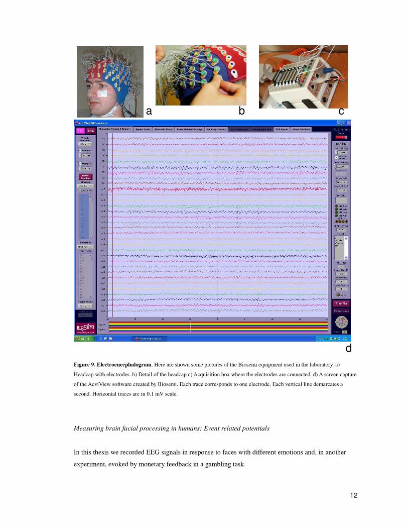
12
Figure 9. Electroencephalogram. Here are shown some pictures of the Biosemi equipment used in the laboratory. a)
Headcap with electrodes. b) Detail of the headcap c) Acquisition box where the electrodes are connected. d) A screen capture
of the AcviView software created by Biosemi. Each trace corresponds to one electrode. Each vertical line demarcates a
second. Horizontal traces are in 0.1 mV scale.
Measuring brain facial processing in humans: Event related potentials
In this thesis we recorded EEG signals in response to faces with different emotions and, in another
experiment, evoked by monetary feedback in a gambling task.
a b c
d

13
To study facial emotion processing we estimated a classical brain signal referred to as the N170,
which shows a negative peak at approximately 140-200 ms post-stimulus, and has been shown to be
involved in the processing of faces (Rossion and Jacques 2008). It is also present, although with a
lesser amplitude, when humans are exposed to familiar objects, such as houses, trees and cars.
Figure 10. The N170 is a negative component recorded from posterior lateral electrode sites following the presentation of
faces and object categories (here pictures of cars). It peaks at about 160–170 ms following stimulus onset and is recorded
between 130 ms and 200 ms. It is most prominent at the lowest occipito-temporal electrode sites. The component is larger in
response to faces than objects in both hemispheres, with usually a larger response in the right hemisphere. Taken from
(Rossion and Jacques 2008).
The N170 component is sensitive to stimulus type (facial or other, see figure 10) (Rossion, Gauthier
et al. 2002; Goffaux, Gauthier et al. 2003; Itier and Taylor 2004; Rousselet, Mace et al. 2004; Thierry,
Pegna et al. 2006; Churches, Baron-Cohen et al. 2009; Ibanez, Gleichgerrcht et al. 2010). The N170 is
affected by emotional valence (Eimer and Holmes 2002; Pizzagalli, Lehmann et al. 2002; Batty and
Taylor 2003; Ashley, Vuilleumier et al. 2004; Galli, Feurra et al. 2006; Sprengelmeyer and Jentzsch
2006; Blau, Maurer et al. 2007; Hendriks, van Boxtel et al. 2007; Vuilleumier and Pourtois 2007;
Montalan, Caharel et al. 2008; Chammat, Foucher et al. 2010). Moreover, the N170 amplitude can be
modulated by interference (e.g., two opposite valence stimuli classifications). In particular, it is
14
sensitive to the compatibility between the stimulus and background, indicating that it may reflect an
interaction between executive functioning and emotional processing (Galli, Feurra et al. 2006; Righart
and de Gelder 2006; Righart and de Gelder 2008; Fruhholz, Fehr et al. 2009; Gonzalez-Garrido,
Ramos-Loyo et al. 2009). Studies of brain topography have localized the cortical source of the N170
in the fusiform gyrus (FG) (Itier and Taylor 2004; Sadeh, Podlipsky et al. 2010). In summary, the
N170 component can be considered as a neural marker of early face-selective processing that is
modulated by affective valence and contextual cues.
We used an ERP design of a dual valence task (DVT) to study the ERP stimulus and emotional
discrimination in healthy participants and psychiatric patients. In the DVT, faces, words or
simultaneous face-word stimuli are presented. Participants are asked to classify the stimuli according
to their emotional valence. The modulation of the N170 by three factors (stimulus type, valence and
interference) was quantified.
Measuring brain feedback processing in humans: Event related potentials
To study monetary reward processing in a gambling task a potential called feedback error related
negativity (fERN) was estimated. A robust error related negativity (ERN) component is observed after
errors are committed during various choice tasks, even when the participant is not explicitly aware of
it. An event-related potential is also observed following the presentation of negative feedback stimuli
in a cognitive task indicating the outcome of a response, often called the fERN. fERN is a product of
prediction error signals carried by the dopamine system arriving to the anterior cingulate
cortex indicating that events have gone worse than expected (Holroyd and Coles 2002)
We recorded event-related potentials (ERPs) from human participants as they performed the RDGT.
Participant choices were followed by feedback that indicated the monetary gains or losses that
resulted. The RDGT elicits a feedback error-related negativity (fERN) modulated by reward valence
and a P3 sensitive to reward magnitude (Gehring and Willoughby 2002; Yeung and Sanfey 2004).
15
ERP recordings.
In all the experiments presented in this thesis, EEG signals were sampled at 500 Hz from a Biosemi
128-channel system. Data outside the frequency band, which ranged from 0.1 Hz to 100 Hz, were
filtered out during recording. A band-pass digital filter (below 0.3 Hz and over 30 Hz) was applied
off-line to remove unwanted frequency components. During recording, the reference was set by
default using linked mastoids but it was then re-referenced off-line to average electrodes. Two bipolar
derivations were designed to monitor vertical and horizontal ocular movements (EOG). Continuous
EEG data were segmented from 200 ms prior to the stimulus to 800 ms after stimulus onset. All
segments with eye movement contamination were removed from further analysis using an automatic
procedure (Gratton, Coles et al. 1983) and a visual procedure. Artifact-free segments were averaged to
obtain ERPs.
N170 Source Localization.
Dipole source models of the N170 component for each condition were estimated using an Automatic
Relevance Determination (ARD) algorithm. Source estimation from EEG data can be problematic but
the use of ARD resolves this by regularizing the solution space using a parameterized data-dependent
prior distribution. A special type of ARD was used, known as Bayesian Learning (SBL) (Wipf and
Nagarajan 2009). An Average Lead Field was used as the head model, built from a sample of 305
MRIs of typical participants. Possible solutions were constrained for location to the cortical surface
but were not constrained for orientation. This head model is useful for source localization when
individual MRI data are not available (Valdes-Hernandez, von Ellenrieder et al. 2009). Given that
temporal differences occur between participants and stimulus types, the local minimum within the
N170 window was considered for each of them. First, the average signal for the N170 representative
electrodes A9, A10, A11, A12 (left) and B6, B7, B8, B9 (right) was obtained, from within a 167-229
ms time window for faces and simultaneous stimuli, and from within a 182-284 ms time window for
word stimuli. Next, data from a time window of 55 ms (7 samples at 128Hz) centered on the local
minimum of this average was extracted for each participant and for all electrodes (128 channels).
Extracted data from all participants was stored in a separate matrix for each stimulus type. A diagonal
matrix of 0.25 �V2 was used as a prior estimate of the matrix of noise covariance. Finally, sources
were localized using the noise matrix, the data matrix and the lead field matrix with the ARD
algorithm. In this manner, dipoles for faces, simultaneous stimuli, and words were obtained
separately. To visually represent the dipoles, the cortex was coregistered with the average image
provided by the Montreal Neurological Institute, ICBM-152. For simplicity, only the dipole
magnitude during the time window is shown.

16
N170 Data analysis.
Matlab software and EEGLab toolbox were used for off-line processing and analysis of EEG data.
Regions of interest (ROIs) were used to analyze the scalp topography of the ERP components (Oken
and Chiappa 1986), which is recommended for dense arrays since it improves statistical power.
Following the analysis used in previous studies, each N170 ROI (left and right) consisted of four
electrodes around T8 and T7 and lateral posterior sites (Rossion and Jacques 2008): the N170 ROIs
were A9, A10, A11 and A12 for the left and B6, B7, B8 and B9 for the right hemisphere.
Consistently, those electrodes showed maximal activity, as shown by inspection of the topographical
maps. For ERP analysis, the 160-220 ms time window for N170 was visually selected for mean
amplitude analysis. Although signal plots show the overall averages of ERPs for each data cell,
statistical tests were performed separately on data for each participant using R software
(http://www.r-project.org).
Accuracy, reaction times (RTs) and ERPs waveforms were separately averaged for faces, words and
simultaneous stimuli and analyzed using a repeated measures ANOVA with the following within-
subject factors: ‘stimulus type’ (two levels: ‘faces’, ‘words’) and ‘valence’ (two levels: ‘positive’ or
‘negative’). In addition, for the simultaneous stimuli, the valence factor was analyzed and
‘compatibility’ was considered as an additional factor, at two levels: ‘compatible’ (positive face plus
positive word or negative face plus negative word) and ‘incompatible’ (negative face plus positive
word or positive face plus negative word). Finally, for ERPs data, the factor ‘hemisphere’ was
considered (two levels: ‘left’ and ‘right’). For all post hoc comparisons, Tuckey’s HSD test was
performed.
To obtain correlations between ERPs and neuropsychological performance, N170 global scores of
valence, interference and stimulus-type discrimination were calculated for accuracy, RTs and ERPs
results. Global scores were tested for correlations with all of the neuropsychological tests (general
neuropsychology, social cognition and executive functioning) using Spearman’s rank, corrected for
multiple comparisons using Tuckey’s HSD test. A significance level of p< 0.05 was used for all
reported results.
Global Scores of accuracy, RT and ERPs.
In order to obtain correlations between ERPs and neuropsychological performance, global scores were
calculated for accuracy, RT and ERP results, as follows.
1) Stimulus discrimination: For the discrimination, face and word stimuli responses were used. The
17
difference between the two was calculated by subtracting the scores for word stimuli from those for
face stimuli (face-word). To estimate the interference of words on faces, the difference between face
and simultaneous stimuli was calculated by subtracting the scores for simultaneous scores from those
for face stimuli (face-simultaneous).
2) Valence discrimination: these indices were calculated by subtracting the results for positive stimuli
from those for negative stimuli (negative-positive).
3) Compatibility: For the simultaneous stimuli data, the difference was calculated between results
from the compatible condition (e.g. positive face with positive word) and the incompatible condition
(e.g. negative face with positive word) by subtracting one from the other (compatible-incompatible).
4) In addition, overall accuracy and overall RT were included as global scores.
fERN and P3 recordings. Source estimation.
Source modeling. Rather than using a single dipole model (e.g., ACC), we estimated the cortical
current density mapping of fERN/P3 using a distributed model of 10000 current dipoles. Finally, we
reported the activation of the cingulate cortex (anterior, medial and posterior sections) the valence
(wins minus losses) at fERN, and the magnitude (large minus small) at P3.
Orientation and dipole locations were fit to the Montreal Neurological Institute’s standard brain
model. Next, they were adapted to the standard geometry of the EEG sensor net (BrainSuite
software). All subsequent processing (i.e., source analysis and visualization) was obtained using
BrainStorm software (http://neuroimage.usc.edu/brainstorm/). An extension of the overlapping-
spheres analytical model computed EEG forward modeling. EEG data using dynamic statistical
parametric maps (dSPM) estimated cortical current maps based on the weighted minimum-norm
current estimate (wMNE). We computed an activation threshold from signal baseline.
We separately analyzed ROIs at the anterior, medial and posterior cingulate cortex (aCC, mCC and
pCC, respectively) in both hemispheres using a Tzourio-Mazoyer partition (Tzourio-Mazoyer,
Landeau et al. 2002). Evoked responses in each ROI were indexed as the absolute power of all current
sources for each ROI. Next, we reported the mean values of the three ROIs for valence (wins minus
losses) at the fERN latency and magnitude (large minus small) at the P3 latency (see Figure 24B; grey
shadows).
18
Neuropsychology
Neuropsychology is a discipline that uses structured tests and questionnaires (neuropsychological
tests) that measure human cognitive functions, usually designed to understand its relation with a
specific brain function in healthy subjects. It was developed after the First World War, and some of its
tests are nowadays standards, routinely used in the laboratory. They usually involve the systematic
administration of clearly defined procedures, typically administered to a single person working with
an examiner in a quiet formal environment, free from distractions. Some famous neuropsychological
tests are: the Wechsler Adult Memory Scale (WMS), the Wechsler Adult Intelligence Scale (WAIS),
and the Wechsler Intelligence Scale for Children (WISC).
Clinical neuropsychology is the application of neuropsychological techniques to the assessment,
management, and rehabilitation of people who have suffered illness or injury (particularly to the
brain), which has caused cognitive impairments. Clinical neuropsychology is an important
complementary tool to diagnose mental deficit or illness. Some of them have been specially designed
to diagnose a specific disorder as is the case for autism or ADHD. Most of these tests are
standardized, meaning that they have been administered to a large group of healthy individuals before
being used in clinical cases.
Neuropsychological tests. General Neuropsychology
.
Memory. Rey Verbal List Test (RVLT) (Rey 1958 ).
The standard RVLT starts with a list of 15 words, which an examiner reads aloud at the rate of one
per second. The patient is asked to repeat all the words he or she can remember, in any order
(Immediate Recall). This procedure is carried out a total of five times. Then the examiner presents a
second list of 15 words, allowing the patient only one attempt at recall. Immediately following this,
the patient is asked to remember as many words as possible from the first list (Delayed Recall). After
an interval of 20 minutes, the examiner reads a list of 50 words, and the patient is asked to tell which
of the words was in the firs list (Recognition).
Arithmetic Test (Wechsler 1997).
The arithmetic test is a subtest of the WAIS III. It consists of 20 questions asked in the form of
arithmetic problems. Subjects have a time limit to answer. The test ends when two consecutives
19
incorrect responses are given. The level of difficulty is increased from question to question. The
arithmetic test requires attentional resources and working memory. It assesses the level of
concentration and arithmetic reasoning.
Neuropsychological tests. Executive Functioning.
To assess participants’ executive functioning, a set of several tests were performed. The ‘INECO
Frontal Screening’ test (IFS) (Torralva, Roca et al. 2009) assesses frontal lobe functioning. The IFS
yields a global score that is based on several subtasks: ‘motor programming’, ‘conflicting
instructions’, ‘verbal inhibitory control’, ‘proverb interpretation’, ‘backwards digit span’ and ‘spatial
working memory’. ‘Backward Digit Span’ (Wechsler 1997) and the ‘Trail Making Test B’ (TMT B)
(Partington 1949) were used to assess attentional flexibility, sequencing and planning skills. The
‘Forward Digit Span’ test (Wechsler 1997) was used to assess Working Memory.
The IFS is a brief screening tool based on the extensive clinical experience of INECO staff in their
work with patients with executive dysfunction. The IFS incorporates classical subtests designated to
assess executive functioning. It presents very good internal consistency and correlates significantly
with classical tests of executive function. Its administration takes less than 10 minutes to complete,
providing a clear profile of executive functioning.
Digit Span (Wechsler 1997).
It consists of two tasks administered independently. In Forward Digit Span, the subject repeats
numbers in the same order as read aloud by the examiner. For Backward Digit Span, the subject
repeats numbers in reverse order of that presented aloud by the examiner. The length of the list
increases one digit per trial. It is a useful measure to estimate concentration, attention, immediate
recall and working memory.
Trail Making Test (TMT) (Partington 1949).
This test requires participants to connect a series of circles with a pen (figure 11). In Part A, the
circles are numbered from 1 to 25, and participants must connect them in order. Part B contains
circles numbered from 1 to 13 and circles lettered from A to L, and participants must connect the
circles in order by alternating from numbers to letters (i.e., 1-A-2-B-3-C, etc.). The scores for both
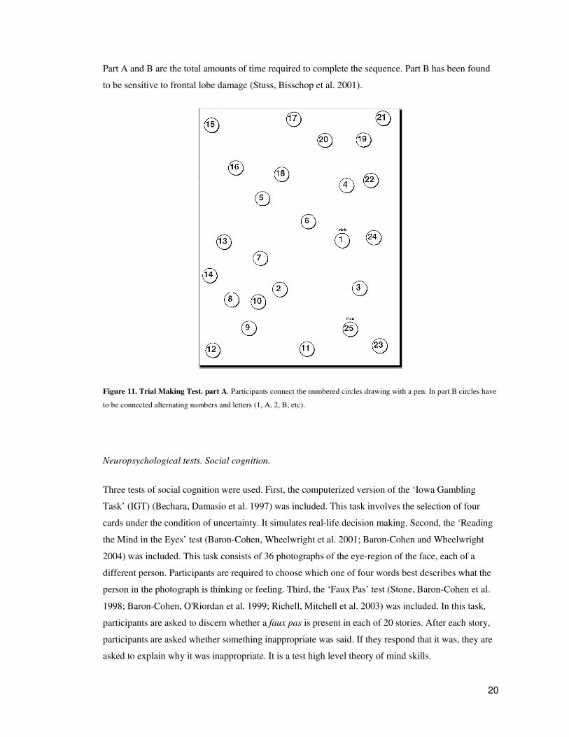
20
Part A and B are the total amounts of time required to complete the sequence. Part B has been found
to be sensitive to frontal lobe damage (Stuss, Bisschop et al. 2001).
Figure 11. Trial Making Test. part A. Participants connect the numbered circles drawing with a pen. In part B circles have
to be connected alternating numbers and letters (1, A, 2, B, etc).
Neuropsychological tests. Social cognition.
Three tests of social cognition were used. First, the computerized version of the ‘Iowa Gambling
Task’ (IGT) (Bechara, Damasio et al. 1997) was included. This task involves the selection of four
cards under the condition of uncertainty. It simulates real-life decision making. Second, the ‘Reading
the Mind in the Eyes’ test (Baron-Cohen, Wheelwright et al. 2001; Baron-Cohen and Wheelwright
2004) was included. This task consists of 36 photographs of the eye-region of the face, each of a
different person. Participants are required to choose which one of four words best describes what the
person in the photograph is thinking or feeling. Third, the ‘Faux Pas’ test (Stone, Baron-Cohen et al.
1998; Baron-Cohen, O'Riordan et al. 1999; Richell, Mitchell et al. 2003) was included. In this task,
participants are asked to discern whether a faux pas is present in each of 20 stories. After each story,
participants are asked whether something inappropriate was said. If they respond that it was, they are
asked to explain why it was inappropriate. It is a test high level theory of mind skills.

21
Reading the Mind in the Eyes (Baron-Cohen, Wheelwright et al. 2001; Baron-Cohen and
Wheelwright 2004)
In this standardized multiple-choice test participants are shown 36 black-and-white photos of the eye
region of different persons. Subjects are asked to choose which of four words best describes what the
person in the photograph is thinking or feeling (e.g., “terrified,” “upset,” “arrogant,” and “annoyed”;
see figure 12) . The individual in each photo displays a particular emotional or cognitive state. This
task depends in part of Theory of Mind, the ability to attribute mental states
beliefs, intents, desires, pretending, knowledge to oneself and others and to understand that others
have beliefs, desires and intentions that are different from one's own.
Figure 12. Reading the mind in the eyes test. In each trial, one face is shown with four words in the corners. Subjects have
to choose the word that best describe what the person in the photograph is thinking or feeling.
Faux Pas Test (Stone, Baron-Cohen et al. 1998; Baron-Cohen, O'Riordan et al. 1999; Richell,
Mitchell et al. 2003)
In this test the subject is read a story where one of the characters says something that may or may not
contain something that it would be better not to say; that is, a social faux pas. Subjects are exposed to
ten stories with a faux pas and ten stories without a faux pas. After each story, the subject is asked
whether something inappropriate was said. An additional control question is included to evaluate if
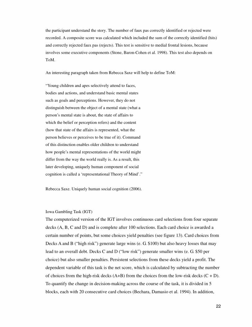
22
the participant understand the story. The number of faux pas correctly identified or rejected were
recorded. A composite score was calculated which included the sum of the correctly identified (hits)
and correctly rejected faux pas (rejects). This test is sensitive to medial frontal lesions, because
involves some executive components (Stone, Baron-Cohen et al. 1998). This test also depends on
ToM.
An interesting paragraph taken from Rebecca Saxe will help to define ToM:
“Young children and apes selectively attend to faces,
bodies and actions, and understand basic mental states
such as goals and perceptions. However, they do not
distinguish between the object of a mental state (what a
person’s mental state is about, the state of affairs to
which the belief or perception refers) and the content
(how that state of the affairs is represented, what the
person believes or perceives to be true of it). Command
of this distinction enables older children to understand
how people’s mental representations of the world might
differ from the way the world really is. As a result, this
later developing, uniquely human component of social
cognition is called a ‘representational Theory of Mind’.”
Rebecca Saxe. Uniquely human social cognition (2006).
Iowa Gambling Task (IGT)
The computerized version of the IGT involves continuous card selections from four separate
decks (A, B, C and D) and is complete after 100 selections. Each card choice is awarded a
certain number of points, but some choices yield penalties (see figure 13). Card choices from
Decks A and B (“high risk”) generate large wins (e. G. $100) but also heavy losses that may
lead to an overall debt. Decks C and D (“low risk”) generate smaller wins (e. G. $50 per
choice) but also smaller penalties. Persistent selections from these decks yield a profit. The
dependent variable of this task is the net score, which is calculated by subtracting the number
of choices from the high-risk decks (A+B) from the choices from the low-risk decks (C + D).
To quantify the change in decision-making across the course of the task, it is divided in 5
blocks, each with 20 consecutive card choices (Bechara, Damasio et al. 1994). In addition,
23
we compared participants’ net score on the first (1+2) and last (4+5) blocks. IGT is sensitive
to ventromedial prefrontal cortex lesions (Bechara, Damasio et al. 1994).
Figure 13. Iowa gambling Task. Participants have to choose cards from any deck. Each time the subject choose a deck, the
program showed how much money they gain (or loose) for that card and the total amount of money they have. The rule that
subjects have lo learn is that two decks give big gains but also big loses, and in average are less convenient that the other two
decks (small gains and small loses, but in average more net gain).
The Rapid Decision Making Under Risk (RDMUR) Task.
We designed a simplified computer gambling task based on a modified version of blackjack (Slovic
1966; Fernandez-Duque 2007). At the beginning of the task, participants read the following
instructions on screen: “In this deck, there are 10 cards. Nine cards are “good,” and one is “bad”.
You will win one dollar for each good card you draw; however, if you draw the bad card, then you will
lose everything, and the game will end. You will keep whatever money you win, so try to play as well
as possible. Choose one card at a time by clicking on it.” Participants are told that the Joker is the bad
card and that it is randomly placed within the set of ten cards. Finally, participants are asked to play
only once but could stop at any time to collect their prize. The task ended when participants stopped
or drew the bad card. Using a mouse, participants could either select a face down card or stop the
game by clicking the “check-out” box. Participants are unaware that any of the first 8 choices led to
wins; in other words, the bad card was always the ninth card (Fernandez-Duque 2007). Because the
24
expected value in this task is the highest after turning five cards (Slovic 1966), rational decision
makers should stop after turning the fifth card.
For those tests for which they were available, standardized norms were used. Otherwise, scores were
compared with published data from healthy participants. Scores on the ‘Digit Repetition’, ‘Digit
Span’ and ‘Arithmetic’ tests were compared with the standardized scores for the Wechsler Adult
Intelligence Scale-III (WAIS-III) (Wechsler 1997). RVLT scores were compared with Schmidt’s data
(Schmidt 1996). TMT scores were compared with data from Torralva (Torralva, Gleichgerrcht et al.
2010). Scores on the IFS, ‘Faux Pas’ and ‘Reading the Mind in the Eyes’ tests were compared with
data published by Torralva (Torralva, Roca et al. 2009). Scores on the IGT were compared with
Bechara’s results (Bechara, Damasio et al. 1997) .
25
Rapid face perception interacts with a complex system of
emotional processing and social behavior
The results are structured in three sections, each corresponding to an independent experiment. Each
section in turn is organized as follows: 1) The hypothesis which justifies and motivates the
experiment, 2) Specific Methods for the particular experiment and 3) Description of the results and
brief summary of the main observations.

2
Chapter I
The cortical potential N170 is consistently associated with
emotion, social cognition and executive functions.
Section I
H1) A cortical electrophysiological marker of the processing of facial emotion (N170) is associated
with individual differences in complex social cognition skills.
This section has been done in collaboration with the following colleagues: Andrés Canales-Johnson,
Hugo Urquina, Raphael Guex, Esteban Hurtado, Alejandro Blenkmann, Nicolás von Ellenrieder,
Facundo Manes and Agustin Ibañez, under the supervision of Professor Mariano Sigman.
SPECIFIC MATERIALS AND METHODS FOR THIS EXPERIMENT
A major part of the results on the first section are, in a way, control experiments. This means a
validation of the paradigm, to measure a clear neural signal, that this signal is modulated by the
experimental factors in the expected trend, and that the neural sources are in correspondence with that
measured by other experimenters.
In this section, healthy subjects completed the DVT while EEG recorded its scalp activity.
Psychophysical (performance and RT) and ERP (N170 modulation by facial emotion) results of the
DVT were correlated with three types of neuropsychological tests: general neuropsychology,
executive functions and social cognition.
Participants
Twenty healthy participants, six females (30.9+2.3 years old, with 16.6+0.6 years of formal
3
education) completed the DVT. In a subsequent session the participants had voluntarily completed a
neuropsychological battery that comprised tests of general neuropsychological functioning, executive
functioning and social cognition. A questionnaire was administered to all participants to rule out
hearing, visual, psychiatric or neurological deficits. The N170 component has been reported in
neuropsychiatric studies with both right- and left-handed participants [e.g.(Turetsky, Kohler et al.
2007; Gunji, Inagaki et al. 2009)]. As the current paradigm is used in subsequent experiments of this
thesis with neuropsychiatric populations, a small number of left-handed participants were included to
reflect the individual variability found in individuals with psychiatric diagnoses. All participants
completed the tasks voluntarily and signed an informed consent form in agreement with the Helsinki
declaration.
Neuropsychological Assessment.
A subset of 16 participants (four females, mean age 32.5+2.7, mean years in education 17.3+0.7)
received a battery of neuropsychological tests comprising tests of general neuropsychology (i.e., the
Word Accentuation Test, the Rey Verbal Learning Test [RVLT] and the Wechsler Adult Intelligence
Scale [WAIS]), executive functioning (i.e., the INECO Frontal Screening test [IFS], the Go/No Go
test, the Backward Digit Span test [BDS], the Trail Making Test B [TMT-B], the Forward Digit Span
test [FDS]), and social cognition (i.e., The Faux pas test, the reading the mind in the eyes test and the
Iowa gambling task).
General Neuropsychology.
Participants were assessed using a neuropsychological battery designed to cover various basic
cognitive functions. General neuropsychology was included in order to test general cognitive
processes. Memory was assessed by the Rey Verbal Learning Test (RVLT) (Rey 1958 ), which
includes measures of verbal learning, retention and immediate recall. Phonological and semantic
fluency were assessed using the Controlled Oral Word Association test [COWAT (Benton, Hamsher
et al. 1994)]. An arithmetic test from the Wechsler Adult Intelligence Scale (WAIS) (Wechsler 1997)
was also included.
4
Executive functions
To assess participants’ executive functioning, a set of several tests was compiled. The ‘INECO
Frontal Screening’ test (IFS) (Torralva, Roca et al. 2009) was used to assess frontal lobe functioning.
‘Backward Digit Span’ (Wechsler 1997) and the ‘Trail Making Test B’ (TMT B) (Partington 1949)
were used to assess attentional flexibility, sequencing and planning skills. The ‘Forward Digit Span’
test (Wechsler 1997) was used to assess Working Memory.
Social cognition.
Three tests of social cognition were used. First, the computerized version of the ‘Iowa Gambling
Task’ (IGT) (Bechara, Damasio et al. 1997) was included. This task involves the selection of four
cards under the condition of uncertainty. It was designed to simulate real-life decision making.
Second, the Reading the Mind in the Eyes (RMET) test (Baron-Cohen, Wheelwright et al. 2001;
Baron-Cohen and Wheelwright 2004) was included. This task consists of 36 photographs of the eye-
region of the face, each of a different person. Participants are required to choose which one of four
words best describes what the person in the photograph is thinking or feeling. Third, the ‘Faux Pas’
test (Stone, Baron-Cohen et al. 1998; Baron-Cohen, O'Riordan et al. 1999; Richell, Mitchell et al.
2003) was included. In this task, participants are asked to discern whether a faux pas is present in each
of 20 stories. After each story, participants are asked whether something inappropriate was said. If
they respond that it was, they are asked to explain why it was inappropriate. It is a test of the capacity
to identify something that would be (unintentionally) hurtful or insulting to another person..
RESULTS
Behavioral measures of the Dual Valence Task
RTs were shorter to face stimuli than word stimuli, to positive stimuli than to negative stimuli, and to
compatible simultaneous stimuli than to incompatible simultaneous stimuli when face valence was
positive (see figure 14 ). All participants performed with greater than 80% accuracy on all of the
subtasks of the dual valence task (see table 1).
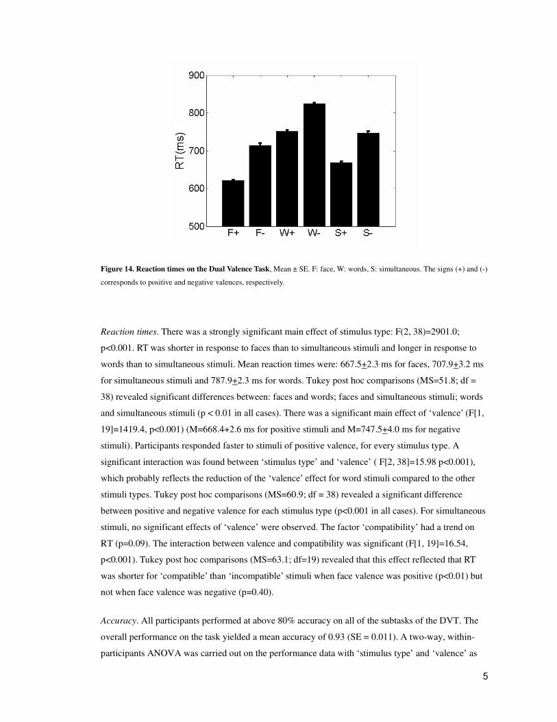
5
Figure 14. Reaction times on the Dual Valence Task, Mean ± SE. F: face, W: words, S: simultaneous. The signs (+) and (-)
corresponds to positive and negative valences, respectively.
Reaction times. There was a strongly significant main effect of stimulus type: F(2, 38)=2901.0;
p<0.001. RT was shorter in response to faces than to simultaneous stimuli and longer in response to
words than to simultaneous stimuli. Mean reaction times were: 667.5+2.3 ms for faces, 707.9+3.2 ms
for simultaneous stimuli and 787.9+2.3 ms for words. Tukey post hoc comparisons (MS=51.8; df =
38) revealed significant differences between: faces and words; faces and simultaneous stimuli; words
and simultaneous stimuli (p < 0.01 in all cases). There was a significant main effect of ‘valence’ (F[1,
19]=1419.4, p<0.001) (M=668.4+2.6 ms for positive stimuli and M=747.5+4.0 ms for negative
stimuli). Participants responded faster to stimuli of positive valence, for every stimulus type. A
significant interaction was found between ‘stimulus type’ and ‘valence’ ( F[2, 38]=15.98 p<0.001),
which probably reflects the reduction of the ‘valence’ effect for word stimuli compared to the other
stimuli types. Tukey post hoc comparisons (MS=60.9; df = 38) revealed a significant difference
between positive and negative valence for each stimulus type (p<0.001 in all cases). For simultaneous
stimuli, no significant effects of ‘valence’ were observed. The factor ‘compatibility’ had a trend on
RT (p=0.09). The interaction between valence and compatibility was significant (F[1, 19]=16.54,
p<0.001). Tukey post hoc comparisons (MS=63.1; df=19) revealed that this effect reflected that RT
was shorter for ‘compatible’ than ‘incompatible’ stimuli when face valence was positive (p<0.01) but
not when face valence was negative (p=0.40).
Accuracy. All participants performed at above 80% accuracy on all of the subtasks of the DVT. The
overall performance on the task yielded a mean accuracy of 0.93 (SE = 0.011). A two-way, within-
participants ANOVA was carried out on the performance data with ‘stimulus type’ and ‘valence’ as

6
independent factors. No significant main effects were found: F(2, 38)=0.30; p=0.73 for ‘stimulus type’
and (F(1, 19)=3.03, p=0.09 for ‘valence’. There was no significant effects of ‘compatibility’: F(1,
19)=0.08, p=0.76 and no significant interaction effects For simultaneous stimuli, performance was
better for positive than for negative faces (M=0.94+0.01 and M=0.92+0.01, respectively): F(1,
19)=5.96, p<0.05.
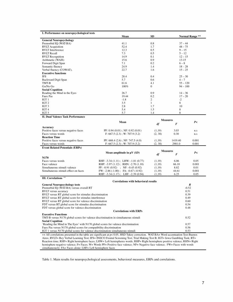
7
I. Performance on neuropsychological tests Mean SD Normal Range **
General Neuropsychology Premorbid IQ (WAT-BA) 41.1 0.8 37 – 44 RVLT Acquisition 52.4 1.7 48 – 75 RVLT Interference 12.3 0.5 9 – 15 RVLT Recall 7.3 0.5 5 – 12 RVLT Recognition 14.9 0.1 12 – 15 Arithmetic (WAIS) 15.6 0.9 13-15 Forward Digit Span 7.1 0.2 6 – 8 Semantic fluency 24.9 1.4 18 – 28 Verbal fluency (COWAT), 22.7 1.6 15 – 25
Executive functions IFS 28.4 0.4 25 – 30 Backward Digit Span 5.7 0.6 4 – 7 TMT-B 61.6 4.1 55 – 120 Go/No Go 100% 0 94 – 100 Social Cognition Reading the Mind in the Eyes 26.7 0.9 14 – 36 Faux Pas 19.44 0.2 17 – 20 IGT 1 -1.8 2 -2 IGT 2 3.5 1 8 IGT 3 2.8 1.7 10 IGT 4 5.4 1.5 8 IGT 5 5.7 1.4 8
II. Dual Valence Task Performance Measures
Mean df F
P<
Accuracy Positive faces versus negative faces PF: 0.94 (0.01) ; NF: 0.92 (0.01) (1,19) 3.03 n.s Faces versus words F: 667.5 (2.3) ; W: 707.9 (3.2) (2, 38) 0.30 n.s Reaction Time Positive faces versus negative faces PF: 668.4 (2.6) ; NF: 747.5 (4.0) (1,19) 1419.40 0.001 Faces versus words F: 667.5 (2.3) ; W: 707.9 (3.2) (2, 38) 2901.0 0.001
Event Related Potentials (ERPs) Measures
Mean amplitude in µV (SD) df F
P<
N170 Faces versus words RHF: -3.34 (1.11) ; LHW: -1.81 (0.77) (1,19) 6.06 0.05 Face valence RHP: -3.97 (1.12) ; RHN: -2.70 (1.10) (1,19) 66.10 0.001 Simultaneous stimuli valence PF: -0.91 (0.92) ; NF: -0.45 (0.92) (1,19) 8.82 0.01 Simultaneous stimuli effect on faces FW: -2.86 (-1.00) ; FA: -0.67 (-0.92) (1,19) 44.61 0.001 RHF: -3.34 (1.17) ; LHF: -2.39 (0.94) (1,19) 6.25 0.05
III. Correlations ++ Correlations with behavioral results
General Neuropsychology tests R Premorbid IQ (WAT-BA) versus overall RT -0.52 RVLT versus accuracy 0.51 RVLT versus RT global score for stimulus discrimination 0.39 RVLT versus RT global score for stimulus interference 0.49 RVLT versus RT global score for valence discrimination 0.60 FDT versus RT global score for stimulus discrimination 0.54 FDT versus global score for valence discrimination 0.48 Correlations with ERPs Executive Functions TMT-B versus N170 global scores for valence discrimination in simultaneous stimuli 0.52
Social Cognition ‘Reading the Mind in The Eyes’ with N170 global scores for valence discrimination 0.57 Faux Pas versus N170 global scores for compatibility discrimination 0.56 IGT 1 versus N170 global scores for valence discrimination simultaneous stimuli 0.53
++ All correlations presented in the table are significant at p< 0.05, HSD Tukey correction. WAT-BA= Word accentuation Test-Buenos Aires; RVLT= Rey Verbal Learning Test; IFS= INECO Frontal Screening Test; Trial Making Test B; IGT= Iowa Gambling Task; RT= Reaction time; RHF= Right hemisphere faces; LHW= Left hemispheres words; RHP= Right hemisphere positive valence; RHN= Right hemisphere negative valence; F= Faces; W= Words PF= Positive face valence; NF= Negative face valence; FW= Faces with words simultaneously; FA= Faces alone; LHF= Left hemisphere faces
Table 1. Main results for neuropsychological assessments, behavioral measures, ERPs and correlations.
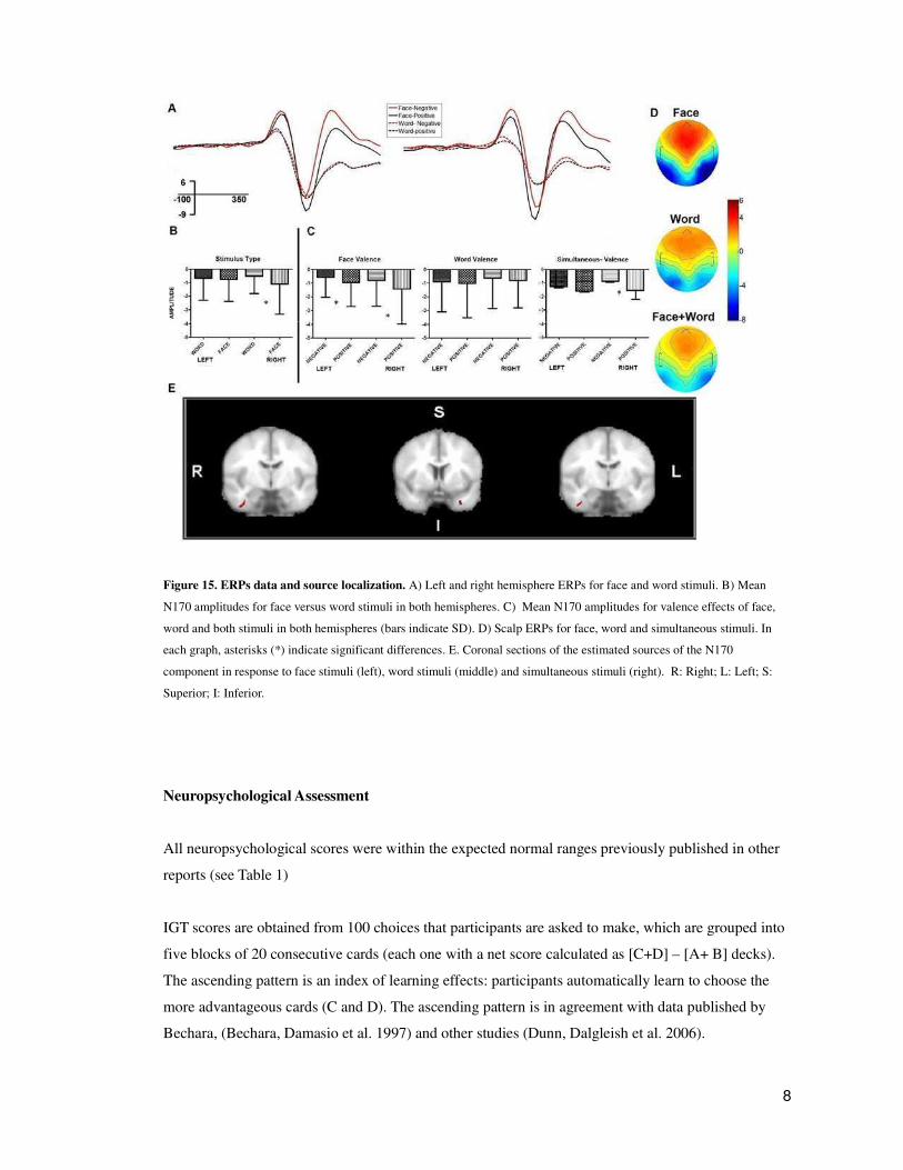
8
Figure 15. ERPs data and source localization. A) Left and right hemisphere ERPs for face and word stimuli. B) Mean
N170 amplitudes for face versus word stimuli in both hemispheres. C) Mean N170 amplitudes for valence effects of face,
word and both stimuli in both hemispheres (bars indicate SD). D) Scalp ERPs for face, word and simultaneous stimuli. In
each graph, asterisks (*) indicate significant differences. E. Coronal sections of the estimated sources of the N170
component in response to face stimuli (left), word stimuli (middle) and simultaneous stimuli (right). R: Right; L: Left; S:
Superior; I: Inferior.
Neuropsychological Assessment
All neuropsychological scores were within the expected normal ranges previously published in other
reports (see Table 1)
IGT scores are obtained from 100 choices that participants are asked to make, which are grouped into
five blocks of 20 consecutive cards (each one with a net score calculated as [C+D] – [A+ B] decks).
The ascending pattern is an index of learning effects: participants automatically learn to choose the
more advantageous cards (C and D). The ascending pattern is in agreement with data published by
Bechara, (Bechara, Damasio et al. 1997) and other studies (Dunn, Dalgleish et al. 2006).
9
N170 Source Localization.
The cortical source of the N170 component was estimated for each stimulus type: 1) Faces, 2) Words,
and 3) Simultaneous Stimuli (see figure 15D). For face stimuli, the source was located at the FG
anterior division, with a peak in the right hemisphere (at coordinates 41,-8,-36). Compared with words
and simultaneous stimuli, the face stimuli elicited a higher intensity (145.97 nA.m).
For word stimuli, the mean source was located at the border between the temporal pole and temporal
fusiform cortex, anterior division, in the left hemisphere (coordinate -30,4,-37), with an intensity of
62.11 nA.m. A second source of lower amplitude was found in the temporal fusiform cortex, anterior
division, in the right hemisphere (coordinate 41,-7,-37, intensity=25.04 nA.m).
Finally, for simultaneous stimuli, two principal sources were identified, located bilaterally. The higher
source was located in the temporal fusiform cortex, anterior division, in the right hemisphere
(coordinates 41,-7,-37; intensity=48.3 nA.m). The other source was observed in the fusiform cortex,
posterior division in the left hemisphere (coordinates -31,-12,-36; intensity= 22.93 nA.m).
N170
The findings from the ERP data followed expected trends for the main experimental factors: words
and faces showed left-right asymmetries; the N170 discriminated face valence in both hemispheres
(with greater sensitivity in the right hemisphere); the N170 did not discriminate word valence; and
N170 amplitude decreased when faces were accompanied by words. In brief, the N170 showed an
emotional valence effect for faces but not for words, similarly to those findings from other paradigms
that test processing of facial and semantic emotional stimuli (Schacht and Sommer 2009).
N170. Stimulus type effects (Faces versus words).
The comparison of ERPs for faces and words (i.e., stimulus type) revealed no significant main effects
when collapsed across both hemispheres (see Figure 15.A, 15.B). In accordance with previous studies
that found opposite lateralization of faces and words (Bentin, Mouchetant-Rostaing et al. 1999;
Rossion, Joyce et al. 2003), we found a significant interaction between stimulus type and hemisphere
(F[1, 20] = 6.06, p<0.05). Post hoc comparisons (MS = 1.82, df = 20) revealed that the N170
discriminated stimulus type only within the right hemisphere (right hemisphere: -3.34+1.11 µV for
faces and -1.81+0.77 µV for words, p < 0.05; left hemisphere: -2.39+0.90 µV for faces and -
2.31+0.72 µV for words, p = 0.99) (Table 1)
10
N170. Face valence effects.
A significant effect of valence was found (F[1, 20] = 66,10, p<0.01), which reflects an increased
N170 amplitude in response to positive faces as compared to negative faces (-3.35+0.96 µV and -
2.38+0.95 µV, respectively). There was no significant main effect of hemisphere (F[1, 20] = 2.056,
p=0.167) and no significant interaction between face valence and hemisphere ( F[1, 20] = 11.01,
p<0.01). Post hoc comparisons (MS = 0.16, df = 20) revealed that face valence was better
discriminated by the N170 in the right hemisphere (positive: -3.97+1.12; negative: -2.70+1.10,
p<0.001) than in the left hemisphere (positive:-2.7+0.81 µV; negative: -2.04+0.89, p<0.05). See Table
1 and figure 15.A
N170. Word valence effects
Word valence was not discriminated by N170 (F(1,20) = 0.55, p = 0.47). The interaction between
word valence and hemisphere was not significant (F[1, 20] = 2.84, p = 0.11).
N170. Simultaneous stimuli valence effects
The N170 discriminated the face valence of simultaneous stimuli (F[1, 19] = 8.82, p<0.01) having a
mean of -0.91 (+0.92 µV SD) and -0.457 (+0.920 µV SD) for positive and negative faces, respectively
(positive >negative, as was the case for face stimuli).
There were no significant main effect differences between the hemispheres for simultaneous stimuli
(F[1, 19] = 0.08, p = 0.78), but there was a significant interaction between valence and hemisphere
(F(1, 19) = 4.88, p<0.05). Post hoc comparisons (MS = 0.27, df = 19) revealed that face valence was
discriminated within the right hemisphere when presented simultaneously with words (p<0.01 for
right hemisphere and p = 0.65 for left hemisphere).
Simultaneous stimuli compatibility
The N170 was not subject to any significant effects of compatibility (F[1, 18]=0.03, p=0.87) and nor
was there any significant interaction effect between compatibility and hemisphere (F[1, 19]=0.06,
p=0.81). The interaction between valence and compatibility was not significant (F[1, 18]=0.07,
p=0.79) either and nor was the three-way interaction between hemisphere, valence and compatibility
(F[1, 18]=2.48, p=0.13).

11
Simultaneous stimuli effect on faces
A robust effect was found when comparing the N170 ERP evoked by faces and by simultaneous
stimuli (F[1, 19]=44.61, p<0.001). The mean data show that the amplitude of the N170 was lower
when faces were presented simultaneously with words compared to when they presented alone (M=-
2.86+-1.00 and M=-0.67+-0.92, respectively). The same comparison revealed a significant interaction
between stimulus type and hemisphere (F[1, 19]=6.25, p<0.05). Tukey post hoc comparisons (MS =
0.50, df = 19) revealed that, as reported elsewhere (Brandeis, Lehmann et al. 1995; Eimer and Holmes
2002), N170 amplitude is higher in the right hemisphere than in the left in response to faces (M=-
2.39+0.94 µV for left hemisphere and M=-3.34+1.17 µV for right hemisphere; p<0.01) but there was
no significant difference between the hemispheres in response to simultaneous stimuli (p=0.89).
In brief, N170 results, in conjunction with source localization, confirm that the N170 component was
correctly estimated (Eimer and Holmes 2002; Batty and Taylor 2003; Ashley, Vuilleumier et al. 2004;
Blau, Maurer et al. 2007; Sadeh, Podlipsky et al. 2010). Main N170 results are summarized in Table
1.
Correlations
General neuropsychology
Behavioral measures. Correlations were found between general memory (as measured by the Rey
Verbal Learning Test) and overall accuracy (r=0.51); RTs global score for stimulus discrimination
(RTs face-RTs word) (r=0.39); RTs global score for stimulus interference (RTs face-RTs simultaneous)
(r=0.49); and RTs global score for valence discrimination (RTs positive-RTs negative) (r=0.60).
Working memory (as measured by the Forward Digit Span Test) correlated with the RTs global score
for stimulus discrimination (RTs face/RTs word) (r=0.54) and the RTs global score for valence
discrimination RTs (RTs positive/RTs negative) (r=0.48).
Executive Functioning
Behavioral measures. Scores on the INECO Frontal Screening Test correlated with the RTs global
scores for stimulus discrimination (RTs face-RTs word) (r=0.54) and with the RTs global scores for
valence discrimination (RTs positive-RTs negative) (r=0.54).
ERPs measures. Scores on the Trial Making Test B correlated positively with the N170 global scores
for valence discrimination in simultaneous stimuli (r=0.52).
12
Social cognition
ERPs measures. Scores from the Iowa Gambling Task block 1 correlated significantly with the ERPs
global scores for valence discrimination in simultaneous stimuli (r=0.53). Scores on the reading the
mind in the eyes test correlated significantly with the N170 global scores for valence discrimination
(positive-negative) (r=0.57). Scores on the Faux pas test correlated with the N170 global scores for
compatibility discrimination (compatible/incompatible) (r=0.56). Correlation results are summarized
in Table 1.
In summary, all the significant correlations have the expected sign, and are, as a group, very
consistent.
Memory correlated positively with many basic behavioral skills important for the DVT (RT stimulus
and type discrimination)
N170 emotional valence discrimination correlated with a social cognition task (RMET).
N170 stimulus type discrimination correlated with an executive functions task (IFS).
N170 valence discrimination in simultaneous stimuli (word interference over faces) correlated with an
executive functions task (TMT-B)
N170 compatibility discrimination in simultaneous stimuli correlated with a social cognition task with
an important executive component (Faux Pas).

2
Section II
The previous experiment showed that complex social cognition and executive functions are associated
to N170 facial emotion modulation. The next experiment follows on this path by inquiring the
following hypothesis:
H2a) BD patients present cortical deficits in processing emotional facial information.
H2b) these deficiencies are associated with clinical measures.
This section includes experimental results obtained in collaboration with Agustín Ibáñez, Hugo
Urquina, Sandra Báez, Micaela do Nascimento, Raphael Guex, Esteban Hurtado, Alejandro
Blenkmann, Leandro Beltrachini , Alicia Lischinsky, Teresa Torralva, Fernando Torrente, Marcelo
Cetkovich and Facundo Manes. This work has been done under the supervision of Professor Mariano
Sigman.
SPECIFIC MATERIALS AND METHODS FOR THIS SECTION
In this section BD patients and matched controls completed the DVT while EEG recorded its scalp
activity. Psychophysical (performance and RT) and ERP (N170 modulation by facial emotion) results
of the DVT were correlated with neuropsychological tests and clinical neuropsychology.
Participants
Thirteen adult participants with BD, five females, (M=40.1, SD=2.5 years old), twelve right handed,
and 13 healthy controls matched for age (M=39,30, SD=2.51), gender and handedness completed a
full clinical, neuropsychological end electrophysiological evaluation. A questionnaire was given to
healthy participants to rule out hearing, visual, psychiatric or neurological deficits. All participants
gave signed, informed consent in agreement with the Helsinki declaration.
Patient’s criteria and recruitment process
All participants with fulfilled DSM-IV criteria for BD. Diagnosis were made by three experts (AL,
3
FT, and MC). The clinical protocol included questionnaires of informant-based version of Depression
Inventory II [BDI-II; (Beck, Steer et al. 1996)] and the Young Mania Rating Scale YMRS (Young,
Biggs et al. 1978), to assess depression and mania, respectively. To obtain scores of impulsivity and
anxiety, the BIS11 (Barrat Impulsivity Scale BIS11; (Patton, Stanford et al. 1995)], and the STAI trait-
state (Spielberger, Gorsuch et al. 1970) were considered. To exclude possible comorbid ADHD we
applied the ADHD Rating Scale questionnaire for adults and children (Barkley and Murphy 1998). All
patients were euthymic bipolar patients type II without comorbidity. Patients were not receiving
antipsychotic medication and didn’t have change of medication type or dosage over a period of 4
months.
Neuropsychological Assessment
General Neuropsychology
General neuropsychology evaluated participants’ basic cognitive functioning. Memory was evaluated
using the Rey Verbal Learning Test (Rey 1958 ). Attention and concentration were assessed using the
Trail Making Test A [TMT-A (Partington 1949)]. Phonological and semantic fluency were assessed
using the Controlled Oral Word Association test [COWAT (Benton, Hamsher et al. 1994)]. An
arithmetic test, Wechsler Adult Intelligence Scale III [WAIS III; (Wechsler 1997)] was also included.
Executive Functioning
Several tests evaluate executive functioning. The INECO Frontal Screening (Torralva, Roca et al.
2009) was used. Backward Digit Span and TMT-B (Partington 1949) were used to assess attentional
flexibility, attentional speed, sequencing and planning skills. Numbers Key and Searching Symbols
(Wechsler 1997) were used to evaluate visual perception and organization, visual scanning and the
efficient production of multiple motor responses. Ordering Letters and Numbers (Letters & Numbers
hereafter) was used to assess mental manipulation and working memory (Wechsler 1997). Finally, a
working memory index was derived from performance on the Digit Span, Arithmetic, and Letter–
Number Sequencing subtests (Hill, Elliott et al. 2010).
Social Cognition
Two social cognition tasks related to theory of mind were included. First, the Faux Pas test [FPT,
(Stone, Baron-Cohen et al. 1998)] which evaluates hurtful or insulting social situations. Second, the
Reading the Mind in The Eyes [RMET (Baron-Cohen, Wheelwright et al. 2001)], which assesses
individual differences in the ability to infer the affective mental states of other humans.
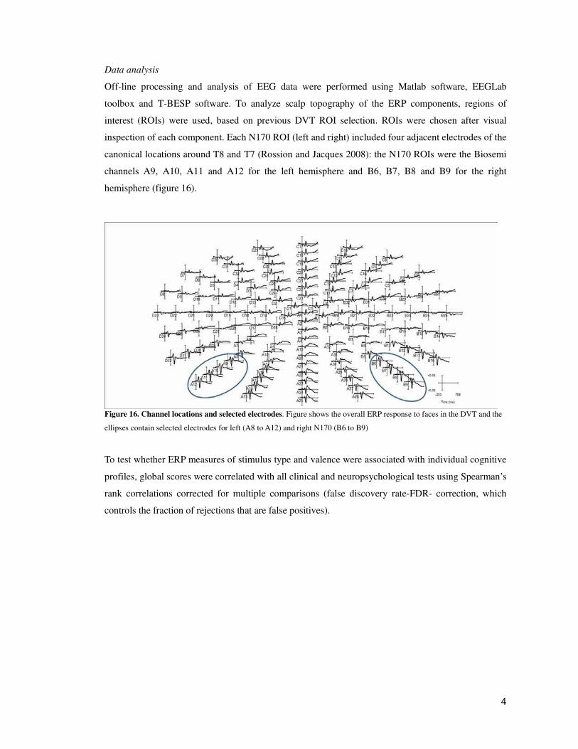
4
Data analysis
Off-line processing and analysis of EEG data were performed using Matlab software, EEGLab
toolbox and T-BESP software. To analyze scalp topography of the ERP components, regions of
interest (ROIs) were used, based on previous DVT ROI selection. ROIs were chosen after visual
inspection of each component. Each N170 ROI (left and right) included four adjacent electrodes of the
canonical locations around T8 and T7 (Rossion and Jacques 2008): the N170 ROIs were the Biosemi
channels A9, A10, A11 and A12 for the left hemisphere and B6, B7, B8 and B9 for the right
hemisphere (figure 16).
Figure 16. Channel locations and selected electrodes. Figure shows the overall ERP response to faces in the DVT and the
ellipses contain selected electrodes for left (A8 to A12) and right N170 (B6 to B9)
To test whether ERP measures of stimulus type and valence were associated with individual cognitive
profiles, global scores were correlated with all clinical and neuropsychological tests using Spearman’s
rank correlations corrected for multiple comparisons (false discovery rate-FDR- correction, which
controls the fraction of rejections that are false positives).
5
RESULTS
Demographic and clinical assessment
Table 2 shows the overall results from the demographic, clinical, and neuropsychological assessments.
Regarding demographic data, no differences regarding age (F(1, 24) =0.056, p=0.81), gender
(X2(1)=0.15, p=0.69), educational level (F(1, 24)=0.26, p=0.61), or handedness (X2(1)=0.00, p=1)
were observed between groups.
No differences were observed between groups for clinical measures of impulsivity (F(1, 24)=2.18,
p=0.15), depression (F(1, 24)=0.33, p=0.56), or mania (F(1, 24)=0.24, p=0.62). However, compared
to controls group, the BD patients showed significant differences for the anxiety scale STAI- State
(F(1, 24,)=19.86, p<0.001) and STAI- Trait (F(1, 24)=16.03, p<0.01).
Neuropsychological assessment
General Neuropsychology
Significant differences were observed in attention and concentration assessed with the TMT-A
(F(1,24)=8.89, p=0.006) and forward digits span (F(1,24)8.45, p<0.01). No group differences
regarding memory were observed for the RAVLT immediate recall (F(1,24)=0.38, p=0.54), delayed
recall (F(1,24)=0.1, p=0.9) or recognition phase (F(1,24)=0.25, p=0.87).
The groups did not differ on most tests of executive functioning, including the IFS total score
(F(1,24)=1.85, p=0.18), backwards digit span (F(1,24)=1.60, p=0.21), arithmetic evaluation -WAIS III
(F(1,24)=0.56, p=0.45), TMT-B (F(1,24)=0.88, p=0.35) and letters and numbers task (F(1,24)=0.21,
p=0.64). However, compared to controls the phonological fluency task (F(1,24)=5.39, p<0.05) yielded
lower scores in the BD group.
Social Cognition
Performance on FPT was significantly reduced in BD (F(1, 24)=12.14, p< 0.001) suggesting failures
in theory of mind process. For the RMET, no differences between groups were observed (F(1,
24)=2.68, p=0.11).
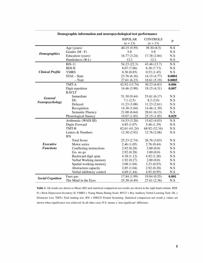
6
Demographic information and neuropsychological test performance
BIPOLAR
(n = 13) CONTROLS
(n = 13) P
Age (years) 40.15 (9.59) 39.30 (8.5) N.S Gender (M : F) 5:8 5:8 N.S Education (years) 16.77 (3.24) 17.38 (2.84) N.S
Demographics
Handedness (R:L) 12:1 12:1 N.S
BIS-11 54.23 (22.3) 43.46 (13.7) N.S BDI-II 8.07 (7.06) 6.38 (7.73) N.S YMRS 0.30 (0.85) 0.53 (1.45) N.S STAI – State 23.76 (6.16) 14.15 (4.77) 0.0001
Clinical Profile
– Trait 27.61 (6.23) 18.61 (5.19) 0.0005
TMT-A 42.92 (13.74) 30.23 (6.83) 0.006 Digit repetition 14.46 (3.90) 19.15 (4.31) 0.007 RAVLT Immediate 51.30 (9.44) 53.61 (6.17) N.S
DL 7.1 (2.5) 8.2 (2.0) N.S Delayed 11.23 (3.08) 11.23 (2.61) N.S Recognition 14.38 (1.04) 14.46 (1.39) N.S Semantic Fluency 21.00 (6.64) 28.61 (6.13) N.S
General
Neuropsychology
Phonological fluency 19.07 (1.85) 25.15 (1.85) 0.029
Arithmetic (WAIS III) 14.53 (3.20) 15.62 (4.03) N.S Digits Forward 4.85 (1.07) 5.46 (1.39) N.S TMT-B 82.61 (41.24) 68.92 (32.34) N.S Letters & Numbers 12.30 (2.92) 12.76 (2.08) N.S IFS Total Score 25.23 (2.74) 26.76 (3.03) N.S Motor series 2.46 (1-05) 2.76 (0.44) N.S Conflicting instructions 2.92 (0.28) 3.00 (0.0) N.S Go- no go 2.92 (0.28) 3.00 (0.0) N.S Backward digit span 4.38 (1.12) 4.92 (1.26) N.S Verbal Working memory 1.92 (0.27) 2.00 (0.0) N.S Spatial working memory 3.08 (1.04) 3.23 (0.93) N.S Abstraction capacity 2.85 (1.04) 2.92 (0.28) N.S
Executive
Functions
Verbal inhibitory control 4.69 (1.44) 4.92 (0.95) N.S
Faux pas 17.84 (1.99) 19.84 (0.55) 0.001 Social Cognition
The Mind in the Eyes 25.30 (4.49) 27.61 (2.36) N.S
Table 2. All results are shown as Mean (SD) and statistical comparison test results are shown in the right-hand column. BDI-
II = Beck Depression Inventory II; YMRS = Young Mania Rating Scale; RVLT = Rey Auditory Verbal Learning Task; DL =
Distractor List; TMT= Trail making test; IFS = INECO Frontal Screening. Statistical comparison test result p values are
shown when significance was achieved. In all other cases N.S. means a ‘non significant’ difference.
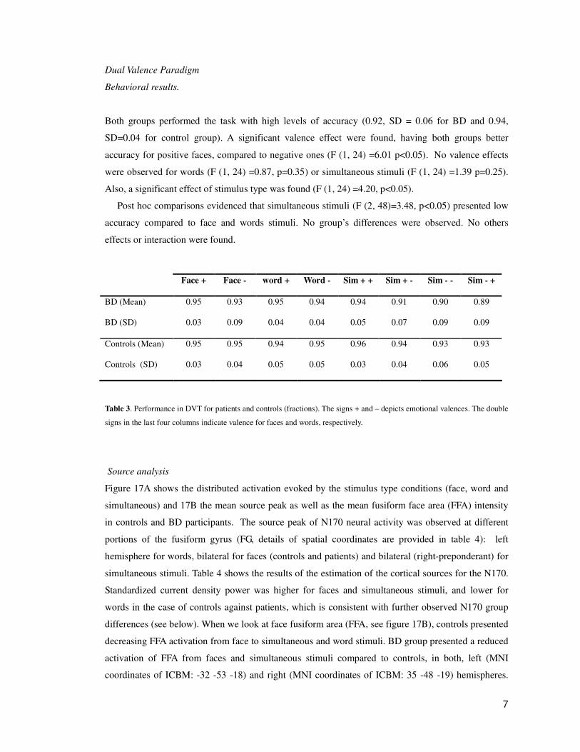
7
Dual Valence Paradigm
Behavioral results.
Both groups performed the task with high levels of accuracy (0.92, SD = 0.06 for BD and 0.94,
SD=0.04 for control group). A significant valence effect were found, having both groups better
accuracy for positive faces, compared to negative ones (F (1, 24) =6.01 p<0.05). No valence effects
were observed for words (F (1, 24) =0.87, p=0.35) or simultaneous stimuli (F (1, 24) =1.39 p=0.25).
Also, a significant effect of stimulus type was found (F (1, 24) =4.20, p<0.05).
Post hoc comparisons evidenced that simultaneous stimuli (F (2, 48)=3.48, p<0.05) presented low
accuracy compared to face and words stimuli. No group’s differences were observed. No others
effects or interaction were found.
Face + Face - word + Word - Sim + + Sim + - Sim - - Sim - +
BD (Mean) 0.95 0.93 0.95 0.94 0.94 0.91 0.90 0.89
BD (SD) 0.03 0.09 0.04 0.04 0.05 0.07 0.09 0.09
Controls (Mean) 0.95 0.95 0.94 0.95 0.96 0.94 0.93 0.93
Controls (SD) 0.03 0.04 0.05 0.05 0.03 0.04 0.06 0.05
Table 3. Performance in DVT for patients and controls (fractions). The signs + and – depicts emotional valences. The double
signs in the last four columns indicate valence for faces and words, respectively.
Source analysis
Figure 17A shows the distributed activation evoked by the stimulus type conditions (face, word and
simultaneous) and 17B the mean source peak as well as the mean fusiform face area (FFA) intensity
in controls and BD participants. The source peak of N170 neural activity was observed at different
portions of the fusiform gyrus (FG, details of spatial coordinates are provided in table 4): left
hemisphere for words, bilateral for faces (controls and patients) and bilateral (right-preponderant) for
simultaneous stimuli. Table 4 shows the results of the estimation of the cortical sources for the N170.
Standardized current density power was higher for faces and simultaneous stimuli, and lower for
words in the case of controls against patients, which is consistent with further observed N170 group
differences (see below). When we look at face fusiform area (FFA, see figure 17B), controls presented
decreasing FFA activation from face to simultaneous and word stimuli. BD group presented a reduced
activation of FFA from faces and simultaneous stimuli compared to controls, in both, left (MNI
coordinates of ICBM: -32 -53 -18) and right (MNI coordinates of ICBM: 35 -48 -19) hemispheres.
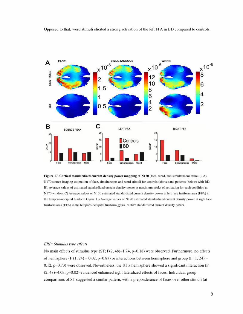
8
Opposed to that, word stimuli elicited a strong activation of the left FFA in BD compared to controls.
Figure 17. Cortical standardized current density power mapping of N170 (face, word, and simultaneous stimuli). A).
N170 source imaging estimation of face, simultaneous and word stimuli for controls (above) and patients (below) with BD.
B). Average values of estimated standardized current density power at maximum peaks of activation for each condition at
N170 window. C) Average values of N170 estimated standardized current density power at left face fusiform area (FFA) in
the temporo-occipital fusiform Gyrus. D) Average values of N170 estimated standardized current density power at right face
fusiform area (FFA) in the temporo-occipital fusiform gyrus. SCDP: standardized current density power.
ERP: Stimulus type effects
No main effects of stimulus type (ST; F(2, 48)=1.74, p=0.18) were observed. Furthermore, no effects
of hemisphere (F (1, 24) = 0.02, p=0.87) or interactions between hemisphere and group (F (1, 24) =
0.12, p=0.73) were observed. Nevertheless, the ST x hemisphere showed a significant interaction (F
(2, 48)=4.03, p=0.02) evidenced enhanced right lateralized effects of faces. Individual group
comparisons of ST suggested a similar pattern, with a preponderance of faces over other stimuli (at
9
both hemispheres in controls and at right hemisphere in BD. Figure 18A shows the ERPs stimulus
type modulation for both groups. In controls, no effects for hemisphere (F (1. 25) = 0.33, p= 0.56) or
interactions between ST and hemisphere (F (2. 50) = 1.75, p = 0.18) were found. A significant
stimulus type effect (F (2. 50) = 10.41, p=0.0001) was observed. Compared to words, post hoc
comparisons over this interaction showed N170 amplitude enhancement in both hemispheres for faces
(left p<0.001; right p<0.001) and simultaneous stimuli (left p<0.01; right p<0.005) respectively.
BD group not shown significant effects of stimulus type (F (2.50) = 1.63 p= 0.20). However, the
stimulus type x hemisphere showed a significant interaction (F (2.50) = 3.90, p = 0.02). Post hoc
comparisons performed over this interaction (Tukey HSD, df MS = 1.51, df = 50.00) showed
enhanced of N170 amplitude in the left hemisphere (faces - simultaneous stimulus; p = 0.01) and
similar results compared faces and words in the right hemisphere (p<0.002).

10
Faces
Source peak Left FFA Right FFA
MNI Coordinates SCDP SCDP
X Y Z
Anatomical Description (HOCSA) Harvard-Oxford Cortical
Structural Atlas) Mean Mean Mean
Control -18 -78 -17 32% Occipital Fusiform Gyrus; 19% Lingual Gyrus.
28.07 16.20
14.87
BD -21 -93 -8
28% Left Occipital Pole, 5%; Occipital Fusiform Gyrus, 5 % Lateral Occipital Cortex, inferior division
8.49 2.01
4.02
Simultaneous
Source peak Left FFA Right FFA
MNI Coordinates SCDP SCDP
X Y Z
Anatomical Description (Harvard-Oxford Cortical Structural
Atlas) Mean Mean Mean
Control 19 -85 -18
33% Right Occipital Fusiform Gyrus, 8% Lingual Gyrus, 4% Occipital Pole; 3% Lateral Occipital Cortex, inferior division
13.71 7.29
7.56
BD -24 -86 -20
35% Left Occipital Fusiform Gyrus, 8% Lateral Occipital Cortex, inferior division, 3% Occipital Pole, 3% Lingual Gyrus
9.30 1.99
3.76
Words
Source peak Left FFA Right FFA
MNI Coordinates SCDP SCDP
X Y Z
Anatomical Description (Harvard-Oxford Cortical Structural
Atlas) Mean Mean Mean
Control -11 -85 -18 25% Left Occipital Fusiform Gyrus, 22% Left Lingual Gyrus
8.42 4.78
1.52
BD -13 -89 -19
33% Left Occipital Fusiform Gyrus, 10% Lingual Gyrus, 5% Occipital Pole 2% Left Lateral Occipital Cortex, inferior division
9.46 5.96
0.17
Table 4. Estimation of the N170 neural generators. FFA: Face fusiform area in the temporo-occipital fusiform gyrus. MNI
coordinates for FFA: left (-32 -53 -18) and right (35 -48 -19). SCDP: Standardized Current Density Power. MNI coordinates
are provided by ICBM.
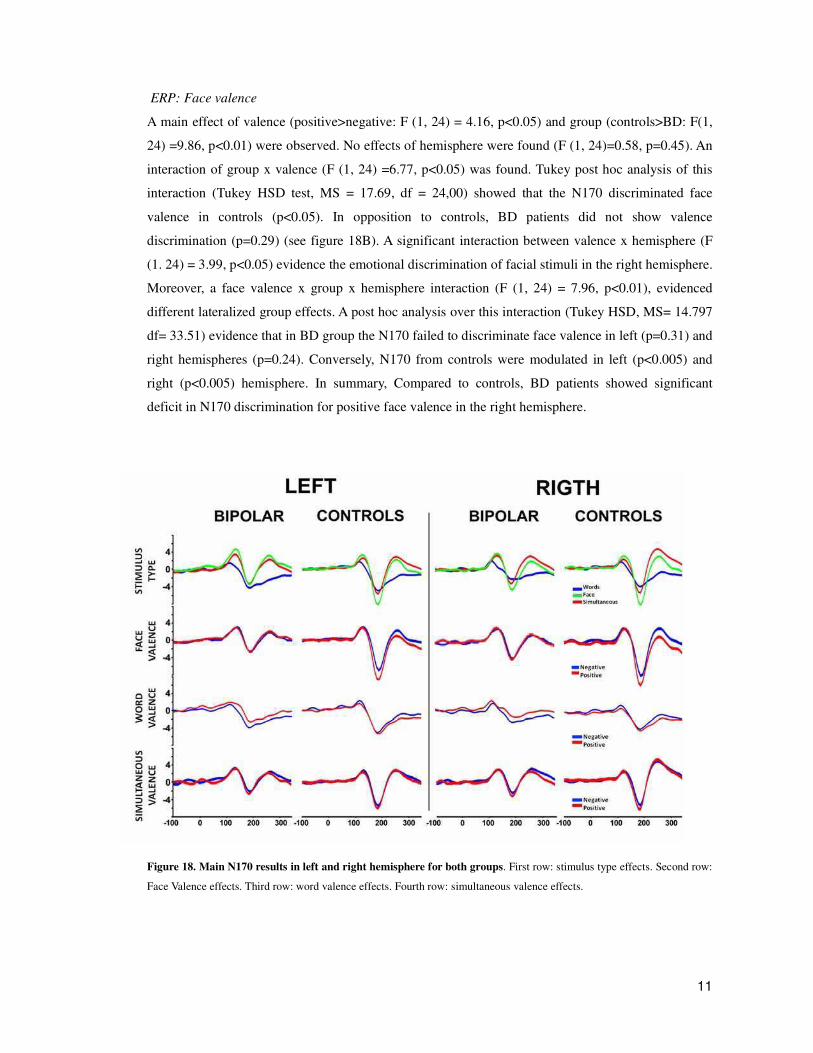
11
ERP: Face valence
A main effect of valence (positive>negative: F (1, 24) = 4.16, p<0.05) and group (controls>BD: F(1,
24) =9.86, p<0.01) were observed. No effects of hemisphere were found (F (1, 24)=0.58, p=0.45). An
interaction of group x valence (F (1, 24) =6.77, p<0.05) was found. Tukey post hoc analysis of this
interaction (Tukey HSD test, MS = 17.69, df = 24,00) showed that the N170 discriminated face
valence in controls (p<0.05). In opposition to controls, BD patients did not show valence
discrimination (p=0.29) (see figure 18B). A significant interaction between valence x hemisphere (F
(1. 24) = 3.99, p<0.05) evidence the emotional discrimination of facial stimuli in the right hemisphere.
Moreover, a face valence x group x hemisphere interaction (F (1, 24) = 7.96, p<0.01), evidenced
different lateralized group effects. A post hoc analysis over this interaction (Tukey HSD, MS= 14.797
df= 33.51) evidence that in BD group the N170 failed to discriminate face valence in left (p=0.31) and
right hemispheres (p=0.24). Conversely, N170 from controls were modulated in left (p<0.005) and
right (p<0.005) hemisphere. In summary, Compared to controls, BD patients showed significant
deficit in N170 discrimination for positive face valence in the right hemisphere.
Figure 18. Main N170 results in left and right hemisphere for both groups. First row: stimulus type effects. Second row:
Face Valence effects. Third row: word valence effects. Fourth row: simultaneous valence effects.
12
ERP: Word valence
For word stimuli, no effects of valence (F (1, 24) = 3.07, p=0.09) group (F (1.24) = 2.79, p=0.10), or
hemisphere (F (1, 24) = 2.54, p=0.12) were observed. Additionally, no interactions between
hemisphere x group (F (1, 24) =0.21, p=0.64) or valence x hemisphere (F (1, 24) = 0.26, p=0.60) were
found. Nevertheless, a group x valence interaction was observed (F (1, 24) = 5.62, p<0.05). In BD
patients an enhanced N170 amplitude for negative words was observed (p<0.01). In addition the
amplitude of negative valence words in BD was significant enhanced compared to controls (p<0.05).
No other effects were observed. Therefore, compared to control group, the N170 amplitude in BD
patients presented an early semantic valence modulation (enhancement of N170 to negative words).
ERP: Simultaneous Stimuli
Main effects of group (controls>BD; F (1, 24) =6.46, p<0.05) and valence (positive >negative; F (1,
24) =4.33, p=0.04) were observed. No effect of hemisphere (F (1, 24) = 0.14, p= 0.70) was present.
An interaction group x valence F (1, 24) =5.23, p<0.05 indicated different valence processing of
simultaneous stimuli in both groups. A post-hoc comparison over this last interaction (Tukey HSD
test, MS=3.56, df=24.00) yielded emotional discrimination of simultaneous stimuli in controls
(positive>negative; p<0.05), but not in BD (p=0.42). No other effects or interactions were significant.
Table 5 shows means and SD for these conditions.
In summary, N170 stimulus type discrimination was observed in both groups. BD patients showed
N170 deficits in facial and simultaneous stimuli emotional modulation. Furthermore, only BD patients
showed an early N170 discrimination for word valence in the left hemisphere (negative>positive).
Correlations with clinical profile
In BD, face valence was inversely correlated with mania (r = -0.63 p< 0.01) positively with
depression scores (r = 0.59, p<0.05). Word valence was associated with anxiety (STAI Trait; r = 0.60,
p<0.05). In control subjects, only word valence was associated with mania scores (r = 0.64, p<0.05).
13
BD mean (SE) Controls mean (SE)
Left Right Left Right
a. Stimulus Type Effects
Faces -0.65 (4.98) -1.71 (4.31) -5.57 (2.18) -5.42 (3.15)
Words -2.15 (4.30) -1.06 (2.65) -3.83 (2.80) -3.23 (2.91)
Simultaneous -0.88 (3,08) -1.17 (2,58) -3.86 (3,13) -3.97 (3,90)
b. Face Valence Effects
Positive -0.31 (4.76) -1.56 (4,50) -6.94 (3,49) -7.09 (3,45)
Negative -1.00 5.27) -1.85 (4.38) -4.20 (2.67) -3.75 (4.02)
c. Word Valence Effects
Positive -1.28 (4.64) -0.39 (3.62) -3.90 (2.75) -3.36 (2.91)
Negative -3.02 (4.20) -1.72 (2.58) -3.75 (3.12) -3.09 (3.10)
d. Simultaneous Valence Effects
Positive -1.2 (4.05) -1.55 (3.22) -4.20 (2.57) -4.46 (3.24)
Negative -0.55 (3.14) -0.79 (2.63) -3.51 (1.93) -3.49 (3.00)
Table 5. N170 amplitude values in response to stimulus type and valence factors.
General neuropsychology
In BD Patients, word valence showed a positive correlation with attention (r = 0.59, p<0.05). In
controls, word valence correlated significantly with semantic fluency (r = 0.58, p<0.05).
Executive Functions
In BD Patients, face valence presented a positive correlation with working memory (r = 0.58 p<0.05).
In controls, stimulus type showed a positive correlation with several IFS subtests: IFS total score (r =
0.67, p<0.05); backward digit span (r = 0.62 p = 0.02); abstraction capacity (r = 0.62, p<0.05) and
verbal inhibitory control (r = 0.63, p<0.05).
14
Theory of Mind
In BD stimulus type showed a positive correlation with FPT (r = 0.55, p<0.05). Word valence also
correlated positively with FPT (r = 0.61, p<0.05) and RMET (r = 0.56, p<0.05). In controls, stimulus
type showed a correlation with RMET (word valence; r = 0.63, p<0.05).
In summary, N170 face emotional discrimination correlated with mania and depression (with an
opposed sign) in BD patients.
Neuropsychological measures have the expected sign. Theory of mind tasks scores improve with
better N170 stimulus type discrimination and word valence in BD. Executive functions tasks scores
improve associated with an increase in N170 face valence and stimulus type discrimination in BD.

2
SECTION III:
In Sections I and II, we showed that face rapid processing indexed by the N170 is associated to
neuropsychological measures in healthy subjects and clinical measures in BD. The purpose of this
experiment is to explore face processing in ADHD by examining the following hypotheses:
H3a) ADHD patients present cortical deficits in processing emotional facial information.
H3b) These deficiencies are associated with executive functioning scores
The following colleagues have collaborated in the experiments shown in this section: Agustin Ibáñez,
Hugo Urquina, Teresa Torralva, Fernando Torrente, Esteban Hurtado, Raphael Guex, Alicia
Lischinsky, and Facundo Manes. This work has been done under the supervision of Professor Mariano
Sigman.
SPECIFIC MATERIALS AND METHODS FOR THIS EXPERIMENT
The aim of this experiment is to identify cortical markers of emotion processing in adult ADHD and
explore their relation to individual neuropsychological profiles.
Adult participants with ADHD and controls classified stimuli according to its emotional valence
(positive or negative).
Participants
Ten adult participants with ADHD, one female, (M=33.1, SD=3.6 years old), three left-handed,
completed the dual valence task. Ten healthy controls, matched for gender, age (M=33.0, SD=3.8
years old), handedness, and years of education were recruited (see below and table 6). Participants
with ADHD and controls received a thorough neuropsychological battery that comprised measures of
general neuropsychology, executive functioning and social cognition. A questionnaire was given to
healthy participants to rule out hearing, visual, psychiatric or neurological deficits. All participants
gave signed, informed consent in agreement with the Helsinki declaration.
3
Participant criteria and recruitment process
All participants with ADHD fulfilled DSM-IV criteria for ADHD. Diagnosis was made by three
experts (Alicia Lischinsky, Fernando Torrente and Facundo Manes). Participants were recruited from
amongst the patients of the Institute of Cognitive Neurology (INECO, Buenos Aires, Argentina), from
the Adult ADHD Clinic. From the initial set, 8 patients presented with ADHD combined type
(ADHD/C) and 2 patients presented with predominantly inattentive type (ADHD/I). All patients were
taking methylphenidate medication which was suspended on the day of ERP recordings. ADHD
diagnosis based on the DSM-IV criteria was assessed using the following protocol for adults:
1). ADHD Rating Scale for Adults (Barkley and Murphy 1998) in patient and informant versions. It
identifies current and retrospective childhood symptoms corresponding to DSM-IV characterization of
ADHD.
2). Depression Inventory II (BDI-II; Beck et al., 1996) and the Young Mania Rating Scale YMRS;
(Young et al., 1978), to assess depression and mania, respectively. Scales were administered to
patients and controls.
3) Neuropsychological assessment (see methods and next section for details).
Neuropsychological Assessment
Both participants with ADHD and controls received a comprehensive neuropsychological battery that
lasted approximately 120 minutes. It included general neuropsychology, executive functioning, and
social cognition.
General Neuropsychology
General neuropsychology evaluated participants’ basic cognitive functioning (see chapter 1 for
details). Memory was evaluated using the Rey Verbal Learning Test [RVLT (Rey 1958 )] that
comprised verbal learning, immediate and delayed recall and a distractor list. Attention and
concentration were assessed using the Trail Making Test A [TMT-A, (Partington 1949)]. Phonological
and semantic fluency were assessed using the Controlled Oral Word Association test [COWAT
(Benton, Hamsher et al. 1994)]. An arithmetic test, Wechsler Adult Intelligence Scale III [WAIS III
(Wechsler 1997)] was also included.
Executive Functioning
Several tests were compiled to evaluate executive functioning. The INECO Frontal Screening
(Torralva et al., 2009) was used to assess frontal lobe function indexed by several subtasks: Motor
Programming, Conflicting Instructions, Verbal Inhibitory Control, Abstraction, Backwards Digit Span,
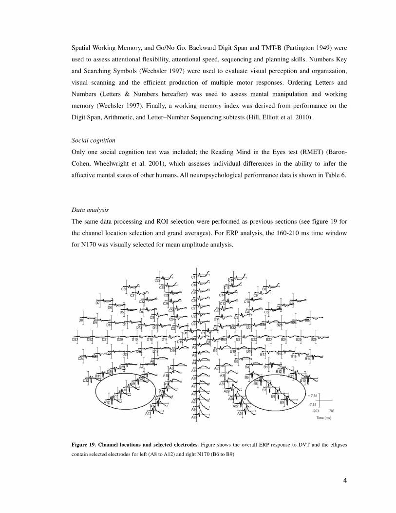
4
Spatial Working Memory, and Go/No Go. Backward Digit Span and TMT-B (Partington 1949) were
used to assess attentional flexibility, attentional speed, sequencing and planning skills. Numbers Key
and Searching Symbols (Wechsler 1997) were used to evaluate visual perception and organization,
visual scanning and the efficient production of multiple motor responses. Ordering Letters and
Numbers (Letters & Numbers hereafter) was used to assess mental manipulation and working
memory (Wechsler 1997). Finally, a working memory index was derived from performance on the
Digit Span, Arithmetic, and Letter–Number Sequencing subtests (Hill, Elliott et al. 2010).
Social cognition
Only one social cognition test was included; the Reading Mind in the Eyes test (RMET) (Baron-
Cohen, Wheelwright et al. 2001), which assesses individual differences in the ability to infer the
affective mental states of other humans. All neuropsychological performance data is shown in Table 6.
Data analysis
The same data processing and ROI selection were performed as previous sections (see figure 19 for
the channel location selection and grand averages). For ERP analysis, the 160-210 ms time window
for N170 was visually selected for mean amplitude analysis.
Figure 19. Channel locations and selected electrodes. Figure shows the overall ERP response to DVT and the ellipses
contain selected electrodes for left (A8 to A12) and right N170 (B6 to B9)
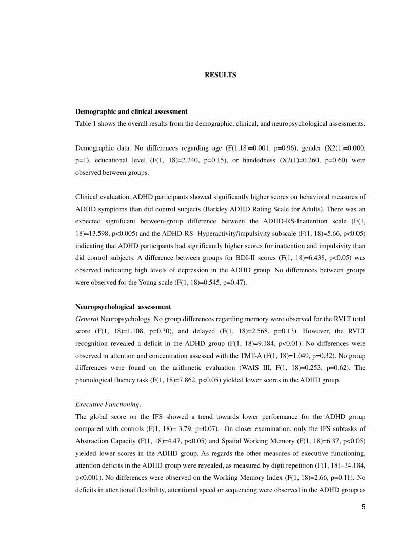
5
RESULTS
Demographic and clinical assessment
Table 1 shows the overall results from the demographic, clinical, and neuropsychological assessments.
Demographic data. No differences regarding age (F(1,18)=0.001, p=0.96), gender (X2(1)=0.000,
p=1), educational level (F(1, 18)=2.240, p=0.15), or handedness (X2(1)=0.260, p=0.60) were
observed between groups.
Clinical evaluation. ADHD participants showed significantly higher scores on behavioral measures of
ADHD symptoms than did control subjects (Barkley ADHD Rating Scale for Adults). There was an
expected significant between-group difference between the ADHD-RS-Inattention scale (F(1,
18)=13.598, p<0.005) and the ADHD-RS- Hyperactivity/impulsivity subscale (F(1, 18)=5.66, p<0.05)
indicating that ADHD participants had significantly higher scores for inattention and impulsivity than
did control subjects. A difference between groups for BDI-II scores (F(1, 18)=6.438, p<0.05) was
observed indicating high levels of depression in the ADHD group. No differences between groups
were observed for the Young scale (F(1, 18)=0.545, p=0.47).
Neuropsychological assessment
General Neuropsychology. No group differences regarding memory were observed for the RVLT total
score (F(1, 18)=1.108, p=0.30), and delayed (F(1, 18)=2.568, p=0.13). However, the RVLT
recognition revealed a deficit in the ADHD group (F(1, 18)=9.184, p<0.01). No differences were
observed in attention and concentration assessed with the TMT-A (F(1, 18)=1.049, p=0.32). No group
differences were found on the arithmetic evaluation (WAIS III, F(1, 18)=0.253, p=0.62). The
phonological fluency task (F(1, 18)=7.862, p<0.05) yielded lower scores in the ADHD group.
Executive Functioning.
The global score on the IFS showed a trend towards lower performance for the ADHD group
compared with controls (F(1, 18)= 3.79, p=0.07). On closer examination, only the IFS subtasks of
Abstraction Capacity (F(1, 18)=4.47, p<0.05) and Spatial Working Memory (F(1, 18)=6.37, p<0.05)
yielded lower scores in the ADHD group. As regards the other measures of executive functioning,
attention deficits in the ADHD group were revealed, as measured by digit repetition (F(1, 18)=34.184,
p<0.001). No differences were observed on the Working Memory Index (F(1, 18)=2.66, p=0.11). No
deficits in attentional flexibility, attentional speed or sequencing were observed in the ADHD group as
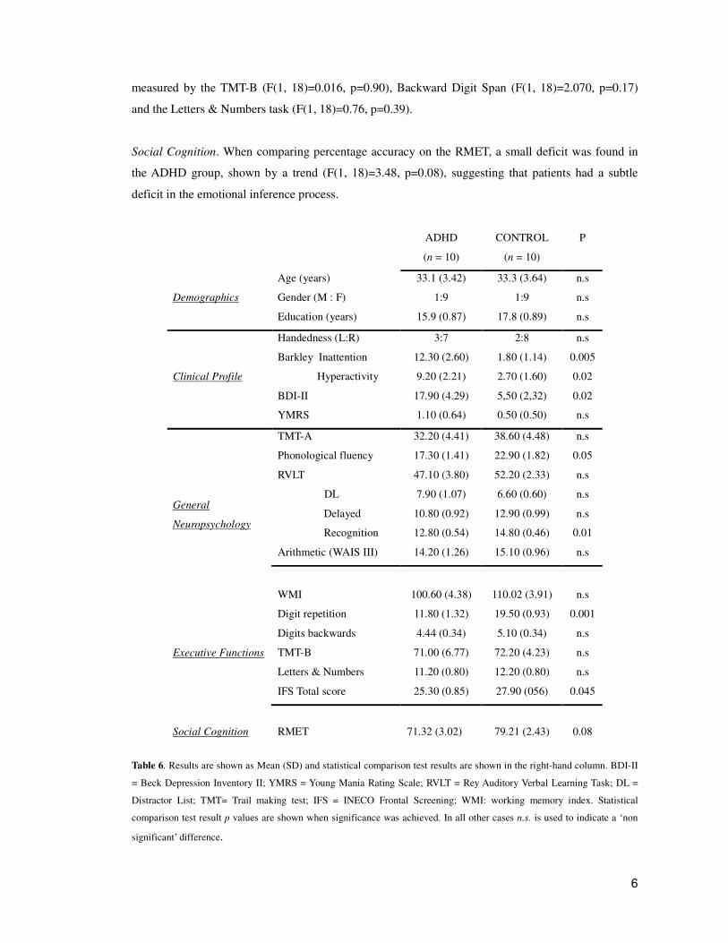
6
measured by the TMT-B (F(1, 18)=0.016, p=0.90), Backward Digit Span (F(1, 18)=2.070, p=0.17)
and the Letters & Numbers task (F(1, 18)=0.76, p=0.39).
Social Cognition. When comparing percentage accuracy on the RMET, a small deficit was found in
the ADHD group, shown by a trend (F(1, 18)=3.48, p=0.08), suggesting that patients had a subtle
deficit in the emotional inference process.
Table 6. Results are shown as Mean (SD) and statistical comparison test results are shown in the right-hand column. BDI-II
= Beck Depression Inventory II; YMRS = Young Mania Rating Scale; RVLT = Rey Auditory Verbal Learning Task; DL =
Distractor List; TMT= Trail making test; IFS = INECO Frontal Screening; WMI: working memory index. Statistical
comparison test result p values are shown when significance was achieved. In all other cases n.s. is used to indicate a ‘non
significant’ difference.
ADHD
(n = 10)
CONTROL
(n = 10)
P
Age (years) 33.1 (3.42) 33.3 (3.64) n.s
Gender (M : F) 1:9 1:9 n.s
Demographics
Education (years) 15.9 (0.87) 17.8 (0.89) n.s
Handedness (L:R) 3:7 2:8 n.s
Barkley Inattention 12.30 (2.60) 1.80 (1.14) 0.005
Hyperactivity 9.20 (2.21) 2.70 (1.60) 0.02
BDI-II 17.90 (4.29) 5,50 (2,32) 0.02
Clinical Profile
YMRS 1.10 (0.64) 0.50 (0.50) n.s
TMT-A 32.20 (4.41) 38.60 (4.48) n.s
Phonological fluency 17.30 (1.41) 22.90 (1.82) 0.05
RVLT 47.10 (3.80) 52.20 (2.33) n.s
DL 7.90 (1.07) 6.60 (0.60) n.s
Delayed 10.80 (0.92) 12.90 (0.99) n.s
Recognition 12.80 (0.54) 14.80 (0.46) 0.01
Arithmetic (WAIS III) 14.20 (1.26) 15.10 (0.96) n.s
WMI 100.60 (4.38) 110.02 (3.91) n.s
General
Neuropsychology
Digit repetition 11.80 (1.32) 19.50 (0.93) 0.001
Digits backwards 4.44 (0.34) 5.10 (0.34) n.s
TMT-B 71.00 (6.77) 72.20 (4.23) n.s
Letters & Numbers 11.20 (0.80) 12.20 (0.80) n.s
Executive Functions
IFS Total score 25.30 (0.85) 27.90 (056) 0.045
Social Cognition
RMET
71.32 (3.02)
79.21 (2.43)
0.08
7
Dual Valence Paradigm
Behavioral results
Both groups performed the task with an accuracy of 89% or more (see table 7 for mean and SD). The
performance on the overall task was very similar for both groups: 0.915%, SD=0.02 for the ADHD
group and 0.907%, SD=0.02 for the control group (F(1, 18)=0.04, p=0.89). For faces, word and
simultaneous stimuli, no differences were found for stimulus type (F(2, 36)=1.8, p=0.17) or group
differences (F(1, 18)=0.08, p=0.77). For faces, no main effects of valence (F(1, 18)=0.07, p=0.79) or
group ( (1, 18)=0.95, p=0.34) were found. For words, no main effects for valence (F(1, 18)=0.26,
p=0.61) or group (F(1, 18)=0.003, p=0.95) were found. Finally, for simultaneous stimuli, an effect of
valence was observed (accuracy: positive>negative, F(2, 18)=4.10, p<0.05) but no group effect (F(2,
17)=0.55, p=0.58). No congruency effects (F(2, 18)=0.06, p=0.94) or group differences were observed
(F(2, 18)=0.55, p=0.58) for simultaneous stimuli comparing the congruency of face and word valence.
There were no interactions between any of the previously mentioned factors. Table 2 shows the
descriptive statistics.
Face + Face - Word + Word - sim ++ Sim +- sim-- Sim -+
ADHD mean 0.92 0.93 0.90 0.90 0.93 0.91 0.90 0.90
ADHD SD 0.09 0.05 0.06 0.10 0.07 0.05 0.05 0.05
Controls mean 0.92 0.90 0.91 0.9 0.92 0.89 0.89 0.88
Controls SD 0.07 0.07 0.06 0.07 005 0.08 0.07 0.07
Table 7. Performance of the DVT for patients and controls. The signs + and – depict emotional valences. The double signs in
the last four columns indicate valence for faces and words, respectively.
Summarizing the behavioral results, accuracy was high across all conditions for both groups, and
despite the small mean differences between conditions, no significant effects were observed for
stimulus type in either group. For valence effects, only a small difference was obtained in
simultaneous stimuli for both ADHD and control groups (performance was better for positive than for
negative valence). No other effects yielded significant differences.
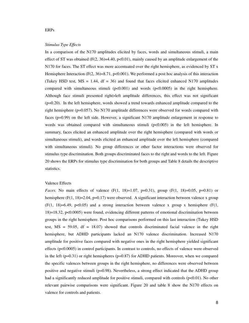
8
ERPs
Stimulus Type Effects
In a comparison of the N170 amplitudes elicited by faces, words and simultaneous stimuli, a main
effect of ST was obtained (F(2, 36)=4.40, p<0.01), mainly caused by an amplitude enlargement of the
N170 for faces. The ST effect was more accentuated over the right hemisphere, as evidenced by ST x
Hemisphere Interaction (F(2, 36)=8.71, p<0.001). We performed a post hoc analysis of this interaction
(Tukey HSD test, MS = 1.44, df = 36) and found that faces elicited enhanced N170 amplitudes
compared with simultaneous stimuli (p<0.001) and words (p<0.0005) in the right hemisphere.
Although face stimuli presented right>left amplitude differences, this effect was not significant
(p=0.20). In the left hemisphere, words showed a trend towards enhanced amplitude compared to the
right hemisphere (p=0.057). No N170 amplitude differences were observed for words compared with
faces (p=0.99) on the left side. However, a significant N170 amplitude enlargement in response to
words was obtained compared with simultaneous stimuli (p<0.005) in the left hemisphere. In
summary, faces elicited an enhanced amplitude over the right hemisphere (compared with words or
simultaneous stimuli), and words elicited an enhanced amplitude over the left hemisphere (compared
with simultaneous stimuli). No group differences or other factor interactions were observed for
stimulus type discrimination. Both groups discriminated faces to the right and words to the left. Figure
20 shows the ERPs for stimulus type discrimination for both groups and Table 8 details the descriptive
statistics.
Valence Effects
Faces. No main effects of valence (F(1, 18)=1.07, p=0.31), group (F(1, 18)=0.05, p=0.81) or
hemisphere (F(1, 18)=2.04, p=0.17) were observed. A significant interaction between valence x group
(F(1, 18)=6.49, p<0.05) and a strong interaction between valence x group x hemisphere (F(1,
18)=18.32, p<0.0005) were found, evidencing different patterns of emotional discrimination between
groups in the right hemisphere. Post hoc comparisons performed on this last interaction (Tukey HSD
test, MS = 59.05, df = 18.07) showed that controls discriminated facial valence in the right
hemisphere, but ADHD participants lacked an N170 valence discrimination. Increased N170
amplitude for positive faces compared with negative ones in the right hemisphere yielded significant
effects (p<0.0005) in control participants. In contrast to controls, no effects of valence were observed
in the left (p=0.31) or right hemispheres (p=0.87) for ADHD patients. Moreover, when we compared
the specific valences between groups in the right hemisphere, no differences were observed between
positive and negative stimuli (p=0.98). Nevertheless, a strong effect indicated that the ADHD group
had a significantly reduced amplitude for positive stimuli, compared with controls (p<0.01). No other
relevant pairwise comparisons were significant. Figure 20 and table 8 show the N170 effects on
valence for controls and patients.
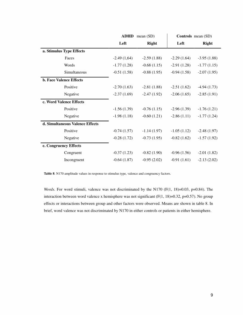
9
Table 8. N170 amplitude values in response to stimulus type, valence and congruency factors.
Words. For word stimuli, valence was not discriminated by the N170 (F(1, 18)=0.03, p=0.84). The
interaction between word valence x hemisphere was not significant (F(1, 18)=0.32, p=0.57). No group
effects or interactions between group and other factors were observed. Means are shown in table 8. In
brief, word valence was not discriminated by N170 in either controls or patients in either hemisphere.
ADHD mean (SD) Controls mean (SD)
Left Right Left Right
a. Stimulus Type Effects
Faces -2.49 (1,64) -2.59 (1.88) -2.29 (1.64) -3.95 (1.88)
Words -1.77 (1.28) -0.68 (1.15) -2.91 (1.28) -1.77 (1.15)
Simultaneous -0.51 (1.58) -0.88 (1.95) -0.94 (1.58) -2.07 (1.95)
b. Face Valence Effects
Positive -2.70 (1.63) -2.81 (1.88) -2.51 (1.62) -4.94 (1.73)
Negative -2.37 (1.69) -2.47 (1.92) -2.06 (1.65) -2.85 (1.91)
c. Word Valence Effects
Positive -1.56 (1.39) -0.76 (1.15) -2.96 (1.39) -1.76 (1.21)
Negative -1.98 (1.18) -0.60 (1.21) -2.86 (1.11) -1.77 (1.24)
d. Simultaneous Valence Effects
Positive -0.74 (1.57) -1.14 (1.97) -1.05 (1.12) -2.48 (1.97)
Negative -0.28 (1.72) -0.73 (1.95) -0.82 (1.62) -1.57 (1.92)
e. Congruency Effects
Congruent -0.37 (1.23) -0.82 (1.90) -0.96 (1.56) -2.01 (1.82)
Incongruent -0.64 (1.87) -0.95 (2.02) -0.91 (1.61) -2.13 (2.02)
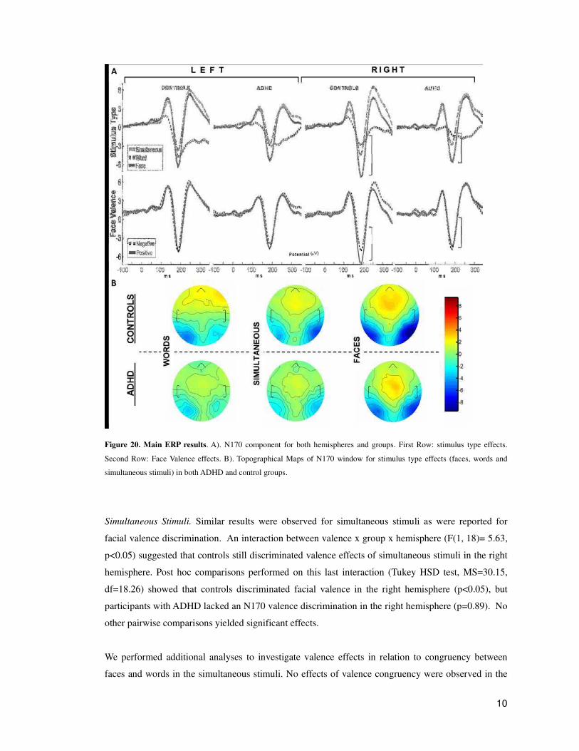
10
Figure 20. Main ERP results. A). N170 component for both hemispheres and groups. First Row: stimulus type effects.
Second Row: Face Valence effects. B). Topographical Maps of N170 window for stimulus type effects (faces, words and
simultaneous stimuli) in both ADHD and control groups.
Simultaneous Stimuli. Similar results were observed for simultaneous stimuli as were reported for
facial valence discrimination. An interaction between valence x group x hemisphere (F(1, 18)= 5.63,
p<0.05) suggested that controls still discriminated valence effects of simultaneous stimuli in the right
hemisphere. Post hoc comparisons performed on this last interaction (Tukey HSD test, MS=30.15,
df=18.26) showed that controls discriminated facial valence in the right hemisphere (p<0.05), but
participants with ADHD lacked an N170 valence discrimination in the right hemisphere (p=0.89). No
other pairwise comparisons yielded significant effects.
We performed additional analyses to investigate valence effects in relation to congruency between
faces and words in the simultaneous stimuli. No effects of valence congruency were observed in the
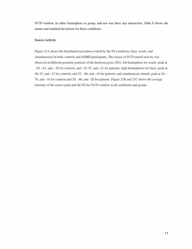
11
N170 window, in either hemisphere or group, and nor was there any interaction. Table 8 shows the
means and standard deviations for these conditions.
Source Activity
Figure 21A shows the distributed activation evoked by the ST conditions (face, words, and
simultaneous) in both, controls and ADHD participants. The source of N170 neural activity was
observed at different posterior portions of the fusiform gyrus (FG): left hemisphere for words, peak at
–30, –81, and – 20 for controls, and –25, 87, and –21 for patients; right hemispheres for faces, peak at
40, 67, and –12 for controls, and 25, –86, and –18 for patients; and simultaneous stimuli, peak at 26, –
76, and –16 for controls and 20, –86, and –20 for patients. Figure 21B and 21C shows the average
intensity of the source peak and the FG for N170 window in all conditions and groups.
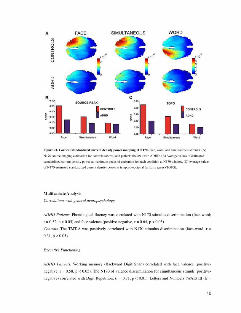
12
Figure 21. Cortical standardized current density power mapping of N170 (face, word, and simultaneous stimuli). (A)
N170 source imaging estimation for controls (above) and patients (below) with ADHD. (B) Average values of estimated
standardized current density power at maximum peaks of activation for each condition at N170 window. (C) Average values
of N170 estimated standardized current density power at temporo-occipital fusiform gyrus (TOFG).
Multivariate Analysis
Correlations with general neuropsychology.
ADHD Patients. Phonological fluency was correlated with N170 stimulus discrimination (face-word;
r = 0.52, p < 0.05) and face valence (positive-negative, r = 0.64, p < 0.05).
Controls. The TMT-A was positively correlated with N170 stimulus discrimination (face-word; r =
0.31, p < 0.05).
Executive Functioning
ADHD Patients. Working memory (Backward Digit Span) correlated with face valence (positive-
negative, r = 0.58, p < 0.05). The N170 of valence discrimination for simultaneous stimuli (positive-
negative) correlated with Digit Repetition, (r = 0.71, p < 0.01), Letters and Numbers (WAIS III) (r =
13
0.68, p < 0.05) and the Working Memory Index (r = 0.54, p < 0.02).
Controls. The TMT-B correlated positively with the N170 valence discrimination for simultaneous
stimuli (r = 0.589, p<0.05) (r = 0.64, p < 0.05). The N170 valence discrimination for simultaneous
stimuli correlated with the Working Memory Index (r = 0.68, p < 0.01).
Theory of Mind
ADHD Patients. RMET scores correlated significantly with N170 face valence discrimination (happy
– angry; r = 0.51, p < 0.05).
Controls. RMET scores correlated significantly with N170 face valence discrimination (happy –
angry; r = 0.32, p < 0.05).
In summary, all the emergent correlations are meaningful and have the expected sign.
N170 face emotional discrimination correlated in both groups with a social cognition task (RMET)
As occurred in BD, N170 face emotional discrimination also correlated with executive functions in
ADHD (2 tasks) and controls (1 task).
And finally, N170 face valence in the presence of interfering words, correlated with two tasks of
executive functions.

Chapter II: Error monitoring of monetary reward is affected in
bipolar disorder and ADHD
H4a) BD and ADHD patients have cortical deficits in the processing of monetary reward.
H4b) The deficitary modulation of monetary reward processing in both patients correlate with
clinical measures and executive functions performance.
This section includes experimental work done in collaboration with Agustin Ibanez, Marcelo
Cetkovich, Hugo Urquina, Sandra Baez, Juan Kamienkowski, Teresa Torralva, Fernando Torrente,
Sergio Strejilevich, Julia Teitelbaum, Esteban Hurtado, Raphael Guex, Alicia Lischinsky, Facundo
Manes, under the supervision of Professor Mariano Sigman.
SPECIFIC MATERIALS AND METHODS FOR THIS EXPERIMENT
This section assesses decision-making using the behavioral and neural correlates of different tasks in
adults with ADHD, those with euthymic BD and matched healthy controls. We included affective,
risky and rapid-decision gambling paradigms. Specifically, we used the Iowa gambling task [IGT;
(Bechara, Damasio et al. 1994)], a simple task of rational decision-making under risk [RDMUR
(Fernandez-Duque 2007)] and a rapid-decision gambling task (RDGT) that elicits neurophysiological
processes involved in the rapid evaluation of the motivational effect of events (Gehring and
Willoughby 2002). We recorded event-related potentials (ERPs) from human participants as they
performed the RDGT. Participant choices were followed by feedback that indicated the monetary
gains or losses. The RDGT elicits a feedback error-related negativity (fERN) modulated by reward
valence and a P3 sensitive to reward magnitude (Gehring and Willoughby 2002; Yeung and Sanfey
2004). We also estimated the neural sources of these components. Finally, to assess the relationship
between decision-making tasks and individual differences, a multivariate analysis examined the
extensive clinical and neuropsychological participant profiles.
2
Participants
Fifty participants (BD: n=13; ADHD: n=12; controls: n=25) received a full clinical assessment and
neurocognitive profile, and their ERPs were recorded. Patients in the BD and ADHD groups were
selected from the outpatient population of the Institute of Cognitive Neurology using the following
inclusion criteria: 1) aged between 18 and 55 years old; 2) diagnosed with Type-I /II BD or adult
ADHD according to the DSM-IV using the Structured Clinical Interview for DSM-IV (SCID); and 3)
euthymia scores less than or equal to 8 points according to the Montgomery-Asberg Depression
Rating Scale (MDRS) and less than or equal to 6 according to the Young Mania Rating Scale (YMRS)
for at least 8 weeks and with no change in medication type or dosage over 4 months. Patients did not
receive antipsychotics. Exclusion criteria were 1) other Axis-I diagnoses, except for generalized
anxiety disorder and 2) a history of mental retardation, neurological disease, or any clinical condition
that might affect cognitive performance. We assessed all participants using a standard diagnostic
process that included neurological, neuropsychiatric and neuropsychological examinations. All
patients with ADHD were taking methylphenidate, which was suspended on the day of the ERP
recordings because this medication improves task performance (Volkow, Wang et al. 2004).
We recruited 25 healthy controls matched for sex, age, handedness, and years of education from a
larger pool of volunteers who did not have a history of drug abuse or a family history of
neurodegenerative or psychiatric disorders. All participants provided written informed consent in
agreement with the Helsinki declaration.
Clinical, symptomatic and neuropsychological assessment
All participants completed a series of psychiatric and behavioral questionnaires to establish a clinical
symptom profile that included depression, mania, impulsivity, anxiety, attention and
hyperactivity/impulsivity scores. The Beck Depression Inventory II (Beck 1996 ) and the
Montgomery-Asberg Depression Rating Scale (Montgomery 1979) rated depression. The Young
Mania Rating Scale (Young, Biggs et al. 1978) and the Barratt Impulsiveness Scale (Barratt 1959)
rated mania and impulsivity, respectively. The State-Trait Anxiety Inventory (Spielberger, Gorsuch et
al. 1970) rated anxiety. We obtained an ADHD symptom profile from the inattention and
hyperactivity/impulsivity scores of the ADHD Rating Scale for Adults (Barkley and Murphy 1998).
A general neuropsychology test evaluated participants’ basic attention and memory processes. Several
tests, including the INECO Frontal Screening, evaluated executive functioning. Digit and symbol
searching and forward digit span tasks evaluated attention, visual scanning and the efficient
3
production of multiple motor responses. The Rey Auditory Verbal Learning Test, which is composed
of verbal learning, immediate and delayed recall and a distractor list, evaluated memory.
Several tests evaluated executive functioning. The INECO Frontal Screening assessed frontal lobe
function via several subtasks: motor programming, conflicting instructions, go/no-go, backward digit
span, spatial working memory, abstraction capacity and verbal inhibitory control. Trail Making B
assessed attentional flexibility and attentional speed. Backward digit span, letter-number sequencing
and an arithmetic test assessed mental manipulation and working memory. A go/no-go task that
included correct, incorrect and omitted responses as percentages and reaction time assessed inhibitory
control. We also included a phonological fluency task.
Data analyses
An ANOVA and Tukey’s HSD post-hoc comparisons (when appropriate) compared demographic,
neuropsychological and reaction time data across all three groups. The X2-test examined categorical
variables (e.g., sex). For the RDGT, we averaged accuracy and the ERP amplitudes for wins and
losses (valence factor) as well as for large and small values (magnitude factor). We included a
between-subjects factor for the group (patients with BD, those with ADHD and controls). Offline
processing and EEG data analysis were performed using Matlab. After a valence and electrode
position analysis of the fERN (Gehring and Willoughby 2002; San Martin, Manes et al. 2010), we
selected the FCz site for all analyses based on the higher win-loss amplitude discrimination. A 225-
281 ms timeframe for fERN and a 372-464 ms timeframe for P3 were selected for mean amplitude
analysis. Although the P3 has more of a central distribution, its effects are reliable at FCz
(Nieuwenhuis, Holroyd et al. 2004; Yeung and Sanfey 2004).
To perform multivariate comparisons between the ERPs and the neuropsychology tests, we calculated
global scores for (a) valence (fERN: wins minus losses) and (b) magnitude (P3: large minus small).
Spearman’s rank correlations examined these global scores with regard to all clinical and
neuropsychological tests after correcting for multiple comparisons (at p<0.05).
4
RESULTS
Table 9 shows the results from the demographic, clinical, and neuropsychological assessments.
Demographic data
We did not observe between-group differences with regard to age (F[2, 47]=1.50, p=0.23), sex
(X2[2]= 0.00, p=1.00) or education level (F[2, 47]=1.50, p=0.23).
Clinical and neuropsychological evaluation
Clinical evaluation
Patients with ADHD had higher scores of inattention, hyperactivity/impulsivity and depression than
controls. In addition, patients with ADHD and those with BD had higher levels of trait anxiety than
controls.
There was an expected between-group significant difference for the ADHD-RS-Inattention subscale
(F[2, 47]=12.44, p<0.001) and the ADHD-RS-Hyperactivity/impulsivity subscale (F[2, 47]=8.90,
p<0.001). Post-hoc comparisons (Tukey’s HSD test, MS=24.75; df =47.00) showed that participants
with ADHD had significantly higher inattention and hyperactivity/impulsivity scores compared to
those with BD (p=0.02, p=0.03, respectively) and controls (both p<0.001). We observed a between-
group difference for BDI-II scores (F[2, 47]=5.63, p<0.01). Post-hoc comparisons (Tukey’s HSD test,
MS=77.28; df =47.00) revealed higher levels of depression for participants with ADHD (p<0.005)
compared to controls. In addition, we observed a between-group difference for MADRS scores (F[2,
47]=3.37, p=0.42). Post-hoc comparisons (Tukey’s HSD test, MS=16.30; df =47.00) revealed more
severe depressive symptoms for patients with ADHD (p=0.04) compared to controls.
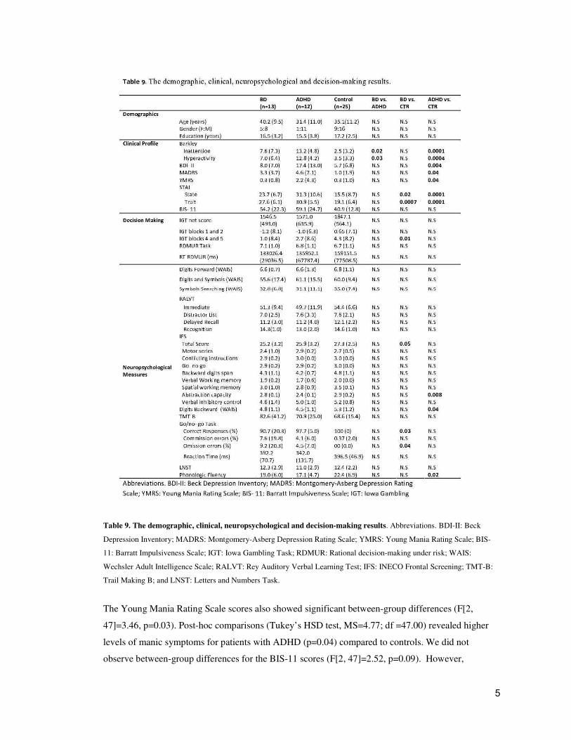
5
Table 9. The demographic, clinical, neuropsychological and decision-making results. Abbreviations. BDI-II: Beck
Depression Inventory; MADRS: Montgomery-Asberg Depression Rating Scale; YMRS: Young Mania Rating Scale; BIS-
11: Barratt Impulsiveness Scale; IGT: Iowa Gambling Task; RDMUR: Rational decision-making under risk; WAIS:
Wechsler Adult Intelligence Scale; RALVT: Rey Auditory Verbal Learning Test; IFS: INECO Frontal Screening; TMT-B:
Trail Making B; and LNST: Letters and Numbers Task.
The Young Mania Rating Scale scores also showed significant between-group differences (F[2,
47]=3.46, p=0.03). Post-hoc comparisons (Tukey’s HSD test, MS=4.77; df =47.00) revealed higher
levels of manic symptoms for patients with ADHD (p=0.04) compared to controls. We did not
observe between-group differences for the BIS-11 scores (F[2, 47]=2.52, p=0.09). However,
6
significant differences between groups for STAI- State subscale (F(2,47)=13.57, p<0.001) and STAI-
Trait subscale (F(2,47)=16.85, p<0.001) were observed. State subscale posthoc comparisons (Tukey
test, HSD, MS=74.28; df =47.00) showed that BD (p=0.02) and ADHD (p<0.001) participants had
significantly higher scores than control subjects. Also, post hoc comparisons (MS=38.27; df =47.00)
showed higher scores for Trait subscale in BD (p<0.001) and ADHD (p<0.001) patients compared
with the control group.
Neuropsychological assessment
The global score of the executive-function INECO Frontal Screening showed significant differences
between groups (F[2, 47]= 3.53, p < 0.05). Specifically, patients with BD had lower go/no-go IFS
subscale scores compared to controls and lower abstraction capacity IFS subscale scores than with
ADHD. Furthermore, we observed impairments in patients with ADHD with regard to executive
control and working memory. Posthoc comparisons (Tukey test, HSD, MS=7.54; df =47.00) showed
lower performance for the BD group compared with controls (p < 0.05). On abstraction capacity IFS
subscale, significant differences between groups were observed (F(2, 47)=4.93, p < 0.05). Posthoc
comparisons (Tukey test, HSD, MS=0.24; df =47.00) showed lower performance in the ADHD group
(p<0.01) compared with controls. On the Go-no go Task, accuracy on go trials (F(2, 47)=3.31, p <
0.05) and omission responses percentage (F(2, 47)=3.17, p < 0.05), showed significant differences
between groups. Posthoc comparisons on accuracy (MS=111.49; df =47.00) showed lower
performance for the BD group compared with controls (p < 0.05). Also, post hoc comparisons (Tukey
test, HSD, MS=120.69; df =47.00) showed that BD had significantly higher omission responses
percentage than did control subjects (p < 0.05). No differences were observed on either the
commission responses percentage (F(2, 47)=2.17, p=0.12) or the reaction time (F(2, 47)=2.63,
p=0.08).
Regarding the other measures of executive functioning, the score on verbal Phonologic Fluency Task
showed significant differences between groups (F(2, 47)=3.86, p < 0.05). Posthoc comparisons
showed lower performance for the ADHD group compared with controls (p<0.01). The score on the
Backward Digit Span showed a trend towards lower performance for the ADHD group (F(2, 47)=
3.14, p=0.05). In contrast, no differences were observed between groups on the TMT-B (F(2,
47)=1.10, p=0.34), or the Letters and Numbers task (F(2, 47)=1.36, p=0.26).
The neuropsychological assessment of decision-making (IGT and RDMUR)
The IGT net score did not reveal a between-group difference (F[2, 47]=1.36, p=0.26). Furthermore,
7
we did not observe an interaction between block and group. To compare the initial and final blocks,
we performed a separate analysis between the average of Blocks 1-2 and 3-4. Although an ANOVA
did not find group differences in Blocks 1-2 (F[4, 90]=1.05, p=0.38), it did for Blocks 4-5 (F[4,
90]=3.50, p < 0.01). Post-hoc comparisons (MS=57.75, df = 47) revealed that patients with BD had
impaired performances compared to controls (p=0.01, see Figure 22.A).
When comparing RDMUR tasks (Figures 22.B and 22.C), we did not observe significant between-
group differences with regard to total score (F[2, 47]=0.64, p=0.52) or total reaction time (F[2,
47]=1.07, p=0.34).
The neurophysiological measures of Decision-Making
Reaction time. We did not observe main effects of valence or magnitude, nor did we observe group
effects or interactions. However, patients with ADHD had longer response times in general compared
to controls. Regarding overall RTs, a group effect was obtained (F(2, 47)=3.47, p < 0.05). Post hoc
comparison performed over this effect (Tukey HSD test, MSE = 1798, df = 47) evidenced that ADHD
patients made longer responses (M=1039ms, SD=122) than to controls (M= 650ms SD=84). No
differences were observed between BD (M=825ms, SD=117) and controls. No main effects or
interactions of valence and magnitude were observed in reaction times.
RDGT: ERPs
fERN. We did not observe main effects of valence (F[1, 47]=3.30, p=0.07) or magnitude (F[1,
47]=0.15, p=0.69); however, as expected, we observed significant valence x group (F[2, 47]=3.62, p <
0.05) and magnitude x group interactions (F[2, 47]=5.11, p<0.005). To analyze the simple effects for
control participants as well as those with ADHD and those with BD, we examined the fERN
component of each group separately (see Table 10 for descriptive statistics).
Controls. We did not observe an effect of magnitude (F[1, 11]=0.34, p=0.85); however, as expected, a
significant effect of valence (F[1, 11]=10.69, p<0.01) revealed less positive amplitudes on trials with
losses than those with wins. In addition, we observed a significant valence x magnitude interaction
(F[1, 11]=11.52, p<0.01). Post-hoc comparisons (MS=2.27; df =47.00) revealed that amplitudes after
a large win were more positive than those after large (p<0.001) and small losses (p < 0.05).
Patients with BD. We did not observe an effect of valence (F[1, 12]=0.29, p=0.59); however, we did
find a significant effect of magnitude (F[1, 12]=7.50, p < 0.05), revealing that the amplitudes
associated with large reward were more positive than those associated with smaller ones. There was
not a significant valence x magnitude interaction (F[1, 12]=0.70, p=0.41).
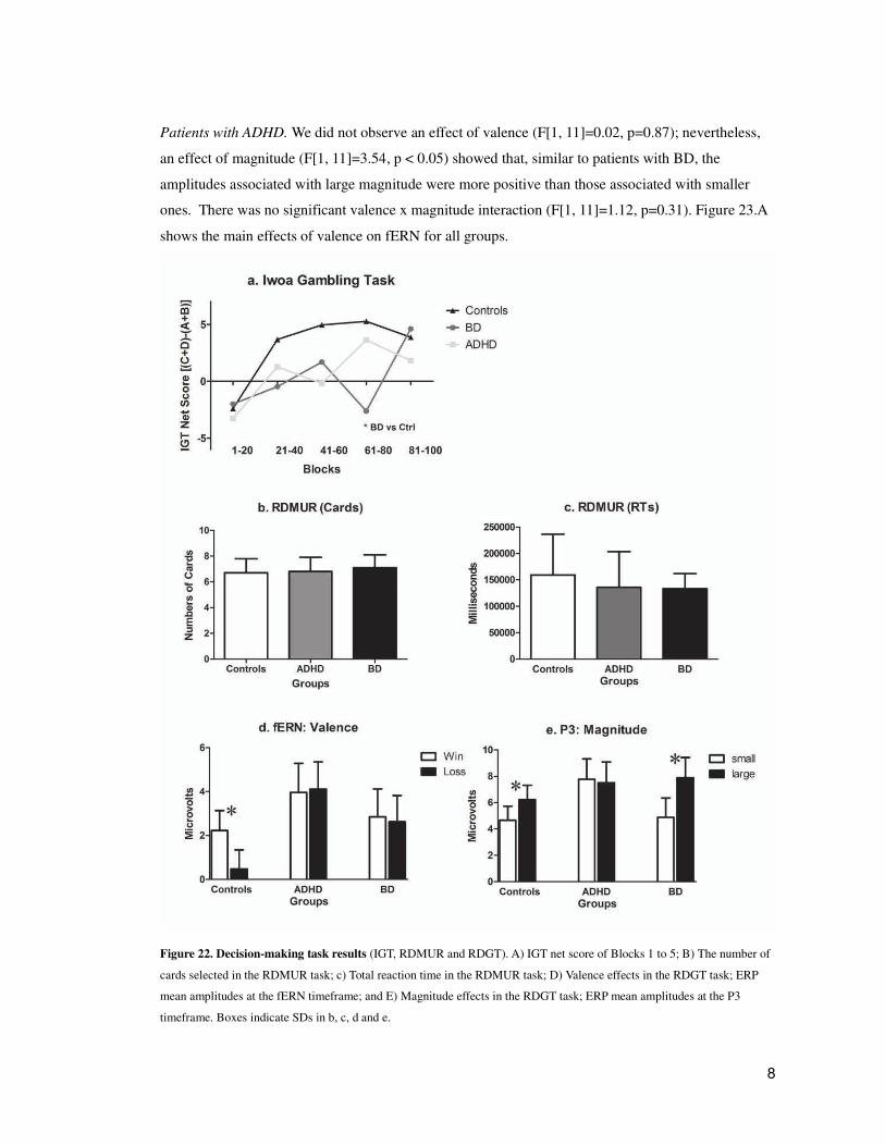
8
Patients with ADHD. We did not observe an effect of valence (F[1, 11]=0.02, p=0.87); nevertheless,
an effect of magnitude (F[1, 11]=3.54, p < 0.05) showed that, similar to patients with BD, the
amplitudes associated with large magnitude were more positive than those associated with smaller
ones. There was no significant valence x magnitude interaction (F[1, 11]=1.12, p=0.31). Figure 23.A
shows the main effects of valence on fERN for all groups.
Figure 22. Decision-making task results (IGT, RDMUR and RDGT). A) IGT net score of Blocks 1 to 5; B) The number of
cards selected in the RDMUR task; c) Total reaction time in the RDMUR task; D) Valence effects in the RDGT task; ERP
mean amplitudes at the fERN timeframe; and E) Magnitude effects in the RDGT task; ERP mean amplitudes at the P3
timeframe. Boxes indicate SDs in b, c, d and e.
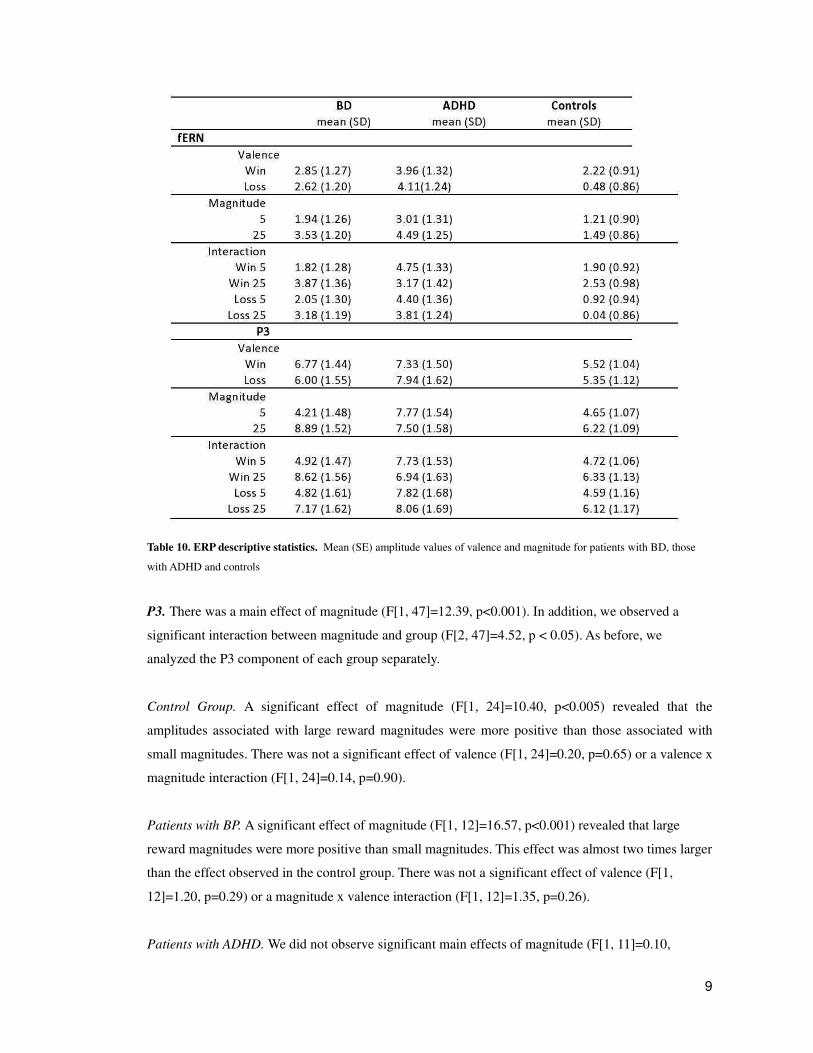
9
Table 10. ERP descriptive statistics. Mean (SE) amplitude values of valence and magnitude for patients with BD, those
with ADHD and controls
P3. There was a main effect of magnitude (F[1, 47]=12.39, p<0.001). In addition, we observed a
significant interaction between magnitude and group (F[2, 47]=4.52, p < 0.05). As before, we
analyzed the P3 component of each group separately.
Control Group. A significant effect of magnitude (F[1, 24]=10.40, p<0.005) revealed that the
amplitudes associated with large reward magnitudes were more positive than those associated with
small magnitudes. There was not a significant effect of valence (F[1, 24]=0.20, p=0.65) or a valence x
magnitude interaction (F[1, 24]=0.14, p=0.90).
Patients with BP. A significant effect of magnitude (F[1, 12]=16.57, p<0.001) revealed that large
reward magnitudes were more positive than small magnitudes. This effect was almost two times larger
than the effect observed in the control group. There was not a significant effect of valence (F[1,
12]=1.20, p=0.29) or a magnitude x valence interaction (F[1, 12]=1.35, p=0.26).
Patients with ADHD. We did not observe significant main effects of magnitude (F[1, 11]=0.10,
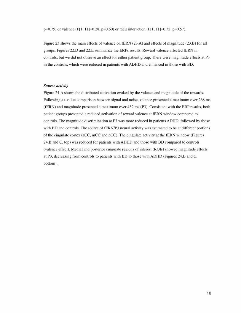
10
p=0.75) or valence (F[1, 11]=0.28, p=0.60) or their interaction (F[1, 11]=0.32, p=0.57).
Figure 23 shows the main effects of valence on fERN (23.A) and effects of magnitude (23.B) for all
groups. Figures 22.D and 22.E summarize the ERPs results. Reward valence affected fERN in
controls, but we did not observe an effect for either patient group. There were magnitude effects at P3
in the controls, which were reduced in patients with ADHD and enhanced in those with BD.
Source activity
Figure 24.A shows the distributed activation evoked by the valence and magnitude of the rewards.
Following a t-value comparison between signal and noise, valence presented a maximum over 268 ms
(fERN) and magnitude presented a maximum over 432 ms (P3). Consistent with the ERP results, both
patient groups presented a reduced activation of reward valence at fERN window compared to
controls. The magnitude discrimination at P3 was more reduced in patients ADHD, followed by those
with BD and controls. The source of fERN/P3 neural activity was estimated to be at different portions
of the cingulate cortex (aCC, mCC and pCC). The cingulate activity at the fERN window (Figures
24.B and C, top) was reduced for patients with ADHD and those with BD compared to controls
(valence effect). Medial and posterior cingulate regions of interest (ROIs) showed magnitude effects
at P3, decreasing from controls to patients with BD to those with ADHD (Figures 24.B and C,
bottom).
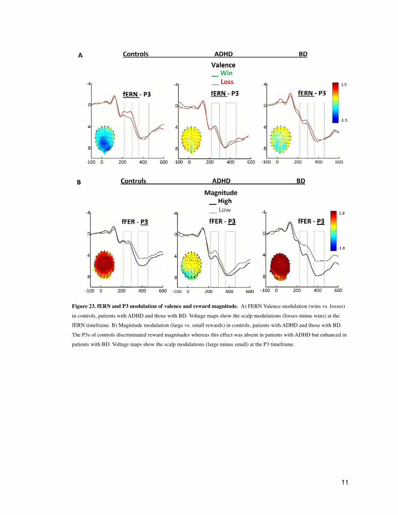
11
Figure 23. fERN and P3 modulation of valence and reward magnitude. A) FERN Valence modulation (wins vs. losses)
in controls, patients with ADHD and those with BD. Voltage maps show the scalp modulations (losses minus wins) at the
fERN timeframe. B) Magnitude modulation (large vs. small rewards) in controls, patients with ADHD and those with BD.
The P3s of controls discriminated reward magnitudes whereas this effect was absent in patients with ADHD but enhanced in
patients with BD. Voltage maps show the scalp modulations (large minus small) at the P3 timeframe.
12
Figure 24. Cortical current density mapping of valence and reward magnitude. A. The source estimation of distributed
valence dipoles (fERN, left) and magnitude effects (P3, right) for controls, patients with ADHD and those with BD. Color-
map values represent the t-values of comparisons between signal and noise. B. A time-series of the absolute power activation
evoked by valence and reward magnitudes at the anterior, medial and posterior cingulate cortex (aCC, mCC, pCC). C. The
average values of absolute power at aCC, mCC and PCC for the valence and magnitude effects for all groups. We obtained
the ROIs at aCC, mCC and pCC using a Tzourio-Mazoyer partition.
13
Correlations (Clinical/Neuropsychological Assessments and ERPs)
Control Group. Impulsivity (BIS-11: r=-0.32) and depression (MADRS: r=-0.48) were negatively
correlated with fERN win/loss discrimination. ADHD-RS-Inattention subscale scores were positively
correlated with P3 amplitudes of magnitude discrimination (r=0.40). Working memory (backward
digits span) was negatively correlated with P3 magnitude discrimination (r=-0.40).
Patients with BD. Depression level (MADRS total score, r=-0.47 and Beck-II, r=-0.41) was
negatively correlated with win/loss discrimination. Anxiety scores (STAI- Trait) were positively
correlated with P3 magnitudes discrimination (r=0.61). Inhibitory control (incorrect responses on a
go/no-go task) was positively correlated with fERN win/loss discrimination (r=0.36) and with P3
magnitude discrimination (r=0.43). Go/no-go reaction times were negatively correlated with fERN
win/loss discrimination (r=-0.39).
Patients with ADHD. Significantly high ADHD-RS-Inattention (r=-0.55) and ADHD-RS-
Hyperactivity-impulsivity subscale scores (r=-0.39) were negatively correlated with fERN win/loss
discrimination. With regard to executive functions, the IFS total score was positively correlated with
fERN win/loss discrimination (r=0.43). Working memory (numbers and letters, r=0.54; WAIS-
working memory index, r=0.41) and attention (WAIS-digit score; r=0.58) were also positively
correlated with fERN win/loss discrimination.
In summary, both patients presented an impaired valence modulation of the fERN, but a modulation in
magnitude of the monetary reward, in contrast to controls. In relation to P3, Controls and BD, but not
ADHD, presented and effect of valence.
fERN presents all meaningful and consistent correlations.
In controls fERN win/loss discrimination correlates negatively with impulsivity and depression
scores.
In Bipolar patients fERN win/loss discrimination correlates negatively with depression and slow
reaction time in inhibitory control, and positively with inhibitory control performance (both executive
functions).
In ADHD patients fERN win/loss discrimination correlate negatively with inattention and
hyperactivity scores and positively with performance in four executive functions tasks.
P3 did not present meaningful correlations.

2
General Discussion
The set of experiments presented here coherently provide support for a view in which rapid facial
recognition is not a modular process, independent of cognitive, emotional and social representations.
First, we showed that the N170 was affected by social cognition scores. It must be emphasized that
the relevance of this finding is that the N170 is a very rapid component (only 170 ms) of face
recognition which has been observed even in subliminal presentations. This has led to think that it is a
feed-forward, automatic encoding of a face which only later on interacts with other systems that
provide social and emotional information. Instead, our data show that even this very early component
is affected by such cognitive features. The results of this thesis then establish that the global influence
of a broad network that acts very rapidly in the first encoding stages of perception (section I).
In what follows we discuss in detail how these results relate to controversies on face recognition and
emotion and social processing, their more general implications for the organization of brain
computations and clinical implications.
Taking the results in section I, we can conclude that the broad network that takes part in early face
perception is implicated in complex social cognition, including theory of mind, but also in frontal
executive functions, reflecting a close interaction between both systems. This result also support the
view of a brain network with dynamic transitions between emotional and more cognitive states, a
version of which establishes that same brain areas may account for cognitive or emotional responses
depending on the context or task demands (Pessoa 2008). In fact, two psychiatric disorders that
present shared social cognition and frontal deficits show an impaired N170 in response to facial
emotions (sections II and III).
A similar association occurs between low level rapid processing of monetary feedback and executive
function tasks (Chapter II). A rapid and short (millisecond scale) brain potential (fERN) evoked by
monetary feedback showed a robust and consistent association with frontal functioning clinical
measures in ADHD, depression in BD, and both types of measures in controls. In particular, fERN
valence modulation correlated negatively with depression scores in BD, negatively with inattention
and hyperactivity scores in ADHD and negatively with both depression and impulsivity in controls.
This result suggests that rapid feedback processing is embedded in a complex frontal network that
takes part in planning, action monitoring, and global control of other networks, severely affected in
ADHD. But it also takes part in emotional state regulation in BD. Note that in both patients fERN
valence modulation is also associated with executive functions measured by neuropsychology.

3
The results of this thesis show that early perception may not be modular, but distributed. The currently
dominant view among many cognitive psychologists proposes two broad sets of processes: those that
are controlled and those that are automatic. The dichotomous scheme is summarized in a recent
review (Lieberman 2007), which enumerates the various properties attributed to controlled and
automatic processing. Controlled processes have long been assigned a host of other attributes: They
are slow, effortful, reflective, arise late in evolution and development, and often involve language-
based declarative reasoning and reflective thinking. Automatic processes are thought to be faster,
spontaneous, reflexive, shared in common with a wide range of species and dominant early in
development, and often involve emotions (Adolphs 2009). This view influenced another view, which
argues that emotional processes as facial perception are automatic and central to social evolution, and
hence evolved to a dedicated module. Some evidences support this view, mainly the existence of the
FFA. However, the FFA also can be activated by nonface objects provided that subjects acquire
substantial expertise with them, such as birds, cars, or butterflies in experts for those categories
(Gauthier, Skudlarski et al. 2000). Although the disproportionate activation by faces argues for a
domain-specific module specialized to process a particular category of stimuli (faces) (Kanwisher
2000), the other data argue for a particular type of processing rather than processing for a particular
stimulus category (Tarr & Gauthier 2000). Other imaging data have argued that faces are never
represented in a single cortical region, but in a distributed region of cortex considerably more
extensive than the FFA (Haxby et al. 2000)
Our findings provide evidence towards a distributed but stable network of social cognition, whose
interactions occurred at a short time-scale, with strong feedback connections. It includes low-level
components (as the rapid extraction of facial emotion) and high order processors (e.g. those that
manipulate other’s state of mind and social context).
The amygdala may be a nodal component in the maintenance and regulation of this network, in part
because its massive connectivity with other brain areas and also for its nodal position within the
network. Studies with bilateral amygdala lesioned patients support this view (Buchanan, Tranel et al.
2009).
In healthy subjects, the N170 response to facial emotional valence, but not to stimulus type, is
associated with three social cognition tasks: the reading the mind in the eyes test, Faux pas test and
the first block of the Iowa Gambling Task. Better social skills are related to greater N170 valence
discrimination. First, scores on a measure of theory of mind related to emotional inference (RMET)
correlated significantly with the N170 emotional discrimination. This result suggests that the more
basic theory of mind processes (e.g., emotional inference) are supported, at least in part, by early brain
activity that is sensitive to facial emotional valence. Second, scores on the Faux pas test (FPT)

4
correlated with the N170 compatibility discrimination. The compatibility effect may be associated
with the cognitive ability to make inferences about others’ mental states at a more complex level.
Additionally, the FPT involves dealing with a high number of cognitive and affective components,
including inferences about others’ mental states and contextual cues (Riveros, Manes et al. 2010), as
compared to the RMET (Stone, Baron-Cohen et al. 1998; Baron-Cohen, Wheelwright et al. 2001;
Ochsner 2008; Ahmed and Stephen Miller 2010). Our pattern of association for the two tests follows
the direction of this distinction (the FPT as associated with more complex ToM processes and the
RMET with more basic emotional stages). Finally, we found that only the first block of the Iowa
Gambling Task, which contains five blocks, correlated significantly with the ERPs emotional
discrimination for simultaneous stimuli. This first block consists of an exploration of the cards used in
the task. It reflects decision making under total uncertainty (because participants are unaware of the
cards’ properties) and is related to emotional decision making. This association between emotional
processing and the first IGT block is relevant because the subsequent four blocks of the IGT cannot be
considered to reflect decision making under conditions of ambiguity, but rather, they reflect decision
making under conditions of risk (Dunn, Dalgleish et al. 2006). Research that was conducted using
participants with neuropsychiatric diagnoses also supports this distinction (Torralva, Roca et al. 2009).
The current findings suggest that basic emotional discrimination is related to an implicit and
emotional ability to make decisions in an ambiguous context.
The N170 emotion discrimination in simultaneous stimuli in healthy subjects correlated positively
with Trial Making Test B (TMT-B). This result suggests that there is an association between
simultaneous task segregation, and the physiological segregation needed to focus on a face and ignore
a word. Moreover, TMT-B is a test that is sensitive to frontal lobe damage (Gouveia, Brucki et al.
2007), suggesting that the early ERPs discrimination of face valence in the presence of interfering
stimuli, which occurs in many cognitive processes that require executive control, depends on frontal
lobe executive functioning.
Consistent with previous reports, the main source of the N170 was estimated in the right FG for faces
(Rossion and Gauthier 2002); (Rossion, Joyce et al. 2003) and in the left FG for words (Maillard,
Barbeau et al. 2010); (Rossion, Joyce et al. 2003). In addition, simultaneous stimuli (face-word)
elicited a bilateral (right predominant) activation of the fusiform cortex. These results highlight the
role of the lateralized FG in object recognition of category-specific visual information (Rossion, Joyce
et al. 2003).
Two psychiatric disorders that present anatomical and functional connection deficits in some shared
areas implicated in social cognition (e.g. amygdala and ACC) showed an affected brain processing of
facial emotions and reward processing in a gambling task. These results are in line with our previous
findings, suggesting that the social cognition network is heavily disbalanced in these disorders. Our
5
findings have an important clinical relevance, because they can be used to design and develop
electrophysiological techniques to help in the detection of pathologies at early stages, its management
and treatment. Facial processing may be altered before the full manifestation of these disorders during
development, especially for BD. The same principle can be applied to the fERN, which showed very
consistent associations with frontal deficits (e.g. executive functions) and clinical state. The fERN
reward discrimination covaries in a consistent way with depression in BD and inattention and
hyperactivity scores in ADHD.
BD presented a reduced N170, and a reduced facial emotional modulation (in both, facial and
simultaneous stimuli). Furthermore, BD patients presented an early cortical discrimination of negative
words valence, suggesting the activation of negative bias in the semantic stimuli processing. N170
source analysis evidenced reduced BD fusiform gyrus activation. An important result is the
association of deficiencies in facial emotional modulation and clinical measures, in particular indices
of mania and depression (inversely and directly correlated, respectively). Nevertheless, BD presented
a normal accuracy in the DVT, may be due its easiness, relying on redundant circuits to solve the task.
Regarding words, BD group showed amplitude enhancement of N170 negative valence in the left
hemisphere, suggesting an early attentional bias toward negative information. The reactivity toward
negative semantic information is consistent with the increased perception of allocation to negative
than to positive emotional cues (Leppanen 2006; Eimer and Holmes 2007). Additionally, enhanced
recognition of negative facial expression, such us disgust (Harmer, Grayson et al. 2002), have been
reported in euthymic BD (Malhi, Lagopoulos et al. 2007).
In agreement with previous studies (Henry, Van den Bulke et al. 2008), we found that BD patients
showed higher levels of anxiety. According with previous reports, the pattern of cognitive functioning
revealed that BD patients showed deficits in the attentional domain (Elshahawi, Essawi et al. 2011)
and some failures in executive tasks (Torralva, Roca et al. 2009). Also, as previously demonstrated
(Martino, Strejilevich et al. 2010), we found lower performance in BD group on FPT, suggesting
failures in theory of mind process.
The social perceptual component of theory of mind consists in the capability to perceive mental states
of others based on observable information like facial expressions, and include the capacity to
distinguish between people and objects (Tager-Flusberg and Joseph 2003). Accordingly, the results
exposed in this thesis showed that stimulus type and word valence are associated with theory of mind
measures, which may be related with the deficits observed in BD patients in these processes. Thus
early abnormal semantic and facial processing would influence object recognition and emotional
categorization process, affecting the building blocks of further impaired social cognition skills (de
Almeida Rocca, de Macedo-Soares et al. 2008).
Abnormalities in prefrontal cortex (PFC) and amygdala circuitry appear to be critical factors in the
6
dysregulation of affective disorders (Hariri, Bookheimer et al. 2000; Rich, Fromm et al. 2008).
Although ADHD showed high accuracy on the DVT, the adults with ADHD showed deficits in N170
emotion discrimination for facial stimuli. In particular ADHD presented reduced N170 amplitude for
positive stimuli in the right hemisphere.
Notably, in ADHD participants, N170 emotion processing was associated with performance on an
emotional inference theory of mind task and N170 for simultaneous stimuli was associated with
executive functioning, especially working memory.
Recently, it has been proposed that in ADHD, a possible reduction in amygdala activity [see (Plessen,
Bansal et al. 2006)] in response to positive stimuli may lead to reduced activation of the reward
system and in turn to impaired processing of positive emotional stimuli (Herrmann, Schreppel et al.
2009). ADHD appears to involve predominantly right hemispheric dysfunction [for a review see (Barr
2001) and (Booth, Burman et al. 2005)]. Impaired emotional facial processing is the most consistently
reported form of social cognitive impairment in ADHD (Uekermann, Kraemer et al. 2010).
Participants with ADHD presented deficits in recall performance on the RAVLT, as well as some
executive impairment, which is not a new issue (Torralva, Gleichgerrcht et al. 2010). In addition, we
found a subtle deficit in theory of mind indexed by the RMRT, and this task correlated with cortical
deficits in face valence. At the same time, in the ADHD patients the ERPs for simultaneous stimuli
valence discrimination were associated with higher levels of executive functioning and working
memory. Both executive and theory of mind deficits in ADHD have been reported elsewhere, and
these deficits are often both associated with the disorder (Kalmar, Wang et al. 2009; Uekermann,
Kraemer et al. 2010).
The finding of a combined executive and social impairment in ADHD is consistent with current neural
models of cognition (Pessoa 2009) and particularly with dysfunction of frontostriatal structures in
ADHD [for reviews (Bush, Valera et al. 2005; Marsh and Williams 2006; Uekermann, Kraemer et al.
2010)]. We identified brain markers of impaired facial emotion discrimination in participants with
ADHD. Those deficits were related to subtle differences in theory of mind and executive functioning,
supporting the frontostriatal dysfunction hypothesis of ADHD.
Decision Making
Patients with ADHD presented a neural pattern indicative of deficient valence (fERN) and reward
7
magnitude learning (P3). This pattern was associated with clinical evaluations of impulsivity,
hyperactivity and inattention as well as impairments in executive function and working memory.
These results are consistent with the clinical features of ADHD with regard to decision-making: If the
learning of valence and reward magnitude from the environment is impaired, then information
concerning which decisions are most important will be reduced. Thus, decisions will be based on
impulsivity or will not have a learned strategy.
Patients with BD presented a pattern of cortical modulation based on the saliency of reward
magnitudes regardless of learning via feedback. There was no fERN valence modulation; conversely,
only the magnitude of the reward affected this variable. This results are consistent with the hypothesis
that there is reduced sensitivity to emotional reward or punishment contexts in BD (Chandler,
Wakeley et al. 2009). The ERP pattern of the data of chapter II was associated with mood states (i.e.,
clinical measures of depression and anxiety) and inhibitory control.
We found reduced activity in the cingulate cortex (aCC, mCC and pCC) in both patient groups
compared to controls. This activity was especially reduced for patients with BD at the fERN (valence)
and those with ADHD at the P3 (magnitude). These results suggest that one of the main circuits
associated with decision-making, the so called “action selection-monitoring system”, is impaired for
both groups but at different stages.
The results are relevant in numerous aspects. First, the behavioral measures of affective and risky
gambling tasks are not sensitive enough to assess these disorders’ well-known deficits in decision-
making. Second, both patient groups show an abnormal neural processing of valence and reward
magnitudes to a gambling task but that this pattern was associated with different clinical and
neuropsychological profiles. Finally, these results are consistent with the models of cingulate cortex
activation to reward, action selection and action monitoring.
Higher levels of inattention, hyperactivity and impulsivity were associated with reduced fERN
win/loss discrimination in patients with ADHD, which confirms the association between impulsivity
and decision-making these patients (Malloy-Diniz, Fuentes et al. 2007). In addition, executive
function was associated to decision-making-related ERPs, which confirms previous reports of an
association between inhibitory control, decision making and working memory (Mantyla, Still et al.
2010). The latter result suggests that difficulties in sustaining attention when updating working
memory affect the decision-making of patients with ADHD. Likewise, this results are consistent with
reports of reduced responses to rewards and reinforcements in children with ADHD (Iaboni, Douglas
et al. 1997; Crone, Jennings et al. 2003), which suggests an impaired sensitivity to learning via
feedback (Luman, Oosterlaan et al. 2005).

8
As expected, patients with ADHD had higher inattention and hyperactivity/impulsivity scores than
those with BD and controls. Patients with ADHD also had higher levels of depression, which is
common in this clinical population and congruent with previous reports (Torralva, Gleichgerrcht et al.
2010).
Chapter II results reveal a clinical association between neural substrates and the common, well-known
impairments of decision-making in patients with ADHD and those with BD.
At a theoretical level, chapter II results highlight the role of monitoring systems and their relevance in
decision and reward processing. Although the orbitofrontal cortex is one of the most relevant areas in
decision-making, a network that includes monitoring systems and reward processing has been
revealed (Rushworth, Behrens et al. 2007; Rangel 2008; Rangel, Camerer et al. 2008; Kable and
Glimcher 2009; Gleichgerrcht, Ibanez et al. 2010). The role of the cingulate cortex in selection and
monitoring, along with that of the amygdala and basal ganglia in reward systems, should be affected
in patients with ADHD and those with BD [(Wang, Kalmar et al. 2009); for a review of the latter
group, see (Marchand and Yurgelun-Todd 2010)].

9
10
REFERENCES Adolphs, R. (2009). "The social brain: neural basis of social knowledge." Annu Rev Psychol
60: 693-716. Adolphs, R. (2010). "What does the amygdala contribute to social cognition?" Ann N Y Acad
Sci 1191: 42-61. Adolphs, R., D. Tranel, et al. (1994). "Impaired recognition of emotion in facial expressions
following bilateral damage to the human amygdala." Nature 372(6507): 669-72. Ahmed, F. S. and L. Stephen Miller (2010). "Executive Function Mechanisms of Theory of
Mind." J Autism Dev Disord. Amico, F., J. Stauber, et al. (2011). "Anterior cingulate cortex gray matter abnormalities in
adults with attention deficit hyperactivity disorder: a voxel-based morphometry study." Psychiatry Res 191(1): 31-5.
Ashley, V., P. Vuilleumier, et al. (2004). "Time course and specificity of event-related potentials to emotional expressions." Neuroreport 15(1): 211-6.
Astikainen, P. and J. K. Hietanen (2009). "Event-related potentials to task-irrelevant changes in facial expressions." Behav Brain Funct 5: 30.
Atkinson, A. P. and R. Adolphs (2011). "The neuropsychology of face perception: beyond simple dissociations and functional selectivity." Philos Trans R Soc Lond B Biol Sci 366(1571): 1726-38.
Barkley, R. A. and K. R. Murphy (1998). Attention-Deficit Hyperactivity Disorder: A Clinical Workbook. New York.
Baron-Cohen, S., M. O'Riordan, et al. (1999). "Recognition of faux pas by normally developing children and children with Asperger syndrome or high-functioning autism." J Autism Dev Disord 29(5): 407-18.
Baron-Cohen, S. and S. Wheelwright (2004). "The empathy quotient: an investigation of adults with Asperger syndrome or high functioning autism, and normal sex differences." J Autism Dev Disord 34(2): 163-75.
Baron-Cohen, S., S. Wheelwright, et al. (2001). "The "Reading the Mind in the Eyes" Test revised version: a study with normal adults, and adults with Asperger syndrome or high-functioning autism." J Child Psychol Psychiatry 42(2): 241-51.
Barr, W. B. (2001). "Schizophrenia and attention deficit disorder. Two complex disorders of attention." Ann N Y Acad Sci 931: 239-50.
Barratt, E. (1959). "Anxiety and impulsiveness related to psychomotor efficiency." Percept Mot Skills 9: 191-198.
Barton, J. J., R. L. Hefter, et al. (2007). "Investigations of face expertise in the social developmental disorders." Neurology 69(9): 860-70.
Batty, M. and M. J. Taylor (2003). "Early processing of the six basic facial emotional expressions." Brain Res Cogn Brain Res 17(3): 613-20.
Beck, A. T., R. A. Steer, et al. (1996). "Comparison of Beck Depression Inventories -IA and -II in psychiatric outpatients." J Pers Assess 67(3): 588-97.
Beck, A. T., Steer, R., Brown, G.K. (1996 ). Manual for the Beck Depression Inventory-II. San antonio, TX, The Psychological Corporation.
Bechara, A., A. R. Damasio, et al. (1994). "Insensitivity to future consequences following damage to human prefrontal cortex." Cognition 50(1-3): 7-15.
Bechara, A., H. Damasio, et al. (1997). "Deciding advantageously before knowing the advantageous strategy." Science 275(5304): 1293-5.
Bechara, A. and M. Van Der Linden (2005). "Decision-making and impulse control after frontal lobe injuries." Curr Opin Neurol 18(6): 734-9.
Bentin, S., Y. Mouchetant-Rostaing, et al. (1999). "ERP manifestations of processing printed
11
words at different psycholinguistic levels: time course and scalp distribution." J Cogn Neurosci 11(3): 235-60.
Bentin, S. and B. S. Peled (1990). "The contribution of task-related factors to ERP repetition effects at short and long lags." Mem Cognit 18(4): 359-66.
Benton, A. L., K. Hamsher, et al. (1994). Multilingual Aphasia Examination Iowa City, AJAAssociates.
Bickart, K. C., C. I. Wright, et al. (2010). "Amygdala volume and social network size in humans." Nat Neurosci 14(2): 163-4.
Blau, V. C., U. Maurer, et al. (2007). "The face-specific N170 component is modulated by emotional facial expression." Behav Brain Funct 3: 7.
Booth, J. R., D. D. Burman, et al. (2005). "Larger deficits in brain networks for response inhibition than for visual selective attention in attention deficit hyperactivity disorder (ADHD)." J Child Psychol Psychiatry 46(1): 94-111.
Brand, M., K. Labudda, et al. (2006). "Neuropsychological correlates of decision-making in ambiguous and risky situations." Neural Netw 19(8): 1266-76.
Brandeis, D., D. Lehmann, et al. (1995). "Mapping event-related brain potential microstates to sentence endings." Brain Topogr 8(2): 145-59.
Brotman, M. A., A. E. Guyer, et al. (2008). "Facial emotion labeling deficits in children and adolescents at risk for bipolar disorder." Am J Psychiatry 165(3): 385-9.
Bruce, V., P. R. Green, et al. (1996). Visual perception (3rd ed.). Buchanan, T., D. Tranel, et al. (2009). The Human Amygdala. New York. Bush, G., E. M. Valera, et al. (2005). "Functional neuroimaging of attention-
deficit/hyperactivity disorder: a review and suggested future directions." Biol Psychiatry 57(11): 1273-84.
Calvo-Merino, B., D. Glaser, et al. (2005). "Action Observation and Acquired Motor Skills: An fMRI Study with Expert Dancers." Cerebral Cortex 15(8): 1243-1249.
Clark, T. F., P. Winkielman, et al. (2008). "Autism and the extraction of emotion from briefly presented facial expressions: stumbling at the first step of empathy." Emotion 8(6): 803-9.
Cody, M. W. and B. A. Teachman (2010). "Post-event processing and memory bias for performance feedback in social anxiety." J Anxiety Disord 24(5): 468-79.
Crone, E. A., J. R. Jennings, et al. (2003). "Sensitivity to interference and response contingencies in attention-deficit/hyperactivity disorder." J Child Psychol Psychiatry 44(2): 214-26.
Chammat, M., A. Foucher, et al. (2010). "Reading sadness beyond human faces." Brain Res. Chandler, R. A., J. Wakeley, et al. (2009). "Altered risk-aversion and risk-seeking behavior in
bipolar disorder." Biol Psychiatry 66(9): 840-6. Chang, K. D. (2010). "Course and impact of bipolar disorder in young patients." J Clin
Psychiatry 71(2): e05. Christodoulou, T., M. Lewis, et al. (2006). "The relationship of impulsivity to response
inhibition and decision-making in remitted patients with bipolar disorder." Eur Psychiatry 21(4): 270-3.
Churches, O., S. Baron-Cohen, et al. (2009). "Seeing face-like objects: an event-related potential study." Neuroreport 20(14): 1290-4.
Damasio, A. R. (1996). "The somatic marker hypothesis and the possible functions of the prefrontal cortex." Philos Trans R Soc Lond B Biol Sci 351(1346): 1413-20.
Damasio, H., T. Grabowski, et al. (1994). "The return of Phineas Gage: clues about the brain from the skull of a famous patient." Science 264(5162): 1102-5.
Davidson, R. J. and W. Irwin (1999). "The functional neuroanatomy of emotion and affective style." Trends Cogn Sci 3(1): 11-21.
12
de Almeida Rocca, C. C., M. B. de Macedo-Soares, et al. (2008). "Social dysfunction in bipolar disorder: pilot study." Aust N Z J Psychiatry 42(8): 686-92.
Degabriele, R., J. Lagopoulos, et al. (2011). "Neural correlates of emotional face processing in bipolar disorder: an event-related potential study." J Affect Disord 133(1-2): 212-20.
Delaloye, C., F. de Bilbao, et al. (2009). "Neuroanatomical and neuropsychological features of euthymic patients with bipolar disorder." Am J Geriatr Psychiatry 17(12): 1012-21.
Doyle, M. C. and M. D. Rugg (1998). "Word repetition within- and across-visual fields: an event-related potential study." Neuropsychologia 36(12): 1403-15.
Dunn, B. D., T. Dalgleish, et al. (2006). "The somatic marker hypothesis: a critical evaluation." Neurosci Biobehav Rev 30(2): 239-71.
Dziobek, I., M. Bahnemann, et al. (2010). "The role of the fusiform-amygdala system in the pathophysiology of autism." Arch Gen Psychiatry 67(4): 397-405.
Eimer, M. and A. Holmes (2002). "An ERP study on the time course of emotional face processing." Neuroreport 13(4): 427-31.
Eimer, M. and A. Holmes (2007). "Event-related brain potential correlates of emotional face processing." Neuropsychologia 45(1): 15-31.
Elshahawi, H. H., H. Essawi, et al. (2011). "Cognitive functions among euthymic bipolar I patients after a single manic episode versus recurrent episodes." J Affect Disord 130(1-2): 180-91.
Ernst, M., A. S. Kimes, et al. (2003). "Neural substrates of decision making in adults with attention deficit hyperactivity disorder." Am J Psychiatry 160(6): 1061-70.
Fernandez-Duque, D. (2007). "Actor/observer asymmetry in risky decision making." Judgment and Decision Making 2: 1:8.
Fernandez-Duque, D. and S. E. Black (2005). "Impaired recognition of negative facial emotions in patients with frontotemporal dementia." Neuropsychologia 43(11): 1673-87.
Fiorentini, C. and P. Viviani (2011). "Is there a dynamic advantage for facial expressions?" J Vis 11(3).
Frodl, T., J. Stauber, et al. (2010). "Amygdala reduction in patients with ADHD compared with major depression and healthy volunteers." Acta Psychiatr Scand 121(2): 111-8.
Fruhholz, S., T. Fehr, et al. (2009). "Early and late temporo-spatial effects of contextual interference during perception of facial affect." Int J Psychophysiol 74(1): 1-13.
Galli, G., M. Feurra, et al. (2006). ""Did you see him in the newspaper?" Electrophysiological correlates of context and valence in face processing." Brain Res 1119(1): 190-202.
Gauthier, I., P. Skudlarski, et al. (2000). "Expertise for cars and birds recruits brain areas involved in face recognition." Nat Neurosci 3(2): 191-7.
Gehring, W. J. and A. R. Willoughby (2002). "The medial frontal cortex and the rapid processing of monetary gains and losses." Science 295(5563): 2279-82.
Getz, G. E., P. K. Shear, et al. (2003). "Facial affect recognition deficits in bipolar disorder." J Int Neuropsychol Soc 9(4): 623-32.
Gleichgerrcht, E., A. Ibanez, et al. (2010). "Decision-making cognition in neurodegenerative diseases." Nat Rev Neurol 6(11): 611-23.
Glimcher, P. W. and A. Rustichini (2004). "Neuroeconomics: the consilience of brain and decision." Science 306(5695): 447-52.
Goffaux, V., I. Gauthier, et al. (2003). "Spatial scale contribution to early visual differences between face and object processing." Brain Res Cogn Brain Res 16(3): 416-24.
Gonzalez-Garrido, A. A., J. Ramos-Loyo, et al. (2009). "Visual processing in a facial emotional context: an ERP study." Int J Psychophysiol 71(1): 25-30.
Gospic, K., E. Mohlin, et al. (2011). "Limbic justice--amygdala involvement in immediate
13
rejection in the Ultimatum Game." PLoS Biol 9(5): e1001054. Gouveia, P. A., S. M. Brucki, et al. (2007). "Disorders in planning and strategy application in
frontal lobe lesion patients." Brain Cogn 63(3): 240-6. Gratton, G., M. G. Coles, et al. (1983). "A new method for off-line removal of ocular artifact."
Electroencephalogr Clin Neurophysiol 55(4): 468-84. Green, M. J., C. M. Cahill, et al. (2007). "The cognitive and neurophysiological basis of
emotion dysregulation in bipolar disorder." J Affect Disord 103(1-3): 29-42. Gschwind, M., G. Pourtois, et al. (2011). "White-Matter Connectivity between Face-
Responsive Regions in the Human Brain." Cereb Cortex. Gunji, A., M. Inagaki, et al. (2009). "Event-related potentials of self-face recognition in
children with pervasive developmental disorders." Brain Dev 31(2): 139-47. Guyer, A. E., E. B. McClure, et al. (2007). "Specificity of facial expression labeling deficits
in childhood psychopathology." J Child Psychol Psychiatry 48(9): 863-71. Hariri, A. R., S. Y. Bookheimer, et al. (2000). "Modulating emotional responses: effects of a
neocortical network on the limbic system." Neuroreport 11(1): 43-8. Harmer, C. J., L. Grayson, et al. (2002). "Enhanced recognition of disgust in bipolar illness."
Biol Psychiatry 51(4): 298-304. Hassel, S., J. R. Almeida, et al. (2008). "Elevated striatal and decreased dorsolateral
prefrontal cortical activity in response to emotional stimuli in euthymic bipolar disorder: no associations with psychotropic medication load." Bipolar Disord 10(8): 916-27.
Haxby, J. V., E. A. Hoffman, et al. (2000). "The distributed human neural system for face perception." Trends Cogn Sci 4(6): 223-233.
Hendriks, M. C., G. J. van Boxtel, et al. (2007). "An event-related potential study on the early processing of crying faces." Neuroreport 18(7): 631-4.
Henry, C., D. Van den Bulke, et al. (2008). "Affective lability and affect intensity as core dimensions of bipolar disorders during euthymic period." Psychiatry Res 159(1-2): 1-6.
Herrmann, M. J., T. Schreppel, et al. (2009). "Emotional deficits in adult ADHD patients: an ERP study." Soc Cogn Affect Neurosci 4(4): 340-5.
Herzmann, G., O. Kunina, et al. (2010). "Individual differences in face cognition: brain-behavior relationships." J Cogn Neurosci 22(3): 571-89.
Hileman, C. M., H. Henderson, et al. (2011). "Developmental and individual differences on the P1 and N170 ERP components in children with and without autism." Dev Neuropsychol 36(2): 214-36.
Hill, B. D., E. M. Elliott, et al. (2010). "Can we improve the clinical assessment of working memory? An evaluation of the Wechsler Adult Intelligence Scale-Third Edition using a working memory criterion construct." J Clin Exp Neuropsychol 32(3): 315-23.
Holroyd, C. B. and M. G. Coles (2002). "The neural basis of human error processing: reinforcement learning, dopamine, and the error-related negativity." Psychol Rev 109(4): 679-709.
Hurtado, E., A. Haye, et al. (2009). "Contextual blending of ingroup/outgroup face stimuli and word valence: LPP modulation and convergence of measures." BMC Neurosci 10: 69.
Iaboni, F., V. I. Douglas, et al. (1997). "Psychophysiological response of ADHD children to reward and extinction." Psychophysiology 34(1): 116-23.
Ibanez, A., E. Gleichgerrcht, et al. (2010). "Early Neural Markers of Implicit Attitudes: N170 Modulated by Intergroup and Evaluative Contexts in IAT." Front Hum Neurosci 4: 188.
Ibanez, A., E. Hurtado, et al. (2011). "Facial and semantic emotional interference: A pilot
14
study on the behavioral and cortical responses to the dual valence association task." Behav Brain Funct 7(1): 8.
Ibanez, A., V. Lopez, et al. (2006). "ERPs and contextual semantic discrimination: degrees of congruence in wakefulness and sleep." Brain Lang 98(3): 264-75.
Inoue, Y., Y. Tonooka, et al. (2004). "Deficiency of theory of mind in patients with remitted mood disorder." J Affect Disord 82(3): 403-9.
Itier, R. J. and M. J. Taylor (2004). "Source analysis of the N170 to faces and objects." Neuroreport 15(8): 1261-5.
Jamrozinski, K., O. Gruber, et al. (2009). "Neurocognitive functions in euthymic bipolar patients." Acta Psychiatr Scand 119(5): 365-74.
Jollant, F., S. Guillaume, et al. (2007). "Psychiatric diagnoses and personality traits associated with disadvantageous decision-making." Eur Psychiatry 22(7): 455-61.
Kable, J. W. and P. W. Glimcher (2009). "The neurobiology of decision: consensus and controversy." Neuron 63(6): 733-45.
Kalmar, J. H., F. Wang, et al. (2009). "Relation between amygdala structure and function in adolescents with bipolar disorder." J Am Acad Child Adolesc Psychiatry 48(6): 636-42.
Kanai, R., B. Bahrami, et al. (2011). "Online social network size is reflected in human brain structure." Proc Biol Sci 279(1732): 1327-1334.
Keysers, C. and D. I. Perrett (2004). "Demystifying social cognition: a Hebbian perspective." Trends Cogn Sci 8(11): 501-7.
Klassen, L. J., M. A. Katzman, et al. (2010). "Adult ADHD and its comorbidities, with a focus on bipolar disorder." J Affect Disord 124(1-2): 1-8.
Kleinhans, N. M., T. Richards, et al. (2011). "fMRI evidence of neural abnormalities in the subcortical face processing system in ASD." Neuroimage 54(1): 697-704.
Kluver, H. and P. Bucy (1937). "“Psychic blindness” and other symptoms following bilateral temporal lobectomy in rhesus monkeys " Am J Physiol 119: 352–3.
Kluver, H. and P. Bucy (1939). "Preliminary analysis of functions of the temporal lobes in monkeys." Arch Neurol Psychiatry 42: 979–997.
Lahera, G., J. M. Montes, et al. (2008). "Theory of mind deficit in bipolar disorder: is it related to a previous history of psychotic symptoms?" Psychiatry Res 161(3): 309-17.
Leopold, D. A. and G. Rhodes (2010). "A comparative view of face perception." J Comp Psychol 124(3): 233-51.
Leppanen, J. M. (2006). "Emotional information processing in mood disorders: a review of behavioral and neuroimaging findings." Curr Opin Psychiatry 19(1): 34-9.
Lieberman, M. D. (2007). "Social cognitive neuroscience: a review of core processes." Annu Rev Psychol 58: 259-89.
Luman, M., J. Oosterlaan, et al. (2005). "The impact of reinforcement contingencies on AD/HD: a review and theoretical appraisal." Clin Psychol Rev 25(2): 183-213.
Luman, M., J. A. Sergeant, et al. (2010). "Impaired decision making in oppositional defiant disorder related to altered psychophysiological responses to reinforcement." Biol Psychiatry 68(4): 337-44.
M'Bailara, K., J. Demotes-Mainard, et al. (2009). "Emotional hyper-reactivity in normothymic bipolar patients." Bipolar Disord 11(1): 63-9.
Maillard, L., E. J. Barbeau, et al. (2010). "From perception to recognition memory: time course and lateralization of neural substrates of word and abstract picture processing." J Cogn Neurosci 23(4): 782-800.
Makris, N., J. Biederman, et al. (2007). "Cortical thinning of the attention and executive function networks in adults with attention-deficit/hyperactivity disorder." Cereb Cortex 17(6): 1364-75.
15
Makris, N., S. L. Buka, et al. (2008). "Attention and executive systems abnormalities in adults with childhood ADHD: A DT-MRI study of connections." Cereb Cortex 18(5): 1210-20.
Malhi, G. S., B. Ivanovski, et al. (2007). "Neuropsychological deficits and functional impairment in bipolar depression, hypomania and euthymia." Bipolar Disord 9(1-2): 114-25.
Malhi, G. S., J. Lagopoulos, et al. (2007). "Is a lack of disgust something to fear? A functional magnetic resonance imaging facial emotion recognition study in euthymic bipolar disorder patients." Bipolar Disord 9(4): 345-57.
Malloy-Diniz, L., D. Fuentes, et al. (2007). "Impulsive behavior in adults with attention deficit/ hyperactivity disorder: characterization of attentional, motor and cognitive impulsiveness." J Int Neuropsychol Soc 13(4): 693-8.
Mantyla, T., J. Still, et al. (2010). "Decision Making in Adults With ADHD." J Atten Disord. Marchand, W. R. and D. Yurgelun-Todd (2010). "Striatal structure and function in mood
disorders: a comprehensive review." Bipolar Disord 12(8): 764-85. Marsh, P. J. and L. M. Williams (2006). "ADHD and schizophrenia phenomenology: visual
scanpaths to emotional faces as a potential psychophysiological marker?" Neurosci Biobehav Rev 30(5): 651-65.
Martinez-Aran, A., J. Scott, et al. (2009). "Treatment nonadherence and neurocognitive impairment in bipolar disorder." J Clin Psychiatry 70(7): 1017-23.
Martino, D. J., S. A. Strejilevich, et al. (2010). "Decision making in euthymic bipolar I and bipolar II disorders." Psychol Med: 1-9.
Maurer, U., B. Rossion, et al. (2008). "Category specificity in early perception: face and word n170 responses differ in both lateralization and habituation properties." Front Hum Neurosci 2: 18.
Montalan, B., S. Caharel, et al. (2008). "Sensitivity of N170 and late positive components to social categorization and emotional valence." Brain Res 1233: 120-8.
Montgomery, S., Asberg, M (1979). "A new depression scale designed to be sensitive to change." Br j psychiatry 134: 382-389.
Nieuwenhuis, S., C. B. Holroyd, et al. (2004). "Reinforcement-related brain potentials from medial frontal cortex: origins and functional significance." Neurosci Biobehav Rev 28(4): 441-8.
Ochsner, K. N. (2008). "The social-emotional processing stream: five core constructs and their translational potential for schizophrenia and beyond." Biol Psychiatry 64(1): 48-61.
Oken, B. S. and K. H. Chiappa (1986). "Statistical issues concerning computerized analysis of brainwave topography." Ann Neurol 19(5): 493-7.
Olson, M. A. and R. H. Fazio (2004). "Reducing the influence of extrapersonal associations on the Implicit Association Test: personalizing the IAT." J Pers Soc Psychol 86(5): 653-67.
Ongur, D. and J. L. Price (2000). "The organization of networks within the orbital and medial prefrontal cortex of rats, monkeys and humans." Cereb Cortex 10(3): 206-19.
Parr, L. A., B. M. Waller, et al. (2008). "Facial expression categorization by chimpanzees using standardized stimuli." Emotion 8(2): 216-31.
Partington, J. (1949). "Detailed instructions for administering Partington's pathways test." Psychological Service Center Journal 1: 46–48.
Patton, J. H., M. S. Stanford, et al. (1995). "Factor structure of the Barratt impulsiveness scale." J Clin Psychol 51(6): 768-74.
Pavuluri, M. N., M. M. O'Connor, et al. (2007). "Affective neural circuitry during facial emotion processing in pediatric bipolar disorder." Biol Psychiatry 62(2): 158-67.
16
Pessoa, L. (2008). "On the relationship between emotion and cognition." Nat Rev Neurosci 9(2): 148-58.
Pessoa, L. (2009). "How do emotion and motivation direct executive control?" Trends Cogn Sci 13(4): 160-6.
Petroni, A., F. Baguear, et al. (2010). "Motor resonance may originate from sensorimotor experience." J Neurophysiol 104(4): 1867-71.
Phelps, E. A. and J. E. LeDoux (2005). "Contributions of the amygdala to emotion processing: from animal models to human behavior." Neuron 48(2): 175-87.
Pinker, S. (2010). "Colloquium paper: the cognitive niche: coevolution of intelligence, sociality, and language." Proc Natl Acad Sci U S A 107 Suppl 2: 8993-9.
Pizzagalli, D. A., D. Lehmann, et al. (2002). "Affective judgments of faces modulate early activity (approximately 160 ms) within the fusiform gyri." Neuroimage 16(3 Pt 1): 663-77.
Plessen, K. J., R. Bansal, et al. (2006). "Hippocampus and amygdala morphology in attention-deficit/hyperactivity disorder." Arch Gen Psychiatry 63(7): 795-807.
Posner, J., B. J. Nagel, et al. (2011). "Abnormal amygdalar activation and connectivity in adolescents with attention-deficit/hyperactivity disorder." J Am Acad Child Adolesc Psychiatry 50(8): 828-37 e3.
Pourtois, G., L. Spinelli, et al. (2010). "Temporal precedence of emotion over attention modulations in the lateral amygdala: Intracranial ERP evidence from a patient with temporal lobe epilepsy." Cogn Affect Behav Neurosci 10(1): 83-93.
Rangel, A. (2008). "Consciousness meets neuroeconomics: what is the value of stimulus awareness in decision making?" Neuron 59(4): 525-7.
Rangel, A., C. Camerer, et al. (2008). "A framework for studying the neurobiology of value-based decision making." Nat Rev Neurosci 9(7): 545-56.
Rey, A. (1958 ). L'Examen Clinique en Psychologie. Paris, Press Universitaire de France. Rich, B. A., S. J. Fromm, et al. (2008). "Neural connectivity in children with bipolar disorder:
impairment in the face emotion processing circuit." J Child Psychol Psychiatry 49(1): 88-96.
Rich, B. A., M. E. Grimley, et al. (2008). "Face emotion labeling deficits in children with bipolar disorder and severe mood dysregulation." Dev Psychopathol 20(2): 529-46.
Richell, R. A., D. G. Mitchell, et al. (2003). "Theory of mind and psychopathy: can psychopathic individuals read the 'language of the eyes'?" Neuropsychologia 41(5): 523-6.
Righart, R. and B. de Gelder (2006). "Context influences early perceptual analysis of faces--an electrophysiological study." Cereb Cortex 16(9): 1249-57.
Righart, R. and B. de Gelder (2008). "Recognition of facial expressions is influenced by emotional scene gist." Cogn Affect Behav Neurosci 8(3): 264-72.
Riveros, R., F. Manes, et al. (2010). "Context-sensitive social cognition is impaired in schizophrenic patients and their healthy relatives." Schizophr Res 116(2-3): 297-8.
Robinson, L. J. and I. N. Ferrier (2006). "Evolution of cognitive impairment in bipolar disorder: a systematic review of cross-sectional evidence." Bipolar Disord 8(2): 103-16.
Rosen, H. R. and B. A. Rich (2010). "Neurocognitive correlates of emotional stimulus processing in pediatric bipolar disorder: a review." Postgrad Med 122(4): 94-104.
Rossion, B. and I. Gauthier (2002). "How does the brain process upright and inverted faces?" Behav Cogn Neurosci Rev 1(1): 63-75.
Rossion, B., I. Gauthier, et al. (2002). "Expertise training with novel objects leads to left-lateralized facelike electrophysiological responses." Psychol Sci 13(3): 250-7.
Rossion, B. and C. Jacques (2008). "Does physical interstimulus variance account for early
17
electrophysiological face sensitive responses in the human brain? Ten lessons on the N170." Neuroimage 39(4): 1959-79.
Rossion, B., C. A. Joyce, et al. (2003). "Early lateralization and orientation tuning for face, word, and object processing in the visual cortex." Neuroimage 20(3): 1609-24.
Rosso, I. M., W. D. Killgore, et al. (2007). "Reduced amygdala volumes in first-episode bipolar disorder and correlation with cerebral white matter." Biol Psychiatry 61(6): 743-9.
Rosvold, H. E., A. F. Mirsky, et al. (1954). "Influence of amygdalectomy on social behavior in monkeys." J Comp Physiol Psychol 47(3): 173-8.
Rousselet, G. A., M. J. Mace, et al. (2004). "Spatiotemporal analyses of the N170 for human faces, animal faces and objects in natural scenes." Neuroreport 15(17): 2607-11.
Rugg, M. D., R. E. Mark, et al. (1997). "ERP repetition effects in indirect and direct tasks: effects of age and interitem lag." Psychophysiology 34(5): 572-86.
Rushworth, M. F. and T. E. Behrens (2008). "Choice, uncertainty and value in prefrontal and cingulate cortex." Nat Neurosci 11(4): 389-97.
Rushworth, M. F., T. E. Behrens, et al. (2007). "Contrasting roles for cingulate and orbitofrontal cortex in decisions and social behaviour." Trends Cogn Sci 11(4): 168-76.
Sadeh, B., I. Podlipsky, et al. (2010). "Event-related potential and functional MRI measures of face-selectivity are highly correlated: a simultaneous ERP-fMRI investigation." Hum Brain Mapp 31(10): 1490-501.
San Martin, R., F. Manes, et al. (2010). "Size and probability of rewards modulate the feedback error-related negativity associated with wins but not losses in a monetarily rewarded gambling task." Neuroimage 51(3): 1194-204.
Scott, J. and M. Pope (2003). "Cognitive styles in individuals with bipolar disorders." Psychol Med 33(6): 1081-8.
Schacht, A. and W. Sommer (2009). "Emotions in word and face processing: early and late cortical responses." Brain Cogn 69(3): 538-50.
Schepman, S., L. Weyandt, et al. (2010). "The Relationship Between ADHD Symptomology and Decision Making." J Atten Disord.
Schmidt, M. (1996). Rey Auditory and Verbal Learning Test: A handbook. Los Angeles, CA, Western Psychological Services.
Semple, D. (2005). Oxford Hand book of Psychiatry. Slovic, P. (1966). "Risk-taking in children: Age and sex differences." Child Development 37:
169-176. Smith, C. D., N. F. Lori, et al. (2009). "MRI diffusion tensor tracking of a new amygdalo-
fusiform and hippocampo-fusiform pathway system in humans." J Magn Reson Imaging 29(6): 1248-61.
Spielberger, C. D., R. L. Gorsuch, et al. (1970). Manual for the state-trait anxiety inventory. . Sprengelmeyer, R. and I. Jentzsch (2006). "Event related potentials and the perception of
intensity in facial expressions." Neuropsychologia 44(14): 2899-906. St Onge, J. R., C. M. Stopper, et al. (2012). "Separate prefrontal-subcortical circuits mediate
different components of risk-based decision making." J Neurosci 32(8): 2886-99. Stone, V. E., S. Baron-Cohen, et al. (1998). "Frontal lobe contributions to theory of mind." J
Cogn Neurosci 10(5): 640-56. Stuss, D. T., S. M. Bisschop, et al. (2001). "The Trail Making Test: a study in focal lesion
patients." Psychol Assess 13(2): 230-9. Suzuki, K., G. Sugihara, et al. (2011). "Reduced acetylcholinesterase activity in the fusiform
gyrus in adults with autism spectrum disorders." Arch Gen Psychiatry 68(3): 306-13. Tager-Flusberg, H. and R. M. Joseph (2003). "Identifying neurocognitive phenotypes in
18
autism." Philos Trans R Soc Lond B Biol Sci 358(1430): 303-14. Tate, A. J., H. Fischer, et al. (2006). "Behavioural and neurophysiological evidence for face
identity and face emotion processing in animals." Philos Trans R Soc Lond B Biol Sci 361(1476): 2155-72.
Thierry, G., A. J. Pegna, et al. (2006). "An event-related potential component sensitive to images of the human body." Neuroimage 32(2): 871-9.
Torralva, T., E. Gleichgerrcht, et al. (2010). "Neuropsychological functioning in adult bipolar disorder and ADHD patients: A comparative study." Psychiatry Res 186(2-3): 261-6.
Torralva, T., M. Roca, et al. (2009). "A neuropsychological battery to detect specific executive and social cognitive impairments in early frontotemporal dementia." Brain 132(Pt 5): 1299-309.
Torralva, T., M. Roca, et al. (2009). "INECO Frontal Screening (IFS): a brief, sensitive, and specific tool to assess executive functions in dementia." J Int Neuropsychol Soc 15(5): 777-86.
Turetsky, B. I., C. G. Kohler, et al. (2007). "Facial emotion recognition in schizophrenia: when and why does it go awry?" Schizophr Res 94(1-3): 253-63.
Tzourio-Mazoyer, N., B. Landeau, et al. (2002). "Automated anatomical labeling of activations in SPM using a macroscopic anatomical parcellation of the MNI MRI single-subject brain." Neuroimage 15(1): 273-89.
Uekermann, J., M. Kraemer, et al. (2010). "Social cognition in attention-deficit hyperactivity disorder (ADHD)." Neurosci Biobehav Rev 34(5): 734-43.
Valdes-Hernandez, P. A., N. von Ellenrieder, et al. (2009). "Approximate average head models for EEG source imaging." J Neurosci Methods 185(1): 125-32.
Volkow, N. D., G. J. Wang, et al. (2004). "Evidence that methylphenidate enhances the saliency of a mathematical task by increasing dopamine in the human brain." Am J Psychiatry 161(7): 1173-80.
Volman, I., K. Roelofs, et al. (2011). "Anterior prefrontal cortex inhibition impairs control over social emotional actions." Curr Biol 21(20): 1766-70.
Vuilleumier, P. and G. Pourtois (2007). "Distributed and interactive brain mechanisms during emotion face perception: evidence from functional neuroimaging." Neuropsychologia 45(1): 174-94.
Wang, F., J. H. Kalmar, et al. (2009). "Functional and structural connectivity between the perigenual anterior cingulate and amygdala in bipolar disorder." Biol Psychiatry 66(5): 516-21.
Wechsler, D. (1997). Wechsler Adult Intelligence Scale-III: Administration and scoring manual. San Antonio.
Wessa, M. and J. Linke (2009). "Emotional processing in bipolar disorder: behavioural and neuroimaging findings." Int Rev Psychiatry 21(4): 357-67.
Williams, L. M., D. F. Hermens, et al. (2008). "Misinterpreting emotional expressions in attention-deficit/hyperactivity disorder: evidence for a neural marker and stimulant effects." Biol Psychiatry 63(10): 917-26.
Wingo, A. P. and S. N. Ghaemi (2007). "A systematic review of rates and diagnostic validity of comorbid adult attention-deficit/hyperactivity disorder and bipolar disorder." J Clin Psychiatry 68(11): 1776-84.
Wipf, D. and S. Nagarajan (2009). "A unified Bayesian framework for MEG/EEG source imaging." Neuroimage 44(3): 947-66.
Yeung, N. and A. G. Sanfey (2004). "Independent coding of reward magnitude and valence in the human brain." J Neurosci 24(28): 6258-64.
Young, R. C., J. T. Biggs, et al. (1978). "A rating scale for mania: reliability, validity and sensitivity." Br J Psychiatry 133: 429-35.

19