Procedure Protocols
98
Procedure Protocols 1
Transcript of Procedure Protocols

Procedure Protocols
1

2
Appendix Abdominal Pain-3 Acid Base Disorders-4-6 ALS Attends-7 Amputations-8 Anaphylactic Shock-9-10 Asthma-11 Automatic Defibrillation-12 Behavioral/Psych Disorders- 13-14 Burns- 15-16 Cardioversion- 17-18 Chest Decompression/Tension Pnumothorax Decompression- 19
Chest Injuries-20 Coma-21 CPAP-22-24 COPD-Respiratory Distress-25 Crotalid Envenomation (Snake Bite)-26-28 Croup / Epiglottitis-29 Drowning / Near Drowning-30 End Title CO2 Detector-31-32 Endotracheal Intubation-33-35 Extracation-36 Fluid Challenge-37 Glascow Coma Scale-38-39 Hazmat Calls-40 Head Trauma-41 Hypertensive Emergencies-42 Hyperthermia-43 Hypoglycemia-44-45 Hypothermia-46 IO insertion-47-48 Ischemic Heart Disease-49-50
King LDT-S Airway-50 Maintenance of IV Infusions-51 Metabolic/Electrolyte Disorders-53-54 Multiple Casualty Incidents (MCI)-55 Nasotracheal Intubation-56-57 Naso/Orogastric Tube Placement-58 Oral Intubation of Trauma Patient-59 Orthostatic Vital Signs-60 Patient Assessment-61-62 Patient History-63 Pediatric Cardiac Arrest-64-65 Conditions Requiring Cardiopulmonary Assessment-66-68 Pediatric Shock Resucitation-69-73 Peripheral IV Insertion-74-75 Physician Involvement on Scene-76 Poisons and Overdoses-77-80 Pulmonary Edema-81 Pulse Oximitry-82 Renal Failure-83-84 Restraints-85 Resuscitation Guidelines-86 Seizures-87 Shock Cardiogenic-88 Shock Hypovolemic-89 Shock Neurogenic-90 Shock Septic-91 Splinting-92-93 Stroke/CVA-94 Syncope-95 Thoracic Aortic Dissection-96 Trauma Penetrating-97

ABDOMINAL PAIN
SPECIFIC INFORMATION NEEDED: 1. Complete history of present pain: Quality, onset, duration, etc. 2. Significant associated symptoms. 3. Past medical history relevant to complaint.
SUBJECTIVE AND OBJECTIVE FINDINGS:
1. Vital signs: obtain orthostatics when possible. 2. Abdomen: presence of tenderness, distention, guarding, rigidity, or absence of bowel
sounds, pulsatile mass. 3. Emesis: amount, description, presence of blood. 4. Bowels: presence of blood, last bowel movement, diarrhea. 5. Women: menstrual history, possibility of pregnancy. 6. Urination: dysuria, frequency, inability to void, hematuria.
TREATMENT:
1. Position of comfort unless trendelenburg for shock. 2. N.P.O. 3. Volume expander: large bore, Normal Saline.
A. TKO. B. Fluid challenge of 500 cc if hypotensive. (reassess) C. Or, as indicated for severe shock.
4. 02, as indicated. 5. Cardiac monitor. 6. Transport, monitor vitals closely for changes.
PRECAUTIONS:
1. Abdominal pain may be the first sign of internal bleeding. 2. Be prepared for hypovolemic shock, monitor vitals closely. 3. Myocardial infarction can mimic acute abdominal pain with vomiting.
3

Revised 7/2013
ACID – BASE DISORDERS (CTN)
pH - The concentration of hydrogen ions in plasma. Normal is 7.36 7.43. If the pH is less than 7.36 then the patient is acidotic and we need to determine if it is metabolic or respiratory in etiology. If the pH is greater than 7.43, then the patient is alkalotic and one needs to determine if it is metabolic or respiratory in etiology. One also needs to determine if the metabolic or respiratory acidosis or alkalosis is compensated or uncompensated. pCO2 - Partial pressure of carbon dioxide (CO2) in mmHg. The CO2 is a volatile acid and produces a respiratory acidosis when elevated and a respiratory alkalosis when low. Normal is 34-43 mmHg. CO2 fluctuations produce a minute to minute change in pH. Total C02- This is the amount of bicarbonate (HCO3) in plasma. Normal is 22-30 meq/L. When the value is less than 22 meq/L then the patient has a metabolic acidosis. When the value is greater than 30 meq/L then the patient has a metabolic alkalosis. The value is also reported on a Chem Panel as the total CO2 (HCO3) The value is venous rather than arterial, however and will be 1 - 2 meq/L lower than the arterial blood gas value. Changes in total CO2 occur over hours as opposed to minutes. Base excess or deficit - (BE or -BE)
This value reflects the amount of bicarbonate relative to other acids. It is reported as a base excess (too much bicarbonate) or a base deficit (too little bicarbonate) at NCMC. Some hospitals report this as a base excess, but report a negative value (-BE). The important point is to only give bicarbonate for a base deficit or a negative base excess value (-BE). Metabolic Acidosis
When the total CO2 (bicarbonate) is less than 18, it is best to classify the acidosis as mild (22-18 meq/L), Moderate (17-12 meq/L), or severe (_ 12 meq/L). The anion gap is helpful in determining whether the acidosis is from an endogenous metabolic derangement (DKA, renal disease, shock, etc.) or from an exogenous toxin (aspirin, methanol, ethylene glycol, CO). To calculate the anion gap use:
AG = NA+ - - (CL- + HC03) If the AG is greater than _ 16 then the mnemonic below can be used to form a differential diagnosis. If the AG is _ 25 then one of the following is likely present.
4

M Methanol U uremia D DKA P poisons (cyanide, CO) I NH/Iron L Lactic E Ethylene glycol S Salicylates Treatment
It is aimed at correcting the underlying cause. In general fluids can be given liberally except when renal failure or CHF are present. Pressors, antibiotics, and other specific toxicologic antidotes should be used when clinically indicated. Bicarbonate should be given when the pH is < 7.2 from a metabolic cause only. Because alkaline overshoot, sodium overload, and paradoxical CNS acidosis can become problems, ½ - 1 meq/kg of bicarbonate should be given at a time followed by reassessment. The worse the acidosis, the more liberally bicarbonate may be used. Metabolic Alkalosis
It is diagnosed when the total CO2 is > 30 meq/L. It should be thought of as saline responsive or saline unresponsive. The most common causes of saline responsive alkalosis are dehydration, over diuresis, and excessive loss of gastric secretions. Most of these patients will be hypokalemic, dehydrated and hypochioremic. Table 6-8 shows the classification of metabolic alkalosis. Table 6-8. Classification of Metabolic Alkalosis Saline – Responsive Alkaloses Saline – Unresponsive Alkaloses Diuretic therapy Renal alkalosis Poorly reabsorbable anion therapy Normotensive (carbenicillin, PCN, sulfate, phosphate) Bartter’s syndrome Posthypercapnia Severe Potassium depletion Gastrointestinal alkalosis Hypertensive
Gastric alkalosis Endogenous ineralocorticoids Intestinal alkalosis (chlorine diarrhea) Primary aldosteronism Liddle’s syndrome
Exogenous alkali Exogenous mineralocorticoids
NaHCO3 (baking soda) antacids, sodium citrate GI - induced transfusions Contraction alkalosis Treatment
5

Saline should be given if saline responsive alkcalosis is present. Potassium at 20 - 40 meq/L of saline should also be given if hypokalemia is present. If the patient appears well hydrated and/or in doubt then give small incremental amounts of fluid. Base contact may be helpful in sorting out these etiologies. Respiratory Acidosis
The pCO2 will be elevated above 45 mmHg when this is present. Hypoventilation from a CNS cause (toxin, trauma), primary respiratory problem (COPD, asthma, pneumonia) or iatrogenic from insufficient mechanical ventilation are etiologies for this. Treatment
Most CNS causes will require intubation unless Narcan or Romazicon reverses the depression. Most primary respiratory cases may be given a trial of respiratory therapy (Albuterol, Racemic Epi, 02, suction, etc.) as clinically indicated. Iatrogenic respiratory acidosis should be treated by increasing the tidal volume and/or rate of ventilation. Respiratory Alkalosis
Any stressful situation be it from shock, sepsis, trauma, or psychogenic cause may precipitate hyperventilation. Other frequent causes include: CNS disorders, pulmonary embolism, asthma, and hepatic failure. One must be careful not to overlook compensatory hyperventilation when a metabolic acidosis is present. The pCO2 (usually < 30 mmHg), clinical picture, and total CO2 (HCO3) are helpful in sorting out the various etiologies. Treatment
If psychogenic hyperventilation is present, then paper bag breathing or a mask equivalent plus sedatives may be used. Respiratory alkalosis from the other causes are treated by addressing the primary problem. Revised 7/2013
6

ADVANCE LIFE SUPPORT PERSONNEL ATTENDS
The patient attendant for the following cases will be Advanced Life Support Providers when
available:
Patients requiring cardiac monitoring for any reason Shortness of breath Abdominal pain with abnormal vital signs or greater than 45 years of age Acute bleeding with Systolic BP < 90 or Pulse > 110 Acute CVA Trauma patients that meet the “red” criteria Patients requiring IV medications (exception glucose) Altered Mental Status of unknown etiology
The EMT Intermediate may attend to patients whose treatment requirements do not extend
beyond their scope of practice. Unless there are two EMT’s on the call and no ALS within 5 minutes. Patient transport should not be delayed for the approval of ALS. Rendezvous in the direction of the hospital should be an option.
Paramedics will be requested in all Core-O, unresponsive patients, severe respiratory distress,
tricyclic overdose, drowning, major incident standby, i.e. Search and Rescue or SWAT calls and serious trauma (trauma red) patients. If a paramedic is not available then an EMT-I may proceed with ALS interventions within their scope of practice. If ALS is not available or causes a delay in rapid transport, the EMT-B will maintain airway, apply oxygen, defribrillate as indicated per AED, and transport immediately. ALS may rendevouz to meet ambulance if 1) it does not delay transport, 2) it will be beneficial to the patient, i.e., airway management, IV access
Revised 7/2013
7

AMPUTATIONS
SPECIFIC INFORMATION NEEDED A. Mechanism of amputation. B. Time of injury. C. Past medical history.
TREATMENT A. Bleeding controlled.
B. O2, as indicated. C. IV, volume expander (NS), if indicated. D. Dress wound. E. Splint, as indicated. F. Transport the amputated part:
1. wrap part in saline moistened gauze. 2. place in a sterile container. 3. place container on ice. Do not put the amputated part directly on ice or in
water. Teeth should go in milk if possible and DO NOT debreed. G. TRANSPORT PATIENT TO CPMC FACILITY, AND CALL IN AS SOON AS POSSIBLE. For any extremity amputation, patients with distal tips or finger/toes can be transported to EMCH.
SPECIFIC PRECAUTIONS:
A. Re-implantation remains an experimental procedure. B. Recommendations for re-implantation attempts:
1. Sharply incised wounds. 2. Young, stable patients. 3. Amputated thumb. 4. Multiple digit amputations. 5. Proximal level of amputation. C. Suggested contraindications for re-implantation: 1. Severe crush injury. 2. Presence of other severe injuries to major systems. 3. Contamination. 4. Distal to PIP joint of digit. 5. Distal to IP joint of thumb. 6. Single digit amputations, except thumb. 7. Older patients. 8. Emotionally unstable patients. D. Bring in all amputated parts when practical. Don’t make the judgment about reimplantation in the field.
8

Revised 7/2013
ANAPHYLACTIC SHOCK SYNDROME
GENERAL INFORMATION: Anaphylactic shock may occur after exposure to drugs, blood products, extracts of pollen, foreign serum, insect stings, iodinated contrast materials, vaccines, local anesthetics, and even certain food products to which the patient has been sensitized.
CLINICAL PRESENTATION:
The two most common clinical patterns are severe respiratory distress and profound shock. Respiratory embarrassment may occur from laryngeal edema, severe bronchospasm, or both. In this setting, shock is often secondary to the profound hypoxia, but vascular collapse may occur in the absence of respiratory symptoms. Symptoms of anaphylaxis include: apprehension, generalized urticaria and pruritus, edema, cyanosis, wheezing, choking, coughing, paresthesia and loss of consciousness. Death may occur within minutes.
TREATMENT PROTOCOL:
A. Maintaining an adequate airway is essential. 1. Endotracheal intubation with assisted ventilation may be necessary in severe, unrelenting bronchospasm. 2. Cricothyrotomy may be required in cases of severe laryngeal edema.
B. All patients will have IV access and cardiac monitoring. C. Expand the intravascular volume rapidly, with NS. Large losses of fluid from the intravascular compartment are the rule and must be replaced to restore tissue perfusion. D. Drug therapy:
1. Utilizing Epinephrine is the drug of choice for the initial treatment of all reactions. 2. SEVERE REACTIONS: blood pressure less than 90 mmHg with associated signs of shock, i.e. tachycardia, pallor cold, clammy, diaphoretic, depressed level of consciousness, and or obvious wheezing.
A. Adults: for severe reactions that include hypotension, severe bronchospasm, or both:
a. 3 cc of 1:10,000 dilution should be given immediately IV. This dose may be repeated every 5 to 15 minutes until an adequate response occurs. Endotracheal administration of the same dose or 0.5 cc of 1:1,000 dilution may be effective sublingual if IV or ET routes are not available.
B. Pediatric: The peds dose should not exceed the adult dose and is based on 0.1 cc/kg of 1:10,000 IV or ET or 0.01 cc/kg of 1:1,000 dilution sublingual.
3. Less severe reactions: Epi 1:1000 0.01 cc/kg sq up to 0.3cc may be given for angioedema of uvula, lips, and face without hypotension.
9

E. In cases where hypotension or respiratory compromise is not a significant problem, the subcutaneous or intramuscular use of epinephrine may be indicated, realizing that it’s onset of action will be delayed 5 to 10 minutes with a peak in 20 minutes, and may be repeated every 20-30 minutes.
1. Adults: Epinephrine 0.3 cc of a 1:1,000 dilution SQ or IM. 2. Pediatrics: Epinephrine 0.01 cc/kg of a 1:1,000 dilution SQ or IM. Not to exceed the adult dose. 3. Consider: Benadryl or Dopamine. 4. Consider the use of Epi drip.
F. Benadryl:
1. 50 mg. Slow IV push or deep 1m (second line down 2. 1mg/kg in peds slow IV or deep IM
Revised 7/2013
10

ASTHMA
SPECIFIC INFORMATION NEEDED: A. A history of chief complaint: known allergies, recent exposures to allergens. B. Past medical history. C. Medications at home, including use of 02, inhalers and nebulizers. D. Recent respiratory infections.
SPECIFIC PHYSICAL FINDINGS:
A. Subjective. 1. SOB. 2. Cough. 3. Itching, rashes or edema.
B. Objective. 1. LOC. 2. Positional preference. 3. Skin color. 4. Dyspnea, use of accessory muscles. 5. Breath sounds.
TREATMENT:
A. ABC’S. B. Position for ease of respirations. C. 02: High flow. D. Monitor cardiac rhythm. E. Consider: 1. Terbutaline 0.25 cc SQ every 30 minutes X 2 doses. 2. Proventil - >1 yr age = 0.5 ml/3cc NS <1 yr age = 0.25ml/3ccNS 3. In severe resp distress: >1 yr age = 1.0 ml/3cc NS <1 yr age = 0.5 ml/3cc NS and
may be given continuously. 4. Atrovent a) >2 years 500 mcg / 2.5 cc NS b) <2years 20Omcg/1.25ccNS 5. Terbutaline - 0.1 mg/kg in 2 cc NS (max 2.5 mg) maybe nebulized if Albuterol is
not effective.
SPECIAL PRECAUTIONS: A. Don’t overlook the possibility of anaphylaxis. Be suspicious if the patient is hypotensive. B. Consider COPD, pulmonary edema, or pulmonary embolus as a cause of wheezing. C. Asthmatics in severe distress may have a tension pneumothorax.
11

Revised 7/2013
AUTOMATIC DEFIBRILLATION SPECIFIC FINDINGS
A. Patient Assessment 1. Pulseless 2. Apneic 3. Patient is to weigh more than 90 pounds and be over 12 years old
B. Patient should be placed on hard surface, or backboard TREATMENT
A. Assess/maintain airway, initiate CPR until AED available B. Turn on AED, apply patches C. Verbalize patient status, scene and identification for the audio recording D. Follow AED prompts E. If no shock is advised, continue with CPR and BLS treatments F. Re-analyze every minute G. Continue to follow AED prompts until the arrival of ALS or until ready to transport H. Indicate arrival of ALS crew I. Print an event summary tape for report and save audio tape
SPECIFIC PRECAUTIONS/REMINDERS A. EMT Basic responders operate the AED in AED MODE ONLY B. Document the event by recording EKG strips both during and after the call. C. Turn in all appropriate paperwork
This includes 1. the completed trip report 2. all EKG strips
Revised 7/2013
12

BEHAVIORAL/PSYCHIATRIC DISORDERS
SPECIFIC INFORMATION NEEDED: Usually from a relative or friend. Encourage them to leave a phone number or come to the hospital.
A. History of emotional and behavioral problems - suicidal history. B. Current medications: drug or alcohol abuse. C. Current medical problems i.e., diabetes, etc. D. Acute changes in behavior.
SPECIFIC PHYSICAL FINDINGS:
A. Subjective: 1. Feelings of anxiety, depression, persecution, etc. 2. Feelings of aggression or violent behavior. 3. Hallucinations - delusions. 4. Thoughts of suicide.
B. Objective: 1. Vital signs. 2. Level of consciousness. 3. Appearance. 4. Speech. 5. Suicide attempts with resulting trauma or medical problems. 6. Outwardly directed violence, menacing behavior. 7. Environmental clues. (empty pill bottles, etc.)
TREATMENT:
A. Establish calm and continue communications. Do not allow the patient to be in charge. B. Be specific in inquiring about suicidal attempts and feelings. C. Restrain face down (be careful not to cause airway compromise) or on side, if indicated. D. If the patient has had behavioral changes always consider organic basis:
1. 02, as indicated. 2. IV volume expander. 3. Draw bloods. If possible, check blood glucose level, if less than 60 mg./dl give
D50%. 4. Give D50% if unable to check blood glucose. 5. Narcan. 6. Monitor cardiac rhythm. 7. Transport all medicine containers to the hospital.
13

SPECIFIC PRECAUTIONS: A. Always consider your own safety first. B. If patient is determined to be a safety risk even though restrained, you may ask for
law enforcement to accompany you. C. Obtain mental health hold from the base physician by phone or from the police prior to transporting against the patient’s will. D. Patient will be restrained as per Patient Restraining for safety Purposes.
* *NOTE: If the patient is restrained, search the patient and belongings for weapons, drugs, or medical information, before you put them in the ambulance with you if possible.
Revised 7/2013
14

BURNS SPECIFIC INFORMATION NEEDED:
A. Type of burn: thermal, chemical or electrical. B. Environment: closed or open space. C. Accompanying explosion or toxic fumes. D. Significant medical history: cardiac or pulmonary. E. Age of patient: severity factor greater if over 35 or under 10 years.
OBJECTIVE FINDINGS:
A. Vital signs and LOC. B. Size of the burn: rule of nines. C. Degree of burn: first, second or third. D. Evidence of respiratory burn:
1. Soot or erythema of mouth. 2. Singed nasal hairs. 3. Respiratory distress, cough, hoarseness, sooty sputum.
E. Associated trauma. TREATMENT:
A. General care: 1. Removal clothing and constricting items, including rings if possible. 2. Provide adequate airway.
A. High flow 02. B. Non-rebreather mask in respiratory associated burns. C. Intubate if acute respiratory distress or possibility of airway involvement.
3. Provide a volume expander: large bore, normal saline or LR. 4. Cardiac monitor. 5. Position: elevate head. 6. Keep warm: prevent hypothermia. 7. Dressings:
A. Minor burns: clean, moist dressing. B. Major burns: dry, clean sheets.
8. Pain control: Morphine/Fentanyl per protocol. 9. Transport: monitor closely for arrhythmia, resp distress, hypovolemia.
B. Specific Care: 1. Chemical burns of the eye.
A. Tetracaine (if available). B. Irrigate with normal saline for at least 15 minutes..
15

2. Parkland Burn Formula for fluid replacement:
% Burn area X weight (KG) = Fluid replacement 1st 8 hours after the burn 4
This formula does not apply to patients in shock. Patients in shock need aggressive fluid replacement above and beyond this formula.
SPECIFIC PRECAUTIONS:
A. Death from burns in the first 24 hours are due to respiratory burns or severe fluid loss. B. Leave all blisters intact. C. Consider carbon monoxide in a closed space bum. D. Consider cause of burn:
1. Myocardial infarct or cardiac arrhythmia. 2. Child abuse. 3. Suicide.
E. Consider inhalation injury in all bums, closed or open space. F. Don’t forget to consult appropriate burn chart (Adult vs Peds)
Revised 7/2013
16

CARDIOVERSION (EMT-P Only)
INDICATIONS:
Unstable tachyarrhythmia. Unstable is defined as the presence of BP less than 75 systolic, altered LOC, pulmonary edema, and/or moderate chest pain or shortness of breath PRECAUTIONS:
A. Precautions for defibrillation apply. B. A patient who is talking to you is probably perfusing adequately. They will remember
a Cardioversion for a long time - and so will you, therefore, sedate C. If the defibrillator does not discharge on the “sync” with ventricular tachycardia, turn
off the sync switch and refire. The waves may not have enough amplitude to trigger the sync mechanism.
D. If a sinus rhythm is achieved only transiently with cardioversion, subsequent cardioversion at a higher setting will be of no additional value. Leave the energy setting the same; consider alteration of other variables.
E. Beware of patients with chronic atrial fibrillation. They will not cardiovert easily and are almost certainly decompensated for another reason. Ask for a history of “irregular heart beat”.
F. Sinus tachycardia is a symptom of an underlying problem. The patient must be treated for the underlying cause; cardioversion is not indicated. Initial treatment should be as for shock if perfusion is poor.
TECHNIQUE: A. Turn the synchronizer switch to “on” position. B. Set the charge at 25 to 200 watt/sec. C. Charge defibrillator. D. Place defib pads on chest. E. Apply paddles to the chest as for defibrillation. F. While applying firm pressure, hold firing buttons depressed until synchronizer fires
defibrillator. G. If no firing occurs and the patient is in ventricular tachycardia, turn off the “synch”
swith and refire. H. If firing occurs, but the rhythm does not convert:
1. In supraventricular tachycardia, consult with MD for orders. 2. In ventricular tachycardia, increase the charge in 50 watt/sec increments
recharge defibrillator, and try again. I. If the patient is cardioverted into or progresses to ventricular fibrillation,
immediately: 1. Increase charge to defibrillation level. 2. Recharge defibrillator. 3. Turn off ‘sync” switch. 4. Defibrillate.
17

SIDE EFFECTS AND SPECIAL NOTES:
A. Erythema or irritation of the skin will occur, particularly if good lubrication and skin contact are not achieved.
B. Cardioversion is rarely indicated in children C. Tachycardias are particularly devastating in patients with artificial valves which
cannot move fast. D. Ventricular fibrillation and asystole are rare complications of cardioversion and
usually occur in the setting of a digitalis toxic patient. E. In the patient with ventricular tachycardia without vital signs, defibrillate immediately
rather than attempting cardioversion. TREATMENT: If time permits, sedation should be considered prior to cardioversion. Patients who are symptomatic and critical need cardioversion immediately. A. Full monitoring B. IV, NS, fluid challenge of 150cc may help convert patient. C. High flow O2 D. Versed 2mg-4mg Revised 7/2013
18

CHEST DECOMPRESSION/TENSION
PNEUMOTHORAX DECOMPRESSION
(EMT-P, EMT-I)
A. If a pneumothorax is recognized with clinically significant chest injuries the patient
should be transported with intubation and needle decompression utilized when appropriate.
INDICATIONS:
A. Tension pneumothorax, must strongly suspect this prior to procedure. Decreased breath sounds alone are not an indication. Signs of a tension pneumothorax:
1. Shift of mediastinum. 2. Hypotension. 3. Distended neck veins. 4. Increased resistance to bagging/ventilating the patient. 5. Persistent cyanosis and respiratory distress.
PROCEDURE:
A. Insert a 10 gauge angio-cath at the midclavicular line at the 2nd intercostal space on the side of the suspected tension pneumothorax. Insert the needle at the top margin of the rib to avoid the neurovascular bundle that travels along the lower margin of the ribs. Remove the needle and leave the catheter in place. Aspiration of air with a large syringe may be helpful in evacuation of air.
B. An alternative location for needle insertion would be the midaxillary line at the level of the nipple, only if access to the 2nd ICS was not available for some reason.
NOTE: If the patient is not intubated with assisted positive pressure ventilations a Heimlich valve must be attached to the needle.
COMPLICATIONS:
A. Pneumothorax in the normal chest. B. Bleeding from an intercostal artery/vein. C. Liver, spleen or bowel perforation in the mid-axillary approach. D. Bleeding from a great vessel if the appropriate landmarks are not utilized in the
anterior midclavicular approach.
19

Revised 7/2013
CHEST INJURIES
SPECIFIC INFORMATION NEEDED: A. Mechanism of injury. B. Past medical history.
SPECIFIC PHYSICAL FINDINGS: A. Subjective:
1. Pain: location and description. 2. SOB.
B. Objective: 1. Dyspnea. 2. Movement of chest wall. 3. Chest wounds, impaled objects. 4. Breath sounds. 5. Subcutaneous emphysema and/or crepitus. 6. Hemoptysis. 7. Tracheal shift. 8. Jugular vein distention. 9. LOC. 10. Heart tones. 11. Pulse pressure. 12. Revised Trauma Score.
TREATMENT: A.. ABC’s. B. C-spine precautions if indicated. C. Bleeding control. D. 02, as indicated. E. Consider intubation with positive pressure ventilation. F. IV, volume expander (NS). Rate dependent on situation. G. Monitor cardiac rhythm. H. Cover open chest wounds with defib pads or Vaseline gauze. I. Stabilize flail segments with intubation and chest decompression as indicated. J. Stabilize impaled objects. K. Draw bloods if possible. SPECIFIC PRECAUTIONS: A. Chest injuries are commonly associated with hypovolemia. B. Impaled objects obstructing the airway may be removed. Death in Blunt trauma
20

Revised 7/2013
COMA SPECIFIC INFORMATION NEEDED: A. Present history:
1. Onset and progression of condition. 2. Trauma clues: near stairs or scalp hematoma. 3. Associated symptoms. 4. Surroundings: i.e., pill bottles, gas fumes, Medic-alert tags.
B. Past history: 1. Chronic medical condition: diabetes, epilepsy, stroke, fever, headache, or
nausea/vomiting. 2. Current medications: drug or alcohol use/abuse.
OBJECTIVE FINDINGS: A. Vital signs. B. Glasgow Coma Scale. C. Glasgow changes – reassess for:. D. Possible trauma findings. Revised Trauma Score if indicated. E. Medic-alert tags. TREATMENT: A. General:
1. Airway as indicated. 2. 02, as indicated. 3. Spinal immobilization as indicated for suspected trauma. 4. W,NSatTKO. 5. Draw bloods. Check Diascan level if able. Administer 50 cc (1 Amp) D50% if
Diascan < 60. If between 60-80, use at crew discretion. Repeat if needed. 6. Give D50% if unable to check blood glucose level. 7. Narcan.
B. Specific:
1. Aggressive airway management. If gag reflex is absent, nasal or oral intubation with 100% O2.
PRECAUTIONS: A. General:
1. Watch for airway complications. Secretions, vomiting, and inadequate tidal volume are common.
2. Hypoglycemia may present as a neurological deficit or a stroke-like picture. 3. Cardiac arrhythmias are a common cause of syncopal episodes in aged and elderly
patients. Revised 7/2013
21
CONTINUOUS POSITIVE AIRWAY PRESSURE (CPAP) EMT EMT-A EMT-I EMT P
OVERVIEW:
There is sufficient evidence that CPAP (Continuous Positive Airway Pressure) and BiPAP (Bi-level Positive Airway Pressure) are effective in preventing intubation and decreasing mortality in patients with Acute Respiratory Failure in properly selected patients. BiPAP is more effective because it delivers CPAP but also senses when an inspiratory effort is being made and delivers a higher pressure during inspiration. It however isn’t practical for field use.
CPAP delivers a continuous positive air pressure, most frequently at about 10cm H2O. This is delivered throughout the respiratory cycle and has been described as being similar to breathing with your head stuck out the window of a moving car. There are multiple reasons why this might improve breathing:
1. Counteracts a patient’s intrinsic PEEP 2. Decreases preload and afterload in CHF 3. Improves lung compliance in CHF 4. Decreases the work of breathing
Intrinsic PEEP (positive end expiratory pressure) is the concept that in patients with severe COPD the lung does not fully empty due to obstruction in the airway resulting in a positive pressure in the airways at “end expiration”. Therefore in order for the COPDer to be able to breath adequately, they must first overcome this positive airway pressure (normally around 5cm H2O), before they can generate a negative pressure needed to inhale more air. Providing CPAP at 10cm H20 overcomes the patients’ instinct pressures making it much easier for the patient to breath, thus reducing the need for intubate. The same holds true with asthma, pneumonia, and CHF patients.
In congestive heart failure (CHF), where distress is caused by increased vascular pressure from the failing left ventricle which forces interstitial fluid into the alveoli. This fluid not only impedes oxygen exchange, but it also washes out the surfactant that holds alveolar sacs open, allowing them to collapse. CPAP increases pressure in the lungs and holds open collapsed alveoli, pushes more oxygen across the alveolar membrane, and forces interstitial fluid back into the pulmonary vasculature. This improves oxygenation, ventilation and ease of breathing. In addition, the increased intrathoracic pressure decreases venous return to the heart and reduces the overwhelming preload (pressure in the ventricles at the end of diastole). It also lowers the pressure that the heart must pump against (afterload), both of which improve left ventricular function.
Continuous Positive Airway Pressure (CPAP) has been shown to rapidly improve vital signs, gas exchange, the work of breathing, decrease the sense of dyspnea, and decrease the need for endotracheal intubation in the patients who suffer from shortness of breath from congestive heart failure and acute cardiogenic pulmonary edema. CPAP is also shown to improve dyspnea
22

associated with pneumonia and chronic obstructive pulmonary disease (asthma, bronchitis and emphysema). In patients with CHF, CPAP is thought to improve hemodynamics by reducing preload and afterload.
Noninvasive ventilatory support is emerging in the prehospital setting as an effective treatment option for patients who need some support for breathing but can still maintain an airway. CPAP is emerging as a noninvasive option for EMS providers to provide respiratory support through a mask rather than an ET tube. It can get patients through their crisis without their having to be intubated, or at least buy some time until intubation is required INDICATIONS: 1. Any patient who is in respiratory distress with signs and symptoms consistent with asthma, CHF, COPD, or pneumonia AND who:
A. Is awake and oriented and able to follow commands B. Is over the age of 12 and is able to be fitted with a CPAP mask C. Has the ability to maintain an open airway D. Has a respiratory rate greater than 25 breaths per minute E. Has an SPO2 less the 94% at any time F. Has a systolic blood pressure above 90 mmHg G. Uses accessory muscles during respirations
CONTRAINDICATIONS:
1. Respiratory arrest/apnic 2. Suspected pneumothorax or chest trauma 3. Patients with inadequate control of their airway 4. Patients with tracheotomy 5. Patients actively vomiting 6. GI bleeding or recent gastric surgery 7. B/P less then 90mmHg
PRECAUTION:
1. The most common problem is anxiety; a few patients will not tolerate it despite coaching. In these cases, it should be removed. 2. Because CPAP increases intrathoracic pressure, there is a theoretical risk of hypotension and a pneumothorax. You must continually reassess for these conditions. 3. CPAP can rapidly deplete portable oxygen supplies, especially if the FiO2 is increased. It is important to monitor the amount of pressure available in both portable and on-board tanks. 4. Once CPAP is started, it should be continued, so it may be a good idea to move the patient to the ambulance before applying it. Give the hospital staff advance notice so they can have their equipment ready. 5. Monitor patient for gastric distension which may lead to vomiting
23

PROCEDURE: 1. EXPLAIN THE PROCEDURE TO THE PATIENT! If they begin to fight it because of
anxiety, coach them through it. Within a few breaths they should relax realizing it’s making them better.
a. Example: “You are going to feel some pressure from the mask but this will help you breath easier.”
2. Ensure adequate O2 supply before starting 3. Make sure the patient does not have a pneumothorax! 4. Place the patient on continuous pulse oximetry and a cardiac monitor 5. Using 10cm H2O of peep activate the CPAP device 6. Place the mask over the patients mouth and nose providing 100% O2 7. Secure the mask using the straps and ensure a tight seal 8. Continue to coach the patient into letting the machine work for them 9. Monitor continuously for air leaks 10. Assess vital signs every 5 minutes and if the B/P drops below 90mmHg discontinue the
treatments 11. Notify ER a.s.a.p. so that they can be ready to switch the patient over to their device 12. Documentation on the patient care record should include:
a. CPAP level (10cmH2O) b. FiO2 (100%) c. SpO2 q5 minutes d. Vital Sign q5 minutes e. Response to treatment f. Any adverse reactions
EFFECTIVE 9/2007 Revised 09/13
24

COPD - RESPIRATORY DISTRESS SPECIFIC INFORMATION NEEDED: A. History and chief complaint. B. Past medical history. C. Medications at home, including the use of 02 and inhalers. D. Recent respiratory infections. SPECIFIC PHYSICAL FINDINGS: A. Subjective:
1. Pain: location, description. 2. SOB: at rest, with exertion, orthopnea, Paroxysmal Nocturnal Dyspnea. 3. Cough: productive, characteristics of sputum. 4. Fever.
B. Objective: 1. Level of consciousness. 2. Breath sounds. 3. Skin color. 4. Barrel chested appearance, clubbing of fingers. 5. Dyspnea and use of accessory muscles.
TREATMENT: A. ABC's B. Position for ease of respirations. C. 02: 2L, nasal cannula. High flow by non-rebreather only if severely hypoxic, and only as a
temporary measure prior to intubation. D. Encourage pursed lip breathing. E. IV: as indicated. F. Monitor cardiac rhythm. G. Consider:
1. Proventil - >1 yr age = 0.5ml/3ccNS <1 yr age = 0.25ml/3cc NS In severe resp distress: >1 yr age = 1ml/3cc NS <1 yr age = O.5m1/3ccNS
2. Atrovent 500 mcg / 2.5 cc NS 3. Terbutaline. 4. CPAP
SPECIAL PRECAUTIONS: A. Consider pulmonary edema, asthma, pulmonary embolus, or pneumothorax as cause of
dyspnea in these patients. B. Administer 02 prudently, but do not withhold 02 from any patient that needs it. C. Be prepared to intubate when high flow 02 therapy is initiated. D. Marked confusion is often quickly followed by respiratory arrest. Be prepared to
nasally intubate the patient before respiratory arrest occurs. E. High oxygen can cause respiratory arrest in patients who are chronically hypoxic.
25

Revised 7/2013
CROTALID ENVENOMATION (SNAKE BITE)
The Western Diamond Back Rattle Snake is the only Crotalid that is native to Colorado and the surrounding areas. Crotalid venom causes local tissue injury, systemic vascular damage, hemolysis, fibrinolysis and neuromuscular symptoms such as nausea, vomiting, oral paresthesia or unusual tastes, giddiness, muscle twitching or altered level of consciousness. CLINICAL FEATURES:
Up to 25% of bites maybe “dry” and no venom effects will be seen. Size of the snake, age and wt of the victim, time elapsed since the bite, characteristics of the bite (number of fang marks, location and depth) and the patients general health will all effect the clinical appearance. Every envenomation will be unique and it is important to assess the patient frequently in order to evaluate the progression of the envenomation.
Using a pen, draw a line around the outer edge of the affected area. (redness & edema). Envenomation causes clotting problems
GRADING OF ENVENOMATION:
MILD • Swelling, erythema or ecchymosis limited to area of the bite. (approximately
5 inches) • Systemic sign and symptoms are not present • Coagulation parameters are all normal.
MODERATE
• Swelling, erythema or ecchymosis present and extending away from the local site.
• Systemic signs and symptoms present, but not life threatening. • Coagulation parameters are abnormal, but bleeding is absent.
SEVERE
• Swelling or ecchymosis involving most of the extremity and is spreading rapidly.
• Systemic signs and symptoms are markedly abnormal including hypotension, altered LOC, and evidence of shock.
• Coagulation parameters are very abnormal with serious bleeding present or platelets _ 20,000
TREATMENT:
26

1. Immobilize extremity and place just below heart level 2. ICE SHOULD NOT BE USED. 3. A superficial lymphatic constriction band (rubber band) may be of some use if
extended wilderness rescue is anticipated. 4. 2 large bore IV lines 5. Full monitoring 6. High flow 02 7. Appropriate ALS / Shock resuscitation and pain management as indicated 8. VS at least every 15 minutes 9. Physical exam at least every 30 minutes to include distal pulses, extent of edema,
signs of bleeding, level of consciousness and peripheral perfusion. ANT1VENOM USE (FYI ONLY)
Polyvalent Crotalid Ovine Fab (CroFab) is a purified antibody derived from sheep, similar in action to Digoxin Fab (Digibind). Skin testing does not need to be performed and it appears to be safer and perhaps more effective than the old horse serum Antivenom. It should be given for moderate to severe envenomations only. The initial dose is 4-6 vials (for both Peds and Adults) in 250 ml of NS infused over 1 hour. Subsequent doses are administered at 2-4 vial increments every six hours based on venom effects and progression of symptoms. Anaphylaxis has not been reported but should be monitored for. A delayed serum sickness reaction 3-14 days after initial infusion occurs in 5-10% of patients and is seen as malaise, fever, arthralgia and rash. Compartment syndrome is a rare complication of a snake bite and is manifested as severe pain in the extremity associated with tense swelling, loss or diminished pulses and sensory changes. This is a medical emergency and requires a STAT orthopedic consult.
HOW SUPPLIED
750 mg Fab per vial
27

Revised 7/2013
28

CROUP / EPIGLOTTITIS SPECIFIC INFORMATION NEEDED:
A. History of chief complaint. B. Past medical history. C. Medications at home.
SPECIFIC PHYSICAL FINDINGS: A. Subjective:
1. Fever. 2. Cough. 3. Dyspnea. 4. Pain on swallowing.
B. Objective: 1. Level of consciousness. 2. Positional preference. Don’t force the patient to lie down. 3. Skin color and temperature. 4. Upper airway obstruction: indicated by inspiratory noises, hoarseness,
drooling, stridor, barking cough. 5. Breath sounds.
TREATMENT: A. ABC’s. B. Position for ease of respirations: minimize activity and agitation of the patient. C. 02: High flow by non-rebreather mask. (may not tolerate on face) 1. Can try nebulizer saline if they will take it to help reduce swelling
SPECIAL PRECAUTIONS:
A. Do not place anything in the child’s mouth or attempt to visualize the larynx. B. Prepare to assist ventilation’s if respiratory failure or total obstruction occurs. C. Increased activity and anxiety in the child increases the risk of total airway
obstruction. D. If it becomes necessary to assist ventilation’s, attempt basic airway management first,
i.e., mouth to mask, bag mask. These methods of airway management are usually quite successful as opposed to attempting what is usually a very difficult intubation in this type patient.
PARENT CONSIDERATION:
At the crew’s discretion, if deemed necessary to keep the child calm and to facilitate optimum patient care, a parent may accompany the child. This is the sole discretion of the crew. Revised 7/2013
29

DROWNING/NEAR DROWNING SPECIFIC INFORMATION NEEDED: A. Present history:
1. Time submerged. 2. Fresh or salt water. 3. Diving accident. 4. Temperature of water. 5. Resuscitative measures prior to arrival.
OBJECTIVE INFORMATION: 1. Vital signs. 2. Neurological status. 3. Respiratory complications.
A. Rales. B. Respiratory distress/Apnea.
4. Trauma signs. TREATMENT: A. General:
1. Clear airway. 2. C-Spine immobilization prior to removal from water. If any suggestion of neck injury. 3. 02 high flow non-rebreather mask. 4. IV normal saline, TKO. 5. Monitor cardiac rhythm. 6. Keep warm. 7. All submersions should be transported.
B. Specific: 1. Assist ventilations: nasal or oral intubation. 2. Highflow O2. 3. N.G. Tube for unconscious patient (also D50%, Narcan,draw bloods). 4. Cardiac arrests should be treated appropriately.
PRECAUTIONS: A. General:
1. Late coma, respiratory and metabolic complications are possible within the first 24 hours. All submersions need medical attention!!
2. Normal saline is IV fluid of choice since it produces less cerebral edema then D5W. Revised 7/2013
30

(FEF) END-TIDAL CO2 DETECTOR
PURPOSE: End-tidal CO2 detector attaches to an endotracheal tube and ambu bag to detect ranges of end-tidal CO2 by color comparison, thereby verifying correct endotracheal tube placement.
INDICATIONS:
A. All Nasotracheal / Orotracheal intubations. USE:
A. Do not remove end caps from device until ready to use. B. Initial color indicator should match purple color labeled “check”. If the color is not
the same, DO NOT USE. C. Insert endotracheal tube. Inflate cuff. D. Remove caps from detector and attach to ET tube and ambu bag. E. Ventilate patient with SIX BREATHS. F. Compare color of indicator on EXPIRATION to color chart on dome. G. Intubated, non-arrested patient: 6 BREATHS! COLOR RANGE A (purple)
1. ET tube in esophagus. Remove tube and re-intubate. COLOR RANGE B (tan)
1. Possible returned CO2 or low pulmonary perfusion. 2. Deliver 6 additional breaths. 3. If color shifts to Range A, re-intubate. 4. If color stays in Range B, confirm tube placement by other method.
COLOR RANGE C (yellow) 1. ET tube placement correct. 2. Auscultate breath sounds. 3. Secure tube. 4. Observe for color changes.
H. Intubated, arrested patient: 6 BREATHS!
COLOR RANGE A (purple) 1. ET tube in esophagus or inadequate perfusion. 2. Confirm correct tube placement by auscultation breath sounds.
COLOR RANGE B (tan) 1. Retained CO2 or ET tube in trachea. 2. Deliver 6 additional breaths. 3. If color shifts to Range A, re-intubate. 4. If color remains in Range B, auscultate breath sounds and confirm tube
placement.
31
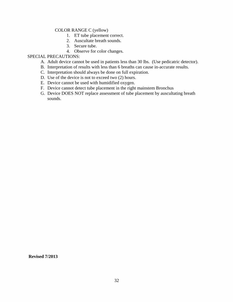
COLOR RANGE C (yellow) 1. ET tube placement correct. 2. Auscultate breath sounds. 3. Secure tube. 4. Observe for color changes.
SPECIAL PRECAUTIONS: A. Adult device cannot be used in patients less than 30 lbs. (Use pedicatric detector). B. Interpretation of results with less than 6 breaths can cause in-accurate results. C. Interpretation should always be done on full expiration. D. Use of the device is not to exceed two (2) hours. E. Device cannot be used with humidified oxygen. F. Device cannot detect tube placement in the right mainstem Bronchus G. Device DOES NOT replace assessment of tube placement by auscultating breath
sounds. Revised 7/2013
32

ENDOTRACHEAL INTUBATION: (EMT-P, EMT-I)
INDICATIONS: In most cases, orotracheal intubation provides definitive control of the airway. It’s purpose includes:
A. To ventilate the patient. B. To deliver high concentrations of oxygen. C. To suction secretions and maintain airway patency. D. To prevent aspiration of gastric contents. E. To prevent gastric distention. F. To administer positive pressure, especially during expiration. G. To administer drugs during resuscitation for absorption through the lungs. H. To allow a faster rate of cardiac compression during CPR.
PRECAUTIONS:
A. Never use intubation as a primary treatment of respiratory arrest in the field. Airway management should be accomplished first with pocket mask, or bag-valve mask as needed.
B. Do not pry the laryngoscope against the teeth. The jaw should be lifted with direct upward traction on the laryngoscope.
C. Suction must be ready. Regurgitation is a common, especially when the esophagus is entered and the tube must be replaced.
TECHNIQUE:
A. Assemble the equipment while continuing ventilation:
1. Choose the tube size (see below). 2. Introduce the stylet and be sure it stops 1/2” short of the tube’s end. 3. Assemble laryngoscope and check the light. 4. Connect and check suction.
B. Position the patient: neck flexed forward, head extended backward. Occiput should be
level with or higher than back of shoulders. C. Give 4 good ventilations before starting procedure. D. Insert laryngoscope to right of midline. Move it to midline, pushing tongue to left and
out of the view. E. Lift up on the blade to expose posterior pharynx.
33

F. Identify the epiglottis: tip of curved blade should sit in vallecula (in front of epiglottis), straight blade should slip over epiglottis.
G. With gentle further traction to straighten the airway, identify trachea from cords, arytenoid cartilage.
H. Insert tube from right side of mouth, along blade into trachea UNDER DIRECT VISUALIZATION.
I. Inflate cuff with 5-1 0ml of air or until there is no air leak around the tube, clamp if necessary to secure against leaks.
J. Check position of tube by listening for breath sounds over the stomach and then on both sides of chest.
K. Note proper position and secure tube. L. Place ET C02 detector (should turn yellows within six breaths)
COMPLICATIONS AND SPECIAL NOTES:
A. Esophageal intubation should be avoided by direct visualization of the cords and endotracheal tube tip throughout the procedure. Listen carefully and check to see that the patient looks better (pupils constricting, color improving) when ventilating.
B. Intubation of the right main stem bronchus is very common. If no breath sounds are heard on the left, withdraw the tube slightly until bilateral breath sounds are heard. Initial depth = 3 times ETT size
C. To determine tube size: 0 years 3.0-3.5mm 2 years 4.5mm 4 years 5.0mm 6 years 6.0mm 8 years 6.5mm 10 years 7.0mm Adult female 7.5mm Adult male 8.0mm
16 + age in years = ETT size. (4)
D. Never interrupt ventilations more than 15 seconds for intubation. If you cannot perform the procedure in this amount of time, you must initiate and maintain other methods of ventilary assistance
E. There is seldom reason to replace an esophageal airway once it is place. However, if there is a compelling reason to prefer endotracheal intubation, the procedure must be
undertaken while the esophageal obturator airway is still in place. If the patient is ventilating satisfactorily, do not compromise the patients condition.
F. REMEMBER: Endotracheal intubation is not the procedure of choice in the first minutes of a resuscitation. It is a secondary procedure only. Most persons can be adequately ventilated with a pocket mask or with a bag valve mask with oropharyngeal or nasopharyngeal airway until there is enough control of the situation to perform the intubation in a controlled and prompt manner.
Revised 7/2013
34

EXTERNAL PACEMAKER
(EMT-P, EMT-I) INDICATIONS:
A. Sinus bradycardia (<40 min) with a blood pressure <80 systolic and a decreased mental status, unresponsive to Atropine.
B. Complete heart block with a BP <80 systolic, unresponsive to Atropine. C. Asystole. D. Mobitz type II second-degree heart block.
PRECAUTIONS:
A. May cause skeletal muscle contractions in the chest wall, that may require analgesia or sedation.
B. Ventricular fibrillation may occur during pacing and should be treated with prompt defibrillation.
TECHNIQUE: ESTABLISH IV, NS, TKO THEN:
A. Electrodes should be applied to clean dry skin. B. If excessive chest hair is present, it should be clipped rather than shaved. C. The preferred placement is the anterior-posterior position:
1. The negative electrode is placed on the left anterior chest, halfway between the xiphoid process and the left nipple (V2-3area).
2. The positive electrode is placed on the left posterior chest, beneath the scapula and lateral to the spine.
D. An alternative placement is the anterior-anterior: 1. The negative electrode is placed on the left chest, over the forth intercostal
space in the mid-axillary line. 2. The positive electrode is placed on the anterior right chest in the subclavicular
area. This placement is less desirable because it interferes with defibrillation and also tends to cause pectoral muscle stimulation.
E. Select the pacing mode: Synchronous: for bradycardia. Asynchronous: non-demand for asystole.
F. Select the pacing rate - 70 is usual. G. Set the current at minimum. H. Activate the pacemaker. Adjust the current upward, observing the patient and EKG.
1. Electrical capture is usually evident by a wide QRS and a tall broad T-wave. In some patients, it may be less obvious, noted only by a change in the QRS configuration.
Revised 7/2013
35

EXTRICATION PROTOCOL Morgan County Ambulance Service personnel are prohibited from participating in the extrication of patients from vehicles involved in traffic accidents, industrial or farm machinery entanglement or any other situation that requires the use of special equipment in order to gain access to the patient compartments or alterations to free someone from entangling. Simple extrication, which includes transferring a patient to a back board while still in a vehicle, that does not require disentanglement, is permitted. Safety of the rescuers is the primary concern in all extrication situations. Any situation that is felt to be dangerous or which posses a threat to rescuers safety, should not be proceed with until additional qualified help has arrived regardless of patient condition. SPECIAL CONSIDERATIONS: Anyone who has been injured in an accident that needs to be extricated and who cannot be freed easily without the use of special equipment should be assessed immediately upon arrival for:
1. Level of conscienousness 2. Airway concerns 3. Hemodynamics by palpating pulses
Extrication should be allowed to continue without hindrance once it’s been determined that there are no life threats to the entrapped patients that can be addresses by ambulance personnel. Further patient evaluations should be delayed until the person has been freed from entrapment. If, however, the extrication process has become time consuming, periodic reassessments should be done. Treating life threats prior to extrication is essential for patient outcomes. If access to a patient can be obtained without risk of injury to the rescuers, than such access should be obtained and life saving procedures should be performed immediately. Communication with the incident commander should be done explaining the need to immediately treat all life threats. If possible the extrication procedures should be continued and precautions should be taken to protect the patient and rescuers from flying debris while these procedures are being preformed. Once these procedures have been completed and secured, the rescuers should remove themselves from the patient compartment if the patient’s condition allows with periodic reassessment of the patient for complications and or deterioration in condition. Revised 7/2013
36

FLUID CHALLENGE A. HYPOVOLEMIC SHOCK: (trauma or medical) - with blood pressure less than 90 systolic:
1. Run IV wide open - check the blood pressure after every 300 cc's. 2. Start second IV and use pressure infusers until BP over 100 systolic 3. Reduce IV rate to TKO, if BP and pulse normal and stable.
B. ORTHOSTATIC HYPOTENSION: (BP fall 20 mmHg or pulse rise 20/Min)
1. Run IV wide open. 2. Check BP after every 300 cc's. 3. Reassess lung sounds with FC
* = Patients over 65 and patients with known or suspected heart condition
1. Check BP, pulse and breath sounds at lung bases after every 300cc. 2. If the pulse rises, the patient develops SOB, rales or pulse oximetry drops, make IV
TKO. C. PEDIATRIC PATIENTS: (12 and under)
1. Check BP and pulse after every 20 cc/kg (1/4 blood volume of the child.) A. Reassess administer additional 20cc/kg if uneffective. May repeat x3 or total of
60cc/kg 2. After 60cc/kg, patient needs blood products contact ER ASAP to be ready with blood
RULE OF THUMB TO HELP RMEMBER HOW MUCH FLUID: The first place you check for a child who is hemodynamically compromised is the bottom of the feet. Cap refill >3 sec is a bad sign. HOW MUCH FLUID? Count the fingers and toes = 10+10 = 20cc/kg the reassess. HOW MANY TIMES DO I REPEAT? 20cc/kg X3 or 3 strikes and your out. They now need blood products. Revised 7/2013
37

GLASGOW COMA SCALE
A score of eleven (11) or less indicates that the patient most likely has a severe injury. Coma is defined as no response and no eye opening or a score of seven or less. The Glasgow Coma Scale may not be useful in patients who are severely hypovolemic or intoxicated or in infants or children.
INFANT CHILD/ADULT EYE OPENING 4 SPONTANEOUSLY SPONTANEOUSLY 4 3 TO SPEECH TO COMMAND 3 2 TO PAIN TO PAIN 2
1 NO RESPONSE NO RESPONSE 1_______ BEST VERBAL RESPONSE 5 COOS, BABBLES ORIENTED 5 4 IRRITABLE CRIES CONFUSED 4 3 CRIES TO PAIN IN APPROPRIATE WORDS 3 2 MOANS, GRUNTS INCOMPREHNSIBLE 2 ____1 NO RESPONSE NO RESPONSE 1________ BEST VERBAL RESPONSE 6 SPONTANEOUS OBEYS COMMANDS 6 5 LOCALIZES PAIN LOCALIZES PAIN 5 4 WITHDRAWS FROM PAIN WITHDRAWS FROM PAIN 4 3 FLEXION (DECORTICATE) FLEXION 3 2 EXTENSION (DECEREBRATE) EXTENSION 2
______1 NO RESPONSE NO RESPONSE 1__________ ______ TOTAL* *TOTAL=_______________
* GCS < 8= INTUBATE!! Severe Head Injury = GCS of 8 or less Moderate Head Injury = GCS of 9 - 12 Minor Head Injury = GCS of 13 - 15 All severe head injuries need to be intubated. Consider RSI Moderate head injuries should be intubated when appropriate, particularly if combative and will not follow commands.
A change in GCS of 2 is worrisome and should be dealt with appropriately.
38

REVISED TRAUMA SCORE
RESPIRATORY RATE 10-29 4 30 OR> 3 6-9 2 1-5 1 NONE 0____________ SYSTOLIC BP 90 OR> 4
76-89 3 50-75 2 1-49 1
NO PULSE 0____________ ADD GLASGOW COMA GCS TRAUMA PTS CONVERTED SCALE 13-15 4 9-12 3
6-8 2 4-5 1
3- 0___________ TOTAL=__________________ Revised 7/2013
39

HAZMAT CALLS HISTORY
1. Any patient that has been exposed to any type of Hazmat material requires decontamination prior to being placed in an ambulance
2. Any call with possibility of exposure to Hazmat should request the Fire Department to also respond and should follow the Fire Chief’s/Incident Command’s direction
ASSESSMENT
1. Scene safety is priority. Never approach down wind, if there is suspicion of explosives stage at least one mile away. Never approach a possible hazmat scene that placards or substance has not been identified.
2. Receive all available information from dispatch prior to responding 3. Respond according to Fire Chief’s request after patient has been decontaminated 4. Proceed with care ONLY after patient has been decontaminated
TREATMENT
1. Assess for airway and maintain as needed 2. Provide oxygen 3. consider need for intubation 4. Transport to nearest facility after notifying them of Hazmat patient 5. Do not enter medical facility if patient remains contaminated until authorized
NOTE: PATIENTS THAT WILL BE FLOWN BY HELICOPTER NEED TO BE COMPLETELY DECONTAMINATED PRIOR TO LOADING WHICH WILL INCLUDE:
1. REMOVING ALL CLOTHING 2. LANDING ZONE MUST BE A SAFE DISTANCE TO ASSURE NO CONTACT
WITH ELEMENTS OF HAZARDOUS MATERIAL 3. AVOID DOWN WIND LANDINGS, APPROACHES AND LOW AREAS WHICH
VAPORS MAY ACCUMULATE Revised 7/2013
40

HEAD TRAUMA
SPECIFIC INFORMATION NEEDED: A. Subjective:
1. History: Mechanism of injury, estimated force involved, change in level of consciousness, amnesia. Was a helmet worn with associated bike injury?
2. Past history: Medical problems, medications. B. Objective:
1. Complete primary and secondary survey. 2. Vital signs. 3. Glasgow Coma Scale.
TREATMENT:
A. Immobilize C-Spine. B. Airway management: as per Airway Protocol. C. 02, high flow by non-rebreather mask. D. Control bleeding. E. IV, NS, TKO, or as indicated to maintain adequate blood pressure. F. Monitor VS and Glasgow Scale as indicated. G. Blood Pressure Control - If hypertensive with head trauma: Attempt to keep BP 150-
180 Systolic and Diastolic less than 110 but greater than 80. SPECIFIC PRECAUTIONS:
A. Assume C-Spine with head injuries. B. All unconscious patients require intubation. C. If diabetes or drug overdose are a possibility, administer D50% and/or Narcan if
depressed level of consciousness is present, draw bloods prior to administration if possible.
D. Scalp lacerations may result in profound hypovolemia. Consider the possibility of a skull fracture.
Death from Blunt trauma Revised 7/2013
41

HYPERTENSIVE EMERGENCIES
DEFINITION: BP 180/120 or greater AND 1. Chest pain or 2. Shortness of breath or 3. Localized weakness or paralysis or 4. Altered mental state or level of consciousness or 5. Severe headache.
MEDICAL HISTORY:
1. Previous high blood pressure. 2. Previous heart attack or heart failure. 3. Previous stroke. 4. Previous kidney failure. 5. Diabetes. 6. Medications.
PHYSICAL ASSESSMENT:
1. Blood pressure, pulse and respirations. 2. Mental status, LOC, (CVA Encephalopathy). 3. Chest auscultation - Rales? (CHF). 4. Extremity exam - Paralysis? Edema? (CVA, CHF). 5. Monitor - Rhythm (AMI).
TREATMENT:
1. Oxygen, 6 liters/mm: aggressive airway management as indicated. 2. Monitor cardiac rhythm - treat arrhythmias, chest pain. 3. IV TKO - (sitting position). 4. Narcan IV, if depressed level of consciousness, withhold D50 if you suspect a
Intracranial bleed. 5. Reassurance of the conscious patient may be all that is needed to reduce blood
pressure. 6. Monitor BP and P every 5 minutes during transport. 7. NTG SL
8. Neuro BP Goals: 150-180 Systolic. 90-110 Diastolic.
9. Cardiac BP Goals: 100-120 Systolic <105 Diastolic Revised 7/2013
42

HYPERTHERMIA
SPECIFIC INFORMATION NEEDED: 1. Environmental conditions. 2. Onset - sudden vs. gradual. 3. Exercise induced. 4. Medical history and medications. 5. Toxin / drug induced.
SPECIFIC PHYSICAL FINDINGS:
1. Heat exhaustion: A. Minor LOC changes. B. Dizziness - nausea - headache. C. Skin hot and moist.
2. Heat stroke: A. Confusion - irrational behavior - loss of consciousness. B. Convulsions. C. Hypotension. D. Skin warm, but dry.
TREATMENT:
1. 02 as indicated. 2. IV: NS, large bore, as indicated to support BP after 200 fluid challenge. 3. Remove all clothing 4. Cool with wet sheets. 5. Consider Versed for seizures. 6. Monitor cardiac rhythm. 7. Monitor vital signs.
SPECIFIC PRECAUTIONS:
1. Heat exhaustion can progress to heat stroke if untreated. 2. Wet sheets on patient without good air flow wilt tend to increase temperature. 3. Definite cooling will need ice water bath. Do not let cooling in the field delay your
transport. Cool patient enroute. By removing ALL clothing and dousing them with water, open windows or use fans.
4. Do not cool to the point of shivering, as this will increase their temperature. Revised 7/2013
43

HYPOGLYCEMIA ( INSULIN SHOCK)
SPECIFIC INFORMATION NEEDED: 1. Onset of symptoms (sudden vs gradual). 2. Duration. 3. Known diabetic and/or other significant medical history (medical alert tags or
relatives). 4. Present history:
A. Last insulin (amount and time) or hypoglycemic agent. B. Last meal: presence of vomiting or diarrhea. C. Increased physical or emotional stress.
SPECIFIC PHYSICAL FINDINGS:
1. Vital signs (tachycardia). 2. Level of consciousness. 3. Neurological findings (due to hypoglycemia of CNS):
A. Inability to concentrate. B. Headache. C. Bizarre behavior. D. Convulsions. E. Imitation of other nervous disorders; i.e., CVA, coma.
4. Skin color, temperature, hydration. 5. Effects of Epinephrine release:
A. Anxiety. B. Sweating. C. Nausea. D. Trembling.
TREATMENT:
1. Assess and support airway, breathing and circulation. 2. Start IV: NS, as indicated. 3. Draw bloods; attempt to obtain a red top tube for later glucose testing. 4. Diascan if time permits. 5. Administer 50 cc (1 Amp) D50%. if Diascan < 60. If between 60-80, use at crew
discretion. Repeat if needed. 6. Transport and monitor closely.
SPECIFIC PRECAUTIONS:
1. Hypoglycemia can present as seizures, coma, mental problems, alcohol intoxication, confusion or stroke like picture with focal deficits (particularly elderly pts).
2. Patients who are elderly or who have been hypoglycemic for prolonged periods of time, may also be slow to respond to D50%.
3. If the diabetic patient is unconscious, it may be difficult to decide between diabetic coma and insulin shock. If the precise nature of the patient’s condition is in question,
44

SUGAR SHOULD BE GIVEN TO ANY UNCONSCIOUS OR SEMICONSCIOUS DIABETIC. If the condition turns out to be hypoglycemia, the administration of sugar will result in rapid improvement and may be lifesaving. If the condition is diabetic coma little harm will be done by giving sugar. The signs and symptoms of hypoglycemia are those of an adrenaline outpouring; diabetic coma signs are those of dehydration.
4. Patients on Beta-blockers may not have signs of Epinephrine release associated with
hypoglycemia. Revised 7/2013
45

HYPOTHERMIA
SPECIFIC INFORMATION NEEDED: 1. Length of exposure. 2. Wind and wet exposure. 3. Environmental conditions. 4. Any drugs including alcohol used.
SPECIFIC PHYSICAL FINDINGS:
1. Lethargic - LOC - irrational - combative - paradoxical - clothes removal. 2. Evidence of local injury. 3. Body temperature: less than 95 degrees (35 degrees C) is significant.
TREATMENT: 1. O2 as indicated. 2. IV: NS, large bore, TKO, draw bloods. 3. Administer 50 cc (1 Amp) D50% if Diascan < 60. If between 60-80, use at crew
discretion. Repeat if needed. 4. Monitor cardiac rhythm. 5. Monitor vital signs. 6. Keep dry - out of the wind. 7. Insulate from the cold: warm blankets, warm fluids. 8. For frost bite:
A. Remove all previous coverings of injured area. Avoid rubbing and breaking blisters.
B. Protect injured areas with warm clothing. SPECIFIC PRECAUTIONS:
1. General: A. Fibrillation is likely when core temperature is between 85-88 degrees, so be
gentle with the patient in transport. Conversion is probable with a temperature of 88 degrees F or greater and corrected acidosis.
B. Rapid correction of acidosis can lead to ventricular fibrillation. Re-warming will usually correct acidosis without use of Bicarb.
C. Hypothermia may be a sign of hypoglycemia. D. Airway irritation may produce V-fib in hypothennic patients. E. Bretylium is preferred over Lidocaine for ventricular arrhythmias since it
doesn’t suppress automatically. (FYI) 2. Local:
A. Thawing is extremely painful; and should only be performed under controlled conditions.
B. Do not allow the patient to ambulate once the limb has started to thaw. C. Do not allow limb to thaw if there is a chance that the limb may refreeze.
Partial re-warming is worse than none. Revised 7/2013
46

INTRAOSSEOUS INFUSION INSERTION
(EMT-P, EMT-I, AEMT) INDICATIONS:
A. Illness: shock, cardiac arrest, widespread bums, massive trauma. B. Mental status: patient must be unconscious or a local anesthetic used. C. Access: unable to start a peripheral line after two (2) attempts.
Peripheral IV is always attempted first, Intraosseous second. CONTRAINDICATIONS:
A. Tibial and femoral fractures on the same leg. TECHNIQUE:
A. Gather all necessary equipment: 1. Disposable intraosseous needle. 2. Appropriate intravenous fluid and IV tubing.
B. Select the site: 1. First choice: Tibia - one finger width (1-3cm) below the tibial tuberosity on the
anteromedial surface. 2. Second choice: Humeral Head- Refer to Vidacare guidelines for placement
C. Prepare insertion site with provodine iodine (Betadine). 1% plain Lidocaine should be infiltrated locally and down to the periosteum.
D. Hold the needle shaft with your non-dominant hand, apply downward pressure on the needle with the dominant hand, using a rotary motion at the same time. Angle of entry may be 90 degrees to the bone or 45 degrees away from the nearest joint. Entrance into the bone marrow is heralded by a sudden loss of resistance, when the needle is felt to pop into the marrow space.
E. Confirmation of proper needle position can only be determined by visible aspiration of marrow contents (dark blood) with a 10cc syringe. Easy flow of IV solution does not confirm the correct placement (e.g. needle only in periosteum, needle completely through the bone). Watch for tissue swelling and compartment syndrome in these instances.
F. If properly placed, the needle will be secure (it will be solidly lodged in the bone); the IV tubing should be securely taped. It should be stabilized at all times. Only a dressing is needed at the needle site. The intraosseous puncture site should be guarded constantly on scene and enroute to the hospital to ensure the patient’s lifeline.
G. If the first tibial attempt is unsuccessful, a second attempt in the femur of the same leg should be attempted.
H. Only one leg will be utilized in the field prior to ER arrival, unless further attempts on the other leg are cleared by Medical Control.
COMPLICATIONS:
47

A. IO insertion may result in leakage of infused fluid into the surrounding tissues,
creating an infiltrate which may lead to compartment syndrome. B. Bone fractures (pushing too hard while not twisting the needle). C. Osteomyelitis (0.5% incidence) occurs in septic patients, use of Intraosseous lines
beyond 24 hours, and infusion of hypertonic solutions (e.g. bicarbonate). D. Needle broken off in the bone (1 reported case - needle left in the bone without
subsequent complications). E. Growth plate and marrow damage from Intraosseous infusions are largely un-studied.
SPECIAL NOTES:
A. Venous access in children can be extremely difficult. Intraosseous infusion provides quick and reliable access to the venous circulation. Infused sub-stances are passed from the marrow cavity into the sinusoids, to large medullary venous channels, to nutrient and emissary veins, and finally into the systemic circulation.
B. All solutions or drugs normally administered IV may be administered Intraosseously. C. Only one Intraosseous attempt is permissible on each bone, since successful needle
placement on the second try in the same bone will result in fluids or drugs draining out of the first hole instead of into the venous circulation.
Revised 7/2013
48

ISCHEMIC HEART DISEASE PATHOPHYSIOLOGY:
Myocardial ischemia is a condition in which myocardial demand exceeds the capacity of the coronary artery supply. This imbalance can result from a variety of conditions but the most commonly results from segmental atherosclerotic obstruction of the coronary arteries. Ischemic symptoms may result when demand exceeds supply such as during exertion, accelerated hypertension, or a dysrhythmia or when the obstruction becomes critical. The chest pain syndromes are stable angina, acute myocardial infarction, and silent ischemia. Myocardial salvage and the prevention of electrical instability and hemodynamic compromise are the principal reasons for early diagnosis and treatment. SPECIFIC INFORMATION NEEDED:
A. Pain: nature, onset, duration, location, radiation, and aggravating and alleviating factors.
B. Associated symptoms: Nausea, vomiting, diaphoresis, dyspnea, or near syncope. C. Past medical history including old electrocardiograms if available. D. Medications.
PHYSICAL FINDINGS:
A. Vital signs. B. Profusion and presence of diaphoresis. C. Breath sounds - rales, rhonchi, or wheezing. D. Jugular venous distention (JVD).
12-LEAD ELECTROCARDIOGRAM (FYI ONLY)
It is the best adjunctive test for assessing a patient with ischemic heart disease (IHD). A completely normal EKG does not exclude acute myocardial infarction (AMI) and often, time and serial EKG's are needed to preclude the diagnosis of IHD. AMI maybe seen as ST segment elevation, depression, t-wave inversion, hyper acute t-waves, or the development of a new bundle branch block (BBB) or new pathologic Q-waves. A Q-wave MI (transmural MI) generally have acute ST segment elevation and develop Q-waves with time. Subendocardial MI (non Q-wave MI) or ischemia generally show ST segment depression and/or T-wave inversion. Any acute change in the EKG from the old should be considered ischemic in etiology until proven otherwise. ST segment changes seen on the monitor are nondiagnostic since monitors are not calibrated and standardized to the same degree that 12-lead EKG’s are. 12-lead EKG leads that correspond to ischemia or infarction of specific areas of the heart are as follows:
A. Inferior II, Ill, AVF. B. Anteroseptal: Vi, V2, V3, V4. C. Lateral: I, AVL, V5-V6.
DIAGNOSTIC CRITERIA FOR CHEST PAIN SYNDROMES:
These are important clinical subsets of the spectrum of IHD. They may all be associated with severe Coronary Artery Disease and some overlap.
49

A. Stable angina: rarely lasts longer than 15 min. Brought on by stress relieved with rest. Does not usually awaken from sleep.
B. Unstable angina: pain lasting longer than 15 min. Angina that has changed pattern and become more frequent or incompletely relieved with nitroglycerin, or angina at rest.
C. AMI: chest pain present for at least 30 minutes with ST segment elevations of 0.1 mV in two contiguous leads.
D. Silent ischemia: symptoms and signs such as weakness, pulmonary edema, hypotension or dysrhythmia without chest pain.
DIFFERENTIAL DIAGNOSIS:
GE reflux, thoracic dissecting aneurysms, pulmonary emboli, pericarditis, pleurisy, spontaneous pneumothorax, pneumonia, and chest wall pain are all differential diagnostic considerations.
TREATMENT:
A. Full cardiac and pulse oximetry monitoring. B. At least one IV with NS. C. Position of comfort.
D. Baby Aspirin should be chewed and swallowed on all suspected MI (if not already given)
E. Oxygen to keep pulse oximetry greater than 92%. F. Nitro spray or sublingual nitro every 5 min until pain relieved or three given. G. Morphine/Fentanyl IV titrated and/or NTG IV starting a 5-10 micrograms per minute
titrated to pain and blood pressure systolic greater than or equal to 90. NTG drips should be increased by 5 micrograms at a time, then reassess B/P. Keep increasing until pain is gone or B/P starts to drop <90 systolic. NTG dose given 5-300mcg/min
**** The decision to use MS vs NTG IV depends upon transport time, response to initial nitroglycerin and clinical impression. COMPLICATIONS OF ACUTE MYOCARDIAL INFARCTION:
Both transmural and subendocardial MFs produce ventricular irritability and patients will be at risk for spontaneous ventricular fibrillation for ventricular tachycardia. In anterior wall MI's, heart blocks and left ventricular failure are the most frequent complications. Lateral wall MI’s, are most frequently complicated by ventricular failure. Inferior wall MI's can produce symptomatic bradycardia due to the vagotonic affect. Additionally, one can see right ventricular infarct syndromes with hypotension that is based on a reduction in preload. Nitroglycerin can exacerbate the reduction in filling pressure often resulting in marked hypotension. The treatment for this is to temporarily stop the nitro drip and give incremental boluses of normal saline. Revised 7/2013
50

KING LDT-S AIRWAY (EMT-P, EMT-I, AEMT, EMT- IV, EMT-B)
Placement of the King LDT-S should not delay CPR, Basic airway management, use of defibrillator, or other necessary patient care. INDICATIONS:
Provides method for administering sufficient ventilation when endotracheal intubation with conventional ET Tube is not successful or available.
CONTRAINDICATIONS Responsive patients with a gag reflex
Patients with know esophageal disease
Patients who have ingested caustic substances
TECHNIQUE / PROCEDURE Begin artificial respirations taking usual precautions to open airway
Check King LDT-S for correct size based on patient height
Prepare King LDT-S for insertion
Place the head in the “Sniffing” position
Hold King LDT-S airway in dominate hand, hold the mouth open and apply chin lift
Rotate 45° to 90° so blue line is touching the corner of the mouth advance beyond the tongue
DO NOT FORCE DEVICE!!!
As the tube passes under the tongue, rotate the tube back to midline so the blue line faces the chin
Advance until the proximal opening of the gastric access lumen is aligned with the teeth or gum
Inflate the cuff with the appropriate amount of volume to seal the airway. Attach BVM and ventilate
While ventilating, withdraw the King LDT-S airway until there is minimal airway pressure & large tidal volume delivery is present.
Confirm placement by auscultation of breath sounds, chest rise and fall, lack of epigastric sound, capnography, and pulse ox.
Revised 7/2013
51

MAINTENANCE OF IV INFUSIONS
I. Purpose For the continuous maintenance of IV infusions during ground transport
II. Procedure
Only the following medications/ infusions are allowed, and to be administered with an infusion pump, see following protocols for information for specific infusions
Antibiotics Dopamine Lidocaine Magnesium Sulfate (CTN) Heparin Nitroglycerin Terbutiline (CTN) Thrombolytic (CTN)
IV infusions are not to be initiated by Paramedics with the exception of Dopamine, Lidocaine, and Amiodarone
The Paramedic must ensure the patency of the IV line EKG monitoring is mandatory Documentation will include
Drug name and concentration Physician ordering drug treatment Rate and amount of drug administered during transport
In the event of infiltration, the IV infusion is to be discontinued, Base physician contact is required
Contact Base physician for any medication infusion not listed Revised 7/2013
52

METABOLIC/ELECTROLYTE DISORDERS
DKA This results from endogenous production of ketones brought on by insufficient insulin
combined with a stress. The hyperglycemia usually exceeds 300 mg/dl and the acetone is positive
The pathophysiology of DKA includes profound dehydration, varying degrees of acidosis, and various electrolyte abnormalities including hypokalemia. One always needs to search for a precipitant of DKA such as infection, acute MI, etc. Pediatric consideration: Caution should be used in pediatric DKA not to overhydrate them. An initial 20 cc/kg NS bolus over 1 hour. Treatment
Fluid resuscitation is the mainstay of therapy. It is more important than insulin and most patients are profoundly hypovolemic. Typically, an adult will need 3-4 L of NS and a child 20 cc/kg of NS in the first hour. Insulin is usually given IV as a drip at 0.1 u/kg/hr IV. When the glucose falls below 250 mg/dl then fluids are changed to D5 ‘/2 NS with potassium. Generally 20-40 meq of KCL are added to each liter of NS. NO INSULIN BOLUS!!!!! THEY NEED FLUID, FLUID, FLUID Hyperkalemia
This probably the most life-threatening of the electrolyte disorders. As the potassium rises progressively, first T-waves will become peaked, followed by bundle branch blocks, junctional rhythms, IVR and followed by an asystolic arrest or ventricular fibrillation. This generally occurs when serum potassium levels exceed 6.5 mg/L. Succsynolcoline will raise K+ levels, therefore should be used with caution in burn patients or massive crush injuries.
No calcium if Hyperkalemia is from Digoxin toxicity! Calcium is the drug of choice (Rule of 10’s): 10cc of 10% solution over 10 minutes NOTE: 1) high concentrations of calcium suddenly reaching the heart can cause fatal cardiac arrest. 2) direct IV push can cause hypotension. 3) ECG monitoring is a must during and after administration to watch for hypercalcemia associated with prolonged QT interval and with inverted t waves. 4) be ready for seizure
Hypokalemia It is less life-threatening than Hyperkalemia, but can produce profound weakness, an
ileus, or precipitate digoxin toxicity when potassium is less than 3.0 meq/L. KCL should be replaced at no more than 10-15 meq/hr. No more than 40 meq of KCL should be added to each liter of saline for maintenance infusions. Usually 40-50 meq of KCL is needed to raise the serum potassium by 1 meq/L.
53

Hyponatremia It can produce profound changes in mental status, focal neurologic deficits, and seizures.
Symptoms are related to the rate of the drop. Someone may be relatively asymptomatic with a sodium of 120 meq/L if it occurred slowly over days to weeks. Causes of hyponatremia are:
Sweating, vomiting, diarrhea Third space sequestration (bums, peritonitis, pancreatitis) Diuretics Aldosterone deficiency Ketonuria
Salt-losing nephropathies, Bartter’s syndrome, Renal failure
Cirrhosis Cardiac failure Renal failure
Inappropriate antidiuretic hormone secretion Sickle cell or “reset osmostat” syndromes Physical and emotional stress or pain Myxedema, Addison’s and Sheehan’s syndromes
Pseudohyponatremia = hyperproteinemia, hyperlipidemia, hyperglycemia
Adult patients who appear dehydrated and hypovolemic should be given NS liberally. If edema and volume overload are present then NS should be given at 50-100 ml/hr. Rarely, hyponatremia is so profound that 3% saline should be given. Severe neurologic impairment and/or seizures will require a 300 ml bolus in an adult or 10 ml/kg in a child of 3% saline. Children can experience hyponatremia from swimming pools or pools high in chlorine. They swallow the chlorine which effects sodium intake. If kids neurologically act bizarre for unexpected reasons, this could be a cause. However, other sources should be ruled out as well, i.e,, poisoning, hypoglycemia. Revised 7/2013
54

MULTIPLE CASUALTY INCIDENTS (MCI) SCENARIO #1
When Morgan County Ambulance is called to a MCI, Morgan County Ambulance will accept the patient who has been triaged as the most critical patient for immediate transport to the facility as directed by incident command.
SCENARIO #2 When Morgan County Ambulance is first on scene, the highest-level EMT will assume the role of triage until an EMT-P arrives at which time the EMT-P is responsible for triage. That EMT-P should remain on scene until all patients have been accounted for and transported or there is only one critical patient at which time the EMT-P should go with the critical patient putting someone else in charge of triage. SCENARIO #3
When Air Life is called to a MCI at which Air Life is the first ALS agency in attendance, the flight paramedic will assume triage duties.
The flight nurse should accompany the most critical patient in transport to the hospital. The flight paramedic will remain at the scene until the arrival of the ground ALS unit, or until the remaining casualties have been triaged and it has been determined that ALS attendance is not required.
Death in mass casualty Revised 7/2013
55

NASOTRACHEAL 1NTUBATION
(EMT-P Only) Use endotracheal tube that is approximately a half size smaller than oral.
Adults: 7.0 Female, 7.5 Male. INDICATIONS:
Respiratory distress in the patient that is still breathing; but with decreasing level of consciousness or impending respiratory arrest.
ADVANTAGES:
Less traumatic to conscious patients, less likely to cause neck injury, patients tolerate it better.
PROCEDURE:
A. Protect C-spine during the procedure if indicated. B. Insert 1 dropper full of Neosynephrine in both naris. C. Insert approximately 5 cc 2% Xylocaine jelly into the nostril. Advise the patient of
what you are going to do if the patient is awake. D. Insert ET tube straight in the nostril - do not force. E. If significant resistance is met, try the other side. Don’t forget the Neosynephrine
and Xylocaine! F. DO NOT USE THE STYLET! G. Slowly advance the tube into the posterior pharynx. H. Place your ear or use a bamm on the distal tube to listen for breath sounds. I. When breath sounds are the loudest, you are just above the vocal cords. J. Place one hand on the Trachea to put slight pressure and sense alignment. K. With a smooth movement, advance the tube at the beginning of inspiration. L. If breath sounds are not audible, withdraw to the level of the loudest breath sounds
and repeat. If problems are encountered, check your timing and make sure insertion is smooth, you may have to reposition the head. This procedure requires finesse, not strength.
M. After successful intubation (patient is unable to talk), listen for breath sounds bilateral.
N. Secure the tube with umbilical tie. O. Place an ET CO2 detector on (should turn yellow within 6 breaths) SPECIAL NOTE: Nasotracheal Intubation is NOT contraindicated in severe mid-facial trauma. The performance of nasotracheal intubation in severe mid-face trauma requires that the person performing the procedure be proficient in nasal intubation and be able to identify ABNORMAL resistance or lack of in these specific patients. Direct visualization of the ET tube in the posterior pharynx is required. If any abnormal resistance or other factor is encounter, the procedure should be terminated and alternate means of airway control should be explored. (Oral intubation (RSI) is the preferred method when CVA is suspected and the use of thrombolytics is a possibility) (FYI ONLY). Revised 7/2013
56

(EMT-P Only) INDICATIONS:
For use as a temporary airway when other common methods of airway control have been unsuccessful in both medical and trauma patient. It may be particularly useful in patients with acute airway obstruction of a foreign body, traumatic injury of infection etiology unrelieved by other methods.
PRECAUTIONS:
A. This form of airway management is only temporary and CO2 levels will build up to unacceptable levels in approximately 30 to 45 minutes.
B. This form of airway management does not protect the airway against aspiration. INSERTION PROCEDURE:
A. Identify the cricothyroid membrane. B. Position a 10 gauge over the needle IV catheter, attached to a 12cc syringe, at a 90
degree angle to the neck. C. While exerting negative pressure in the syringe, insert the catheter tip into the center
of the cricothyroid membrane, advancing slowly until the needle tip enter the trachea, and air is aspirated into the syringe.
D. Once the needle tip enters the trachea, advance the needle/catheter approximately ½ cm more to assure the entry of the catheter and not just the needle tip. The needle is then withdrawn slightly into the catheter so that the sharp tip is no longer exposed.
E. The needle/catheter/syringe assembly is then aimed caudally at 30 to 45 degree angle, and the location is again confirmed with the aspiration of air.
F. The catheter is then advanced over the needle, removing the needle. Confirm the position once more, and recheck for any sign of subcutaneous air.
VENTILATION PROCEDURE:
A. Ventilation is achieved by attaching a 3.0 ET Tube connector to Angio hub, and using a BVM to ventilate patient.
B. Ventilation ratio should be 1:5; one second for inhalation, then exhalation for four (4) seconds to allow for exhalation. Continue this 1:5 ratio in a cyclic fashion.
NOTE: You must allow for exhalation Revised 7/2013
57

NASOGASTRIC / OROGASTRIC TUBE PLACEMENT (EMT-P ONLY)
INDICATIONS:
A. Distended abdomen in severe abdominal pain. B. Unconscious patients with protected airway via ET C. Cardiac Arrest with protected airway via ET D. > 20% BSA burns. Interfacility only.
PRECAUTIONS:
A. Do not force NG tube. B. Avoid epistaxis by using lubricant and gentle technique. C. Extreme caution in head injury. Discontinue if any resistance.
TECHNIQUE: A. Inspect the tube (salem sump –FR) to make sure you have all the components needed and to make sure the tube is not damaged.
B. Have patient sit upright if possible C. Estimate the length of tube. Measure from corner of the mouth, around the er down to the umbilicus. Make note of your mark. D. Select the larger naris (usually the right) E. Lubricate the distal end of the tube with viscus lidocaine. A drip of neosynepherine can help with bleeding. F. Slighly flex the neck to narrow the upper airway. G. Insert the tube in a horizontal posterior direction with gentle pressure. DO NOT PUSH the tube upward, this may damage the cribform plate. H. Encourage the ptient to swallow as the tube is going down. Advance to your measured mark. I. Using a 60cc Tumi syringe, try to aspirate stomach content. J. Inject 60cc of air into the tube and auscultate over the stomach for placement K. Tape/secure the tube
Revised 7/2013
58

ORAL INTUBATION OF THE TRAUMATIZED PATIENT (IN - LINE STABILIZATION)
(EMT-P, EMT-I)
INDICATIONS: 1. To provide a patent airway in the obtunded trauma patient. 2. Can be utilized before cricothyrotomy is attempted. 3. Useful for patients with suspected C-spine injury in whom nasal intubation is
contraindicated or not possible. 4. It is particularly useful in the pediatric trauma patient.
PRECAUTIONS AND CONTRAINDICATIONS:
1. To be performed properly and effectively, this procedure requires two people. 2. The method employs “in-line” or “axial” stabilization, not traction; traction on the C
spine is the field could be disastrous. 3. Do not become fixated with orally intubating the patient; if after 2-3 attempts, proceed
with the Cricothyrotomy, particularly in maxillofacial trauma, or in patients with copious and continuous emesis-secretions in the airway which can make landmark identification difficult if not impossible. CRICOTHYROTOMY IS NOT A PROCEDURE ALLOWED BY OUR PROTOCALS AS OF NOW
4. The patient must be hyperventilated with 100% 02 before, between, and after intubation attempts to avoid hypoxia.
5. It is not intended to replace cricothyrotomy, but is provided to offer an alternative to cricothyrotomy for those patients who fit the criteria for ILS intubation.
6. Unless completely comfortable with the procedure, it should not be attempted on patients with known or obvious signs of C-spine injury.
TECHNIQUE: 1. Prepare the equipment while ensuring that the patient is being properly ventilated with
BVM. 2. The patient should be positioned on LSB with straps secured and head in neutral
position. 3. The C-collar must be removed during the stabilization/intubation phase. Lateral
immobilization however, should remain in place.(i.e. towel rolls, head-bed) 4. The assistant positions himself/herself so that he can immobilize the patient’s head,
while at the same time not to interfere with the intubation. 5. The assistant places her/his hands on each side of the patients head at approximately
the ears or TMJ’s, firmly grasping and immobilizing the head from a flexion/rotational movement.
6. From a position directly behind the patient’s head, orally intubate the patient without actually lifting the patient’s head with the laryngoscope.
7. Cricoid pressure (Sellick Maneuver) should be done until the intubation is complete. Revised 7/2013
59

ORTHOSTATIC VITAL SIGNS
INDICATIONS: 1. Potential hypovolemia: vomiting, diarrhea, bleeding. 2. Abdominal pain. 3. Possible internal hemorrhage. 4. Syncope. 5. Abdominal or chest trauma. PRECAUTIONS: 1. May not be applicable in trauma situations due to spinal immobilization. 2. Be prepared for syncope when the patient is moved upright. TECHNIQUE: 1. Patient is supine position for minimum of three (3) minutes before taking lying blood pressure
and pulse. 2. Then stand the patient and wait one (1) minute before repeating BP and pulse. 3. If the patient is unable to stand, sit him upright with legs dangling over the cot. SPECIAL NOTES AND EVALUATION: 1. Orthostatic changes are not valid if:
A. Taken initially upright, then supine. B. Taken upright with only the head of the cot elevated and the legs not dangling.
2. Significant orthostatic changes include: A. Pulse increase greater than 20 beats per minute when moved upright. B. Systolic or Diastolic pressure (Systolic most significant) drop greater than 20 mmHg
when upright. C. Pallor, diaphoresis or faintness when upright.
3. Significant changes indicate volume depletion (dehydration or blood loss). 4. Considerations:
A.Young people tend to compensate for intravascular volume loss by maintaining blood pressure and increasing pulse rates until 20-25% of blood volume is lost, at which point, their blood pressure drops drastically.
B. Elderly patients have less reserve and may mount little, if any, tachycardic response to volume loss and will drop their pressure early. C. Medications may inhibit the body from it’s normal tachycardic response to volume loss (i.e. beta blockers) and others may prevent or slow the normal vasoconstriction response to postural changes in order to maintain blood pressure when standing. Revised 7/2013
60

PATIENT ASSESSMENT ENVIRONMENTAL: A. Recognize environmental hazards. B. Identify number of patients. Initiate a triage system if appropriate. C. Recognize mechanism of injury or chief complaint, position of patient. D. Identify self. E. Call for backup; initiate communication. PRIMARY SURVEY: A. Airway and C-spine immobilization if trauma: open, check for adequacy, note potential
problems. Neck immobilization should be completed immediately following this if appropriate
B. Breathing: respiratory noises and effort, skin color, behavior. C. Circulation: stop exsanguinating hemorrhage, note presence and quality of pulse. D. Responsiveness: note initial level. Ex- AVPU (Alert, Verbal stimuli, Painful stimuli,
Unresponsive) E. Shock: Watch for cool, clammy pale skin, thirst or agitation. SPECIAL NOTES: - should take thirty (30) seconds or less for assessment.
SECONDARY SURVEY: A. Neck: evaluate for trauma and mechanism of injury and immobilize if question. B. Head and face:
1. Palpate for deformities, asymmetry, blood or pain. 2. Recheck airway for potential compromise, dentures, loose or avulsed teeth, proper
occlusion. 3. Eyes: pupils (equal or unequal, responsiveness to light), foreign bodies, contact
lenses, lacerations, blurred or lost vision. 4. Nose: deformity, bleeding or discharge. 5. Ears: bleeding or discharge.
C. Chest:
1. Recheck for deformity or tenderness if not already immobilized. 2. Note wounds, JVD, use of neck muscles for respiration, altered voice and medical
alert tags. 3. Palpate for tenderness, wounds, fractures, unequal rise of chest, or crepitus. 4. Have patient take deep breath: recheck wounds, symmetry of breathing.
D. Abdomen: 1. Inspect for wounds, ecchymosis. 2. Palpate for tenderness, rigidity.
61

E. Pelvis: 1. Palpate and compress for tenderness, instability.
F. Shoulders/Upper Extremities:
1. Palpate symmetrically for wounds, fractures, tenderness. 2. Check for distal CMS, medical alert tags. 3. Check for weakness (have patient squeeze your hands if no obvious fractures present). 4. If exam is normal, gently move arms to check overall function.
G. Lower Extremities: 1. Palpate symmetrically for wounds, fractures, tenderness. 2. Check for distal CMS. 3. Check for weakness (have patient push feet against your hands if no obvious fractures
present). 4. If exam is normal, gently move legs to check overall function.
H. Back: 1. If spinal injury is possible, inspect and palpate for wounds, fractures or tenderness. 2. Recheck for motor or sensory deficits, as appropriate.
SPECIAL NOTES: - should take one (1) to two (2) minutes to complete.
- should be systematic, though exact order may vary. - do not interrupt unless ABC deterioration is noted. - auscultate abdomen or check if appropriate. - obtain quantitative vital signs after secondary survey unless the patient
shows signs of shock. VITAL SIGNS: A. Obtain first quantitative set of VS within 5 minutes if practical. Pulse, blood pressure, and
respiratory rate. B. Repeat according to the patient’s condition. At least once every fifteen (15) minutes. C. Note neurological status. Monitor level of consciousness particularly. D. Glasgow Coma Scale. A decrease of two is a worrisome sign. E. Revised Trauma Score. GROUND TIMES: A. All Trauma scene ground times should be 10 minutes or less. B. All Medical scene ground times should be 15 minutes or less. Revised 7/2013
62

PATIENT HISTORY
MEDICAL: A. Chief complaint: Questioning to include, when appropriate;
1. Onset. 2. Quality. 3. Quantity. 4. Duration. 5. Relief/aggravation. 6. Associated symptoms. 7. With pain: location, radiation.
B. Associated complaints: questions as for chief complaint. C. Relevant past medical history. D. Medication and drugs: prescription, OTC, recreational. E. Survey of surroundings for evidence of drug abuse, mental functioning and family problems. TRAUMA: A. Mechanism of injury:
1. Cause. 2. Implements. 3. Trajectory. 4. Force. 5. With vehicle: speed, condition (windshield, steering wheel, seat belts).
B. Patient complaints: work up as with medical complaints. C. Relevant past medical history. D. Medication and drugs: Prescription, OTC, recreational. SPECIAL NOTES: A. Do not let gathering of information distract you from management of life threatening
problems. B. Appropriate questioning can provide valuable information while establishing your authority,
competence and rapport with the patient. C. History is commonly obtained while performing the secondary survey. Assistance is often
used for gathering information from the patient or bystanders. D. Do not forget to use bystanders to confirm information obtained from the patient and to
provide facts when the patient cannot. History from the scene is invaluable; you are the only one who can obtain this.
E. Over-the-counter (OTC) medications (including aspirin) and birth control pills are frequently overlooked by the patient and rescuer.
F. Consider medical causes for trauma, particularly in the single-person accidents. G. Patients are people in need. They ask for help, but they also deserve your respect, kindness,
and consideration. Your interaction with them demands professionalism and confidentiality. H.. Utilize local rescue units to maximize efficiency in gathering information and providing
patient care. Make them feel part of the team. Revised 7/2013
63

PEDIATRIC CARDIAC ARREST
TREATMENT:
1. Pediatric arrest is treated essentially the same as the adult with all treatments (drugs, procedures) modified for size (usually based on kg). We tend to want to pick them up and run with them. You must work them where they are initially to give them the best chance.
2. Oxygenation is the most important therapy and should have priority. Most pediatric arrests are due to hypoxemia (not cardiac disease).
3. Use Broselow Tape for weights and dosages. A. Immediate airway: Breathing management.
1. Mask to mouth. BVM initially, then intubate. 2. If airway obstruction, Heimlich first: back blows or ABD thrusts. 3. Intubate: Rule of thumb, tube size is equivalent to the size of the small finger of
patient. ET suction catheter is twice the size of ET tube (French) 4. Needle Cricothyrotomy, if indicated. Realize that this will have long lasting
implications. 5. If unable to ventilate, “hard to bag”, with endotracheal tube and diminished breath
sounds, consider chest decompression, but only after kinked or obstructed ET tube is ruled out.
B. Initiate CPR:
1. Compression rate: 120/min, neonate, 100/mm infant & child. 2. Ventilation rate: 5 compressions; 1 ventilation (40-60 min infant).
C. Start IV:
1. Normal saline, micro drip or buretrol. 2. Rate TKO: give pediatric fluid challenge if you suspect trauma or dehydration. (20
cc/kg) May repeat x3 3. Best sites: Antecubital, external jugular, dorsum of wrist or foot. (forehead in infants) 4. IO.
D. Cardiac Monitor: E. Treat arrhythmias per protocol:
1. Common drugs used and dosage: A. Epinephrine: 0.01-0.03 mg/kg every 5 minutes of 1:10,000. B. Atropine: 0.2 mg/kg. Minimum dose 0.1mg, max dose 0.5mg for a child. New
born dose is 0.03mg/kg C. Sodium Bicarb 1.0 meq/kg initially, then 1/2 of initial dose every 10 minutes.
Neonate: 2meq/kg of 4.2% solution
64

2. Defibrillation:
A. 2 watt/sec/kg Double if unsuccessful and repeat as needed. This wattage is the same for the biphasic (ZOLL) as it is for the monophasic monitors
B. Anterior-Posterior position for paddle placement. C. Never blindly defibrillate a child.
3. KEEP THEM WARM!!!
SPECIAL PRECAUTIONS:
1. Most common cause of pediatric arrest: A. Drowning B. Trauma
C. SIDS. D. Upper respiratory obstruction (Croup, Epiglottitis, foreign body).
2. Most common seen arrhythmia: asystole/Bradycardia due to hypoxia, ventricular
fibrillation is rare in children.
3. Normal Vital Signs in children:
A. Blood pressure: 80 plus 2 times the age in years = Systolic. Diastolic = 2/3 Systolic.
B. Pulse Rates:
6 months or less — less than 160. 6 years or less = less than 140. 6 years or greater = less than 120.
C. Tachypnea = Respiratory rate over 40.
4. Defibrillation: 2 watt/sec/kg. If unsuccessful, energy dose maybe doubled.
Respiratory Distress - When kids are retracting, the higher up the retraction the more ominous the condition. Subclavin retractions more worrisome than diaphragmatic. ET Tube selection- size of the little finger or 16 plus their age in years divided by 4 ET suction cath = ET tube size times 2 Fluid calculation = Count their fingers and toes, there should be 20. (20cc/kg, reassess) How many times do you give it? 3 strikes and your out or (60cc/kg) Then move to blood products, how much? Blood is thicker than water so cut it in half (10cc/kg) NOTE: WHEN IN DOUBT USE YOUR ADJUNCTS TO FIGURE OUT V/S AND DRUG DOSING.
65

CONDITIONS REQUIRING CARDIOPULMONARY ASSESSMENT:
Respiratory rate greater than 60.
- Heart rate greater than 180; less than 80 (under 5 years). greater than 160 (over 5
years).
- Respiratory distress.
- Trauma.
- Bums.
- Cyanosis.
- Failure to recognize parents.
- Diminished level of consciousness.
- Seizures.
- Fever with petechiae.
- Being transferred to an ICU.
TREATMENT: These patients will receive high flow oxygen by face mask or blow-by, cardiac monitor, pulse oximetry, spinal precautions when indicated, and an IV of normal saline. If the patient is critical and compensated shock present as many possible peripheral IV's should be attempted by available personnel. If after one minute total and no access is obtained, than at least one, but preferable two, interosseus lines should be placed. Initial intubation should be considered when the primary problem is thought to be respiratory or when there is a depressed level of consciousness secondary to head trauma or toxic ingestion. The lethargic tachypneic patient in shock from other causes usually improves rapidly with aggressive resuscitation and may not require intubation. The exception may be the septic shock patient. After any intervention, reassessment is vital. Improvement should follow; if not, then more aggressive therapy or alternative therapy is indicated. All patients with seizures or depressed level of consciousness should have rapid assessment of blood glucose. Dextrose 25% 2 cc/kg IV should be given if the glucose is less than or equal to 80, or if it cannot be assessed. SPECIFIC THERAPY FOR VARIOUS SHOCK STATES:
66

The initial fluid for resuscitation is normal saline. This should be given in 20 cc/kg increment boluses as rapidly as possible . Reassessment of heart rate, level of consciousness, perfusion, blood pressure, respiratory rate, and liver size should be done after each 20 cc/kg bolus. Three types of shock 1. Hypovolemic Shock:
A. Dehydration - boluses should be given in 20 cc/kg increments up to 60 cc/kg. If the patient remains in shock after 60 cc/kg, then medical control should be contacted. 60cc/kg should resuscitate most hypovolemic shock from dehydration patients and if it does not then perhaps the patient is in septic shock and may require albumin, FFP or Dopamine. (Special Note: Pediatric DKA is treated less aggressively. See Metabolic / Electrolyte Disorders)
B. Hemorrhagic boluses of 20 cc/kg up to 60 cc/kg should be given. If the patient
remains in shock following this, then packed red blood cells in 10 cc/kg boluses is needed. Medical control should be contacted when blood needs to be given A.S.A.P.
2. Distributive Shock:
A. Septic - 20 cc/kg boluses up to 60 cc/kg should be given. If shock persists after this time, then medical control should be contacted. Dopamine should be considered..
B. Neurogenic - same as above. C. Anaphylactic - fluid resuscitation is the same as above.
Epinephrine should be used very early in this situation. Shock with a palpable pulse should be treated initially with half the IV dose of 1:10,000 of epinephrine while an epinephrine drip is readied. If the pulse is lost then full intravenous doses of 1:10;000 epinephrine should be used. Epinephrine is titrated to improve perfusion as evidenced by increased level of consciousness, increased blood pressure and improved capillary refill.
3. Cardiogenic: a high index of suspicion is needed in order to recognize this. Patients with
known congenital heart disease usually does not represent a diagnostic dilemma. Medical control should always be contacted when dealing with patients in cardiogenic shock.
A. Arrhythmogenic Shock - supraventricular tachycardia and the bradycardias are the
two most common types. Heart rates greater than or equal to 220 are usually caused by SVT. If shock is present then immediate cardioversion with 0.5 joules/kg following sedation should be done. If the patient is stable, a consultation with medical control should be obtained. Heart rates in the range of 200-220 may receive a cautions trial of normal saline at 10 cc/kg. If sinus tachycardia is present then the heart rate will decrease following that. If an SVT is present then there will be no change in the heart rate. Bradycardia is caused by primary cardiac problems such as a person with congenital heart disease it should be treated with Atropine. Bradycardia from hypoxic
67

ischemic injury, i.e. near drowning, should be treated with Epinephrine. SPECIFIC DRUG THERAPY: 1. Dopamine: it possesses both alpha and beta agonist properties. Its primary action is to
enhance contractility in a dose related way. Increased vascular resistance (alpha) is achieved with doses of 10-20 mcg/kg/min. Increased renal blood flow is obtained with doses less than 5 mcq/kg/mm. Indications are septic shock not responsive to
60 cc/kg of normal saline, hypoxic ischemic injuries who are normotensive, and cardiogenic shock not caused from an arrhythmia. The “Rule of Sixes" should be used to mix Dopamine drips. Six times the body weight in kilos is the milligram dose added to make 100 cc’s of NS.. One ml/hr delivers 1 mcg/kg/min. Dopamine should be started at 10 mcg/kg/minute and titrated to desired effect.
2. Epinephrine: it has very potent alpha and beta effects. It is tolerated well by children.
Indications include all shock states unresponsive to fluid and 20 mcg/kg/min of Dopamine. It should be used primarily in anaphylactic shock and following hypoxic ischemic injuries when hypotension is present.
SPECIAL NOTE: Scene time should not be prolonged in order to resuscitate the pediatric patient. Initial stabilization focusing on airway, breathing and circulation should be done followed by 1st line ACLS and all other resuscitation should be done while enroute. Do not go before you have addressed the airway and 1st line ACLS. Revised 7/2013
68

PEDIATRIC SHOCK RESUCITATION
Shock is defined as a state of hypo-perfusion. Compensated shock is defined as a state where there is clinical evidence of hypo-perfusion, yet a normal B/P is present. Three types of shock: 1. Hypovolemia: Etiologies include dehydration states from, profound diarrhea, vomiting, DKA, toxic ingestions or hemorrhage from traumatic injuries. 2. Distributive: Etiologies include sepsis, certain toxic ingestions, spinal cord injuries, or
anaphylaxis. Hypo-perfusion results from a loss of vascular resistance (venous and arterial dilation) and an increase in vascular permeability.
3. Cardiogenic: Etiologies include arrythmias, both tachy and bradicardias, hypoxic ischemia injures (HII) following resuscitation, i.e., near drowning, primary cardiac dysfunction, i.e., tamponade, cardiac contusions.
Assessment 1. Vital signs a. Heart rate, respiratory rate, B/P will be taken on all pediatric patients. (Temperature should be taken prior to transport from hospital to hospital.) b. Vital signs should be taken at least every 15 minutes. They should be taken every 5 minutes if therapeutic interventions have been taken. c. Blood pressure cuff should be 2/3 the length of the upper arm. Approximate (systolic) should be 80 plus 2 times age in years; (diastolic) 2/3 the systolic pressure. HR RESP RATE URINE OUTPUT Infant 120/160 40-60 2cc/kg/hr Pre-School 100/120 30-40 Children > 2y/0 1cc/kg/hr Children 80/100 20-30 2. Physical Exam a. Neuro Status – LOC, interaction with parents and staff, response to pain, eye contact. b. Respiratory Status – Air movement, breath sounds, work of breathing, stridor, cyanosis
69

Retraction – Respiratory distress in kids will sometimes present with retractions. It is the body’s response in using accessory muscles. Four types of Retraction: Abdominal mild distress Intercostal moderate distress Sternal severe distress Supraclavicular ominous sign c. Cardiovascular status 1. Cap refill – soles of feet, this should be the first thing you look at. Less than 2 sec normal. > 2 sec means they are either cold or have hemodynamic compromise. NOTE: Do not wait for B/P and HR to change before transport. 2. Ability to palpate the radial and dorsal pedis pulse d. Secondary survey 1. Signs of external trauma 2. Symmetric movement of extremities 3. Abdominal exam to include palpation of the liver (normally not present). 4. Skin exam or signs of purpura and pstechiae 5. Palpation of extremities for evidence of fracture.
CONSTANT REASSESMENT IS CRUCIAL
CONDITIONS REQUIRING RAPID CARIOPULMONARY ASSESSMENT
HR >180 or <80 (under 5 y/o) Respiratory rate >60 Respiratory distress (retractions) Trauma Burns Cyanosis Failure to recognize parents Decreased LOC Seizures Fever and petechiae Any pediatric patient being transported to another hospitals ICU
70

Treatment 1. High flow O2 by mask or blow by 2. Cardiac monitor, pulse ox 3. Spinal precautions if indicated 4. IV of NS NOTE: If patient is critical, IV’s should be attempted numerous times, simultaneously by numerous people if compensated shock is present. If after “1” minute (total) no IV is accessed then at least one, but preferably 2 IO should be placed. 5. Intubation should be considered when the primary problem is thought to be
respiratory, or when there is a depressed LOC secondary to head trauma, or toxic ingestion.
NOTE: The lethargic tachypnic patient in shock from other causes will usually improve rapidly with aggressive resuscitation, and may not require intubation. (the exception may be a patient in septic shock) 6. After any intervention, you must reassess. Improvement should follow treatment, if not, more aggressive treatment or alternative therapy is indicated. Start with the basics, if no improvement get more aggressive, i.e,. 02, Bag, ET, Drugs. All patients with seizures or decreased LOC should have a rapid assessment of blood sugar, D25 should be given if sugar is <80mgdl or if it can’t be assessed, administer 2cc/kg I.V. THERAPY FOR VARIOUS STAGES OF SHOCK Normal saline is the initial fluid of choice. It should be given 20cc/kg increments as rapidly as possible. Reassess HR,. LOC, Resp rate, Cap refill, B/P and liver size after each 20 cc/kg bolus.
Hypovolemic Shock 1. Dehydration: Bolus with NS 20cc/kg increments up to 60 CC/kg. If patient
remains in shock after 60 cc/kg contact base control immediately. 60 cc/kg should resuscitate most hypovolemic shock patients that are dehydrated. If it does not, consider the patient to be septic. Use Dopamine 2 – 20mcg/kg, FFP or Albumin.
2. Hemorrhagic – Bolus 20 cc/kg up to 40 cc/kg. If the patient remains shocky after 40 cc/kg, they need PRB 10 cc/kg, contact base control to be standing by with PRB. Continue with NS at 20 cc/kg increments.
71

Distributive Shock 1. Septic – 20 cc/kg up to 60 cc/kg. Contact base if shock persists and consider Dopamine. 2. Neurogenic 20 cc/kg up to 60 cc/kg. Contact base if shock persists and consider Dopamine, FFP or Albumin.
3. Anaphylactic – Fluid resuscitation is the same as above. EPI should be used very early in this situation. Shock with a palpable pulse should be treated with half the IV dose of 1:10,000 while EPI drip is prepared. If pulses are lost then full dose of EPI 1:10,000, follow ACLS guidelines. EPI is used in anaphylaxis to improve perfusion, evident by increase LOC, increased B/P and improve cap refill
Cardiogenic A high index of suspicion is needed in order to recognize this. Patient with known congenital heart disease do not present the diagnostic dilemma. While rare, young infants can present with undiagnosed heart disease (usually from VSD or coarctation of the aorta). Contact medical control ASAP when this is suspected. 1. Arrythmogenic shock
a. SVT and bradicardia are the two most common. HR of 220 or greater are usually SVT. If shock is present, immediately cardiovert at 0.5 joules/kg following sedation. If the patient is
stable contact base as soon as possible. HR 200-220 may receive a cautious trial of NS at 10 cc/kg. ST will usually respond to fluid bolus while SVT will not.
b. Bradycardias caused by congenital defects should be treated
with Atropine. Bradycardia caused by Hypoxic Ischemia, i.e., near drowning, should be treated with EPI
2. Primary Myocardial Dysfunction a. Fluid should be used very cautiously in these patients. If lungs are clear, O2 sats are good and the liver edge is not palpable, then give 10 cc/kg bolus of NS, reassess, then repeat if needed. If there is evidence of pulmonary edema, (wet lungs, chest x-ray, palpable liver edge), then use Dopamine
72

Drug Therapy
1. Dopamine: it possesses both alpha and beta antagonist properties. Its primary action is to enhance contractility in a dose related way. Increased vascular resistance (alpha) is achieved with doses of 10-20 mcg/kg/min. Increased renal blood flow is obtained with doses less than 5 mcg/kg/min. Indications are septic shock not responsive to 60 cc/kg of normal saline, hypoxic ischemic injuries who are normal tensive, and cardiogenic shock not caused from an arrhythmia. The “Rule of Sixes” should be used to mix Dopamine drips. Six times the body weight in kilos is the milligram dose added to make 100 ccs. One ml/hr delivers 1 mcg/kg/min. Dopamine should be started at 10 mcg/kg/minute and titrated to desired effect
2. Epinephrine: Indications include decreased B/P refractory to 60 cc/kg NS and Dopamine 10 meg/min. Primary use is for anaphylactic shock and hypoxic Ischemic Injury. Use EPI early with anaphylactic. It has very potent alpha and beta effects. It is tolerated well by children. Indications include all shock states unresponsive to fluid and 10 mcg/kg/min of Dopamine. It should be used primarily in anaphylactic shock and following hypoxic Ischemic injuries when hypotension is present. The “Rule of Sixes” should also be used when mixing an epinephrine drip -0 .6 x body weight in kg is the milligram dose added to make 100 ccs. 1 cc/hr delivers 0.1 mcg/kg/min
3. Antibiotics: (CTN) This should be given early if septic shock is suspected.
Claforan or Rocephin 50 mg/kg IV should be given. Antibiotics should have already been given or should be given while enroute during an interfacility transfer.
NOTE: Scene time should not be prolonged in order to resuscitate the pediatric patient. Initial stabilization focusing on airway, breathing and circulation should be done followed by resuscitation while enroute. Reviewed 07/13
73

PERIPHERAL IV LINE INSERTION (EMT-P, EMT-I, AEMT, EMT-IV)
INDICATIONS:
A. Administer fluids for volume expansion. B. Administer drugs.
PRECAUTIONS:
A. Do not start IV’s peripheral to a fracture site or through skin damaged with more than Erythema or superficial abrasion.
B. Make certain the IV solution is the correct one for the indication. TECHNIQUE: A. Have all supplies and equipment needs ready to go prior to attempting an IV B. Select appropriate IV Cath based on patients presenting anatormy C. Apply tourniquet tightly to the bicep arm. An additional tourniquet can be applied to the forearm to restrict blood flow even more while searching for difficult sites
1. Patting, rubbing, milking proximal do distal and warm packs are also ways to enhance difficult sites. D. Start distally and work your way up the arm. AC IV’s are inappropriate if hand or forearm views are available.
E. Clean the site with an alcohol swab F. Hold traction by pulling the skin distal to the site in a distal direction. 1. If you hold traction by “clamping” the area, you will be more likely to be through the back of the vein because of flattening the vein G. Insert the cath at a 30-50 degree angle to lessen the chance of going through the back of the vein. 1. Make sure the bevel is up 2. Do not slide the catheter on and off the needle prior to insertion. This could cause catheter sheering H. Once you have a flush, advance the needle a little further, (approximately 1- 2mm), then advance the catheter I. Attach IV tubing and secure with tape J. Check for infiltration for the IV not running K. If the line infiltrates, it after you have started another line EXTERNAL JUGULAR VEIN (PARAMEDIC ONLY):
1. Position the patient: supine, head down (this may not be necessary or desirable if congestive heart failure or respiratory distress). Turn the patient’s head to opposite side from procedure.
2. Cleanse as previously described.
74

3. Align the cannula in the direction of the vein, with the point aimed toward the ipsilateral shoulder (on the same side).
4. Make puncture midway between the angle of the jaw and the midclavicular line, “tourniqueting” the vein lightly with one finger above the clavicle.
S. Proceed as per guidelines.
The most difficult problem with IV insertion is to know when to try and when to stop trying. More time is wasted at the scene making the patient a sieve than you will ever realize while involved in the process at the scene. Know yourself as well as your transport time and stability of the patient. When in doubt, consult with Medical Control.
TROUBLE SHOOTING IV’S Q: What do I do if the catheter is in and blood is freely flowing from it but it won’t advance A. Try hooking up the line and floating it in. If it won’t float in but fluid is running with no
signs of insertion, tape it and report this to the staff taking the patient. Q: What should I do if I miss the line but the patient still needs an IV A: Try again, but do not pull the 1st catheter out yet. If you do it tends to bleed which means
you to need to dress it, which takes time away from your next attempt. Once you have the line or have the line or have decided you can’t get one, then pull out the caths and dress
Q: Should I apply a 2x2 with pressure over the IV site when pulling the cath out A: No, pressure cause friction which burns as you pull out the cath. Hold the 2x2 over the
site and apply pressure after the cath has been pulled. Q: How many times should I try A: As many as it takes if the patient is in real need of a line, if not then 2-3 attempts should
be enough Q: Where should I start the IV A: You should always start as low as possible. Using the AC should never be your 1st or 2nd
choice attempt, unless the patient needs an immediate line and nothing else is visible. Do not use the AC as a crutch, you will get better by missing 100 IV’s verses always getting the most easy one.
Q: What if I can’t see a site A: You need to be good at finding sites by feeling for them. Practice on yourself, spouse,
kids Q: How do you find them by feeling A: You should use your index and middle finger and walk them “across” the arm or hand.
Walk them side by side and 2-3 cm at a time. You will feel the vein spring up as you apply pressure with the opposite finger. This is one procedure that requires alot--alot---alot of practice.
Revised 7/2013
75

PHYSICIAN INVOLVEMENT ON SCENE
1. PHYSICIAN IS A BYSTANDER: A. Determine if the physician is willing to assume the responsibility for patient care. If
so, they must accompany the patient to the hospital. B. Confirm ALL orders with the base physician. C. In the event of conflict, follow the orders of the base physician.
2. PHYSICIAN IS THE PATIENT’S PHYSICIAN:
A. Confirm identity as the patient’s physician (by circumstance, i.e. doctor’s office or history from the patient or relative). Identification is not required.
B. Determine if the physician is willing to assume responsibility for patient care. C. Confirm and document the physician’s orders with the base physician if the orders are
outside of written protocols. D. In the event of conflict between the patient’s physician and the base physician, require
the physician himself to administer contraindicated treatment, confirming and documenting all events with the base physician.
E. In the event of conflict or change in the patient’s condition, follow the orders of the base physician.
NOTE: Assume responsibility means they go with you and the patient in the ambulance to the hospital. Revised 7/2013
76

POISONS AND OVERDOSES
SPECIFIC INFORMATION NEEDED: 1. Type of ingestion: what, when and how much. Bring container, if possible. 2. Reason for ingestion: accident versus suicide. 3. Past history: medications, psychological disorder, disease.
SPECIFIC PHYSICAL FINDINGS:
1. Vital signs, pupillary response. 2. Level of consciousness. 3. Neurological status. 4. Vomitus, breath odor. 5. Gag reflex.
TREATMENT:
1. INGESTION’S: A. Assess and support ABC’s. B. 02 as indicated. C. IV: NS, TKO or as indicated to support BP. D. Administer Narcan and/or D50%, if altered level of consciousness. E. Monitor vital signs and cardiac rhythm.
2. EXTERNAL CONTAMINATION:
A. Protect self while decontaminating the victim. B. Flush skin/eyes with copious amounts of water. C. Remove clothes. Place in a plastic bag.
SPECIAL PRECAUTIONS:
1. Inhalation poisoning is particularly dangerous to the rescuers. 2. Contact the base physician, if unfamiliar with ingested drugs or poison. 3. Appropriate use of restraints as deemed necessary for crew safety.
77

POISONS/OVERDOSES
TRICYCLIC ANTIDEPRESSANTS (TCA’s): 1. GENERAL - Can cause very rapid (mins.) decline in the patient status; an awake patient may
progress rapidly to coma and require intubation. 2. CLINICAL FEATURES - Main manifestations may include:
A. Tachycardia. B. Hypotension. C. Widened QRS. D. A multitude of arrhythmias. E. Seizures. F. Coma. G. Respiratory arrest.
3. SPECIAL TREATMENT CONSIDERATIONS: A. NaHCO3 - 1 to 2 mEq/kg IV - for hypotension, arrhythmias, widened QRS (> 100 msec). B. Lidocaine, Amioderone for arrhythmias. C. Valium or Versed for seizures. D. Epinephrine if shock persists.
CAUSTICS - CORROSIVES - ALKALINE:
1. GENERAL - In oral ingestion’s, emesis should not be induced. 2. CLINICAL FEATURES:
A. Oral and esophageal mucous membrane bums. B. Usually with spontaneous emesis.
3. TREATMENT CONSIDERATIONS: A. AVOID emesis or lavage. B. Oxygen.
CALCIUM CHANNEL BLOCKER OVERDOSE:
1. GENERAL - Includes Diltiazem, Nifedipine, Verapamil are known as calcium channel blockers.
2. CLINICAL FEATURES; A. Cardiovascular effects: Decreased BP & pulse, AV dissociation, Sinus arrest,
asystole. B. Respiratory - Pulmonary edema. C. Neurological - Lethargy, confusion, seizures. D. G.I. - Nausea and Vomiting. E. Endocrine - Hyperglycemia.
78

3. SPECIAL TREATMENT CONSIDERATIONS: A. Calcium Chloride 10% - IV, give 10 to 20 cc IV in adults over 5 minutes and
may repeat doses as needed based on the clinical picture. MUST monitor EKG.
B. Glucagon - 0.03 mg/kg IV (Pediatrics) 5-10 mg IV over one (1) minute if 16 C. Dopamine - for persistent decreased BP. D. Atropine - CaCL2 - Isuprel - for heart block. E. Valium or Versed for seizures. F. Lidocaine, Cardioversion - for tachycardia.
BETA-BLOCKERS:
1. GENERAL - drugs like Inderal and in toxic doses, the cardiovascular effects are the most serious.
2. CLINICAL FEATURES: A. Cardiovascular effects: Decreased BP & Heart rate, complete heart block,
shock. B. Respiratory - Increased airway resistance/bronchospasm. C. Neurologic - HA, lethargy, seizures, coma. D. Endocrine - Hypoglycemia.
3. SPECIAL TREATMENT CONSIDERATIONS: A. Glucagon - 0.03 mg/kg IV (Pediatrics) 5-10 mg IV over one (1) minute if >16
y/o.. B. Atropine. C. Administer 50 cc (1 Amp) D50% if Diascan < 60. If between 60-80, use at
crew discretion. Repeat if needed. D. Valium orVersed for seizures. E. Epinepbrine, Terbutaline, Proventil, Atrovent for severe bronchospasm.
HYDROCARBONS/PETROLEUM DISTILLATES:
1. GENERAL - ingestion’s generally cause problems primarily because of aspiration at the time of ingestion and subsequent pneumonitis. It takes large volumes to cause system absorption problems but is occasionally seen.
2. CLINICAL FEATURES: A. Respiratory - Aspiration pneumonitis with early tachypnea, rales, wheezes. B. CNS - transient depression or excitement, seizures, coma. C. Cardiovascular - dysrhythmia’s - especially with inhalants
3. SPECIAL TREATMENT CONSIDERATIONS: A. Decontamination remove from the environment.Remove the clothes. flush the skin, eyes, with water, NS. B. DO NOT INDUCE EMESIS! C. 02 with assisted ventilations as needed.
INSECTICIDES - ORGANOPHOSPHATES - CARBONATES - CHLORINATES:
1. GENERAL -significant effects may be experienced by skin absorption, inhalation, or
79

ingestion.
2. CLINICAL FEATURES: A. Cardiovascular - Decreased HR & BP, increased HR also common. B. Respiratory - dyspnea, bronchorrhea. C. Neurologic - HA, seizures, coma. D. G.I. - salivation, diarrhea. E. Misc. lacrimation, miosis, muscle cramps/fasciculation.
3. SPECIAL TREATMENT CONSIDERATIONS:
A. Atropine - use in 2 to 5 mg doses every 15 minutes until complete clearing of
bronchial/pulmonary rales. “Atropinization” - doses in children are 0.05 mg/kg every 5 to 10 minutes
(total doses of up to 2000 mg have been used in some cases).
B. Valium or Versed for seizures. C. Decontamination - remove to a safe environment.
- irrigation with H2O, NS.
Revised 7/2013
80

PULMONARY EDEMA
SPECIFIC INFORMATION NEEDED: A. History of chief complaint. B. Past medical history. C. Medications at home.
SPECIFIC PHYSICAL FINDINGS:
A. Subjective: 1. SOB: at rest, with exertion, orthopnea or Paroxysmal Nocturnal Dyspnea. 2. Pain; location and description. 3. Cough: characteristics of sputum.
B. Objective: 1. Level of consciousness. 2. Skin color, diaphoresis. 3. Dyspnea, use of accessory muscles. 4. Breath sounds, especially moist rales. 5. Peripheral or dependent edema. 6. Jugular vein distention.
TREATMENT:
A. ABC’s. B. Position for ease of respirations. C. 02: High flow with non-rebreather mask. D. IV: NS TKO. E. Monitor cardiac rhythm. F. Consider:
1. NTG 2. CPAP 3. Proventil. 4. Morphine. 5. Intubation with Peep (start at 5 cm / H2O).
SPECIAL PRECAUTIONS:
A. Consider COPD, Asthma, or Pulmonary Embolus as causes of dyspnea in these patients. When these uncertainties exist, notify base physician for diagnostic/therapeutic intervention.
B. Marked confusion is often quickly followed by respiratory arrest. Be prepared to nasally intubate the patient before respiratory arrest occurs.
Revised 7/2013
81

PULSE OXIMETRY
DEFINITION: Measurement of the oxygen saturation of the blood by applying a lighted electronic probe to a finger or toe.
INDICATIONS: May be applied by people trained at the EMT Basic level and higher
A. To diagnose hypoxemic states. B. To determine adequacy of oxygen therapy.
CONTRAINDICATIONS: A. None.
METHOD: A. Turn machine on. B. Apply electronic probe to finger or toe. C. Obtain reading of oxygen saturation (02-SAT). D. If reading is less than 90%, consider increasing inspired oxygen concentration/flow
rate. E. If 100% oxygen is being administered and reading is less than 90%, consider
intubation and ventilation with 100% 02. Also consider intensive medical therapy for underlying condition (CHF, COPD, etc.).
NOTE: Not every patient requires to be put on oxygen and or a pulse ox. Focus and treatment should be with the patient and not the equipment. COMPLICATIONS: If your pulse ox reading does not match your patient’s presentation the pulse ox may not be reading correctly as in the case of: a. poor circulation – increases B/P & Heart Rate using fluid and resuscitation medicine. b. cold extremities – warm the extremity by wrapping with towels and hot packs c. too much sunlight – cover the probe with a towel d. improper placement – check placement e. Nail polish – remove polish with polish remover Revised 7/2013
82

RENAL FAILURE
Types 1. PRE RENAL - dehydration, CHF, burns, DKA, shock (prolonged), reduced perfusion of
kidney form any cause. 2. RENAL - disease of the kidney. Glomerulonephritis, diabetic nephropathy, A1’N,
nephrotoxic drugs, multiple pyelonephritis. 3. POST RENAL - obstruction at the bladder or ureters, plugging of tubules with uric acid,
calcium and myoglobin. Reversible Causes of Renal Failure C - CHF H - Hypertension O - Obstruction P - Potassium I - Infection of kidney or sepsis elsewhere N - Nephrotoxic drugs C - Hypercalcemia U - Elevated uric acid D - Dehydration Pre renal vs. Renal Creat < 5 usually > 5 Bun/creat > 20:1 Bun/creat < 20:1 Acute onset Chronic or delayed Assessment
Volume Status - is patient volume overloaded or hypovolemic? Use HR, BP, Orthostatics, presence/absence of edema, lab (bun/creat, elevated uric acid), lung exam, CXR, past history and clinical exam.
CHF - Reduced renal blood flow secondary to reduced cardiac output. Patients get volume overloaded because of this and many times they have pre-existing kidney disease. Therefore, both renal and pre renal conditions usually exist. Preload reducers such as NTG, Nipride, MS, Lasix and Labetalol are the mainstay of therapy.
Pearls - Young, previously healthy patients with acute renal failure without signs and symptoms of volume overload may be given fluids judiciously.
83

Older patients with pre-existing CHF and Diabetes Mellitus who appear volume overloaded are better treated with Lasix and NTG.
Many times both renal and pre renal conditions exist in which case small fluid boluses are best used in addition to Lasix.
If patients have pulmonary edema and are anuric then treatment options include high dose NTG, Nipride, followed by dialysis.
Remember, renal disease can cause coma, acidosis, Hyperkalemia, CHF, anemia, and often patients are more prone to sepsis. Treatment
o Foley catheter when indicated o Volume when indicated o Labetalol for HTN o Lasix (40 mg - 80 mg - 160 mg IV q 20 minutes) o NTG when indicated o Treat Hyperkalemia (HCO3, CaCL, insulin and D50) o HCO3 when pH _ 7.20 o Dialysis when indicated o Treatment of other conditions (e.g. CHF, sepsis) o Dopamine - low dose for increased renal perfusion. o Consider Nipride
. . . . Revised 7/2013
84

RESTRAINTS
Specific Findings A. Patient Assessment
1. Emotional or behavioral problems 2. Drug or alcohol abuse 3. Current medical problems 4. Acute changes in behavior 5. Patient or crew safety
Treatment
1. Establish communication 2. Administer oxygen if indicated 3. Treat injuries or illness according to protocol 4. Types of restraints to be used are limited to soft restraints (ie: Kerlix)
Specific Precautions
1. Safety First! 2. Assure that adequate personnel are on scene and available to assist 3. Document type of restraints, time applied and reason for restraints 4. Check patients CTC distal to restraints every 10 minutes 5. Hand cuffs and other ‘hard restraints’ are to be applied by law enforcement only. 6. Law Enforcement should accompany all patients who are in custody. (ie: Patients
that are under arrest). 7. If a patient is restrained, there should be two EMS providers in the back of the
ambulance for transport safety if one attendant feels unsafe. 8. NEVER transport a patient prone with his wrists and ankles tied together behind his
back unless law enforcement refuses to restrain in another manner. If transported this way they must be accompanied by law enforcement. Make every attempt to restrain in another position if possible.
9. Maintain awareness of patients airway at all times Revised 7/2013
85

RESUSCITATION GUIDELINES
1. INDICATIONS FOR RESUSCITATION: A. Drowning with submersion less than 60 minutes. B. Hypothermia. C. Patient is pregnant and believed to be 20 weeks or greater in gestation. D. All other patients not covered in sections 2 and 3. E. Any patient, regardless of cause, who is in PEA should be transported with full resuscitative
efforts. 2. INDICATIONS FOR WITHHOLDING RESUSCITATION WITHOUT CONSULTING WITH
MEDICAL CONTROL: No resuscitation efforts need to be performed on patients found pulseless and apnic when the
below conditions are met. A. Decapitation. B. Decomposition. C. Third degree burns over 90% of total body surface area. D. Dependent lividity and rigor mortis. E. Blunt trauma F Trauma arrest when there are other patients G. Injuries incompatible with life H Presence of valid CPR directive signed by a physician which directs that CPR not be
administered. (AFTER CONSULTING MEDICAL CONTROL) 3. INDICATIONS FOR TERMINATING RESUSCITATIVE EFFORTS:
These efforts may be terminated only after contact with Medical Control. A. Penetrating Trauma:
No spontaneous pulse, respirations, and presence of asystole after appropriate interventions such as intubation, fluid resuscitation, and needle decompression.
B. Medical Arrest (no evidence of trauma) C. No return of spontaneous pulse or respirations and presence of asystole following 10 minutes
of full ACLS resuscitation in the adult and 30 minutes of full PALS resuscitation in the pediatric patient. This does not include hypothermic patients or drowning in cold water.
D. Any patient found in PEA. 4. ADDITIONAL CONSIDERATIONS:
A. Mass casualty incidents are not covered by these guidelines. B. Any variations from the above guidelines due to location, transport time, family concerns,
etc., should be dealt with on an individual basis after consultation with Medical Control. C. If the situation appears to be a crime scene, then we should disturb the scene as little as
possible during our interventions. D. In the adult medical or trauma arrest situation that meet the aforementioned criteria, it is
preferable to not transport these patients when efforts are futile. Extenuating circumstances such as family concerns must be taken into account and addressed at the scene with appropriate family members. Medical Control will be contacted before terminating efforts in these situations.
E. If it’s been determined that a patient meets the criteria for withholding resuscitation efforts, DO NOT put them on the cardiac monitor. Resuscitation efforts can not be withheld for any patients found in PEA.
NOTE: SEE “CORONER” PROTOCOL FOR MORE RESUSCITATION GUIDELINES REVISED 7/2013
86

SEIZURES
SPECIFIC INFORMATION NEEDED: A. Seizure history. B. Current and past medical history. C. Current medications. D. Recent history of trauma.
SPECIFIC PHYSICAL FINDINGS:
A. Subjective: 1. Complaints of injury. 2. Witnessed seizure activity prior to arrival.
B. Objective: 1. Seizure activity (type). 2. Obvious altered level of consciousness. 3. Incontinence. 4. Related trauma. 5. Odor of alcohol present. 6. Environmental clues (empty pill bottles, etc.).
TREATMENT:
A. ABC’s. B. Consider nasal intubation. C. Suction as needed. D. High flow 02 for seizing patients; 02 as indicated for postictal patients. E. IV,NS,TKO. F. Draw bloods to check blood glucose. If less than 60mgdl give D50%. G. If unable to draw bloods, give D50%. H. Valium as indicated for Status Epilepticus, followed by versed refractory to valium I. Monitor cardiac rhythm.
SPECIAL PRECAUTIONS:
A. Do not force anything between teeth. B. Do not restrain the patient, only to prevent injury. C. Do not force an oral airway or ET tube.
Revised 7/2013
87

SHOCK CARDIOGENIC
DIFFERENTIAL FEATURES: A. Setting: acute MI, chest trauma (particularly blunt) B. Findings:
1. Marked tachy - or bradyrhythmia. 2. Neck vein distention in the upright position. 3. Wet lungs.
TREATMENT:
A. Sit patient upright or in a position of comfort. B. 02, high flow 10 - 15 liters per minute, assist ventilations if necessary. C. IV: volume expander, large bore, TKO. D. Monitor cardiac rhythm. E. Evaluate and treat arrhythmias. F. Consider tension pneumothorax and treat accordingly. G. If BP less than 80 systolic and patient appears critical:
1. Intubate. 2. Consider IV fluid bolus of 200 ml (5 mi/kg). 3. Consider Dopamine 4. Consider Epi or Intermittent IV doses of 100-200 mcg (1-2 cc 1:10,000) if
Dopamine is not effective. H. Monitor vital signs and level of consciousness during transport.
SPECIAL PRECAUTIONS:
A. Cardiogenic shock is poorly treated in the field and in the hospital. In patients with an acute MI, the most treatable factor is associated hypovolemia (from sweating, vomiting, diuretics, poor intake and right ventricular infarct). For this reason a trial of fluids is indicated.
B. In a trauma victim, your first glance should be to the hands or feet to look for evidence of peripheral vasoconstriction and shock. Your second glance should be to the neck veins to rule out the rare cardiogenic origin before treatment for hypovolemia. (positive JVD)
Revised 7/2013
88

SHOCK HYPOVOLEMIC
DIFFERENTIAL FEATURES: A. Causes:
1. Blunt or penetrating trauma to: chest, abdomen, pelvis, major peripheral vessels.
2. Bums. 3. Dehydration caused by: vomiting, diarrhea, inadequate intake, fever
uncontrolled diabetes. 4. Blood loss caused by: GI Bleed, vaginal bleeding, ruptured ectopic pregnancy.
B. Findings:
1. Thirst, weakness, confusion, tachypnea, anxiety, abnormal vital signs. 2. Orthostatic symptoms and changes in VS (pulse rise greater than 20 beats per
minute, or systolic BP drop more than 20 mmHg from lying to sitting or standing).
3. Peripheral vasoconstriction. TREATMENT:
A. Control obvious bleeding. B. Elevate legs. C. 02, high flow, assist ventilations as necessary, consider intubation as indicated. D. Conserve body heat. E. Two (large bore if possible) IV, volume expander, large bore, two sites; TKO if
patient appears stable and systolic BP greater than 90. 500 ml wide open if BP is less than 80 and signs of shock. Titrate fluids to BP above 90.
F. Monitor VS and level of consciousness closely during transport. G. Rapid transport is indicated.
SPECIAL PRECAUTIONS:
A. Vital signs in hypovolemic shock can be misleading. You must have a high index of suspicion. Do not wait for the blood pressure to drop out before you make your diagnosis. The pulse will often give you an earlier indication. Particularly in young, previously healthy adults, the pulse and blood pressure may remain normal unless you stress the patient by sitting him up.
B. NOTE: 1. Aggressive C-spine immobilization. 2. Aggressive airway management 3. Fluids should be wide open, titrate to keep BP greater than 90. 4. If 4000 cc is given and it is not hemorrhagic in nature, consider septic shock
and initiate Dopamine. Titrate to BP greater than 90. Revised 7/2013
89

SHOCK NEUROGENIC
DIFFERENTIAL FEATURES: A. Causes: Spinal cord trauma results in loss of peripheral sympathetic effects
(vasoconstriction and cardiac stimulation) with un-checked parasympathetic stimulation (vasodilatation and bradycardia).
B. Findings: 1. Peripheral vasodilatation with warm, dry, pink skin below the level of spinal
cord injury. 2. Slow, bradycardia, full heart rate/pulse. 3. BP less than 90 mmHg systolic. 4. Labored breathing with use of accessory muscles. 5. Level of consciousness reveals an awake, alert patient. 6. Patient may be hypothermic or quickly become so. 7. Muscular paralysis corresponding to the level of injury. 8. Priapism.
C. Treatment:
1. C-spine immobilization. 2. High flow 02. Assist ventilations as indicated. Consider intubation. 3. Two (large bore if possible) IV, volume expander, with fluid challenges in 500
cc increments to titrate BP greater than 90.(typically requires 3-4 liters of NS) 4. Trendelenburg, if possible. 5. Prevent hypothermia. 6. Rapid transport. 7. Consider dopamine if unresponsive to fluids.
SPECIAL PRECAUTIONS:
A. In the face of trauma, hypotension may be the result of hypovolemia, spinal cord injury, or a combination of both. Remember the physical findings are distinctly different, but initial treatment is the same.
Revised 7/2013
90

SHOCK SEPTIC
DIFFERENTIAL FEATURES: A. CAUSES: Septic shock results from overwhelming massive infection, usually a gram
negative bacteria. 1. Endotoxins released by the bacteria interfere with normal cell metabolism and
cause damage to surrounding tissue, thereby causing release of vasoactive substances. These substances cause vasodilatation and increased vascular permeability to fluid. This causes a shift of intravascular fluid to extravascular space (edema) and subsequent hypotension.
B. FINDINGS: EARLY STAGES: (“warm stages”) may last 30 minutes to 16 hours
1. Vasodilatation with warm flushed skin. 2. Tachycardia with full pulse. 3. BP is normal to slightly decreased initially. 4. Respiratory rate is normal. 5. Mental status: restless, confused. 6. Clear lungs. 7. Chills may be present. 8. Trace edema and petechiae.
LATE STAGES: (“cool stage”) 1. Cool, pale, cyanotic skin. 2. Tachypnea with rapid respiratory failure from pulmonary edema.
3. Tachycardia. 4. BP less than 90 mmHg.
C. TREATMENT:
1. Support respirations. A. High flow 02. B. Assist ventilations as indicated.
2. Cardiovascular support. A. Position-supine, elevate legs B. Two (large bore if possible) IV, volume expander. Fluid
challenge if BP less than 90. Consider dopamine. 3. Transport.
SPECIAL CONSIDERATIONS:
1. Because the patient with septic shock has a systemic infection, the early signs and symptoms are not unlike the flu.
2. Although hypotension in septic shock is initially treated as in all forms of shock, the desired treatment is rapid initiation of antibiotic therapy to treat the ineffective agent. This requires symptomatic support and rapid transport.
Revised 7/2013
91

SPLINTING
INDICATIONS: A. Pain, edema or deformity in the extremity or the spine which may be due to fracture
or dislocation. Splinting decreases pain and protects blood vessels and soft tissue from further injury.
B. Spine should be splinted in all unconscious trauma patients because of the possibility of occult spine injury.
C. Cervical immobilization should be used on all significant head trauma and or anyone who complains of neck pain or back pain following a traumatic event..
PRECAUTIONS;
A. All patients with altered level of consciousness must be examined thoroughly and treated very conservatively because fractures may not be recognized by the patient.
B. Always note circulation and neurologic exam distal to suspected injury prior to and after splinting the patient.
C. Make sure the obvious injury is also the only one. It is particularly easy to miss fracture proximal to the obvious one.
D Do not move or transport patient before splinting unless condition is critical or environmental hazards exist.
E. Never deliberately test for crepitus. TECHNIQUE:
A. Extremity splinting: 1. Check distal CMS prior to movement. 2. Identify and dress open wounds prior to splinting. 3. Avoid sudden or unnecessary movement of fracture site to minimize pain and
soft tissue damage. 4. Severely angulated mid-shaft fractures may be straightened by gentle,
continuous traction if necessary for immobilization, extrication or transport. 5. Joint injuries should be immobilized in the position found. 6. With an open fracture, retraction of the bone ends is not desired. 7. Maintain gentle, continuous axial stabilization and support during splinting. 8. Pad splint to prevent pressure points. 9. Immobilize joints above and below the possible fracture with splint. 10. Splint should not compromise circulation but should be secure enough to
prevent movement during transport. 11. Check distal CMS before and after splint application and during transport.
B. Traction splinting:
1. Follow principles for extremity splinting. 2. Measure the splint length prior to application. 3. Position ischial pad appropriately (empty pockets if needed). 4. Secure the groin strap first, carefully! 5. Maintain continuous traction and support throughout the procedure. 6. Position straps on the leg.
92

7. Select the proper size ankle hitch (if appropriate). 8. Titrate the proper amount of traction to patient’s comfort. 9. Secure the leg straps. 10. Check CMS before and after application and during transport. 11. Avoid this method whenever the patient’s pain is aggravated by the application
(particularly likely with injuries about the hip and knee joint). C. Spinal Immobilization:
1. Apply following primary assessment, if indicated. 2. Use two persons in application if at all possible. 3. Apple gentle continuous stabilization in neutral axis of spine. Do not use force
to straighten. 4. Obtain secure immobilization by choice of equipment. 5. Advise the patient of procedure and purpose before and during application. 6. Complete secondary survey and splint fractures prior to movement of the
patient when possible. 7. Document neurological findings. 8. Roll the patient as a unit onto immobilization device. 9. Apply continuous gentle axial stabilization during movement. Do not use force
to straighten spine. 10. Use tape and straps to secure the patient effectively and allow turning as a unit
for airway control. 11. Recheck neuro status after movement and as transporting. 12. Instruct bystanders or assistant for continued monitoring of airway and
effectiveness of immobilization. COMPLICATION AND SPECIAL NOTES:
A. Traction splints should be used only if the leg can be straightened easily. Beware of hip dislocations and angulated knee injuries; these can be aggravated by forced application of traction splints.
B. When in doubt, splint. Do not be deceived by the absence of deformity or disability. Fractured limbs often retain some ability to function.
C. With suspected spine or cervical fractures, monitor BP and P closely for development of neurogenic shock. Low BP with a slow pulse is characteristic.
D. MAST suit can be used effectively as a long-leg air splint, or to stabilize a pelvis fracture. Pelvic fractures can also be splinted by tightly tying a sheet around the pelvic area.
E. Splinting body parts together can be a very effective way of immobilizing; arm to trunk or leg to leg. Padding will increase comfort; this method can be very useful in children when traction devices and pre-made splints do not fit.
F. Tightly typing a sheet around someone’s pelvis will help stabilize a pelvic fracture
All acute trauma patients will have complete spinal immobilization whether C-spine has been cleared or not Revised 7/2013
93

STROKE (CEREBRAL VASCULAR ACCIDENT)
SPECIFIC INFORMATION NEEDED: A. Stroke history: Onset of symptoms, progress of symptoms, headache, vomiting,
previous CVA, or TIA. B. Medical history: Head injury, diabetes, hypertension, heart disease, smoking, and
medications. C. Should be thought of as a “brain attack”, similar to a heart attack.
SPECIFIC PHYSICAL FINDINGS:
A. Vital signs. B. Level of consciousness/ Glasgow coma scale. C. Adequacy of respirations. D. Hemiparesis or hemiplegia. E. Cardiac rhythm.
TREATMENT:
A. Airway: Insure patency- intubation may be indicated. Suction PRN. 1) Do not nasally intubate if at all possible. (RSI is indicated.- FYI ONLY)
B. High flow 02. C. IV, volume expander.
SPECIAL PRECAUTIONS:
A. Head injuries (with C-spine injury), hypoglycemia, postictal state, and drug overdoses may all mimic CVA.
B. CVA patients often have other cardiovascular problems; acute MI with arrhythmias, severe hypotension and pulmonary edema. Be alert for these possibilities.
C. Treat hypertension per Hypertension protocol D. Rapid transport if patients initial signs and symptoms are within the 3 hour window to receive thrombolytic therapy.
Revised 7/2013
94

SYNCOPE
SPECIFIC INFORMATION NEEDED: A. History of the event: onset, duration, seizure activity, precipitating factors. B. Past history: medications, diseases, prior syncope. C. Associated symptoms: vertigo, nausea, chest or abdominal pain, vomiting blood,
vaginal or rectal bleeding, diarrhea, fever or heat exposure. SPECIFIC PHYSICAL FINDINGS:
A. Orthostatic vital signs. B. Cardiac rhythm. C. Respiratory status. D. Level of consciousness. E. Neurological status. F. Signs of trauma.
TREATMENT:
A. 02 as indicated. B. IV, volume expander, TKO or as indicated. C. Monitor cardiac rhythm and vital signs during transport. D. C-spine precautions as indicated.
SPECIAL PROCEDURES:
A. Syncope by definition: A brief but reversible loss of consciousness usually less that 90 seconds.
B. Most syncope in young patients (under 30) is vasovagal not cardiac. Trendelenburg position should be sufficient to restore vital signs and level of consciousness to normal.
C. Syncope while in a recumbent position is almost always cardiac. D. Syncope of recent onset in middle-aged or elderly patients is often cardiac and
deserves special concern. Acute GI bleeding may also present with syncope. E. Syncope in patients over 40 should always be transported and monitored for
arrhythmias and BP changes. Revised 7/2013
95

THORACIC AORTIC DISSECTION
FEATURES: Pathophysiology: Intimal tears in the thoracic aorta produce a dissection of a column of
blood. This dissection causes signs and symptoms such as severe chest and back pain, focal neurologic deficits, acute ischemic cardiac pain, and acute pulmonary edema. Those involving the proximal aorta or Type A usually require surgery and those involving the distal thoracic aorta or Type B are usually managed medically. The overall mortality for this disease is very high and they are probably the most critically ill patients you will take care of. SPECIFIC PHYSICAL FINDINGS:
A. Acute onset of “tearing, ripping or knife-like”, chest pain which radiates to the back. The pain is worse at the onset and is often accompanied by vasovagal symptoms such as diaphoresis, nausea and vomiting, near syncope, and severe apprehension.
B. The association of focal neurologic symptoms. C. Chest pain unrelieved by nitroglycerin and/or large doses of morphine (>20 mg in 2
hrs). D. Absence of EKG criteria for acute MI. (rarely the dissection may occlude the
coronary arteries. RCA> than L main) E. Chest x-ray may show widened mediastinum.
DIAGNOSTIC CRITERIA:
A. CT of the aorta. B. An aortagram.
TREATMENT:
A. Full monitoring. B. Early intubation if pulmonary edema and obtundation are present. C. High flow 02. D. Two IV’s - at least one of which is NS and large bore. E. Morphine sulfate / Fentanyl titrated to pain, BP 85-100 Systolic and level of
consciousness. F. Rapid transport to facility with a CT and thoracic surgeon.
Revised 7/2013
96

TRAUMA PENETRATING
TEN MINUTE PROTOCOL FOR PENETRATING TRAUMA 1. Should be used for all penetrating trauma to the neck, chest or abdomen. 2. A maximum often (10) minutes on scene, unless there are extenuating circumstances. 3. Activate appropriate Trauma Red Team from field when able and appropriate. 4. Two large bore IV's, preferably enroute, normal saline wide open. 5. Oxygen by mask or cannula: advanced airway maneuvers as dictated by the patient
circumstances. ACLS initiate with 1st line medications. 6. Vital signs. 7. Glasgow Coma Score and Revised Trauma Score. NOTE: If there are injuries or signs incompatible with life and there is the potential for the scene being a crime scene ask the officers if they wish for things to be left as they are, including the patient. If the decision is made to work the patient, all efforts must be made to preserve the crime scene. a. minimal numbers of rescuers b. only handle the patient c. do not turn lights/tv/stereos on or off d. do not move any weapons e. touch only your equipment and the patient
BLUNT TRAUMA
Resuscitation efforts should be withheld for anyone who has suffered cardiac arrest following a blunt traumatic event. Revised 7/2013
97

98