PRMT5 is required for lymphomagenesis triggered by...
44
Li et al CD-14-0625R 1 PRMT5 is required for lymphomagenesis triggered by multiple oncogenic drivers Yan Li 1,8 , Nilesh Chitnis 1 , Hiroshi Nakagawa 2 , Yoshiaki Kita 3 , Shoji Natsugoe 3 , Yi Yang 4 , Zihai Li 4 , Mariusz Wasik 5,6 , Andres J. P. Klein-Szanto 7 , Anil K. Rustgi 2,6 , and J. Alan Diehl 1, 8 . 1 Department of Cancer Biology, University of Pennsylvania Perelman School of Medicine, Philadelphia, Pennsylvania, USA. 2 Division of Gastroenterology, Departments of Medicine and Genetics and Abramson Cancer Center University of Pennsylvania Perelman School of Medicine, Philadelphia, Pennsylvania, 3 Department of Digestive Surgery, and Breast and Thyroid Surgery Kagoshima University School of Medicine, Sakuragaoka, Kagoshima 890- 8520, Japan. 4 Department of Microbiology and Immunology, Medical University of South Carolina, Charleston, SC 29425. 5 Department of Pathology and Laboratory Medicine, University of Pennsylvania, Philadelphia, Pennsylvania 19104, 7 Fox Chase Cancer Center, Philadelphia PA, 19111, USA. 6 Abramson Cancer Center, University of Pennsylvania Perelman School of Medicine, Philadelphia, Pennsylvania, USA. 8 Current address, Department of Biochemistry and Molecular Biology, Medical University of South Carolina, Charleston, SC 29425. Running Title: Unmasking PRMT5 neoplastic activity Key words: Cyclin D1, CDK4, PRMT5, MEP50, Arginine Methylation. Correspondence: J. Alan Diehl, 86 Jonathan Lucas Street, Hollings Cancer Center, HCC-709, Charleston, SC 29425. Phone: 843-792-1449; Email: [email protected] The authors declare no conflict of interest Research. on May 22, 2018. © 2015 American Association for Cancer cancerdiscovery.aacrjournals.org Downloaded from Author manuscripts have been peer reviewed and accepted for publication but have not yet been edited. Author Manuscript Published OnlineFirst on January 12, 2015; DOI: 10.1158/2159-8290.CD-14-0625
Transcript of PRMT5 is required for lymphomagenesis triggered by...
Li et al CD-14-0625R
1
PRMT5 is required for lymphomagenesis triggered by multiple
oncogenic drivers
Yan Li1,8
, Nilesh Chitnis1, Hiroshi Nakagawa
2, Yoshiaki Kita
3, Shoji Natsugoe
3, Yi Yang
4, Zihai
Li4, Mariusz Wasik
5,6, Andres J. P. Klein-Szanto
7, Anil K. Rustgi
2,6, and J. Alan Diehl
1, 8.
1Department of Cancer Biology, University of Pennsylvania Perelman School of Medicine,
Philadelphia, Pennsylvania, USA. 2Division of Gastroenterology, Departments of Medicine and
Genetics and Abramson Cancer Center University of Pennsylvania Perelman School of
Medicine, Philadelphia, Pennsylvania, 3Department of Digestive Surgery, and Breast and
Thyroid Surgery Kagoshima University School of Medicine, Sakuragaoka, Kagoshima 890-
8520, Japan. 4Department of Microbiology and Immunology, Medical University of South
Carolina, Charleston, SC 29425.5Department of Pathology and Laboratory Medicine, University
of Pennsylvania, Philadelphia, Pennsylvania 19104, 7Fox Chase Cancer Center, Philadelphia PA,
19111, USA. 6Abramson Cancer Center, University of Pennsylvania Perelman School of
Medicine, Philadelphia, Pennsylvania, USA. 8Current address, Department of Biochemistry and
Molecular Biology, Medical University of South Carolina, Charleston, SC 29425.
Running Title: Unmasking PRMT5 neoplastic activity
Key words: Cyclin D1, CDK4, PRMT5, MEP50, Arginine Methylation.
Correspondence: J. Alan Diehl, 86 Jonathan Lucas Street, Hollings Cancer Center, HCC-709,
Charleston, SC 29425. Phone: 843-792-1449; Email: [email protected]
The authors declare no conflict of interest
Research. on May 22, 2018. © 2015 American Association for Cancercancerdiscovery.aacrjournals.org Downloaded from
Author manuscripts have been peer reviewed and accepted for publication but have not yet been edited. Author Manuscript Published OnlineFirst on January 12, 2015; DOI: 10.1158/2159-8290.CD-14-0625
Li et al CD-14-0625R
2
ABSTRACT
Protein arginine transferase 5(PRMT5) has been implicated as a key modulator of
lymphomagenesis. Whether PRMT5 has overt oncogenic function in the context of
leukemia/lymphoma and whether it represents a therapeutic target remains to be established. We
demonstrate that inactivation of PRMT5 inhibits colony-forming activity by multiple oncogenic-
drivers including cyclin D1, c-MYC, NOTCH1 and MLL-AF9. Furthermore, we demonstrate
that PRMT5 overexpression specifically cooperates with cyclin D1 to drive lymphomagenesis in
a mouse model revealing inherent neoplastic activity. Molecular analysis of lymphomas,
revealed that arginine methylation of p53 selectively suppresses expression of crucial pro-
apoptotic and anti-proliferative target genes thereby sustaining tumor cell self-renewal and
proliferation and bypassing the need for the acquisition of inactivating p53 mutations. Critically,
analysis of human tumor specimen reveal a strong correlation between cyclin D1 overexpression
and p53 methylation supporting the biomedical relevance of this pathway.
STATEMENT OF SIGNIFICANCE
We have identified and functionally validated a crucial role for PRMT5 for the inhibition of p53-
dependent tumor suppression in response to oncogenic insults. The requisite role for PRMT5 in
the context of multiple lymphoma/leukemia oncogenic drivers suggests a molecular rationale for
therapeutic development.
Research. on May 22, 2018. © 2015 American Association for Cancercancerdiscovery.aacrjournals.org Downloaded from
Author manuscripts have been peer reviewed and accepted for publication but have not yet been edited. Author Manuscript Published OnlineFirst on January 12, 2015; DOI: 10.1158/2159-8290.CD-14-0625
Li et al CD-14-0625R
3
INTRODUCTION
Arginine methylation is becoming increasingly appreciated as an important mechanism of
post-transcriptional control (1). Proteins targeted by arginine methylation contribute to a variety
of cellular processes including transcriptional regulation, chromatin regulation, RNA processing
and DNA damage repair (2-4). The type II protein arginine transferase, PRMT5, has been most
thoroughly characterized with regard to its function as a histone 3 and 4 methyltransferase.
Methylation of H3R8 and H4R3 is associated with transcription repression (5-7). However,
PRMT5 also targets multiple soluble proteins, including components of the spliceosome, PIWI
proteins (8, 9), RelA (10), EGFR (11), E2f1 (12) and p53 (13), thereby potentially impacting
multiple cellular signaling events.
Cyclin D1, together with its binding partners cyclin dependent kinase 4 and 6 (CDK4/6),
forms an active complex that promotes cell cycle progression by phosphorylating and
inactivating the retinoblastoma protein (14). Aberrant expression and/or regulation of the D
cyclins (D1, D2, D3) has been linked to loss of cell cycle control and is considered a driving
event in many malignancies. Accumulating evidence has implicated dysregulation of cyclin D1
nuclear export and ubiquitin-dependent degradation during S-phase as key events in the genesis
of neoplastic events. Cyclin D1 nuclear export and polyubiquitylation depend upon
phosphorylation of a specific threonine residue (Thr-286) (15). The oncogenicity of D1T286A, a
constitutively nuclear mutant, has most thoroughly been examined in the context of the Eμ-
D1T286A transgenic mouse model (16, 17). In this model, D1T286A expression is targeted to
the lymphoid compartment by the immunoglobulin enhancer, thereby providing an expression
pattern analogous to that observed in human mantle cell lymphoma (MCL) (18). Analysis of
early-stage tumors reveals that nuclear accumulation of D1T286A/CDK4 triggers DNA damage
Research. on May 22, 2018. © 2015 American Association for Cancercancerdiscovery.aacrjournals.org Downloaded from
Author manuscripts have been peer reviewed and accepted for publication but have not yet been edited. Author Manuscript Published OnlineFirst on January 12, 2015; DOI: 10.1158/2159-8290.CD-14-0625
Li et al CD-14-0625R
4
and activation of the ATM/CHK2/p53 checkpoint pathway, which leads to p53-dependent
apoptosis (16, 19, 20). A latency period of 4 to 21 months is required for the accumulation of
cooperating mutations to counter p53 surveillance before lymphoma can develop (19). The
clinical relevance of mutations that specifically disrupt phosphorylation-dependent degradation
and nuclear export of cyclin D1 is highlighted by their occurrence in human cancers (21, 22).
p53 is the central regulator of cell fate following numerous stresses including genotoxic
stress and oncogene activation (23, 24). The tumor suppressor properties of p53 have been linked
to its function as a transcription factor that regulates the expression of target genes linked with
cell cycle arrest, apoptosis, senescence and DNA repair (25, 26). Tumor suppressor activities of
p53 have also been attributed to its capacity to target Bax at the mitochondria, thereby directly
regulating pro-apoptotic functions in a transcription-independent fashion (27). More than 60% of
human primary tumors exhibit mutations in the p53 gene (28). In contrast, hematological
malignancies exhibit a low frequency of p53 mutation (29, 30), implicating the existence of
alternative mechanisms for bypassing p53-dependent tumor suppression.
We provide evidence for a direct link between PRMT5-dependent arginine methylation
of p53, reduced expression of pro-apoptotic p53 transcriptional targets and hematologic
malignancy. This mechanism is engaged by multiple drivers of hematologic malignancy where it
serves as key regulatory event that directly alters promoter engagement by p53, providing a new
mechanism by which a p53 modification contributes to neoplastic transformation.
Research. on May 22, 2018. © 2015 American Association for Cancercancerdiscovery.aacrjournals.org Downloaded from
Author manuscripts have been peer reviewed and accepted for publication but have not yet been edited. Author Manuscript Published OnlineFirst on January 12, 2015; DOI: 10.1158/2159-8290.CD-14-0625

Li et al CD-14-0625R
5
RESULTS
Cyclin D1T286A and PRMT5 cooperatively induce an aggressive T-cell
lymphoma/leukemia.
To directly assess the potential of PRMT5 to drive neoplastic growth, we chose to first
assess whether PRMT5 would cooperate with a cancer-derived allele of cyclin D1 to drive
lymphomagenesis; this strategy was fueled by previous reports of PRMT5 overexpression in
cyclin D1-driven malignancy (5). Initially, 5-FU-treated bone marrow HSPCs transduced with
retroviral supernatants encoding PRMT5 and cyclin D1T286A were injected into lethally
irradiated, syngeneic C57BL/6 mice. Surprisingly, recipient mice reconstituted with HSPCs
overexpressing only D1T286A developed fatal pancytopenia with a remarkable reduction in the
white blood cells, red blood cells and platelet counts by 2-weeks post reconstitution (Fig S1A;
Fig 1A). The spleen and thymus of D1T286A reconstituted mice exhibited significant atrophy
(Fig S1B). These results indicated failure of bone marrow reconstitution by D1T286A. However,
all animals transplanted with cells co-expressing D1T286A and PRMT5 survived hematopoietic
failure and succumbed to leukemia/lymphoma by 170 days with a median survival age of 147
days (Fig 1A). Macroscopic examination of tumor-burdened mice revealed thymic, splenic and
liver involvement; involvement of peripheral blood leukocytosis and increased blast circulation
in bone marrow was also readily apparent (Fig 1B-D). Histologic analyses revealed extensive
infiltration of lymphoblastoid cells within liver, spleen, thymus, lung and kidney and almost
complete effacement of the normal tissue architecture (Fig 1E). D1T286A/PRMT5 chimeric
mice (n=7) exhibited accumulation of CD4+ lymphocytes in the bone marrow and spleen (Fig
1F-G). Tumor cells were GFP+/NGFR
+ demonstrating maintenance of transgenes (Fig 1F). The
tumors analyzed were CD3+TCR Vβ
+ CD4
+ CD8
- (Fig S2A and primarily CD25
neg CD69
neg Fig
Research. on May 22, 2018. © 2015 American Association for Cancercancerdiscovery.aacrjournals.org Downloaded from
Author manuscripts have been peer reviewed and accepted for publication but have not yet been edited. Author Manuscript Published OnlineFirst on January 12, 2015; DOI: 10.1158/2159-8290.CD-14-0625
Li et al CD-14-0625R
6
S2B) consistent with their identity as mature T cells. T-cell clonality was further assessed
through both immunophenotypic analysis and PCR-based analysis of the T-cell receptor Vβ
repertoire (TCR- Vβ -R) (Table S1; Fig S1D). Whereas CD4+ T cells from a wild type mouse
used a variety of Vβ chain as expected, those from the tumor-bearing mice did not exhibit
outgrowth of a monoclonal TCR V clone suggesting the tumors are oligoclonal. However,
because these results could reflect technical issues pertaining to antibody selectivity, we further
addressed the suggested oligoclonal nature of tumors. The clonality of the TCR repertoires of 22
individual Vβ gene families (from Vβ 1-20, with the subfamilies Vβ 8.1, 8.2 and 8.3) was
assessed by a PCR amplification assay. An oligoclonal pattern was observed in all tumors
derived from D1T286A+PRMT5 mice (Fig S1D). In addition, the CD4+ tumor cells have
phenotypes of memory T cells (CD44high
CD62Llow
, Fig S2C). Interestingly, PRMT5 alone was
not sufficient for transformation (Fig 1A; Fig S1C). The generation of mitotic spreads from
dispersed tumors and normal lymphocytes revealed chromosomal gains (>40N) and increased
chromatid breaks associated specifically with the tumor (Fig S2D-E) demonstrating that co-
expression of PRMT5 had not reduced DNA damage associated with D1T286A expression (5).
To ensure the phenotype reflected neoplastic growth, cells from the bone marrow of
primary leukemia/lymphoma burdened mice were transplanted into sub-lethally irradiated
secondary and tertiary recipients. All secondary recipients receiving more than 1x10⁵ cells
succumbed to CD4+ leukemia/lymphoma with an average latency of 62 days (1x10
6, brown) and
79 days (1x10⁵, black) (Fig S2F). Notably, 1x10
4 cells were sufficient following secondary
transplantation for disease manifestation, albeit with reduced penetrance (60%) and longer
latency (15–20 weeks) (blue, Fig S2F). Tertiary recipients died rapidly between 18-27 days (red,
Fig S2F). The immunophenotype of the leukemia/lymphoma cells in secondary recipients
Research. on May 22, 2018. © 2015 American Association for Cancercancerdiscovery.aacrjournals.org Downloaded from
Author manuscripts have been peer reviewed and accepted for publication but have not yet been edited. Author Manuscript Published OnlineFirst on January 12, 2015; DOI: 10.1158/2159-8290.CD-14-0625
Li et al CD-14-0625R
7
analogous with that of primary disease, with most cells retaining high expression of CD4 (Fig
S2G). In addition, the tumor cells were Lin-, c-Kit
+ and Sca-1
+ (Fig S2H). Collectively, this work
demonstrates that PRMT5 can function as a driver oncogene in the context of nuclear cyclin D1.
MEP50 phosphorylation is required for D1T286A-dependent neoplastic transformation.
Cyclin D1T286A-dependent regulation of PRMT5 reflects phosphorylation of MEP50 on
Thr-5 (5). If MEP50 phosphorylation serves as the point of integration for D1T286A, then
inhibition of MEP50 phosphorylation should inhibit tumorigenesis. Since endogenous MEP50
levels remain stable after D1T286A and PRMT5 transduction (Fig 1H), we co-expressed either
wild type MEP50 or MEP50T5A (an alanine mutation previously shown to make
PRMT5/MEP50 complexes refractory to regulation by D1/CDK4 (5) with D1T286A/PRMT5.
D1T286A/PRMT5/wtMEP50 mice developed CD4+ leukemia/lymphoma with complete
penetrance and reduced latency relative to D1T286A/PRMT5 (Fig 2A-C; log rank test p=0.01).
In striking contrast, mice reconstituted with D1T286A/PRMT5/MEP50T5A) died within 2
weeks of transplant (Fig 2A) similar to that observed with D1T286A alone. This again likely
reflects hematopoietic failure, as survival can be supported with normal bone marrow. Mice
reconstituted without sorting (under these conditions 70% of cells were normal BM) did not die
and survive within the observation period. FACS revealed that TA+MEP50T5A+PRMT5 cells
are eliminated by 4 weeks post transplantation (Fig 2D). These results support a model wherein
MEP50-phosphorylation serves as the point of integration of cyclin D1 with PRMT5 to drive
neoplastic growth.
PRMT5 is required for leukemia/lymphoma driven by multiple oncogenes.
Research. on May 22, 2018. © 2015 American Association for Cancercancerdiscovery.aacrjournals.org Downloaded from
Author manuscripts have been peer reviewed and accepted for publication but have not yet been edited. Author Manuscript Published OnlineFirst on January 12, 2015; DOI: 10.1158/2159-8290.CD-14-0625

Li et al CD-14-0625R
8
Given the critical function of PRMT5 in D1-dependent lymphomagenesis, we ascertained
whether PRMT5 contributes to neoplastic outgrowth triggered by other oncogenic drivers such
as NOTCH 1 (intercellular domain, ICN), c-MYC and MLL-AF9. Ectopic expression of PRMT5
failed to accelerate disease, increase the penetrance of leukemia/lymphoma or alter the
phenotype of tumors driven by the ICN domain of NOTCH1, c- MYC, or MLL-AF9 (Fig 3A,
Fig S3A-D). However, upon examination of PRMT5 levels in tumors versus normal splenic
lymphocytes, we noted a significant increase in endogenous PRMT5 levels that greatly exceeded
that achieved by retroviral transduction (Fig 3B; Fig S3E).
The significant increase in endogenous PRMT5 highlighted a potential requirement for
increased PRMT5 activity downstream of NOTCH1, c-MYC, and MLL-AF9. To specifically
assess a potential requisite role for PRMT5 in the context of these oncogenic drivers, we utilized
either a dominant negative PRMT5 allele (PRMT5) or an shRNA validated as Prmt5-specific
(5). Prmt5 knockdown and PRMT5 overexpression were confirmed in transduced HSPCs prior
to transplantation into irradiated mice (Fig 3C-D). Surprisingly, Prmt5 knockdown and
PRMT5 overexpression were without effect on the latency of disease (Fig 3A). However, upon
western analysis, we noted that c- MYC and ICN driven tumors had restored PRMT5 expression
in cells transduced with shPrmt5 (Fig 3C; note PRMT5 expression post transplantation).
Likewise in tumors driven by cells transduced with myc-tagged PRMT5, PRMT5 expression
was undetectable (Fig 3D). Similar results were observed with MLL-AF9 (Fig S3F). These
results demonstrate strong selection to maintain PRMT5 expression. To independently assess
the requisite role for PRMT5 in neoplastic growth driven by c-MYC and ICN, we evaluated the
impact of PRMT5 expression on colony expansion and HSC renewal in methylcellulose.
Consistent with a requisite functional role in c-MYC and ICN driven tumor cell proliferation and
Research. on May 22, 2018. © 2015 American Association for Cancercancerdiscovery.aacrjournals.org Downloaded from
Author manuscripts have been peer reviewed and accepted for publication but have not yet been edited. Author Manuscript Published OnlineFirst on January 12, 2015; DOI: 10.1158/2159-8290.CD-14-0625
Li et al CD-14-0625R
9
survival, PRMT5 significantly inhibited colony formation during serial passage (Fig 3E).
Similar results were observed when PRMT5 was co-expressed with MLL-AF9 (Fig S3G).
Collectively, these results suggest that PRMT5 plays a critical role in supporting neoplastic
transformation in this setting.
The PRMT5 methyltransferase inhibits Cyclin D1T286A induced apoptosis in a MEP50
phosphorylation-dependent manner.
The strong cooperative generation of a leukemic phenotype in mice transplanted with
HSPC co-expressing D1T286A and PRMT5 was in stark contrast to mice transplanted with
HSPC expressing only D1T286A, wherein all mice died within the first 14 days of
transplantation (Fig 1A). This result prompted us to carry out a more extensive analysis of the
mechanistic contribution of PRMT5 to D1T286A driven disease. Tumorigenesis driven by
constitutively nuclear cyclin D1 mutants is opposed by p53-dependent apoptosis (16). While
apoptosis can be overcome through genetic ablation of p53 in mice (19), wild type p53 is
retained in 70% of human MCL cases (31). Sequencing of Tp53 in lymphomas that arise in E-
D1T286A single transgenic mice reveals retention of wild-type Tp53 in 40% of resultant tumors
(16), suggesting the existence of uncharacterized mechanisms that permit the bypass of p53-
dependent tumor suppression. To gain insights into the regulation of p53 in cyclin D1-driven
neoplasia, we generated bone marrow chimeras using HSPC derived from p53+/+ or p53-/- mice.
HSPC were transduced with retroviruses expressing wild-type cyclin D1 or D1T286A. In
contrast to mice reconstituted with p53+/+ HSPCs expressing D1T286A, which die by 14 days
post reconstitution (Fig 1A; Fig 4A), use of p53-/- donors expressing D1T286A permitted
hematopoietic reconstitution (Fig 4A-B). To determine whether this reflected graft failure, we
Research. on May 22, 2018. © 2015 American Association for Cancercancerdiscovery.aacrjournals.org Downloaded from
Author manuscripts have been peer reviewed and accepted for publication but have not yet been edited. Author Manuscript Published OnlineFirst on January 12, 2015; DOI: 10.1158/2159-8290.CD-14-0625
Li et al CD-14-0625R
10
examined the contribution of D1T286A-expressing p53-/- HSPCs to recipient bone marrow in a
competitive setting. We transplanted a mixture of transduced (GFP+) and control HSPCs at
around 1:1 ratio into recipient mice and analyzed donor contribution to bone marrow cells one
month later. Donor p53+/+ cells transduced with cyclin D1 contributed to ~55% of bone marrow
cells one month post transplant, whereas p53+/+ cells transduced with D1T286A contributed to
only 5% of recipient bone marrow cells (Fig 4B). Therefore D1T286A impairs the engraftment
of HSPCs, and this is dependent on the presence of p53. Previous studies showed that expression
of D1T286A causes p53-dependent apoptosis (19). Consistent with this, transduction of wild-
type HSPCs with a retrovirus encoding D1T286A triggered extensive p53-dependent apoptosis,
while infection with an equivalent MOI of virus encoding wild-type cyclin D1 was without effect
(Fig 4C).
Mice transplanted with p53-/- HSPCs that express D1T286A developed aggressive and
widespread lymphomas involving the lung, liver, kidney, spleen, thymus and bone marrow, with
a significantly reduced latency compared to recipients reconstituted with p53-/- HSPCs (p=0.003
by log rank test; Fig 4D; Fig S4A-E;). Mice reconstituted with p53-/- HSPCs expressing cyclin
D1 developed malignancies not significantly different from those transplanted with p53-/-
HSPCs in latency, frequency and molecular phenotype (p = 0.36, Fig 4D;Fig S4A),
demonstrating that wild-type D1 provided no overt oncogenic activity even on the p53-/-
background. Malignancies were not observed in mice transplanted with p53+/+ or p53+/+ cells
expressing D1 (Fig 4D). Critically, mice transplanted with syngeneic HSPCs co-overexpressing
a dominant negative allele of p53 (p53DN) and nuclear D1T286A developed CD4+ lymphoma
by 180 days (Fig 4E; Fig S4F). Taken together, these data suggest that p53 inactivation
contributes to sustained growth in the presence of D1T286A, thereby facilitating tumorigenesis.
Research. on May 22, 2018. © 2015 American Association for Cancercancerdiscovery.aacrjournals.org Downloaded from
Author manuscripts have been peer reviewed and accepted for publication but have not yet been edited. Author Manuscript Published OnlineFirst on January 12, 2015; DOI: 10.1158/2159-8290.CD-14-0625
Li et al CD-14-0625R
11
The synergism of PRMT5 and D1T286A in cells harboring wild type p53 prompted us to
ascertain whether PRMT5 attenuated p53-dependent apoptosis. Co-infection of HSPCs with
viruses encoding PRMT5 and D1T286A reduced apoptosis of D1T286A-expressing cells to a
similar degree as a dominant p53DN (Fig 5A). Expression of D1T286A and PRMT5 was
confirmed by western blot and expression of the IRES-linked GFP and mCherry markers (Fig
S5A-B). Expression of a catalytically inactive PRMT5, PRMT5 (32), failed to protect
D1T286A HSPCs from death (Fig 5A), thereby demonstrating the requirement for PRMT5
methyltransferase function.
These findings support a model wherein PRMT5 inactivates p53, thereby promoting
survival of D1T286A-expressing cells. To further interrogate this model, the ability of bone
marrow derived HSPCs to form colonies in methylcellulose was determined (Fig 5B). Both
PRMT5 and p53DN increased the number of colonies generated by D1T286A expressing cells
through 5 rounds of serial replating. These results demonstrate that increased PRMT5
methyltransferase activity is as effective as dominant negative p53 with regard to inhibiting
D1T286A triggered apoptosis and increasing self-renewal and cell transformation by D1T286A
in vitro (Fig 5B) or in vivo (Fig 1A).
MEP50, being the direct substrate of D1T286A/CDK4, should be required for inhibition
of apoptosis. Expression of wild type MEP50 decreased D1T286A-dependent apoptosis, as did
co-expression of MEP50 and PRMT5. In contrast, MEP50T5A failed to inhibit D1T286A-
induced apoptosis (Fig 5C). Consistent with this, expression of MEP50T5A inhibited the
PRMT5-dependent increase in serial replating ability (Fig 5D). These data are consistent with
the impact of MEP50T5A on D1T286A/PRMT5 driven lymphoma (See Fig 2A) and suggest a
model where D1T286A-dependent activation of PRMT5, via MEP50 phosphorylation, is
Research. on May 22, 2018. © 2015 American Association for Cancercancerdiscovery.aacrjournals.org Downloaded from
Author manuscripts have been peer reviewed and accepted for publication but have not yet been edited. Author Manuscript Published OnlineFirst on January 12, 2015; DOI: 10.1158/2159-8290.CD-14-0625
Li et al CD-14-0625R
12
necessary for inhibition of p53-dependent apoptosis.
Cyclin D1T286A/CDK4 promotes PRMT5-dependent methylation of p53.
PRMT5/MEP50-dependent inhibition of apoptosis in D1T286A expressing cells implies
that tumors driven by D1T286A/PRMT5 should maintain wild-type p53. Indeed, DNA
sequencing of D1T286A/PRMT5 expressing tumors revealed intact, wild type p53 in 12 of 12
tumors analyzed. To mechanistically interrogate D1T286A/PRMT5 regulation of p53, we
determined assessed the ability of D1T286A phosphorylated MEP50/PRMT5 to methylate p53.
Wild type p53 or a mutant p53 harboring arginine to lysine mutations positions corresponding to
reported sites of PRMT5 methylation: (333, 335, 337, denoted p53RK(13); were utilized as
substrates. PRMT5/MEP50 catalyzed methylation of wild type p53, while methylation was
abrogated in the p53RK mutant (Fig S6A). Catalytically dead PRMT5 also failed to methylate
p53 (Fig S6B). To assess whether D1T286A-dependent phosphorylation of PRMT5/MEP50
induced p53 methylation, immunopurified PRMT5/MEP50 complexes were mixed with purified,
active cyclin D1T286A/CDK4 kinase and ATP in CDK4 kinase buffer. PRMT5/MEP50
complexes were then washed extensively and mixed with recombinant p53 and 3H-SAM.
Similar to published results using a histone H4 substrate (5), D1T286A/CDK4 kinase triggered a
significant increase in PRMT5-dependent methylation of p53 (Fig S6C). To confirm this reflects
phosphorylation of MEP50 on Thr-5, we performed an analogous experiment, using
PRMT5/MEP50, PRMT5/MEP50T5A or PRMT5/MEP50T5D complexes (MEP50T5D is a
phosphomimetic mutant in which threonine 5 was mutated to asparagine (D) to mimic
constitutive phosphorylation) (Fig S6D). While D1T286A/CDK4 increased catalysis by
PRMT5/MEP50, the MEP50T5A complexes retained only basal activity. By contrast,
Research. on May 22, 2018. © 2015 American Association for Cancercancerdiscovery.aacrjournals.org Downloaded from
Author manuscripts have been peer reviewed and accepted for publication but have not yet been edited. Author Manuscript Published OnlineFirst on January 12, 2015; DOI: 10.1158/2159-8290.CD-14-0625
Li et al CD-14-0625R
13
PRMT5/MEP50T5D complexes exhibited activity equivalent to phosphorylated wild-type
PRMT5/MEP50 complexes and were refractory to further activation.
To independently address D1T286A-enhanced PRMT5 activity towards p53, we
generated a p53-me2 specific antibody that specifically recognizes p53 symmetrically
methylated on arginines 333, 335, 337 (Fig S5C-D). Using this antibody, elevated methyl p53
levels were noted following expression of D1T286A (indicated with double arrows) in NIH3T3
cells that harbor wild-type p53 compared to that in untransfected cells (single arrows) (Fig S6E).
We also noted that co-transfection of PRMT5 with D1T286A (triple arrows compared to double
arrows) resulted in a strong increase in p53-me2 staining (Fig S6F). Importantly, p53-me2 and
PRMT5 were nuclear demonstrating that methylation of p53 did not trigger nuclear exclusion
(Fig S6E-F).
D1T286A-dependent activation of PRMT5/MEP50 inhibits p53-dependent induction of
pro-apoptotic genes.
Tumor suppression by p53 can reflect either transcription-dependent (nuclear) activities
and/or transcription-independent (cytoplasmic) activities (27, 33). No differential targeting of
p53 to the cytoplasm was observed suggesting a transcription-dependent impact (Fig S6E-F).
PRMT5-dependent methylation of p53 occurs within the p53 oligomerization domain suggesting
a direct influence on p53 DNA binding and transcriptional output. To test this, we assessed the
gene expression profiles of PRMT5, D1T286A or D1T286A+PRMT5 in HSCPs using a
quantitative PCR array containing 84 p53 target genes. D1T286A expression triggered varying
induction of p53 target genes, including factors involved in apoptosis, cell cycle and DNA repair
(Fig 6A). PRMT5 expression alone had little or no impact on expression of these genes. In
Research. on May 22, 2018. © 2015 American Association for Cancercancerdiscovery.aacrjournals.org Downloaded from
Author manuscripts have been peer reviewed and accepted for publication but have not yet been edited. Author Manuscript Published OnlineFirst on January 12, 2015; DOI: 10.1158/2159-8290.CD-14-0625

Li et al CD-14-0625R
14
contrast, co-expression of PRMT5 with D1T286A antagonized induction of a majority of the
genes induced by D1T286A alone (Fig 6A). We independently confirmed that D1T286A
expression triggered strong increases in mRNAs encoding Apaf1, Bax, Pmaip1 (Noxa), Casp9,
and Gadd45a (Fig 6B). We also noted increased expression of Bbc3 (Puma), which was not
represented on the array (Fig 6B). Importantly, co-expression of PRMT5 with D1T286A
reduced expression of these pro-apoptotic genes. Decreased expression did not reflect the
absence of D1T286A (Fig 1H; Fig S5A). We also noted a strong increase in D1T286A-
dependent increase in Cdkn1a expression, which was also antagonized by PRMT5 (Fig 6B).
Chromatin immunoprecipitation (ChIP) was performed using bone marrow cells isolated
from 5-FU treated C57BL/6 mice transduced with D1T286A, PRMT5 or D1T286A+PRMT5 to
address p53 recruitment. Expression of D1T286A resulted in a significant increase in p53
occupancy on all promoters tested (Fig 6C). While expression of PRMT5 alone failed to
influence p53 occupancy, co-expression with D1T286A significantly reduced p53 occupancy on
Cdkn1a, Apaf1, and Bax promoters, but had no statistically significant effect on p53 occupancy
on the Pmaip1 promoter (Fig 6C).
Recent work has also demonstrated that E2f1, analogous to p53, can be methylated by
PRMT5 (34); here methylation appears to destabilize E2f1 thereby reducing its transcriptional
activity and ultimately expression of pro-apoptotic genes. Most of the genes induced by
D1T286A are targets of both E2f1 and p53(12, 34). To determine whether E2f1 might
differentially engage promoters cooperatively with p53 following D1T286A expression, we
performed ChIP using an E2F1-specific antibody. D1T286A transduction resulted in small but
significant recruitment of E2F1 to the Apaf-1, and Cdkn1a promoter regions; recruitment to the
Cdkn1a promoter was sensitive to PRMT5 (Fig S7A). Strikingly D1T286A triggered a much
Research. on May 22, 2018. © 2015 American Association for Cancercancerdiscovery.aacrjournals.org Downloaded from
Author manuscripts have been peer reviewed and accepted for publication but have not yet been edited. Author Manuscript Published OnlineFirst on January 12, 2015; DOI: 10.1158/2159-8290.CD-14-0625

Li et al CD-14-0625R
15
stronger induction of E2f1 recruitment to the Pmaip1 promoter and this was entirely reversed by
PRMT5 (Fig S7A) suggesting that E2f1 likely contributes more to Pmaip1 expression. No
enrichment of E2F1 was observed at the Bax promoter (Fig S7A).
PRMT5-dependent dimethylation of histone H4 arginine 3 (H4R3) is associated with
transcriptional repression (5-7). We determined whether direct histone methylation might also
contribute to PRMT5-mediated gene silencing of Apaf-1, Cdkn1a, Bax and Pmaip1by
performing ChIP with an antibody specific for dimethylated histone H4R3 and using primers
specific to the proximal promoter regions (~500bp upstream of the first coding exon). Cyclin
D1T286A and PRMT5 co-expression resulted in increased methylation of H4R3 at both the
Apaf1 and Pmaip1 promoters (Fig S7B). These data suggest that Apaf1 and Pmaip1 suppression
reflects coordinated repressive histone modification and reduced occupancy of E2f1 and p53 all
of which are PRMT5/D1T286A dependent.
Elevated p53 arginine methylation in human cancer.
If arginine methylation antagonizes p53 activity in response to expression of oncogenic
D1 alleles, tumors should exhibit increased p53me2. We utilized the antibody reactive against
p53 dimethylated on arginines 333,335,337 (Fig S5C-D) to assess p53me2 status in tumors.
Immunoblot of tumor lysates revealed a marked induction of p53me2 in the D1T286A/PRMT5
but not D1T286A/p53DN lymphoid tumors (Fig 7A). These data support a mechanism wherein
survival of D1T286A expressing cells is driven by and may require PRMT5-dependent
methylation of p53. We also examined p53me2 in murine lymphomas driven by either ICN or c-
MYC. Increased arginine p53me2 was noted in all ICN and c-MYC driven tumors examined
(Fig 7B-C).
Research. on May 22, 2018. © 2015 American Association for Cancercancerdiscovery.aacrjournals.org Downloaded from
Author manuscripts have been peer reviewed and accepted for publication but have not yet been edited. Author Manuscript Published OnlineFirst on January 12, 2015; DOI: 10.1158/2159-8290.CD-14-0625
Li et al CD-14-0625R
16
We next examined p53 arginine methylation status in primary human cancers wherein D1
is a driver. Immunohistochemical staining of lymph nodes sections from human MCL patients
revealed a marked increase of p53me2 relative to normal lymph node (Fig 7D). Increased
staining was readily apparent in 6 of 8 primary MCL specimens. PRMT5 overexpression (Fig
S8A) and increased symmetrical dimethylation histone H4R3 (Fig S8B), a marker for PRMT5
methyltransferase activity were also noted in these samples. Forty percent of esophageal
squamous cell carcinoma (ESCC) is associated with nuclear cyclin D1 as a driver (35).
Immunohistochemistry in tissue microarrays (TMAs) containing paired tumor and adjacent non-
neoplastic clinical specimen revealed 44% of the ESCCs exhibited concurrent high p53me2 and
high nuclear D1 (Fig 7E); 46% simultaneously exhibited high expression of PRMT5 and high
nuclear D1 (Fig S8C-D). Finally, we also assessed p53me2 status in a panel of T-cell
leukemia/lymphoma-derived cell lines relative to normal peripheral lymphocytes. Arginine
methylation of p53 was readily detected in cancer cell lines with wild type p53, while cell lines
harboring mutant p53 exhibited low to undetectable arginine methylation (Fig 7F). These data
support the utilization of PRMT5-dependent methylation of p53 as an alternative to the
acquisition of inactivating p53 mutations.
Research. on May 22, 2018. © 2015 American Association for Cancercancerdiscovery.aacrjournals.org Downloaded from
Author manuscripts have been peer reviewed and accepted for publication but have not yet been edited. Author Manuscript Published OnlineFirst on January 12, 2015; DOI: 10.1158/2159-8290.CD-14-0625

Li et al CD-14-0625R
17
DISCUSSION
PRMT5 levels are frequently high in human lymphoid cancers and it is thought to
contribute directly to manifestation of the malignancy. Although high levels of PRMT5 are
associated with increased proliferation (36), mechanistic insights into its contribution will be
critical to determine its potential as a therapeutic target. We have assessed the capacity of
PRMT5 to regulate lymphomagenesis triggered by four distinct oncogenic drivers: Cyclin D1, c-
MYC, NOTCH1 (ICN), and MLL-AF9. Notably, overexpression of PRMT5 was found to
cooperate with an oncogenic allele of D1 (D1T286A). Intriguingly, co-expression of PRMT5
failed to notably increase the penetrance or decrease onset of disease triggered by any of the
other three driver oncogenes. However, expression of a PRMT5 dominant negative allele or
knockdown of endogenous PRMT5 significantly inhibited the ability of all three to induce
neoplastic growth in the colony-forming assay. While seemingly a paradox, this likely reflects
the capacity of c- MYC, NOTCH and MLL-AF9 to potently induce high levels of endogenous
PRMT5 expression thereby abrogating the need for co-expression. Collectively, these data
identify PRMT5 as a point of convergence during lymphomagenesis.
PRMT5 antagonizes p53-dependent tumor suppression.
While cyclin D1 has been considered a driver oncogene since its identification as the
PRAD1 oncogene in parathyroid adenoma and the BCL1 oncogene in mantle cell lymphoma (37,
38), we have only recently gained insights to the molecular underpinnings whereby it triggers a
neoplastic switch. Current evidence suggests that failure to inactivate the nuclear D1/CDK4
kinase during S-phase, dysregulates the “once and only once” regulation of DNA replication
initiation, a direct consequence of Cdt1 stabilization (19). Activation of PRMT5 is a key to this
Research. on May 22, 2018. © 2015 American Association for Cancercancerdiscovery.aacrjournals.org Downloaded from
Author manuscripts have been peer reviewed and accepted for publication but have not yet been edited. Author Manuscript Published OnlineFirst on January 12, 2015; DOI: 10.1158/2159-8290.CD-14-0625
Li et al CD-14-0625R
18
phenotype in that it generates repressive histone marks within the Cul4A/B promoter. The loss of
Cul4A/B ultimately leads to overexpression of Cdt1, a key component of the replication
licensing machinery (39). This is in turn sensed by the DSB checkpoint effector ATM (20) that
phosphorylates and activates p53.
The activation of p53 in this context is highly apoptotic (Figures1-2) (40) and it
effectively inhibits tumorigenesis triggered by D1T286A (19). Inactivating mutations in p53
occur at a surprisingly low frequency in animal models of cancer driven by cyclin D1 and even
lower frequency in human MCL (31). The retention of wild-type p53 is generally associated with
the loss of key effectors such as p19ARF
(p14ARF
in human cells) or with the overexpression of
MDM2 (18, 40-42). Since neither of these mechanisms was observed in D1T286A-driven
tumors (16); data herein), we considered alternative mechanisms for bypassing p53 function.
Among the potential non-histone targets of PRMT5/MEP50 is p53. Given that arginine
methylation was suggested to modify the pro-apoptotic response of p53, we considered whether
D1T286A-dependent activation of PRMT5/MEP50 might directly inhibit p53 and p53-dependent
cell death thereby precluding the selection for inactivating p53 mutations. Consistent with this
hypothesis, co-expression of PRMT5 with D1T286A bypassed p53-dependent apoptosis and
resulted in the rapid acquisition of an aggressive CD4+ lymphoma that retained wild-type p53.
The need for co-expression of PRMT5 in this system likely reflects its very low expression in
HSCs relative to progenitor lineages. This is in contrast to the Eμ-D1T286A model where
D1T286A expression is targeted to lineage committed IgM+/IgD
low B-lymphocytes that express
PRMT5 (3). In the latter model, PRMT5 levels are at a threshold wherein phosphorylation-
dependent increases in its function are sufficient for disease manifestation.
Research. on May 22, 2018. © 2015 American Association for Cancercancerdiscovery.aacrjournals.org Downloaded from
Author manuscripts have been peer reviewed and accepted for publication but have not yet been edited. Author Manuscript Published OnlineFirst on January 12, 2015; DOI: 10.1158/2159-8290.CD-14-0625

Li et al CD-14-0625R
19
Importantly, we also noted increased PRMT5 expression and p53 methylation in primary
human MCL and in esophageal squamous cell carcinoma. The physiological importance of
arginines R333, 335 and 337 in the regulation of p53 function is further emphasized by their
mutation in Li Fraumeni families that present a wide spectrum of tumors (43, 44).
Similar to D1T286A, we noted increased p53 methylation on arginines targeted by
PRMT5 in NOTCH and MLL-AF9 triggered tumors, but not those generated by c- MYC
expression, suggesting that PRMT5 may contribute to p53 inactivation in tumors where NOTCH
or MLL-AF9 function as drivers, thereby alleviating selection for p53 mutation. In the context
of c- MYC, it has already been established that p53 mutation or biallelic deletion of Arf are the
primary genetic events for p53 bypass (42). Importantly however, our data suggest that PRMT5
may represent a unique therapeutic target in multiple neoplastic settings. In point of fact,
PRMT5 inhibition can induce lymphoma cell death (45); and has also been suggested as a
therapeutic target for glioblastoma (46). Collectively, the data support a model wherein PRMT5
exhibits broad pro-proliferative and pro-survival activities and wherein the precise mode of
action by PRMT5 likely reflects genetic and perhaps tissue specificity.
PRMT5/MEP50-mediated methylation modifies p53-dependent transcription.
An open question that remains is why do certain oncogenic drivers utilize mechanisms to
bypass p53-dependent tumor suppression such as methylation (e.g. cyclin D1), while others
select for mutation of p53 (e.g. c-MYC). One possibility is that each mechanism, while reducing
p53 function, permits the maintenance of key p53 functions that are important for tumor
progression. Data demonstrating that mutant p53 alleles frequently have neopmorphic activities
and thus do not equate with p53 deletion support this conclusion (28). Likewise, methylated p53,
Research. on May 22, 2018. © 2015 American Association for Cancercancerdiscovery.aacrjournals.org Downloaded from
Author manuscripts have been peer reviewed and accepted for publication but have not yet been edited. Author Manuscript Published OnlineFirst on January 12, 2015; DOI: 10.1158/2159-8290.CD-14-0625
Li et al CD-14-0625R
20
while having significantly reduced transcriptional activity at many gene targets (Cdkn1a and
Apaf1), exhibits less sensitivity at other targets (e.g. Pmaip1). A second more applicable
consequence of maintenance of wild type p53 might reflect in how tumors respond to therapeutic
intervention. With D1 driven tumors such as MCL, inhibition of PRMT5 should not only
directly impact many transcriptional programs, but should also permit functional reactivation of
wild type p53 and p53-dependent tumor suppressive activities. The development of PRMT5
selective inhibitors will allow further investigation of these concepts in many distinct tumor
contexts.
Research. on May 22, 2018. © 2015 American Association for Cancercancerdiscovery.aacrjournals.org Downloaded from
Author manuscripts have been peer reviewed and accepted for publication but have not yet been edited. Author Manuscript Published OnlineFirst on January 12, 2015; DOI: 10.1158/2159-8290.CD-14-0625

Li et al CD-14-0625R
21
METHODS
Cell Culture
HEK 293T (obtained from and authenticated by American Type Cell Culture) and NIH3T3 cells
(a gift from Charles J. Sherr St Jude, authenticated by Southern Blot) were cultured in DMEM
supplemented with 10% fetal bovine serum (FBS) and 1% Pen/Strep. HSB2, LOUCY, 8402,
PF382, TALL-1 (obtained from and authenticated by American Type Cell Culture) cells were
maintained in RPMI-1640 containing 10% FBS, 1% Glutamine and 1% Pen/Strep supplemented
with 0.05mM 2-mercaptoethanol. MOLT-3, MOLT-4 and CEM cells (obtained from and
authenticated by American Type Cell Culture) were maintained in RPMI-1640 containing 10%
FBS and 1% Pen/Strep.
Plasmids and Retroviruses
Cyclin D1 and D1T286A were flag-tagged and subcloned into MSCV-IRES-GFP (MigR1). The
6X myc tagged human PRMT5 from pCS2-PRMT5 and myc-tagged human MEP50 from
pcDNA3-myc-MEP50 (5) were subcloned into pcDNA3, MSCV-IRES-tNGFR (tNGFR), or
MSCV-IRES-mCherry (mCherry subcloned with MigR1and pCS2-mCherry) vectors. p53R175H
(p53DN) was subcloned into tNGFR and mCherry vectors. PRMT5 and p53RK mutants were
generated with QuikChange Site-Directed Mutagenesis Kit (Stratagene) according to the
manufacturer’s instructions. All clones were sequenced in their entirety. Retroviral supernatants
were generated by transient transfection of 293T cells with Lipofectamine.
Bone Marrow Transplantation
All animal experiments were conducted in compliance with Animal Care and Use
Research. on May 22, 2018. © 2015 American Association for Cancercancerdiscovery.aacrjournals.org Downloaded from
Author manuscripts have been peer reviewed and accepted for publication but have not yet been edited. Author Manuscript Published OnlineFirst on January 12, 2015; DOI: 10.1158/2159-8290.CD-14-0625
Li et al CD-14-0625R
22
Committee of University of Pennsylvania and Medical University of South Carolina. Bone
marrow transplantation (BMT) experiments were performed as previously described (47).
Briefly, bone marrow cells were collected from 6-8-week-old C57BL/6 or B6.129S2-
Trp53tm1Tyj/J (p53-/-) (The Jackson Laboratory) mice 4 days after intravenous administration
of 5-fluorouracil (5-FU; 150 mg/kg) and retrovirally transduced ex vivo in the presence of IL-3,
IL-6, and stem cell factor (SCF). Retroviral supernatants with equal titers were used to produce
similar transduction efficiencies. GFP+ NGFR
+ cells (0.5-1 × 10
6) were then injected
intravenously into lethally irradiated (900 rads) B6 recipients. Chimeric mice were maintained
on antibiotics for 2 weeks.
FACS
Single cell suspensions prepared from bone marrow and spleen were stained on ice in PBS plus
2% FBS and analyzed on FACSCalibur, FACSVerse, LSRII or FACSAria (BDBiosciences).
Files were analyzed in Flowjo (TreeStar).
Cell and Colony Growth in Methylcellulose
For assessing the total hematopoietic progenitor cell activity, bone marrow was harvest from 5-
fluorouracil treated C57BL/6 mice. After red blood cell lysis using ACK lysis buffer (Lonza),
2x10⁴ nucleated cells were plated in triplicates into methylcellulose medium (MethoCult 3234,
Stem Cell Technologies) supplemented with 50 ng/ml Flt3L, 50 ng/ml SCF, 10 ng/ml IL-3, 10
ng/ml IL-6, and 10 ng/ml IL-7 (Stem cell Technologies). The colony number was counted 7 days
after replating.
Research. on May 22, 2018. © 2015 American Association for Cancercancerdiscovery.aacrjournals.org Downloaded from
Author manuscripts have been peer reviewed and accepted for publication but have not yet been edited. Author Manuscript Published OnlineFirst on January 12, 2015; DOI: 10.1158/2159-8290.CD-14-0625
Li et al CD-14-0625R
23
Immunohistochemistry and Immunofluorescence
Tissue was fixed in 4% buffered formalin and subsequently was dehydrated, paraffin embedded,
and sectioned. Tissue sections were immunostained as described previously (19), with meR-p53
at 1: 200 dilution and PRMT5 at 1: 150 dilution as the primary antibody. For
immunofluorescence, cells were fixed, blocked and immunostained as described (48).
p53 Signaling Pathway PCR Array
Total RNA was extracted using the RNeasy Micro kit (Qiagen), and used as template to
synthesize cDNA (RT2 First Strand Kit) for quantitative RT-PCR (qRT-PCR) analysis with the
Mouse p53 Signaling Pathway PCR Array (PAMM-027; SuperArray Bioscience Corporation).
Primers for Apaf1, Cdkn1a, Bax, Pmaip1, Casp9, Gadd45a, and Bbc3 were generated according
to the RT² qPCR Primer Assay, Qiagen.
Western Blot and Chromatin Immunoprecipitation (CHIP)
Western blot was carried out as previously described (5). Chromatin was prepared using truChIP
Low Cell Chromatin Shearing Kit (Covaris) and sheared into 200–700 bp fragments using a
Covaris S2 instrument (duty cycle, 2%; intensity, 3; 200 cycles per burst; 4 min).
Immunoprecipitation was performed using the IgG, p53 (FL-393), E2F-1 (C-20) and H4R3
(Abcam) antibodies with a Quick Chip Kit (Imgenex, San Diego, CA). Quantification of the
precipitated DNA was determined with qPCR (Qiagen, QuantiTect SYBR Green Mastermix) and
normalized with the input genomic DNA. Primers used are: Apaf-1 (E2F-1) forward, 5′-
TAGTTTTGTAGGCACACAGCTCTAAATAGGAG-3′, Apaf-1 (E2F-1) reverse, 5′-
CGGATGAGTTTGCTCACACCCTCCACC-3′; Pmaip1 (E2F-1) forward, 5′-
Research. on May 22, 2018. © 2015 American Association for Cancercancerdiscovery.aacrjournals.org Downloaded from
Author manuscripts have been peer reviewed and accepted for publication but have not yet been edited. Author Manuscript Published OnlineFirst on January 12, 2015; DOI: 10.1158/2159-8290.CD-14-0625
Li et al CD-14-0625R
24
GCCCCAGCAATGGATACGA -3′, Pmaip1 (E2F-1) reverse, 5′-
TGCTCAACCCCCAAATTGCT -3′. The other primers are from Qiagen EpiTect ChIP qPCR
Primers.
Statistical Analysis
Statistical significance was determined by Student's t test using the Excel software; p < 0.05 was
considered statistically significant.
Research. on May 22, 2018. © 2015 American Association for Cancercancerdiscovery.aacrjournals.org Downloaded from
Author manuscripts have been peer reviewed and accepted for publication but have not yet been edited. Author Manuscript Published OnlineFirst on January 12, 2015; DOI: 10.1158/2159-8290.CD-14-0625

Li et al CD-14-0625R
25
ACKNOWLEDGEMENTS
The authors thank Nancy Speck for advice and technical expertise for BM reconstitution
experiments and Zhaorui Lian for technical assistance.
GRANT SUPPORT
This work was supported by CA11360 (JAD) and P01-CA098101 (JAD, AKR, HN, AJPK). We
thank NIH-P30-DK050306, the Molecular Pathology and Imaging, Molecular Biology and Cell
Culture Core Facilities.
Research. on May 22, 2018. © 2015 American Association for Cancercancerdiscovery.aacrjournals.org Downloaded from
Author manuscripts have been peer reviewed and accepted for publication but have not yet been edited. Author Manuscript Published OnlineFirst on January 12, 2015; DOI: 10.1158/2159-8290.CD-14-0625
Li et al CD-14-0625R
26
REFERENCES
1. Bedford MT, Clarke SG. Protein arginine methylation in mammals: who, what, and why.
Mol Cell. 2009;33(1):1-13. PubMed PMID: 19150423.
2. Bedford MT, Richard S. Arginine methylation an emerging regulator of protein function.
Mol Cell. 2005;18(3):263-72. PubMed PMID: 15866169.
3. Wang L, Pal S, Sif S. Protein arginine methyltransferase 5 suppresses the transcription of
the RB family of tumor suppressors in leukemia and lymphoma cells. Mol Cell Biol.
2008;28(20):6262-77. Epub 2008/08/13. doi: MCB.00923-08 [pii]
10.1128/MCB.00923-08. PubMed PMID: 18694959; PubMed Central PMCID: PMC2577430.
4. Yang Y, McBride KM, Hensley S, Lu Y, Chedin F, Bedford MT. Arginine methylation
facilitates the recruitment of TOP3B to chromatin to prevent R loop accumulation. Mol Cell.
2014;53(3):484-97. PubMed PMID: 24507716.
5. Aggarwal P, Vaites LP, Kim JK, Mellert H, Gurung B, Nakagawa H, et al. Nuclear cyclin
D1/CDK4 kinase regulates CUL4 expression and triggers neoplastic growth via activation of the
PRMT5 methyltransferase. Cancer Cell. 2010;18(4):329-40. Epub 2010/10/19. doi:
10.1016/j.ccr.2010.08.012. PubMed PMID: 20951943; PubMed Central PMCID: PMC2957477.
6. Pal S, Vishwanath SN, Erdjument-Bromage H, Tempst P, Sif S. Human SWI/SNF-
associated PRMT5 methylates histone H3 arginine 8 and negatively regulates expression of ST7
and NM23 tumor suppressor genes. Mol Cell Biol. 2004;24(21):9630-45. Epub 2004/10/16. doi:
10.1128/MCB.24.21.9630-9645.2004. PubMed PMID: 15485929; PubMed Central PMCID:
PMC522266.
7. Zhao Q, Rank G, Tan YT, Li H, Moritz RL, Simpson RJ, et al. PRMT5-mediated
methylation of histone H4R3 recruits DNMT3A, coupling histone and DNA methylation in gene
silencing. Nat Struct Mol Biol. 2009;16(3):304-11. Epub 2009/02/24. doi: nsmb.1568 [pii]
10.1038/nsmb.1568. PubMed PMID: 19234465.
8. Vagin VV, Wohlschlegel J, Qu J, Jonsson Z, Huang X, Chuma S, et al. Proteomic
analysis of murine Piwi proteins reveals a role for arginine methylation in specifying interaction
Research. on May 22, 2018. © 2015 American Association for Cancercancerdiscovery.aacrjournals.org Downloaded from
Author manuscripts have been peer reviewed and accepted for publication but have not yet been edited. Author Manuscript Published OnlineFirst on January 12, 2015; DOI: 10.1158/2159-8290.CD-14-0625
Li et al CD-14-0625R
27
with Tudor family members. Genes Dev. 2009;23(15):1749-62. Epub 2009/07/09. doi:
gad.1814809 [pii]
10.1101/gad.1814809. PubMed PMID: 19584108; PubMed Central PMCID: PMC2720255.
9. Kirino Y, Kim N, de Planell-Saguer M, Khandros E, Chiorean S, Klein PS, et al.
Arginine methylation of Piwi proteins catalysed by dPRMT5 is required for Ago3 and Aub
stability. Nat Cell Biol. 2009;11(5):652-8. Epub 2009/04/21. doi: ncb1872 [pii]
10.1038/ncb1872. PubMed PMID: 19377467; PubMed Central PMCID: PMC2746449.
10. Wei H, Wang B, Miyagi M, She Y, Gopalan B, Huang DB, et al. PRMT5 dimethylates
R30 of the p65 subunit to activate NF-kappaB. Proc Natl Acad Sci U S A. 2013;110(33):13516-
21. Epub 2013/08/02. doi: 1311784110 [pii]
10.1073/pnas.1311784110. PubMed PMID: 23904475; PubMed Central PMCID: PMC3746871.
11. Hsu JM, Chen CT, Chou CK, Kuo HP, Li LY, Lin CY, et al. Crosstalk between Arg 1175
methylation and Tyr 1173 phosphorylation negatively modulates EGFR-mediated ERK
activation. Nat Cell Biol. 2011;13(2):174-81. Epub 2011/01/25. doi: ncb2158 [pii]
10.1038/ncb2158. PubMed PMID: 21258366; PubMed Central PMCID: PMC3048027.
12. Zheng S, Moehlenbrink J, Lu YC, Zalmas LP, Sagum CA, Carr S, et al. Arginine
methylation-dependent reader-writer interplay governs growth control by E2F-1. Mol Cell.
2013;52(1):37-51. Epub 2013/10/01. doi: S1097-2765(13)00639-4 [pii]
10.1016/j.molcel.2013.08.039. PubMed PMID: 24076217.
13. Jansson M, Durant ST, Cho EC, Sheahan S, Edelmann M, Kessler B, et al. Arginine
methylation regulates the p53 response. Nat Cell Biol. 2008;10(12):1431-9. Epub 2008/11/18.
doi: ncb1802 [pii]
10.1038/ncb1802. PubMed PMID: 19011621.
14. Sherr CJ, Roberts JM. Living with or without cyclins and cyclin-dependent kinases.
Genes Dev. 2004;18(22):2699-711. PubMed PMID: 15545627.
Research. on May 22, 2018. © 2015 American Association for Cancercancerdiscovery.aacrjournals.org Downloaded from
Author manuscripts have been peer reviewed and accepted for publication but have not yet been edited. Author Manuscript Published OnlineFirst on January 12, 2015; DOI: 10.1158/2159-8290.CD-14-0625
Li et al CD-14-0625R
28
15. Alt JR, Cleveland JL, Hannink M, Diehl JA. Phosphorylation-dependent regulation of
cyclin D1 nuclear export and cyclin D1-dependent cellular transformation. Genes Dev.
2000;14(24):3102-14. PubMed PMID: 11124803.
16. Gladden AB, Woolery R, Aggarwal P, Wasik MA, Diehl JA. Expression of constitutively
nuclear cyclin D1 in murine lymphocytes induces B-cell lymphoma. Oncogene. 2006;25(7):998-
1007. PubMed PMID: 16247460.
17. Lin DI, Barbash O, Kumar KG, Weber JD, Harper JW, Klein-Szanto AJ, et al.
Phosphorylation-dependent ubiquitination of cyclin D1 by the SCF(FBX4-alphaB crystallin)
complex. Mol Cell. 2006;24(3):355-66. PubMed PMID: 17081987.
18. Jares P, Colomer D, Campo E. Molecular pathogenesis of mantle cell lymphoma. J Clin
Invest. 2012;122(10):3416-23. PubMed PMID: 23023712.
19. Aggarwal P, Lessie MD, Lin DI, Pontano L, Gladden AB, Nuskey B, et al. Nuclear
accumulation of cyclin D1 during S phase inhibits Cul4-dependent Cdt1 proteolysis and triggers
p53-dependent DNA rereplication. Genes Dev. 2007;21(22):2908-22. Epub 2007/11/17. doi:
10.1101/gad.1586007. PubMed PMID: 18006686; PubMed Central PMCID: PMC2049193.
20. Vaites LP, Lian Z, Lee EK, Yin B, DeMicco A, Bassing CH, et al. ATM deficiency
augments constitutively nuclear cyclin D1-driven genomic instability and lymphomagenesis.
Oncogene. 2014;33(1):129-33. Epub 2013/01/16. doi: 10.1038/onc.2012.577. PubMed PMID:
23318439.
21. Benzeno S, Lu F, Guo M, Barbash O, Zhang F, Herman JG, et al. Identification of
mutations that disrupt phosphorylation-dependent nuclear export of cyclin D1. Oncogene.
2006;25(47):6291-303. Epub 2006/05/30. doi: 1209644 [pii]
10.1038/sj.onc.1209644. PubMed PMID: 16732330.
22. Moreno-Bueno G, Rodriguez-Perales S, Sanchez-Estevez C, Hardisson D, Sarrio D, Prat
J, et al. Cyclin D1 gene (CCND1) mutations in endometrial cancer. Oncogene.
2003;22(38):6115-8. PubMed PMID: 12955092.
Research. on May 22, 2018. © 2015 American Association for Cancercancerdiscovery.aacrjournals.org Downloaded from
Author manuscripts have been peer reviewed and accepted for publication but have not yet been edited. Author Manuscript Published OnlineFirst on January 12, 2015; DOI: 10.1158/2159-8290.CD-14-0625
Li et al CD-14-0625R
29
23. Halazonetis TD, Gorgoulis VG, Bartek J. An oncogene-induced DNA damage model for
cancer development. Science. 2008;319(5868):1352-5. Epub 2008/03/08. doi: 319/5868/1352
[pii]
10.1126/science.1140735. PubMed PMID: 18323444.
24. Brady CA, Jiang D, Mello SS, Johnson TM, Jarvis LA, Kozak MM, et al. Distinct p53
transcriptional programs dictate acute DNA-damage responses and tumor suppression. Cell.
2011;145(4):571-83. Epub 2011/05/14. doi: S0092-8674(11)00312-6 [pii]
10.1016/j.cell.2011.03.035. PubMed PMID: 21565614; PubMed Central PMCID: PMC3259909.
25. Sperka T, Wang J, Rudolph KL. DNA damage checkpoints in stem cells, ageing and
cancer. Nat Rev Mol Cell Biol. 2012;13(9):579-90. Epub 2012/08/24. doi: nrm3420 [pii]
10.1038/nrm3420. PubMed PMID: 22914294.
26. Vousden KH, Prives C. Blinded by the Light: The Growing Complexity of p53. Cell.
2009;137(3):413-31. Epub 2009/05/05. doi: S0092-8674(09)00459-0 [pii]
10.1016/j.cell.2009.04.037. PubMed PMID: 19410540.
27. Chipuk JE, Kuwana T, Bouchier-Hayes L, Droin NM, Newmeyer DD, Schuler M, et al.
Direct activation of Bax by p53 mediates mitochondrial membrane permeabilization and
apoptosis. Science. 2004;303(5660):1010-4. PubMed PMID: 14963330.
28. Muller PA, Vousden KH. Mutant p53 in Cancer: New Functions and Therapeutic
Opportunities. Cancer Cell. 2014;25(3):304-17. Epub 2014/03/22. doi:
10.1016/j.ccr.2014.01.021. PubMed PMID: 24651012.
29. Xu-Monette ZY, Medeiros LJ, Li Y, Orlowski RZ, Andreeff M, Bueso-Ramos CE, et al.
Dysfunction of the TP53 tumor suppressor gene in lymphoid malignancies. Blood.
2012;119(16):3668-83. Epub 2012/01/26. doi: blood-2011-11-366062 [pii]
10.1182/blood-2011-11-366062. PubMed PMID: 22275381; PubMed Central PMCID:
PMC3335376.
Research. on May 22, 2018. © 2015 American Association for Cancercancerdiscovery.aacrjournals.org Downloaded from
Author manuscripts have been peer reviewed and accepted for publication but have not yet been edited. Author Manuscript Published OnlineFirst on January 12, 2015; DOI: 10.1158/2159-8290.CD-14-0625
Li et al CD-14-0625R
30
30. Peller S, Rotter V. TP53 in hematological cancer: low incidence of mutations with
significant clinical relevance. Hum Mutat. 2003;21(3):277-84. Epub 2003/03/06. doi:
10.1002/humu.10190. PubMed PMID: 12619113.
31. Cheung KJ, Horsman DE, Gascoyne RD. The significance of TP53 in lymphoid
malignancies: mutation prevalence, regulation, prognostic impact and potential as a therapeutic
target. Br J Haematol. 2009;146(3):257-69. Epub 2009/06/09. doi: 10.1111/j.1365-
2141.2009.07739.x. PubMed PMID: 19500100.
32. Branscombe TL, Frankel A, Lee JH, Cook JR, Yang Z, Pestka S, et al. PRMT5 (Janus
kinase-binding protein 1) catalyzes the formation of symmetric dimethylarginine residues in
proteins. J Biol Chem. 2001;276(35):32971-6. Epub 2001/06/20. doi: 10.1074/jbc.M105412200
M105412200 [pii]. PubMed PMID: 11413150.
33. Sykes SM, Stanek TJ, Frank A, Murphy ME, McMahon SB. Acetylation of the DNA
binding domain regulates transcription-independent apoptosis by p53. J Biol Chem.
2009;284(30):20197-205. Epub 2009/06/06. doi: 10.1074/jbc.M109.026096. PubMed PMID:
19494119; PubMed Central PMCID: PMC2740446.
34. Cho EC, Zheng S, Munro S, Liu G, Carr SM, Moehlenbrink J, et al. Arginine methylation
controls growth regulation by E2F-1. EMBO J. 2012;31(7):1785-97. Epub 2012/02/14. doi:
emboj201217 [pii]
10.1038/emboj.2012.17. PubMed PMID: 22327218; PubMed Central PMCID: PMC3321197.
35. Okano J, Snyder L, Rustgi AK. Genetic alterations in esophageal cancer. Methods Mol
Biol. 2003;222:131-45. Epub 2003/04/25. doi: 10.1385/1-59259-328-3:131. PubMed PMID:
12710684.
36. Karkhanis V, Hu YJ, Baiocchi RA, Imbalzano AN, Sif S. Versatility of PRMT5-induced
methylation in growth control and development. Trends Biochem Sci. 2011;36(12):633-41. Epub
2011/10/07. doi: 10.1016/j.tibs.2011.09.001. PubMed PMID: 21975038; PubMed Central
PMCID: PMC3225484.
Research. on May 22, 2018. © 2015 American Association for Cancercancerdiscovery.aacrjournals.org Downloaded from
Author manuscripts have been peer reviewed and accepted for publication but have not yet been edited. Author Manuscript Published OnlineFirst on January 12, 2015; DOI: 10.1158/2159-8290.CD-14-0625

Li et al CD-14-0625R
31
37. Xiong Y, Connolly T, Futcher B, Beach D. Human D-type cyclin. Cell. 1991;65(4):691-
9. PubMed PMID: 1827756.
38. Motokura T, Bloom T, Kim HG, Juppner H, Ruderman JV, Kronenberg HM, et al. A
novel cyclin encoded by a bcl1-linked candidate oncogene. Nature. 1991;350(6318):512-5.
PubMed PMID: 1826542.
39. Blow JJ, Gillespie PJ. Replication licensing and cancer--a fatal entanglement? Nat Rev
Cancer. 2008;8(10):799-806. PubMed PMID: 18756287.
40. Meek DW. Tumour suppression by p53: a role for the DNA damage response? Nat Rev
Cancer. 2009;9(10):714-23. PubMed PMID: 19730431.
41. Wade M, Li YC, Wahl GM. MDM2, MDMX and p53 in oncogenesis and cancer therapy.
Nat Rev Cancer. 2013;13(2):83-96. PubMed PMID: 23303139.
42. Eischen CM, Weber JD, Roussel MF, Sherr CJ, Cleveland JL. Disruption of the ARF-
Mdm2-p53 tumor suppressor pathway in Myc-induced lymphomagenesis. Genes Dev.
1999;13(20):2658-69. PubMed PMID: 10541552.
43. McBride KA, Ballinger ML, Killick E, Kirk J, Tattersall MH, Eeles RA, et al. Li-
Fraumeni syndrome: cancer risk assessment and clinical management. Nat Rev Clin Oncol.
2014;11(5):260-71. PubMed PMID: 24642672.
44. Ribeiro RC, Sandrini F, Figueiredo B, Zambetti GP, Michalkiewicz E, Lafferty AR, et al.
An inherited p53 mutation that contributes in a tissue-specific manner to pediatric adrenal
cortical carcinoma. Proc Natl Acad Sci U S A. 2001;98(16):9330-5. Epub 2001/08/02. doi:
10.1073/pnas.161479898. PubMed PMID: 11481490; PubMed Central PMCID: PMC55420.
45. Chung J, Karkhanis V, Tae S, Yan F, Smith P, Ayers LW, et al. Protein arginine
methyltransferase 5 (PRMT5) inhibition induces lymphoma cell death through reactivation of the
retinoblastoma tumor suppressor pathway and polycomb repressor complex 2 (PRC2) silencing.
J Biol Chem. 2013;288(49):35534-47. Epub 2013/11/06. doi: M113.510669 [pii]
10.1074/jbc.M113.510669. PubMed PMID: 24189068; PubMed Central PMCID: PMC3853299.
Research. on May 22, 2018. © 2015 American Association for Cancercancerdiscovery.aacrjournals.org Downloaded from
Author manuscripts have been peer reviewed and accepted for publication but have not yet been edited. Author Manuscript Published OnlineFirst on January 12, 2015; DOI: 10.1158/2159-8290.CD-14-0625

Li et al CD-14-0625R
32
46. Yan F, Alinari L, Lustberg ME, Katherine Martin L, Cordero-Nieves HM, Banasavadi-
Siddegowda Y, et al. Genetic Validation of the Protein Arginine Methyltransferase PRMT5 as a
Candidate Therapeutic Target in Glioblastoma. Cancer Res. 2014;74(6):1752-65. Epub
2014/01/24. doi: 0008-5472.CAN-13-0884 [pii]
10.1158/0008-5472.CAN-13-0884. PubMed PMID: 24453002.
47. Chiang MY, Shestova O, Xu L, Aster JC, Pear WS. Divergent effects of supraphysiologic
Notch signals on leukemia stem cells and hematopoietic stem cells. Blood. 2013;121(6):905-17.
Epub 2012/11/02. doi: blood-2012-03-416503 [pii]
10.1182/blood-2012-03-416503. PubMed PMID: 23115273; PubMed Central PMCID:
PMC3567338.
48. Xu K, Shimelis H, Linn DE, Jiang R, Yang X, Sun F, et al. Regulation of androgen
receptor transcriptional activity and specificity by RNF6-induced ubiquitination. Cancer Cell.
2009;15(4):270-82. Epub 2009/04/07. doi: S1535-6108(09)00073-7 [pii]
10.1016/j.ccr.2009.02.021. PubMed PMID: 19345326; PubMed Central PMCID: PMC2848969.
Research. on May 22, 2018. © 2015 American Association for Cancercancerdiscovery.aacrjournals.org Downloaded from
Author manuscripts have been peer reviewed and accepted for publication but have not yet been edited. Author Manuscript Published OnlineFirst on January 12, 2015; DOI: 10.1158/2159-8290.CD-14-0625
Li et al CD-14-0625R
33
FIGURE LEGENDS
Figure 1. PRMT5 cooperates with D1T286A to drive T-cell leukemia/lymphoma. (A)
Kaplan-Meier survival analysis of mice reconstituted with FACS purified (GFP-NGFR double
positive) BM-derived HSC transduced with MigR1 and tNGFR vectors (-), MigR1-cyclin D1
(D1), MigR1-cyclin D1T286A (T286A) and tNGFR-6xmyc-PRMT5 (PRMT5) retroviruses. (B)
Representative photographs of involved organs derived from T286A+PRMT5 mice. (C) White
blood cell (WBC) counts from the peripheral blood of the indicated genotype; *p<0.05. (D)
Wright-Giemsa-stained peripheral blood and bone marrow single cell suspensions from WT and
T286A+PRMT5 mice. Arrows indicate white blood cells in the WT and lymphoma cells in the
T286A+PRMT5 mice blood smear. Scale bar, 30 μm. (E) Histology of the spleen, liver, thymus,
lung and kidney of tumor burdened mice of the indicated genotype. Scale bar, 1,000μm. (F)
Representative FACS of bone marrow and spleen. (G) Quantification of data from F: **p<0.01
(H) Western analysis of spleen lysates prepared from the indicated genotype.
Figure 2. MEP50 promotes D1T286A+PRMT5-induced T-cell lymphoma in mice. (A-C) 5
-FU–treated bone marrow cells were transduced with MigR1-D1T286A (T286A), tNGFR-
PRMT5, mCherry-MEP50 or mCherry-MEP50T5A retroviruses as indicated. GFP-NGFR-
mCherry triple positive or GFP-NGFR double positive cells were purified and transplanted into
recipient mice. (A) Survival depicted by Kaplan-Meier survival analysis. (B) FACS of bone
marrow and spleen derived single cell suspensions. (C) Summary of the data in B; **p<0.01.
(D) 5 -FU–treated bone marrow cells were transduced with T286A, PRMT5 and MEP50T5A
retroviruses and transplanted into recipient mice. Bone marrow cells were isolated 2 and 4 weeks
following bone marrow transplantation (BMT) and GFP-NGFR-mCherry triple positive cells
Research. on May 22, 2018. © 2015 American Association for Cancercancerdiscovery.aacrjournals.org Downloaded from
Author manuscripts have been peer reviewed and accepted for publication but have not yet been edited. Author Manuscript Published OnlineFirst on January 12, 2015; DOI: 10.1158/2159-8290.CD-14-0625

Li et al CD-14-0625R
34
were quantified by FACS. **p<0.01compared to percentage of triple positive cells before BMT.
Figure 3. PRMT5 is required for lymphoma/leukemia driven by NOTCH 1(ICN) and c-
MYC. 5-FU–treated bone marrow cells were transduced with empty MigR1 vector (-) or with
vectors encoding the indicated cDNAs and transplanted into recipient mice. (A) Kaplan-Meier
survival curves. (B) Western analysis of PRMT5 in ICN1 or c-MYC plus PRMT5 induced
lymphoma/leukemia. (C) 5-FU–treated bone marrow cells were transduced with shPRMT5 and
sh-control viral supernatants followed by MigR1-c-MYC (MYC) or MigR1-ICN1 (ICN1). Cells
were transplanted into recipient mice and bone marrow isolated 2~3 months post-transplantation.
Western analysis depicts PRMT5 expression in bone marrow before BMT and in spleen after
BMT. (D) 5-FU–treated bone marrow-derived HSPC were transduced with MigR1-c-MYC
(MYC) and tNGFR-myc-PRMT5∆ retrovirus or ICN and myc-PRMT5 as indicated. Western
analysis performed as indicated. (E) 5-FU–treated bone marrow cells were transduced with
MigR1-ICN1 (ICN1) and tNGFR-myc-PRMT5∆ (left) or MigR1-c-MYC (MYC) and tNGFR-
myc-PRMT5∆ retroviruses (right) and plated into methycellulose media. Colonies were
subjected to 5 rounds of serial replating. Colony quantification following each plating is
provided. * p< 0.05, ** p< 0.01.
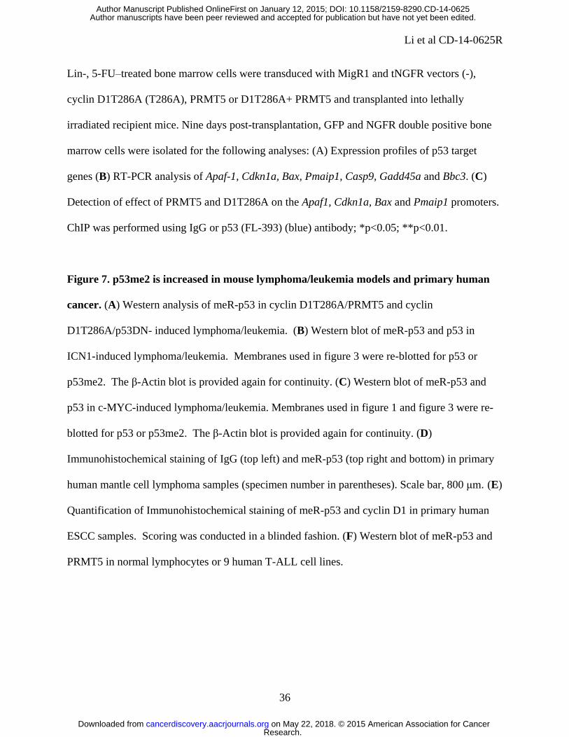
Figure 4: Cyclin D1T286A triggers p53-dependent apoptosis of BM-derived lymphocytes.
5-FU–treated bone marrow cells (p53+/+ or p53-/- donor mice) were transduced with either
MigR1 empty vector (-), MigR1-cyclin D1wt (D1) or MigR1-cyclin D1T286A (T286A)
retroviruses. (A) GFP+ cells were sorted and transplanted into irradiated (900 rads) recipient
mice. (B) GFP+:GFP- (1:1) were transplanted into recipient mice and bone marrow cells were
Research. on May 22, 2018. © 2015 American Association for Cancercancerdiscovery.aacrjournals.org Downloaded from
Author manuscripts have been peer reviewed and accepted for publication but have not yet been edited. Author Manuscript Published OnlineFirst on January 12, 2015; DOI: 10.1158/2159-8290.CD-14-0625

Li et al CD-14-0625R
35
isolated 1 month after transplantation and GFP+ cells were quantified by FACS. (C) Cells were
transplanted into recipient mice and bone marrow cells were isolated 9 days after transplantation.
GFP+ cells were gated and the apoptotic cells identified by FACS for Annexin V positive cells.
Top: Representative FACS profile. Bottom: Quantification of 3-independent experiments
(**p<0.01.). (D) 5-FU–treated bone marrow cells (from p53+/+ or p53-/- donor mice) transduced
with indicated retroviruses, were sorted for GFP, and transplanted into lethally irradiated
recipients. Survival is illustrated by a Kaplan-Meier analysis. (E) 5-FU–treated bone marrow
cells transduced with indicated retroviruses, were sorted for GFP-NGFR double positive
populations, and transplanted into lethally irradiated recipients. Survival is illustrated by a
Kaplan-Meier analysis.
Figure 5: PRMT5/MEP50 inhibits Cyclin D1T286A-dependent apoptosis and potentiates
colony outgrowth. 5-FU–treated bone marrow was transduced as follows: MigR1, tNGFR and
mCherry empty vectors (-), MigR1-cyclin D1 (D1), MigR1-cyclin D1T286A (T286A), tNGFR-
PRMT5 (PRMT5), tNGFR- PRMT5 (PRMT5), tNGFR- p53DN (p53DN), mCherry-MEP50
(MEP50), mCherry-MEP50T5A (T5A). Cells were isolated for analysis 9 days post-
transplantation. (A and C) GFP and mCherry double positive (A) or GFP, NGFR and mCherry
triple positive (C) cells were gated and apoptotic cells were identified by a fluorescence-labeled
Annexin V. Data are mean ± SD (**p<0.01). (B and D) Double (B) or triple positive cells (D)
were purified and plated in methylcellulose cultures. Total colony numbers were scored 7 days
after each plating.
Figure 6. Analysis of differential gene expression profiling by PRMT5 and D1T286A.
Research. on May 22, 2018. © 2015 American Association for Cancercancerdiscovery.aacrjournals.org Downloaded from
Author manuscripts have been peer reviewed and accepted for publication but have not yet been edited. Author Manuscript Published OnlineFirst on January 12, 2015; DOI: 10.1158/2159-8290.CD-14-0625
Li et al CD-14-0625R
36
Lin-, 5-FU–treated bone marrow cells were transduced with MigR1 and tNGFR vectors (-),
cyclin D1T286A (T286A), PRMT5 or D1T286A+ PRMT5 and transplanted into lethally
irradiated recipient mice. Nine days post-transplantation, GFP and NGFR double positive bone
marrow cells were isolated for the following analyses: (A) Expression profiles of p53 target
genes (B) RT-PCR analysis of Apaf-1, Cdkn1a, Bax, Pmaip1, Casp9, Gadd45a and Bbc3. (C)
Detection of effect of PRMT5 and D1T286A on the Apaf1, Cdkn1a, Bax and Pmaip1 promoters.
ChIP was performed using IgG or p53 (FL-393) (blue) antibody; *p<0.05; **p<0.01.
Figure 7. p53me2 is increased in mouse lymphoma/leukemia models and primary human
cancer. (A) Western analysis of meR-p53 in cyclin D1T286A/PRMT5 and cyclin
D1T286A/p53DN- induced lymphoma/leukemia. (B) Western blot of meR-p53 and p53 in
ICN1-induced lymphoma/leukemia. Membranes used in figure 3 were re-blotted for p53 or
p53me2. The β-Actin blot is provided again for continuity. (C) Western blot of meR-p53 and
p53 in c-MYC-induced lymphoma/leukemia. Membranes used in figure 1 and figure 3 were re-
blotted for p53 or p53me2. The β-Actin blot is provided again for continuity. (D)
Immunohistochemical staining of IgG (top left) and meR-p53 (top right and bottom) in primary
human mantle cell lymphoma samples (specimen number in parentheses). Scale bar, 800 μm. (E)
Quantification of Immunohistochemical staining of meR-p53 and cyclin D1 in primary human
ESCC samples. Scoring was conducted in a blinded fashion. (F) Western blot of meR-p53 and
PRMT5 in normal lymphocytes or 9 human T-ALL cell lines.
Research. on May 22, 2018. © 2015 American Association for Cancercancerdiscovery.aacrjournals.org Downloaded from
Author manuscripts have been peer reviewed and accepted for publication but have not yet been edited. Author Manuscript Published OnlineFirst on January 12, 2015; DOI: 10.1158/2159-8290.CD-14-0625
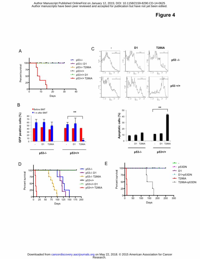
Published OnlineFirst January 12, 2015.Cancer Discovery Yan Li, Nilesh Chitnis, Hiroshi Nakagawa, et al. oncogenic driversPRMT5 is required for lymphomagenesis triggered by multiple
Updated version
10.1158/2159-8290.CD-14-0625doi:
Access the most recent version of this article at:
Material
Supplementary
http://cancerdiscovery.aacrjournals.org/content/suppl/2015/01/10/2159-8290.CD-14-0625.DC1
Access the most recent supplemental material at:
Manuscript
Authoredited. Author manuscripts have been peer reviewed and accepted for publication but have not yet been
E-mail alerts related to this article or journal.Sign up to receive free email-alerts
Subscriptions
Reprints and
To order reprints of this article or to subscribe to the journal, contact the AACR Publications
Permissions
Rightslink site. Click on "Request Permissions" which will take you to the Copyright Clearance Center's (CCC)
.http://cancerdiscovery.aacrjournals.org/content/early/2015/01/10/2159-8290.CD-14-0625To request permission to re-use all or part of this article, use this link
Research. on May 22, 2018. © 2015 American Association for Cancercancerdiscovery.aacrjournals.org Downloaded from
Author manuscripts have been peer reviewed and accepted for publication but have not yet been edited. Author Manuscript Published OnlineFirst on January 12, 2015; DOI: 10.1158/2159-8290.CD-14-0625