Print 001.tif (1 page) - Census of Marine Zooplankton - Home€¦ · 1963 Printed at the National...
92
Transcript of Print 001.tif (1 page) - Census of Marine Zooplankton - Home€¦ · 1963 Printed at the National...


A KEY FOR THE IDENTIFICATION OF THE MORE COMMON PLANKTONIC COPEPODA
OF INDIAN COASTAL WATERS
BY
L. R. ICASTURIRANGAN ( Central Marine Fisheries Research Ins t i t~~te , Mandapani Camp )
PUBLICATION NO. 2 Indian National Committee on Oceanic Research
Edited b y : N. K. PANTKKAR
COUNCIL OF SCIENTIFIC & INDUSTRIAL RESEARCH, NEW DELHI
1 9 6 3
Price Rs. .OO ( I~iland ), p or 3 6 ( Foreign ).

Publicatiott No. 2 q f the Indian National Cornmiitee on Oceanic Research. " TheIndian Scientific Programmes, 1962-65" published in 1962 is numbered as Pz~blicotion No. 1 o f the Indian ~l~atiottal Conunittee on Oceanic Research.
The Council of Scientific & Industrial Research New Delhi
1963
Printed at the National Printing Works, 10, Daryaganj, Delhi.

FOREWORD
India is an active participant in the International Indian Ocean Expedition. The National Programme which is co-ordinated by the Council of Scientific St Industrial Research has been developed by the Indian National Committee on Oceanic Research. The Expedition is stimulating a considerable amount of work on marine sciences in this country. I t is appropriate that the Indian National Committee is sponsoring publications of direct interest in the Expedition. T h e present volume by Shri L.R. Kasturirangan which is a field guide for the identi- fication of planktonic copepods is one of several publications which the Council of Scientific and Industrial Research propose to publish ~vithin the coming few years.
Copepods which are very small crustaceans form the most in~portanr element in the zooplankton and consist of many hundreds of species. Their correct identification is by no means easy. he more critical work On the group is essefitially a matter for the specialist hut a great deal of obser- vational ~vork can Ile done by junior workers. Publications of this type will not only enlarge :he scope of marine biological observations made a t different places but also encourage close; studies of planktonic groups. The 1iteratul.c. on this group is very scattered and attempts have, therefore, Ixen made to bring the essent;al taxcnomic material within the frame- \vork of this small pul,lica~ion so as to help preliminary analysis and sorting of plankton.
I commend this volume to the scientific public in the hope that it \ v i l l encourage specialists in other groups also to come forward ~v i th such field guides and tllus create greater interest ir: field biology.
S. E-IUSAIN ZAHEER Director- Getlerul
C.S.I. R.

INTRODUCTION
This key has been prepared for the use mainly of non-specialists in the group Copepoda, to aid them in the ready identification of the commoner species occurring in marine plankton. I n framing the key an attempt ha, been made to avoid excessive technicality on the one hand and too gre?t artificiality on the other. It \vill be seen that the species are treated in a linear order that is not very different from the taxonoinic order. The ~l~orphological characters used for differentiation are, as far as possible, such as could he verified by non-specia!ists and offel less of difficulty in the actual working. The characters are those of the sexually mature individuals only, since the larval and copepodite stages are excluded from the purvlelv of the key.
The "commonness" of a species has reference to the magnitude of the numerichl, temporal and geographical distribution and all three aspects have been borne in mind in deciding which are the species to be included. Yet no sharp line can be drawn separating the common from the less common species, since the latter grade into the Ibrmer. I t is hoped that the ninetynine species listed belo~v would l ~ e found to be a representative '
and useful selection. All these species have been examined, at one time or another, 11y the author in the course of his studies at Madras, Eozhikode, Cochin and Mandapam.
The form and detai!~ of the fifth pair of legs of copepods are very distinctive for each species and with differentiating characters for the malt=; and the females. While this is broadly rue of the several sub- orders, i t is especially true in the case of suborder Calanoida, \vhich comprises the bulk c,f the planktonic species. The general form of the body and of the fifth pair of legs is invariably suficient to guide one to the correct determination and consequentl:~ the figures provided nearly always include them. The student ~vould require some skill to manipulate

the copepod under the microscope so as to bring the fifth pair of legs into clear view and this could be acquired in course of time'through practice. The correctness of the species determined, should be verified, as far as possible, by reference to the full morphological description of the species to be found in a systematic ~vork. Some of the terms used in the key are explained below to avoid ambiguity and this section should be read through bpfore the key is actually employed.
Body length. The length from the anterior margin of the head region to the posterior margin of the caudal rami excluding the caudal setae.
Body regions. The three terms defined below are useful to refer to three regions of the body of the copepod visibly distinguishable from one another. They do not correspond exactly to the morphological bsdy-divisions, viz. head, thorax and abdomen. Authorities sometimes debate where to locate the boundary between one morphological region and another, or the number of segments included in one morphological region or the homology between regions called by the same name in different classes of crustacea. The use of the terms cephalosome, mctasome and urosome is quite free of morphological implications and makes for precision in referring to the different body re,' wlons.
Cephalosome. The anterior unsegmei~ted region of the body that includes not only the head but also, at the least, the segment of the maxillipeds. One or more of the segments corresponding to the anterior pairs of swimming legs may also be merged in the cephalosome. The appendages of the head-region are the first pair of antennae, thc second pair of antennae, the mandibles, the first maxillae and the second maxillae. In some of the older works the second maxillae are referred to as the first pair of maxillipeds and the true maxillipeds are referred to as the seco~id pair of maxillipeds. This practice is out of date.
Metasome. The segmented region of the body immediately posterior to the cephalosome and anterior to the urosome. I t is separated from the latter by a distinct articulation which admits of free movements. The metasome includes a variable number of segments, never more than five, corresponding to the swimming legs. The reduction in the number of the obvious segments is due, either to the merging of the anterior segments in the cephalosome, or the posterior segments in the ~lrosome, or to the coalescence of some of the segments with one another. The metasome includes the greater number, but not all, of the segments of the body corresponding to the thorax.
Urosome. The posteriormost region of the body of the copepod, usually narrower than the rest of the body and marked off from the metasome by a distinct articulation in virtue of which the urosome can be

freely moved about like a tail. The urclsome includes, at the least, the genital segment (i.e. the last thoracic segment) and the post-genital se,ments numbering 1 to 5 (i.c. the abdominal segments). The last or anal segment bears the caudal rami. One or more of the pre-senital segments corresponding to the 5th and 4th pairs of legs may also be illcorporated in the urosome through the for\\lard shifting of the point of articuiation.
Caudal rami. A pair of laminar structures a t the' posterior end of the anal segment, movably articulated with the latter and each provided typically with six setae. The older name, caudal furca, is now-a-days discarded.
The eye. Paired compound eyes are absent in the copepoda except in the Arguloida. The eye when present is the median eye or nauplius eye, composed of three ocelli close to one another, two of them being dorso-lateral in position and one, median-ventral. The well- - developed eyes of Poutella or Cog1caezrs are instances of high elaboration of the simple ocelli.
Geniculation. Modification of the first anterinae for prehension or grasping, through the formation of an elbo~v or hinge.
The preparation of this key was originally suqgested by Dr N.K. Panik- kar, at present Director, Indian Programme of Iniernational Indian Ocean Expedition, C.S.I.K., and the author is grateful to him for continued interest and encouragement. The author is keenly appreciative of the help received fkom many colleagues and the benefit derived from discussing with them problems relating to work on the copepoda and he wishes to acknowledge in particular his gratitude to Dr S. Jones and Dr R.R. Prasad. Grateful thanks are due to Prof. P.K. Menon of Presidency College, Madras, for . affording library facilitiei. Among the ~nany publications that have been found useful or indispensable, the chief works consulted ~411 be found listed at the end. The itebt to the papers of Lt. Col. R.B. Seymour-Sewell is one to be acknov-ledged not only by the present author but by every student of the copepoda of the Indian Ocean. I t was the intention in the beginning to prepare a fresh set of sketches for every species to be included in this key as G.O. Sdrs did for the Crustacea of Norway. But this had to b~ abandoned and the majority of sketches ha-?e been adapted from published ~rorks as acknowledged in the legends to the figures.

SYSTEMATIC LIST OF SPECIES
ORDER COPEPODA
Suborder CALANOIDA
Family Calanidae Calanus tenuicornis Dana A"annoca1anus minor (Claus) Canthocalanus Pauper (Giesbrecht) Undinula vulgaris (Dana)
variety typica, v. giesbrechti, v. z ~ l a n i c a . Sewell Undinula darwini (Lubbock)
variety synmetrica, v. intermedia, v. ypica. Sewell
Family Eucalanidae Rhincalanus cornutus Dana Rhincalanus nasutus Giesbrecht Eucalanus elongatus (Dana) Eucalanus attenuatus (Dana) Eucalanus pseudattenuatus Sewell Eucalanus monachus Giesbrecht Eucalanus crasszu Giesbrzcht Eucalanus subcrarsus Giesbrecht
Family Pseudocalanidae Calocalanus pavo Dana
Family Paracalanidae Paracalanus parvus (Claus) Paracalanus aculeatus Giesbrecht Acrocalanus gibber Giesbrecht Acrocalanus gracilis Giesbrecht
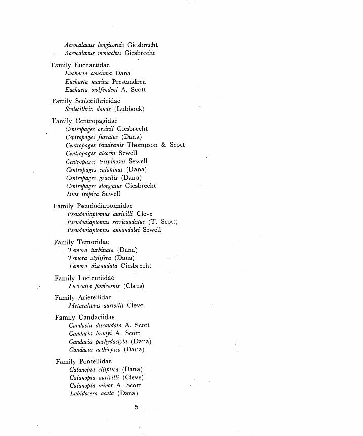
Acrocalai~us longicorilis Giesbrecht Acrocalanus monachus Giesbrecht
Fzmily Euchaetidae Euchaeta concinnn Dana Euchaeta marina Prestandrea Euchaeta woLfe12deni A. Scott
Family Scolecithricidae Scolecithrix danae (Lubbock)
I:amily Centropagidae Centropages orsinii Giesbrecht Centropages furcatus (Dana) Cetztropages tenuiremis Thompson & Scott Ce7ztfopages alcocki Sewell Centropages 1?ispitzosu: Sewell Centropages calaninus (Dana) Centrapages gracilis (Dana) Centropages elongatus Giesbrecht Isias tropica Sewell
Family Pseudodiaptomidae Pseudodiaptomus aurivilli Cleve Pseudodiaptomus serricaudatus (T. Scott) Psertdodiaptomus nnnatzdalei Se~vell
Family Temoridae Temora turbinata (Dana) Temora styLCera (Dana) Temofn discaudnta Giesbrecht
Family Lucicutiidae Lucicutia Javiconlis (Claus)
Family Alietellidae A4etacalanrts aurivillt ~ i e v e
Family Candaciidae Candacia discaudata A. Scott Candacia bradyi A. Scott Candacia pachydacpla (Dana) Candacza aethiopica (Dana)
Family Pontellidae Calanopia elliptica (Dana) Calanopia auriuilli (Cleve) Calanopia minor A. Scott Labidocera u u t n (Dana)
5
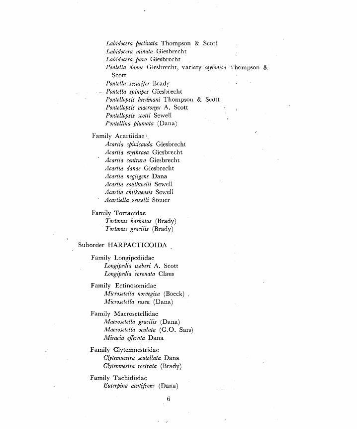
Labiducela pectitiala Thompson & Scott Labidocela mitzuta Gieshrecht Lahidoceta pauo Giesbrecht Ponlella danae Giesbl echt, variety ceylon7:ca Thompson P;
Scott Pontella seczir$er Brad :, Po~atella spit~zpes Gieshrecht Pontellopsis herdnlntzi Thompson Rr Scott Poizlellopsis macroiyx A. Scott Potltellopsis siotti Selvell Po)ztelliila plziitzatn (Dana)
Family Acartiidac . Acarlia spilzicauda Giesbrecht Acartia egithraea Giesbrecht Acartia centrum Giesbrecht Acartia daitae Gies1)recllt Acartia negligetzs Dana Acartia southiwelli Selvell dcartia chilkaetzsis Sewell Acartiella sewelli Steuer
Family Tortanidae Tortanus bgrbatus (Brady) Tortalzzls gracilis (Brady)
Suborder HAKPACTICOIDA
Family Loligipediidac Loxgipedia zeleberi A. Scott Longipedia coiamztn Claus
Family Ectinosolnidae Mi(-rosefella noruegica (Boeck) , il.licrosetella rosea (Dana)
Family Macrosetellidae Macrosetella gracilis (Dana) Macrosetella oculata (G.O. Sars) Miracia e$eel.cota Dana
Family Clytemnestridae Clytemnestrd scutellata Dana Cbtemnestra ro~trata (Brady)
Family Tachidiidae Euf~rpi t la actlt;fron.r (Dana)

Family AiIetidae M e f i s ~~oz~sseaunzei (Richard)
Suborder CY CLOPOIDA
Fami!y Oithonidae Oithona spinirosfris Claus (= 0. atlajztica Farran) Oithotza plumifera Baird Oitllona similis Claus (= 0. helgoiaildica Claus) Oithona rigida Giesbrecht Oithona brevicornis Giesbrecht , Oithotza lineal is Giesbrecht
Family Oncaeidae Oncaea uenusta Philippi Oncaea conifera Giesbrecht
Family Corycaeidae Corycaeus speciosus Dana Corycaeus danae Giesbrecht ( = C. crassiusculus Dana) Corrycaeus catus F. Dahl Corycella gibbula Giesbrecht Copilia uitrea (Haeckel) Copilia mirabilis Dana Copilia quadrata Dana
Family Sapphirinidae Sapfihiritza ouatolanceolata Dana Sapphiri~za aurolzitens Claus Sapphirina nigromaculata Dana
Family Bomolochidae Bomolochus species

KEY
I Paired compound eyes never present.. . . . . . . . . . . . . . . . . . . . . . . . . . . 2 Paired compound eyes present ; 5th and 6th legs absent ; the corres-
ponding segments merged in the abdomen which is leaf-like, unsegmented and notched or bilobed posteriorly
-ARGULOI DA
2 Second antennae and mouth parts present ; developmental stages usually free-swimming ; adults free-swimming or ectoparasitic
. . . . . . . . . . . . . . . . . . . . . . . . . . . . . . . . . . . . . . . . . . . . . . . . . . . . . . only 3
Second antennae and mouth parts absent in the adult which is fi-ee- swimming ; developmenta1 stages parasitic
-I\.IONSTRILLOIDA
3 Body not depressed and habits not ectoparasitic except in very raie instances ; nletasome ends behind the segment of the 4th or 5th pair of legs ; sternal fork not present . . . . . . . . . . . . . . . . . . . . . . . . . . 4
Body depressed and habits cctoparasitic ; when the articulation between metasome and urosome is ~1~11-marked at all, it is just behind the segment whicll forms a waist at the anterior margin of the urosolne ; sternal fork present between the bases of the nlaxillipeds. . . . . . . . . . . . . . . . . . . . . . . . . . . . . . . . . . . . CALIGOIDA
4 Urosome includes not only the geilital and abdominal segments but one further segment bearing the 5th pair of legs ; first antennae of the male, if geniculate, geniculate on both sides. . . . . . . . . . . . . . . .5
Urosome includes the genital and abdominal segments only ; first antennae of the male, if geniculate, geniculate on one side only, commonly the right side.. . . . . . . . . . . . . . . . . . . . .CALANOIDA 6
Note : Members of the first three suborders 11a:.c not beex] included in this Key, being mostly less common cpecics.

5 Body usually cylindrical, the metasome passing into the urosomc without abrupt change in width; 1)asal segment of the fifth Iegs usually showing an inner expansion ; males distinguished from the females in all cases by the geniculation of the first antennae ; er.;sacs
. . . usually unpaired, carried underneath. HARPACTICOIDA 1 19 -.
Body usually depressed with the metasome much wider than the urosome; hasal segment of the fifth legs without an inner expan- sion; geniculation of the fir.qt antennae of the male is usual but not i~~variable: eggsacs paired, carried laterally or subdorsally . . . . . . . . . . . . . . . . . . . . . . . . . . . . . . . . . . . . . . . . . CYCLOPOIDA 129
/
G Swimming legs 1 to 4 with 3-segmented excpodites and endopodites ... 7
Swimming legs 2, 3 and 4 with 3-segmented exopodites and endopo- dites, but swimming leg 1 with less than 3 segments in the endo- podites or in both rami . . . . . . . . . . . . . . . . . . . . . . . . . . . . . . . . . . . .17
Swimming legs 3 and 4 with 3-segmented exopodites and endopodites 11ut swimming legs 1 and 2 with less than 3 segments in some of the rami . . . . . . . . . . . . . . . . . . . . . . . . . . . . . . . . . . . . . . . . . . . . . . . . . . . . 39
Swimmlng legs 1 to 4 ~vi th 3-segmented exopodites but most or all of the endopodites are 2-segmented through reduction or coalescellce
............................................ ofthesegments 45
S\vimming legs 1 to 4 with 2-segmented exopodites and endopodites except for the exopodite of the 1st leg which is 3-segmented ; the ?-segmented condition of the exopodites is due to the partial or complete disappearance of the articuIation between the basal and the middle segments. . . . . . . . . . . . . . . . . . . . . . . . . . . . . . . . . . . . . .66
7 Fifth legs with 3-segmenteti rami in 1,oth seses and substantially . . . . . . . . . . . . . . . . . . . . . . . . . . . similar to the s~vimming legs.. .8
Fifth legs substantially similar to the s\vimining legs in the females only, i.e. 3-segme~?.ted and fuliy setose; the male fifth legs are always
. . . . . . . . . . . . . . . considerably different from the srvimming legs.. 13
Fil'ih legs of both sexes markedly different from the swimming legs. .58
8 Basipod of 1st legs without a hook and seta arrangement on the anterior face ; inner margin of basipod 1 of 5th legs in both sexes denticulate or srlrate in most species (though not in C. tenuicornis) 9
Rasipod of 1st legs with a hook and seta arrangement on the anterior face ; inner margin of basipod 1 of 5th legs smooth in hoth sexes.
:CANTHOCALANUS 12 . . . . . . . . . . . . . . . . . . . . . . . . . . . . . . . . . . . .
9 Females only: Urosome 4-segmented ; 5th legs symmetrical, fully setose . . . . . . . . . . . . . . . . . . . . . . . . . . . . . . . . . . . . . . . . . . . . . . . . . . . . . . I 0

Males only: Urosome 5-segmented ; 5th legs unlike on the two sides, the left leg being usually longer through the greater elongation of the two proximal exopodite segments, the terminal segment being rather short . . . . . . . . . . . . . . . . . . . . . . . . . . . . . . . . . . . . . . . . . . , , . , , . 1 1
10 First antennae extend beyond caudal rami by about half body length ; length 1 .9 to 2.5 mni.:
Q Cala~zus tenziicor~zis Dana (Fig. la, b)
First ant'ennae reach caudal rami; length 1 .9 to 2.0 mm. : C ,Ira~ztzocnlanus mitzo~ (Claus) (Fig. 2a, b)
11 Fifth legs as figured, with eropodites devoid of plumose setae on botl. the right and the left leg ; left leg not longer than the right leg ; length 1 .9 mm. :
$ Calarzus tenuicornis Dana (Fig. Ic, d)
Fifth legs as figwed, with a few plumose setae present on the right exopodite ; left leg distinctly longer than the right leg ; external
Fig. 1. Calanus Y~uiconzzs Dana : (a) Female, doi.aal viear. ( h ) Female, 5th leg of one side. (c) Male, dorsal view. (d) Male, 5th pair of legs, anterior face. Setae absent on the exopodites; setae praent on the endo- podites hut not shoivn irr the figllre. (After Giesbrechi).

Fig. 3. Canthocaia~iur pauper (Giesbrecht). (a) Female, dorsal vie\,. (b) Female, 1st leg, anterior face. (c) Female, 1st leg, larel-21 view. (d) hlale, lateral vie\+.. (e ) Male, 5th pail of legs, posterior face. ( f ) Male, left 5th leg, in flexed position. (a. after ColePx: b. to f. after Wolknden).
11

marginal spines greatly enlarged on the left exopodite ; length 1.70 mm.:
8 Nant~ocalat~us minor (Claus) (Fig. 2c, d)
12 Urosome 4-Segmented ; cxopodites of 5th legs with plumose setae ; length 1 . 7 mm :
Q Canthocaranus Pauper (Giesbrecht) (Fig. 3a, b, r) Urosome 5-segmented ; exopodites of 5th legs without pluniose setae ;
the left exopod often flexed outwards to assume a hammer-like form ; left endopod with 2 terminal setae only; length 1 . 4 mm. :
$ C'atzthocalataus pauper (Giesbrecht) (Fiq. 3d, e, f )
13 Fifth legs of the female ~vithout an inwardly pointed spine on the exopods ; the left fifth leg of the male is very greatly lengthened and modified to form a grasping organ ; 1st antenna not geniculate on either side in the male.. ................. .UNDTNULA 14
Fifth legs of the female with a prominent inwardly pointed spine on the middle eyopod segment ; in the male, the left exopod is 2-segmented ; the right exopod is 3-segmented and forms a strong chela ; 1st antenna geniculate on the right side in the male .................................. CENTROPAGES (part) 48
14 Outer margin of second exopod segment of second legs deeply notched in both sexes; usually over 2 mm. in length. .................... . I5
Outer margin of second exopod segment of second legs without notch ; length below 2 mm.. . . . . . . . . . . . . . . . . . . . . . . . . . . . . . . . . . . . . . . . .I6
Fig. 4. Unditzula uulgavis (Dana). Female : (a) Dorszl view. (b) Lateral view from the left, variety Mica. (c) Lateral view from the left v a r i e ~ giesbrechti. (d) 2nd leg, showing deep notch on 2nd exopod segment. (8)
Right 5th leg, anterior face. (a. original b.c.d. after Sewell; e, after Colefax.

15 Posterior margin of metasome drawn out into spines ; urosome 4- segmented ; 5th legs similar tc the 4th legs ; length 1 .8 to 2.6 mm. :
9 Undinula vulgaris (Dana) (Fig. 4)
Posterior margin of metasome rounded ; Urosome 5-segmtLlted ; 5th legs highly modified as figured, folded like a Z when not extended; metasome more slender than in the female ; length 2.0 to 2 . 3 mm. :
$ Undi?zula vulgaris (Dana) (Fig. 5a,b)
Note : The female U. n~l l sa~is is distinguished into three varieties as follows by Sewell (1929) :
Variety typica: metasome ending in a single down\vard bent spine on both right and left sides (Fig. 4b)
Variety giesbrechti: metasome ending in a single downurard bent spine on the right side but a doublc spine present on the left side, of which the upper spine points straight backwards and the lower one is downwardly curved (Fig. 4c)
Variety zeylanica: metasome drawn out on the right hand side into a single spine which is thickened and points straight backwards, but a double spine on the left, similar to thc one in v. giesbrechti.
Fig. 5. Undinula vulgar2 (Dana) 8 (a) Dorsal view. ( b ) Left 5th leg. Undinula darwini (Lubbock) 8 (c) Left 5th leg. (b. & c. after Wolfenden, a. original).
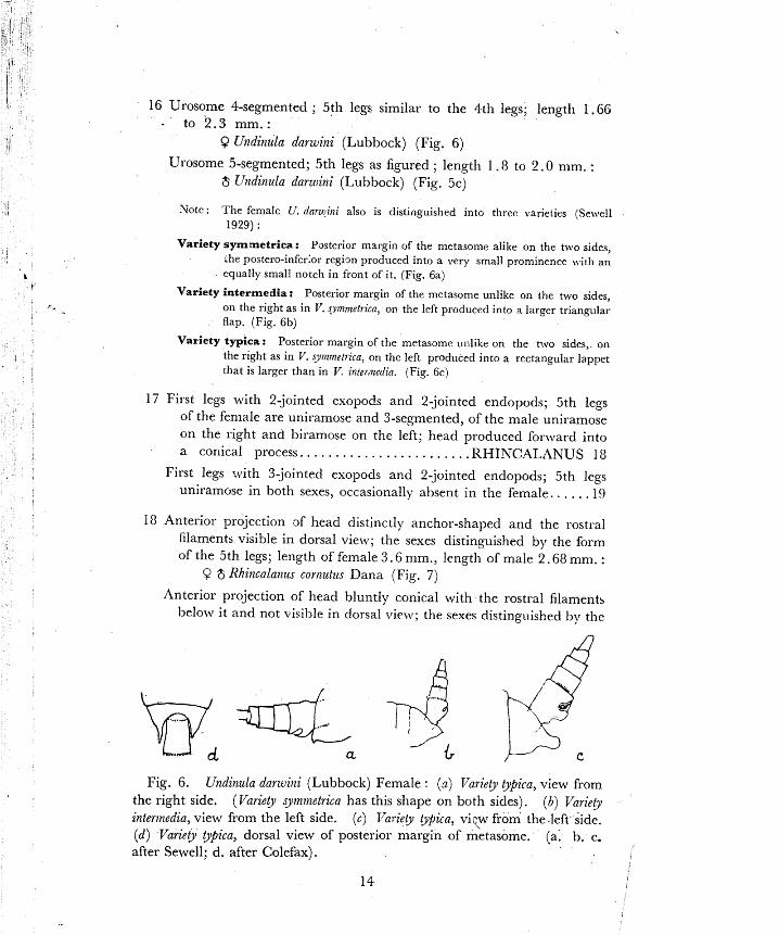
16 Urosome 4-segmented ; 5th legs similar to the 4th legs; length 1 .GG . to 2 . 3 mm. :
9 Undinula darwini (Lubbock) (Fig. 6)
Urosome 5-segmented; 5th legs as figured ; length 1 .8 to 2.0 mm. : $ 1Jndinula darujini (Lubbock) (Fig. 5c)
Note: The female U. dnnrtini also is distinguished into three varieties (Se\vell 1929) :
Variety symmetrica : Posterior margin of the metasome alike on the two sides, .he postero-inferlor regign produced into a very small prominence with an equally small notch in front of it. [Fig. 6a)
Variety intermedia: Posterior margin of the metasome unlike on the two sides, on the right as in IT. <vtnrnetricn, 01, the left produced into a larger triangular flap. (Fig. 6b)
Variety typica : Posterior margin of the nletasome unlike o n the two sides, on the r ~ g h t as in V. g~mmet~zca, on the left produced into a rectangular lappet that is larger than in V. inte~neil in. (Fig. 6 c )
17 First legs with 2-jointed exopods and 2-jointed endopods; 5th legs of the female are uniramose and 3-segmented, of the male uniramose on the right and biramose on the left; head produced forward into a coriical process.. . . . . . . . . . . . . . . . . . . . . . ,liHINCALANUS 18
First legs :vith 3-.jointed exopods and 2-jointed endopods; 5th legs uniramose in both sexes, occasionally absent in the female. . . . . . l 9
18 Anterior projection .af head distinctly anchor-shaped and the rostral filament5 visible in dorsal view; the sexes distinguished by the form of the 5th legs; length of female 3 .6 mm., length of male 2.68 mm. :
9 is Khi~zcalai~zts cornzlt~is Dana (Fig. 7)
Anterior projection of head bluntly conical with the rostral filaments hrlo\v it and not visible in dorsal \ . i e~v; the sexes distinguished l ~ y the
Fig. 6. U?zdinula darwitli (Lubbock) Female : (a) l'ariety Mica, view from the right side. (Variety sy~nvzetrica has this shape on both sides). (b) 17ariety
interirredia, view from the left side. (c) Variety @pica, vij:~i from the.left side. (d) Variety Qpica, dorsal view of posterior margin of metasome. (a. b. c.
after Sewell; d , after Colefax).
14

form of the 5th legs; length of female 3.9 to 5.1 mm., length of male 3.5 to 3 .8 mm.:
$ ;S Rhincala~zus nasutus Giesbrecht (Fig. 8 )
....... 19 Outer margin of 2nd, 3rd and 4th exopodites not toothed.. 20 . . . . . . . . . Outer margin of 2nd, 3rd and 4th exopodites toothed.. .32
20 Head triangular; caudal rami fused to the anal segment particularly in the females; -body more than 4 times as long as the greatest
. . . . . . . . . . . . . . . . . . . . . . . . . . . . . . . . . . . breadth. EUCALANUS 2 1
Head not triangular; caudal rami not fused to the anal segment; body usually much less than 4 times as long as the greatest breadth . . . . . . . . . . . . . . . . . . . . . . . . . . . . . . . . . . . . . . . . CALOCALANUS 3 1
21 Females only : urosome 3-or 4-segmented ; caudal rami fused to the . . . . . . . . . . . . . . . . . . . . . . . . . . . . anal segment; 5th legs absent. .22
Males only : urosome 5-segmented; caudal rami indistinctly separated . . . . . . . from the anal segment, 5th legs present, asymmetrical.. .27
I Fig. 7. Rhinacala~zus cornutffi Dana. (a ) Female, dorsal view. (b) Female,
5th leg of one side. (cj Male, 5th pair of legs, anterior face. (after Wilson).

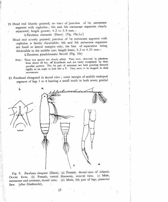
22 Urosome 4-segmented; an enlarged caudal seta present on the right side, posterior margin of metasome rounded in the Indo-Pacific forms but two blunt teeth present in the Atlantic form ; length 4 .4 to 8.25 mm.:
Q Ez~calanus clongatz~s {Dana) (Fig, 9 i,b)
Urosome 3-segmented; an enlarged caudal seta present on the left . . . . . . . . . . . . . . side; posterior margin of metasome rounded.. 23
Note : E. elongatzis is remarkable for its large size and 4-segmented urosome in the female. It was observcd on several occasions at Kozhikode in plankton from 18 km. off the coast but always in few numbe~s.
23 Genital segment longer than broad; head indented on either side . . . . . . . . . . . . . . . . . . . . . . . . . . . . . . . . . . . on the frontal margin. 24
Genital segment broader than long; head not indented on either side . . . . . . . . . . . . . . . . . . . . . . . . . . . . . . . . . . . . . . . . . . . . . . . . . . . . 25
Fig. 8. Rhincalalztls rzasutus Giesbrecllt. (a ) Female, dorsal view. ( b ) Female, head-end, ventral view. (c) Female, urosome, dorsal view. (d) Female, 5th pair of legs. (e) Male, 5th pair of legs, posterior face. (after Sars).
21 Head end l>luntly pointed; 110 trace of junction of 1st lnetasome segment with csphalon ; 4tn and 5th metasoqe segments clearly separated; length greater, 4 . 2 to 5.9 mm. :
Q Ezccaln~rzrs altenzcatus (Dana) (Fig. I Oa,l,,c)
Head end acutely pointed; junction of 1st metasome segment with cephalon is faintly discernible; 4th and 5th metasome segments are fused at lateral margins only, the line of separation being discernible in the middle i~ne ; length lesser, 3 .3 to 1 . 2 5 inm. :
Q E~rcalanzts pseudat tcnt~ntrrs Sewell (Fig. 10e)
These two species are closely allied. They were observed in plankton from about 18 km. off Kozhikode and are easily recognized by their
peculiar posture. The 1st pair of antennae are held pointing forward rigidly at an angle to look like a Y. They seem to be sluggish in their movements.
25 Forehead elongated in dorsal vie~v ; outer margin of middle endopod segment of legs 1 to 4 bearing a small tooth in both sexes; genital
Fig. 9. Eucalat~us eloiz~qnius (Dana). (a) Female. dorsal view of Atlantic Ocean form. (0) Female, rostra1 filaments, vent,-a1 view. (c) Male, metasome and urosome, dorsal view. ( d ) Male, 5th pair of legs; posterior face, (after Giesbrecht) .
17
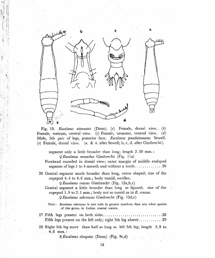
Fig. 10. Eucalanus attenuatus [Dana). (a ) Female, dorsal view. (b) Female, rostrum, ventral view, (c) Femal?, urosome, ventral view. (d) Male, 5th pair of legs, posterior face. Eucalarus p~eudattenuatus Sewell. (e) Female, dorsal view. (a. & e. after Sewell; 11, c , d. after Giesbrecht).
segment only a little broader than long; length 2.50 mm. : Q Eucalanus monachzts Giesbrecht (Fig. 1 1 a)
Forehead rounded in dorsal view; outer margin of middle eridopod segment of legs 1 to 4 smooth and without a tooth. . . . . . . . . . . . . .26
26 Genital segment much broader than long, onion shaped; size of the copepod 4.4 to 4.6 mm.; body tumid, swollen.
Q Eucalanw crassus Giesbrecht (Fig. 12a,b,c) Genital segment a little broader than long as figured; size of the .
copepod 1.9 to 2.1 mm.; body not so tumid as in E, crassus. Q Eucalanus subcrasszls Giesbrecht (Fig. 1 2d,e)
Note: Eucalanus subcrassus is met with in greater numbers than any other species of this genus in Indiart coastal waters.
27 Fifth legs preserit on both sides.. . . . . . . . . . : . . . . . . . . . . . . . . . . . .28 . . . . . . . . . . Fifth legs present on the left only; right 5th leg absent. -29
28 Right 5th leg more than half as long as left 5th leg; length 3 .9 to 4.8 mm.:
8 Eucalanus elongatus (Dana) (Fig. 9c,d)

Right 5th leg less than half as long as left 5th leg; head not as pointed as in the female E. a/fenuatus; length 3.1 to 3.25 mm. :
$Eucalanus att~nzlatus (Dana) (Pig. 10d)
Note : The male of E pseudatletlunilts is distingltishable f ~ o m the allied species chiefly by its slightly smaller sizr. I n cither species. the males could be assigned to the species of the females along with which tliey ocru~rrd in the tow net collection.
29 ,4 small tooth present on the outer margin of the mi'ddle endopod segment of nvirnming legs 2, 3 and 4; length 2 .OO mm. :
$ Ez~caln~zzis tnolzachus Giesbrecht (Fig. 1 l b,c)
Outer margin of the middle endopod segment of swimming legs 2, 3 and 4 smooth and vrithout tooth likc ~rojection . . . . . . . . . . . . . . . .30
30 Fourth metasome segment bea& tu-o tartile setae on each side as figured; length 2.6 mm. :
$ Ezicalatzus crassus Giesbrecht (Fig. I ld,e)

Fourth metasome segment ~vithout conspicuous laterally placed setae; length 1 .7 to 1.8 mm
$ Eucalanus subcrassus Giesl~recht
31 Urosome 2-segmented; caudal ram; turned outwards at right angles to the body axis, each ramus bearing four equal plumose setae: 5th legs 4-segmented, symmetrical; length 0.88 to 1 . 2 mm. :
Q Calocala~zus pavo (Dana) (Fig. 13 & i4a)
Urosome 5-segmented; caudal rami parallel to the body axis and not provided with large plumose setae; 5th legs asymmetrical, right leg, 4-segmented, left leg 5-segmented and much longer than the right; length 1.04 mm. :
$ Calocalmus pavo (Dana) (Fig. 1411 & c)
32 Terminal segment of the exopodites of legs 2, 3 and 4 is separated into a proximal and a distal portion by the outer marginal spine such that the proximal portion is at least twice as long as the distal portion; 2nd antenna of the female with the exopodite (;-segmented)
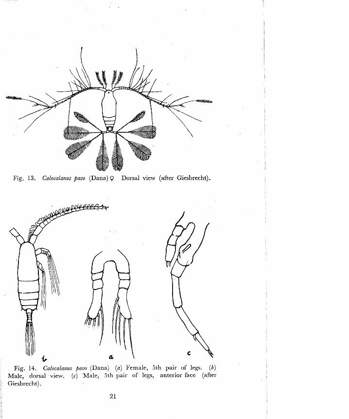
Fig. 13. Calocalatlus pavo (Dana) Q Dorsal view (after Giesbrecht).
Fig. 14. Calocalatzz~s pavo (Dana) (a) Female, 5th pair of legs. (b) Male, dorsal view. (c) R/lale, 5th pair of legs, anterior face (after Giesbrecht).
as long as the endopodite (?.-segmented) ; 5th legs present in the female . . . . . . . . . . . . . . . . . . . . . . . . . . . . . . . . . . PARACALANUS 33
Terminal segment of the exopodites of legs 2, 3 and 4 is separated into a proximal and a distal portion by the outer rnarginal spine such that the proximal portion is less than tuice a; long as the distal portion; 2nd antenna of the female with the exopodite (7- segmented) shorter than the endopodite (2-segmknted); 5th legs
I absent in the female.. . . . . . . . . . . . . . . . . . . . .ACROCALANUS 36 ----___-, 33 First antennae not generally reaching beyond the caudal rami, surface
of basipod 1 of legs 1 to 4 beset by hairs and bristles. . . . . . . . . . . . . .34
First antennae reaching beyond the caudal rami; surface of basipod 1 of legs 1 to 4 naked (except for one plumose seta) though hairs and bristles occur on the segments of the exopod and endopod. .. .35
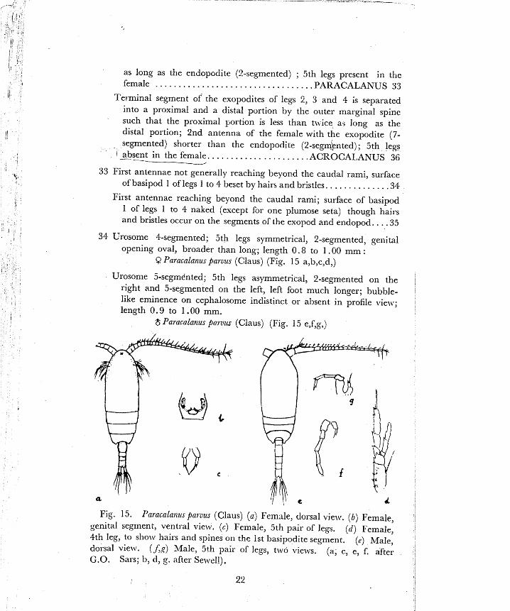
34 Urosome 4-sepmen~ed; 5th legs symmetrical, 2-segmented, genital opening oval, broader than long; length 0.8 to 1 .OO mm :
Q Paracalanus paruus (Claus) (Fig. 15 a,b,c,d,)
Urosonle 5-segmdnted; 5th legs asymmetrical, 2-segmented on the right and 5-segmented on the left, left foot much longer; bubble- like eminence on cephalosome indistinct or absent in profile vie\\.; length 0.9 to 1 .OO mm.
$ Paracalarzus parurn (Claus) (Fig. 15 e,f,g,)
Fig. 15. I'aracalanuspatuus (Claus) (a ) Fem~le , dorsal view. (6) Female, genital segment, ventral view. (c) Female, 5th pair of legs. (d) Female, 4th leg, to show hairs and spines on the 1st basipodite segment. (e) Male, dorsal view. (Jg) Male, 5th pair of legs, t~vo vie~vs. (a, r, e, f'. after G.O. Sars; b, d, g. after Sewell).

Fig. 16. Paracalanu~ aculeatu-s Giesbrecht. ( a ) Female, 4th leg, to show absence of hairs and spines on the 1st basipodite segment. (6) Female, genital segment, ventral view. (c) Female, 5th leg. (d) Male, lateral view. (e) Male, 5th pair of legs. (a, b, c. after Sewell; d, e. after Colefax).
Fig. 17. dc)acniaasrp (a) 11. gibber Gitsbrecht, lateral view. (b) il.
gibber Giesbrecht, 4th leg. (c) A. gracilis Giesb~echt, !atera1 view. ( d ) A. longicorrris Giesbrecht. lateral view. (e) A. lo~~giconzis Giesbrecht, 4th leg ( f ) A . monechu-s Gieibrecht, lateral view. (a, c, d, f, after Wolfcn-
den; b, e, after Se~zell). 2 3
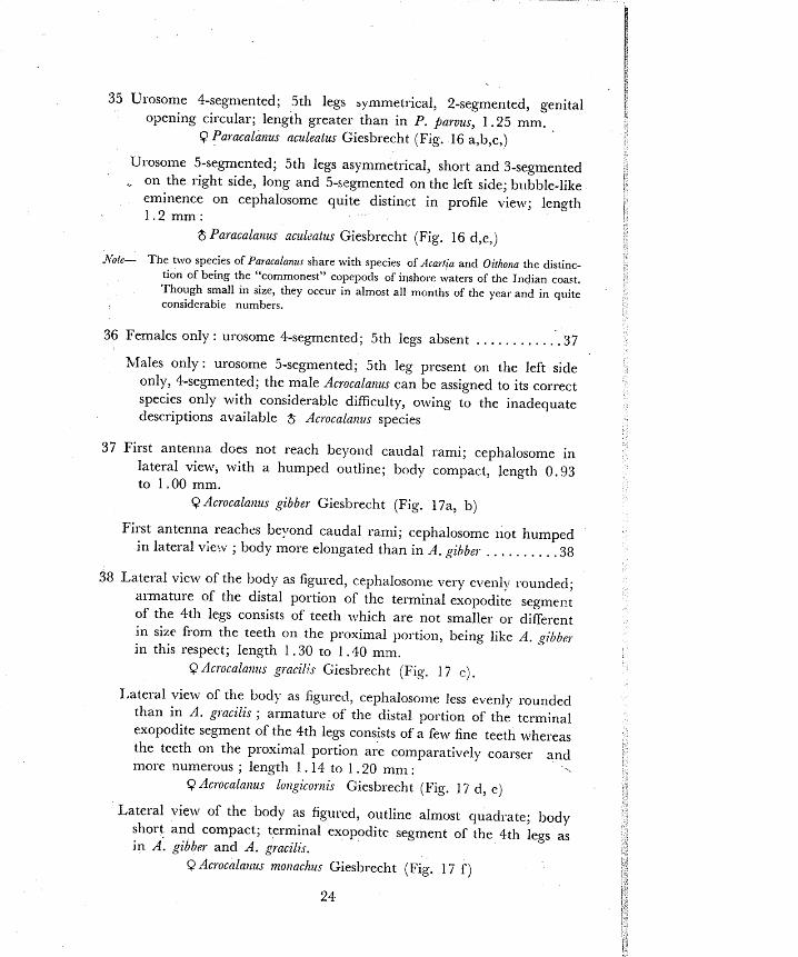
35 Usosome 4-segmented; 5th legs symmetrical, 2-segmented, genital opening circular; length greater than in P. parvus, 1.25 mm.
Q Paracalalzus aculeatus Giesbrecht (Fig. 16 a,b,c,)
Urosome 5-segmented; 5th legs asymmetrical, short and 3-segmented . on the right side, long and 5-si-grnented on the left side; bl~bble-like
eminence on cephalosome quite distinct in profile view; length 1 . 2 m m :
$ Paracalatzus aculzntus Gieshrecht (Fig. 16 d,e,)
A'oie- The two speries of Paracalanus share with species of Acartza and Oithona the distinc- tion of being the "commonest" cupepods of ir~shore waters of the Indian coact. Though small In size, they occur in almost all months of the yeat and in quite considerabie numbers.
36 Females only : urosome $-segmented; 5th legs absent . . . . . . . . . . . . 3 7
Males only : urosome 5-segmented; 5th leg present on the left side only, $-segmented; the male Acrocalalzzis can be assigned to its correct species only with considerable difficulty, owing to the inadequate descriptions available $ Ac~ocalatzvs species
37 First antenna does not reach beyoild caudal rami; cephalosome in lateral view, with a humped outline; body compact, length 0.93 to 1.00 mm.
9 Acrocalatzus gibber Giesbrecht (Fig. 17a, b)
First antenna reaches beyond caudal rami; cephalosome not humped in lateral v;e:v ; body more elongated than in A. gihbel- . . . . . . . . . .38
38 Lateral view of the body as figured, cephalosome very cvenly sounded; armature of the distal portion of the terminal exopodite segment of the 4th legs consists of teeth which are not smaller or different in size from the teeth on the proximal portion, being like A. gibber in this respect; length 1.30 to 1.40 mm.
Q Acl-ocala~zus gracilis Giesbrecht (Fig. 1 7 c) . Lateral vie\% of the bod>- as figured, cephalosome less evenly rounded
than in A . gl-acilis ; armature of the distal portion of the terminal exopodite segment of the 4th legs consists of a few fine teeth whereas the teeth on the proximal portion are comparatively coarser and more numerous ; length 1 -14 to 1 .20 mm :
Q Acroca1a)tus lo~zgicorrzis Giesbrecht (Fig. 1 7 d, e)
Lateral view of the body as figured, outline almost quadrate; body short and compact; terminal exopodite segment of the 4th legs as in A. gibb~l- and A . gracilis.
9 Acrocalnt~us monachur Giesbrecht (Fig. 17 f )

Fig. 18. Euchaeta conci?~na Dana. ( a ) Female, dorsal view. (6) Female, urosome, dorsai view. (c) Female, genital segment, view from the right. (d) Female, genital segment, ventral view. (e) Female, genital segment, view from the left ( f ) Female, 2nd leg, exopodite. (g) Male, right 5th leg. ( h ) Male, left 5th leg. ( j ) Male, left 5th leg, terminal part, enlarged view. ( k ) Male, left 5th leg, terminal part another view, more 8
enlarged. (a, g, h, j, after Colefax: b, c, d, e, f. after Wolfenden; k. after A. Scott.)
Fig. 1s. Euchaeta marina Prestandrea Q (a ) Genital segment, view from the left. . (b) Genital segment, view from ventral aspect. (c) Genital segment, vie\\ from the right. ( d ) 2nd leg, exopodite. (after Wolfenden).
2 5

Fig. 20. Ezichaeta nzaritza Prestandrea 6 (a) View from the right. ( 6 ) 5th pair of legs, anterior face. (c) Left 5th leg, terminal part, enlarged. (d) Left 5th leg, terminal part, another view, crlarged. (a. after Giesb- recht; b, c. after Wolfend~n; d. after A. Scott).
33 Shvimming leg, 1 and 2 with 2- and 3-segmented ~xopodites respec- tively and 1-segmented endopodites; posterior margin of metasome lounded, covered ventrally with tufts of hairs; 1st antennae moderately long with numerous stiff setae; head-end pointed, 1~1th a frontzl projection dorsal to the rostra1 projection, the two together const;:uting a charact~ristic profile view. . . . . . . . EUCHAETA 40
Swixming legs 1 and 2 with 3-segmented exopodites and 1 and 2 segmented endopodites respectively; posterior margin of metasome rounded, without tufts of hairs; 1st antennae not extending beyond the metasome. . . . . . . . . . . . . . . . . . . . . . . . . . . SCOLECITHRIX 44
40 Females only: urosome 4-segmented, anal segment very short; 5th 1egsaSsent . . . . . . . . . . . . . . . . . . . . . . . . . . . . . . . . . . . . . . . . . . . . . . . . . . . 1
Males only: urosolne 5-segmented, anal segr:lent very short; l)ody more slender than ir, the female; 5th legs large, conspicuous, biramose on the right side and uniramose on the left, the left h o t terminating in a complicated "hand", which transfers. the spermato-
. . . . . . . . . . . . . . . . . . . . . . . . . . . . . . . . . . . . . . . . . . . . . . . . . . . . phore 43

Fig. 21. Euclzaeta wolfeitdeili A. Scott ( a ) Ferrale, dorsal view. ( 6 ) Genital segment, ventral vie^^. (6) Genital segment, view from the right. (d) Genital segment, view from the left. ( e ) 2nd leg, exopodite. ( f ) Male, left 5th leg, terminal part, enlarged view. (a. to e. after Wolfenden; f, after A. Scott)

Fig. 22. Scolecithrix danae (Lubbock) ( a ) Female, dorsal view. ( 6 ) Female, urosome, lateral view to show shovel-like ventral projection on genital segment. (c) Male, dorsal view. jd) Male, 5th pair of legs, ante- rior face. (a. after Vqilson; b. after Giesbrecht, c, d. after Colefax).
Fig. 23. Centropage~, orsbii Giesbrecht ( a ) Female, lateral view. (6) Female, 5th pair of legs, posterior face. (cj Male, Iateral view. (d) hlale, 5th pair of legs, posterior face. (after Wolfenden).

41 Genital segment provided on the right side ~ ~ i t h a blunt peg-like projection which is curved to~zrards tail end; in the 2nd le the terminal exopod segment bears three external marginal spines, all three being quite short and of equal length; in the same leg, the middle exopod segment bears a single external marginal spine which is so long as to reach beyond the base of the spine above it; Length 3.75 mm.
Q Euchaeta conci~zna Dana (Fig. 18a to f ) - Genital segment without peg-like prqjection; in the 2nd leg the terminal
exopod segment bears three external marginal spines of which spine No. 2 is always longer than spines 1 and 3; in the same leg, the middle exopod segment bears a single external marginal spine which does not reach beyond the base of the spine above i t . . . .42
42 Posterior margin of metasome more produced on the right side than on the left; genital segment v,ith i r reg~~lar outline on the right side; i:l the terminal exopod segment of the 2nd leg external marginal spine No. 2 is very long and reaches as far as spine No. 3; length 2.25 to 3.9 mm.
Q Ezlchaeta nlaritla Prestandrea (Fig. 19) Posterior m ~ r g i n of metasome of the two sicles appears symmetrical
in dorsal view; genital segment \vith a small round button-like protuberance at the posterior margin on the right side; in the terminal exopod segment of the 2nd leg, external marginal spine No. 2 is less than half as long as necessary to reach spine No. 3; length 3.30 mm.
Q Euchaeta wolfetzdetzi A. Scott (Fig. 2 1 a to e)
43 Toothed process at the end of the left 5th leg is armed ~vi th iine teeth as figured; length 2.6 mm.
$ Euchneta conclrzna Dana (Fig. 18g to k) Toothed process at the end of the left 5th leg is some~:hat broader
and set with coarse teeth as figured; length 2.8 to 3 . 2 mm. $ Euchaeta marina Prestandrea (Fig. 20)
Toothed process at the end of the left 5th leg is armed lvith fine teeth and the apex is notched as figured; length 2.7 mm.
8 Erlchaeta wolfetzdeiii A. Scott (Fig. 2 I f )
44 Fifth legs lacking; genital segment with a shovel-like ventral projec- tion; length 2.2 mm.
Q Scolecithrix datzae (Lubbock) (Fig. 22a, b) Fifth legs biramose on the left with 3-jointed exopod and one jointed
endopod borne at the tip of the much elongated second basal segment; uniramose on the right as figured; length 2.0 to 2.15 mm.
$ Scolecithrix donne (Lubbock) (Fig. 22c, d)

a
Fig. 24. Ce7it~opages f i tcatus (Dana) (a) Female, dorsal view. (b) I
Female, proximal five segments of 1st antenna. (c) Female, 5th pair of 1
legs, posterior face. (d) Male, urosome and part of metasome, dorsal view. ( e ) Male, 5th pair uf legs, posterior face. (a, d. after Giesbrecht, c. after Colefax; 11, e. original). I
Fig. 25. Centrapages teauirenzis Thompson & Scott [a) Female. dorsal 1
view. (b) Female, 5th pair of legs, posterior face. (c) Male, urosome, ' I I
dorsal view. (S) Male, 5th pair of legs, posterior face. (a, c. after 1
Thompson & Scott;-b, d. Original). ;I i r

45 Endopodltes of legs 1 to 4 are 2-segmented in both sexes. ....... ,46 Endopodites of legs 2 to 4 are 2-segmented hut endopodite of leg 1 is
3-segmented. . . . . . . . . . . . . . . . . . . . . . . . . . . . . . . . . . . . . . . . . . . . . . .94
Endopodites of legs 2 to 4 are 2-segmented but endopodite of leg 1 is either 2- or 3-segmented, the latte* condition being exceptional and
. . . . . . . . . . . . . . . . peculiar to the males of some o; the species. : .117
46 The endopodites of the 5th legs are 3-segmented and fully furnished with plumose setae; the 2-segmented appearance of the endopods of legs 1 to 4 is secondary owing to the fusion of ihp proximal segment with the middle segment, partially in leg 4 and more completely in legs 3, 2 and 1 . . . . . . . . . . CENTROPAGES (part) 47
The endopodites of the 5th legs are either rudimentary or absent, the 2-segmented condition of the endopodites of legs 1 to 4 gives no
. . . . . . . . . . . . . . . . . . . . . . . . indication of secondary derivation. .74
47 Urosome 3-segmented; 5th legs as figured; length 1.7 mm. Q Centropages orsinii Giesbrecht (Fig. 23a, b)
Urosome 4-segmented; 5th legs as figured; l e n ~ t h 1.3 to 1.5 mm. $ Centropag~s orsinii Giesbrecht (Fig. 237, d)
48 Posterior corners of metasome drawn out into strong spiniform . . . . . . . . . . . . . . . . . . . . . . . . . . . . . . . . . . . . . . . . . . . . . . . projections 49
Posterior corners of metasome without strong spiniform projections. .51
49 A prominent backwardly curved spine is present in the mid-dorsal line near the posterior margin of the cephalosome; 5th legs as figured; 1-ngth of female 1.3 lo 1.4 mm., of male 1.2 to 1.3 mm.
Q $ Centropages dorsispinatus Thompson 8t Scott (Fig. 26) . . . . . . . . . . . . . . . . . . . . . . No spine is present in the mid-dorsa! line. -50
50 Posterior margin of metasome provided with two smaller, more dorsally placed spines in addition to the two large ones; a tooth present on the anterior margin of segments 1, 2 and 5 of 1st antennae; eye, red in colour, in continual movement in the living condition: 5th legs as figured; length of female 1.9 mm., of male 1 .5 to 1.7 mm.
I? $ Centrojages furcatus (Dana) (Fig. 24)
Posterior margin of metasome ~ ~ i t h o u t additional spines; no teeth present on the anterior margin of 1st antennae; eye not in move- ment; 5th !egs as figured; length of female ?. 0 mm., of male 1.8 mm.
Q 6 Centropag6s tenuinmis Thompson Pr. Scctt (Fig. 25)
51 Anterior part of body comparatively broad and compact, as in . . . . . . . . . . . . . . . . . . . . . . . . . . . . . . . . . . . . . . . . . . C. dorsispinaius. .52
Anterior part of body comparatively long and slender, as in C. j%rcatus . . . . . . . . . . . . . . . . . . . . . . . . . . . . . . . . . . . . . . . . . . . . . 53

52 Posterior margin of metasome with three small tooth-like projections visible in lateral view only ; 5th legs in female of normal type, in male as figured; length of female 1 . O O to 1 .20 mm.; of male 1 .0 mm.
Q $ Centrapages tiispintlsus Sewell (Fig. 27d, e, f )
Posterior margin of metason5e with only one small tooth, visible in lateral view only; 5th legs as figured; length of female 1.1 to 1.4 mm., df male 1 . 0 to 1 . 2 mm .
Q $ Cetztrojages a k s c k i Sew~ell (Fig. 27a, b,c)
53 Females only: urosome 3-segmented; 1st antennae zlike on the t5vo . . . . . . . . . . . . . . . . . . . . . . . . . . . . . . . . . . . . . . . . . . . . . . . . . . . . . sides 54
Males only: urosome 4-segmented; 1st antennae genicuiate on the . . . . . . . . . . . . . . . . . . . . . . . . . . . . . . . . . . . . . . . . . . . . . . . . . . . right 56
Fig. 26. Ceiltropages do?sispilaatus Thompson 8r. Scott ( a ) Female, dorsal I
-- -- view. (b) Female, 1st antenna, proximal section to show teeth present on antenna1 segme~ts 2, 5, 10 and 11. ( 6 ) Female, cephalosome, lateral I
view. (d) Female, 5th leg of one side. ( e ) Male, urosome. ( f ) Male, 5th pair of legs, posterior face. (after Thompson Sr Scott). I
32 I ~
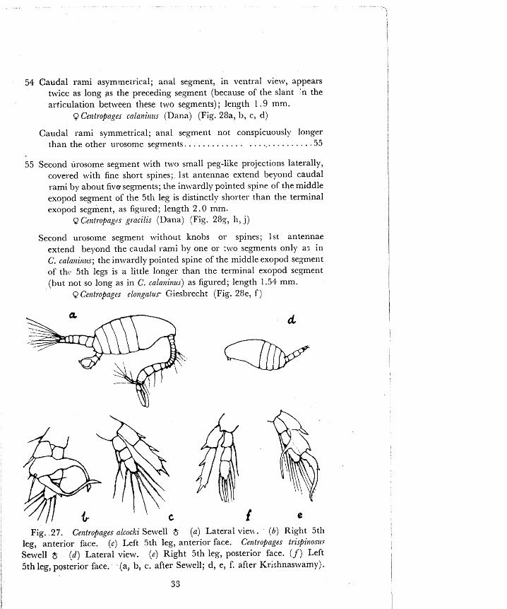
54 Caudal rami asymme~rical; anal segment, in ventral view, appears twice as long as the preceding seqment (because of the slant 'n the articulation between t!lese two segments) ; length 1 .9 mm.
Q Cerztropnges cnla?zirizts [Dana) (Fig. ZSa, 11, c, d)
Caudal rami symmetrical; anal segment not conspicuously longer than the other urosome seyments. . . . . . . . . . . . . . . . .,. . . . . . . . . .55
55 Second urosome segment with t'izlo small peg-like projections laterally, covered with fine short spines; 1st antennae extend beyond caudal rami by about five segments; the in\z,ardly pointed spine of the middle exopod segment of the 5tli leg is distinctly shorter than the terminal exopod segment, as figured; length 2 . 0 mm.
Q Centropag<>s gracilis (Dana) (Fig. 287, 11, j)
Second urosome segment ~rithout knobs or spines; 1st antennae extend beyond the caudal rami by one or t~vo segments only a; in C. cala~zit~us; the in\\rardly pointed spine of the middle exopod segment of th~- 5th legs is a little longer than the terminal exopod se, wment (but not so long as in C. calaninus) as figured; length 1.511 mm.
Q Centropages elongarus Giesl~recht (Fig. 28e, f )
Fig. 27. Cetztropagees alcacki Sew~ell $ (a) Lateral vie^). (6) Right 5th leg, anterior face. ( c ) Left 5th leg, anterior face. Centropages trispinoszt.r Sewell $ { d ) Lateral view. (e) Right 5th leg, posterior face. ( f ) Left 5th leg, posterior face. (a, b, c. after Sewell; d, e, f. after Krfshnaswamy).

Fig. 28. Cent~opages cala~linus (Dana) Q (a) Dorsal view. (6) Urosome, ventral view. (c) Urosome, lateral view. ( d ) 5th leg of one side. G n t - ropages elongatus Giesbrecht. 8 (e) Urosome, ventral view. ( f ) 5th leg of one side. Centropages gracilis (Dana) 9 (g) Dorsal view. (h) Vrosome, ventral view. ( j ) 5th leg of one side. (a, d, g, j, after Colefax; b, c, e, f, h. after Wolfenden).
Fig. 29. Centropages calaninus (Dana) $ (a) Chela of right 5th leg, anterior face, (b) Exopodite of left 5th leg; anterior face. Centropages gracilis (Dana) $ (c) Chela of right 5th leg, posterior face. (d) Exopodite of left 5th leg, posterior face. Centfopages elongatu~ Giesbrecht. (e) 5th pair of legs, posterior face. ( f ) Exopodite of left 5th leg, view from outer side. (a, b, c, d. after Giesbrecht : e, f, after Wolfenden).

56 Right 5th leg of the male ~vi th the claw-like terminal segment of the . . . . . . . . . . . . . . . . . exopod comparatively long and bent into a 1' .57
Right 5th leg of the male with the cla\v-like termiilal segment of the exopod comparatively short and not bent, as depicted in figure; body length 1.50 mm.
$ Ce?ztropages eloiigatzrs Giesbrecht (Fig. 29e, f )
57 Thumb of chela is curved and shorter than the middle exopod segment from which it springs; distal arm of the V-shaped terminal segment is straight; body length 1.8 mm.
$ Cenfropages calankzw (Dana) (Fig. 29a, b)
Thumb of chela is straight and longer than the middle exopod segment from which it springs; distal arm of the V-shaped terminal segment
I 1
is curved outwards, proximal arm broadened inwards triangularly; , body length 1.70 inm.
$ Bntropages g)acilis (Dana) (Fig. 29c, d)
Note : Thew three species of Centrapages were observed during 1949-53 at Kozhikode in plankton collected from approximately 18 km. off the coast.
. . . . . . . . . . . . . . . . . . . . . . 58 Fifth legs clearly biramose in both sexes. .59
Fifth legs unira~nose in the female and usually in the maIe too, with 1 1
only indistinr-t indications of the endopod in the male, if at ~ 1 1 . . . . . . . . . . . . . . . . . . . . . . . . . . . . . . . . . . PSEUDODIAPTOh4US 61
*:7
s >< Fig. 30. h a s tropics Sewell. (a) Female, dorsal view, (b) Female, e
genital segment, view from the left (c) Female, genital segment, vcntral >.- 'r view. (d) Female, 5th leg of one side. (e) hlale, urosome, dorsal view.
( f ) Male, 5th pair of legs, anterior face. (after Sewell). 1-a \ %
3 5 *e d-
' &$ -$

Fig. 31. Pseudodiaptonzus auriuilli Cleve. Q (a) Dorsal view. ( b ) View from the left. (c) 5th pair of legs. ( d ) 5th pair of legs, another view. (original).
Fig. 32. Pseudodiaptomus aurivilli Cleve 6 ( a ) Dorso-lateral view. (b ) 5th pair of legs, antelior face. (c) 5th pair of legs, another view of anterior face. (Original).

Fifth legs clearly uniralnose in both sexes, 2-segmented in the female and 5-segmented in the male.. . . . . . . . . . . .METACALANUS 73
59 Fifth legs of the female with 3-segmented exopodites and very shc. . 1 -segmented endopodites; of the male, \vith t\vo segmented exopodites, endopodites absent in the right foot, 1-segmented in the left foot, the right foot larger than the left.. . . . . . . . . . . . ISIAS 60
Fifth legs of the female with plumoee setae, with 3-segmented exopodites and ~lery short 3- or 2-segmented endopodites; of the male, both rami 3-segmented in the left foot and two segmented in the right foot, right exopodite prehensile, sub-chelate. 9.. LUCICUTIA 72
60 Urosome 3-segmented; 1st antenna alike on the two sides; genitaI segment with asymmetrical lateral margins; length 1.25 mm.
Q Isias tl-opica Sewell (Fig. 30a to d)
Urosome 5-segmented; 1st antenna geniculate on the right side; the third urosome segment with a projection 3n the right; length 1.25 mm.
3 Isias trojica Sewell (Fig. 30e, f )
Note : This species was observed in Cochin harbour waters in 1956-57 during monsoon months.
61 Posterior margin of metasome drawn out into prominent spine-like projections, one on each side. . . . . . . . . . . . . . . . . . . . . . . . . . . . . .62
Posterior margin of metasome not drawn out into prominent projec- tions . . . . . . . . . . . . . . . . . . . . . . . . . . . . . . . . . . . . . . . . . . . . . . . . . . . . . 63
62 Urosome 4-segmented; 1st antennae alike on the two sides; 5th legs as figured; length 1.2 mm.
Q Pseudodiaptomus auriuilli CIeve (Fig. 3 1)
Urosome SPsegmented; 1st antennae genicuIate on the right side;
5th legs highly complex, as figured; length 0.93 mm. $ Pseudodiaptomus aurivilii Cleve (Fig. 32) .
Note: I t is open to doubt whether Ps. inertoni Fruchtl is a valid species, distinct froin Ps. au~zuilli Cleve. In the specimens examined on nunlerous occasions, the 8 5th legs agreed with published figures of Ps. inerlotri, while the Q corresponded more to the descriptions of Ps, au~liiilli. The specific name which has priority is used here. This species and Ps. se~lirnudatus are verv commonly observed in iashore plankton collections.
63 Females only : urosome 4-segmented; 1st antennae alike on the two sides . . . . . . . . . . . . . . . . . . . . . . . . . . . . . . . . . . . . . . . . . . . . . . . . . . . . . . 64
Males only: urosome 5-segmented; 1st antennae geniculate on 'the . . . . . . . . . . . . . . . . . . . . . . . . . . . . . . . . . . . . . . . . . . . . . . . . right side 65
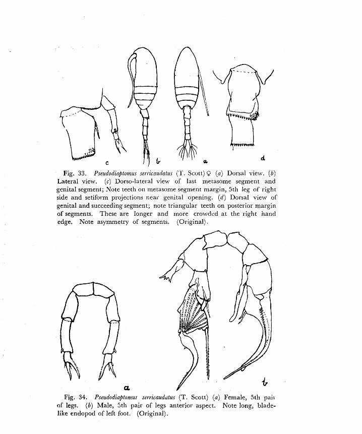
Fig. 33. Pseudodi@tomus se~ricnu(latus ('1'. Scott) 9 ( a ) Dorsal view. ( b ) Lateral view. ( c ) Dorso-lateral view of last metasome segment and genital segment; Note teeth on metasome segment margin, 5th leg of right side and setiform projections near genital opening. ( d ) Dorsal view of genital and succeeding segment; note triangular teeth on posterior margin of segments. These are longer and more crowded at the right nand edge. Note asymmetry of segments. (01.iginal).
F i g . 34. PseudodiaPtomus serricaudutus (T. Scott) ( a ) Female, 5th pail of legs. (b) Male, 5th pair of legs anterior aspect. Note long, blade- like endopod of left foot. (Originai).

64 Genital segment with a prominent spine on each side pointing out- wards; 5th legs as figured; length 1.18 mm.
$? PseudodiaPtomus annandalei Sewell (Fig. 35a, b, c) Note: Ps. annandalei was observed on a number of occasions in Cochin harbour w;::rs
and on a few occasions in Kozhikode during monsoon months.
Genital segment without laterally pointing spines, slightly asymme- trical, the posterior margin being more produced backwards on the right than on the left; all urosome segments with a regular row of triangular teeth on posterior margin; 5th legs as figured, length 0.9 to 1.2 mm.
Q Pseudodiaptomus serricaudatus (T. Scott) (Fig. 33, 34a)
65 Fifth legs uniramose on both sides as figured; length 1.09 ~ n m . $ Pseudodiaptomus annandalei Sewell (Fig. 35d)
Fifth legs highly complex as figured; left leg bears a long blade-like endopod; length 0.9 to 1.1 mm.
8 Pseudodiaptomus serricaztdatus (T. Scott) (Fig. 34b)
56 Body short, compact, head-end massive, caudal rami over six times . . . . . . . . . . . . . . . . . . . . . . . . . . . as long as broad.. .TEMORA 67
67 Females only; urosome 3-segmented; 5th legs 3-segmented and . . . . . . . . . . . . . . . . . . . . . . . . . . . . . . . . . . . . . . . . . . . . . . . symmetrical 68
Males only : urosome 5-segmented; 5th legs 3-segmented, asymmetri- . . . . . . . . . . . . . . . . . . . . . . . . . . . . cal, the left leg forming a chela. .70
a ~
Fig. 35. Pseudodiaptonucs annalldalei Sewell (a) Fema!e, dorsal view.
( b ) Female, caudal ramus and setae. ( c ) Female, 5th lcg of one side* ( d ) Male, 5th pair of legs, anterior face. (a. after Sewell; b, c. after Brehm; d. Original).

68 Posterior ma~gin of rnetasome rounded; length 1.50 mm. Q Te?no?n tzlrbinate (Dana) (Fig. 36a, b)
Poste~ior margin of metasome drawn out into spines which are . . . . . . . . . . . . . . . . . . . . . . . . . . . . . . . . . . . . . . . . . . . . . symmetrical. .69
h 69 Caudal rami symmetrical; postero-lateral corners of ceph:tlosome
expanded into laminae; length 1.35 mm. Q Temora sQl$e,a (Dana) (Fig. 38a, b)
Candal rami strongly asymmetrical; length 1.90 mm. L Q Tenzora discaudata Giesbrccht (Fig. 3721, b, c)
TO Posterior maigin of metasome rounded; details of 5th legs as figured; length 1.40 rnm.
$ Teniota 1117 bitzata (Dana) (Fig. 36c)
Posterior margin of metasome drawn out into spines which are not symmetrical, the left spine being larger. . . . . . . . . . . . . . . . . . . . . . . . 7 1
.G
Fig. 36. Ternova tubiilala (Dana) ( a ) Female, dorsaI view. ( 6 ) Female, 5th leg of one side. ( 2 ) Male, 5th pair of Iegs, anterior face. (after Giesbrecht) .

71 Asymmetry of metasomal spines is slight; details of 5th legs as figured, the middle segment of the light leg being very short and the end segment (end claw) being short as compared to T. discazcdata; length 1.30 mm.
$ Tenlola sl_vli$e~a (Dana) (Fig. 38c, d) Asymmetry of metasomal spines is pronounced; details of 5th legs
as figured, the middle and end segments of the right leg being much longer as compared to T. s~lzjefela ; length 1.80 mm.
$ Tenzora discaudata Giesbrecht (Fig. 37 d, e) 72 Urosome 4-segmented; 1st antennae alike on the t\vo sides; 5th legs
symmetrical, simila~ to mimrning legs 3 and 4 but differing in one
Fig. 37. 'Temura discaudata Giesbrecht. (a) Female, dorsal view. (b ) Iemale, anal segment and caudal i-ami, different specimen. (c) Female, 5th leg of one side. (d) Male, part of metasome and iirosome, dorsal view. ( e ) Male, 5th pair of legs, anterior face. (a, b, c, e, after Giesbrecht; d,original) .
4 1

respect, the inner distal angle of the middle exopod segment is furnished with a large awl-shaped spine; length 1 .4 to 1.75 mm.
9 Lucicutia Jl'avicornis (Claus) (Fig. 39 a, b)
Urosome 5-segmented; 1st antenna geniculate on the left side but not conspicuously; 1st antennae of both sides richly furnished with aesthetasks along anterior margin; 5th legs asymmetrical, right foot prehensile; length 1 . 3 to I .7 mm.
$ Lucicutia JEa+cornis (Claus) (Fig. 39 c, d)
Note: This species was observed on a few occasions at Kozhikode in plankton from 18 km. off the shore. I t is a strikingly beautiful species.
73 Body short and compact as figured; end segment of 5th legs lamellar in the female and in the form of a slender sniooth claw in the male; length of female G . 63 mm. of male 0.55 mm.
hfetacalanus aurivilli Cleve (Fig. 40)
Note: This small copepod is quite numerous and occurs frequently in the plankton of the Gulf of Mannar during most months of the year. It was observed very seldom in plankton of the west coast.
74 Head-end truncate and rectangular without a distinct rostrum or rostra1 filaments; outer margins of exopods toothed in legs 1 to 4 . . . . . . . . . . . . . . . . . . . . . . . . . . . . . . . . . . . . . . . . . . . . . CANDACIA 75
Head-end not truncate or rectangular; outer margins of exopods not toothed . . . . . . . . . . . . . . . . . . . . . . . . . . . . . . . . . . . . . . . . . . . . . . . . . . 82
Fig. 38. Temora stylzfera (Dana) (a ) Female, dorsal view. ( b ) Female, 5th leg of'one side. (cj Male, border of. metasome and urosome, dorsal view. (d) Male, 5th pair of legs, anterior face. (after Giesbrecht).

Fig. 39. Lzaicut~a /iauiconzir (Claus) (a) Female, dorsal view. (b) Female, 5th leg of one side. (c) Male, dorsal view. (d ) Male, 5th pair of legs, posterior aspect. (after Giesbrecht) .
Fig. 40. Ahtocala~ms auriuilli Cleve (a) Female, doisal view. (b) Female, lateral view. (c) Female, urosome, dorsal vierv. (d) Female, i st antenna. ( r ) Female, 5th palr of legs. ( f ) Male, Left antenna of 1st pair. Not.- aesthetasks and incipient peniculation of the two terminal segments. (g) Male, 5th pair of legs, posterior aspect. (c, e, g. aftcr Thompson Pc Scott; the rest, after Cleve). .

75 Females only : urosonle 3-segmented; 1st antennae symmet~.ical 76 Males only: urosome 5-segmented, 1st antenna geniculate on the
right . . . . . . . . . . . . . . . . . . . . . . . . . . . . . . . . . . . . . . . . . . . . . . . . . . . . 79 76 Anal segment and caudal rami asymmetrical; 5th legs long slender,
tipped with three teeth close together; length 1.94 mm. Q Catzdacia discauduta A. Scott (Fig. 41 a to d)
Anal segment and caudal rami symmetrical . . . . . . . . . . . . . . . . . . . . . .77 77 Middle segment of urosome. bears a curved pointed spine in median
ventral position; 5th legs slender, tipped with a single tooth; length 1.8 min.
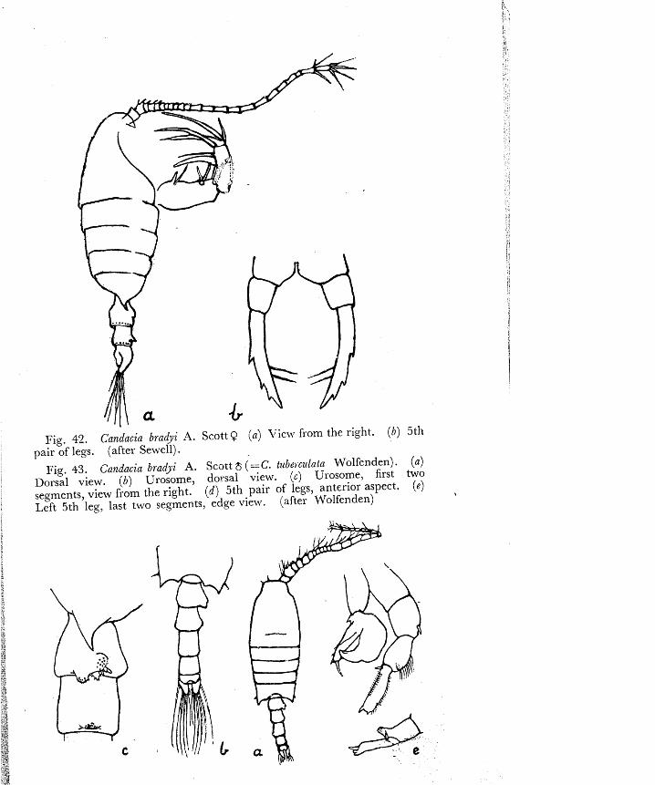
Q Catldacia h a 4 i A. Scott (Fig. 42) Middle segment of urosome without pointed ventral spine . . . . . . . .78
78 Genital segment bears long pointed processes on either side as figured; these are not symmetrical; 5th legs bear three blunt teeth at the tip; length 2.4 to 3.00 mm.
Q Candacia pachydaclyla (Dana) (Fig. 44 a to d) Genital segment bears a short flat process, one on either side symmetri-
cally placed; tip of each 5th leg bears several thin, sharp-pointed teeth; length 2.6 mm.
Q Catzdccia aethiopoca (Dana) (Fig. 44 e to h) Abte: 111 almost every instance when observed, Candacia aethiopica had an opaque
bIack colour over the entire dorsal surface. I: occurred in plankton from 18 km. off the coast at Kozhikode.
Fig. 41. Ca~ldacia dissaudata A. Scott. ( a ) Female, dorsal view. ( 6 ) Female, part of metasome and urosome, lateral view. (c) Female, 5th pair of legs. (d) Female, tip of 5th leg. ( e ) Male, urosome, dorsal view. ( f ) Male, genital segment, view from the right. (g) MaIe. 5th pair of legs, posterior view. (after A. Scott).
' 1 '
Fig. 42. Ca~zdacia bm&i A. Scott9 (a) Vie\* from the right. (6) 5th pair of legs. (after Sewell).
Fig. 43. i h n i c i o bradji A. Scott 6 (==C. tubeicziiata Wolfenden). (a ) Dorsal view. (6) Urosome, dot-sal view. (c ) Urosome, first two segments, view from the right. (d ) 5th pair of legs, anterior aspect. (e) Left 5th leg, last two segments, edge view. (after Wolfenden)
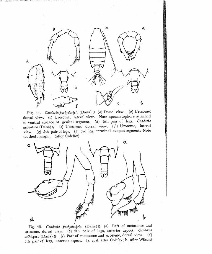
Fig. 44, Candacia pachydactyla (Dana) 9 (a) Dorsal view. (b) Urosome, dorsal view. (c) Urosome, lateral view. Note spermatophore attached to ventral surface of genital segment. (d) 5th pair of legs. Candacia aethiopica (Dana) Q (e) Urosome, dorsal view. c ' f ) Urosome, lateral view. (g) 5th pair of legs. (h) 3rd leg, terminal exopod segment; Note toothed margin. (after Colefax) .
Fig. 45. Calzdacia pach~ldactyla (Dana) $ (a) Part of metasome and urosome, dorsal vielv. (6) 5th pair of legs, anterior aspect. Candacia 3
aethiopica (Dana) 5 (c) Part of metasome and urosome, dorsal view. ( d ) 5th pair of legs, anterior aspect. (a, c, d. after Colefax; b. after Wilson)

79 Metasome corners symmetrical.. . . . . . . . . . . . . . . . . . . . . . . . . . . . . -80
Metasome corners asymmetrical, more produced on the right. . . . . .81
80 Anal segment and caudal rami asymmetrical though less so than il the female; details of 5th legs as figured; process on the right of the genital segment comparatively simple; length 1.8 mm.
$ Candacia discaudata A. Scott (Fig. 41 e, f, g)
Anal segment and caudal rami symmetrical; details of 5th legs as figuret?; process on the right of the genital segment more compli- cated as figured; a slight eminence present on the right side of the following segment also; length 1 . 9 mm.
$ Candacia bra+i A. Scott (Fig. 43)
81 Metasome corner of righf. side prolonged to reach beyond the genital segment; a bluntly pointed process present on the right of the genital segment; details of 5th legs as figured; length 2.3 to 2.6 mm.
$ Candacia jachydactyla (Dana) (Fig. 45 a, b)
Metasome corner of right side is only a little longer than the left and does not reach beyond genital segment; a flat outgrowth present on the right of the genital segment; details of 5th legs as figured; length 2.4 mm.
8 C a n d x i a aefhiojica (Dana) (Fig. 45 c, c!)
82 Body not usually pellucid; anterior lip not greatly enlarged; 5th legs not very slender or spine like ; endopod of 2nd nritenna 2-segmented
. . . . . . . . . . . . . . . . . . with distal segment shorter than proximal .83
Body fusiform and usually highly pellucid; anterior lip large, promi- nent, three-lobed; 5th legs of females usually spiniform; endopod of 2nd antcnna either of one short segment or two long segments,
. . . . . . . . . . . . . . . . . . . . . . . . . . . . . . . . distal as long as proximal .I05
. . . . . . . . . . . . . . . . . . . 83 Cuticular eye-lenses not present CALANOPIA 84 1 One pair of cuticular Lye-lenses present dorsally on the cephalosome
. . . . . . . . . . . . . . . . . . . . . . . . . . . . . . . . . . . . . . . . . . . LABIDOCERA 89
84 Females only: urosome 2-segmented; 1st antenna not genicidate; . . . . . . . . . . . . . . . . . . . . . . . . . . . . . . . . . . . . . 5th legs $-segmented. .85
Males only : urasorne 5-segmented, 1st antenna geniculate on the right . . . . . . . . . . . . . . . . . . . . . . side; right 5th leg chelate, prehensile. .87
85 Left 5th leg longer than the right; length of body 1.9 mm. 9 Calanopia ell@tica (Dana) (Fig. 46 a, b, c)
. . . . . . . . . . . . . . . . . . . . . Left 5th leg symmetrical with right 5th leg .86
86 Terminal seta of 5th leg is very long and slender; tip of terminal
segment bears 2 long pointed teeth outer to the seta; length 1 .34 mm. Q Cala7iojia au~iv i l l i Cleve (Fig. 47 a, b, c)

Terminal seta of 5th leg is moderately long, stoutish, tip of terminal segment bears a single tooth, outer to the seta; length 1.4 mm.
Q Cnlanopia minor A. Scott (Fig. 48 a, h, c)
87 Second urosome segment bears a sinall but distinct tooth-like projection on the posterior margin of the right side; details of 5th legs as figured; length 1.8 mm.
$ Calanopia ~llipiica (Dana) (Fig. 46 d, e)
. . . . . . . . . . Second urosome segment bears no tooth-like projection. .88
88 Inner margin of second segment of left 5th leg is raised into an eminence tipped with a small fiat plate; length 1 .12 mm.
$ Calailofiia a/il-ioilli Cleve (Fig. 47 d)
Inner margin of second segment of left 5th leg is raised into an eminence tipped .ivith a small curved pointed tooth; length 1.2 mm.
$ Calarlopia minor A. Scott (Fig. 48 d, e)
89 Anterior margin of head with a median pointed hook curved to the . . . . . . . . . . . . . . . . ventral aspect; body size large, 2 . 8 to 3.4 inm. .90
Anterior margin of head without a hook, body size lesser, 2.1 mm. orless . . . . . . . . . . . . . . . . . . . . . . . . . . . . . . . . . . . . . . . . . . . . . . . . . . . . 91
Fig. 46. Calanopia elliptica (Dana) [a ) Female, dorsal view. ( b ) Female, part of metasome and genital segment, left aspect. jc) Female, 5th pair of legs, posterior aspect. (d) Male, urosome, dorsal view. ( e ) Male, 5th pair of legs. posterior aspect. (after A. Scott)

Fig. 47. Calanojia auriviiii Cleve ( a ) Female, dorsal view. (b) Femalr, part of metasome and genital segment, lateral view. (c) Female, 5th pair of legs. (d) Male, 5th pair of legs, posterior aspect. (after A. Scott.)

90 Urosome 3-segmented; 1st antennae symmetrical; corners of metasome symmetrical; eye-lenses not so large as in the male; 5th legs as figured; length 3 .OO to 3.40 mm.
Q Labidocera acuta (Dana) (Fig. 49) Urosome 5-segmented, 1st antellna geniculate on the right side;
corners of metasome unlike on the two sides as figured; eye-lenses larger than in the female; 5th legs as figured; length 2.8 to 3 . 3 mm.
$ Labidocer-n acuta (Dana) (Fig. 49)
91 Females only . urosome 3-segmented; 1st antennae symmetrical. . . .92 Males only: urosome 5-segmented, 1st antenna geniculate on the
right side . . . . . . . . . . , . . . . . . . . . . . . . . . . . . . . . . . . . . . . . . . . . . . . 9 3
92 Carriers of metasome drawn out into down curved points that are dearly perceived in lateral view; details of urosome and 5th legs as figured; length 2.1 mm.
Q Labidoce~a pecfinata Thompson st Scott (Fig. 50) Posterior margin of metasome rounded, with a very small projection
present on the right side oniy, not visible in dorsal view; details of urosome and 5th legs as figured; length 2.1 mm.
9 Labidocera mi~zzifa Giesbrecht (Fig. 52 a to d) Corners or metasome ending in points hut not drawn out; urosome
condensed, very short, with a peg-like projection on the right side; 5th legs as figured; length 1.9 mm.
Q Lahidocera pavo Giesbrecht (Fig. 53 a, b) Rote- L. pecti~lata is the commonest Labidocera in coastal plankton.
Fig. 49. Labidocera acuta (Dana) (a) Fimale, dorsal view. (b) Female, 5th pair of legs. (c) Male, dorsal view. ( d ) Male, 5th pair of legs, anterior aspect. (original)
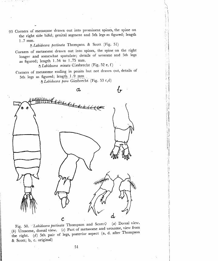
93 Corners of metasol~le dralvn out into prominent spines, the spine on the right side 13ifid; genital segmcnt and 5th legs as figured; length 1 . 7 mm.
8 Labidocera pectiilata Thompson Kr Scott (Fig. 51)
Corners of metasome dialvn out into spines, the spine on the light longer and somelvhat spatulate; details of ulosome and 5th legs as figured; length 1.54 to 1 .75 mm.
3 Labidoc~la minuta Giesbrecht (Fig. 52 e, f )
Co~ne ls of metasome eildillg in poiilts but not dra~vn out, details of 5th legs as figured; length 1 .9 lnin
$ Lnbidocejn pnz)o ~iesb lech t (Fig. 53 c,d)

u Fig. 51. Labidocera fiectiliata Tbompsan and Sco t t5 ( a ) Dorsal view.
( 6 ) Ventral view to show 5th pair of legs in position, metasome corners and spine on the genital segment. (c) Right 5th leg, chela. ( d ) LeR
5th leg. (a, c, d. after Seuiell; b , original)
Fig. 52. Labidoceru minufa Giesbrecht (a ) Frmale, dnrsal view. ( 6 ) Female, part of Inetasome and urosome, ventral view. (c) Female, part of metasome and urosome, dorsolateral view. (d) Female, 5th pair of legs. (e) Male, prr t of metasome and urosome dorsal view. (f) Male, 5th pair of legs, posterior aspect. (a, c, f. after Colefax; b, d, e. after

Pig. 53. Labidocera fauo Giesbrecht ( a ) Female, dorsal view of meta- some corners and urosome. ( 6 ) Female, 5th pair of legs. ( c ) Male, dorsal view. ( d ) Male, 5th pair of legs. (after Sewell)
Fig. 54. Pontella danae Giesbrecht. ( a ) Female, dorsal view, variely &nica. (b) Female, urosome, dorsal view, variety ceylonica. (c) Femde, 5th pair of legs, variety cpylonica. ( d ) Female, rostrum, variety cylonzca. ( e ) Male, ri ght 5th leg. ( f ) Male, ?euninal segments of left 5th leg. (a, b, c, d. after Thompson and Scott; e, f. after Giesbrecht)

Fig. 5 5 . Poiltella securifeer Brady Q (a) Dorsal view. ( 6 ) 5th leg of one side. Polltella spinipes Giesbrecht Q (c) Dorsal view. (d) 5th leg of one side. (after \Yilson)
Fig. 56. Pontella securzfer Brady 6 (a) Oorsal view. ( 6 ) Rostrum, lateral view to show ventral eye and rostra1 lens. (c) 5th pair of legs, anterior aspect. (a, c. after Colefax; b. original)
54

94 ~ ~ t h the caudal rami distinctly separated from the anal segment.. . .95
Left caudal ramus distinctly separated from the anal segment hut the right caudal ramus more or less fused with it . . . . . . . . . . . . . . . ,103
95 One pair of dorsal eye-lenses are present, larger in the males, rostral
lens also usually present; rostral rami comparatively short, lateral margins of the head with a hook on each side . . . .PONTELLA 96
~ o r s j l 3nd rostral eye-lenses quite absent; rostral rami comparatively
long and slender; head without lateral hooks . . . . . . . . . . . . . . . . . . . . . . . . . . . . . . . . . . . . . . . . . . . . . . . . . . . . . . . . . . PONTELLOPSIS 100
96 Females only: urosome 2-segmented, genital segment with variocs outgro~lths and excrescences concealing the urosome segments in dorsal view; urosome and caudal rami more or less asymmetrical; 1st antennae aIike on the two sides . . . . . . . . . . . . . . . . . . . . . . . . . .97
Males only : urosome 5-segmented, the segments clearly visible in dorsal view ; urosome and caudal rami little or not at all asymme- trical; 1st antenna geniculate on the right side . . . . . . . . . . . . . . . .99
97 Corners of metasome only a little asymmetrical, the left one being a little longer; right ca:ldal ramus distinctly larger and hearing a vertical crest-like extension visible in lateral view; left 5th leg longer with 2 outer spines 011 exopodite, one of which is distinct; length 3.4 mm.
Q P,xzlella danae Giesbrecht, variety ceylotzica, Thompson 8: Scott (Fig. 54 a to d)
Corners of rnetasome strongly asymmetrical, the left lobe reaching the middle of the left caudal rarnus; right caudal ramus larger; 5th
. . . . . . . . . . . . . . . . . . . . . . . . . . . . . . . . legs not unequal in length. .98
98 Genital segment with two or three finger-like outgrowths on the dorsal surface; exopods of 5th legs strongly curved, with 4 outer spinules; lengrh 4.4 mm.
Q Pontella secul-ifer Bradv (Fig. 55a, b)
Genital segment with transverse corrugations on the dorsal surface; right caudal'ramuq larger hut its setae much shorter as compared to left side; exopods of 5th legs strongly curved, vith 3 outer spinules; length 4.5 mm
9 Po~ztella slji~zipes (iiesbrecht (Fig. 55c, d)
99 In the chela of the right 5th leg, the terminal claw b~a r son its inner margin, about half way along its length, three rounded processes; the "hand" bears three short more or less triangular processes;
55

the "thumb" is long, smooth and simple; it has no corrugated \
appendage; body length 3.2 to 4.4 mm. $ Pontella spinipes Giesbrecht (Fig. 57)
I n the chela of the right 5th leg, the terminal claw bears no rounded processes near the middle of its length; but onlytwo setae, proxi- mally placed; the "hand" bears one, long, pointed process and one very short rounded process; the "thumb" is short; it has a corru- gated appendage; body length 4.3 mm.
$ Pontella sec~rrifeel- Brady (Fig. 56)
Details of the 5th legs as figured, terminal claw and "hand" of the right 5th leg very similar to P. se;urifeer but the thumb has no corrugated process; tip of left 5th leg as figured; body length 3.1 mm.
$ Potztella daizae Giesbrecht (Fig. 54 e, f )
100 Females only : urosome 2-segmented, 1st antennae symmetrical. . . . 101
Males only : urosome 5-segmented; 1st antenna geniculste on the right side . . . . . . . . . . . . . . . . . . . . . . . . . . . . . . . . . . . . . . . . . . . . . . . . 1 0 2
Fig. 57. Pontella spinipes Giesbrecht 8 (a ) View from the right. ( 6 ) Chela of right 5th leg. (c) Left 5th leg. (after Sewellj

101 Metasome corners symmetrical; genital segment with outgrowths as
figured, the spine on the ;ight posterior corner being the largest,
caudal rami quite symmetrical; length 1 .90 mm; the male undes-
cribed yet. Q Pontellojvis herdnzani Thompson 8 Scott (Fig. 58)
Metasome corners symmetrical; genital segment with a different arrangement of outgrowths as figured, the 1eft.posterior spine being the largest, caudal rami sligh.1~ asymmetrical; length 1 . 7 to 1 .97 . .
+. - ->
mm Q Pontellop~is maclolplr A. Scott (Fig. 39 a, 1))
Metasome corners as)mmetrical, the left colner beariqc a longer roiection; genital segment with t\ro long spinous projections on
4 5 - the right margin as Bguied, right caudal ramus slightly more
posterior in position than the left, lerrgth 1 6 to 1 .9 mnl. 9 Pontellopsis scotti Sewell (Fig. 60 a. h)
Fig. 58. Pontellopsis hridmani Tllompson & Scott. Q (a) Dorsal view ( b ) 5th pair of legs. (after Sewell).
5 7
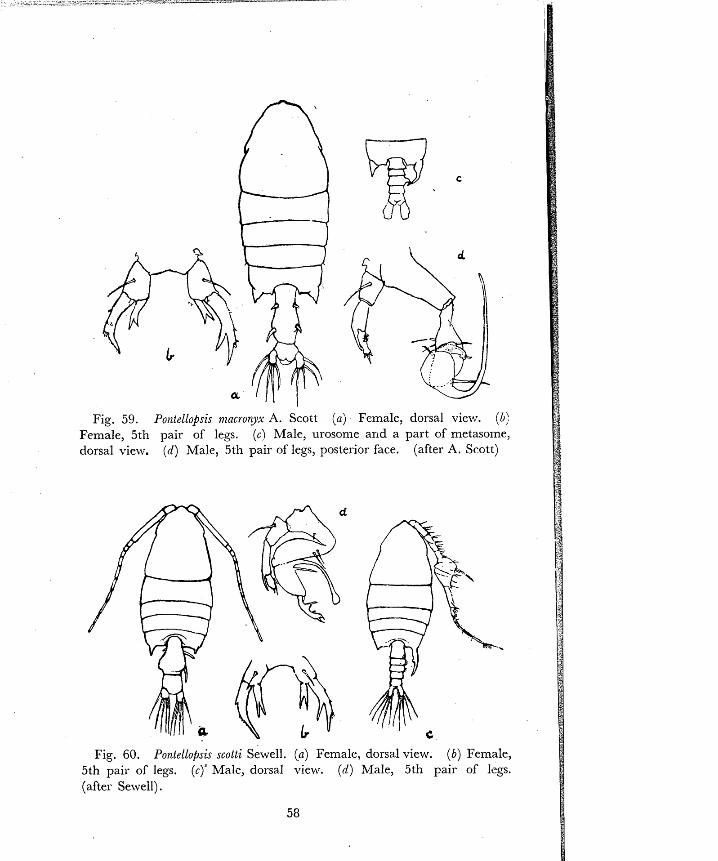
Fig. 59. Poi~tellopsis nzacro?yx A. Scott (a) Female, dorsal view. ( 6 : Female, 5th pair of legs. (6) Male, urosome and a part of metasome, dorsal view. (d) Male, 5th pair of legs, posterior face. (after A. Scott)
Fig. 60. PontelloPsis scotti Sewell. ( a ) Female, dorsal view. ( 6 ) Female, 5th pair of legs. (c)' Male, dorsal view. (d) Male, 5th pair of legs. (after Sewell).

102 Right metasomal spine dilated at base and curved inwards as figured; 3rd urosome segment with a peg-like projectioll on the right margin; in the chela of the right 5th leg, the fixed claw is very long, straight and pointed; length 1.67 mm.
8 Pontellopsis maci-oigx A. Scott (Fig. 59 c, d)
Right metasomal spine slightly curved, shaped as in the figure; 2nd and 3rd urosome segments each with zt group of minute teeth on the right margin; fixed clalv cT the chela curved, its tip spatulate; length 1 .42 mm.
$ Pontello~sis scolli Sewell (Fig. 60 c, d)
103 Body short and 1-obust; no dorsal 'eye-lenses; plumose setae of 2nd antennae and mandibular palps are very long and collspicuous . . . . . . . . . . . . . . . . . . . . . . . . . . . . . . . . . . . . . . . . . . . ONTELLINA104
I04 Urosome 2-segmented; 1st antennae alike on two sides; 5th legs a figured; length 1 . G to 1.9 mnl.
9 Purrtellitla pluinatn (Dana) (Fig. 6 1 a, b)
Urosome .?-segmented; 1st antenna geniwlated on the right side; 5th legs as figured; length 1.5 to 1 ..65 inm.
8 Po~2tellina p~u?ndla (Dana) (Fig. 61 c)
brote-- Pontellina filunzatu occurrcd frequently in the plankto.1 from 18 km. off the coast at Kozhikode. It is easily recognized by the large setae borne on the 2nd antennae and mandibular palps which are used in swimming. The movements 01- Pontellina recall the Happii~g moveilleilts of tllc wirgs of a bird in flisl~t.
Fig. 61. Pontellina pluinata (Dana). ( a ) Female, dorsal view. (b) Female, 5th pair of legs. (c) Male, 5th pair of legs. (a, b. after Colefax,
c . after Wilson).

I I
Fig- 62. (a ) -4cartia spi?mauda Giesbrecbt Q Dorsal vie\\.. (b j Acartia ey t iraen Giesbrecht Q Dorsal view. I C ) Aca~i ia cent?rm Giesiil-ech t Q Dorsal view. (aiter Gieshrrchtj.
Fig. 6 3 ( a ) Acartia rginicauda Giesbrecht Q 2nd arrtenna. (b ) Bcartia rpi?zicairda Giesbrecht Q 5th leg. (i) Acartia ewhraea Giesbrecht Q 5th leg. (d) Acartia centrum Giesbrecht Q 5th leg. (a. original; h, r , d. after Giesbrecht)

105 Second antenna with two-jointed endopod in which the distal segment is as long as the proximal (Fig. 63 b), 5th legs ofthe female always uniramose, slender and spine-like. . . . . . . . ACARTIA 10fi
S,econd antenna with a short one-jointed endopod that is fused to the basal segment, and a one-jointed exopod (Fig. 69 b); 5th legs of the female biramose, spine-like. . . . . . . . . . . . . . ACARTIELLA 116
106 Posterior margin of metasome drawn out into spines . . . . . . . . . . . . 107
. . . . . . . . . . . . . . Posterior margin of metasome without large spines. .I14
107 Two prominent metasomal spines present and in addition two smaller spines present, situated more dorsally and nearer the middle line
Two prominent metasomal spines present but no additional spines. . 113
108 Females only: urosome 3-segmented; 1st antennae alike on two sides . . . . . . . . . . . . . . . . . . . . . . . . . . . . . . . . . . . . . . . . . . . . . . . . . . . . 169
Males only: urosome 5-segmented, 4th segment very short, second segment the largest, 1st segment with lateral genital aperture, 1st antenna of the right side is geniculate but very irldistinctlv. . . . . . 11 1
109 The 2nd urosome segment bears tw-o small inconspicuous spines placed close together; terminal claw of 5th leg thickened a little at the base, slightly curved, smooth, and without any notch; length 1.0 to 1 .4 mm.
Q A c a ~ t i a erythraea Giesbrecht (Fig. 62b, 6 3 ~ )
The 2nd urosome segment bears two spines that are compai-able in size and position to the spines on the first urosome segment. . . . . . l l 0
110 The spines of the metasome corners are as large as in A. eythraea; terminal claw of 5th leg swollen at the base, smooth, straight and with a distinct notch as figured; length 1.20 to 1.24 mm.
Q Acartia centrura Giesbrecht (Fig. 62c, 63d)
The spines of the metasome corners are smaller than in A . erythraea and A. centrura; terminal claw of 5th leg scarcely widened at base, straight, without notch and with serrations on distal half as figured; length 1 .O to 1.3 mm.
Q Acartia sfl inic~ud~z Giesbrecht (Fig. 62a, 63b)
flo"iote- Great care is required to distinguish from one another the abobe three species of Acartia. While A. centrura appears to be less frequent than the other two, A. erythraea js the most abundant at Madras, and A. sfiinicauda is a t least 'as common as A , eathraea at Kozhikode and Cochin.

11 1 Second iirosome segment wider than long with two pails of prominent spines, inner pair as large as the outer; details-of 5th legs as figured, length 1.0 to 1 .3 mm.
d Acartia eythraea Giesbrecht (Fig. 64L, 6511) Second urosome segment almost as wide is long. . . . . . . . . . . . . . . . . . I12
Fig. 64- ( a ) Acari:a ~Pinicauda Giesbrecht, 8 Urosome, dorsal vielv. ( 6 ) Acartia e ~ t h r a e a Giesbrecht d Urosome, dorsal view. (c) Acarfin ceii/r?lra Giesbrccht 8 Urosome, dorsal viciv. (after Giesbrecht).
Fig. 65. (a ) Acarlia spinicnada Giesbrecht g, 5th pair. of legs, anterior. view. ( 6 ) Acartia erythram Giesbrecht 8, 5th pair of leW posterior (c) Acartia ce~itrura Giesbrecht 8, 5th pair of legs, anterior (after Giesbrecht) .
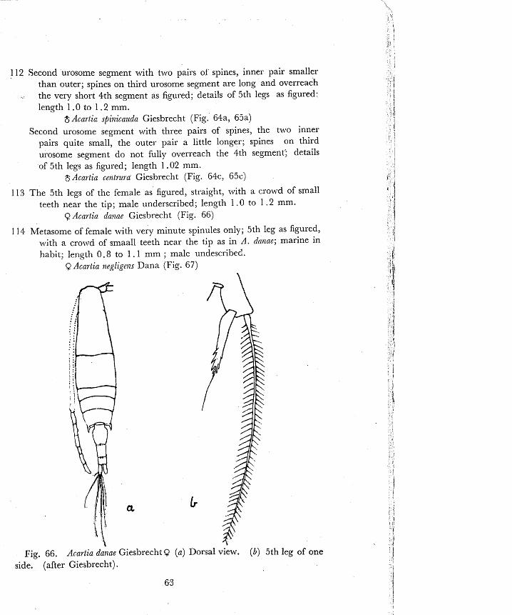
112 Second urosome segment with two pairs of' spines, inner pair smaller than outer; spines on third urosome segment are long and overreach
- the very short 4th segment as figured; details of 5th legs as figured. length 1 .O to 1.2 mm.
$ Acartic spitzicauda Giesbrecht (Fig. 64a, 65a) Second urosome segment with three pairs of spines, the two inner
pairs quite small, the outer pair a little longer; spines on third urosome segment do not fully overreach the 4th segment'; details of 5th legs as figured; length 1.02 mm.
$ Acartia c e n t n ~ ~ a Giesbrecht (Fig. 64c, 65c)
113 The 5th legs of the female as figured, straight, ~vi th a cro~vd of small teeth near the tip; male underscribed; length 1 . 0 to 1 . 2 mm.
Q Acartia daxae Giesbrecht (Fig. 66)
114 Metasome of female with very minute spinules only; 5th leg as figured, with a croxvd of smaall teeth near the tip as in A. dclllnc; marine in habit; length 0.8 to 1 . I mm ; male undescribei.
Q Acartia negligeizs Dana (Fig. 67)
Fig. side.
66. Acartia danae Giesbrecht (after Giesbrecht) .
Dorsal view. (b) 5th leg of one

Metasome, posterior margin smooth or with minute s~inules only; habitat estuarine and brackish water; both sexes known. . . . . . . . I 1 5
115 Dorsal view of female and details of 5th legs as figured; female 5th legs, smooth, slightly curved and without notch, as in A. erythraea; length of 90 .73 to 0.80 mm; of 80 .70 to 0.75 mm.:
Q 8 Acartia southwelli Se .veil (Fig. 68, a,b,c) DomJ view of female, of male urosome and details of 5th legs as
figured; female 5th legs straight, with a notch as in A. cetztuura but set with short hairs on both margins; length I . 0 to 1 . 1 lnm in both sexes
9 8 Acartia chilkaensis Sewell (Fig. 68 d to h) 116 Urosome 3-segmented, anal segment very short; right caudal ramus
a little longer than the left; details of 5th legs as figured; length A .38 to 1.57 mm.
Q Acartiella sewelli Steuer (Fig. 69a,b,c) Urosome 4-segmented; right caudal ramus longer than the left, this
inequality being more pronounced than in the female; details of the 5th legs as figured, right leg longer and with a short process arising from its basal segment; length 1 .27 to 1 .5 1 mm.
$ Acartiella sezoelli Steurr (Fig. 69d, e) Note :- 'These three species were obsemed in Cochin harbour waters in rnonsooll mon~hs.
When the salinity is i t its lowest and zooplankton eleme-lrs are atmost entirely absent, A. sewel!i is seen to occur as the sole zooplankton element and is therefore noteworthy. Its occurrence in Indian coastal areas appears to be a new record, since it was described from Burma.
Fig. 67. Acallia negligens Dana Q ( a ) Dorsal view. (b) 5th leg of one side (only one half of seta is shown). (aftzr Giesbrecht).
64

Fig. 68. Acartia southwelli Se~vell. (a) Female, dorsal view. (b) Female, 5th pair of legs. (c) Male, 5th pair of legs, anterior face. Acartia chilkaensis Sewell (d) Female, dorsal view. (e) Female, urosome, lateral view. ( f ) Female, 5th pair of legs. (g) Male, 5th pair of legs, posterior face. (h) Male, urosome and a part of mztasome, dorsal view. (a, h, c. after Sewell; d. to h. after Steuer).
Fig. 69 , Acartiella sewellz Steuer (a) Female, dorsal view. ( b ) Female, 2nd antenna. (c) Female, 5th pair of legs. (d) Male, 5th pair of legs, posterior face. ( e ) Male, urosome, dorsal view. (after Stever),
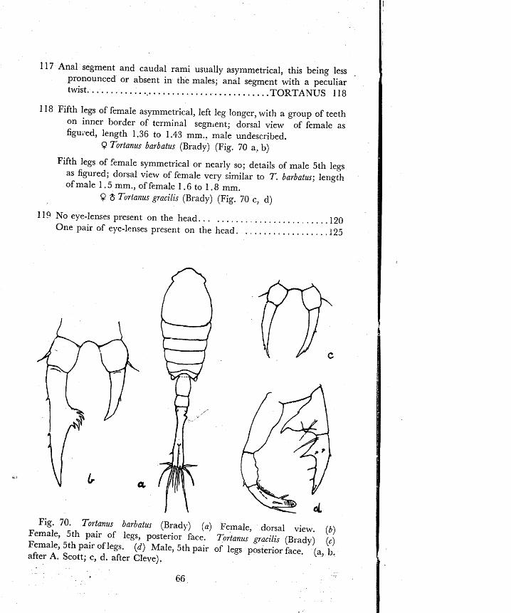
117 Anal segment and caudal rami usually asymmetrical, this being less pronounced or absent in the males; anal segment with a peculiar twist. . . . . . . . . . . . .~.. . . . . . . . . . . . . . . . . . . . . . . . . TORTAWUS 118
118 Fifth legs of female asymmetrical, left leg longer, with a group of teeth on inner border of terminal segment; dorsal view of female as figuced, length 1.36 to 1.43 mm.. male undescribed.
9 Tortanus barbatus (Brady) (Fig. 70 a, b)
Fifth legs of female symmetrical or nearly so; details of inale 5th legs as figured; dorsal view of female very similar to T. barbatus; length ofmale 1.5mm.,offemale 1 .6 to 1.8 mm.
$? 6 Tortataus grncilis (Brady) (Fig. 70 c, d)
119 No eye-lenses present on the head.. . . . . . . . . . . . . . . . . . . . . . . . . I20 One pair of eye-lenses present on the head. . . . . . . . . . . . . . . . . ,125
Fig. 70. Tortanu barbatu~ (Brady) ( a ) Female, dorsal view. (b) Female, 5th pair of legs, posterior face. Tortanus pracilis (Brady) [c) Female, 5th pair of legs. (d ) Male, 5th pair of legs posterior face. (a, b. after A. Scott; c, d. after Cleve).

120 Fifth pair of legs with the basal segment possessi~lg the inward . . . . . expansion characteristic of the Harpacticoida (Fig. 72 d) .I21
. Fifth pair of legs without the characteristic inward expansion.. . l26
121 Endopods of 2nd legs greatly elongated; inward expansion of 5th legs . . . . . . . . . . . . . . . narrow, curved and pointed. LONGIPEDI A 122
Endopods of 2nd legs not elongated; body fusiform; caudal setae very . . . . . . . . . . . . . . . . . . . . . . . . . . . . . . . . . . . . . . . . . . . . . . . . . . . . . long 123
122 Anal operculum with the central tooth a little longer than the two teeth on each side (Fig. 71 e) : female as depicted, length 0.95 mm.; male undescribed.
Q Longipedia webesi A. Scott (Fig. 71 a to e)
Anal operculumn with the central tooth much longer than the two teeth on each side (Fig. 71 g); general appearance much as in L. rueberi, length of 0.8 to 1.30 mm.; of male 1.08 mm.
9 $ Longibedia corouata Claus (Fig. 7 1 f, g)
Fig. 71. Lotzgipedia webefi A. Scott9 (a ) Dorsal view. ( 6 ) Lateral Y view. (c) 2nd leg of one side. ( d ) 5th leg of one side. (e) Anal oper- i
1 culum. Longipedia coronata Claus ( f ) Male, 5th leg. (g) Female, anal 1 operculum. (a, c, d, f, after Sewell; b, e, g. after A. Scott). 1
J

Fig. 72. Microsetella aorvegica (Boeck) (a) Dorsal view of 0. (6 ) Lateral view of Q . (c) F~male, 2nd antenna. (d) Female, 5th leg of one side. (e) Male, lateral view. (after G.O. Sars).
- Fig. 73. Microsetella rosen (Dana) Q (a) Lateral view. (6) 5th leg of
one side. (after Giesbrecht) .

123 Caudal rami as long as broad, inconspicuous, 2nd antennae with 3-segmented exopodite (Fig. 72 c) ; small in size, less than
- 0.85 mm ............................................... 124
Caudal rami slender, cylindrical, over 4 times as long as broad; 2nd antennae without any exopodite; larger in size, over 1.0 mm.; caudal setae about as long as the body; length of female 1.4 to 1.5 mm., of male 1.16 to 1.30 mm.
Q $ hlncrosetella gracilis (Dana) (Fig. 74)
Fig. 74. iVfacrosetella gracilis (Dana) (a) Female, dorsal view. (b) Male, lateral view. (c) Female, 5th leg of one side. ( d j Male, 5th leg of one side. (a, b. after Giesbrecht; c. d. after Wilson).
Fig. 75. Macrosetella oculata (G.O. Sars) (a) Female, lateral view. (b) Female, 5th leg of one side. (c) Male, 5th leg of one side. (after Wilson).

t Fig. 76. , i ~ ~ i l a c i a effcrala Dana ( a ) Female, lat-ral view. (b) Female,
dorsal view. ( c ) Female, 5th leg of one side. (d) Male, 5th leg of one side. (after Wilson).
124 The inward expansion of the basal of the 5th leg b ~ a r s one short and one long seta; caudal setae about as long as the body; length of female 0.35 to 0.53 mm., of -ale 0.33 to 0.42 mm.
9 $ Microsetella ~zorvegicr (Boeck) (Fig. 7 2 )
The inward expansion of the basal of the 5th leg bears two equally long setae; caudal setae 1+ times as long as the body; length of female 0.64 to 0.85 mm.; of male 0.56 mm.
Q $ A.licrosete!la rosea (Dana) (Fig. 73)
125 Second antennae without an exopodite; apical caudal setae more than twice as long as caudal raini ; body proportions as figured, length of female 1.2 to 1.35 mm., of male 1.15 to 1.30 mm.
Q 8 1LIacroselella oculala (G.O. S lrs) (Fig. 75)
Second antennae with a small one-jointed exopodite; &pica1 caudal setae only a little longer than the caudal rami; body proportion as figured; length of female 1.75 to 2.10 mm. of male 1.4 to i .6 mm
Q $ .Lliracia efferata Dana (Fig. 7 6 )
126 Body depressed, with angular projections at the posterior corners of the cephalosome and the next three segments; 5th legs long, narrow, 2-segmented and tipped with setae. . . . . . . . . . . . . . . . . . . . . . . .12?
Body not depressed 5th legs plate-like.. . . . . . . . . . . . . . . . . . . . . . .128

127 Exopod of 2nd antenna represented by two long setae; 1st antenna 5-segmented; caudal r an i twice as long as broad; apical caudal setae very long in the males only, quite short in the fema1es;length of female 1.07 to 1.30 mm., of male about the same
9 8 Clytemjzestra scutellata Dana (Fig. 77; 78 a to d)
Exopod of 2nd antenna represented by one long seta; 1st antenna 7-segmented through the fusion of two terminal segments; caudal rami hardly as long as broad; lengih of female 0.5 to 0.87 mm., of male 0.4 to 0.7 mm
9 $ C&fenztzestra rostrata (Brady) (Fig. 78 e, f, g)
Note: The three most frequently seen Harpacticoids in inshore plankton are (in decreasing order of frequency) EuterFina act~t$rons, Clytemnestra scutellata and ibfacrosetella gracilis. Clytetnnestra scutellata is remarkable for its rapid movements. Each of these three species may, on occasion, occur in "swarm" condition eclipsin~ all other zooplankton elements in numbers.
Fig. 77. Clytemnestrascutellatd Dana, female (a) Dorsalvie~r. (b) Lateral view. (c) 5th leg of one side. (after G.O. Sars).
7 1
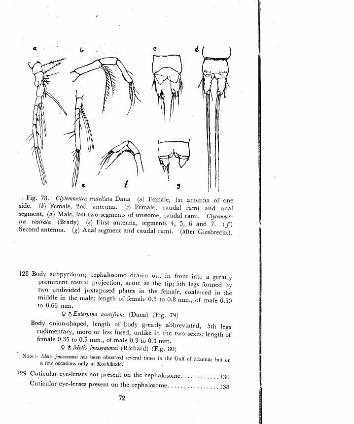
Fig. 78 . Cbtetntzestra sczllel!atiz Dana ( a ) Feinalc, 1st antenna of one side. ( 6 ) Female, 2nd antenna. ( c ) Femalc, caudal rami and anal segment, ( d ; Male, last two segments of urosome, caudal rami. Clyternnes- tra rostrata (Brady) (e) First antenna, segments 4, 5, G and 7. ( f ) Second antenna. (g) Anal segment and caudal rami. (after Giesbrecht).
128 Body suhpyriform; cephalosome dra\.vn out in fiont into a gl.eatly prominent rostra1 projection, .acuie at the tip; 5th legs formed by two undivided juxtapcsed p!ates in the female, coalesced in the middle in the male; length of femalc 0.5 to 0.8 mm., of male 0.50 to 0.66 mm.
9 $Euterpina ncut$-om (Dana) (Fig. 79)
Body onion-shaped, length of body greatly al~breviated, 5th legs rudimentary, Inore or less fused, unlike in the two sexes; length of female 0.35 to 0.5 mm., of male 0.3 to 0.4 mm.
Metis jousseaumei (Richard) (Fig. 80)
Note :- ~Clel~s j~usseatmtei has been observed several timcs in the Gulf of ivlannar h ~ l t on a few occasions only at Kor-hikode.
129 Cuticular eye-lenses not present on the cephalosoine. . . . . . . . . . . 130
Cuticular eye-lenses present on the cephalosonle. . . . . . . . . . . . . . . . I38

Fig. 79. Euterpina acutifioizs (Dana) (a) Female, dorsal view. (b) Female, lateral view. (c) Female, 1st antenna. { d ) Female, 5th pair of legs. (e) Male, 1st antenna of one side. ( f ) Male, 5th pair of legs. (after G.O. Sars).

I30 Body not depressed, of cyclopoid form, maxillipeds normal. . . . . . I 3 1
Body depressed, leaf-like and transparent as in Sapphirina.. . . . . . . I40 Body depressed but of cyclopoid form; maxillipeds specialized in both
. . . . . . . . . . . . . . . . . . . . . . . . . . . . . . . . . . . . . . . . . . . . . . . . . . sexes. .152
131 Genital segment only a little Iarger than the other urosome segments; maxillipeds and 2nd maxillae slender, covered with numerous
. . . . . . . . . . . . . . . . . . . . . . . . . . . . . . . spiny bristles. OITHONA 132
Genital segment conspicuously enlarged; succeeding segments in- conspicuous; maxillipeds 3-segmented, subchelate, forming powerful grasping organs in both sexes. . . . . . . . . . . . . . . . . ONCAEA 137
132 Females only : metasome very slender, fusiform; head terminating in frcnt in a pointed rostrum ; 1st antennae not geniculate on either side, bearing numerous setae; urosome 5-segmented: genital
. . . . . . segment a little wider than the other urosome segments. .I33
Males only : metasome not so s le~der as in the female, smaller in length; head-end truncate, without a rostrum; each 1st antenna twice geniculate, with fewer setae than in the female; urosonle 6-segmented; genital segment wider than other urosome segments, this being more pronounced than in the female.. . . . . . . . . . . . ,136
133 Rostrum visible in dorsal view; body length over 1 mm; outer marginal spines of the terminal cxopod segments of legs 1 to 4 number 2, 2, 1, 1; two apical setae greatly elongated in each caudal ramus. . . . . . . . . . . . . . . . . . . . . . . . . . . . . . . . . . . . . . . . . . . . . . . . . . . 134
Rostrum bent down, not visible in dorsal view; body length less than i mm.; outer marginal spine formula variable; apical setae not so greatly elongated. . . . . . . . . . . . . . . . . . . . . . . . . . . . . . . . . . . . . . . .I35
Rostlum not visible in dorsal view; body length 1.2 mm.; metasome and cephalosome together being shorter than urosome (11 :13); metasome only about 3 times wider than urosome; outer marginaI spines of the terminal exopod segmcnts of legs 1 to 4 number 3, 2, 1, 1; male undescribed.
Q Oithona linearis Giesbrecht (Fig. 83 c)
.34 Conspicuously coloured plumose setae are present on the outer margin of second basipod segment of legs 2, 3 and 4 and they project out- wards and upwards, being visible in dorsal view if undamaged, the enlarged caudal setae are also plumose and conspicuous; genital segment beats a b u n ~ h of fine hairs on each side ventrally; length 1.0 to I .5 mm.
C Oithorzn plnmzj2ern Baird (Fig 81 b, c)
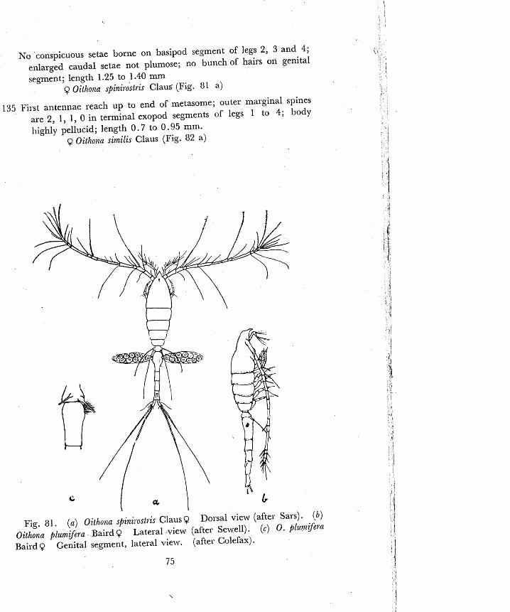
No conspicuous setae borne on basipod segment of legs 2, 3 and 4; enlarged caudal setae not plumose; no bunch of hairs on genital segment; length 1.25 to 1.40 mm
Q Oithona spini~ostris Claus (Fig. 81 a)
135 First anten~lar reach up to end of metasome; outer marginal spines are 2, 1, 1, 0 in terminal exopod segments of legs 1 to 4; body highly pellucid; length 0.7 to 0.95 mm.
Q Oithotza similis Claus (Fig. 82 a)
Fig. 8 1 . (a) Oltholia spini;ostris Claus Q Dorsal view (after Sars) . (b ) Oithotla fihrnij2ra Baird Q Lateral view (after Sewell). (c) 0. plumqera Baird Q Genital segment, lateral view. (after Colefax) .
7 5

First antennae reach up to end of 3rd metasome segment only; outer marginal spines are 3, 3, 3, 2 in terminal exopod segments of legs 1 to 4; body usually with much pigment; caudal setae coarsely plumose, forming a fan; length 0.75 to 0.85 mm.
Q Oithona rigida Giesbrecht (Fig. 82 c, d)
First antennae reach up to 2nd metasorne segment only; outer marginal spines are 3, 3, 3, 2 in terminal exopod segments of legs I to 4; the two longer setae on each caudal ramus show a peculiar crossed arrangement; length 0.6 mm.
Q Oithona brevicosnis Giesbrecht (Fig. 83 a)
136 Fixt antennae twice geniculate, the first segment beyond the proximal elbow, sheathing the base of the segment beycnd it; the 1st segment beyond the distal elbow bearing a small ~emicircular process (Fig. 82 13); outer marginal spines are 2, 2, 2, 2 in terminal exopod segments of leg!: 1 to 4; length 0.5 to 0.7 mm.
$ Oithonn similis Claus (Fig. 82 b)
Fig. 82. ( a ) O~thorza similis Claus Q Dolsal view. (after Sass) ( b ) 0ithot:a simtlis Claus 8 1st antenna to show sheathing base of I st segment after 1st geniculation aild semicircular process on 1st segment after 2nd geniculation (after W.M. Wheeler). (c) Oithona rigida Giesbrecht Q Dorsal view. (after Cleve) . (d) Oifhona zgida Giesbrech t $2 Lateral view. (after Cleve) .

First antennae as described above; outer marginal spines are 2, 3, 3, 2 in terminal exopod segments of legs 1 to 4; length 0.75 to 1 .0 mm
$ Oithotza plumifeel-a Baird
First antennae twice geniculate; only a tra.ce of the sheathing base is found : semicircular process absent on 1st segment beyond distal elbow; outer marginal spines are 2, 3, 3, 2 in terminal exopod segments of legs 1 to 4; length 0.75 mm. to 0.85 mm.
$ Oithona spiniel-ostris Claus
First anten,lae twice geniculate; sheathing base appears to be present; semicircular process absent; outer marginal spines are 3, 3, 3, 2 in terminal exopod segments of legs 1 to 4; length 0 .7 mm.
$ Oitholla el-igida Giesbrecht
First antennae twice geniculate; neither pro2imal sheath nor distal semicircular process is present; outer marginal spines are 3, 3, 3, 2 in terminal exopod segments of legs 1 to 4; length 0.55 mm.
2; Oithona brevicornis Gieshrecht (Fig. 83 h)
Note :- Oithona rigida is the commonest species of Oithona in inshore waters. 0. pluinifera and 0. s,binir.ost,is were observed very often at Kozhikode in plankton From 18 km. off the shore. They have been ohserved also at C o c l ~ i n ~ Madras ant1 Rlandapam
Fig. 83. ( a ) Oithona bel-evicornis Giesbrecht Female Dorsal view. (6) Oithona bel-evicornis Giesbrecht Male Dorsal view. (c) Oithona linearis Giesbrecht Female Dorsal view. (a, b, after Wilson; c. after Wolfenden).

137 Second free metasome segment raised into a prominent hump on the dorsal middle line; outline of the genital segment in both sexes as figured; length of female 0.75 to 1 .25 mm., of male 0.6 to 0 .8 mm.
Q 8 Ot~caea conifera Giesbrecht (Fig. 84 d to g)
Second free metasome segment not raised into a hump; anterior part of the body obovate in the female, less wide in the male; genital segment more conspicuously enlarged in the male than in the female and follo~ved by 3 (in the female) 01 4 (in the male) segments which are closely telescoped together; length of female 1.1 to 1.27 mm, of male 0 .7 to 1 .OO mm.
Q 3 O~zcaea verzusta Philippi (Fig. 84 a, k, c)
. . . 138 Body moderately depressed or not at all; caudal rami styliform. 139
Body greatly depressed and flattened, most so in the males: caudal rami broadly lamellate, without elongated setae.. . . . . . . . . . . . . . . . . . . . . . . . . . . . . . . . . . . . . . . . . . . . . . . . . . . . . . . . . SAPPHIRINA 148
Fig. 84. Oncaea vetlusta Philippi. (a) Female, dorsal view. ( b ) Male, dorsal view. (c) Male, urosome, ventral view. Otlcaea coiz&Gru Giesbrecht (d) Female, dorsal view. (e) Female, lateral view. ( f) Female, urosome, ventral. (g) Male, urosome, ventral view. (a, b, c: f. g. after Giesbrecht; d, e, after Farran).
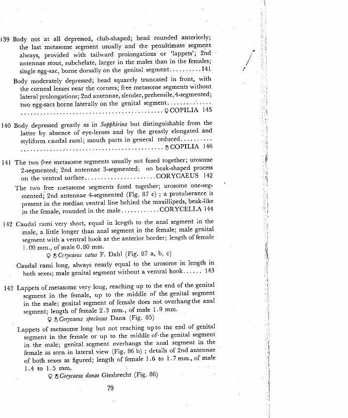
139 Body not at all depressed, club-shaped; head rounded anteriorly; the last metasome segment usually and the penultimate segment aIways, provided with tailward proiongations or 'lappets'; 2nd antennae stout, subchelate, larger in the males than in the females; single egg-sac, borne dorsally on the genital segment.. . . . . . . . .I41
Body moderately depressed; head squarely truncated in front, with the corneal lenses near the corners; free metasome segments without lateral prolongations; 2nd antennae, slender, prehensile, 4-segmented;
. . . . . . . . . . . . . . two egg-sacs borne laterally on the genital segment. . . . . . . . . . . . . . . . . . . . . . . . . . . . . . . . . . . . . . . . . . . . . 3 COPILIA 145
140 Body depressed greatly as in Sapjhirina but distinguishable from the latter by absence of eye-lenses and by the greatly elongated and styliform caudal rami; mouth parts in general reduced.. . . . . . . . . . . . . . . . . . . . . . . . . . . . . . . . . . . . . . . . . . . . . . . . . . . . . $COPILIA 146
141 The two free metasome segments usually not fused together; urosome 2-segmented; 2nd antennae 3-segmented; no beak-shaped process on the ventral surface.. . . . . . . . . . . . . . . . . . . . .CORYCAEUS 142
The two free 1:letasome segments fused together; urosome one-seg- -
mented; 2nd antennae 4-segmented (Fig. 8 7 e) ; a protuberance is present in the median ventral line behind the maxillipeds, beak-like
. . . . . . . . . in the female, rounded in the male. .CORYCEL,LA 144
142 Caudal rami very short, eclusrl in length to the anal segment in the male, a little longer than anal segment in the female; male genital segment with a ventral hook at the anterior border; length of female i .OO mm., of male 0.80 mm.
Q $Ccrycaeus cutus F . Dahl (Fig. 87 a, 11; C)
Caudal rami long, always nearly equal to the urosome in length in both sexes; male genital segment without a ventral hook. . . . . . 143
143 Lappets of metasome very long, reaching up to the end of the genital segment in the female, up to the middle of the genital segment in the male; genital segment of female does not overhangthe anal segment; length of female 2 . 3 mm., of male 1 . 9 mm.
Q $ Corycaeus specioszrs Dana (Fig. 85)
1-appets of metasome long but not reaching upto the end of genital segment in the female or up to the middle of-the genital segment in the male; genital segment overliangs the anal segment in the female as seen in lateral view (Fig. 86 b) ; details of 2nd antennae of both sexes as figured; length of female 1.6 to 1 . 7 mm , of male 1.4 to 1.5 mm.
Q $ Corycaeus danae Giesbrecht (Fig. 86)

Fig. 85. Colycaeus speciosus Dana ( a ) Female, dorsal view. (b) Male, dorsal view. (after Giesbrecht).
Fig. 86. C'ol_ycaeus danae Giesbrecht ( a ) Female, dorsal view. ( b ) Female, lateral view-note the slanting junction between genital and anal segments. (c) Female, 2nd antenna (note 3 segments). Id) Male, dorsal view. ( e ) Male, 2nd antenna. (a, b, c, e. after Sewell, d. after Colefax).
80

144 Outline of the body, particularly the genital segment, in lateral view as figured; length of female 0.85 to 1 .OO mm., of male 0.82 to 0.95 mm.
Q $ Corycella gibbzila Giesbrecht (Fig. 87d, e, f )
145 Distance between eye-lenses less than the lens diameter; length of anal segment 1$ times its least width : elldopod as long as firs: exopod segment in 4th leg; length 3.2 to 5.4 mm.
Q Copilia vitrea (Haeckel) (Fig. 88 a, 11, c)
Distance between eye-lenses less than twice lens diameter; length of anal segment 5 times its least width; endopod as long as first exopod segment in 4th leg; length 2.2 to 4.. 1 mm
QCopilia mirabilis Dana (Fig. 88, d, e, f )
Distance between eye-lenses approximately three times the lens diameter; length of anal segment 24 times its least width; endopod distinctly shorter than first exopod segment in 4th leg; length 2.2 to 4.4 mm.
3 Copilia quarlrata Dana (Fig. 88 g, 11, j)
Fig. 87." Corycneus catzls F. Dahl. (a) Female, dorsal view. (b) Male, dorsal view. (c) Male, urosome, lateral view (note hook). Corycella gibbula Giesbrecht (d) Female, lateral view (note ventral beak). (e) Female, 2nd antenna (note 4 segments). ( f ) Male, lateral view. (a, b, c, d, f. after Colefax; e. after Wolfenden).

Fig. 88. Copilia vitrea (Haeckel) Q (a) Dorsal view. (b) Urosome, ventral view. (c) 4th leg. Copilia mirabilis Dana Q ( d ) Dorsal view. ( e ) Urosome, ventral view. ( f ) 4th leg. Copilia quadrata Dana Q (g) Dorsal view. ( h ) Urosome, ventral view. (j) 4t1~ leg. (after Lehnhofer).
Fig. 89 (a) Copilia uitrea (Haeckel) $ dorssl view. Copilia mirabilis Dana, 5 dorsal view (after Brady) Dana, 8 dorsal view. (after Giesbrecht).
82
(after Colefax) (b) ( 2 ) Copilia yuadrata

Fig. 90. Copilia iritrea (Haeckel) $ jaj maxilliped; Copilia mirabilis Dana$ (b) maxilliped of forma typica (c) maxilliped of forma platyo~lyx; Copilia quadrats Dana $ ( d ) maxilliped (after Lehnhofer)
ir
146 Length of cephalosome is more than 213 of breadth . . . . . . . . . . 147 Length of cephalosome is 213 of breadth or less; details of maxilliped
(in t~ o forms) as figured; length 3 .2 to 6 .1 mm. $ Copilicl mirabilis Dana (Fig. 89 b; 90 b, c)
147 Anterior border of cephalosome evenly rounded; maxillipeds as figured, terminal segment whip-like, 2nd segment comparatively large; length 5.5 to 9.0 mm.
$ Copilia vitrea (Haeckel) (Fig. 89 a; YOa)
A:~rel-ior border of cephaloqome flat or slightly incurved; maxillipeds as figured; terlninal segment is a curved claw, 2nd segment is long and narrow; length 3.5 to 5.7 mm.
8 Copilia quad~ata Dana (Fig. 89 c; 90 d)
148 Females only : urosome distinctly narrower than the metason~e; 5- seginrnted, or 6-segmented when the genital segment is distinctly divided into two; 1st urosome segment is not concealed; it bears the 5th pair of legs which are small and alike i11 both sexes; dorsal surface not as iridescent as in the males. . . . . . . . . . . . . . . . . . . . . ,149
Males only : urosome \vide, only a little narrower than metasome; alw~ays 6-segmented; ! s t urosome segment concealed; epimeral plates well developed on urosome segments 2 to 5 ; dorsal surface of body usually iridescent or brightly tinted or both. . . . . . . . . . I50
149 Outline of body long and narrow as figured; 4th leg wit11 the endopod as long as the exopod; length 2 . 3 mm.
Q Snpphiriila ovatolatlceolata Dana (Fig. 91 a, b)

Fig. 91. Sapphilitla ovatolatzreolata Dana 9 ( a ) Dorsal view7 ( b ) 4th leg. Sapphirina auro7ziten.r Claus Q (c ) Dorsal view. ( d ) 4th leg. Sapphiritza tzigro~naculatc Dana Q (e) Dorsal view. If) 4th leg. (a, b, after Colefax, c, d, f. after Wilson; e. after Giesbrecht).
Fig. 92. ( a ) Sapphiritza ovatolanceolata Dana $ Dorsal view. ( b ) SaMhirina aurotzitetx Claus 8 Dorsal view. (c) Sapphiritza ~zigromaculata Dana $ Dorsal view. (a, c. after Giesbrecht; b. after Colef'dx).

Fig. 93. B o r n o l o ~ ~ ~ u s sp. ( a ) Female, dorsal view. (L) Female, maxilliped. (c) Male, dorsal view (at greater magnification than the female). (d) Male, maxilliped (original).
Outline of body as figured; 4th leg with the endopod shorter than the exopod but over 4 its length; length 2 .3 mm.
9 Sapph i~ ina aurotzifetzs Claus (Fig. 91 c, d)
Outline or the body as ligured; 4th leg wit11 the endopad only one half as long as exopod; length 1 .') to 2 .0 mm.
Q Sapphiii~za t2igronznculafa Dana (Fig. 91 e, f )
150 'Third and fourth metasome segments ah \vide as cephalosome; coriieal lenses not visible in dorsal view, length 4. 1 mm.
$ S a f i h i ~ i i l a ouatolatzceola[a Dana (Fig. 92 a)
'l'hird and fourth metasolne seginents n a l * r o ~ ~ e r than cephalosone; corneal lenses visible in dorsal vie\v . . . . . . . . . . . . . . . . . . . . . . . . 151
15 1 Caudal rami hardly 16 times as long as broad, length 2 . 2 mm. $ Saj$hi~inn nzlronite~zs Claus (Fig. 92 b)
Caudal rami a t least 1 ', times as long as broad; length 2.05 to 2.45 n1 n1.
$ Sapphi~ii la nig~omaculata Dana (Fig. 92 c)
152 Maxillipeds in the female turned forward outside the other mouth parts and more or less fused with the head, the apical claw S shaped; in the male not fused to the head; gill-parasites of fishes often found
free-swimming in the plankton; Q d.6 to 0.75 mm.; iS 0.5 to 0.70 mm.
Q 3 Bomolochus species (Fig. 93)
Sl'ote: The figures given are those of an undercribed species observed frequently in plankton. Though the number of segments in swimming legs and in the urosome appeared to be fewer than in the adult, a t least the male depicted seemed to be sexually mature.
Brady, G.S. 1883. Report on the Copepoda collected by H.R.I.S. "Challenger" during the years 1873-1876. Challenger Refiorts 8, London.
Brehm, 1'. 1953. Indische Diaptomiden, Pseudodiaptomiden and Cladocelen. Oster- reiche ~oologische <eitschrl/t, 4.
Cleve, P.T. 1901. Plankton from the Indian Ocean and Malay Archipelago IT. Su. Vet-Akaa. Hnndl. 35, Stockholm.
Dakin, W.J. & Col~fax, A.N. 1940. Plankton of the Australian coastal waters off New South Wales, Part I. Pzrblzcations of the Uniu. of Sydney, Dei,. of <oolo,ag, Monograph N o . 1.
Farran, G.P 1936. Copepoda. Great Batrier Reef E~peAt ion 1928-29, Scientzfic Repotts 5, 3, London.
Giesbrecht, W. 1892. Pela_~ische Copepoden, Fauna und FLolotz d e ~ G o b s ion Areapel, 19, Berlin.
Krishnaswamy, S. 1951. Notes on the undescribed males of two species of Copepoda 3'. IVashLzgton Ac. SC. 41.
1957. Studies on the Copepoda of Madras. Pub. by the Univ. of A4adras.
Lehnhofe:., K. 1926. Copepoda : Copilia. Tl'issenschaftliche Ergebnisse der Deutschert Tiefsee-Exfieditian auf dem daa';fer "Va1di1'ia" 1898-1899, 23, 3, Jena.
Sars, G.O. 1903-1921. All Account of the Crustacea of Norway. Uergen. \'ols. IV to \'I11 on Copepoda.
Scott, A. 1909. The Copepoda of thc Siboga Expedition 1099-1900, Si6oaaExpedile Ahnograph 29, Leiden.
Sewell, R.B.S. 1912. Notes on the surface-living Cvpcpoda oC the Ba); ofBengal, 1 Kr 11. Rec. Indian Af i~s . 7.
1914. Notes on the surface Copepoda of the Gulf of hfannar. Spoliu <tylutlica. 9.
1924. A preliminary note on some newr speclea of Copepoda. Rec. It:diun .Mus. 16.
- 1924. Fauna of the Chilka Lake, Crustacea Copepoda. i\fenl. Indiun X ~ I I S . 5.
- 1929. The Copepocla of Indian Seas, Calanoida Amphaskandria. izfenfi. Indic~z MUJ. 10.
1932. The Copepoda cf Indian Seas, Calanoida Hetclarthrandria. Ibzd. 10. 1947. The Free-swimming Planktonic Copepoda : Systematic Account. 'r/te Scient$,- Reports o f the John Murray Expcdzlzon 1933-34, 8.
-- 1951. Thc embionts and parasites of the Planktonic Copepoda of the Arabian Sea. Ibzd. 9.

Stcuer, A. 1934. Two new copepods of the genus Acartia fror,~ Burma. Rec. Indian M u s . 36.
Thompson, I.C. & Scott, A. 1903. Report on the Copepoda collected by Prof. Herdman a t Ceylon in 190'2. Relort on Ceylon Pearl Oyster Fisheries and Marine Biology. Royal Society, London, Part I , No. 7
Wilson, C.B. 1932a. The copepods of the M'oods Hole Region, Massachusetts. Bull. U.S. Nut. M u s . 158.
1932b. The copepod crustaceans of Chesapeake Bay. Proc. U.S.NaNat. M u s . 80.
\Yolfenden, R.N. 1906. Notes on the collection ol copepoda. Fauna and Geoxr!,hy a/ the A.laldi~,e and Laccuclir,e Archifielagoes. 2, Cambridge.