Principles of Rhythmic Motor Pattern Generation · PHYSIOLOGICAL REVIEWS Vol. 76, No. 3, July 1996...
31
PHYSIOLOGICAL REVIEWS Vol. 76, No. 3, July 1996 Printed in U.S.A. Principles of Rhythmic Motor Pattern Generation EVE MARDER AND RONALD L. CAIABRESE Volen Center, Bmndeis University, Waltham, Massachusetts; and Department of Biology, Emory University, Atlanta, Georgia I. Introduction II. What Kinds of Behaviors Are Controlled by Pattern Generators? III. Circuit Building Blocks A. Identification of neurons in pattern-generating networks B. Intrinsic cellular properties C. Properties of synaptic connections D. Network modules: small networks IV. Specific Cases: Physiological and Computational Approaches A. Elemental pattern generators: intrinsic membrane properties and reciprocal inhibition B. Intersegmental coordination c. circuit switching V. Conclusions A. Reevaluating the concept of command neurons B. Organizational rules 688 688 689 692 693 693 693 701 705 708 708 709 Marder, Eve, and Ronald L. Calabrese. Principles of Rhythmic Motor Pattern Generation.Physiol. Rev. 76: 687- 717, 1996.-Rhythmic movements are produced by central pattern-generating networks whose output is shaped by sensoryand neuromodulatory inputs to allow the animalto adapt its movements to changing needs.This review discusses cellular, circuit, and computational analyses of the mechanisms underlying the generation of rhythmic movementsin both invertebrate and vertebrate nervous systems. Attention is paid to exploring the mechanisms by which synaptic and cellular processes interact to play specific roles in shaping motor patterns and, consequently, movement. I. INTRODUCTION Although the role of single neuron oscillators and oscillatory circuits in the production of rhythmic move- ments has been appreciated for a long time (57, 99, 100, 123, 267), recent experimental and theoretical work has suggested that oscillatory processes are important in vi- sual processing (107), olfaction (e.g., Refs. 172,173), mem- ory formation (177), and a wide variety of other sensory, motor, and cognitive tasks. The widespread significance of oscillatory processes in multiple brain computations makes it crucial to understand how single neuronal oscil- lators influence the dynamics of the circuits in which they operate, as well as to understand the mechanisms by which oscillatory circuits function. The central pattern-generating circuits that generate rhythmic movements in many animals have been inten- sively studied for a long time, and there is a vast and often idiosyncratic literature in this field. Rather than attempt the impossible task of providing an exhaustive review of all preparations, here we have focused attention on sev- eral preparations that illustrate principles of organization in oscillatory networks. By so doing, we hope that the generalities among pattern-generating systems will be more apparent and that these may provide insight to those interested in oscillatory processes in nonmotor systems as well. Obviously, many preparations that we have ne- glected here have also provided fundamental insights into the mechanisms of rhythmic motor pattern generation in animals. Rhythmic movements in animals are controlled by neural networks that provide the timing of motoneuron discharge. The central components of these networks are capable of producing rhythmic patterns of activity, al- though sensory information may be essential for the ap- propriate response of these networks to behavioral requi- sites (see Refs. 57, 113, 123,242 for other recent reviews). Historically, there was a long and protracted contro- versy between those who believed that rhythmic motor patterns result from chains of reflexes, in which each movement was “strung together” from successive activa- tion of reflexes, versus those who believed that they were 0031-9333/96 $15.00 Copyright 0 1996 the American Physiological Society 687
Transcript of Principles of Rhythmic Motor Pattern Generation · PHYSIOLOGICAL REVIEWS Vol. 76, No. 3, July 1996...

PHYSIOLOGICAL REVIEWS
Vol. 76, No. 3, July 1996
Printed in U.S.A.
Principles of Rhythmic Motor Pattern Generation
EVE MARDER AND RONALD L. CAIABRESE
Volen Center, Bmndeis University, Waltham, Massachusetts; and Department of Biology, Emory University, Atlanta, Georgia
I. Introduction II. What Kinds of Behaviors Are Controlled by Pattern Generators?
III. Circuit Building Blocks A. Identification of neurons in pattern-generating networks B. Intrinsic cellular properties C. Properties of synaptic connections D. Network modules: small networks
IV. Specific Cases: Physiological and Computational Approaches A. Elemental pattern generators: intrinsic membrane properties and reciprocal inhibition B. Intersegmental coordination c. circuit switching
V. Conclusions A. Reevaluating the concept of command neurons B. Organizational rules
688 688 689 692 693 693 693 701 705 708 708 709
Marder, Eve, and Ronald L. Calabrese. Principles of Rhythmic Motor Pattern Generation. Physiol. Rev. 76: 687- 717, 1996.-Rhythmic movements are produced by central pattern-generating networks whose output is shaped by sensory and neuromodulatory inputs to allow the animal to adapt its movements to changing needs. This review discusses cellular, circuit, and computational analyses of the mechanisms underlying the generation of rhythmic movements in both invertebrate and vertebrate nervous systems. Attention is paid to exploring the mechanisms by which synaptic and cellular processes interact to play specific roles in shaping motor patterns and, consequently, movement.
I. INTRODUCTION
Although the role of single neuron oscillators and oscillatory circuits in the production of rhythmic move- ments has been appreciated for a long time (57, 99, 100, 123, 267), recent experimental and theoretical work has suggested that oscillatory processes are important in vi- sual processing (107), olfaction (e.g., Refs. 172,173), mem- ory formation (177), and a wide variety of other sensory, motor, and cognitive tasks. The widespread significance of oscillatory processes in multiple brain computations makes it crucial to understand how single neuronal oscil- lators influence the dynamics of the circuits in which they operate, as well as to understand the mechanisms by which oscillatory circuits function.
The central pattern-generating circuits that generate rhythmic movements in many animals have been inten- sively studied for a long time, and there is a vast and often idiosyncratic literature in this field. Rather than attempt the impossible task of providing an exhaustive review of all preparations, here we have focused attention on sev-
eral preparations that illustrate principles of organization in oscillatory networks. By so doing, we hope that the generalities among pattern-generating systems will be more apparent and that these may provide insight to those interested in oscillatory processes in nonmotor systems as well. Obviously, many preparations that we have ne- glected here have also provided fundamental insights into the mechanisms of rhythmic motor pattern generation in animals.
Rhythmic movements in animals are controlled by neural networks that provide the timing of motoneuron discharge. The central components of these networks are capable of producing rhythmic patterns of activity, al- though sensory information may be essential for the ap- propriate response of these networks to behavioral requi- sites (see Refs. 57, 113, 123,242 for other recent reviews).
Historically, there was a long and protracted contro- versy between those who believed that rhythmic motor patterns result from chains of reflexes, in which each movement was “strung together” from successive activa- tion of reflexes, versus those who believed that they were
0031-9333/96 $15.00 Copyright 0 1996 the American Physiological Society 687

688 EVE MARDER AND RONALD L. CALABRESE Volume 76
generated by central neural oscillators. This debate was resolved for many with the demonstration that the salient features of the flight motor pattern in locust could be generated in the absence of patterned sensory input (324). Subsequently, numerous preparations were placed in vitro, with all sensory inputs removed or silenced, and investigators were able to obtain rhythmic motor patterns in preparations containing only the central nervous sys- tem (267). The strong correspondence between these fic- tive motor patterns and those seen in behaving animals caused many to neglect the importance of sensory shaping of motor patterns. Indeed, the importance of sensory con- trol in producing an adaptive motor pattern has been ele- gantly demonstrated in the same animal, the locust, that was used to demonstrate the ability of the central nervous system to generate a fictive motor pattern (242,325). The dynamic interplay between central and sensory mecha- nisms in the generation of adaptive movements is seen in all preparations. In some preparations, sensory informa- tion may be used primarily to initiate or terminate ongoing movements or to modulate cycle period and amplitude in a graded fashion. In others, sensory information provides critical timing cues.
II. WHAT KINDS OF BEHAVIORS ARE CONTROLLED BY PATTERN GENERATORS?
A wide variety of behaviors are generated by rhyth- mic pattern-generating circuits. These include ongoing and stereotyped movements such as breathing, chewing,
wig, running, flying, and swimming. Some movements such as vertebrate respiration and the neurogenic leech heartbeat are ongoing; others are episodic. Some behav- iors are only rarely and briefly seen, as in escape or scratch behaviors (26,26,292). Others, such as swimming in lampreys and fish, can occur for short or long periods of time. Some behaviors such as hatching (20,21) or ecdy- sis (226) occur in development. Rhythmic behaviors also differ in the extent of their modulation by sensory input. Behaviors like respiration need only be modulated in fre- quency and amplitude to meet the animal’s needs. Others such as locomotor rhythms require cycle-by-cycle correc- tive signals to match them to the environment. In limbed vertebrates, movements in which limbs can operate inde- pendently are selected from a repertoire of behaviors (25, 26, 292). In many movements, intersegmental coordina- tion is required, and networks controlling different func- tions must often be coupled. For example, the rhythmic network controlling respiration must be coupled some- how to those controlling running. Most animals use the same muscles in different movements. Some of the central circuitry underlying these movements must involve the shared use of some of the elements that make up these rhvthmic neural networks. Understanding which elements
these are grouped into functional challenges of today’s work.
are shared and how circuits is one of the /
III. CIRCUIT BUILDING BLOCKS
In response to the diversity found in the circuits that generate rhythmic behaviors, Getting (99,100) introduced the term building bloc/~ to describe circuit components that are commonly found in many circuits. Getting’s aim was to describe the “alphabet” of neuronal properties that could be combined in central pattern-generating circuits to produce different functional ensembles of neurons. Getting’s attempt to ascribe functional consequences to individual cellular properties and patterns of connectivity influenced greatly much of the work during the past 10 years and has aided many of us in organizing our ideas concerning circuit operation.
A. Identification of Neurons in Pattern-Generating Networks
One of the most daunting tasks facing motor systems physiologists is the identification of the neurons that form part of the central circuitry that generates rhythmic move- ments. Indeed, the extent to which progress has been made in understanding a given preparation can be largely attributed to the degree to which the component neurons or neuron classes have been successfully identified. Ex- cept in rare cases, almost all of the relevant neurons are found within complex central nervous systems, and they are usually embedded in ganglia, brains, and nuclei that also contain neurons with other functions. Before it is possible to establish the properties of the neurons and their interactions that give rise to rhythmic movements, it is necessary to locate at least a major number of the constituents and to have these in a preparation amenable to both intracellular and extracellular recording during ongoing fictive behaviors. A number of strategies are used to identify and locate the neurons of these central circuits.
In principle, the study of a motor system should start with a description of the behavior. Usually this is accom- plished with video recordings of the animal during natural and unrestrained movement (e.g., Refs. 20, 30, 111, 112, 132, 168). This allows the investigator to determine the natural periods, durations, and variations in the behavior as it unfolds without experimental perturbation. The ulti- mate challenge of the motor system physiologist is to explain these unfettered movements.
Once a behavior is characterized, it is necessary to determine the sequence of muscles activated to produce it. Often implanted electromyogram (EMG) electrodes are used to characterize the sequences of muscles during be- havior (e.g., Refs. 112, 256). This can be straightforward; in other cases, obtaining this information can be ex-

July 1996 RHYTHMIC MOTOR PATTERN GENERATION 689
tremely difficult. This is especially true when numerous muscle groups are activated simultaneously in a complex movement or in the case of animals such as mollusks where body wall muscles are not organized into discrete bundles. However, in the absence of knowledge of the sequence of muscles activated, it can be difficult to deter- mine the motor patterns required to make the movements and, therefore, to identify the motoneurons whose activity needs to be controlled.
The motoneurons that innervate a given muscle are usually not difficult to find. Most frequently, a combina- tion of anatomic and physiological techniques allows the investigator to locate the neurons in the central nervous system that project directly to a muscle and innervate it. Although it is possible to make errors of identification if motoneurons innervating different muscles are electri- cally coupled, if the muscles are difficult to record from, or if the motoneurons cannot be stimulated individually, these errors result from technical limitations, not an inad- equate conceptual framework.
Identification of the interneurons that participate in the generation of rhythmic movements depends on sev- eral criteria. Investigators look for 1) interneurons that synapse directly on the relevant motoneurons, 2) neurons that are active in time with the rhythmic motor pattern, and 3) neurons that initiate, terminate, or change the ex- pression of an ongoing rhythmic motor pattern. However, in practical terms, these approaches have intrinsic difE- culties. For example, the interneurons that influence a behavior may not actually fire in time with that behavior. This is especially important as we come to appreciate the importance of neuromodulatory and neurosecretory influences that may modify the excitability of neurons over long time scales (123, 229, 230). Additionally, inter- neurons that are important in programming a behavior may be a part of a large set of interneurons, with overlap- ping connections and functions, so that perturbations of a single neuron may have little or no influence on the ongoing motor patterns. These two problems have, in practical terms, significantly hampered interneuronal identification in most vertebrate nervous systems and have complicated work even in invertebrate nervous sys- tems.
Recent appreciation that pattern-generating neurons can change their activity patterns (75, 136, 137, 156, 201, 202, 311-313) seriously complicates the identification of interneurons that form pattern-generating circuits in both invertebrate and vertebrate nervous systems. Because neurons cannot be reliably identified on the basis of their activity patterns alone, cells with different patterns of activity cannot be assumed to be different classes of cells. Moreover, because the same neuron can display different patterns of activity, a single recording will also be inade- quate for classification (209). The advent of slice prepara- tions in the analysis of vertebrate motor systems has
opened up the possibility of making simultaneous re- cordings from two or more neurons and offers the prom- ise of further advances in this area.
B. Intrinsic Cellular Properties
When isolated from all synaptic inputs, some neurons are silent, some show tonic unpatterned activity, some fire bursts of action potentials, and some display plateau potentials.
1. Neuronal oscillators and plateau neurons
Neurons with slow oscillatory properties are found in all nervous systems (145, 178, 279, 293, 294). Neural oscillations are now thought to play significant roles in memory formation (143, 177), sensory processing (see Ref. 107 for a recent review), as well as in the generation of rhythmic movements.
Some neurons such as R15 in Aplysia and the ante- rior burster (AB) neuron in the crustacean stomatogastric ganglion (STG) are robust single-cell oscillators and gen- erate periodic bursts of action potentials followed by si- lent interburst intervals. Such neurons maintain relatively fixed periods unless perturbed and can be entrained or reset in a 1:l fashion within a limited frequency range, while other stimulus patterns result in other patterns of phase locking or loss of coordination (17, 51, 164, 200, 249, 250, 257, 261).
Single-cell oscillators have been extensively studied by theorists. A number of points relevant to the nervous system have emerged in modeling studies. First, by chang- ing the balance of conductances in model oscillators, it is usually possible to obtain a range of behaviors from the “same” model, that is, silent, oscillatory, or excitable (174, 179, 257, 261). Second, using “realistic” conduc- tance-based models, it is possible to produce membrane potential oscillations, or bursts, using a variety of different membrane conductances (19,24,27,48-50,52,73,88,118, 133,210,258,300). In real neuronal and muscle oscillators, many different combinations of ion channels are used to produce oscillatory activity (144, 300). Because the de- tailed waveform of the oscillator depends critically on the specific properties and types of the conductances found in the cell, each neuronal oscillator may have a unique frequency range and response to injected and synaptic current (286).
An intriguing result is seen in recent work of a model of R15 in Aplysia (48). This model can show different modes of activity, depending on initial conditions, without changing the parameters of any of the currents and can be persistently switched among these modes by a transient perturbation, such as that produced by a synaptic input. The interaction between the effects of modulators that change the balance of conductances and the mode
690 EVE MARDER AND RONALD L. CALABRESE Vobume 76
switches that can occur in the absence of changes in any of the parameters in the model was explored further (42, 49).
Modeling studies show that some properties of oscil- lators are generic, while other properties of oscillators, which may be critical to understanding their role in neural circuits, are not common to all oscillators (164, 291). All oscillators, by definition, can be reset or entrained. How- ever, neural oscillators may respond differently to steady current injection or synaptic inputs. Skinner and co-work- ers (285,286) varied the balance of the two conductances in the simple Morris-Lecar model (210). In some parame- ter ranges, the oscillator maintains a relatively constant burst duration, and as it is depolarized, it increases in frequency. In other parameter ranges, the burst duration is sensitive to the level of injected current, and the cycle frequency first increases and then decreases in response to depolarization (285, 286). Therefore, these two types of oscillators will respond to synaptic inputs in different ways. Moreover, modulatory substances that change the relative strengths or properties of one or more of the currents in a neuronal oscillator may alter qualitatively the oscillator’s response to synaptic inputs (e.g., Ref. 159).
Neurons that display plateau properties make rapid transitions between two relatively stable membrane po- tentials (182). When such a neuron is at a hyperpolarized resting potential, a short pulse of depolarizing current will trigger a sustained depolarization (plateau) that will long outlast the stimulus. The time constants of inactivation of slow inward currents play an important role in de- termining the duration and stability of the plateau. When in the plateau, a short hyperpolarization will terminate the plateau.
The ability to produce plateaus may be strongly in- fluenced by the pattern of activity in a neuron. In HeZti (248), P cells fire single action potentials when stimulated at low frequencies, but high frequencies of stimulation induce sustained plateaus. In this case, the transition is explained by Ca2+-dependent inactivation of the outward currents that are responsible for spike repolarization (63). Other neurons may require a prior period of inhibition to initiate a plateau. If the neuron’s resting potential is relatively depolarized so that the inward current sus- taining the plateau is substantially inactivated, then hyper- polarization will be required to deinactivate this current before a plateau can be produced (e.g., Ref. 8).
Plateau properties in follower neurons decrease the importance of synaptic strength in networks (182, 183) because even modest hyperpolarizations or depolariza- tions can trigger transitions between states. In the ex- treme case of perfect bistability, a neuron with plateau properties will only make the transition between its two stable membrane potentials when perturbed. However, many neurons spontaneously make the transition between a hyperpolarized resting potential and a depolarized pla-
teau associated with rapid firing. Many neurons make the transition in the opposite direction spontaneously due to inactivation of the currents that support the depolarized plateau. Recent work in both vertebrate and invertebrate preparations is showing that plateau properties are a com- mon feature in many neurons of motor circuits (18, 62, 138-141, 160, 254, 262).
In some oscillatory neural networks, it is clear that plateau properties are crucial to pattern generation (5, 8, 9, 234). They permit rapid transitions between inactive and active periods upon termination or escape from inhi- bition and provide sustained periods of depolarization during which they release transmitter (5,46, 72). In other cases, plateaus may be crucial for determining the phase relationships among pattern-generating neurons (e.g., Ref. 222).
The ability to produce plateaus is highly modified by modulatory inputs. The activation of plateau properties may be the mechanism by which a rhythmic motor system is activated by neural inputs (18, 77,221,262,312). Modu- lation of plateau properties may influence the specific phase relationships in a rhythmic motor pattern (87, 222) or may alter its frequency (262).
2. Intrinsic properties are voltage dependent
Injected current and synaptic potentials can appreci- ably change the f?ring patterns of many neurons. Trivially, silent neurons often fire tonically when depolarized. More interestingly, some neurons burst when depolarized, while others may only generate bursts or plateau properties when hyperpolarized (178). Some neurons, such as those found in thalamocortical networks, switch between differ- ent modes of activity, depending on their membrane po- tential (175, 198, 199, 294) which in turn is dependent on modulatable leak channels. The effects of membrane potential on the firing patterns of neurons result from the steady-state activation and inactivation properties of the specific mixes of membrane currents that are present at each membrane potential.
Many neurons have slow conductances that influence their behavior and those of networks over long time scales. One such conductance is &,, an hyperpolarization activated inward current that plays an important role in many bursting and plateauing neurons (4,73,79,104,161, 162, 198, 290). This conductance can be “invisible” in some circumstances but can be significant under others. It is important to remember that the electrical properties of a neuron when isolated from all synaptic inputs may not easily predict its activity in a circuit, if the synaptic inputs it receives move its membrane potential to regions where different voltage-dependent conductances are ac- tive than those in which the isolated neuron fires.
3. Modulation of intrinsic membrane properties
Modulatory inputs can markedly change the intrinsic properties of neurons by modifying one or more of the

July 1996 RHYTHMIC MOTOR PATTERN GENERATION 691
voltage-dependent conductances of a neuron or by evok- ing novel conductances (123, 152, 182, 183). Examples of neuromodulatory regulation of bursting and plateau properties abound in the literature (123). The effects of neuromodulatory agents are seen at every level in motor systems, from sensory neuron, to interneuron, to moto- neuron to muscle, in invertebrate and vertebrate prepara- tions.
Sensory neurons play crucial roles in initiating and modifying rhythmic movements. Therefore, modulatory control of the gain of sensory responses will strongly in- fluence movement (54, 280). There are three general mechanisms by which sensory pathways can be modu- lated so that a given stimulus produces a different strength output: 1) modulation of the transduction process itself, 2) modulation of the excitability of the primary sensory neurons so that a given change in conductance produced by the stimulus is more or less effective in activating the primary sensory neuron, and 3) modulation of the release of transmitter from the sensory neuron terminals so that the efficacy of sensory neuron action potentials is altered.
There are now examples of modulation of many dif- ferent types of sensory neurons. Particularly relevant to movement control are proprioceptors, pain and touch re- ceptors, and sensory receptors that respond to wind direc- tion. Crustaceans have several different classes of muscle receptors that respond to stretch and/or muscle tension (236). These include the abdominal muscle receptor or- gans and the oval organs, which provide sensory feedback for the ventilatory system. The response of these sensory receptors to a defined stretch or perturbation is modified by amines and peptides (236). A similar story is found in locusts, where the forewing stretch receptors are modu- lated by octopamine (253).
The effect of modulatory substances on the release of transmitter from sensory neurons has been extensively studied in both vertebrate and invertebrate nervous sys- tems. The release of substance P and other sensory trans- mitters from dorsal root ganglion neurons is modulated by enkephalin and a variety of peptides (90, 212, 327), and the biophysical mechanisms underlying the control of transmitter release from these neurons are extensively studied (83).
Modulation of the efficacy of synaptic strength has been extensively studied in the sensory to motoneuron reflex pathways in Aplysia (43, 127). In this preparation, modulation of the reflex pathway is behaviorally relevant, has been characterized in biophysical and biochemical detail, and is one of the premier illustrations of how modu- latory transmitters can change the gain of synaptic con- nections in a behavioral context (127).
The functional significance of a sensory stimulus de- pends critically on the conductances found in the inter- neurons driven by those sensory neurons. This principle is well illustrated in recent work in the locust flight system
(255), in which bursting properties in flight interneurons act to amplify the effect of proprioceptive inputs.
In almost all rhythmic motor systems, the pattern- generating circuitry is upstream from the motoneurons that actually drive the muscles. Until very recently, the motoneurons in almost all preparations were thought to fire in response to central drive, but not to have any long- lasting regenerative properties that could shape the dura- tion of the muscle activation. However, numerous reports have recently appeared that show clearly that modulatory substances can elicit plateau properties in motoneurons, in both vertebrate and invertebrate preparations (138. 141, 161, 162).
Most vertebrate skeletal muscles fire action poten- tials that serve to trigger muscle contraction, since the surface depolarization is carried into the t-tubule system. In contrast, many invertebrate muscles are multiply inner- vated, and contraction is a graded function of depolariza- tion (142). In these muscles, the dynamic properties of the neuromuscular junctions significantly influence ten- sion and movement. For example, a neuromuscular junc- tion that displays considerable facilitation wilI evoke more tension development subsequent to high-frequency stimulation than it will if the motoneuron is flred more slowly. In preparations such as these, the detailed pat- terning of the motor pattern will strongly shape the move- ment.
Circulating hormones and neuromodulatory cotrans- mitters can strongly modulate the tension development at both vertebrate and invertebrate muscles (23, 32, 33, 44,170,200,295). This can arise if modulatory substances influence the presynaptic release of transmitter, or by di- rect actions on the muscle fibers themselves. Peptides or amines released from motoneurons or circulating as hormones can act directly to modify the voltagedepen- dent membrane properties of the muscle fibers (32, 33). This can influence the amount of tension production as well as the contraction and relaxation rates (252, 314, 315) and can also induce intrinsic oscillatory action in the muscle fibers themselves (170, 176, 200, 295).
The presence of strong oscillatory and/or plateau properties in muscles acts to amplify the response of the muscle to a synaptic input that may be relatively small (200). Therefore, in neuromuscular systems that show considerable direct modulation of the active conduc- tances of the muscles themselves, it is possible to use the same motoneurons and muscle fibers to produce a very large range of forces, with relatively few neurons and muscle fibers. A particularly intriguing illustration of the importance of modulation of the neuromuscular junction is seen in a recent study of the locust ovipositor opener muscle (23). In this preparation, the neurally released pep tide proctolin upregulates the strength of the nerve- evoked muscle contractions driven by the central pattern generator for oviposition digging. However, during the

692 EVE MARDER AND RONALD L. CALABRESE Vohme 76
expression of the behavior, the release of proctolin de- ential release of these substances (170). In several cases, creases, and the movements abate, although the ongoing it is known that peptide cotransmitters appear to be re- motor patterns continue (23). leased only after high-frequency trains of presynaptic ac-
tivity (64, 243, 316, 317), while the small molecule trans-
C. Properties of Synaptic Connections
The critical role of the strength of synaptic interac- tions for the operation of neuronal networks is appreci- ated in both the biological community and among neural network theorists. Indeed, changes in the strength of syn- aptic connections are often considered adequate explana- tions for the changes that underlie plasticity. Not only does this position neglect the critical importance of modu- lation of membrane properties for plasticity in neural net- works, but oftentimes, dynamic features of synaptic trans- mission that are crucial for network operation are ne- glected (100).
Some of the first synaptic connections to be studied were found at vertebrate and invertebrate neuromuscular junctions. These classic preparations have some common features: 1) the synaptic current is rapid, 2) there is a short latency between the presynaptic action potential and the postsynaptic response, 3) there is an anatomically well-defined synaptic structure, and 4) the threshold for transmitter release is at a level of presynaptic membrane potential sufficiently depolarized that presynaptic action potentials are required for transmitter release. Many of the synaptic connections in the networks that generate rhythmic behavior are likely to share these features, but other types of synaptic organization also play significant roles in pattern generation.
1. Time courses of synaptic currents
Typically at vertebrate skeletal muscle neuromuscu- lar junctions, the synaptic currents rise and fall within several milliseconds. Such rapid time course events are also common in the brain. In many preparations, trains of presynaptic action potentials also evoke slow synaptic potentials that follow more rapid events. The best-known example of this is seen in the vertebrate sympathetic gan- glion, where trains of preganglionic action potentials elicit rapid, nicotinic excitatory postsynaptic potentials (EPSPs) followed by a slow inhibitory postsynaptic poten- tial (IPSP), a slow EPSP, and a late slow EPSP, which have time courses from many milliseconds for the slow IPSP and EPSP to minutes for the late slow EPSP (146).
2. Colocalixed neurotransmitters
mitters are released by each action potential. Because in most cases the peptide and amines colocalized with small molecules are responsible for slower time course postsyn- aptic actions (2, 29, 170), this means that the integrative action of a given presynaptic element in a network may change as its pattern of activity changes.
3. Graded release of neurotransmitter
Influx of Ca2’ through voltage-sensitive Ca2’ than- nels is the primary signal for transmitter release. Many neurons only release neurotransmitter in response to a rapid and large depolarization, such as that produced by an action potential, presumably because the Ca2’ currents in the presynaptic terminal require significant depolariza- tion for activation. Other neurons release transmitter at relatively hyperpolarized levels of membrane potential, presumably because their presynaptic terminals have Ca2’ currents that are activated at more hyperpolarized levels of membrane potential (5).
Many pattern-generating networks employ graded synaptic transmission (41, 74, 105, 106, 194, 241, 276). In some preparations, graded transmission controls trans- mitter release by local interneurons that may never spike. In other preparations, neurons may fire action potentials for distance communication while the integration im- portant for pattern generation is primarily graded (106). It has been suggested that graded transmission may play a role in stabilizing pattern-generating networks (163). The strength of graded synaptic connections is subject to considerable modulation (147- 150).
The cable properties of neurons can interact with the threshold for transmitter release to produce interesting “gating” phenomena For example, in neurons of the sto- matogastric nervous system, antidromic spikes may fail to release transmitter while orthodromic spikes do so (203,215). This occurs because the action potentials gen- erated within the neuropil of the STG ride on top of a slow-wave depolarization that brings the cell to threshold for transmitter release. In contrast, antidromic action po- tentials that propagate into the neuropil from distal spike initiation zones depolarize the presynaptic terminals con- siderably less, thus releasing little or no transmitter (190, 203).
4. Electrical coupling
It is now clear that many, if not most, neurons contain Electrical coupling among pattern-generating ele- multiple neurotransmitter substances. One of the interest- ments is common. It has been long appreciated that elec- ing questions for circuit analysis is understanding how trical coupling tends to synchronize the activity of the these multiple neurotransmitters function. It is important coupled neurons. However, there are increasing numbers to determine how activity patterns may result in the differ- of examples in which electrically coupled neurons do not

July 1996 RHYTHMIC MOTOR PATTERN GENERATION 693
fire synchronously and may even fire out of phase (271). A theoretical study (275) illustrates that out-of-phase ac- tivity can be produced at low coupling strengths. More- over, electrically coupled neurons may have quite differ- ent intrinsic membrane properties that contribute to the emergent properties of the networks they form (1, 185, 207). One important lesson for circuit analysis is that neu- rons that are silent and may be below their transmitter release threshold and may nonetheless contribute to pat- tern formation when these neurons are electrically cou- pled to other neurons, as they provide current through the electrical junctions that will influence the activity of their coupled partners (135, 159, 271).
ties of the component neurons and their synaptic interac- tions. The simplest elemental pattern generators employ a pacemaker neuron or neurons that can serve as a source of the rhythm. Synaptic interactions with other network elements can modify this rhythm, but the inherent activity of the pacemaker neuron is dominant. The best-character- ized example of this mechanism is the pyloric rhythm generator of the crustacean stomatogastric nervous system.
Other elemental pattern generators use neurons that have pacemaking properties, which in some cases can be modulated on and off, but reciprocal inhibitory synaptic interactions play a more important role in determining the rhythm. The elemental generators underlying Clione and Xenopus swimming and the leech heartbeat system are thought to represent such cases. The gastric system D. Network Modules: Small Networks
As the connectivity diagrams of neural circuits be- in the stomatogastric nervous system is another example
come characterized, it is clear that each of these has its in which network dynamics are an emergent process from
particular features (100). However, it is ASO clear that the interaction of intrinsic membrane properties and syn
certain two and three cell groupings are ubiquitous and aptic connections .
may serve as computational elements within neural cir- cuits. These elements include such relationships as 1) I* ‘ZJloric Wstem reciprocal inhibition, 2) delayed feed-forward inhibition with feedback excitation, and 3) electrical coupling and one-way inhibition.
Many of our ideas about small-network modules come from invertebrate preparations, where these are easily found and studied. Although vertebrate systems contain many more neurons, it is common to find classes of neurons lumped together and connectivity diagrams drawn among small numbers of “cells” where the cells are now groups of neurons. This illustrates one of the paradoxes that is found throughout the vertebrate system literature. On the one hand, researchers studying complex brain regions often assert that many of the properties of their systems depend on the emergent dynamics of large networks. On the other hand, these workers often draw simplified connectivity diagrams that resemble those that actually describe smaller invertebrate circuits and attempt to explain complex dynamics of large brain regions with these simplified diagrams. For this reason, it is critical to actually determine how the dynamics of small networks depend on the network architecture and the dynamics of the network components.
The simplest mechanism with which to generate a rhythmic motor pattern is to employ a pacemaker neuron or neurons that can serve as a source of the rhythm. The pyloric rhythm of the crustacean STG is one of the best understood central pattern-generating circuits. In this preparation, the ability of a single oscillatory neuron to generate bursts plays a particularly important role in the generation of the rhythm, although the phase relation- ships and frequency of the rhythm depend critically on network interactions and sensory and central modulation.
The pyloric rhythm consists of alternating bursts of activity in the motoneurons that dilate and constrict the pyloric region of the stomach of lobsters and crabs. The purest form of the pyloric rhythm is a five-phase rhythm (Fig. lA). The core of the rhythm is a repeating cycle of activity in which the lateral pyloric (LP), pyloric (PY), and pyloric dilator (PD) neurons fire sequentially. The inferior cardiac (IC) routinely fires in LP time, and the ventricular dilator (VD) often fires in PY time. Additionally, there is a single interneuron, the AB neuron, that fires in time with the PD neurons. The frequency of the pyloric rhythm can vary from -0.2 Hz to -3 Hz but is commonly in the l-Hz range during ongoing behavior (131, 256) or in in vitro preparations (135, 229, 230). IV. SPECIFIC CASES: PHYSIOLOGICAL AND
COMPUTATIONAL APPROACHES An unusual feature of the STG is that the pattern- generating networks are formed by the motoneurons and a few interneurons. Therefore, recordings from the STG
A. Elemental Pattern Generators: Intrinsic motoneurons provide direct information about the motor Membrane Properties and Reciprocal Inhibition pattern, as well as information about the connectivity and
membrane properties that contribute to rhythm and pat- Rhythmic activity within pattern-generating networks
results from the combination of intrinsic electrical proper- tern generation. Moreover, the small number of neurons within the STG (25-32, depending on species) allows the

EVE MARDER AND RONALD L. CALABRESE volume 76
PD
VD
LP
PY
LP
PD
LP
PD
CONTROL PILOCARPINE SEROTON
SDRNFLRhw TRNFLRFIIHZ CCAP RPCH CONTROL
simultaneous recording of a large enough number of ele- ments of the network to make analysis feasible (e.g., Fig. lA). Additionally, it is possible to delete single neurons from the network by photoinactivation subsequent to dye injection (206) and to block pharmacologically many of the chemical synaptic connections within the network (28, 86). With the use of a combination of these tech- niques, it has been possible to establish a connectivity diagram (Fig. lB) for the pyloric rhythm and generate a qualitative description of how the pyloric rhythm is gener- ated (186, 207, 208).
The AB neuron is the usual source of the pyloric rhythm, although when modulatory inputs are left attached to the STG, pyloric-type rhythms can persist after the AB neuron is killed (269). When isolated from the other pyloric network neurons, the AB neuron is able to generate bursts of action potentials (92, 122, 185, 207). The PD neurons depolarize and fire action potentials in time with the AB neuron because they are electrically coupled to the AB neuron. Thus the basic dilator phase of the pyloric rhythm is determined by the intrinsic oscil-
FIG. 1. Pyloric rhythm of crustacean stomatogastric ganglion. A: simultaneous intracellular recordings from 6 cell types that participate in pyloric rhythm. AR, an- terior bursteq PD, pyloric dilator; VD, ventricular dilator; IC, inferior cardiac; LP, lateral pyloric; PY, pyloric neurons. [Modified from Miller (205).] B: connec- tivity diagram of pyloric network of lob- ster Panulirus intmmptus. Resistor symbols denote electrical connections, and circles represent inhibitory chemical synaptic connections. PD and VD neu- rons are cholinergic (solid), and other neurons are glutamatergic (open). [Modi- fied from Hooper and Marder (X35).] C: modulators produce characteristic and different forms of pyloric rhythm. Each panel shows simultaneous intracellular recordings from LP and PD neurons and an extracellular recording from lateral ventricular nerve (lvn). Stomatogastric ganglion was isolated by sucrose block, and preparation was washed between each application. RPCH, red pigment con- centrating hormone; CCAP, crustacean cardioactive peptide. [Adapted from Marder and Weimann (189).]
latory processes in the AB neuron, as shaped by the cou- pling to the PD neurons (1). The remaining neurons in the pyloric rhythm tie at vtious phase relationships that are determined by the synaptic connections among the remaining neurons and their intrinsic membrane proper- ties. The LP and PY neurons are inhibited by the dilator group of the AB and PD neurons, but the LP neuron starts firing before the PY neurons. Factors that have been sug- gested to explain this difference in phase include the strength and time course of the synaptic potentials from the dilator group (86), the properties of transient outward current (IA) in the follower neurons (120, 121, 124, 125, 296), and the Ih in the follower neurons (103, 125, 179).
Although the STG contains only 30 neurons, the nerve that connects the STG to the rest of the central nervous system (stn) has at least 60 fibers (61). Some of the stn fibers are axons from STG neurons; however, -50 stn fibers are projections from somata in the brain and the commissural ganglia (61) that contain a large number of different modulatory substances (181, 188). Different modulatory substances evoke different forms of the pylo-
July 1996 RHYTHMIC MOTOR PATTERN GENERATION 695
ric motor pattern, in which the component neurons fire bursts of varying intensities and varying phase relation- ships (Pig. lC, Refs. 91, 119, 186, 189).
The AB and PD neurons differ in terms of their trans- mitter content (W), their responses to modulators (92, 135, 185, 187), and their intrinsic membrane properties (18,185,208). Each of these differences plays an interest- ing role in explaining how modulatory substances influ- ence pyloric network dynamics.
A number of different substances activate the pyloric rhythm by directly activating the pacemaker AB neuron. These include amines (92, 185), peptides (135), and cho- linergic agonists (187). However, it is important to remem- ber that the frequency of the network depends not only on the frequency of the AH neuron, but also on the proper- ties of the remaining neurons in the network, as has been shown physiologically (135) and theoretically (159, 271).
The differences in the intrinsic properties of the AB and PD neurons play a critical role in shaping the wave- form of the oscillator ensemble. Although the AB neuron is the “pacemaker” for the pyloric rhythm, the properties of the AB neuron are critically shaped by virtue of its electrical coupling with the PD neurons. The isolated AB neuron is a constant burst duration oscillator (l), while the AEYPD electrically coupled ensemble maintains a con- stant duty cycle (1). The mechanism by which the oscilla- tor waveform is transformed by the intrinsic properties of the electrically coupled PD neuron may be important for unde&anding numerous systems in which oscillatory elements are electrically coupled to elements with sig- nifxantly different intrinsic membrane properties.
The AB neuron releases glutamate and the PD neu- rons release acetylcholine. The glutamatergic IPSPs are more rapid than the cholinergic IPSPs (85, 184). There- fore, the relative amounts of AB or PD released neuro- transmitters control the relative duration of the inhibition the pacemaker group produces on the follower neurons (86) and can control the phase of follower neuron activity. Although the AB and PD neurons are electrically coupled, they are differentially modulated, and therefore, their con- tribution to transmitter output can vary as a function of synaptic and modulatory mixes. This provides an interest- ing mechanism by which phase relations of the follower neurons can be simply modulated (86). Other mechanisms for controlling and modulating the phase of the follower neurons depend on the properties of the voltage-depen- dent conductances in the follower neurons (77, 120, 121, 124, 179,221,296). In fact, the “take-home message” from this work on the pyloric rhythm is that the timing of the rhythm depends critically on the interplay between the strength and time course of the synaptic inhibition and the intrinsic membrane currents of the follower neurons. Indeed, modulation of any of these parameters will influ- ence the phase of the follower neuron activity.
The pyloric network in its simplicity illustrates sev-
eral very important lessons for the study of neural net- works.
1) Even in a network that is “completely defined” in terms of the identity of the constituent neurons and their interactions, it is impossible to predict the dynamics of a network without fully understanding the dynamic proper- ties of the individual neurons and their connections. More- over, this process almost certainly requires some compu- tational and or mathematical models.
2) Even very simple networks can produce multiple outputs, if any of the intrinsic or synaptic currents that participate in network function are subject to modulation.
3) The frequency of a pacemaker-driven network is not solely determined by the frequency of the isolated pacemaker, but network interactions influence it as well. Likewise, the phase of each of the components of the network is determined by both synaptic interactions and intrinsic membrane properties.
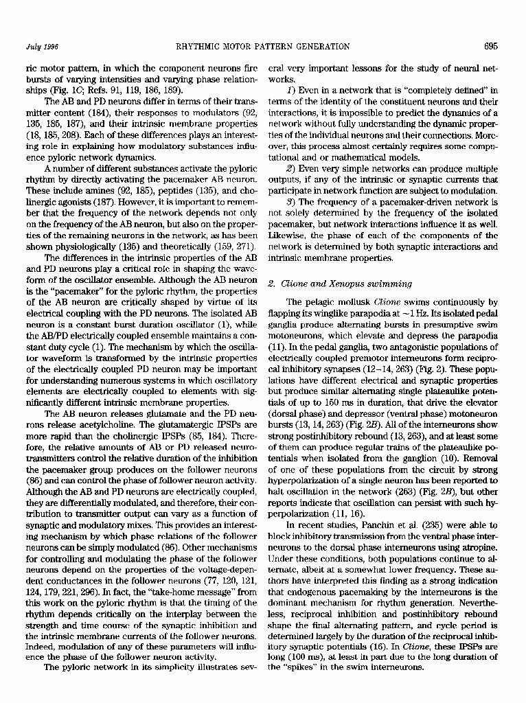
2. Clione and Xkaopus swimming
The pelagic mollusk Clione swims continuously by flapping its winglike parapodia at - 1 Hz. Its isolated pedal ganglia produce alternating bursts in presumptive swim motoneurons, which elevate and depress the parapodia (11). In the pedal ganglia, two antagonistic populations of electrically coupled premotor interneurons form recipro- cal inhibitory synapses (12- 14,263) (Pig. 2). These popu- lations have different electrical and synaptic properties but produce similar alternating single plateaulike poten- tials of up to 150 ms in duration, that drive the elevator (dorsal phase) and depressor (ventral phase) motoneuron bursts (13,14,263) (Pig. 2.B). All of the interneurons show strong postinhibitory rebound (13,263), and at least some of them can produce regular trains of the plateaulike po- tentials when isolated from the ganglion (10). Removal of one of these populations from the circuit by strong hyperpolarization of a single neuron has been reported to halt oscillation in the network (263) (Pig. 223), but other reports indicate that oscillation can persist with such hy- perpolarization (11, 16).
In recent studies, Panchin et al. (235) were able to block inhibitory transmission from the ventral phase inter- neurons to the dorsal phase interneurons using a&opine. Under these conditions, both populations continue to al- ternate, albeit at a somewhat lower frequency. These au- thors have interpreted this finding as a strong indication that endogenous pacemaking by the interneurons is the dominant mechanism for rhythm generation. Neverthe- less, reciprocal inhibition and postinhibitory rebound shape the final alternating pattern, and cycle period is determined largely by the duration of the reciprocal inhib- itory synaptic potentials (16). In Clio~, these IPSPs are long (100 ms), at least in part due to the long duration of the “spikes” in the swim interneurons.

696 EVE MARDER AND RONALD L. CALABRESE Volume 76
A
. A
Dorsal phase Ventral phase muscles musdles
pattern generator of Clione. IN, interneurons; D- 3 7
MN, dorsal motoneurons; V-MN ventral motoneu- rons. Seven IN and eight IN interneurons form reciprocal inhibitory synapses that together with their intrinsic membrane properties lead to oscil- lation in swimming pattern generator. Connec- tions from 12 IN interneurons to 7 IN/8 IN osciIla- tor stabilize oscillation during intense swimming activity. [From Arshavsky et al. (16).] B: oscil.la- tions in reciprocally inhibitory 7 IN and 8 IN inter- neurons cease when one of the interneurons is hyperpolarized strongly with injected current (CM). [From Satterlie (263).] C: synaptic connec-
I l . I
Len muscles Right muscles tionsins wimming pattern generator of Xmpus. iIN, inhibitory interneurons; eIN, excitatory inter-
A neurons; MN, motoneurons. The iIN intemeurons form reciprocal inhibitory synapses across mid- line that together with their intrinsic membrane
FIG. 2. A: synaptic connections in swimming
B
L-MN
1 set <
properties lead to oscillation in swimming pat- tern generator. [From Arshavsky et al. (16).] D: oscillations in a left motoneuron (LMN) and right intemeuron (R-IN) and rhythmic firing in right (R-vr) and left (Lvr) ventral roots during swimming motor pattern. [From Arshavsky et al. (16).] In A and C, excitatory connections are indi- cated by open triangles, inhibitory connections are indicated by solid circles, and electrical junc-
1 1
100 ms tions are indicated by resistor symbols.
The segmental motor pattern that underlies swim- ming in early stage tadpoles from Xenopz~~ bears remark- able similarities to the Clione swim pattern, and the pat- tern-generating networks that give rise to these patterns are organized along parallel lines (16). In each spinal seg- ment, motoneurons innervating axial muscles on each side alternate spikelike activity that produces the alternat- ing lateral undulations of the body associated with swim- ming. In each spinal hemisegment, inhibitory premotor interneurons, which form reciprocal inhibitory connec- tions across the midline (Fig. ZC), produce alternating spikelike potentials (F’ig. 20) that bear a remarkable simi- larity to those produced by the swim pattern-generating interneurons of Clione. Unfortunately, these interneurons have not been well characterized. It is assumed that, like the motoneurons, they fire only once during prolonged depolarization (65,288), and they show strong postinhibi- tory rebound when they are depolarized (288). Rebound is thought to be important in pacing rhythmic activity, and simulated spinal networks of these interneurons gen- erate a robust rhythm based on reciprocal inhibition and rebound. However, other mechanisms certainly contrib- ute to rhythmicity in the network. In recent experiments, K+ currents were blocked with tetraethylammonium or 3,4diaminopyridine and caused stereotypical disruptions in the rhythm, indicating their importance in rhythm gen- eration (302). Moreover, hemisected spinal cord prepara- tions can generate a swimlike rhythm (287); this rhythm
neurons by ipsilateral glycinergic axons (67) and resultant postinhibitory rebound. In such hemisected preparations, some rhythmic neuronal discharge in motoneurons can be evoked by sensory stimulation even when glycinergic inhibition is blocked with strychnine (287), providing some evidence for inherent oscillatory neuronal proper- ties.
Interesting contrasts and similarities between the Xenopus and Clione swim pattern generators highlight principles of organization. In both systems, at least under some conditions, inherent rhythmic activity by itself sus- tains rhythmicity. Postinhibitory rebound is pronounced in both systems, reciprocal inhibition produces altema- tion, and cycle period is determined largely by the dura- tion of the reciprocal inhibitory synaptic potentials (16). In contrast to the Clione swim generator, which operates nearly continuously, that of Xenopus is geared to episodic activity. Excitation necessary to maintain the prolonged depolarization necessary for a swimming bout in Xmopus is provided by activation by sensory input of excitatory intemeurons that use long dual-component [N-methyl-D- aspartate (NMDA) and non-NMDA] synaptic potentials; mutual excitation within the excitatory intemeuron pool sustains activity and leads to a summed steady depolariza- tion of the swim intemeurons (66). Modeling studies indi- cate that non-NMDA-mediated mutual excitation can sus- tain oscillation but that NMDA-mediated mutual excita- tion, enhancing postinhibitory rebound, acts to stabilize
is thought to arise from recurrent inhibition of swim inter- swimming activity and extend its lower frequency range,

swimming wave. On a neural level, each segment is thought to have its own pattern-generating network, and these segmental oscillators must be coordinated longitu-
HN (R 4 9
dinally. While this conception is useful, as we shall discuss below with reference to leech and lamprey swimming, HN (L,4 systems controlling locomotion based on axial muscles can also be viewed as distributed, producing a spatially as well as a temporally varying pattern. This is contrasted to the concentrated Clione system where temporal as- C
July 1996 RHYTHMIC MOTOR PATTERN GENERATION 697
and it steepens the dependence of frequency on synaptic Al A2 drive (259). Excitation within the CZione swim generator is sustained by electrical coupling within the reciprocally inhibitory interneuron pools (12,13). Descending modula- tory interneurons can also provide incremental excitation to initiate swimmin g activity should it stop or to acceler- ate ongoing activity, and during rapid swimming, a new class of depressor swim interneuron is recruited, which feeds back inhibition to the depressor interneurons and excitation to elevator interneurons (14, 15, 264, 265). An- other fundamental difference between the two pattern generators relates to the distributed nature of the undula- B tory swimming inXmp~~ tadpoles. Segmental axial mus- cles must be coordinated longitudinally to produce a
HE IR 5) 1
pects of the pattern predominate. HN (h4) 83
3 3. Leech heartbeat
The rhythmic constrictions (0.1 Hz) of the bilateral heart tubes of the leech, Him~do medicinalis, are paced
HN O-94)
and coordinated by the rhythmically active segmental heart (HE) motoneurons (47) (Fig. 3.B). A network of seven bilateral pairs of the heart @IN) interneurons, one pair of which is located in each of the first seven segmen- tal ganglia of the nerve cord, produces rhythmic activity
FIG. 3. Synaptic connections and oscillations in network of leech he& . t m emeurons. Al: network of 7 identiAed pairs of heart intemeu-
that paces the heart motoneurons (45,47,266) (Fig. 3Al). rons (HN cells) responsible for generation and bilateral coordination of
This network is continuously active in the isolated ner- heartbeat. Cells that make similar connections and oscillate in phase
vous system and produces a fictive motor program that are lumped together. All synaptic connections shown are inhibitory. AZ heartbeat timing oscillator includes 2 elemental oscillators in gunglti 3
can account for the constriction pattern of the hearts ob- and 4. These oscillators are connected via inhibitory connections with
served in situ. The synaptic connections among the inter- ceUs HN[l) and HN(2). B: oscillations in an elemental heartintemeuron
neurons (Fig. 3A1) and from the interneurons to the moto- oscillator. CeUs HN(R,4) and HN(L,4) oscillate in antiphase. CeU HE(R,5) as well as all other ipsilateral heart motoneurons caudal to it
neurons are inhibitory. receive inhibition from ceU HN(R,4). Oscillations in heart motoneurons
The first four pairs of heart interneurons can reset are driven by input from HN cells. HE(R,Q trace shows large inhibitory
ad entrah the rhythm of the entire Pattern-generating postsynaptic potentials during inhibited phase, partially provided by ceU HN(R,4), a& osculates in antiphase witi c& HN(R,#J Da&d he
network of interneurons (Fig. 3A2). The other three marks a potential of -50 mV. C oscillations in model elemental oscilla-
pairs of HN neurons are followers of these anterior tor. Graded and spike-mediated inhibition from each cell to its con&ala&
pairs (247). Two foci of oscillation in this network have eral homologue and intrinsic membrane properties enables cells to pro- duce oscillations in antiphase with a period of -8 s. Amplitude and
been identified in the third and fourth ganglia, where frequency of action potentials are larger than in heart interneurons.
the oscillation is dominated by the reciprocal interac- Dashed line marks a potential of -60 mV. [F’rom Nadim et al. (ZZO).]
tions of the third and fourth pair of HN interneurons, respectively (245). Reciprocally inhibitory synapses pairs can be considered an elemental oscillator. The between the bilateral pairs of HN neurons in these HN interneurons of the first and second ganglia act as ganglia (Fig. 3, A2 and B), combined with an ability of coordinating fibers, serving to link these two elemental these interneurons to escape from inhibition and begin oscillators, thus forming the beat timing oscillator for flring, pace the oscillation (245, 246). Thus each of the system (246) (Fig. 3A2). The beat timing oscillator these two reciprocally inhibitory heart interneuron network projects by inhibitory synapses to the more

698 EVE MARDER AND RONALD L. CALABRESE Vohme 76
posterior heart interneurons, which are involved in intersegmental coordination of the motoneurons (Fig. 3Al).
Several ionic currents have been identified in single- electrode voltage-clamp studies that contribute to the ac- tivity of oscillator heart interneurons. These include, in addition to the fast Na+ current that mediates spikes, two low-threshold Ca2’ currents (5) [one rapidly inactivating (&) and one slowly inactivating (&as>], three outward currents (282) [a fast transient K+ current (Ips and two delayed rectifier-like K+ currents, one inactivating (1& and one persistent (I&], a hyperpolarization-activated in- ward current (&,) (79; mixed Na+/K+, reversal poten- tial = -20 mV) (4), and a recently discovered low-thresh- old persistent Na+ current (Ip) (234). The synaptic inhibi- tion between oscillator interneurons consists of a graded component that is associated with the low-threshold Ca2’ currents (5) and a spike-mediated component that ap- pears to be mediated by an undescribed high-threshold Ca2’ current (283). Spike-mediated transmission is sus- tained even at the high spike frequency observed during normal bursting (223, 224), while graded transmission wanes during a burst, owing to the inactivation of low- threshold Ca2’ currents (5). Blockade of synaptic trans- mission with bicucuhine leads to tonic activity in oscilla- tor heart interneurons and Cs+, which specifically blocks &, and disrupts normal bursting. In reduced Na+ salines, spikes are blocked, and oscillations based solely on graded synaptic transmission occur (8,9). Dynamic clamp studies, in which reciprocal inhibition was artificially re- stored in bicuculline-treated oscillator interneuron pairs, showed that even nonfatiguing inhibition sustains oscilla- tion (284).
Much of this biophysical data was incorporated into a first generation conductance-based model of an elemen- tal (2-tell) oscillator, using standard Hodgkin-Huxley (134) representations of each voltage-gated current. Syn- aptic connections were modeled with a synaptic transfer function that related transmitter release to presynaptic Ca2’ buildup and decline, via low-threshold Ca2’ currents and a Ca2’ removal mechanism, respectively (46,72). The first generation model simulated the essence of the ob- served oscillation and showed the importance of &, in regulating oscillation period through escape from inhibi- tion. It had several flaws, however. Most importantly, there was no specific formulation for spike-mediated transmission, and discrete IPSPs were not observed in the model.
A second generation model has been recently formu- lated that adds spike-mediated synaptic transmission and addresses several other minor flaws of the older model with new experimental data (220, 233). Free parameters in the model were the maximal conductance @ion> for each current (voltage gated or synaptic). The gion values were adjusted to be close to the average observed experi-
mentally. The reversal potential (Ei,J for each current, except leak current (Q, was determined experimentally and was considered fixed. Final selection of parameters to form a canonical model was dictated by model behavior under control conditions, passive response of the model to hyperpolarizing current pulses, and reaction of the model to current perturbations. The model cells were re- quired to fire tonically when all inhibition between them was blocked, because the real neurons fire tonically in bicuculline.
The canonical model generates activity that closely approximates that observed for an elemental oscillator (Fig. 3C). Analysis of current flows during this activity indicates that graded transmission occurs only at the be- ginning of the inhibitory period, acting to turn off the opposite neuron; sustained inhibition of the opposite neu- ron is all spike mediated. The inward currents in the model neurons act to overcome this inhibition and force a transition from off to on. The 11 always acts to drive the membrane toward its reversal potential (-52.5 mv), and &, is slowly activated by the hyperpolarization-associated inhibition, adding a delayed inward current that drives the activation of Ip and eventually the low-threshold Ca2’ currents (Icas and IcaF). These regenerative currents form a plateau that supports burst formation. Because it does not inactivate, Ip provides steady depolarization to sustain spiking, while the low-threshold Ca2’ currents help force the transition to the burst phase and provide graded inhi- bition to silence the opposite neuron, but inactivate as the burst proceeds. Outward currents also play important roles. The K+ current IW, which activates and deactivates relatively slowly and does not inactivate, regulates the amplitude of the depolarized plateau that underlies the burst, whereas IK1, which activates and deactivates rela- tively quickly and inactivates, controls spike frequency. Parameter searches in this model indicate that it is robust to large changes in gion and that the strength of spikes that mediate inhibition and magnitude of &, conductance are particularly important in regulating period (233).
Detailed modeling studies of this types are necessary if we are to understand how reciprocal inhibition interacts with intrinsic membrane properties in other systems to produce oscillation.
-4. General theoretical considerations on half-centers
Half-center oscillations have been studied by theo- rists for many years (e.g., Ref. 244). A very important conclusion from recent theoretical work is that recipro- cal inhibition can produce synchrony (301) when the synaptic inhibition is slow relative to action potential or burst that elicits the synaptic inhibition. Indeed, a large number of behaviors can be obtained from reciprocally inhibitory neurons with noninstantaneous synaptic con- nections when the parameters controlling synaptic re- lease are varied (274).

July 1996 RHYTHMIC MOTOR PATTERN GENERATION 699
A general theoretical framework for understanding how reciprocally inhibitory neurons oscillate was devel- oped by Wang and Rinzel(306). Their model neurons are minimal, consisting of a synaptic conductance that is a sigmoidal function of presynaptic membrane potential with a set threshold and instantaneous kinetics, a constant leak conductance, and a voltage-gated postinhibitory re- bound conductance (gpir). The gpir is derived from a quanti- tative model of a T-type calcium current in thalamic neu- rons (306); it activates rapidly and inactivates slowly and is strongly inactivated at rest so that “hyperpolarization of sufficient duration and amplitude is required to deinac- tivate” gpir to produce “rebound excitation after removal of hyperpolarization.” The authors note that an &, “would have a expression similar to” gpir, and thus their model should be relevant to oscillators that employ &.
Two fundamentally different modes of oscillation ap- pear in the model, “release” and “escape” (306). In the release mode, the inactivation of gpir erodes the depolar- ized or active state of a neuron so that it falls below threshold for synaptic transmission. Consequently, its partner is released from inhibition and rebounds into the active depolarized state. By simply increasing gpir, the es- cape mode can be realized. In the escape mode, once g,, becomes deinactivated by the hyperpolarization associ- ated with inhibition, it activates and overcomes the synap- tic current so that the neuron escapes into the active phase. In the release mode, the transition from the inac- tive state to the active state is controlled by the active presynaptic neuron. In the escape mode, the transition from the inactive state to the active state is controlled by the inactive postsynaptic neuron.
Skinner et al. (285) expanded and clarified this model. Their neurons were correspondingly simple, em- ploying the well-known Morris-Lecar equations (210), representing noninactivating Ca2’ and Kf currents. Each contains a synaptic conductance that is fully acti- vated when the presynaptic membrane potential crosses a set threshold and has instantaneous kinetics. They describe four modes of oscillation, two of which correspond to escape and two of which correspond to release. The submodes are differentiated by whether the releasing cell releases its partner by a transition from the active state to the off state (intrinsic release) or because its membrane potential crosses below threshold for synaptic inhibition (synaptic release), and by whether the escaping cell escapes because it crosses the threshold for transition from the off to the on state (intrinsic escape) or because it crosses the threshold for synaptic inhibition of its partner (synaptic escape). In this model, the period is relatively independent of synaptic threshold over a broad range when the intrin- sic mechanisms operate, while the period is sensitive to changes in synaptic threshold for the synaptic mecha- nisms. For synaptic escape, period varies directly with
threshold, while for synaptic release, period varies in- versely with threshold. As threshold is increased, the system moves through synaptic escape (period in- creases with increasing threshold), intrinsic escape (pe- riod is insensitive to threshold), and synaptic release (period decreases with increasing threshold). Changes in the steepness of the synaptic transfer (sigmoidal function of presynaptic membrane potential) blurs the distinction between the modes and narrows the range of synaptic thresholds over which a constant period is maintained (i.e., the domain of intrinsic escape).
Using tonically firing gastric mill motoneurons f?om the crab STG, Sharp et al. (274) constructed reciprocally inhibitory two-cell networks using artificial synapses (half-sigmoidal presynaptic voltage-postsynaptic conduc- tance relation) produced with the dynamic clamp (273, 274). These networks did not oscillate readily until artifl- cial 1h were added to each with the dynamic clamp. These studies confirm the more theoretical studies of Skinner et al. (285); period is sensitive to changes in synaptic threshold for the synaptic mechanisms, and the domain of intrinsic escape is narrow due the half-sigmoidal synaptic threshold. Moreover, these studies demonstrate that mod- ifications of the synaptic release threshold such as those seen with neuromodulators in the STG (150) are sufficient to move a network from the synaptic escape to the synap- tic release mode of operation.
It appears that the heart interneuron oscillator oper- ates in the escape mode. Whenever &, is sufficiently acti- vated to overcome the waning synaptic current, a transi- tion from the inactive state to the active state occurs (220, 233). However, it is not clear whether this escape should be considered intrinsic or synaptic, because the gradual transition from the active to inactive states, and the grad- ual dependence of synaptic transfer on presynaptic poten- tial, preclude any discrimination. In contrast, the CZione swim oscillator appears to operate in the intrinsic release mode. “Spike” (active state) termination shuts off inhibi- tion and allows the opposite cell to rebound into the active state. Perhaps this mode is more suited to the operational frequency range of this oscillator, which is -10 times faster that the leech heartbeat oscillator.
Considerable attention has been given to exploring the role of reciprocal inhibition and postinhibitory re- bound in many other rhythmic networks, including sev- eral discussed below (3, 6, 7, 34, 93, 95, 112, 117, 261, 277-279, 303, 304).
5. Gastric rhythm
Inside the stomach of crustaceans, there are three teeth that grind and chew food. The lateral teeth and the medial tooth can display a variety of movements, includ- ing ones in which the lateral and medial teeth operate separately, and at least two modes in which the three

EVE MARDER AND RONALD L. CALABRESE Vohme ?‘6
LPG
LG
DG
Int 1
LG
MG
LPG
DG
GM
- Hyperpol Int 10
D LPG
LG
Int 1 - Hyperpol DG
FIG. 4. Gastric mill rhythm of stoma- togastric ganglion of lobster Punuhw-s in- termptus. A: connectivity diagram for gas- tric network. INT 1, intemeuron 1; GM, gastric mill neurons; MG, medial gastric neuron; LG, lateral gastric neuron; DG, dorsal gastric neuron; AM, anterior me- dian neuron; LPG, lateral posterior gastric neuron. Resistor symbols designate elec- trical synapses, and solid circles represent chemical inhibitory synapses. B: extracel- l&r recordings from motoneurons show- ing gastric mill motor pattern. C: simuIta- neous intracellular recordings from LG, DG, and INT 1 and extracellular recording from motor nerve carrying LPG units. ‘Ef- fects of hyperpolarization of lNT 1 (C) and DG (D> during ongoing gastric rhythms elicited by pilocarpine application are shown. Calibrations: 10 mV, 10 s. [From Elson and Selverston (8‘7).]
teeth are coordinately active. Heinzel (128, 129) used an endoscope to visualize the movements of the teeth in re- strained animals. From these data, Heinzel(l28,129) char- acterized two main modes of chewing: the “squeeze” and the “cut and grind.” In the squeeze mode, the cusps of the lateral teeth move toward the cusp of the medial tooth and the medial tooth moves forward, and the cusps meet. In the cut and grind mode, the medial tooth moves back while the lateral teeth move toward the midline. Then the medial tooth moves forward as the lateral teeth move backward and grind their cusps along the surface of the medial tooth.
The motoneurons that move lateral teeth and the me- dial tooth are found in the STG (Fig. 4). The first detailed attempt to understand the generation of the gastric mill rhythm (Fig. 4B) was that of Mulloney and Selverston (216, 217, 270). This work gave the first description of
the connectivity (Fig. 4A) among the motoneurons and a single interneuron, INT 1, in the STG that were considered to constitute the central pattern generator for the gastric mill rhythm.
Unlike the case of the pyloric rhythm, there is no single pacemaker neuron for the gastric mill rhythm (217, 268), but the gastric rhythm appears to be a largely emer- gent network phenomenon. In particular, there are numer- ous sets of reciprocal inhibitory connections among the neurons that participate in the gastric rhythm (Fig. 4A), and the manner in which these are linked together to produce the gastric rhythm has been modeled (261).
As is the case of the pyloric rhythm, the gastric rhythm is richly modulated (87,130,312). However, recent work has shown that the generation of the gastric rhythm is quite complex. Elson and Selverston (87) studied the gastric network in Panulirus ~~&CCM@B in the presence
July 1996 RHYTHMIC MOTOR PATTERN GENERATION 701
of muscarinic cholinergic agonists and showed that the dorsal gastric (DG) neuron generates strong plateau po- tentials in the presence of pilocarpine (Fig. 4C). Similarly, Weimann et al. (312) found that the DG neuron in C. borealis generates strong plateau potentials in the pres- ence of SDRNFLRF-NH2. The importance of plateau po- tentials induced by activation of inputs from anterior gan- glia is clear (222).
Recent work has further complicated and illuminated the circuitry underlying the generation of the gastric rhythm in the crab C. bore&is (59, 60, 231). It is now clear that there are both electrical and chemical synaptic connections between the terminals of identified modula- tory input fibers and the neurons of the gastric mill circuit. Indeed, at least in the crab, it now appears that these local circuit interactions are a necessary part of the connectiv- ity that generates gastric rhythms (59). Thus the gastric rhythm can be best thought of to be an emergent product that depends on synaptic interactions among the STG neu- rons, between STG neurons and modulatory inputs, and the effects of modulators that evoke plateau capabilities in the STG neurons themselves.
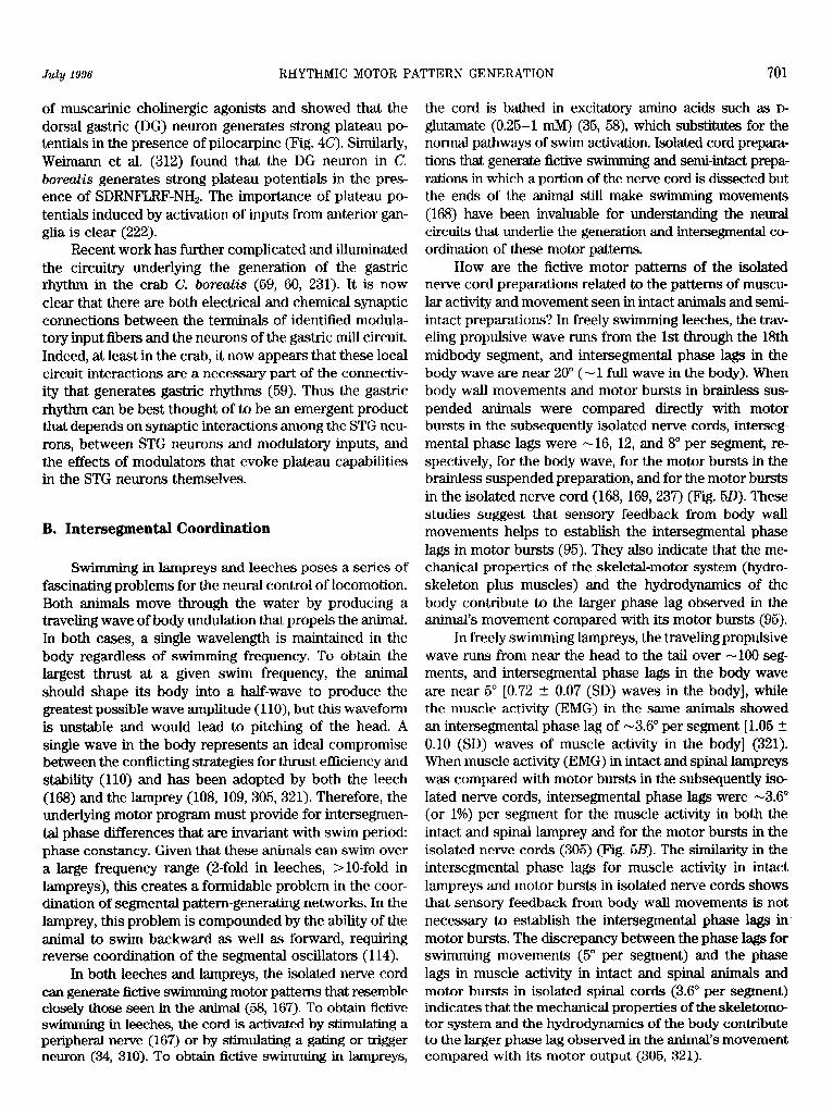
B. Intersegmental Coordination
Swimming in lampreys and leeches poses a series of fascinating problems for the neural control of locomotion. Both animals move through the water by producing a traveling wave of body undulation that propels the animal. In both cases, a single wavelength is maintained in the body regardless of s wimming frequency. To obtain the largest thrust at a given swim frequency, the animal should shape its body into a half-wave to produce the greatest possible wave amplitude (1 lo), but this waveform is unstable and would lead to pitching of the head. A single wave in the body represents an ideal compromise between the conflicting strategies for thrust efficiency and stability (110) and has been adopted by both the leech (168) and the lamprey (108, 109, 305,321). Therefore, the underlying motor program must provide for intersegmen- tal phase differences that are invariant with swim period: phase constancy. Given that these animals can swim over a large frequency range (2-fold in leeches, >lO-fold in lampreys), this creates a formidable problem in the coor- dination of segmental pattern-generating networks. In the lamprey, this problem is compounded by the ability of the animal to swim backward as well as forward, requiring reverse coordination of the segmental oscillators (114).
In both leeches and lampreys, the isolated nerve cord can generate fictive s wimming motor patterns that resemble closely those seen in the animal (58, 167). To obtain fictive swimming in leeches, the cord is activated by stimulating a peripheral nerve (167) or by stimuIating a gating or trigger neuron (34, 310). To obtain fictive swimming in lampreys,
the cord is bathed in excitatory amino acids such as D- glutamate (0.25-l n&l) (35, 58), which substitutes for the normal pathways of swim activation. Isolated cord prepara- tions that generate fictive swimming and semi-intact prepa- rations in which a portion of the nerve cord is dissected but the ends of the animal still make swimming movements (168) have been invaluable for understanding the neural circuits that underlie the generation and intersegmental co ordination of these motor patterns.
How are the fictive motor patterns of the isolated nerve cord preparations related to the patterns of muscu- lar activity and movement seen in intact animals and semi- intact preparations? In freely swimming leeches, the trav- eling propulsive wave runs from the 1st through the 18th midbody segment, and intersegmental phase lags in the body wave are near 20” (- 1 full wave in the body). When body wall movements and motor bursts in brainless sus- pended animals were compared directly with motor bursts in the subsequently isolated nerve cords, interseg- mental phase lags were -16, 12, and 8O per segment, re- spectively, for the body wave, for the motor bursts in the brainless suspended preparation, and for the motor bursts in the isolated nerve cord (168, 169,237) (Fig. So). These studies suggest that sensory feedback from body wall movements helps to establish the intersegmental phase lags in motor bursts (95). They also indicate that the me- chanical properties of the skeletal-motor system (hydro- skeleton plus muscles) and the hydrodynamics of the body contribute to the larger phase lag observed in the animal’s movement compared with its motor bursts (95).
In freely swimming lampreys, the traveling propulsive wave runs from near the head to the tail over -100 seg- ments, and intersegmental phase lags in the body wave are near 5’ [0.72 _ + 0.07 (SD) waves in the body], while the muscle activity (EMG) in the same animals showed an intersegmental phase lag of -3.6’ per segment [1.05 t 0.10 (SD) waves of muscle activity in the body] (321). When muscle activity (EMG) in intact and spinal lampreys was compared with motor bursts in the subsequently iso- lated nerve cords, intersegmental phase lags were -3.6’ (or 1%) per segment for the muscle activity in both the intact and spinal lamprey and for the motor bursts in the isolated nerve cords (305) (Fig. 5B). The similarity in the intersegmental phase lags for muscle activity in intact lampreys and motor bursts in isolated nerve cords shows that sensory feedback from body wall movements is not necessary to establish the intersegmental phase lags in motor bursts. The discrepancy between the phase lags for swimming movements (5” per segment) and the phase lags in muscle activity in intact and spinal animals and motor bursts in isolated spinal cords (3.6” per segment) indicates that the mechanical properties of the skeletomo- tor system and the hydrodynamics of the body contribute to the larger phase lag observed in the animal’s movement compared with its motor output (305, 321).
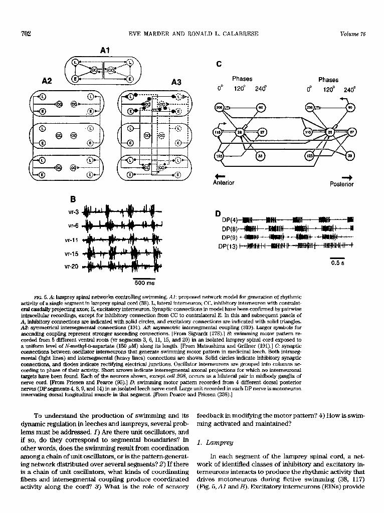
702 EVE MARDER AND RONALD L. CALABRESE Vohm 76
Phases
/ -\ 240’ 0” 1 2o” 240’
+ Anterior
-b Posterior
vr-3
vr-6
vr-11
vr-I 5
vr-20 0.5
500 ms
FIG. 5. A: lamprey spinal networks controlling swimmin g. Al: proposed network model for generation of rhythmic activity of a single segment in lamprey spinal cord (38). L, lateral intemeuron; CC, inhibitory interneuron with contralat- eral caudally projecting axon; E, excitatory intemeuron. Synaptic connections in model have been confirmed by pairwise intracellular recordings, except for inhibitory connection from CC to contralateral E. In this and subsequent panels of A, inhibitory connections are indicated with solid circles, and excitatory connections are indicated with solid triangles. A2 symmetrical intersegmental connections (191). A3: asymmetric intersegmental coupling (319). Larger symbols for ascending coupling represent stronger ascending connections. [From Sigvardt (278).] B: swimming motor pattern re- corded from 5 different ventral roots (vr segments 3, 6, 11, 15, and 20) in an isolated lamprey spinal cord exposed to a uniform level of N-methyl-D-aspartate (150 /IM) along its length. [Prom Matsushima and Grillner (191).] C: synaptic connections between oscillator interneurons that generate swimmin g motor pattern in medicinal leech. Both intraseg- mental (light lines) and intersegmental (heavy lines) connections are shown. Solid circles indicate inhibitory synaptic connections, and diodes indicate rectifying electrical junctions. Oscillator intemeurons are grouped into columns ac- cording to phase of their activity. Short arrows indicate intersegmental axonal projections for which no intemeuronal targets have been found. Each of the neurons shown, except ceU 208, occurs as a bilateral pair in midbody ganglia of nerve cord. [Prom F’riesen and Pearce (96).] D: s wimming motor pattern recorded from 4 different dorsal posterior nerves @P segments 4,8,9, and 14) in an isolated leech nerve cord. Large unit recorded in each DP nerve is motoneuron innervating dorsal longitudinal muscle in that segment. [Prom Pearce and F’riesen (238).]
To understand the production of swimming and its dynamic regulation in leeches and lampreys, several prob- lems must be addressed. 1) Are there unit oscillators, and if so, do they correspond to segmental boundaries? In other words, does the s wimming result from coordination among a chain of unit oscillators, or is the pattem-generat- ing network distributed over several segments? 2) If there is a chain of unit oscillators, what kinds of coordinating fibers and intersegmental coupling produce coordinated activity along the cord? 3> What is the role of sensory
feedback in modifying the motor pattern? 4) How is swim- ming activated and maintained?
1. Lamprey
In each segment of the lamprey spinal cord, a net- work of identified classes of inhibitory and excitatory in- temeurons interacts to produce the rhythmic activity that drives motoneurons during fictive swimming (38, 117) (Fig. 5, Al and B). Excitatory interneurons (EINs) provide

July 1996 RHYTHMIC MOTOR PATTERN GENERATION 703
excitatory drive [mediated by excitatory amino acids @As)] to motoneurons and feedback excitation to two classes of inhibitory interneurons, the lateral interneurons &INS) and crossed interneurons (CCINs) (38, 39). The LINs (260) and CCINs (36) provide ipsilateral and contra- lateral inhibition, respectively, to interneurons and moto- neurons.
Reciprocal inhibitory connections across the midline (commissural connections) between the CCINs are instru- mental in generating oscillation in the segmental network (111, 112, 303). Recent experiments with partially split nerve cord preparations indicate that the commissural reciprocal interactions are essential for normal oscilla- tion. Some rudimentary high-frequency rhythmicity per- sists after the commissural connections are severed, how- ever. Descending reticulospinal neurons activate both NMDA and kainate/DL+amine-3-hydrow-bmethylisoxa- zole-propionic acid receptors on all classes of the inter- neurons and on motoneurons (232) to elicit and sustain oscillation (196). The reticulospinal neurons in turn re- ceive excitatory (as welI as inhibitory) feedback from the spinal circuitry and thus participate in and sustain the rhythmic activity of the network (82).
Sensory input is provided to the segmental pattern- generating networks by intraspinal stretch receptors (edge cells) that sense the stretch associated with the bending movements during swimming (115). Two types of edge cells have been described: one with an ipsilateral axon provides excitation @AA) to ipsilateral interneurons and motoneurons, and the other with a contralateral axon which is inhibitory (glycine) to contralateral CCINs (53, 80,81). These stretch receptors are poised to provide the feedback necessary to coordinate the motor pattern to the ongoing movements by promoting formation of the motor burst ipsilaterally (excitation) when that side is stretched (axial muscle on the opposite side contracted) and terminating it contralaterally (inhibition). Rhythmic activation of the edge cells by bending the nerve cord at either end can entrain the fictive motor pattern over a broad frequency range (-1OS% below to 40% above the rest frequency) (116, 197).
Each of the three types of pattern-generating inter- neurons projects for several segments in the spinal cord (LINs, ipsilaterally and caudally up to 50 segments; CCINs, contralaterally and caudalIy up to 30 segments; EINs, ipsi- laterally and either rostrally or caudally for at least 5 seg- ments), and all synapse with neurons of other segments (36, 38, 39, 260). Although anatomically the distinction between segmental oscillatory networks is blurred, each segment acts as a functional oscillator because just a few segments of spinal cord can produce normal-looking fic- tive swimming motor bursts.
If one views the cord as organized into segmental oscillators, the intersegmental axons of the pattern-gener- ating interneurons provide a substrate for intersegmental
coordination of these oscillators. While excitatory cou- pling is provided by EIN axons that project both rostrally and caudally, inhibitory coupling is provided by LINs and CCINs that project only caudally. Other as yet unde- scribed pathways for coupling of segmental oscillators probably exist, but it is tempting to conclude from the available data that intersegmental coupling is asymmetric and that the inhibitory coupling is descending. Recent experiments (40) with partially split nerve cord prepara- tions suggest that functional length of coupling between segmental oscillators has a length constant of about two segments. Given the paucity of hard data on intersegmen- tal connectivity and synaptic strength in the network, it is perhaps not surprising that two opposing hypotheses to explain intersegmental coordination have been proposed.
The first hypothesis emphasizes the apparent symme- try of excitatory coupling. Intersegmental phase lags and phase constancy are envisioned to result from local excit- ability differences among segmental oscillators (trailing oscillator hypothesis) (114, 191, 192) (Fig. 5AZ). In this model, when the nervous system is intact, descending reticulospinal inputs are thought to activate preferentially the rostral most segmental oscillators of the spinal cord. This leading oscillator then entrains, via symmetrical ex- citatory coupling, the more caudal trailing osciIlators to the appropriate intersegmental phase lag. In this model, regardless of the period of the leading oscillator, the trail ing oscillators will be entrained to the proper phase as long as they have a longer inherent period, yielding phase constancy. In sections of isolated nerve cord, removal of descending inhibition to the rostra&most segmental oscil- lator by section of LIN and CCIN descending axons is envisioned to enhance the excitability of the rostral most oscillator and make it lead. This assumption has been justifiably criticized for being simplistic (278). The hypoth- esis can also accommodate backward swimming; in that case, local sensory input which elicits the backward swim is hypothesized to excite preferentially the caudal-most segmental oscillators, causing them to lead, albeit tran- siently. Modeling studies using semi-realistic model neu- rons organized in a chain of oscillators based on the con- nectivity diagram of Buchanan and Grillner (38) and Grillner et al. (117) and using the simplified connectionist model of Buchanan and co-workers (37,112) and symmet- ric excitatory coupling indicate that the trailing oscillator hypothesis can account for the observed coordination (114, 191, 192). However, this hypothesis is critically de- pendent on symmetry in the intersegmental coupling for the generation of the appropriate intersegmental phase lags and requires that the asymmetry in intrinsic fre- quency be maintained under all modulatory conditions to maintain constant rostral-caudal phase lags.
A second hypothesis (asymmetric coupling) deem- phasizes segmental differences in excitability, because sections of nerve cord from different regions of the nerve

704 EVE MARDER AND RONALD L. CALABRESE Volume 76
cord show no systematic differences in the intrinsic pe- riod of fictive swimmin g (55), although the extrapolation of this observation to the excitability state in intact cord or larger cord sections has been questioned (114). Exten- sive theoretical and mathematical analysis led to a plausi- ble model wherein asymmetric coupling could generate the appropriate phase lags and phase constancy without any excitability differences (56, 165, 166, 278, 319, 320, 323). Experimental evidence pointed to the dominance of the ascending coupling (278). Williams (319) has sug- gested that this asymmetric coupling naturally follows from a simple connectivity rule called synaptic spread: whatever synaptic contacts each neuron makes within its own segment it also makes in neighboring segments (Fig. 5Aa). This simple rule is consistent with the available connectivity data. Williams (319) simulated a chain of os- cillators based on the connectivity diagram of Buchanan and Grillner (38) and Grillner et al. (117) using the simpli- fied connectionist model of Buchanan (37) and this simple intersegmental connectivity rule. This model generates the appropriate front-to-rear phase lags and phase con- stancy as long as the coupling is asymmetric with the ascending coupling dominant (319). The predicted domi- nance of ascending coupling is in contrast to the known descending projections of the inhibitory interneurons (LINs and CCINs), although it is certainly possible that ascending fibers yet to be identified could subserve this function.
Considerable experimental evidence has been gath- ered in support of both hypotheses (asymmetric coupling, Refs. 278,323; trailing oscillator, Refs. 191, 192). Much of this evidence comes from pharmacological experiments in which the excitability of one section of nerve cord relative to another is altered to determine the effect on intersegmental phase lags (191, 192,278). Other evidence comes from attempts to determine whether the limits of entrainment of the motor pattern by edge cell activity evoked by rhythmic bending of the ends of the nerve cord, respectively, are asymmetric (323). More recently, a prediction of the asymmetric coupling model, that there should be a rostral boundary layer where intersegmental phase lags will be nonuniform, has been confirmed experi- mentally (322). To an impartial observer, resolution of the debate will not come from models, no matter how informative they may be, but from difficult to obtain ex- perimental data. It will be critical to determine the length of the coordinating fibers and the strengths of their as- cending and descending synaptic connections.
2. Leech
The leech central nervous system consists of a chain of segmental ganglia, each with -400 (mostly bilaterally paired) neurons, linked by paired connectives. Each gan- glion provides the sensory and motor innervation of the
body wall in its segment. It is natural to ask whether each ganglion contains adequate circuitry to generate the alternating motor bursts required to control the body wall muscles to produce the alternating movements associated with swimming. Isolated single ganglia can produce crude alternating bursts in swim motoneurons only in the pres- ence of exogenously applied neuromodulatory substances such as serotonin (126), or when linked to a chain of ganglia by a small group of fibers in the median connective that gate swimming (307). Thus they contain sufficient circuitry to sustain swim oscillations, and this circuitry can be thought of as composing a segmental swim oscilla- tor. However, the oscillatory neural circuit can also be conceived as intersegmental and distributed (96, 97).
Each ganglion contains several interneurons that are considered part of the swim segmental oscillator because they are rhythmically active during fictive swimming, and because they are able to reset the ongoing fictive motor pattern (93-96, 309) (Fig. 5, C and 0). The segmental oscillator produces a four-phase rhythm in segmental body wall motoneurons. Dorsal excitor motoneurons al- ternate with ventral excitor motoneurons, and dorsal in- hibitor motoneurons alternate with ventral inhibitory mo- toneurons. Each class of motoneurons fires for ~25% of the cycle (169). The interneurons tie in a three-phase rhythm with activity centering around 0, 120, and 240’ (95) and provide both excitatory and inhibitory drive to motoneurons both intra- and intersegmentally to sculpt their rhythmic bursts (251).
In both semi-intact animals and isolated nerve cords, swim activity can be triggered by mechanosensory input that activates identified descending trigger interneurons (34). These neurons in turn activate segmental swimming gating interneurons that provide tonic excitatory drive to the swim oscillator interneurons (227,228,308-310). The swim-gating interneurons receive excitatory and inhibi- tory feedback from the oscillator that sustains the excit- atory state necessary for oscillation (227, 228, 308, 309). Sensory feedback from body wall stretch is thought to play a role in swim generation, (167) and as indicated above, there is evidence that such feedback is important in establishing intersegmental delays in motor activity (237), but the nature of the receptors involved and their connections with swim oscillator interneurons and moto- neurons are unknown.
The oscillator interneurons are highly interconnected by both excitatory and inhibitory synapses (94-96, 309) (Fig. 5C). Sustained excitation from swim-gating interneu- rons and reciprocal inhibition between oscillator interneu- rons, together with postinhibitory rebound, are thought to be instrumental in rhythm generation within a segmental oscillator (93, 95). All of the oscillator interneurons pro- ject to other ganglia, some for at least five segments (96, 251,309), providing the basis for intersegmental coordina- tion of the unit oscillators. The known interganglionic

July 1996 RHYTHMIC MOTOR PATTERN GENERATION 705
synaptic connections of the oscillator interneurons are largely repeats of the intraganglionic connections, con- forming to the synaptic spread rule of Williams (319). They are, moreover, highly polarized and asymmetric; the 0’ phase oscillator interneurons only descend, and the 120 and 240’ phase oscillator interneurons only ascend, the nerve cord. The measured intersegmental synaptic con- nections are as strong as the local ones (94, 96, 309). Because of the extensive intersegmental connections among oscillator neurons and because many of these in- tersegmental connections join with intrasegmental con- nections to form closed inhibitory loops (96,97), the oscil- latory network appears intersegmental and distributed. Nevertheless, for the purpose of discussing intersegmen- tal coordination and extracting principles of organization, it is useful to consider the system as a set of highly cou- pled segmental oscillators.
In the isolated nerve cord, the front-to-rear phase lag between homologous motor bursts in adjacent segments is -8’ or 2% (237) (Fig. 50). When the periods of pairs of ganglia along the nerve cord were compared to determine whether there was a gradient in cycle period, it was found that posterior segments had shorter cycle periods than ante- rior segments (95, 238). According to the trailing oscillator hypothesis described above, this would lead to a rear-to- front phase progression in motor bursts along the cord. ManipuIating this gradient by lowering temperature in a re- stricted portion of the nerve cord produced the expected phase delay in the slowed section (238, 239). Moreover, reducing the coupling between portions of nerve cord by severing one of the paired connectives that carry the inter- segmental axons of the oscillator interneurons produced a revemal of phase relationships across the lesion (238, 239). This result suggests that the asymmetry of the strong cou- pling between segmental oscillators might be counteracting an opposing excitability gradient (95). When coupling was reduced by lesion, the effects of the excitability gradient were expressed, the phase progression of motor bursts along the nerve cord is determined by the relative influence of the “reversed” excitability gradient and the asymmetry of inte~gmental coupling.
Experiments in which rhythmic activity in stretches of nerve cord was blocked (high divalent saline) but con- duction in intersegmental axons was not blocked indicate that intersegmental coordinating information spans at least five segments (238, 239). Further experiments in which intersegmental phase lags were measured in pro- gressively smaller stretches of nerve cord show that inter- segmental phase lag is a function of chain length varying from 8’ in the intact nerve cord to 40’ in a two-ganglion chain. These results emphasize the importance of cou- pling from distant oscillators in establishing the proper phase progression along the nerve cord. Modeling studies of coordination of the segmental oscillators which incor- porated the rear-to-front gradient in cycle period, the
known bidirectional asymmetric intersegmental coupling, and long-range coupling with equivalent strength to ho- mologous connections within the ganglion of origin, repli- cate all the salient features of the experimental data (240).
Studies of intersegmental coordination of swim activ- ity in the leech nerve cord point to a hybrid explanation between “opposing” trailing oscillator and asymmetric coupling hypotheses posed for the lamprey. Not only do these studies illuminate the lamprey analyses, but they offer a satisfying, albeit teleological, explanation for the opposed arrangement apparent in this hybrid. With such an arrangement, the animal may be able to adjust interseg- mental phase lags both up and down by adjusting a single variable, intersegmental coupling strength (95).
3. Other preparations
Considerable experimental effort has be expended to establish the mechanisms of intersegmental coordination in early Xmopus tadpole swimming (299) and crayfish swimmeret beating (214). In the swimmeret system, no gradient of excitability among segmental oscillators is thought to be present (214), and the origin of the interseg- mental phase lags is unclear, although a diverse popula- tion of intersegmental neurons that convey coupling infor- mation has been identified (218,219). Recent experiments in which gradients of excitability were created by applica- tion of cholinergic agonists are interpreted in favor of the asymmetric coupling organization (31).
The early Xmopus tadpole is particularly interesting because so much is known about the segmental oscilla- tors (see sect. ~42). Moreover, unlike any of the other intersegmental control systems reviewed, intersegmental time delays are constant between segmental motor bursts and independent of cycle period (297). A rostrocaudal gradient in tonic excitatory synaptic and phasic inhibitory synaptic drive during swim activity among segmental mo- toneurons has been observed (298,299). If these gradients are also present in premotor spinal interneurons, they would be expected to have an important role in determin- ing the rostrocaudal delays. It is interesting to note that the kinematics of early tadpole swimming show a longitu- dinally uniform intersegmental phase lag and phase con- stancy (68), suggesting that, as in the leech and lamprey, the mechanical properties of the skeletomotor system and hydrodynamics shape the neuromuscular output. As the tadpole develops, the swim segmental pattern generators change, the motor pattern becomes increasingly complex (and unfortunately less well understood), and the neural system achieves phase constancy (281).
C. Circuit Switching
of All animals produce a wide range of behaviors, many
which require different patterns of activation of the

706 EVE MARDER AND RONALD L. CALABRESE Votkwme 76
same motoneurons. Indeed, our leg muscles can be used in walking, running, swimming, bicycle riding, and stand- ing, as well as in voluntary movements. Therefore, one important question is determining how much circuitry is unique in the specification of a given behavior, and how much of the intemeuronal organization underlying these different motor functions is shared (289, 311, 326). On the one hand, one might imagine that there are entirely different groups of neurons that form relatively “hard- wired” circuits for each qualitatively different behavior. In this organizational model, an entirely distinct premotor circuit would exist for each behavior; that is, in animals that both swim and walk, different groups of neurons would form the premotor circuits for walking and for swimming. Alternatively, one might imagine that much of the same premotor circuitry is used to form both the swim and walking motor patterns, but that activation of several unique elements can change the network’s output suffi- ciently to account for a significant number of qualitatively different motor patterns and, consequently, movements.
During the past 10 years, a significant amount of di- rect experimental evidence has come from invertebrate nervous systems that illustrates that many of the same neurons can be employed to form circuits capable of gen- erating more than one behavior. At the same time, a body of circumstantial evidence in a variety of vertebrate prepa- rations argues similarly that many circuit elements may be participating in the generation of more than one behavior.
1. lkitonia swim and rem withdraw&
Ditonia is a mollusk that escapes from prey with a simple dorsal-ventral flexion. In the process of studying the organization of the central pattern generator for this episodic behavior, Getting and Dekin (100-102) first used the term polymorphic circuit. Getting’s essential finding was that the same group of neurons could either generate a simple avoidance reflex or a full rhythmic escape behav- ior, depending on the strength of the sensory stimulus.
In the Fritonia system, a network of dorsal swim interneurons (DSIs) are all linked by excitatory connec- tions to one another and to an interneuron I, which pro- vides strong inhibitory feedback to the DSIs. In response to a weak sensory stimulus, a single DSI can be activated separately, because the DSIs functionally inhibit each other through I. Under these conditions, the animals make a simple defensive withdrawal. However, a strong aver- sive stimulus results in activation of another interneuron, C2, which inhibits I and drives the DSIs to produce syn- chronous bursts. Therefore, the DSIs alternate with the ventral swim intemeurons (VSIs) to produce rhythmic dorsoventral flexions. Under these conditions, the DSIs strongly excite each other (102).
Getting and Dekin (101, 102) argued that it was im- portant to distinguish between the anatomic network con-
necting neurons and the several different functional cir- cuits that could be called into play under different physio- logical conditions. Getting (100) drew three different connectivity diagrams to illustrate 1) the anatomic con- nectivity among the central pattern-generating networks, 2) the functional circuitry he thought operational during the withdrawal behavior, and 3) the functional circuitry during the Tritonia swim.
Recent work on the Tritonia swim circuit (153-155) illustrates that neurons that are part of pattern-generating networks may themselves also have modulatory roles and is adding new components to the swim circuit. In the present view (W. Frost and P. Katz, personal communica- tion), sensory input excites the dorsal ramp interneuron (DRI), a newly identified command interneuron (98). The DRI then excites the DSIs. The DSIs then excite C2 as well as enhance its excitability and the strength of its synaptic connections. The excited C2 then polysynapti- tally excites DRI, creating a positive-feedback loop that sustains swimm’ mg. Alternation is produced by inhibition from the VSI neurons that are also excited by C2.
The DSI neurons have been recently shown to be sero- tonergic (195). The activation of the serotonergic DSI neu- rons increases the amplitude of synaptic co~ections in the swim system (153-155) by what appears to be heterosynap tic facilitation, modulation of transmitter release from the presynaptic terminals (153). Thus inputs to the swim system not only activate it, but in so doing upmodulate the essential circuitry for its operation. This has been termed “intrinsic modulation” (155) to distinguish it from modulatory control that is external to the circuit itself.
In another molluscan preparation, Aplysia, Wu et al. (326) used optical recording methods to record simultane- ously from hundreds of neurons in the abdominal ganglion during spontaneous and evoked siphon withdrawals and argued that the same neurons were used in a variety of different movements.
2. Stomatogastric nervous system
Classically, the stomatogastric nervous system was thought to consist of four separate central pattem-gener- ating networks: those controlling the production of the esophageal rhythm (10-s period), the cardiac sac rhythm (30- to 120-s period), the gastric mill rhythm (5. to 10-s period), and the pyloric rhythm (0.5- to 2-s period). Al- though it was always appreciated that there were synaptic connections among the neurons of these four functional networks (e.g., Refs. 69, 213), these were viewed as con- nections among elements of separate circuits. Studies car- ried out during the last 5 years have led to an entirely different conclusion.
Hooper and Moulins (136,137) studied the firing pat- terns displayed by the VD neuron of the lobster, Jams Uandi. The VD neuron was classically thought to be part

July 1996 RHYTHMIC MOTOR PATTERN GENERATION 707
of the pyloric pattern generator (193) and often fires with the pyloric rhythm (Fig. IA). Hooper and Moulins (136, 137) found that stimulation of a sensory neuron that acti- vated strong cardiac sac activity caused the VD neuron to fire in time with the cardiac sac rhythm. Because the cardiac sac and pyloric rhythms have such different intrin- sic periods, the VD neuron displays dramatically different patterns of firing in these two cases. Hooper and Moulins (136, 137) studied the cellular mechanism induced by the stimulation of the sensory nerve and showed that the abil- ity of the VD neuron to generate plateau properties was decreased after sensory stimulation, allowing the timing of the VD to be governed by the excitatory drive from elements of the cardiac sac pattern generator.
Changes in the strength of synaptic interactions are thought to be responsible for another switch involving neurons of the gastric mill rhythm and the cardiac sac. Dickinson et al. (75) showed that bath application of the peptide red pigment concentrating hormone (RPCH) strongly potentiated the synaptic connections between the IV neurons (cardiac sac) and neurons of the gastric mill circuit. Consequently, in the presence of RPCH, a conjoint rhythm in which the gastric mill and cardiac sac neurons fire together is found (75).
Meyrand et al. (201,202) have taken this theme consid- erably further. These authors found that activation of the pyloric supressor (PS) neurons of the lobster Homamcs gum- rnarus (probably homologous to the IV neurons previously described) produces a novel pattern that employs elements of all four pattern-generating networks previously described. The PS neurons synapse with most of the neurons of the STG. This drive strongly activates some of the neurons and strongly inhibits others, thus actively “dismantling” the integ- rity of the existing networks and creating a new one. The PS neuron activity controls the muscles of the valve between the esophagus and the cardiac sac and constructs a new motor pattern involving the whole foregut in coordinated activity (201, 202).
Many of the neurons of the crab C. bore&s switch between pyloric-timed and gastric-timed activity pat- terns. Weimann et al. (313) found that when the pyloric rhythm was active but the gastric rhythm was not active, many of the neurons that innervate gastric mill muscles fire short pyloric-timed bursts of one or two spikes (Fig. 6A). However, when the gastric rhythm was either spon- taneously active, or evoked by application of the peptide SDRNFLRF-NH2, many of these neurons, as well as sev- eral neurons previously considered parts of the pyloric pattern generating circuit “switched” into gastric time (Fig. 6B) (312, 313). Similar switches can also be pro- voked by stimulation of the gastropyloric receptor neu- rons (156).
To study the possible behavioral consequences of the pyloric/gastric switches seen in crabs, Heinzel et al. (131) used an endoscope to monitor movements of the gastric
mill during pyloric activity and during robust gastric mill movements, either spontaneously occurring or triggered by peptide and other modulator application (131). These experiments showed that the lateral teeth of the gastric mill produced small pyloric-timed movements when the lateral gastric (LG) and IC neurons were firing in pyloric time and demonstrated that even a few spikes/bursts pro- duced sign&ant movements. Moreover, activation of the muscles innervated by the IC produced movements of the lateral teeth, because of the complex mechanical coupling of the stomach muscles and ossicles (131). Thus these studies demonstrated that the pyloric/gastric switches seen in Weimann and co-workers (312,313) are behavior- ally meaningful.
The switches that describe the IV and PS neurons of the cardiac sac are interesting because they involve the activation of a set of neurons that makes a set of strong connections that tend to “drive” the STG neurons into a new firing pattern. In this case, although the local network interactions within the STG shape the influence those in- coming synapses have on the final output of the STG, the major determinant of the new pattern is the set of syn- apses from the IV or PS neurons.
The crab pyloric/gastric switches are somewhat dif- ferent from the switches that involve elements of the car- diac sac system discussed above. Weimann and Marder (311) studied the ability of the neurons that switch activity patterns to reset both ongoing pyloric and gastric rhythms. These experiments showed that when the LG and VD neurons fire in time with the pyloric network, they can reset (Fig. 6C) and entrain the pyloric network. Likewise, when they fire in time with the gastric rhythm, they can reset the gastric network. Thus it appears that changes in intrinsic properties and synaptic efficacy that allow the neurons to change their activity patterns “form” the functional networks and that the switching neurons are true members of different pattern-generating circuits at different times (311).
The crab system has also revealed other interesting features. Nusbaum et al. (231) found synaptic interactions between the terminals of the modulatory inputs and the intrinsic neurons of the STG. One such descending modu- latory input, the modulatory commissural neuron 1
ww um receives inhibitory synaptic feedback at its STG terminals from the commisural gastric (CG) neuron (a major player in the gastric circuit) which can gate the output of MCNl. These local interactions in the STG effec- tively make the modulatory neuron an integral part of the gastric mill pattern generator. Descending activity origi- nating in the CG activates the gastric rhythm, and local STG activity participates in generating the gastric rhythm (59). Thus the distinction between a modulatory input and a pattern-generating element is blurred by local interac- tions. Correspondingly, single elements serve multiple functions.
708 EVE MARDER AND RONALD L. CALABRESE Vohme 76
FIG. 6. Switching neurons of crab Cancer bo- realis. Abbreviations are as in Figures 1 and 4. A: control saline; simultaneous intracellular re- cordings from DG, LG, and LP neurons. Note that DG neuron is silent and LG neuron is fking in time with LP neuron (pyloric time). B: in presence of peptide SDRNFLRF-NH:!, LG neuron is now firing in gastric time, in alternation with DG neuron [seen as burst of action potentials on dorsal gastric nerve (dgn)]. C: depolarization of LG neuron resets pylo- ric rhythm, with phase response curve shown on right. [Modified from Marder and Weimann (189, 31 l).]
VD
C 0 0 8 l 0 0 0
0.6
Pyloric PD
I g OS4 l l Delay
co Oa2 . l .
* iii
3 0
Gastric <p 0.0 --- *me-e-m -
LG E .
I -0.2 Advance 0 -0.4
0.0 0.2 0.4 0.6 0.8 1.0
dvn Stimulus phase
ftt .
t LPPYPD CC
In summary, the conclusions of these studies on V. CONCLUSIONS switching in the stomatogastric nervous system argue that the functional circuit that produces a series of motor pat- terns exists only in a defined modulatory and physiologi- A. Reevaluating the Concept of Command Neurons cal state. Different modulatory and sensory inputs “con- struct” different motor functional circuits (76, 189, 201, 202) as needed for the appropriate behavior of the animal. Descending inputs to pattern-generating networks,
whether they be involved in leech or lamprey swimming (34, 117, 310), control of the crustacean foregut (59, 60,
3. Other prepamtions 69, 77, 78, 201, 221, 231), or any of a myriad of other rhythmic behaviors, often serve to gate specific motor
It is much more difficult to obtain direct experimental patterns on or off. The observation that in invertebrate evidence that interneurons are part of severitl pattern- motor patterns this gating C~JJ be accomplished by stimu- generating circuits in vertebrate systems. However, there lating single neurons led to the concept of motor “com- is a growing body of circumstantiaI evidence supporting mand neurons” (157, 318). The term was first applied to such an organization (22, 151, 204, 211, 289). fibers dissected from the nerve cord of crayfish that when

July 1996 RHYTHMIC MOTOR PATTERN GENERATION 709
stimulated extracellularly induced rhythmic movements of the swimmerets (318).
The concept of command neurons was influential in developing thinking about control of escape and many postural and rhythmic behaviors in vertebrates as well as invertebrates (70, 71, 84, 89, 157, 158, 180,.218, 219, 225).
The command neuron concept came under consider- able scrutiny in the late 197Os, which led to the evolution of a set of rigid criteria for establishing a neuron as a command neuron (171). Kupfermann and Weiss (171) ar- gued that a command neuron must be both necessary and sufficient for the activation of a motor pattern by sensory input. These criteria were so rigid that the concept has largely been abandoned, and the term command neuron has virtually disappeared from the literature on control of rhythmic motor systems. However, the recent discov- ery of the DRI neurons in Tritonia (98) provides a clear example of a neuron that meets all the established criteria for a command neuron. Additionally, the PS neurons in the stomatogastric nervous system (201, 202) also can be thought of as command neurons in the strict sense.
However, the notion that a neuron must be shown to be necessary as well as sufficient for the gating of a partic- ular motor pattern before it can be usefully labeled as a command neuron flies in the face of our current experi- ence. Parallel pathways participating in motor pattern gat- ing, each pathway often with the ability to produce a particular pattern variant, appear to be the rule. Thus the failure of a specific neuron to be always active in association with a given motor pattern is to be expected and in no way negates the possible importance of such elements in the control of behavior. Moreover, local inter- action of command elements with pattern-generating net- works can sustain and even gate activity in the command elements themselves, thus making experimental efforts at determining sufficiency largely irrelevant.
Thus, in retrospect, this attempt at rigor seems to have destroyed the original utility of the concept loosely expressed in the term commandjibw, and it may be time to retrieve it from disuse. Two examples will illustrate how this concept remains useful in discussing motor net- work organization. In the crustacean stomatogastric ner- vous system, descending activity in MCNl initiates oscilla- tory activity in the gastric mill network of the STG, and inhibitory feedback to its terminals in the STG recruits this neuronal component as part of the oscillatory net- work (59). Other modulatory input neurons to the STG can also activate the gastric pattern generator, and pre- sumably recruit the MCNl STG terminals in the process. In the leech, a family of descending interneurons, called trigger neurons, in the subesophageal ganglion integrate mechanosensory input. Transient activity in these neu- rons excites a set of segmental swim-initiating interneu- rons in midbody ganglia. When excited, these interneu- rons, acting in parallel with segmental serotonergic inter-
neurons, initiate the activity in swim oscillator networks in each midbody ganglion. These gating interneurons re- ceive strong excitatory feedback from the oscillator net- works that creates a reverberation that sustains the activ- ity of the oscillator networks. Neither the MCN neurons of the stomatogastric nervous system nor the leech swim trigger or initiation neurons fulfill the “necessity” contin gency discussed above, yet these neurons possess a motor command function and are the embodiment of the modern concept of command neurons. Single control neurons, act- ing alone or in concert with other neurons, that serve to activate whole networks that produce coordinated activ- ity constitute a class of important cells that were first described as command neurons. These neurons in turn receive feedback from the network they activate and par- ticipate to a greater or lesser extent in network function. Thus a modern definition of a command neuron does not imply hierarchical position, nor does it imply necessity, but rather has functional implications.
B. Organizational Rules
All pattern-generating networks depend on intrinsic membrane properties and circuit building blocks to form patterned output. The output of all pattern-generating net- works must be shaped to the needs of the organism. This is accomplished by a variety of control mechanisms that include sensory feedback, hormonal regulation, and mod- ulatory inputs. As data are accumulated on a variety of preparations, it is becoming apparent that there are multi- ple sites of regulation and modulation within these net- works and that each network is regulated by multiple routes.
Although we are starting to understand some of the basic cellular and biophysical mechanisms that are used to build unit oscillators, the organizational principles and the cellular mechanisms that underlie the interactions among networks to produce coordinated behaviors in- volving multiple limbs, posture, and respiration are still largely mysterious. At every level of analysis, the chal- lenge is always to explain the behavior of a system in terms of the behavior of the component elements; that is, we must understand how the many currents in cells inter- act to produce the intrinsic properties of neurons, how neurons interact to produce a defined circuit, and how oscillatory circuits interact to produce coordinated behav- iors. At each of these levels, this will require combined computational and experimental tools and the ability to take information gathered at one level and use it to under- stand the next.
Almost all textbooks in neuroscience introduce mo- tor systems in vertebrates with excellent treatments of spinal reflexes, often leaving students with the idea that movement is still largely determined by reflex pathways.

710 EVE MARDER AND RONALD L. CALABRESE Vdume 76
In this review, we focused on the central control of move- ment. However, sensory feedback is certainly required in all rhythmic behaviorcs to allow the animal to move correctly. The importance of sensory feedback in the orga- nization of the basic form of the motor pattern depends a great deal on the behavior, and this too is always context dependent.
In this review we neglected a discussion of the volun- tary control of rhythmic movements, such as the internally generated decision to start walking in humans. Our even- tual hope would be to understand the most challenging of human motor acts, speech, in which complex cognitive acts must be used to drive neurons and circuits that are also used in simpler behaviors such as breathing. Insights into these processes will require cooperation and coordi- nation among groups of neuroscientists, in much the same way that any coordinated behavioral act requires coordi- nation among multiple, complex neural circuits.
This work was supported by National Institute of Neurolog- ical Disorders and Stroke Grants NS-17813 (to E. Marder) and NSr24072 (to R. L. Calabrese) and by the W. M. Keck Foundation (to E: Marder).
1.
2.
3.
4.
5.
6.
7.
8.
9.
10.
11.
12.
ABBOTT, L. F., S. L. HOOPER, AND E. MARDER. Oscillating net- works: control of burst duration by electrically coupled neurons. Neural Comput. 3: 487-497, 1991. ADAMS, M. E., AND M. O’SHEA. Peptide cotransmitter at a neuro- muscular junction. S&&e ‘Wush. DC 221: 286-288, 1983. ALFORD, S., AND K. A. SIGVARDT. Excitatory neurotransmitters activate voltage-dependent properties in neurons in spinal motor system of lamprey. J. Neumphysiol. 62: 334-341, 1989. ANGSTADT, J. D., AND R. L. CALABRESE. A hyperpolarization- activated inward current in heart interneurons of the medicinal leech. J. Neumphysiol. 9: 2846-2857, 1989. ANGSTADT, .I. D., AND R. L. CALABRESE. Calcium currents and graded synaptic transmission between heart interneurons of the leech. J. Neurosci. 11: 746-759, 1991. ANGSTADT, J. D., AND W. 0. FRIESEN. Modulation of swimming behavior in the medicinal leech. I. Effects of serotonin on the elec- trical properties of swim-gating cell 204. J. Comp. Physiol A Sew. Neural Behuv. Physiol. 172: 223-234, 1993. ANGSTADT, J. D., AND W. 0. FRIESEN. Modulation of swimming behavior in the medicinal leech. II. Ionic conductances underlying serotonergic modulation of swim-gating cell 204. J. Comp. Physiol. A Sens. Neural Behav. Physiol. 172: 235-248, 1993. ARBAS, E. A, AND R. L. CALABRESE. Ionic conductances underly- ing the activity of interneurons that control heartbeat in the medici- nal leech. J. Neurosci. 7: 3945-3952, 1987. ARBAS, E. A, AND R. L. CALABRESE. Slow oscillations in mem- brane potential in intemeurons that control heartbeat in the medici- nal leech. J. Neurosci. 7: 3953-3960, 1987. ARSHAVSKY, Y. I., T. G. BELIAGINA, G. N. ORLOVSKY, Y. PAN- CHIN, G. A. PAVLOVA, AND L-B. POPOVA Control of locomotion in marine moIIusc clione limacina. VI. Activity of isolated neuron of pedal gangha. E&p. Brain Res. 63: 106-112, 1986. ARSHAVSKY, Y. I., I. N. BELOOZEROVA, G. N. ORLOVSKY, Y. PANCHIN, AND G. A. PAVLOVA. Control of locomotion in marine moIIusc Clione limacinu. I. Efferent activity during actual and fic- titious swimming. Exp. Brain Res. 58: 256-262, 1985. ARSHAVSKY, Y. I., I. N. BELOOZEROVA, G. N. ORLOVSKY, Y. PANCHIN. AND G. A. PAVLOVA. Control of locomotion in marine
13.
14.
15.
16.
17.
18.
19.
20.
21.
22.
23.
24.
25.
26.
27.
28.
29.
30.
31.
32.
33.
molIusc Clione limacinu. I. Rhythmic neurons of pedal ganglia. Eq. Brain Res. 58: 263-272, 1985. ARSHAVSKY, Y. I., I. N. BELOOZEROVA, G. N. ORLOVSKY, Y. PANCHIN, AND G. A. PAVLOVA. Control of locomotion in marine mohusc Clione limacinu. III. On the origin of locomotory rhythm. Eq. Brain Res. 58: 273-284, 1985.
ARSHAVSKY, Y. I., I. N. BELOOZEROVA, G. N. ORLOVSKY, Y. PANCHIN, AND G. A. PAVLOVA. Control of locomotion in marine mohusc CZione limacina. IV. Role of type 12 intemeurons. Exp. Brain Res. 58: 285-293, 1985.
ARSHAVSKY, Y. I., G. N. ORLOVSKY, Y. PANCHIN, AND G. A. PAV- LOVA. Control of locomotion in marine mohusc Clione limacina. VII. Reexamination of type 12 interneurons. Exp. Brain Res. 78: 398-406, 1989. ARSHAVSKjl, Y. I., G. N. ORLOVSKY, Y. V. PANCHIN, A. ROBERTS, AND S. R. SOFFE. Neuronal control of swimming locomotion: analy- sis of the pteropod mohusc Clione and embryos of the amphibian Xmpus. Trends Neurosci. 16: 227-233, 1993.
AYERS, J., AND A. I. SELVERSTON. Monosynaptic entrainment of an endogenous pacemaker network a cellular mechanism for von Holst’s magnet effect. J. Comp. Physiol. 129: 5-17, 1979. BAL, T., F. NAGY, AND M. MOULINS. The pyloric centraI pattern
generator in crustacea: a set of conditional neuronal oscihators. J. Comp. Physiol. A Sens. Neural Behuv. Physiol. 163: 715-727,1988. BAXTER, D. A., AND J. H. BYRNE. Ionic conductance mechanisms contributing to the electrophysiological properties of neurons. Curr. Opin. Neurobiol. 1: 105-112, 1991. BEKOFF, A., AND J. A. KAUER. Neural control of hatching: role of neck position in turning on hatching leg movements in post-hatch- ing chicks. J. Comp. Physiol. 145: 495-504, 1982.
BEKOFF, A., AND J. A. KAUER. Neural control of hatching: fate of the pattern generator for the leg movements of hatching in post- hatching chicks. J. Neurosci. 4: 2659-2666, 1984. BEKOFF, A., M. P. NUSBAUM, A. L. SABICHI, AND M. CLIFFORD. Neural control of hatching and walking motor output patterns in
normal and deafferented chicks. J. Neurosci. 7: 2320-2330, 1987. BEIANGER, J. H., AND I. ORCHARD. The locust ovipositor opener muscle: proctohnergic central and peripheral neuromodulation in a centrally driven motor system. J. Exp. Biol. 174: 343-362, 1993. BENSON, J. A, AND W. B. ADAMS. Ionic mechanisms of endoge- nous activity in molluscan burster neurons. In: Neuron& and CeUu- lur OscilZutors, edited by J. Jacklet. New York: Dekker, 1989, p.
87- 120. BERKOWITZ, A., AND P. S. G. STEIN. Activity of descending pro- priospinal axons in the turtle hindhmb enlargement during two
forms of fictive scratching: broad tuning to regions of the body surface. J. Neurosci. 14: 5089-5104, 1994. BERKOWITZ, A., AND P. S. G. STEIN. Activity of descending pro- priospinal axons in the turtle hindlimb enlargement during two forms of fictive scratching: phase analyses. J. Neurosci. 14: 5105- 51 19, 1994.
BERTRAM, R. A computational study of the effects of serotonin on a molluscan burster neuron. Biol. Cybern. 69: 257-267, 1993. BIDAUT, M. Pharmacological dissection of pyloric network of the
lobster stomatogastric ganglion using picrotoxin. J. Neurophysiol. 44: 1089-1101, 1980.
BISHOP, C. A., J. J. WINE, F. NAGY, AND M. R. O’SHEA. Physiologi- cal consequences of a peptide cotransmitter in a crayfish nerve- muscle preparation. J. Neurosi. 7: 1769-1779, 1987.
BOOTHBY, K. M., AND A. ROBERTS. The stopping response of Xenopus his embryos: behaviour, development and physiology. J. Comp. Physiol. A Sens. Neural Behav. Physiol. 170: 171-180, 1992.
BRAUN, G., AND B. MULLONEY. Coordination in the crayfish swim- meret system: differential .excitation causes changes in interseg- mental phase. J. Neurophysiol. 73: 8804385, 1995.
BREZINA, V., C. G. EVANS, AND K. R. WEISS. Enhancement of Ca current in the accessory radula closer muscle of Aplysia califor- nica by neuromodulators that potentiate its contractions. J. Neu-
rosci. 14: 4393-4411, 1994. BREZINA, V., C. G. EVANS, AND K. R. WEISS. Activation of K current in the acessorv radula closer muscle of Ad&a califomzica

July 1996 RHYTHMIC MOTOR PATTERN GENERATION 711
34.
35.
36.
37.
42.
46.
by neuromodulators that depress its contractions. J. Neurosci. 14: 4412-4432, 1994. BRODFUEHRER, P. D., AND W. 0. FRIESEN. From stimulation to undulation: an identified pathway for the control of swimming ac- tivity in the leech. Science Wash. DC 234: 1002-1004, 1986. BRODIN, L. S., S. GRILLNER, AND C. ROVAINEN. NMDA, kainate and quisqualate receptors and the generation of fictive locomotion in the lamprey spinal cord. Brain Res. 325: 302-306, 1984. BUCHANAN, J. T. Identification of interneurons with contralateral caudal axons in the lamprey spinal cord: synaptic interactions and morphology. J. Neurophysiol. 47: 961-975, 1982. BUCHANAN, J. T. Neural network simulations of coupled locomo- tor oscillators in the lamprey spinal cord. Biol. Cybem. 66: 367- 374, 1992. BUCHANAN, J. T., AND S. GRILLNER. Newly identified “glutamate interneurons” and their role in locomotion in the lamprey spinal cord. Science Wash. DC 236: 312-314, 1987. BUCHANAN, J, T., S. GRILLNER, S. CULHEIM, AND M. RISLING. Identification of excitatory interneurons contributing to generation of locomotion in lamprey: structure, pharmacology, and function.
J. Neurophysiol. 62: 59-69, 1989. BUCHANAN, J. T., S. KASICKI, AND D. KOMOROSKI. Swim oscilla- tor coupling signals in lamprey spinal cord. Sot. Neurosci. Abstr. 21: 687, 1995. BURROWS, M., AND M. V. SIEGLER. Graded synaptic transmission between local intemeurons and motor neurons in the metathoracic ganglion of the locust. J. Physiol. Lond. 285: 231-255, 1978. BUTERA, R. J., J. W. CLARK, C. C. CANAVIER, D. A. BAXTER, AND J. H. BYRNE. AnaIysis of the effects of modulatory agents on a modeled bursting neuron: dynamic interactions between voltage
and calcium dependent systems. J. Computut. Neurosci. 2: 19-44, 1995. BYRNE, J. H. Cellular analysis of associative learning. Physiol. Rev. 67: 329-439, 1987. CALABRESE, R. L. Modulation of muscle and neuromuscular junc- tions in invertebrates. Sernin. Neurosci. 1: 25-34, 1989. CALABRESE, R. L., J. D. ANGSTADT, AND E. A. ARBAS. A neural oscillator based on reciprocal inhibition. In: Perspectives in Neural Systen2s and Behuvior, edited by T. J. Carew and D. B. Kelley. New York: Liss, 1989, p. 33-50. CALABRESE, R. L., AND E. DESHU’ITER. Motor pattern generating networks in invertebrates: modeling our way toward understand- ing. lbnds Neurosci. 15: 439-445, 1992. CALABRESE, R. L., AND E. L. PETERSON. Neural control of heart- beat in the leech, Hirudo medicinalis. In: Neural Origin of Rhyth- mic Movements, edited by k Roberts and B. Roberts. Cambridge, UK Cambridge Univ. Press, 1983, p. 195-221.
CANAVIER, C. C., D. A. BAXTER, J. W. CLARK, AND J. H. BYRNE. Nonlinear dynamics in a model neuron provide a novel mechanism for transient synaptic inputs to produce long-term alterations of postsynaptic activity. J. Neumphysiol. 69: 2252-2257, 1993. CANAVIER, C. C., D. A. BAXTER, J. W. CLARK, AND J. H. BYRNE. Multiple modes of activity in a model neuron suggest a novel mech- anism for the effects of neuromodulators. J. Neurophysiol. 72: 872- 882, 1994. CANAVIER, C. C., J. W. CLARK, AND J. H. BYRNE. Simulation of
the bursting activity of neuron R15 in Aplysia: role of ionic cur- rents, calcium balance, and modulatory transmitters. J. Neurophys- iol. 66: 2107-2124, 1991. CARDI, P., F. NAGY, J. R. CAZALETS, AND M. MOULINS. Multimodal distribution and discontinuous variation in period of interacting oscihators in the crustacean stomatogastric nervous system. J. Cmp. Physiol. 167: 23-41, 1990. CHAY, T. R., AND J. KEISER. Minimal model for membrane oscilla- tions in the pancreatic P-cell. Biophys. J. 42: 181-189, 1983. CHRISTENSON, J., F. BONGIANNI, S. GRILLNER, AND T. HijK- FELT. Putative GABAergic input to axons of spinal intemeurons
and primary sensory neurons in the lamprey spinal cord as shown by intraceIlular Lucifer yellow and GABA immunohistochemistry. Bmin Res. 538: 313-318,199l. CLARAC, F., A. EL MANIRA, AND D. CA’ITAERT. Presynaptic con- trol as a mechanism of sensory-motor integration. Curr Opin.
55.
56.
57.
58.
59.
60.
61.
62.
63.
64.
65.
66.
67.
68.
69.
70.
71.
72.
73.
74.
75.
76.
COHEN, A H. The structure and function of the intersegmental coordination system in the lamprey spinal cord. J. Comp. Physiol. A Sens. Neural Behav. Physiol. 160: 181-193, 1987.
COHEN, A. H., P. J. HOLMES, AND R. H. RAND. The nature of the coupling between segmental oscillators of the lamprey spinal generator for locomotion: a mathematical model. J. Math Bill. 13: 345-369, 1982. COHEN, A H., S. ROSSIGNOL, AND S. GRILLNER. Neural Control of Rhythmic Movements in Vertebrates. New York: Wiley, 1988. COHEN, A. H., AND P. WALLEN. The neuronal correlate of locomo- tion in fish. “Fictive swimmin g” induced in an in vitro preparation of the lamprey spinal cord. Exp. Bruin Res. 41: ll- 18, 1980. COLEMAN, M. J., P. MEYRAND, AND M. P. NUSBAUM Presynaptic inhibition mediates a switch between two modes of synaptic trans- mission. Nature Lond. 378: 502-505, 1995.
COLEMAN, M. J., AND M. P, NUSBAUM. Functional consequences of compartmentahzation of synaptic input. J. Neurosci. 14: 6544- 6552, 1994. COLEMAN, M. J., M. P. NUSBAUM, I. COURNIL, AND B. J. CLAI- BORNE. Distribution of modulatory inputs to the stomatogastric
ganglion of the crab, Cancer borealis. J. Comp. N~Tu&. 325: 581- 594, 1992. CONWAY, B. A., H. HULTBORN, 0. KIEHN, AND I. MINTZ. Plateau properties in a-motoneurones induced by intravenous injection of Gdopa and clonidine in the spinal cat. J. Physiol. Lond. 405: 369: 384,1988. CREST, M., E. EHILE, T. PIN, K. WATANABE, AND M. GOLk Pla- teau-generating nerve cells in Helix properties of the repoIarizing
voltage-gated and Ca2%ctivated potassium currents. J. Exp. Bill. 152: 211-241, 1990. CROPPER, E. C., D. PRICE, R. TENENBAUM, I. KUPFERMANN, AND K. R. WEISS. Release of peptide transmitters from a chohnergic motor neuron under physiological conditions. Proc. Natl. Acad. Sci. USA 87: 933-937, 1990. DALE, N. The isolation and identification of spinal neurons that control movement in the Xenopus embryo. Eur. J. Neu~~~sci 3: 1025-1035, 1991. DALE, N., AND A. ROBERTS. Dual-component amino-acid-mediated synaptic potentials: excitatory drive for swimming in Xenolpus em- bryos. J. Physiol. Lond. 363: 35-59, 1985. DALE, N., A. ROBERTS, AND S. R. SOFFE. The anatomy, develop-
ment, physiology and role of glycinergic neurons in the Xenopus embryo spinal cord. In: Glycine Neurotmnsmission, edited by 0. P. Ottersen and J. Storm-Mathisen. Chichester, UK: Wiley, 1990, p. 329-353.
DALE, N. C., AND S. R. SOFFE. Kinematic analysis of amphibian tadpole swimmin g (Abstract). J. Mar. Biol. Assoc. UK 71: 734,199l. DANDO, M. R., AND A. I. SELVERSTON. Command fibers from
the supra-oesophageal ganglion to the stomatogastric ganglion in Panulirus argus. J. Comp. Physiol. 78: 138-175, 1972. DAVIS, W. J., AND D. KENNEDY. Command interneurons control- ling swimmeret movements in the lobster. I. Types of effects on
motoneurons. J. Neurophysiol. 35: l- 12, 1972. DAVIS, W. J., AND D. KENNEDY. Command interneurons control- ling swimmeret movements in the lobster. II. Interaction of effects
on motoneurons. J. Neurophystil. 35: 13-19, 1972. DE SHUTTER, E., J. D. ANGSTADT, AND R. L. CALABRESE. A
model of graded synaptic transmission for use in dynamic network simulations. J. Neurophysiol. 4: 1225-1235, 1993. DESTEXHE, A, A. BABLOYANTZ, AND T. J. SEJNOWSKI. Ionic mechanisms for intrinsic slow oscillations in thalamic relay neu- rons. Biophys. J. 65: 1538-1552, 1993.
DICAPRIO, R. A. Nonspiking intemeurons in the ventilatory central pattern generator of the shore crab, Carcinus maenas. J. Comp. Neurol. 185: 83-106, 1989.
DICKINSON, P. S., C. MECSAS, AND E. MARDER. Neuropeptide fusion of two motor pattern generator circuits. Nature Lond. 344: 155- 158, 1990.
DICKINSON, P. S., AND M. MOULINS. Interactions and combina- tions between different networks in the stomatogastric nervous system. In: Dynamic Biological Networks: 2’Ih.e Stumutogastric Nervous System, edited by R. M. Harris-Warrick, E. Marder, A. I. Neural. 2: 764-769, 1992.

712 EVE MARDER AND RONALD L. CALABRESE Volume 76
Selverston, and M. Mouhns. Cambridge, MA: MIT Press, 1992, p. CPG and also participates in rhythm generation. Sot. Neurosci. 139- 160. Abstr. 20: 1201, 1994.
77. DICKINSON, P. S., AND F. NAGY. Control of a central pattern gener- ator by an identified modulatory intemeurone in crustacea. II. In- duction and modification of plateau properties in pyloric neurones. J. Exp. Biol. 105: 59-82, 1983.
78. DICKINSON, P. S., F. NAGY, AND M. MOULINS. Control of central pattern generators by an identified neurone in crustacea: activation of the gastric mill motor pattern by a neurone known to modulate the pyloric network. J. Exp. Biol. 136: 53-87, 1988.
79. DIFR.ANCESCO, D., AND D. NOBLE. Current If and its contribution to cardiac pacemaking. In: Neuronal and Cellular Oscillators, ed- ited by J. W. JackIet. New York: Dekker, 1989, chapt. 2, p. 31-57.
80. DIPRISCO, G. V., R. DUBUC, P. WALLEN, AND S. GRILLNER. 5Hydroqtryptamine modulates spike frequency regulation in re- ticulospinal neurons involved in the control of locomotion in lam- prey. Neurosci. Lett. 134: 279-283, 1992.
81. DIPRISCO, G, V., P, WALLEN, AND S. GRILLNER. Synaptic effects of intraspinaI stretch receptor neurons mediating movement-re- lated feedback during locomotion. Brain Res. 530: 161-166, 1990.
82. DUBUC, R., AND S. GRILLNER. The role of spinal cord inputs in modulating the activity of reticulospinal neurons during fictive lo- comotion in the lamprey. Brain Res. 483: 196-200, 1989.
83. DUNLAP, K., AND G. D. FISCHBACH. Neurotransmitters decrease the calcium conductance activated by depolarization of embryonic chick sensory neurones. J. Physiol. Lond. 317: 519-535, 1981.
84. EATON, R. C., R. DIDOMENICO, AND J. NISSANOV. Role of the Mauthner cell in sensorimotor integration by the brain stem escape network. Brain Behuv. Evol. 37: 272-285, 1991.
85. EISEN, J. S., AND E. MARDER. Mechanisms underlying pattern generation in lobster stomatogastric ganglion as determined by selective inactivation of identified neurons. III. Synaptic connec- tions of electrically coupled pyloric neurons. J. Neurophysiol. 48: 1392- 1415, 1982.
99. GETTING, P. A. Comparative analysis of invertebrate central pat- tern generators. In: Neural Control of Rhythmic Movements in Vertebrates, edited by A. H. Cohen, S. Rossignol, and S. GriIIner. New York: Wiley, 1988, chapt. 4, p. lOl- 127.
100. GE’ITING, P. A. Emerging principles governing the operation of neural networks. Annu. Rev. Neurosci. 12: 185-204, 1989.
101. GETTING, P. A., AND M. S. DEKIN. Tritonia swimming: a model system for integration within rhythmic motor systems. In: Model Neural Networks and Behavior, edited by A. I. Selverston. New York: Plenum, 1985, chapt. 1, p. 3-20.
102. GETTING, P. A., AND M. S. DEKIN, Mechanisms of pattern genera- tion underlying swimmin g in Tritonia. IV. Gating of central pattern generator. J. Neurophysiol. 53: 466-480, 1985.
103. GOLOWASCH, J., F. BUCHHOLTZ, I. R. EPSTEIN, AND E. MARDER. Contribution of individual ionic currents to activity of a model stomatogastric ganglion neuron. J. Neumphysiol. 67: 341- 349, 1992.
104. GOLOWASCH, J., AND E. MARDER. Ionic currents of the lateral pyloric neuron of the stomatogastric ganghon of the crab. J. Neuro- physiol. 67: 318-331, 1992.
105. GRAUBARD, K. Synaptic transmission without action potentials: input-output properties of a nonspiking presynaptic neuron. J. Neu- rophysiol. 41: 1014- 1025, 1978.
106. GRAUBARD, K., J. A. RAPER, AND D. K. HARTLINE. Graded synap- tic transmission between identified spiking neurons. J. Neurophys- iol. 50: 508-521, 1983.
86. EISEN, J. S., AND E. MARDER. A mechanism for the production of phase shifts in a pattern generator. J. Neumphysiol. 51: 1375- 1393, 1984.
107. GRAY, C. M. Synchronous oscillations in neuronal systems: mecha- nisms and functions. J. Computut. Neurosci. 1: 11-38, 1994.
108. GRAY, J. Studies in animal locomotion. I. The movement of 6sh with special reference to the eel. J. Exp. BioZ. 10: 88-103, 1933.
109. GRAY, J. Studies in animal locomotion. IV. The neuromuscular mechanism of swimming in the eel. J. Exp. Biol. 13: 170-180,1936.
110. GRAY, J. Animal Locomotion. London: Weidenfeld & Nicolson, 1968.
87. ELSON, R. C., AND A. I. SELVERSTON. Mechanisms of gastric rhythm generation in the isolated stomatogastric ganglion of spiny lobsters: bursting pacemaker potentials, synaptic interactions and muscarinic modulation. J. Neuvhysiol. 68: 890-907, 1992.
88. EPSTEIN, I. R., AND E. MARDER. Multiple modes of a conditional neural oscillator. Biol. Cybern. 63: 25-34, 1990.
89. EVOY, W. H., AND D. KENNEDY. The central nervous organization underlying control of antagonistic muscles in the crayfish. ZooZogist 165: 223-238, 1967.
111. GRILLNER, S., J. T. BUCHANAN, AND A. LANSNER. Simulation of the segmental burst generating network for locomotion in lamprey. Neurosci. Lett. 89: 31-35, 1988.
112. GRILLNER, S., J. T. BUCHANAN, P. WALLEN, AND L. BRODIN. Neural control of locomotion in lower vertebrates: from behavior to ionic mechanisms. In: Neural Control of Rhythmic Movements in Vertebrates, edited by A. H. Cohen, S. Rossignol, and S. Grihner. New York: Wiley, 1988, chapt. 1, p. l-40.
113. GRILLNER, S., AND M. KONISHI, (Editors). Neural control. Curr. Opin. Neurobiol. 1: 553-555, 1991.
90. FIELDS, H. L., M. M. HEINRICHER, AND P. MASON. Neurotransmit- ters in nocioceptive modulatory circuits. Annu. Rev. Neurosci. 14: 219-245, 1991.
91. FLAMM, R. E., AND R. M. HARRIS-WARRICK. Aminergic modula- tion in the lobster stomatogastric ganglion. I. Effects on the motor pattern and activity of neurons within the pyloric circuit. J. Neuro- physiol. 55: 847-865, 1986.
114. GRILLNE,R, S., T. MATSUSHINA, T. WADDEN, A. EL MANIRA, AND
P. WALLEN. The neurophysiological bases of undulatory locomo- tion in vertebrates. Sernin. Neurosci. 5: 17-27, 1993.
115. GRILLNER, S., A. D. MCCLELLAN, AND C. PERRET. Entrainment of the spinal pattern generators for swimming by mechanosensitive elements in the lamprey spinal cord in vitro. Brain Res. 217: 380- 386, 1981.
92. FIAMM, R. E., AND R. M. HARRIS-WARRICK. Amine@ modula- tion in the lobster stomatogastric ganglion. II. Target neurons of dopamine, octopamine, and serotonin within the pyloric circuit. J. Neumphysiol. 55: 866-881, 1986.
116. GRILLNER, S., A. D. MCCLELLAN, AND K. A. SIGVARDT. Mechano- sensitive neurones in the spinal cord of the lamprey. Brain Res. 235: 169- 173, 1982.
93. FRIESEN, W. 0. Neuronal control of leech swimming movements. In: Cellular and Neuronal Os&Uators, edited by J. W. Jacklet. New York: Dekker, 1989, p. 269-316.
117. GRILLNER, S., P. WALLEN, L. BRODIN, and A. LANSNER. Neu- ronal network generating locomotor behavior in lamprey: circuitry, transmitters, membrane properties, and simulation. Annu. Rev. Neurosci. 14: 169-169, 1991.
94. FRIESEN, W. 0. Neuronal control of leech swimming movements. II. Motor neuron feedback to oscillator cells 115 and 28. J. Comp. Physiol. A Sens. Neural Behav. Physiol. 166: 205-215, 1989.
95. FRIESEN, W. O., AND R. A. PEARCE. Mechanisms of intersegmental coordination in leech locomotion. Sernin. Neurosci. 5: 41-47,1993.
96. FRIESEN, W. O., M. POON, AND G. S. STENT. Neuronal control of swimming in the medicinal leech. IV. Identification of a network of oscillatory intemeurons. J. Exp. Biol. 75: 25-43, 1978.
97. FRIESEN, W. O., AND G. S. STENT. Generation of a locomotory rhythm by a neural network with recurrent cyclic inhibition. BioZ. Cybern. 28: 27-40, 1977.
118. GUCKENHEIMER, J., S. GUERON, and R. M. HARRIS-WARRICK. Mapping the dynamics of a bursting neuron. PhiZos. Trans. R. Sot. Lond. B Biol. Sci. 341: 345-359, 1993.
119. HARRIS-WARRICK, R. M. Chemical modulation of central pattern generators. In: Neural Control of Rhythmic Movements in Verte- brates, edited by A. H. Cohen, S. Rossignol, and S. Grillner. New York: Wiley, 1988, p. 285-331.
98. FROST, W. N., AND P. S. KATZ. Identification of Tritonia “ramp” intemeuron: DRI neuron relays sensory input to the escape swim
120. HARRIS-WARRICK, R. M., L. M. CONIGLIO, N. BARAZANGI, J. GUCKENHEIMER, AND S. GUERON. Dopamine modulation of tran- sient potassium current evokes phase shifts in a central pattern generator network. J. Neurosci. 15: 342-358, 1995.
121. HARRIS-WARRICK, R. M., L. M. CONIGLIO, R. M. LEVINI, S. GUERON, AND J. GUCKENHEIMER. Dopamine modulation of two

July 1996 RHYTHMIC MOTOR PATTERN GENERATION 713
subthreshold currents produces phase shifts in activity of an identi- Aed motoneuron. J. N~~hysiol. 74: 1404-1420, 1995.
122. HARRIS-WARRICK, R. M., AND R. E. FLAMM. Multiple mechanisms of bursting in a conditional bursting neuron. J. Neurosci. 7: 2113- 2128, 1987.
123. HARRIS-WARRICK, R. M., AND E. MARDER. Modulation of neural networks for behavior. Annu. Rev. Narosci. 14: 39-57, 1991.
124. HARTLINE, D. K. Pattern generation in the lobster (Punulirus) stomatogastric ganglion. II. Pyloric network stimulation. Biol. Cy- bern. 33: 223-236, 1979.
125. HARTLINE, D. K., AND D. V. GASSIE. Pattern generation in the lobster (Panulirus) stomatogastric ganglion. I. Pyloric neuron ki- netics and synaptic interactions. Biol. Cybern. 33: 209-222, 1979.
126. HASHENZADEH-GARGARI, H., AND W. 0. FRIESEN. Modulation of swimming activity in the medicinal leech by serotonin and octo- pamine. Comp. Biochem. Physiol. C Comp. Phurmacol. 94: 295- 302, 1989.
127. HAWKINS, R. D., E. R. KANDEL, AND S. A. SIEGELBAUM. Learning to modulate transmitter release: themes and variations in synaptic plasticity. Annu. Rev. Neurosci. 16: 625-665, 1993.
128. HEINZEL, H. G. Gastric mill activity in the lobster. I. Spontaneous modes of chewing. J. Neurophysiol. 59: 528-550, 1988.
129. HEINZEL, H. G. Gastric mill activity in the lobster. II. Proctolin and octopamine initiate and modulate chewing. J. Neurophysiol. 59: 551-565, 1988.
130. HEINZEL, H. G., AND A. I. SELVERSTON. Gastric mill activity in the lobster. III. The effects of proctolin on the isolated central pattern generator. J. Neu~hysiol. 59: 566-585, 1988.
131. HEINZEL, H.-G., J. M. WEIMANN, AND E. MARDER. The behavioral repertoire of the gastric mill in the crab, Cancer pugumcs: an in vivo endoscopic and electrophysiological examination. J. Neurosci. 13: 1793-1803, 1993.
132. HERMANS, C. O., AND R. A. SAlTERLIE. Fast-strike feeding behav- ior in a pteropod mollusk, clione limacina Phipps. Biol. Bull. 182: 1-7, 1992.
133. HINDMARSH, J. L., AND R. M. ROSE. A model of neural bursting with three coupled fix& order differential equations. Proc. R. Sot. Lond. B Biol. sci. 221: 87-102, 1984.
134. HODGKIN, A. L, AND A. F. HUXLEY. A qualitative description of membrane current and its application to conduction and excitation in nerve. J. Physiol. Lond. 117: 500-544, 1952.
136. HOOPER, S. L., AND E. MARDER. Modulation of the lobster pyloric rhythm by the peptide proctolin. J. Neurosci. 7: 2097-2112, 1987.
136. HOOPER, S. L., AND M. MOULINS. Switching of a neuron from one network to another by sensory-induced changes in membrane properties. science Wash. DC 244: 1587-1589, 1989.
137. HOOPER, S. L., AND M. MOULINS. Cellular and synaptic mecha- nisms responsible for a long-lasting restructuring of the lobster pyloric network. J. Neumhysiol. 64: 1574-1589, 1990.
138. HOUNSGAARD, J., H. HULTBORN, B. JESPERSEN, AND 0. KIEHN. Bistability of cw-motoneurones in the decerebrate cat and in the acute spinal cat after intravenous 5-hydroxytryptophan. J. Physiol. Lord. 406: 346-367, 1988.
139. HOUNSGAARD, J., AND 0. KIEHN. Serotonin-induced bistability of turtle motoneurones caused by a nifedipine-sensitive calcium plateau potential. J. Physiol. Lond. 414: 265-282, 1989.
140. HOUNSGAARD, J., AND 0. KIEHN. Calcium spikes and calcium plateaux evoked by differential polarization in dendrites of turtle motoneurones in vitro. J. Physiol. Lond. 468: 245-259, 1993.
141. HOUNSGAARD, J., AND 0. KJAERULFF. Ca2’-mediated plateau po- tentials in a subpopulation of intemeurons in the ventral horn of the turtle spinal cord. Eur. J. Neural. 4: 183-188, 1992.
142. HOYLE, G. Micscles and Z&&r Neural Control. New York: Wiley, 1983.
143. HUERTA, P. T., AND J. E. LISMAN. Heightened synaptic plasticity of hippocampal CA1 neurons during a cholinergically induced rhythmic state. Nature Lond. 364: 723-725, 1993.
144. HUGUENARD, J. R., AND D. A. MCCORMICK. Simulation of the currents involved in rhythmic oscillations in thalamic relay neu- rons. J. Neuvhysiol. 68: 1373-1383, 1992.
145. JACKLET, J. W. (Editor). Neuronal and Cellular Oscillutors. New York: Dekker, 1989.
146. JAN, Y. N., L. Y. JAN, AND S. W. KUFFLER. A peptide as a possible
transmitter in sympathetic ganglia of the frog. Proc. Nutl. Acad. Sci. USA 76: 1501-1505, 1979.
147. JOHNSON, B. R., AND R. M. HARRIS-WARRICK. Aminergic modula- tion of graded synaptic transmission in the lobster stomatogastric ganglion. J. Neurosci. 10: 2066-2076, 1990.
148. JOHNSON, B. R., J. H. PECK, AND R. M. HARRIS-WARRICK. Tem- perature sensitivity of graded synaptic transmission in the lobster stomatogastric ganglion. J. Exp. Biol. 156: 267-285, 1991.
149. JOHNSON, B. R., J. H. PECK, AND R. M. HARRIS-WARRICK Dfler- ential modulation of chemical and electrical components of mixed synapses in the lobster stomatogastric ganglion. J. Com.p. Phystil. A Sens. Neural. Behuv. Physiol. 175: 233-249, 1994.
150. JOHNSON, B. R., J. H. PECK AND R. M. HARRIS-WARRICK Distrib- uted amine modulation of graded chemical transmission in the pyloric network of the lobster stomatogastric ganglion. J. Neuro- physiol. 74: 437-452, 1995.
151. JOHNSTON, R. M., AND A. BEKOFF. Constrained and flexible fea- tures of rhythmical hindlimb movements in chicks: kinematic pro- files of walking, swimmin g and airstepping. J. Exp. Biol. 171: 43- 66, 1992.
152. KACZMAREK, L. K, AND I. B. LEVITAN (Editors). NW-U&Z- tion: the Biochemical Control of Neuron& Excitubility. New York Oxford Univ. Press, 1987.
153. KATZ, P. S., AND W. N. FROST. Intrinsic neuromodulation in the Tritonia swim CPG: the serotonergic domaI swim intemeurons act presynaptically to enhance transmitter release from interneuron C2. J. Neurosci. 15: 6035-6045, 1995.
154. KATZ, P. S., AND W. N. FROST. Intrinsic neuromodulation in the Tritonia swim CPG: serotonin mediates both neuromodulation and neurotransmission by the dorsal swim intemeurons. J. Neurophys- iol. 74: 2281-2294, 1995.
155. KATZ, P. S., P. A. GETTING, AND W. N. FROST. Dynamic neuro- modulation of synaptic strength intrinsic to a central pattern gener- ator circuit. Nature Lond. 367: 729-731, 1994.
156. KATZ, P. S., AND R. M. HARRIS-WARRICK Recruitment of crab gastric mill neurons into the pyloric motor pattern by mechanosen- sory afferent stimulation. J. Neurophysiol. 65: 1442-1451, 1991.
157. KENNEDY, D., AND W. J. DAVIS. The organization of invertebrate motor systems. In: Handbook of Physiology. l%e Nervous System. CeUular Biology of Neurons. Bethesda, MD: Am. Physiol. Sot., 1977, sect. 1, vol. I, &apt. 27, p. 1023-1087.
158. KENNEDY, D., W. H. EVOY, B. DANE, AND J. T. HANAWALT. The central nervous organization underlying control of antagonistic muscles in the crayfish. II. Coding of position by command fibres. J. Exp. 2001. 165: 239-248, 1967.
159. KEPLER, T. B., E. MARDER, AND L. F. ABBOTT. The effect of electrical coupling on the frequency of model neuronal OscilIators. Science Wash. DC 248: 83-85, 1990.
160. KIEHN, 0. Plateau potentials and active integration in the “final common pathway” for motor behaviour. Trends Neurosci. 14: 68- 73, 1991.
161. KIEHN, O., AND R. M. HARRIS-WARRICK Serotonergic stretch re- ceptors induce plateau properties in a crustacean motor neuron by a dual-conductance mechanism. J. Neurophysiol. 68: 485-495, 1992.
162. KIEHN, O., AND R. M. HARRIS-WARRICK Serotonergic modulation of hyperpolarization-activated inward current and a calciumdepen- dent outward current in a crustacean motor neuron. J. Neumphys- iol. 68: 496-508, 1992.
163. KOCH, V. T., V. S. BASSLER, AND M. BRUNNER. Non-spiking neu- rons suppress fluctuations in small networks. Biol. Cybem. 62: 75- 81, 1989.
164. KOPELL, N. Toward a theory of modeling central pattern genera- tors. In: Neural Control of Rhythmic Movements in V&ebmtes, edited by A. H. Cohen, S. Rossignol, and S. GriIlner. New York Wiley, 1988, chapt. 10, p. 369-413.
165. KOPELL, N., AND G. B. ERMENTROUT. Symmetry and phase cou- pling in chains of weakly coupled oscillators. Commun. Fure Appl. Math. 39: 623-660, 1986.
166. KOPELL, N., AND G. B. ERMENTROUT. Coupled oscillators and the design of central pattern generators. M&h. Biosci. 90: 87-109, 1988.
167. KRISTAN, W. B., JR., AND R. L. CALABRESE. Rhythmic swimming

714 EVE MARDER AND RONALD L. CALABRESE Vokume 76
activity in neurons of the isolated nerve cord of the leech. J. Exp. Biol. 66: 643-668, 1976.
168. KRISTAN, W. B., JR., G. S. STENT, AND C. A. ORT. Neuronal control of swimming in the medicinal leech. I. Dynamics of the swimming rhythm. J. Comp. Physiol. 94: 97- 119, 1974.
169. KRISTAN, W. B., JR., G. S. STENT, AND C. A. ORT. Neuronal control of swimming in the medicinal leech. III. Impulse patterns of the motor neurons. J. Comp. Physiol. 94: 155-176, 1974. -
170. KUPFERMANN, I. Functional studies of cotransmission. Physiol. Rev. 71: 683-732, 1991.
171. KUPFERMANN, I., AND K. R. WEISS. The command neuron con- cept. Behuv. Brain Sci. 1: 3-39, 1978.
172. LAURENT, G., AND H. DAVIDOWITZ. Encoding of olfactory infor- mation with oscillating neural assemblies. Science Wash. DC 265: 1872-1876, 1994.
173. LAURENT, G., AND M. NARAGHI. Odorant-induced oscillations in the mushroom bodies of the locust. J. Neurosci. 14: 2993-3004, 1994.
174. LEMASSON, G., E. MARDER, AND L. F. ABBOTT. Activity-depen- dent regulation of conductances in model neurons. Science Wash. DC 269: 1916-1917, 1993.
176. LERESCHE, N., S. LIGHTOWLER, I. SOLTESZ, D. JASSIK- GERSCHENFELD, AND V. CRUNELLI. Low frequency oscillatory activities intrinsic to rat and cat thalamocortical cells. J. Physiol. Lond. 441: 166-174, 1991.
176. LINGLE, C. The modulatory action of dopamine on crustacean foregut neuromuscular preparations. J. Exp. Biol. 94: 285-299, 1981.
177. LISMAN, J. E., AND M. A. P. IDIART. Storage of 722 short-term memories in oscillatory subcycles. Science Wash. DC 267: 1512-
1516, 1996. 178. LLINAS, R. R. The intrinsic electrophysiological properties of mam-
malian neurons: insights into central nervous system function. Sci- ence Wash. DC 242: 1654-1664, 1988.
179. LOFARO, T., N. KOPELL, E. MARDER, AND S. L. HOOPER. Subhar- monic coordination in networks of neurons with slow conduc- tames. Neural Comp. 6: 69-84, 1994.
180. MA, P. M., B. S. BELTZ, AND E. A. KRAVITZ. Serotonin-containing neurons in lobsters: their role as gain-setters in postural control mechanisms. J. Neumphysiol. 68: 36-54, 1992.
181. MARDER, E. Neurotransmitters and neuromodulators. In: 7X.e Crustacean Stomatogastric System, edited by A. I. Selverston and M. Moulins. Berlin: Springer-Verlag, 1987, p. 263-300.
182. MARDER, E. Plateaus in time. Curr Biol. 1: 326-327, 1991. 183. MARDER, E. Modulating membrane properties of neurons: role in
information processing. In: ExpWng Brain Functions: Models in Namscience, edited by T. A. Poggio and D. A. Glaser. Chichester, UK Wiley, 1993, p. 27-42.
184. MARDER, E., AND J. S. EISEN. Transmitter identification of pyloric neurons: electricaIly coupled neurons use different neurotransmit- ters. J. Niwvphysiol. 51: 1345-1361, 1984.
186. MARDER, E., AND J. S. EISEN. ElectricaIIy coupled pacemaker neurons respond differently to the same physiological inputs and neurotransmitters. J. Neumphysiol. 51: 1362-1374, 1984.
186. MARDER, E., AND S. L. HOOPER. Neurotransmitter modulation of the stomatogastric ganglion of decapod crustaceans. In: Model Neural Networks and Behavior, edited by A. I. Selverston. New York: Plenum, 1986, p. 319-337.
187. MARDER, E., AND P. MEYRAND. Chemical modulation of an oscil- latory neural circuit. In: Neuronal and CeUular OsciUators, edited by J. Jacklet. New York: Dekker, 1989, p. 317-338.
188. MARDER, E., P. SKIEBE, AND A. E. CHRISTIE. Multiple modes of network modulation. Dtsch. 2001. Ges. 87: 177-184, 1994.
189. MARDER, E., AND J. M. WEIMANN. Modulatory control of multiple task processing in the stomatogastric nervous system. In Neurobi- ology of Motor Prvgmmwu? Selection: New Approaches to Mechu- nisms of Behavioral Choice, edited by J. Kien, C. McCrohan, and W. Winlow. Oxford, UK Pergamon, 1992, p. 3-19.
190. MARDER, E., J. M. WEIMANN, T. B. KEPLER, AND L. F. ABBOTT. Computational implications of a serotonin-sensitive region of axo- naI membrane on a dual function motor neuron. In: Neural Sys-
tems: Analysis and Modeling, edited by F. H. Eeckman. Boston: Kluwer, 1992, p. 377-390.
191. MATSUSHIMA, T., AND S. GRILLNER. Neural mechanisms of inter- segmental coordination in lamprey: local excitability changes mod- ify the phase coupling along the spinal cord. J. Neurophysiol. 67: 373-388, 1992.
192. MATSUSHIMA, T., AND S. GRILLNER. Local serotonergic modula- tion of calcium-dependent potassium channels controls interseg- mental coordination in the lamprey spinal cord. J. Neurophysiol. 67: 1683-1690, 1992.
193. MAYNARD, D. M. Simpler networks. Ann. NYAcad. Sci. 193: 59- 72, 1972.
194. MAYNARD, D. M., AND K. D. WALTON. Effects of maintained depo- larization of presynaptic neurons on inhibitory transmission in lob- ster neuropil. J. Comp. Physiol. 97: 215-243, 1975.
195. MCCLELLAN, A. D., G. D. BROWN, AND P. A. GETI’ING. Modulation of swimming in Tritonia: excitatory and inhibitory effects of sero- tonin. J. Comp. Physiol. A Sens. Neural Behuv. Physiol. 174: 257- 266, 1994.
196. MCCLELLAN, A., AND S. GRILLNER. Activation of “fictive swim-
ming” by electrical microstimulation of brainstem locomotor re- gions in an in vitro preparation of the lamprey central nervous system. Brain Res. 300: 357-361, 1984.
197. MCCLELLAN, A. D., AND K. A. SIGVARDT. Features of entrainment of spinal pattern generators for locomotor activity in the lamprey spinal cord. J. Neurosci. 8: 133-145, 1988,
198. MCCORMICK, D. A., AND H.-C. PAPE. Properties of a hyperpolariza- tion-activated cation current and its role in rhythmic oscillation in thalamic relay neurones. J. Physiol. Lond. 431: 291-318, 1990.
199. MCCORMICK, D. A., AND H.-C. PAPE. Noradrenergic and serotoner- gic modulation of a hyperpolarization-activated cation current in thalamic relay neurones. J. Physiol. Lond. 431: 319-342, 1990.
200. MEYRAND, P., AND E. MARDER. Matching neural and muscle oscil- lators: control by FMRFamide-like peptides. J. Neurosci. 11: 1150- 1161, 1991.
201. MEYRAND, P., J. SIMMERS, AND M. MOULINS. Construction of a pattern generating circuit with neurons of different networks. Na- ture Lond. 351: 60-63, 1991.
202. MEYRAND, P., J. SIMMERS, AND M. MOULINS. Dynamic construc- tion of a neural network from multiple pattern generators in the lobster stomatogastric nervous system. J. Neurosci. 14: 630-644, 1994.
203. MEYRAND, P., J. M. WEIMANN, AND E. MARDER. Multiple axonal spike initiation zones in a motor neuron: serotonin activation. J. Neurosci. 12: 2803-2812, 1992.
264. MILLER A. D., S. NONANCKA, S. F. LAKOS, AND L. K. TAN. Dia- phragmatic and external intercostal muscle control during vom- iting: behavior of inspiratory bulbospinal neurons. J. Neurophysiol. 63: 31-36, 1990.
205. MILLER, J. P. Pyloric mechanisms. In: l%e Crustacean Stornatogus- tric System, edited by A. I. Selverston and M. Moulins. Berlin: Springer-Verlag, 1987, p. lOQ- 137.
206. MILLER, J. P., AND A. I. SELVERSTON. Rapid killing of single neu- rons by an irradiation of intracellularly injected dye. Science Wash. DC 206: 702-704, 1979.
207. MILLER, J. P., AND A. I. SELVERSTON. Mechanisms underlying pattern generation in lobster stomatogastric ganglion as deter- mined by selective inactivation of identified neurons. II. Oscillatory properties of pyloric neurons. J. Neurophysiol. 48: 1378-1391,
1982. 208. MILLER, J. P., AND A. I. SELVERSTON. Mechanisms underlying
pattern generation in lobster stomatogastric ganglion as deter- mined by selective inactivation of identified neurons. IV. Network properties of pyloric system. J. Neurophysiol. 48: 1416-1432,1982.
209. MONTGOMERY, E. B., JR., M. H. CURE, S. SAHRMANN, S. R. BUCHHOLZ, L. S. HIBBARD, AND W. M. LANDAU. Neuronal multi-
potentiality: evidence for network representations of physiological function. Brain Res. 580: 49-61, 1992.
210. MORRIS, C., AND H. LECAR. Voltage oscillations in the barnacle giant muscle fiber. Biophys. J. 35: 193-213, 1981.
211. MORTIN, L. I., AND P. S. G. STEIN. Spinal cord segments containing key elements of the central pattern generators for three forms of scratch reflex in the turtle. J. Newosci. 9: 2285-2296, 1989.
212. MUDGE, A. W., S. E. LEEMAN, AND G. D. FISCHBACH. Enkephalin inhibits release of substance P from sensory neurons in culture

July 1996 RHYTHMIC MOTOR PATTERN GENERATION 715
213.
214.
216.
216.
217.
218
219.
220.
221.
222.
223
224
226
226
227.
228.
229.
230.
231.
232.
233.
and decreases action potential duration. Proc. Nuti. Acud. Sci. USA 76: 526-530, 1979. MULLONEY, B. Organization of the stomatogastric ganglion of the spiny lobster. V. Coordination of the gastric and pyloric systems. J. Comp. Physiol. 122: 227-236, 1977. MULLONEY, B., D. MURCHISON, AND A. CHRACHRI. Modular or- ganization of pattern-generating circuits in a segmental motor sys- tem: the swimm erets of crayfish. Semin. Neumsci. 5: 49-57, 1993.
MULLONEY, B., AND A. I. SELVERSTON. Antidromic action poten- tials fail to demonstrate known interactions between neurons. Sci- ence Wash. DC 177: 69-72, 1972.
MULLONEY, B., AND A. I. SELVERSTON. Organization of the stoma- togastric ganglion in the spiny lobster. I. Neurons driving the lateral teeth. J. Covnp. Physiol. 91: l-32, 1974. MULLONEY, B., AND A. I. SELVERSTON. Organization of the stoma- togastric ganglion in the spiny lobster. III. Coordination of the two
subsets of the gastric system. J. Camp. Physiol. 91: 53-78, 1974. MURCHISON, D., AND J. L. LARIMER. Dual motor output intemeu- rons in the abdominal ganglia of the crayhsh Procumbarus clurkii: synaptic activation of motor outputs in both the swimmeret and abdominal positioning systems by single interneurons. J. Exp. Biol. 160: 269-293, 1990. MURCHISON, D., AND J. L. LARIMER. Synaptic interactions among neurons that coordinate swimmeret and abdominal movements in the crayfish. J. Comp. Physiol. A Sens. Neural Behuv. Physiol. 170: 739-749, 1992. NADIM, F., 0. H. OLSEN, E. DE SCHU’ITER, AND R. L. CALA- BRESE. Modeling the leech heartbeat elemental oscillator. I. Inter- actions of intrinsic and synaptic currents. J. Computut. Neurosci. 2: 216-236, 1996. NAGY, F., AND P. S. DICKINSON. Control of a central pattern gener- ator by an identified modulatory intemeurone in crustacea. I. Modu- lation of the pyloric motor output. J. Exp. Biol. 105: 33-58, 1983. NAGY, F., P. S. DICKINSON, AND M. MOULINS. Control by an identified modulatory neuron of the sequential expression of pla-
teau properties of, and synaptic inputs to, a neuron in a central pattern generator. J. Neurosci. 8: 2875-2886, 1988. NICHOLLS, J. G., AND B. G. WALLACE. QuantaI analysis of trans- mitter release at an inhibitory synapse in the CNS of the leech. J. Physiol. Land. 281: 171-185, 1978. NICHOLLS, J. G., AND B. G. WALLACE. Modulation of transmission at an inhibitory synapse in the centraI nervous system of the leech. J. Physiol. Lond. 281: 157-170, 1978. NISSANOV, J., R. C. EATON, AND R. DIDOMENICO. The motor
output of the Mauthner cell, a reticulospinal command neuron. Brain Res. 517: 88-98,199O. NOVICKI, A., AND J. C. WEEKS. Organization of the larval pre- ecdysis motor pattern in the tobacco homworm, lMunducu sextu. J. Comp. Physiol. A Sens. Neu& Behuv. Physiol. 173: 151-162, 1993. NUSBAUM, M. P. Synaptic basis of swim initiation in the leech. III. Synaptic effects of serotonin-containing intemeurons (cell 21 and 61) on swim CPG neurons (cells 18 and 208). J. Exp. Biol. 122:
303-321, 1986. NUSBAUM, M. P., W. 0. FRIESEN, W. B. KRISTAN, JR., AND
R. A PEARCE. Neural mechanisms generating the leech swimming rhythm: swim-initiator neurons excite the network of swim osciha- tor neurons. J. Camp. Physiol. A Sens. Neural Behuv. Physiol. 161: 366-366,1987. NUSBAUM, M. P., AND E. MARDER. A modulatory proctohn-con- taining neuron (MPN). I. Identification and characterization. J. Neu- rosci. 9: 1691-1699, 1989. NUSBAUM, M. P., AND E. MARDER. A modulatory proctohn-con- taining neuron (MPN). II. State-dependent modulation of rhythmic motor activity. J. Neurosci. 9: 1600-1607, 1989. NUSBAUM, M. P., J. M. WEIMANN, J. GOLOWASCH, AND E. MARDER. Presynaptic control of modulatory fibers by their neural
network targets. J. Neurosci. 12: 2706-2714, 1992. OHTA, Y., AND S. GRILLNER. Monosynaptic excitatory amino acid transmission from the posterior rhombencephahc reticular nucleus to spinal neurones involved in the control of locomotion in lamprey. J. Neumphysiol. 62: 1079-1089, 1989.
OLSEN. 0. H.. F. NADIM, and R. L. CALABRESE. Modeling the
234.
235.
236.
237.
238.
239.
240.
241.
242.
243.
244.
245.
246.
247.
248.
249.
250.
251.
252.
253.
254.
RAMIREZ, J. M., AND I. ORCHARD. Octopaminergic modulation of the forewing stretch receptor in the locust, Locustu mi~toriu. J. Exp. Biol. 149: 255-279, 1990.
RAMIREZ, J. M., AND K. G. PEARSON. Octopamine induces bursting and plateau potentials in insect neurones. Brain Res. 549: 332- 337, 1991.
255. RAMIREZ, J. M., AND K. G. PEARSON. Alteration of bursting proper-
leech heartbeat elemental oscillator. II. Exploring the parameter space. J. Computat. Neurosci. 2: 237-267, 1995. OPDYKE, C. A, AND R. L. CALABRESE. A persistent sodium current contributes to oscillatory activity in heart interneurons of the me- dicinal leech. J. Comp. Physiol. A Sens. Neural Behav. Physiol. 175: 781-789, 1994. PANCHIN, Y. V., R. I. SADREEV, AND Y. I. ARSHAVSKY. Control
of locomotion in marine moIIusc clione limucinu X. Effects of acetylchohne antagonists. Exp. Bruin Res. 106: 135-144, 1995. PASZTOR, V. Modulation of sensitivity in invertebrate sensory re- ceptors. Semin. Neumsci. 1: 5-14, 1989.
PEARCE, R. A., AND W. 0. FRIESEN. Intersegmental coordination of leech swimmin g comparison of in situ and isolated nerve cord
activity with body waU movement. Brain Res. 299: 363-366, 1984. PEARCE, R. A., AND W. 0. FRIESEN. Intersegmental coordination of the leech swimmin g rhythm. I. Intrinsic period gradient and coupling strength. J. Neumphysiol. 54: 1444-1469, 1985. PEARCE, R. A., AND W. 0. FRIESEN. Intersegmental coordination of the leech swimmin g rhythm. II. Comparison of long and short
chains of gangha J. Neurophysiol. 64: 1460-1472, 1985. PEARCE, R. A, AND W. 0. FRIESEN. A model for intersegmental coordination in the leech nerve cord. Biol. Cybern. 58: 301-311, 1988.
PEARSON, K. G., AND C. R. FOURTNER. Nonspiking intemeurons in walking system of the cockroach. J. Neurophysiol. 38: 33-52, 1975. PEARSON, K. G., AND J. M. RAMIREZ. ParaUeIs with other inverte- brate and vertebrate motor systems. In: Dynamic Biologicul Net- works: l%e Stomutogustric Nervous System, edited by R. M. Harris- Warrick, E. Marder, A. I. Selverston, and M. Moulins. Cambridge, MA: MIT Press, 1992, p. 263-281.
PENG, Y.-Y., AND J. P. HORN. Continuous repetitive stimuli are more effective than bursts for evoking LHRH release in bullfrog sympathetic ganglia. J. Neurosci. 11: 85-95, 1991. PERKEL, D. H., AND B. MULLONEY. Motor pattern production in reciprocally inhibitory neurons exhibiting postinhibitory rebound. Science Wash. DC 185: 181-183, 1974. PETERSON, E. L. Generation and coordination of heartbeat timing
oscillation in the medicinal leech. I. Oscillation in isolated gangha J. Neurophysiol. 49: 611-626, 1983. PETERSON, E. L. Generation and coordination of heartbeat timing oscillation in the medicinal leech. II. Intersegmental coordination. J. Neurophysiol. 49: 627-638, 1983.
PETERSON, E. L., AND R. L. CALABRESE. Dynamic analysis of rhythmic neural circuit in the leech, Hirudo medicinuk. J. Neum- physiol. 47: 256-271, 1982.
PIN, T., M. CREST, E. EHILE, G. JACQUET, AND M. GOLA. Plateau- generating nerve cells in Helix: morphological and electrophysio logical characteristics. J. Exp. Biol. 162: 189-209, 1990. PINSKER, H. M. Aplysiu bursting neurons as endogenous oscilla- tors. I. Phase-response curves for pulsed inhibitory synaptic input. J. Neurophysiol. 40: 527~543,,1977. PINSKER, H. M. Aplysiu bursting neurons as endogenous osciha- tars. II. Synchronization and entrainment by pulsed inhibitory syn- aptic input. J. Namphysiol. 40: 644-566, 1977.
POON, M., W. 0. FRIESEN, AND G. S. STENT. Neuronal control of swimming in the medicinal leech. V. Connections between o&ha-
tory intemeurons and the motor neurones. J. Exp. Biol. 76: 45- 63, 1978. PROBST, W. C., E. C. CROPPER, J. HEIERHORST, S. L. HOOPER, H. JAFFE, F. VILIM, S. BEUSHAUSEN, I. KUPFERMANN, AND
K. R. WEISS. CAMP-dependent phosphorylation of Aplysiu twitching may mediate modulation of muscle contractions by neu- ropeptide cotransmitters. Proc. Nutl. Acud. Sci. USA 91: 8487-8491, 1994.
716 EVE MARDER AND RONALD L. CALABRESE Vobwne ?‘6
ties in intemeurons during locust @ht. J. Neurophysiol. 70: 2148- 2166, 1993.
256. REZER, E., AND M. MOULINS. Expression of the crustacean pyloric pattern generator in the intact animal. J. Comp. Physiol. A Sens. Neural Behav. Physiol. 153: 17-28, 1983.
257. RINZEL, J., AND G. B. ERMENTROUT. Analysis of neural excitabil- ity and oscillations. In: Methods in Neuronal Modeling, edited by C. Koch and I. Segev. Cambridge, MA: MIT Press, 1989, p. 135- 169.
258. RINZEL, J., AND Y. S. LEE. Dissection of a model for neuronal parabolic bursting J. Math. Biol. 25: 653-675, 1987.
259. ROBERTS, A., M. J. TUNSTALL, AND E. WOLF. Properties of net- works controlling locomotion and significance of voltage depen- dency of NMDA channels: simulation study of rhythm generation sustained by positive feedback. J. Neurophysiol. 73: 485-495,1995.
260. ROVAINEN, C. M. synaptic interactions of identified nerve ceils in the spinal cord of the sea lamprey. J. Comp. Neural. 154: 189-206, 1974.
261. ROWAT, P. IF‘., AND A.. I. SELVERSTON. Modeling the gastric mill central pattern generator of the lobster with a relaxation-oscillator network. J. Neurophysiol. 70: 1030-1053, 1993.
262. RUSSELL, D. F., AND D. K. HARTLINE. Bursting neural networks: a reexamination. Science Wash. DC 200: 453-456, 1978.
263. SA’ITERLIE, R. A. Reciprocal inhibition and postinhibitory rebound produce reverberation in locomotor pattern generator. Science Wash. DC 229: 402-404, 1985.
264. SAlTERLIE, R. A. Reciprocal inhibition and rhythmicity: swimming in a pteropod mollusc. In Na& and CeUular Oscillutors, edited by J. Jacklet. New York: Dekker, 1989, p. 151-171.
265. SAlTERLIE, R. A Neural control of speed changes in an opisto- branch locomotory system. Biol. Bull. 180: 228-233, 1991.
266. SCHMIDT, J., AND R. L. CALABRESE. Evidence that acetylchohne is an inhibitory transmitter of heart interneurons in the leech. J. Exp. Biol. 171: 329-347, 1992.
267. SELVERSTON, A. I. (Editor). Model Neural Networlcs and Behuv- ior. New York: Plenum, 1985.
268. SELVERSTON, A. I. Lobster gastric mill oscillator. In: Neuronal and Cellular Os&&rs, edited by J. Jacklet. New York: Dekker, 1989, p. 339-370.
269. SELVERSTON, A. I., AND J. P. MILLER. Mechanism underlying pat- tern generation in lobster stomatogastric ganglion as determined by selective inactivation of identi.fIed neurons. I. Pyloric system. J. Neumphysiol. 44: 1102-1121, 1980.
270. SELVERSTON, A. I., AND B. MULLONEY. Organization of the stoma- togastric ganglion of the spiny lobster. II. Neurons driving the me- dial tooth. J. Comp. Physiol. 91: 33-51, 1974.
271. SHARP, A. A, L. F. ABBOTT, AND E. MARDER. Artificial electrical gpses in oscillatory neurons. J. Neuqhysiol. 67: 1691-1694,
272. SHARP, A A, M. B. O’NEIL, L. F. ABBOTT, AND E. MARDER. Dynamic clamp: computer-generated conductances in real neurons. J. Neurrophysiol. 69: 992-995, 1993.
273. SHARP, A A, M. B. O’NEIL, L. F. ABBOTT, AND E. MARDER. The dynamic clamp: artificial conductances in biological neurons. lbnds Neumsci. 16: 389-394,1993.
274. SHARP, A. A, F. K. SKINNER, AND E. MARDER. Dynamic clamp constructed two-cell half-center circuits: mechanisms of oscillation and the interaction and modulation of synaptic and intrinsic con- ductances. J. Neurophysiol. In press.
275. SHERMAN, A., AND J. RINZEL. Rhythmogenic effects of weak elec- trotonic coupling in neuronal models. Proc. Nutl. Acad. Sci. USA 89: 2471-2474, 1992.
276. SIEGLER, M. V. Nonspiking intemeurons and motor control in insects. Adv. Insect Physiol. 18: 249-304, 1985.
277. SIGVARDT, K. A. Spinal mechanisms in the control of lamprey swimming. Am. 2001. 29: 19-35, 1989.
278. SIGVARDT, K. A. Intersegmental coordination in the lamprey cen- tral pattern generator for locomotion. Semin. Neurosci. 5: 3-15, 1993.
279. SIGVARDT, K. A., S. GRILLNER, P. WA&N, AND P. A. M. VAN DONGEN. Activation of NMDA receptors elicits fictive locomotion and b&able membrane properties in the lamprey spinal cord. Brain Res. 336: 390-395, 1985.
280. SILLAR, K. T. Synaptic modulation of cutaneous pathways in the vertebrate spinal cord. Semin. Neurosci. 1: 45-54, 1989.
281. SILLAR, K. T., J. F. S. WEDDERBURN, AND J. A. SIMMERS. The development of swimmin g rhythmicity in post-embryonic Xenopus luevis. Proc. R. Sot. Lond. B Biol. Sci. 246: 147-153, 1991.
282. SIMON, T. W., C. A. OPDYKE, AND R. L. CALABRESE. Modulatory effects of FMRF-NH2 on outward currents and oscillatory activity in heart interneurons of the medicinal leech. J. Neurosci. 12: 525- 537, 1992.
283. SIMON, T. W., J. SCHMIDT, AND R. L. CALABRESE. Modulation of high-threshold transmission between heart interneurons of the medicinal leech by FMRF-NH2. J. Neurophysiol. 71: 454-466,1994.
284. SKINNER, F. K., S. GRAMOLL, R. L. CALABRESE, N. KOPELL, AND E. MARDER. Frequency control in biological half-center oscillators. In: Computa’tion in Neurons and Neural Systems, edited by F. H. Eeckman. Boston: Kluwer, 1994, p. 223-228.
285. SKINNER, F. K., N. KOPELL, AND E. MARDER. Mechanisms for oscillation and frequency control in reciprocal inhibitory model neural networks. J. Computat. Neurosci. 1: 69-87, 1994.
286. SKINNER, F. K., G. G. TURRIGIANO, AND E. MARDER. Frequency and burst duration in oscillating neurons and two-cell networks. Biol. Cybern. 69: 375-383, 1993.
287. SOFFE, S. R. Roles of glycinergic inhibition and N-methyl-n-aspar- tate receptor-mediated excitation in the locomotor rhythmic@ of one half of the X~Q,O~ embryos. Eur. J. Neurosci. 1: 561-571, 1989.
288. SOFFE, S. R. Active and passive membrane properties of spinal cord neurons which are rhythmically active during swimming in Xenopus embryos. Eur. J. Neurosci. 2: l- 10, 1990.
289. SOFFE, S. R. Two distinct rhythmic motor patterns are driven by common premotor and motor neurons in a simple vertebrate spinal cord. J. Neurosci. 13: 4456-4469, 1994.
290. SOLTESZ, I., S. LIGHTOWLER, N. LERESCHE, D. JASSIK- GERSCHENFELD, C. E. POLLARD, AND V. CRUNELLI. Two inward currents and the transformation of low frequency oscillations of rat and cat thalamocortical cells. J. Physiol. Lond. 441: 175-197, 1991.
291. SOMERS, D., AND N. KOPELL. Rapid synchronization through fast threshold modulation. Biol. Cybern. 68: 393-407, 1993.
292. STEIN, P. S. G. Spinal cord circuits for motor pattern selection in the turtle. Ann. NY Acad. Sci. 563: 1- 10, 1989.
293. STERIADE, M., D. A. MCCORMICK, AND T. J. SEJNOWSKI. Thala- mocortical oscillations in the sleeping and aroused brain. Science Wash. DC 262: 679-685, 1993.
294. STERIADE, M., A. NUNEZ, AND F. AMZICA. Intracellular analysis of relations between the slow (< 1 Hz) neocortical oscillation and other sleep rhythms of the electroencephalogram. J. Neurosci. 13: 3266-3283, 1993.
295. THOMPSON, K. J., AND R. L. CALABRESE. FMRFamide effects on membrane properties of heart ceils isolated from the leech, Hiruda medicinalis. J. Neurophysiol. 67: 280-291, 1992.
296. TIERNEY, A. J., AND R. M. HARRIS-WARRICK Physiological role of the transient potassium current in the .pyloric circuit of the lobster stomatogastric ganglion. J. Neurophysiol. 67: 599-609, 1992.
297. TUNSTALL, M. J., AND A. ROBERTS. Longitudinal coordination of motor output during s wimming in Xenopu.s embryos. Froc. R. Sot. Lond. B Biol. Sci. 244: 27-32, 1991.
298. TUNSTALL, M. J., AND A. A. ROBERTS. A longitudinal gradient of synaptic drive in the spinal cord of Xenolp embryos and its role in coordination of swimmin g. J. Physiol. Lond. 474: 393-405,1994.
299. TUNSTALL, M. J., AND K T. SILLAR. Physiological and develop- mental aspects of intersegmental coordination in Xenopus embryos and tadpoles. Sernin. Neurosci. 5: 29-40, 1993.
300. TURRIGIANO, G. G., G. LEMASSON, AND E. MARDER. Selective regulation of current densities underlies spontaneous changes in the activity of cultured neurons. J. Neurosci. 15: 3640-3652, 1995.
301. VAN VREESWIJK, C., L. F. ABBOTT, AND G. B. ERMENTROUT. When inhibition not excitation synchronizes neural firing. J. Com- putat. Neurosci. 1: 313-321, 1994.
302. WALL, M. J., AND N. DALE. A role for potassium currents in the generation of the swimmin g motor pattern of Xmpus embryos. J. Neurophysiol. 72: 337-348, 1994.

303. WALLEN, P., 0. EKEBERG, A. LANSNER, L. BRODIN, H. TRAVEN, control of buccal musculature by a serotonergic neuron (metacere-
AND S. GRILLNER. A computer-based model for realistic simula- braI cell) in Aplysia. J. Neurophysiol. 41: 181-203, 1978. tions of neuronal networks. II. The segmental network generating 315. WEISS, K. R., D. E. MANDELBAUM, M. SCHONBERG, AND I. KUPF-
locomotor rhythmicity in the lamprey. J. Neurophysiol. 68: 1939- ERMANN. Modulation of buccal muscle contractility by serotoner-
1950, 1992. gic metacerebral cells in Aplysia: evidence for a role of cyclic
304. WALLEN, P., AND S. GRILLNER. N-methyl-D-aspartate receptor-in- adenosine monophosphate. J. Neurophysiol. 42: 791-803, 1979.
duced inherent activity in neurons active during fictive locomotion 316. WHIM, M. D., AND P. E. LLOYD. Frequency-dependent release of
in the lamprey. J. Neurosci. 7: 2745-2755, 1987. peptide cotransmitters from identified chohnergic motor neurons
305. WALLEN, P., AND T. L. WILLIAMS. Fictive locomotion in the lam- in Aplysia. Proc. Natl. Acad. Sci. USA 86: 9034-9038, 1989.
prey spinal cord in vitro compared with swimming in the intact 317. WHIM, M. D., AND P. E. LLOYD. Neuropeptide cotransmitters re-
and spinal animal. J. Physiol. Lond. 347: 225-239, 1984. leased from an identified cholinergic motor neuron modulate neu-
306. WANG, X. J., AND J. RINZEL. Alternating and synchronous rhythms romuscular efficacy in Aplysia. J. Neurosci. 10: 3313-3322, 1990.
in reciprocally inhibitory model neurons. Neural Comp. 4: 84-97, 318. WIERSMA, C. A. G., AND K. IKEDA. Intemeurons commanding
1992. swimmeret movements in the crayfish, Procumbarus d&iii (Gi-
307. WEEKS, J. C. Neuronal basis of leech swimming: separation of rard). Comp. Biochem. Physiol. 12: 509-525, 1964.
swim initiation, pattern generation and intersegmental coordina- 319. WILLIAMS, T. L. Phase coupling by synaptic spread in chains of
tion by selective lesions. J. Neurophysiol. 45: 698-723, 1981. coupled neuronal oscillators. Science Wash. DC 258: 662 -665,1QQ2.
308. WEEKS, J. C, Synaptic basis of swim initiation in the leech. I. 320. WILLIAMS, T. L. Phase coupling in simulated chains of coupled
Connections of a swim-initiation neuron (cell 204) with motor neu- oscillators representing the lamprey spinal cord. Neural Comp. 4:
rons and pattern-generating “oscihator” neurons. J. Comp. Physiol. 54-558, 1992.
A Sens. Neumd Behuv. Physiol. 148: 253-263, 1982. 321. WILLIAMS, T. L., S. GRILLNER, V. V. SMOLJANINOV, P. WALLEN,
3090 WEEKS, .I. C. Synaptic basis of swim initiation in the leech. II. A S. KASHIN, AND S. ROSSIGNOL. Locomotion in lamprey and trout:
pattern-generating neuron (cell 208) which mediates motor effects the relative timing of activation and movement. J. Exp. Biol. 143:
of swim-initiating neurons. J. Comp. Physiol. A Sens. Neural Be- 559-566, 1989.
hav. Physiol. 148: 265-279, 1982. 322. WILLIAMS, T. L., AND K. A. SIGVARDT. Intersegmental phase lags
310, WEEKS, J. C., AND W. B. KRISTAN, JR. Initiation, maintenance and in the lamprey spinal cord: experimental confirmation of the exis-
modulation of swimmin g in the medicinal leech by the activity of tence of a boundary region. J. Computut. Neurosci. 1: 61-67,1994.
323. a single neuron. J. Exp. Biol. 77: 71-88, 1978.
WILLIAMS, T. L., K. A. SIGVARDT, N. KOPELL, G. B. ERMEN-
311. WEIMANN, J. M., AND E. MARDER. Switching neurons are integral TROUT, AND M. P. REMLER. Forcing of coupled nonlinear oscilla-
members of multiple oscillatory networks. Curr. Biol. 4: 896-902, tors: studies of intersegmental coordination in the lamprey locomo-
1994. tor central pattern generator. J. Newophysiol. 64: 862-871, 1990.
324. 312. WEIMANN, J. M., E. MARDER, B. EVANS, AND R. L. CALABRESE.
WILSON, D. The central nervous control of locust flight. J. Exp.
The effects of SDRNFLRFamide and TNRNFLRFamide on the mo- Biol. 38: 471-490, 1961.
325. tor patterns of the stomatogastric ganglion of the crab Cancer
WOLF, H., AND K. G. PEARSON. Comparison of motor patterns in the intact and deafferented flight system of the locust. J. Comp.
bore&s. J. Exp. Biol. 181: l-26, 1993. Physiol. A Sens. Neural Behav. Physiol. 160: 269-279, 1987. 313. WEIMANN, J. M., P. MEYRAND, AND E. MARDER. Neurons that 326. WU, J., L. B. COHEN, AND C. X. FALK. .NeuronaI activity during
form multiple pattern generators: identification and multiple activ- different behaviors in Aplysia: a distributed organization. Science ity patterns of gastric/pyloric neurons in the crab stomatogastric Wash. DC 263: 820-822, 1994. system. J. Neurophysiol. 65: 111-122, 1991. 327. ZIEGLGANSBERGER, W., AND T. R. TOLLE. The pharmacology of
314. WEISS, K. R., J. L. COHEN, AND I. KUPFERMANN. Modulatory pain signaling. Curr. Biol. 3: 611-618, 1993.
July 1996 RHYTHMIC MOTOR PATTERN GENERATION 717