Principles of protein structurePrinciples of protein structure Biophysical Chemistry 1, Fall 2009...
45
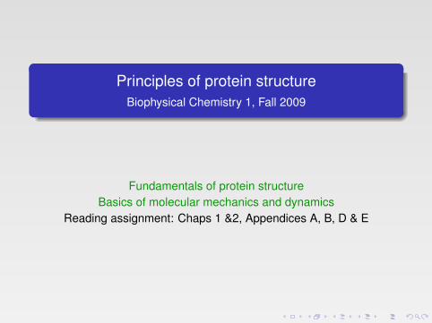
Principles of protein structure Biophysical Chemistry 1, Fall 2009 Fundamentals of protein structure Basics of molecular mechanics and dynamics Reading assignment: Chaps 1 &2, Appendices A, B, D & E
Transcript of Principles of protein structurePrinciples of protein structure Biophysical Chemistry 1, Fall 2009...
Principles of protein structureBiophysical Chemistry 1, Fall 2009
Fundamentals of protein structureBasics of molecular mechanics and dynamics
Reading assignment: Chaps 1 &2, Appendices A, B, D & E

Cell biology: not yet biochemistry!
found. Eukaryotic cells are usually at least 10 times larger than prokaryotic cellsand more complex. In eukaryotic cells, the basic prokaryotic cell structure with theplasma membrane and cytoplasm is upgraded with compartments, also calledorganelles. In the cytoplasm, additional distinctive structures can be found. Insome cases their interior is segregated from each other by a membrane. The mostcommon organelles are: (i) the nucleus storing the cell genetic material and wherereplication and gene expression takes place; (ii) the cytosol, where protein syn-thesis and many essential biochemical reactions take place; (iii) the mitochon-drion, a power plant and energy storage compartment; (iv) the endoplasmaticreticulum and Golgi apparatus, where proteins are packaged and sent to furtherlocations; (v) the lysosomes or vacuoles, where polymeric macromolecules, suchas proteins, are degraded into usable monomers (Fig. 1.3).
It is believed that all organisms on Earth originate from a single kind of uni-cellular organism. Today many millions of different kinds of organisms that donot interbreed with one another can be found and we call them species. They areall successfully adapted to their different environments and in this sense, perfect.However, some of them may not be perfect tomorrow and can thereby become
4 � A Textbook of Structural Biology
FA
FIGURE 1.3 � A schematic picture of an animal cell showing sub-cellular structures, such asnucleus, membrane systems (ER), mitochondrion, etc. (Made by Michael W. Davidson, FloridaState University.)
b541_Chapter-01.qxd 11/20/2008 10:54 AM Page 4

Proteins are polymers of amino acids
SUMOylation). The enzymatic or non-enzymatic modifications of amino acid sidechains in vivo are further described in App. E.
2.1.2 The Protein BackboneIn proteins, the amino acid residues are linked by peptide bonds. The bondbetween the CO and NH groups has a partial double bond character due to a res-onance between the normal form and a form with a double bond between the Cand N. This means that the atoms connecting the two Cα atoms are all in a plane.The ω (omega) torsion angle (Fig. 2.3) for the normal trans peptide bond is closeto 180°. Due to steric interactions, the cis peptide bond (ω ≈ 0°) is much less favor-able. For bonds preceding Pro residues, the cis conformation is more common (cis-prolines). This is discussed in Sec. 9.1.1.1.
Because of the planarity of the peptide bond, the conformation of the poly-peptide backbone can be described by two torsion angles per residue. Theseangles, φ and ψ, have restricted values due to steric clashes between the carbonyloxygen, the hydrogen of the NH group, the hydrogen on the Cα carbon, and theside chain atoms. This was used to define the allowed regions as described by the
Basics of Protein Structure � 15
FA
A
AB
B
C
CDD
ϕ ψ ωC
N
C
H
O R H
C
RH
α
α
H
Cα
Cα
RH
O
C Peptide bond
Peptideplane
123.5°
116°
122°
111°
119.5° 118.5°
1.24
1.0
1.461.33
1.51
120.5°
N
HR
FIGURE 2.3 � The peptide bond. The top left figure shows the definition of a torsion angle. Themiddle figure, where we look along the bond between atoms B and C, shows how we can deter-mine the angles between the bonds AB and CD. The top right drawing shows the names of thetorsion angles. In a trans peptide ω ≈ 180°, since the atoms of the peptide bond tends to forma plane (the amide plane). Bottom: The distances and angles between the atoms of a peptidebond.
b541_Chapter-02.qxd 11/20/2008 10:54 AM Page 15

There are twenty common side chains
2.1.1.1 Side chains and their interactions
The sequence of amino acid side chains gives the proteins unique properties. Theynot only determine the fold of the protein, but they also determine surface prop-erties that are important for selective interactions with other molecules and catal-ysis of chemical reactions.
Depending on the environment of the amino acids, the pKas given inTable 2.1 can be subject to dramatic changes. For example, if two carboxylgroups are close to each other and no positively charged group balances thesenegative charges, their pKas may be raised considerably. In addition to arginine,lysine, aspartic acid and glutamic acid, the side chains of cysteine, histidine,serine, threonine and tyrosine can sometimes be charged in physiologicalenvironments.
The side chains can interact with each other or with the main chain in manydifferent ways (see also App. A). The nonpolar or hydrophobic side chains, mostly
Basics of Protein Structure � 13
FA
Amino Acid Side Chains ( at pH 7)
General Amino Acid Structure
+ H3 N C COO–
H
R
CH2 CH2 CH2 CH2 NH3+
+
Lys
CH2 CH2 CH2 C
Arg NH2
NH2 CH2 C
O
O –
Asp
CH2 CH2 C
O
O –
Glu
Charged Polar Side Chains
His
Uncharged Polar Side Chains
CH2 SH
Cys
OH CH2
Tyr
CH2 OH
Ser
CH
CH3
OH
ThrCH2 C
NH2
OAsn
GlnCH2 CH2 C
NH2
O
Nonpolar Side Chains
Phe Pro
Gly Ala
ValLeu Ile
Met
CH2
N
C
–OOC
H
H
CH2 CH2 S CH3 H CH3
CH
CH3
CH3
CH2 CH
CH3
CH3
CH
CH3
CH2 CH3
NH
N
N H CH 2
Trp
N
CH2
H
FIGURE 2.2 � The 20 different side chains of the amino acids.
b541_Chapter-02.qxd 11/20/2008 10:54 AM Page 13

Ramachandran plots: sidechain torsional potentials
Ramachandran plot (Fig. 2.4). The two main allowed regions in the Ramachandranplot correspond to the two main types of conformation (α helices and β sheets)observed in proteins. A small region corresponding to a left-handed helical con-formation is also allowed. The allowed regions for Gly residues are much largersince there is no side chain to restrict the angles. On the other hand, prolineresidues have a very restricted set of conformations with φ restricted to valuesclose to −60°.
16 � A Textbook of Structural Biology
FA
FIGURE 2.4 � Ramachandran plot. Top: allowed regions based on steric clashes according toRamachandran’s original analysis for alanine (left) and glycine (right). Bottom: Observed anglesbased on about 1000 experimentally determined protein structures, non-glycine residues (left)and glycines (right). The β region is split into two sections. Note that the observed distributionsdiffer considerably from the expected distributions from the steric clashes, especially for glycineresidues. (Reprinted with permission from Hovmöller et al. (2002) Conformations of amino acidsin proteins. (Acta Cryst D58: 768–776. Copyright (2003) Elsevier.)
b541_Chapter-02.qxd 11/20/2008 10:54 AM Page 16

Secondary structure: the α-helix20 � A Textbook of Structural Biology
FA
FIGURE 2.7 � The α-helix. Left: The main chain and Cβ atoms (gray) of an α-helix. The pitch(rise per turn) is 5.4 Å. Right: The same α-helix showing the side chains. The backbone is drawnschematically, with the Cβ atoms pointing towards the N-terminus of the helix (down).
FIGURE 2.8 � A 310 helix from Aplysia limacina myoglobin (PDB: 1MBA) and a π-helix frommethane monooxygenase hydroxylase from Methylococcus capsulatus (PDB: 1MTY).
b541_Chapter-02.qxd 11/20/2008 10:54 AM Page 20

Secondary structure: 310 and π helices
20 � A Textbook of Structural Biology
FA
FIGURE 2.7 � The α-helix. Left: The main chain and Cβ atoms (gray) of an α-helix. The pitch(rise per turn) is 5.4 Å. Right: The same α-helix showing the side chains. The backbone is drawnschematically, with the Cβ atoms pointing towards the N-terminus of the helix (down).
FIGURE 2.8 � A 310 helix from Aplysia limacina myoglobin (PDB: 1MBA) and a π-helix frommethane monooxygenase hydroxylase from Methylococcus capsulatus (PDB: 1MTY).
b541_Chapter-02.qxd 11/20/2008 10:54 AM Page 20

Secondary structure: the β sheets
2.1.4.4 Beta structures
The other type of common secondary structure is the β-sheet. These are formed byextended stretches of the chain, called β-strands, where the CO and NH groupscan form hydrogen bonds to neighboring strands on both sides (App. A.3).β-sheets can be parallel, antiparallel or mixed, depending on the direction of thestrands (Fig. 2.9). In an ideal sheet, all NH groups of internal strands form hydro-gen bonds with CO groups. The strands at the edges will have free NH and COgroups.
Along the polypeptide chain the side chains extend from the sheet on alter-nating sides. Neighboring strands in the β-sheet have neighboring side chainspointing in the same direction — the side chains form lines perpendicular to thechain direction. The β-sheets are not flat but always twisted in the same directions(Fig. 2.10). If one views a sheet along the strands, it has a right-handed twist. Thedegree of twist differs between different sheets.
22 � A Textbook of Structural Biology
FA
FIGURE 2.9 � The hydrogen bonds in a mixed β-sheet. β-sheets are always more or less twisted.
FIGURE 2.10 � An antiparallel β-sheet showing the positions of the side chains on both sides ofthe sheet. Along the strands, the side chains point alternatingly in opposite directions but acrossthe strands neighboring side chains point in the same direction. The side chains on the lowerside of the sheet have been drawn with gray carbon atoms and those on the upper side areshown in black (PDB: 2BU1).
b541_Chapter-02.qxd 11/20/2008 10:54 AM Page 22

Secondary structure: turns
special type of left-handed triple-stranded helix (Fig. 2.12; see Sec. 13.1). This typeof structure is also found in rare cases in soluble proteins such as C1q in the com-plement system.
2.1.4.7 Turns
The reverse turn is another type of regular secondary structure. They areshort and often connect two β-strands. The reverse or β turn ideally has a hydro-gen bond between CO of residue n and NH of residue n + 3 (Fig. 2.13). This typeof tight turn imposes strong restrictions on the conformational angles of residuesn + 1 and n + 2 (Table 2.4). There are a few types of such turns, and in some of
24 � A Textbook of Structural Biology
FA
FIGURE 2.12 � The triple helix of a segment of a collagen. The three chains have a repeatedsequence of proline — hydroxyproline — glycine. The glycine residues allow the chains to comein close contact and form inter-chain hydrogen bonds (PDB: 1Q7D).
FIGURE 2.13 � Two of the most common types of reverse turns, type I’ and II.
b541_Chapter-02.qxd 11/20/2008 10:54 AM Page 24

Motifs, topologies, folds: antiparallel β sheets26 � A Textbook of Structural Biology
FA
FIGURE 2.14 � A β-hairpin and a four-stranded up-and-down sheet.
FIGURE 2.15 � Two up-and-down sheets, the open sheet in the coat protein subunit of phageMS2 (PDB: 2MS2), and the closed cylinder in a bacterial porin, a protein from the outer mem-brane of E. coli (PDB: 2OMF).
FIGURE 2.16 � Up-and-down sheets in a propeller structure: the seven-bladed propeller of theβ-subunit of transducin (PDB: 1GOT).
b541_Chapter-02.qxd 11/20/2008 10:54 AM Page 26
26 � A Textbook of Structural Biology
FA
FIGURE 2.14 � A β-hairpin and a four-stranded up-and-down sheet.
FIGURE 2.15 � Two up-and-down sheets, the open sheet in the coat protein subunit of phageMS2 (PDB: 2MS2), and the closed cylinder in a bacterial porin, a protein from the outer mem-brane of E. coli (PDB: 2OMF).
FIGURE 2.16 � Up-and-down sheets in a propeller structure: the seven-bladed propeller of theβ-subunit of transducin (PDB: 1GOT).
b541_Chapter-02.qxd 11/20/2008 10:54 AM Page 26

Motifs, topologies, folds: Greek key
A special type of a Greek key structure is an extended version called jellyroll.This is an eight-stranded arrangement that mostly forms a β sandwich of two four-stranded sheets (Fig. 2.18).
Proteins mainly composed of β structure normally have antiparallel sheets,but one exception is proteins with β-helix topology, where the chain forms shortβ-strands in a helical manner into a triangular prism (Fig. 2.19).
2.1.5.2 The βαβ unit
Another common motif in proteins is the βαβ unit, formed by two parallel strandsand a connecting helix. This motif exists in two forms, but one of them, the right-handed version, is by far the most common (Fig. 2.20).
Basics of Protein Structure � 27
FA
1 2 3 4
FIGURE 2.17 � A schematic drawing of a Greek key motif and the same motif in a protein(Micrococcal nuclease, PDB: 2SNS). The 5-stranded sheet and the helix in the loop connectingstrands 3 and 4 is an example of the OB (oligonucleotide/oligosaccharide binding) fold foundin many proteins.
65 4 7 2 1 8 3
1 2 3 4
56 7 8
FIGURE 2.18 � Left: A jellyroll topology (top) can be seen as a β-hairpin rolled up. Right: Thecoat protein of the plant virus STNV, a simple jellyroll fold (PDB: 2BUK).
b541_Chapter-02.qxd 11/20/2008 10:54 AM Page 27

Motifs, topologies, folds: jellyroll
A special type of a Greek key structure is an extended version called jellyroll.This is an eight-stranded arrangement that mostly forms a β sandwich of two four-stranded sheets (Fig. 2.18).
Proteins mainly composed of β structure normally have antiparallel sheets,but one exception is proteins with β-helix topology, where the chain forms shortβ-strands in a helical manner into a triangular prism (Fig. 2.19).
2.1.5.2 The βαβ unit
Another common motif in proteins is the βαβ unit, formed by two parallel strandsand a connecting helix. This motif exists in two forms, but one of them, the right-handed version, is by far the most common (Fig. 2.20).
Basics of Protein Structure � 27
FA
1 2 3 4
FIGURE 2.17 � A schematic drawing of a Greek key motif and the same motif in a protein(Micrococcal nuclease, PDB: 2SNS). The 5-stranded sheet and the helix in the loop connectingstrands 3 and 4 is an example of the OB (oligonucleotide/oligosaccharide binding) fold foundin many proteins.
65 4 7 2 1 8 3
1 2 3 4
56 7 8
FIGURE 2.18 � Left: A jellyroll topology (top) can be seen as a β-hairpin rolled up. Right: Thecoat protein of the plant virus STNV, a simple jellyroll fold (PDB: 2BUK).
b541_Chapter-02.qxd 11/20/2008 10:54 AM Page 27

Motifs, topologies, folds: βαβ structures
Parallel sheets in proteins are often built up of βαβ units. In these proteins, theβαβ units are repeated to form all or part of the sheet. The Rossmann fold, foundin many proteins, is built of such βαβ units (Fig. 2.21). It was first found in lactate,maleate, and alcohol dehydrogenase, where a six-stranded parallel sheet is at thecore of the protein.
In the Rossmann fold, the N-terminal strand is in the middle of the sheet. Twoβαβ units form half of the sheet (βαβαβ), and the rest is formed by a similar unitstarting next to the first strand and related by an approximate twofold axis. Becauseof the handedness of the units and the twofold relationship, the helices will end upon opposite sides of the sheet (Fig. 2.21). There are many proteins with similar topol-ogy, sometimes with slight variation in the number and order of the strands.
28 � A Textbook of Structural Biology
FA
FIGURE 2.19 � A β-helix protein: pectin lyase from Aspergillus niger (PDB: 1IDK).
FIGURE 2.20 � Schematic drawing of a βαβ unit. Following the direction of the polypeptide chaina right-hand screw is formed.
b541_Chapter-02.qxd 11/20/2008 10:54 AM Page 28
Another very common fold is the TIM barrel, which got its name from theenzyme, triose phosphate isomerase. This fold is formed by eight β-α units form-ing a cylinder (Fig. 2.22). The eight parallel strands form a closed cylinder, andthe eight helices form a layer outside the β-cylinder. This fold is found in a large
Basics of Protein Structure � 29
FA
N
C
1 2 3 4 5 6
FIGURE 2.21 � Left: Schematic drawing of the topology called the Rossmann fold. Right: TheNAD-binding domain of Sulfolobus solfataricus alcohol dehydrogenase, a typical Rossmann fold(order of strands 654123). The first βαβαβ motif of the domain is in blue, the second in red andthe connection in yellow. The nucleotide is bound at the C-terminal ends of the β-strands at thecenter of the sheet. The phosphates of the NAD molecule are located at the N-termini of twohelices where their binding is favored by the helical dipoles (PDB: 1R37).
FIGURE 2.22 � The TIM barrel of triose phosphate isomerase from Plasmodium falciparum. Theorder of the strands is 12345678. The active site is occupied by a transition-state analog, phos-phoglycolate. In all TIM barrel structures, the active site is found at the C-terminal end of theβ-strands (PDB: 1LYX).
b541_Chapter-02.qxd 11/20/2008 10:54 AM Page 29

Motifs, topologies, folds: TIM barrel
Another very common fold is the TIM barrel, which got its name from theenzyme, triose phosphate isomerase. This fold is formed by eight β-α units form-ing a cylinder (Fig. 2.22). The eight parallel strands form a closed cylinder, andthe eight helices form a layer outside the β-cylinder. This fold is found in a large
Basics of Protein Structure � 29
FA
N
C
1 2 3 4 5 6
FIGURE 2.21 � Left: Schematic drawing of the topology called the Rossmann fold. Right: TheNAD-binding domain of Sulfolobus solfataricus alcohol dehydrogenase, a typical Rossmann fold(order of strands 654123). The first βαβαβ motif of the domain is in blue, the second in red andthe connection in yellow. The nucleotide is bound at the C-terminal ends of the β-strands at thecenter of the sheet. The phosphates of the NAD molecule are located at the N-termini of twohelices where their binding is favored by the helical dipoles (PDB: 1R37).
FIGURE 2.22 � The TIM barrel of triose phosphate isomerase from Plasmodium falciparum. Theorder of the strands is 12345678. The active site is occupied by a transition-state analog, phos-phoglycolate. In all TIM barrel structures, the active site is found at the C-terminal end of theβ-strands (PDB: 1LYX).
b541_Chapter-02.qxd 11/20/2008 10:54 AM Page 29
Motifs, topologies, folds: αhelix packing
number of enzymes (and one non-enzyme, narbonin), many of which are unre-lated in sequence and function.
2.1.5.3 Helix packing
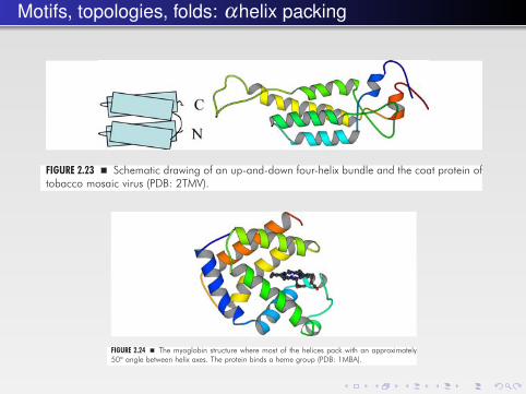
In helical proteins, there is a limited number of ways in which the helices can bepacked. The cause of this is that the side chains of the helices form different ridges,and optimal packing requires that a ridge of one helix is positioned in a groovebetween ridges in the other helix. This leads to angles of about 20° or 50° betweenhelix axes. A common type of helix packing is the four-helix bundle, where two pairsof antiparallel helices are arranged with their helix axis at about 20°. Four-helix bun-dles are found in many proteins. A simple form is the up-and-down bundle (Fig. 2.23),but there are many other forms of bundles formed of helices from one, two or four sub-units. The globin fold is an example of the other main type of helix packing (Fig. 2.24).
30 � A Textbook of Structural Biology
FA
FIGURE 2.23 � Schematic drawing of an up-and-down four-helix bundle and the coat protein oftobacco mosaic virus (PDB: 2TMV).
FIGURE 2.24 � The myoglobin structure where most of the helices pack with an approximately50° angle between helix axes. The protein binds a heme group (PDB: 1MBA).
b541_Chapter-02.qxd 11/20/2008 10:54 AM Page 30
number of enzymes (and one non-enzyme, narbonin), many of which are unre-lated in sequence and function.
2.1.5.3 Helix packing
In helical proteins, there is a limited number of ways in which the helices can bepacked. The cause of this is that the side chains of the helices form different ridges,and optimal packing requires that a ridge of one helix is positioned in a groovebetween ridges in the other helix. This leads to angles of about 20° or 50° betweenhelix axes. A common type of helix packing is the four-helix bundle, where two pairsof antiparallel helices are arranged with their helix axis at about 20°. Four-helix bun-dles are found in many proteins. A simple form is the up-and-down bundle (Fig. 2.23),but there are many other forms of bundles formed of helices from one, two or four sub-units. The globin fold is an example of the other main type of helix packing (Fig. 2.24).
30 � A Textbook of Structural Biology
FA
FIGURE 2.23 � Schematic drawing of an up-and-down four-helix bundle and the coat protein oftobacco mosaic virus (PDB: 2TMV).
FIGURE 2.24 � The myoglobin structure where most of the helices pack with an approximately50° angle between helix axes. The protein binds a heme group (PDB: 1MBA).
b541_Chapter-02.qxd 11/20/2008 10:54 AM Page 30

Proteins often consist of modular domains
from sequence patterns. Each module is a separate unit in the sequence and foldsas a separate domain. An example is tissue plasminogen activator, which is com-posed of five modules of four kinds, one fibronectin type I module, one epidermalgrowth factor (EGF) module, two so-called kringle modules, and the main enzy-matic portion, a serine protease module (Fig. 2.27). Plasminogen is also a mosaicprotein, with five kringle modules preceding a serine protease domain. In someproteins, the number of similar modules is very large. Some of these modules, likethe EGF and the fibronectin type III modules, are found in a large number of pro-teins, and appear to fulfill a similar function in all of them (see Chaps. 11 and 13).Most of these modules are not catalytic, but are able to bind other molecules.
Mosaic proteins are formed by recombination of genetic elements during evo-lution and by different splicing events. Each domain may correspond to a singleexon in the DNA. Gene duplications and differing splicing of these exons willtherefore lead to formation of protein molecules with new combinations of theseexons or domains.
2.1.6.4 Loops and tails
Secondary structure elements are connected by turns or loops. Such loops cansometimes be quite long and protrude from the more compact part of the protein.The loops can be devoid of secondary structure or they can be organized into aβ-ribbon or a helix. Other extensions from a compact structure are amino-terminalor carboxy-terminal tails. These tails can also have a secondary structure. Bothloops and tails usually have structural roles, quite frequently for the stabilizationof oligomeric structures. One example is lactate dehydrogenase where theN-terminal 20 amino acid residues form an arm that folds onto another subunit tostabilize one dimer interaction in the tetrameric enzyme (Fig. 2.28). The relatedenzyme malate dehydrogenase lacks this N-terminal extension and is thereforeoften dimeric.
Coat proteins of viruses (Chap. 15), histones (Sec. 3.2.1) and ribosomal pro-teins (Chap. 8) often also have extended positively charged parts that neutralize
Basics of Protein Structure � 33
FA
Ig Fz Kringle protein kinase
Fnl
EGF
Kringle Serine protease
FIGURE 2.27 � Domain organization of two typical multi-domain proteins, tissue plasminogenactivator (top) and a receptor tyrosine kinase (bottom) as presented in the Pfam database(http://pfam.sanger.ac.uk), where the cylinders are links to the corresponding family.
b541_Chapter-02.qxd 11/20/2008 10:55 AM Page 33
within the same polypeptide, the domains may interact with the correspondingdomain in another polypeptide chain (Fig. 2.26).
Sometimes this can be done in a way that extends the oligomerization in a lin-ear or possibly branched way. The final effect will be aggregation of monomers.
2.1.6.3 Mosaic proteins
A number of eukaryotic proteins (often called “mosaic proteins”) are formed bymany modules. These modules are also functional units and can be identified
32 � A Textbook of Structural Biology
FA
FIGURE 2.25 � A two-domain protein, transducin α, with one domain (red) as an insertion in theother domain, a G-domain (blue). The amino acid sequence is shown in blue and red for themain and inserted domain, respectively.
FIGURE 2.26 � Domain swapping of a protein. Normally the protein is a monomer (top) wherethe two domains interact within the monomer. Bottom: The two domains of the protein interactin the same way with a different monomer.
b541_Chapter-02.qxd 11/20/2008 10:55 AM Page 32

Common protein folds
to form a reasonably well packed hydrophobic core severely restrict the folding. Itis possible that there are many stable folds that do not exist in nature, since theexisting folds are the result of an evolutionary process that has not explored orpreserved all these sequence combinations.
2.2.2 Protein Stability and Dynamics
2.2.2.1 The folding process
The conformation or tertiary structure of a protein is defined by its amino acidsequence and therefore by the nucleotide sequence of the corresponding gene.The direct relation between the conformation and the amino acid sequence of a
40 � A Textbook of Structural Biology
FA
FIGURE 2.31 � Schematic drawings of a number of common protein folds. Top left: α/β domainwith Rossmann fold (3α, 20β-hydroxysteroid dehydrogenase, PDB: 1HDC). Top right:Immunoglobulin constant domain (PDB: 1AQK). Bottom left: TIM barrel (triose phosphateisomerase, PDB: 1YPI). Bottom right: Jellyroll (satellite tobacco necrosis virus coat protein,PDB: 2BUK).
b541_Chapter-02.qxd 11/20/2008 10:55 AM Page 40

Post-translational modifications: glycosylation
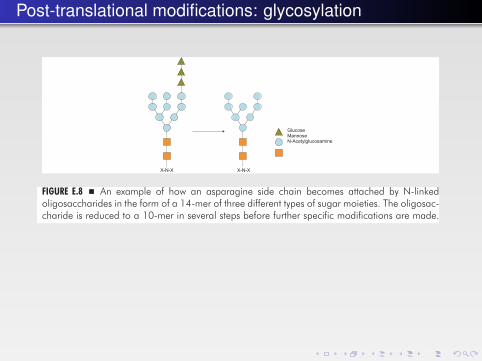
In all N-linked oligosaccharides, N-acetylglucosamine is bound to the sidechain nitrogen of the asparagine (Fig. E.8). The modification here involves a largebranched oligosaccharide, which is prefabricated attached to dolichol, a long lipidthat is firmly bound in the membrane. The oligosaccharide has the composition(glucose)3(mannose)9(N-acetylglucose)2. After the transfer to a protein, the threeglucose residues are removed as well as a specific mannose residue. These
544 � A Textbook of Structural Biology
FA
FIGURE E.7 � Some types of sugar residues that can be attached to proteins. The top left showsthe more extensive formula for fucose where all hydrogens are included. Below is the samefucose without showing the hydrogens directly attached to the ring carbons.
GlucoseMannoseN-Acetylglucosamine
X-N-X X-N-X
FIGURE E.8 � An example of how an asparagine side chain becomes attached by N-linkedoligosaccharides in the form of a 14-mer of three different types of sugar moieties. The oligosac-charide is reduced to a 10-mer in several steps before further specific modifications are made.
b541_Appendix-E.qxd 1/6/2009 9:51 AM Page 544
Post-translational modifications: glycosylation
In all N-linked oligosaccharides, N-acetylglucosamine is bound to the sidechain nitrogen of the asparagine (Fig. E.8). The modification here involves a largebranched oligosaccharide, which is prefabricated attached to dolichol, a long lipidthat is firmly bound in the membrane. The oligosaccharide has the composition(glucose)3(mannose)9(N-acetylglucose)2. After the transfer to a protein, the threeglucose residues are removed as well as a specific mannose residue. These
544 � A Textbook of Structural Biology
FA
FIGURE E.7 � Some types of sugar residues that can be attached to proteins. The top left showsthe more extensive formula for fucose where all hydrogens are included. Below is the samefucose without showing the hydrogens directly attached to the ring carbons.
GlucoseMannoseN-Acetylglucosamine
X-N-X X-N-X
FIGURE E.8 � An example of how an asparagine side chain becomes attached by N-linkedoligosaccharides in the form of a 14-mer of three different types of sugar moieties. The oligosac-charide is reduced to a 10-mer in several steps before further specific modifications are made.
b541_Appendix-E.qxd 1/6/2009 9:51 AM Page 544

Post-translational modifications: phosphorylation
Phosphorylation is central for intracellular and transmembrane signaling and alarge fraction of all proteins undergo phosphorylation. Not only is the phospho-rylation important; it is equally important that the phosphate group can beremoved. This is done by phosphatases. The phosphorylation of intracellularproteins serves as molecular regulation of a range of cellular processes (Chap. 11).The protein kinase family consists of more than 700 proteins. Thus, a significantportion of the proteins of a genome is kinases or phosphorylases. The proteinkinases share a common fold of about 250 residues. Kinases transfer the γ phos-phate of ATP to the substrates. Different residues can be phosphorylated: tyrosine,serine/threonine and more rarely histidine and aspartate. Since kinases playcentral roles in signaling pathways and regulatory mechanisms, they are interest-ing targets for drug design.
Initially, consensus sequences in the substrates were looked for, but with a rap-idly increasing number of kinases and substrates the linear sequence aspect wasnot a reasonable option. The kinases rather interact with several motifs of the sub-strates. Since phosphorylation has a regulatory function, it needs to be very spe-cific. The regulation can be achieved in a number of ways. The kinases themselvesare often activated by phosphorylation. In these cases, the kinases have an activa-tion segment. The kinases switch between two states, on and off (see Chap. 11).In other cases, several domains of the kinase need to interact with the kinase cat-alytic domain, or multisubunit proteins may be involved. One highly regulatedgroup of proteins is the cAMP dependent, cGMP dependent protein kinase C (theAGC family of kinases). Figure E.4 illustrates some of the ways that kinases caninteract with their substrates.
Protein phosphatases belong to several different classes: protein serine-threonine phosphatases, protein tyrosine phosphatases, and dual-specificity phos-phatases. They can be composed of one or several domains. These domains canbelong to a large range of families, and some of the phosphatases are attached to
Appendix E: Protein Modification � 539
FA
Active site
Docking site
FIGURE E.4 � The regulatory role of kinases and phosphatases requires extensive control of theprotein interactions. The enzymes (blue) partly need to be activated but also need to identifystructures other than the part that will be phosphorylated or dephosphorylated. This is due tosubstrate (green) interactions with a docking site that can be part of the kinase domain or belongto different parts of the enzyme.
b541_Appendix-E.qxd 1/6/2009 9:51 AM Page 539

Post-translational modifications: acetylation
The methylation of arginines has become a field of growing interest. A wide rangeof proteins can be modified, including histones and non-histone proteins involved intranscription. Many proteins that interact with nucleic acids can be methylated onarginines. Glycine-arginine-rich motifs are frequently targets for modification.
E.6 Acetylation
Many proteins, including histones, can also be acetylated. Amino groups are thenormal targets for acetylation. This can be the N-terminal α-amino group of anyamino acid or the ε-amino group of lysines (Fig. E.6). In the latter case, the modi-fication can be carried out by histone acetyltransferases (HATs), which can begrouped into a number of families. Subunits of the TFIID and Mediator complexesin the transcription machinery are HATs (see Chap. 7). Many HATs work in largemultiprotein complexes. Several HATs contain actin or actin-related proteins.Histone acetylation, which can regulate transcription of specific genes, is areversible process. Thus, there are also histone deacetylases (HDACs), which canbe classified into several families.
E.7 Carboxylation
Vitamin K-dependent proteins have uniquely modified residues, γ-carboxyglu-tamic acid (Gla). They are formed by post-translational carboxylation of glutamicacid by gamma-glutamyl carboxylase. The proteins to be modified require a
Appendix E: Protein Modification � 541
FA
FIGURE E.6 � Left: An acetylated lysine. This modification is important in the modifications of his-tones. Middle: A carbamylated lysine, which has been found in several enzyme active sites withtwo metals. The elongated residue can bridge between the two metals. Right: A γ-carboxyglu-tamic acid (Gla) residue. This modification is abundant in the blood clotting system.
b541_Appendix-E.qxd 1/6/2009 9:51 AM Page 541

Post-translational modifications: disulfide bonds
A.2 Charge Interactions
Electrostatic interactions are very important in biological systems, but theyare also more abstract. It is not possible to cover electrostatic theory in thisappendix, and we will just mention a few subjects. If we look at a globular pro-tein or at the surface of a lipid membrane we see that charged groups, such asin lysine or arginine residues on proteins and phosphate and ethanolamine orserine groups on lipids, are normally solvated and have nearby counter ions.Not only would burying an isolated ionic group inside the protein or in the coreof the lipid bilayer cause a loss of the solvation energy, but there would also bean enormous electrostatic price to pay. The attraction between two oppositelycharged ions is given by Coulomb’s law, that is, proportional to e2/εr, wheree is the unit charge, r is the distance between the ions considered as pointcharges, and ε is the dielectric constant of the medium in which they arelocated. In water solution ε is roughly 80, whereas it is much lower insidethe protein or lipid bilayer (about 2–20). Thus, if a charge is buried, there is anenormous energetic advantage in burying a suitable opposite charge as closeas possible. This is one of the reasons for the formation of salt-bridges, forexample, and why we need channels in membranes to transport ions throughthe membrane.
Concerning the interactions and binding of ions to charged surfaces on pro-teins or membranes, the theory is more sophisticated. This pertains also to situa-tions where we consider interactions between, for example, two membranes of thesame or different charge or even one neutral and one charged membrane. Osmoticpressures and entropic forces will be involved in such events, and this is outsidethe scope of this book.
Appendix A: Bonds and Energetics of Macromolecules � 487
FA
S
Cβ
βS CFIGURE A.1 � The preferred conformation of a disulfide bond with a 90° angle between theS–Cβ bonds viewed down the S–S bond.
b541_Appendix-A.qxd 12/29/2008 2:46 PM Page 487

Metals in proteins: “blue” copper
Figure 1. Expanded views of geometric and electronic structure of the active sites of plastocyanin (left) and rubredoxin (right). The expanded plastocyanin site is rotatedsuch that the Met‚S-Cu bond is out of the plane of the page. Contour values are set to (0.16, (0.08, (0.04, (0.02, and (0.01 (e/µB3), with positive contours in solid red andnegative values in dashed blue.
AspectsofM
etalSitesin
BiologyChem
icalReviews,1996,Vol.96,No.72241
++
Downloaded by RUTGERS UNIV on September 15, 2009 | http://pubs.acs.org Publication Date (Web): November 7, 1996 | doi: 10.1021/cr9500390

Metals in proteins: Iron-sulfur centers
polynuclear centers. In addition to discrete Rd andFd electron transfer proteins, which often are theultimate electron donors to enzymes, these centersmay also be found within enzyme molecules them-
selves where they form part of the electron transferconduit to the catalytic site.Rubredoxins contain one iron atom, usually fall in
the 6-7 kD range, and are the simplest of the iron-sulfur proteins. Reports of structures are collectedin Table 5;82-86 the resolutions are some of the bestachieved in protein crystallography. The high-spinFeS4 cores of the [FeII,III(S‚Cys)4] coordination unitsare close to tetrahedral, but distortions of the entireunit tend to impose effective D2d symmetry. Thestructures of P. furiosus Rdox and Rdred have beenobtained at 1.8 Å resolution, allowing an assessmentof the structural changes pursuant to electron trans-fer. Mean Fe-S bond distances change by j0.05 Åand S-Fe-S bond angles by j5°.Proteins containing [Fe2S2(S‚Cys)4] units are nu-
merous; the Fe2(µ2-S)2 cores approach D2h symmetry.The structures of six proteins, listed in Table 6,87-97
have been determined in the oxidized state; noreduced protein structure is available. These pro-
Chart 1. Structuresa of Redox Sites in ElectronTransfer Proteins: (A) Rubredoxin (Clostridiumpasteurianum); (B) Ferredoxin (Equisetumarvense); (C) Ferredoxin II (Desulfovibrio gigas);(D) Ferredoxin I (Azotobacter vinelandii); (E)Plastocyanin (Chlamydomonas reinhardtii); (F)Azurin (Alcaligenes denitrificans); (G) stellacyanin(cucumber); and (H) CuA Center in Cytochrome cOxidase (Bovine Heart)
a All structures depicted in these charts were created usingcrystallographic coordinates taken from the Brookhaven ProteinDatabank or by private communication. The color scheme foratoms is as follows: carbon (gray), oxygen (pink), nitrogen (cyan),sulfide and cysteine sulfur (bright yellow), methionine sulfur (lightyellow), zinc (light purple), iron (red), copper (blue), other metals(gold), and hydrogen (white). Hydrogen atoms are not crystallo-graphically defined but have been added to depict the nature ofcertain ligands when known. Structures are cross-referenced inTables 5-8, 9, and 13-18.
Chart 2. (I) Structures of Oxygen-CarryingProteins [Hemocyanin (Limulus polyphemus) in ItsDeoxygenated (A) and Oxygenated (B) Forms;Hemerythrin (Themiste dyscrita) in ItsDeoxygenated (C) and Oxygenated (D) Forms] and(II) Structures of Superoxide Dismutases [(E)Cu/Zn Superoxide Dismutase (Saccharomycescerevisiae); (F) Mn Superoxide Dismutase (HumanKidney)]
2252 Chemical Reviews, 1996, Vol. 96, No. 7 Holm et al.
+ +
Dow
nloa
ded
by R
UTG
ERS
UN
IV o
n Se
ptem
ber 1
5, 2
009
| http
://pu
bs.a
cs.o
rg
Pub
licat
ion
Dat
e (W
eb):
Nov
embe
r 7, 1
996
| doi
: 10.
1021
/cr9
5003
90

Protein families: the SCOP classification
B.2.2.1 SCOP: Structural classification of proteins
In the SCOP (Structural Classification Of Proteins) database developed by A.Murzin and his co-workers (UK), all known protein structures are sorted accord-ing to their fold. This database is mainly based on manual classification of the pro-tein folds and therefore the classifications are subjective. On the other hand, theaccumulated knowledge of protein conformation and the details of the evolutionof specific proteins that is used for the classification are not easily incorporated ina computer program for automatic fold classification.
The levels of the hierarchy (Fig. B.5) are classes, folds, superfamilies, familiesand domains (the individual proteins or protein domains). The various domainswithin a family are assumed to be homologous, i.e. to have a common ancestorfrom which they have diverged. The hypothesis regarding homology of proteindomains with similar structures is based on sequence and/or functional similar-ity. Proteins within one superfamily have the same fold and a related function andtherefore they also probably have a common ancestor, but they differ too much insequence or function to allow a conclusive decision about homology. At the nextlevel, fold, the proteins have the same topology, but there is no evidence of an evo-lutionary relationship except the limited structural similarity.
The SCOP database classifies domains rather than entire proteins. This meansthat multi-domain proteins are divided into their constituent domains. This isvery useful in the many cases where one kind of domain is shared by differentproteins. The high quality of the classifications has made this database into a stan-dard of evolutionary relationships. The database can be searched and has links tothe individual entries in the PDB.
Appendix B: Methods for Fold Comparison � 501
FA
Class All alpha(218)
All beta(144)
Alpha/beta(136)
Alpha+beta(279)
Fold Immuno-globulin-like (23)
Prealbumin(6)
Viral coats(1)
OB fold (10)
Superfamily Immuno-globulin(4)
CuZnsuperoxidedismutase(1)
PapD-like(2)
Fibronectintype III (1)
Family V domains(78)
C1 domains(64)
C2 domains(9)
I domains(45)
FIGURE B.5 � SCOP classification. The example shows the hierarchy for immunoglobulindomains. The number of different subdivisions of a group is shown in parentheses.
b541_Appendix-B.qxd 12/29/2008 2:46 PM Page 501

Protein families: the CATH classification
B.2.2.2 CATH
A similar structure database is CATH, developed by C. Orengo and J. Thornton(UK). It orders every known protein structure hierarchically in classes, architec-tures, topologies (fold families), superfamilies and sequence families (CATHstands for Class, Architecture, Topology, Homologous superfamily; Fig. B.6).
The classification of structures in CATH is to a large extent done by automatedprocedures. The automatic procedure starts with the definition of domains in aprotein. A number of procedures for doing this automatically have been devel-oped. In CATH, the domain definition is the consensus of three different algo-rithms. In many cases, a consensus is not reached and a manual definition has tobe made.
The topologies of the CATH database correspond essentially to the folds of theSCOP classification, but the architecture level (Fig. B.7) does not have any corre-spondence in the SCOP hierarchy. Proteins with the same general arrangement ofsecondary structural elements, for example a β sandwich or an α helix bundle, butpossibly with different connections between the elements are defined as havingthe same architecture.
Due to the differences in classification procedures, some proteins are groupeddifferently in SCOP and CATH. The differences may be due to problems in the
502 � A Textbook of Structural Biology
FA
Class Mainly alpha(5)
Mainly beta(19)
Alpha beta(14)
Irregular(1)
Architecture Sandwich(26)
Barrel (28) Trefoil (1) Roll (28)
Topology Immuno-globulin-like (64)
TumorsuppressorSmad4 (2)
Jelly rolls(21)
Thaumatin(1)
Immuno-globulinfamily(137)
CuZnsuperoxidedismutase(3)
FibronectinType III (53)
Prealbumin(1)
Homologoussuperfamily
Sequencefamily
Immuno-globulindomains
CD2 Interleukin-1receptor
titin
FIGURE B.6 � CATH classification. The immunoglobulins are used as an example again. Notethat prealbumin (transthyretin) is classified as part of the immunoglobulin-like topology in CATH,but is a separate fold in SCOP (Fig. B.5).
b541_Appendix-B.qxd 12/29/2008 2:46 PM Page 502

1901 (and earlier?) ball and stick models

1950s: wire models of proteins

- separate nuclei and electrons
- polarisation, electron transfer and correlation
- can specify electronic state
- can calculate formation energies
- can do chemistry (bond breaking and making)
- variationally bound
- computationally expensive
- typically ~10-100 atoms
- dynamics ~1 ps
QM MOLECULE
Nuclei
Electrons

- no explicit electrons, net atomic charges
- no polarisation, electron transfer or correlation
- conformational energies for ground state
- no chemistry
- semi-empirical force fields
- not variationally bound
- solvent and counterion representations
- typically ~1000-100000 atoms
- dynamics up to ~100 ns
Atoms
Bonds
MM MOLECULE

Some force field assumptions
1 Born-Oppenheimer approximation (separate nuclear andelectronic motion)
2 Additivity (separable energy terms)3 Transferability (look at different conformations, different
molecules)4 Empirical (choose functional forms and parameters based on
experiment)

What does a force field look like?
U = ∑bonds
Kb(b−beq)2 + ∑
anglesKθ (θ −θeq)
2 + ∑impropers
Kw w2
+ ∑torsions
Kφ cos(nφ)+ ∑nonbonded pairs
{4ε
[(σ
r
)12−
(σ
r
)6]+
qiqj
r
}
C
O
N
H
H
H
O
H
H
12
3
formamide
water

Lennard-Jones energy curve
eij

Distance dependence
Electrostatic
Lennard-Jones

AMBER parm94 H atom types
H H bonded to nitrogen atoms
HC H aliph. bond. to C without electrwd.group
H1 H aliph. bond. to C with 1 electrwd. group
H2 H aliph. bond. to C with 2 electrwd.groups
H3 H aliph. bond. to C with 3 eletrwd.groups
HA H arom. bond. to C without elctrwd. groups
H4 H arom. bond. to C with 1 electrwd. group
H5 H arom. bond. to C with 2 electrwd. groups
HO hydroxyl group
HS hydrogen bonded to sulphur
HW H in TIP3P water
HP H bonded to C next to positively charged gr

C sp2 C carbonyl group
CA sp2 C pure aromatic (benzene)
CB sp2 aromatic C, 5&6 membered ring junction
CC sp2 aromatic C, 5 memb. ring HIS
CK sp2 C 5 memb.ring in purines
CM sp2 C pyrimidines in pos. 5 & 6
CN sp2 C aromatic 5&6 memb.ring junct.(TRP)
CQ sp2 C in 5 mem.ring of purines between 2 N
CR sp2 arom as CQ but in HIS
CT sp3 aliphatic C
CV sp2 arom. 5 memb.ring w/1 N and 1 H (HIS)
CW sp2 arom. 5 memb.ring w/1 N-H and 1 H (HIS)
C* sp2 arom. 5 memb.ring w/1 subst. (TRP)
AMBER parm94 C atom types

Force fields in Amber
ff94: widely used (“Cornell et al.), pretty good nucleic acid, toomuch α-helix for proteins
ff99: major recalibration by Junmei Wang and others; basis formost current Amber ff’s
ff99SB: recalibration of backbone potentials for proteins by CarlosSimmerling (“SB”)
ff02r1: polarizable extension for ff99
ff03: new charge model (Yong Duan) + backbone torsions forproteins
ff03ua: united atom extension

Periodic boundary conditions

Example of explicit solvation setup

Minimization and simulated annealing

Molecular dynamics algorithms
x(t +h) = x(t)+ v(t)h +12
a(t)h2 +16
d3xdt3 h3 +O(h4)
x(t−h) = x(t)+−v(t)h +12
a(t)h2− 16
d3xdt3 h3 +O(h4)
x(t +h) = 2x(t)− x(t−h)+a(t)h2 +O(h4) (1)
x(t +h)− x(t) = x(t)− x(t−h)+a(t)h2 +O(h4)
v(t +12
h) = v(t− 12
h)+a(t)h +O(h3) (2)
x(t +h) = x(t)+ v(t +12
h)h +O(h4) (3)
Eq. (1) is the original Verlet propagation algorithm; Eqs. 2 and 3 arethe “leap-frog” version of that. Remember thata = d2x/dt2 = F/m = (∂V/∂x)/m. See pp. 42-47 in Becker &Watanabe.

Regulating temperature
“Temperature” is a measure of the mean kinetic energy. Theinstantaneous KE is
T (t) =1
kBNdof
Ndof
∑i
miv2i
(cf. classical rule of thumb: “kBT/2 of energy for every squareddegree of freedom in the Hamiltonian”)Suppose the temperature is not what you want. At each step, youcould scale the velocities by:
λ =
[1+
h2τ
(T0
T (t)−1
)]1/2
This is the “Berendsen” or “weak-coupling” formula, that has a minimaldisruption on Newton’s equations of motion. But it does not guaranteea canonical distribution of positions and velocities. See Morishita, J.Chem. Phys. 113:2976, 2000; and Mudi and Chakravarty, Mol. Phys.102:681, 2004.

Go model for protein folding

Gaussian network model

Knowledge-based potentials