Principles of immunodetection by Martin Loignon Ph.D. Lady Davis Institute for Cancer Research...
59
Principles of immunodetection by Martin Loignon Ph.D. Lady Davis Institute for Cancer Research Jewish General Hospital
-
Upload
andra-fleming -
Category
Documents
-
view
226 -
download
3
Transcript of Principles of immunodetection by Martin Loignon Ph.D. Lady Davis Institute for Cancer Research...

Principles of immunodetection
by
Martin Loignon Ph.D.Lady Davis Institute for Cancer Research
Jewish General Hospital

• Antibody-based methods allowing the specific: – Detection– Quantification– Localisation
• Of antigens by means of antibody binding
Immunodetection

Aims and Objectives
• Basis of antibody production and antigen interaction
• Conceptualise the different analytical techniques based on this interaction
• Examples of clinical application • Research problems requiring immunoanalyses• Troubleshooting of some common problems

Discovery of antibodies• 1899 *Jules Bordet, Complement and antibody activity in bacteriolysis
• 1900 *Paul Erlich, Antibody formation theory
• 1926 Lloyd Felton & GH Bailey, Isolation of pure antibody preparation
• 1934-8 John Marrack, Antigen-antibody binding hypothesis
• 1941 Albert Coons, Immunofluorescence technique
• 1948 Astrid Fagraeus, Demonstration of antibody production in plasma B cells
• 1959-62 *Rodney Porter et al., Discovery of antibody structure
• 1963 Jaques Oudin et al., antibody idiotypes
• 1964-8 Anthony Davis et al., T and B cell cooperation in immune response
• 1965 Thomas Tomasi et al., Secretory immunoglobulin antibodies
• 1975 *Kohler and Milstein, Monoclonal antibodies used in genetic analysis • 1985 *Tonegawa, Hood et al., Identification of immunoglobulin genes

Generation of an antibody: antigen processing

B cell activation

Structure of an antibody

Antibody and VDJ recombination

Classes of antibodies
Isotype Structure Placentatransfert
Activatescomplement
Additional features
IgMNo Yes First Ab in development and response
IgDNo No B-cell receptor
IgGYes Yes Involved in opsonization and ADCC.
Four subclasses; IgG1, IgG2, IgG3,IgG4
IgENo No Involved in allergic responses
IgANo No Two subclasses; IgA1, IgA2. Also found
as dimer (sIgA) in secretions.

Commercial production of antibodies: polyclonal vs monoclonal
• Host animals ca be used to raise antibodies against a given antigen
• Slected clones from a polyclonal each recognizing a single epitope can be fused to a tumor cell (hybridoma) to proliferate indefinitely

Antigen-antibody interaction
• Antigen: foreign molecules that generate antibodies or any substance that can be bound specifically by an antibody molecule– Proteins, sugars, lipids or nucleic acids
– Natural or synthetic
• Antibody: molecules (protein) responsible for specific recognition and elimination (neutralization) of antigens– Different structures (7-8 classes in mammals)
– Powefull research tools for basic research, clinical applications and drug design

Antigenic determinants
• An antibody will recognize– Epitope: defined segment of an antigen
– Immunoreactivity of epitopes may depend on primary, secondary, tertiary or quaternary structure of an antigen
– Define the possible applications
– Variability of epitopes depends on the species
• Antibodies are antigen themselves

Nature of binding forces
• Hydrogen bonding– Results from the formation of hydrogen bridges between appropriate atoms
• Electrostatic forces– Are due to the attraction of oppositely charged groups located on two protein side
chains
• Van der Waals bonds – Are generated by the interaction between electron clouds (oscillating dipoles)
• Hydrophobic bonds – Rely upon the association of non-polar, hydrophobic groups so that contact with water
molecules is minimized (may contribute up to half the total strength of the antigen-antibody bond)
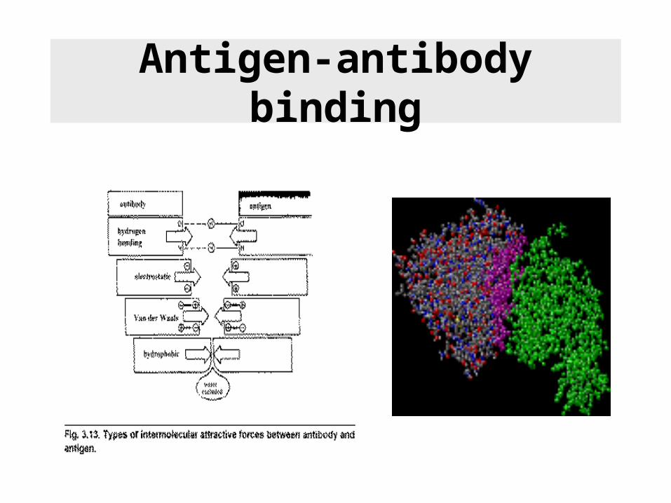
Antigen-antibody binding

Antigen-antibody affinity
The affinity with which antibody binds antigen results from a balance
between the attractive and repulsive forces. A high affinity antibody implies
a good fit and conversely, a low affinity antibody implies a poor fit and a
lower affinity constant

Antigen-antibody interaction: concentration dependence
Concentration of unknown samples are determined from a standard curveSTD concentration values are obtained when the interaction between

Non specific binding
Saturation radioligand binding experiments measure specific radioligand binding at equilibrium at various concentrations of the radioligand.
These experiments are performed to determine receptor number and affinity on cells but also between radiolabeled antigen and Ab.
This can take anywhere from a few minutes to many hours, depending on the ligand, receptor, To, and other experimental conditions.
The lowest concentration of radioligand will take the longest to equilibrate.
When testing equilibration time, therefore, use a low concentration of radioligand (perhaps 10-20% of the KD).
Nonspecific binding is almost always a linear function of ligand concentration.
The analyses depend on the assumption that you have allowed the incubation to proceed to equilibrium.

Dissociation ‘off rate’ experiments
Each ligand-receptor complex dissociates at a random time, so the amount of specific binding follows an exponential dissociation.
Variable Meaning Comment
X Time Usually expressed inunits of sec. or min.
Y Total binding Usually expressed inunits of cpm, mol/mg,sites/cell
Span Differencebetween bindingat time zero andplateau
Specific binding(same units as Y)
Plateau Binding thatdoesn't dissociate
Nonspecific binding(same units as Y).
K Dissociation rateconstant
Expressed In units ofinverse time (inverseof units of X-axis)
T1/2 Half-life 0.69302/k

• General equation for a dose response curve
• It shows response as a function of the logarithm of concentration
• X is the logarithm of agonist concentration and Y is the response
• Log EC50 is the logarithm of the EC50 (effective concentration, 50% of maximal response)
• IC50 (inhibitory conc.)
Sigmoidal dose response curve
10%
90%

• Ligand receptor interaction– Growth factors
– Hormones
• Antibody antigen interaction – RIA, ELISA
• Activity of chemotherapeutics
• Enzymatic activators/inhibitors
Doses response curves

Cross reactivity

One and two sites competition

Laboratory use of antibodies
• Quantitation of an antigen
– RIA, Elisa
• Identification and characterization of protein antigens
– Immunoprecipitation
– Western blotting
• Cell surface labelling and separation
• Localisation of antigens within tissues or cells
• Expression librairies
• Phage display

Detection principles
• Radiolabelled isotopes (antigen)– 125I, 32P, 35S
• Enzymes (Ab)– Peroxydase
• Chromophores (Ab)– Fluorogenic probes (UV, visible or IR)

Peroxydase reaction

RIA: radio immuno assay

Typical RIA standard curve

RIA interference

Elisa: Enzyme-linked immunosorbent assay

Sandwich Elisa

Western blotting

Two dimensional electrophoresis
pH
Mol
ecu
lar
wei
ght
kD
a
1st dimension 2nd dimension
Stable
pH gradient

Immunoprecipitation
ProteomicsWestern Blotting

Immunohistochemistry

• Phosphorylation and dephosphorylation affect the structure and activity of proteins
• Cellular signalling is characterized by cascades of phosphorylation
• Kinases and phosphatases maintain phosphorylated/dephosphorylated state of proteins
• Phospho/Tyrosine/Threonine/ Serine
Phosphospecific antibodies to study cellular signaling

DNA damage inducible cascades

Phosphospecific detections
• Phospho Ser, Thr, Tyr
• Sequence specific (-Ser18 p53)

Antibodies against other post- translational modifications
• Ubiquitination
• Sumoylation
• Acetylation
• Methylation
• Geranylation
• Etc...

• Specific DNA damage (CPD, 6-4PP)
• Sugars
• Lipids
• Vitamins (vit D)
• Iodine
Antibodies against non-protein antigens

• Identification of signaling pathways– Protein modifications
– Signaling partners
• Activity of drugs (lead compounds)
• Lack of specific molecules– Specific ligands (side effects)
– New antibodies
Research requiring immunoanalyses

Cytoskeleton Translati
on
ERK5
ELK1/
TCF
MEF2A-C
ATF2 NFAT4, NFAT
c1
MAX CHOP/ GADD1
53
Transcription Factors
c-jun
SAPKs
Inhibits nuclear
translocation
Effector Kinases
MAPKAP-K2/3
PRAK MSK1/2 MNK1/2 RSKs
p38s
HSP25/27CREB, Histone
H3, HMG14
eIF4E
Chromatin Remodelli
ng
ASK1
Tpl-2 MEKK2
MEKK3
MEKK1
RAF1
SEK1
MKK7
MKK3
MKK6
MEK5
MEK1/2
ERK1/2
MAP3Ks
MAPKs
MEKs
Inhibited by CSAIDS
(Cytokine-Suppressive
Anti-Inflammatory
Drugs)
eg SB203580
Synergize in SAPK
activation
p53
PP2B/Calcineurin
MKP1
CDC25B
CDC2WIP1
Pac1
Pac1
MKP5
MKP4
MKP2
MKP3M3/
6
(Hematopoietic only)
Inhibited by PD98059 (MEK2)
c-Abl
Rac1
dsDNA breaks
Inflammatory cytokines
ATM
MEKK4
TAK1
TAOs
MLKs
UV, MMS
Pyk2 Lyn
SHPTP1
Cdc42Hs
Kinases and signal transduction

Phage display

Bacteriophage structure

Production of recombinant phages

cDNA librairies

Phage display: Ab production
Originally developped to produce monoclonal antibodies, phage display is a simple yet powerful technology that is used to rapidly characterize protein-protein interactions from amongst billions of candidates. This widely practiced technique is used to map antibody epitopes, create vaccines and to engineer peptides, antibodies and other proteins as both diagnostic tools and as human therapeutics

Alternatives to specific antibodies
Gene of interest
Fluoresentproteins
CFP
GFP
YFP
RFP
-FP Ab Direct visualisation
TAGS
His
Myc
Flag
Strep
GST
Affinity -Tag Ab

FRET: Fluorescence resonance energy transfer

Localization of BFP- and RFP-C/EBP protein expressed in mouse 3T3 cells using 2p-FRET microscopy. The doubly expressed cells (BFP-RFP-C/EBP) were excited by 740 nm and the donor (A) and acceptor (B) images of proteins localized in the nucleus of a single living cell were acquired by single scan
Localization of CEBP by FRET

Clinical use of antibodies
• Diagnostic– Detection of peptides and other molecules in various diseases
• Endocrine diseases: hyperinsulinemia, diabetes, hyperparatyroidism
• Tumor antigens (p53 tumor suppressor, PSA, -foetoprotein)
• Antibodies against viral proteins (AIDS, hepatitis)
• Therapeutic – Neutralizing antibodies
• Anti-ErbB2 for breast and ovarian cancer
• Anti-CD20 for B-cell non-Hodgkin's lymphoma
• Antisera and antidotes (viruses and venoms)
• Drug discovery– Identification of therapeutic targets (phage display)

Therapeutic applications
• Neutralizing antibodies– Antidotes and antivenin (snake & spider bites)
– Tumor antigens ErbB-2, melanoma and T-cell leukemia, antibodies coupled to toxins
– Autoimmune antibodies, cytokines TNF- – Antisera aigainst virus, bateria and toxins (vaccine)
– Anti IgE and IgM for allegies (experimental)
– Quantitation of blood peptides (hormones metabolites)
• Activating antibodies– Complement activating for uncontrolled bleeding (hemophilia)

Concentration of serum peptides
• Blood levels of:– Hormones
– Antibodies
– Enzymes
– Metabolites

Detection of HIV proteins by WB
gp160 viral envelope precursor (env)
gp120 viral envelope protein (env) binds to CD4
p31 Reverse Transcriptase (pol)
p24 viral core protein (gag)

Immunodiffusion
Zone of equivalence:formation of large complexes

The problems of chemotherapy
Chemotherapy/radiotherapy
Sensors
Transducers
Cytoplasmic/Nuclear effectors
Chromatin StructureTranscription
DNA repairCell cycle checkpoints
Apoptosis
Drug resistance arisingfrom sensor/transducer
defects
Drug resistance arisingfrom effector defects
DNA Damage
Drug resistance arisingfrom altered drug delivery to target

Physiological roles of antibodies
• Protect against– Viral infections– Bacterial infections– Foreign bodies
• Antigens
• Deleterious in– Autoimmune diseases
• Reumathoid arthritis Lupus• Type 1 diabetes Croh’n
disease
– Graft rejection and hypersensitivity responses

Health care perspectives
• Ab against antigens could lead to diagnostic test or vaccine for several diseases
– BSE (mad cow disease) or human variant Creutzfeldt Jakob. Paramithiotis et al. A prion protein epitope selective for the pathologically misfolded conformation. Nat Med. 2003 Jul;9(7):893-9 Caprion Pharmaceuticals Inc., St-Laurent, Quebec, Canada.
– Vaccine against HIV Crystal structure of a neutralizing human IGG against HIV-1: a template for vaccine design.
Science. 2001 Aug 10;293(5532):1155-9. – SARS
– Nil virus
– Antidotes

Lacking an antibody for your protein or antigen of interest is limiting the progression of your
research!
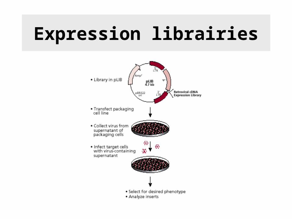
Expression librairies


![Buffering agents and Buffers · [electrophoresis/blotting, affinity chromatography, cell culture and assay, immunodetection,…] Introduction to buffers (solvents, buffering, additives)](https://static.fdocuments.in/doc/165x107/5e5b88d9c3d8e82d8764c404/buffering-agents-and-buffers-electrophoresisblotting-affinity-chromatography.jpg)