Primordial odontogenic tumor: An immunohistochemical profile · Primordial odontogenic tumor: An...
10
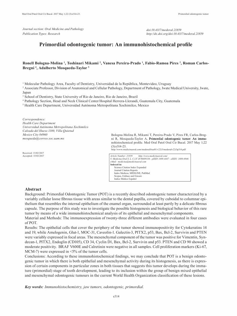
Med Oral Patol Oral Cir Bucal. 2017 May 1;22 (3):e314-23. Primordial odontogenic tumor e314 Journal section: Oral Medicine and Pathology Publication Types: Research Primordial odontogenic tumor: An immunohistochemical profile Ronell Bologna-Molina 1 , Toshinari Mikami 2 , Vanesa Pereira-Prado 1 , Fabio-Ramoa Pires 3 , Roman Carlos- Bregni 4 , Adalberto Mosqueda-Taylor 5 1 Molecular Pathology Area, Faculty of Dentistry, Universidad de la República, Montevideo, Uruguay 2 Associate Professor, Division of Anatomical and Cellular Pathology, Department of Pathology, Iwate Medical University, Iwate, Japan 3 School of Dentistry, State University of Rio de Janeiro, Rio de Janeiro, Brazil 4 Pathology Section, Head and Neck Clinical Center/Hospital Herrera-Llerandi, Guatemala City, Guatemala 5 Health Care Department, Universidad Autónoma Metropolitana Xochimilco, Mexico Correspondence: Health Care Department Universidad Autónoma Metropolitana Xochimilco Calzada del Hueso 1100, Villa Quietud Mexico City 04960 [email protected] Received: 13/02/2017 Accepted: 15/03/2017 Abstract Background: Primordial Odontogenic Tumor (POT) is a recently described odontogenic tumor characterized by a variably cellular loose fibrous tissue with areas similar to the dental papilla, covered by cuboidal to columnar epi- thelium that resembles the internal epithelium of the enamel organ, surrounded at least partly by a delicate fibrous capsule. The purpose of this study was to investigate the possible histogenesis and biological behavior of this rare tumor by means of a wide immunohistochemical analysis of its epithelial and mesenchymal components. Material and Methods: The immunoexpression of twenty-three different antibodies were evaluated in four cases of POT. Results: The epithelial cells that cover the periphery of the tumor showed immunopositivity for Cytokeratins 14 and 19, while Amelogenin, Glut-1, MOC-31, Caveolin-1. Galectin-3, PITX2, p53, Bax, Bcl-2, Survivin and PTEN were variably expressed in focal areas. The mesenchymal component of the tumor was positive for Vimentin, Syn- decan-1, PITX2, Endoglin (CD105), CD 34, Cyclin D1, Bax, Bcl-2, Survivin and p53. PTEN and CD 90 showed a moderate positivity. BRAF V600E and Calretinin were negative in all samples. Cell proliferation markers (Ki-67, MCM-7) were expressed in <5% of the tumor cells. Conclusions: According to these immunohistochemical findings, we may conclude that POT is a benign odonto- genic tumor in which there is both epithelial and mesenchymal activity during its histogenesis, as there is expres- sion of certain components in particular zones in both tissues that suggests this tumor develops during the imma- ture (primordial) stage of tooth development, leading to its inclusion within the group of benign mixed epithelial and mesenchymal odontogenic tumours in the current World Health Organization classification of these lesions. Key words: Immunohistochemistry, jaw tumors, odontogenic, primordial. doi:10.4317/medoral.21859 http://dx.doi.org/doi:10.4317/medoral.21859 Bologna-Molina R, Mikami T, Pereira-Prado V, Pires FR, Carlos-Breg- ni R, Mosqueda-Taylor A. Primordial odontogenic tumor: An immu- . Primordial odontogenic tumor: An immu- nohistochemical profile. Med Oral Patol Oral Cir Bucal. 2017 May 1;22 (3):e314-23. http://www.medicinaoral.com/medoralfree01/v22i3/medoralv22i3p314.pdf Article Number: 21859 http://www.medicinaoral.com/ © Medicina Oral S. L. C.I.F. B 96689336 - pISSN 1698-4447 - eISSN: 1698-6946 eMail: [email protected] Indexed in: Science Citation Index Expanded Journal Citation Reports Index Medicus, MEDLINE, PubMed Scopus, Embase and Emcare Indice Médico Español
Transcript of Primordial odontogenic tumor: An immunohistochemical profile · Primordial odontogenic tumor: An...
Med Oral Patol Oral Cir Bucal. 2017 May 1;22 (3):e314-23. Primordial odontogenic tumor
e314
Journal section: Oral Medicine and PathologyPublication Types: Research
Primordial odontogenic tumor: An immunohistochemical profile
Ronell Bologna-Molina 1, Toshinari Mikami 2, Vanesa Pereira-Prado 1, Fabio-Ramoa Pires 3, Roman Carlos-Bregni 4, Adalberto Mosqueda-Taylor 5
1 Molecular Pathology Area, Faculty of Dentistry, Universidad de la República, Montevideo, Uruguay2 Associate Professor, Division of Anatomical and Cellular Pathology, Department of Pathology, Iwate Medical University, Iwate, Japan3 School of Dentistry, State University of Rio de Janeiro, Rio de Janeiro, Brazil4 Pathology Section, Head and Neck Clinical Center/Hospital Herrera-Llerandi, Guatemala City, Guatemala5 Health Care Department, Universidad Autónoma Metropolitana Xochimilco, Mexico
Correspondence:Health Care Department Universidad Autónoma Metropolitana XochimilcoCalzada del Hueso 1100, Villa QuietudMexico City [email protected]
Received: 13/02/2017Accepted: 15/03/2017
AbstractBackground: Primordial Odontogenic Tumor (POT) is a recently described odontogenic tumor characterized by a variably cellular loose fibrous tissue with areas similar to the dental papilla, covered by cuboidal to columnar epi-thelium that resembles the internal epithelium of the enamel organ, surrounded at least partly by a delicate fibrous capsule. The purpose of this study was to investigate the possible histogenesis and biological behavior of this rare tumor by means of a wide immunohistochemical analysis of its epithelial and mesenchymal components.Material and Methods: The immunoexpression of twenty-three different antibodies were evaluated in four cases of POT.Results: The epithelial cells that cover the periphery of the tumor showed immunopositivity for Cytokeratins 14 and 19, while Amelogenin, Glut-1, MOC-31, Caveolin-1. Galectin-3, PITX2, p53, Bax, Bcl-2, Survivin and PTEN were variably expressed in focal areas. The mesenchymal component of the tumor was positive for Vimentin, Syn-decan-1, PITX2, Endoglin (CD105), CD 34, Cyclin D1, Bax, Bcl-2, Survivin and p53. PTEN and CD 90 showed a moderate positivity. BRAF V600E and Calretinin were negative in all samples. Cell proliferation markers (Ki-67, MCM-7) were expressed in <5% of the tumor cells.Conclusions: According to these immunohistochemical findings, we may conclude that POT is a benign odonto-genic tumor in which there is both epithelial and mesenchymal activity during its histogenesis, as there is expres-sion of certain components in particular zones in both tissues that suggests this tumor develops during the imma-ture (primordial) stage of tooth development, leading to its inclusion within the group of benign mixed epithelial and mesenchymal odontogenic tumours in the current World Health Organization classification of these lesions.
Key words: Immunohistochemistry, jaw tumors, odontogenic, primordial.
doi:10.4317/medoral.21859http://dx.doi.org/doi:10.4317/medoral.21859
Bologna-Molina R, Mikami T, Pereira-Prado V, Pires FR, Carlos-Breg-ni R, Mosqueda-Taylor A. Primordial odontogenic tumor: An immu-. Primordial odontogenic tumor: An immu-nohistochemical profile. Med Oral Patol Oral Cir Bucal. 2017 May 1;22 (3):e314-23. http://www.medicinaoral.com/medoralfree01/v22i3/medoralv22i3p314.pdf
Article Number: 21859 http://www.medicinaoral.com/© Medicina Oral S. L. C.I.F. B 96689336 - pISSN 1698-4447 - eISSN: 1698-6946eMail: [email protected] Indexed in:
Science Citation Index ExpandedJournal Citation ReportsIndex Medicus, MEDLINE, PubMedScopus, Embase and Emcare Indice Médico Español
Med Oral Patol Oral Cir Bucal. 2017 May 1;22 (3):e314-23. Primordial odontogenic tumor
e315
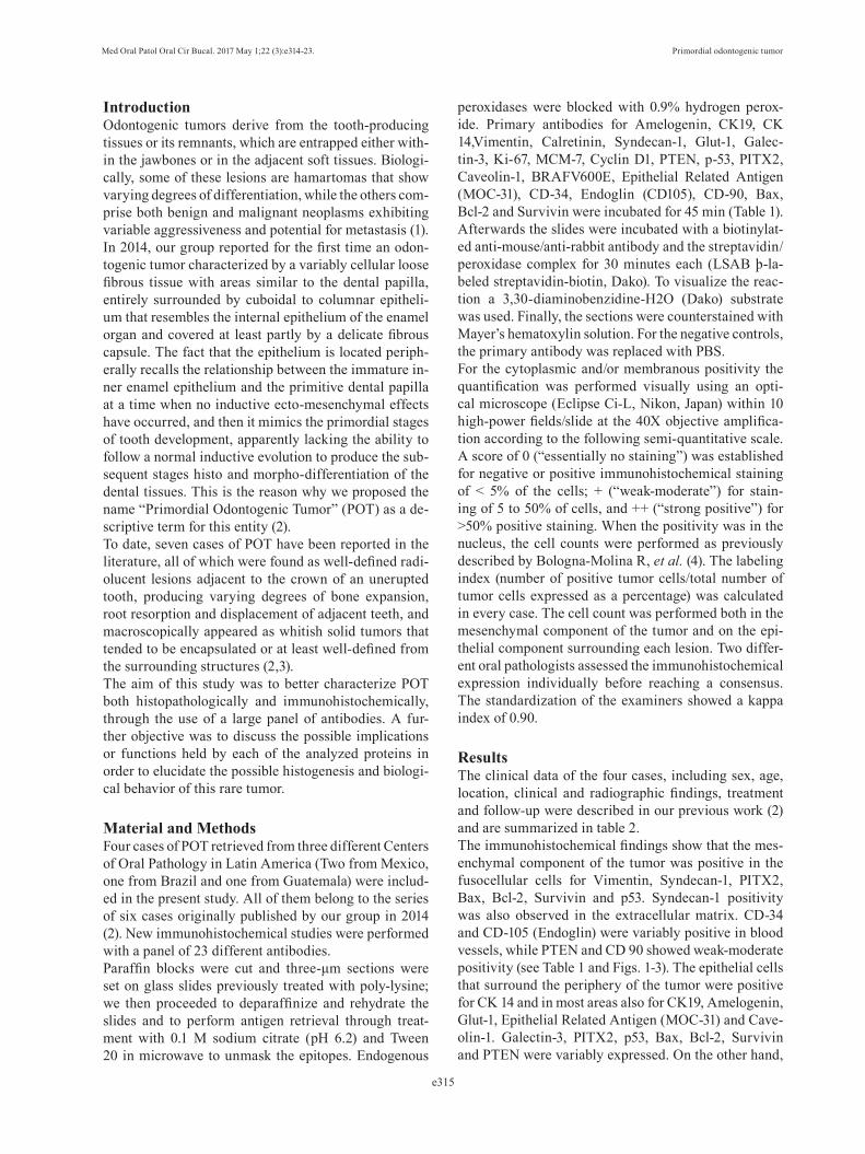
IntroductionOdontogenic tumors derive from the tooth-producing tissues or its remnants, which are entrapped either with-in the jawbones or in the adjacent soft tissues. Biologi-cally, some of these lesions are hamartomas that show varying degrees of differentiation, while the others com-prise both benign and malignant neoplasms exhibiting variable aggressiveness and potential for metastasis (1).In 2014, our group reported for the first time an odon-togenic tumor characterized by a variably cellular loose fibrous tissue with areas similar to the dental papilla, entirely surrounded by cuboidal to columnar epitheli-um that resembles the internal epithelium of the enamel organ and covered at least partly by a delicate fibrous capsule. The fact that the epithelium is located periph-erally recalls the relationship between the immature in-ner enamel epithelium and the primitive dental papilla at a time when no inductive ecto-mesenchymal effects have occurred, and then it mimics the primordial stages of tooth development, apparently lacking the ability to follow a normal inductive evolution to produce the sub-sequent stages histo and morpho-differentiation of the dental tissues. This is the reason why we proposed the name “Primordial Odontogenic Tumor” (POT) as a de-scriptive term for this entity (2).To date, seven cases of POT have been reported in the literature, all of which were found as well-defined radi-olucent lesions adjacent to the crown of an unerupted tooth, producing varying degrees of bone expansion, root resorption and displacement of adjacent teeth, and macroscopically appeared as whitish solid tumors that tended to be encapsulated or at least well-defined from the surrounding structures (2,3). The aim of this study was to better characterize POT both histopathologically and immunohistochemically, through the use of a large panel of antibodies. A fur-ther objective was to discuss the possible implications or functions held by each of the analyzed proteins in order to elucidate the possible histogenesis and biologi-cal behavior of this rare tumor.
Material and MethodsFour cases of POT retrieved from three different Centers of Oral Pathology in Latin America (Two from Mexico, one from Brazil and one from Guatemala) were includ-ed in the present study. All of them belong to the series of six cases originally published by our group in 2014 (2). New immunohistochemical studies were performed with a panel of 23 different antibodies.Paraffin blocks were cut and three-μm sections were set on glass slides previously treated with poly-lysine; we then proceeded to deparaffinize and rehydrate the slides and to perform antigen retrieval through treat-ment with 0.1 M sodium citrate (pH 6.2) and Tween 20 in microwave to unmask the epitopes. Endogenous
peroxidases were blocked with 0.9% hydrogen perox-ide. Primary antibodies for Amelogenin, CK19, CK 14,Vimentin, Calretinin, Syndecan-1, Glut-1, Galec-tin-3, Ki-67, MCM-7, Cyclin D1, PTEN, p-53, PITX2, Caveolin-1, BRAFV600E, Epithelial Related Antigen (MOC-31), CD-34, Endoglin (CD105), CD-90, Bax, Bcl-2 and Survivin were incubated for 45 min (Table 1). Afterwards the slides were incubated with a biotinylat-ed anti-mouse/anti-rabbit antibody and the streptavidin/peroxidase complex for 30 minutes each (LSAB þ-la-beled streptavidin-biotin, Dako). To visualize the reac-tion a 3,30-diaminobenzidine-H2O (Dako) substrate was used. Finally, the sections were counterstained with Mayer’s hematoxylin solution. For the negative controls, the primary antibody was replaced with PBS.For the cytoplasmic and/or membranous positivity the quantification was performed visually using an opti-cal microscope (Eclipse Ci-L, Nikon, Japan) within 10 high-power fields/slide at the 40X objective amplifica-tion according to the following semi-quantitative scale. A score of 0 (“essentially no staining”) was established for negative or positive immunohistochemical staining of < 5% of the cells; + (“weak-moderate”) for stain-ing of 5 to 50% of cells, and ++ (“strong positive”) for >50% positive staining. When the positivity was in the nucleus, the cell counts were performed as previously described by Bologna-Molina R, et al. (4). The labeling index (number of positive tumor cells/total number of tumor cells expressed as a percentage) was calculated in every case. The cell count was performed both in the mesenchymal component of the tumor and on the epi-thelial component surrounding each lesion. Two differ-ent oral pathologists assessed the immunohistochemical expression individually before reaching a consensus. The standardization of the examiners showed a kappa index of 0.90.
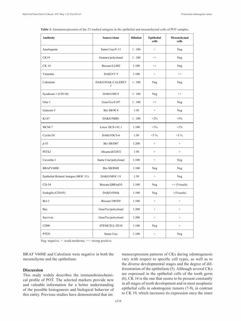
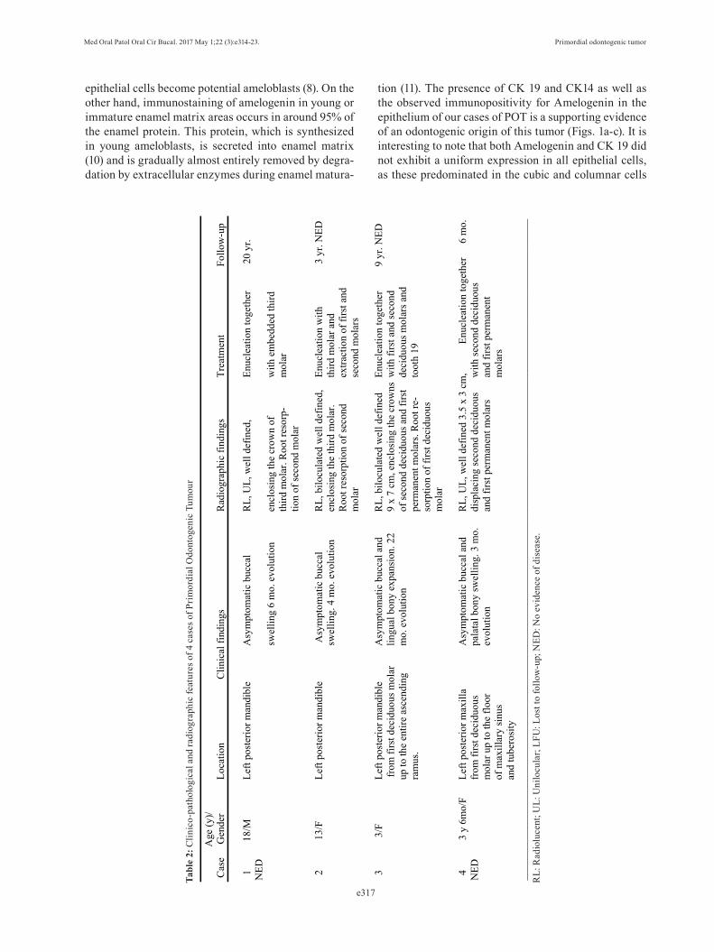
ResultsThe clinical data of the four cases, including sex, age, location, clinical and radiographic findings, treatment and follow-up were described in our previous work (2) and are summarized in table 2. The immunohistochemical findings show that the mes-enchymal component of the tumor was positive in the fusocellular cells for Vimentin, Syndecan-1, PITX2, Bax, Bcl-2, Survivin and p53. Syndecan-1 positivity was also observed in the extracellular matrix. CD-34 and CD-105 (Endoglin) were variably positive in blood vessels, while PTEN and CD 90 showed weak-moderate positivity (see Table 1 and Figs. 1-3). The epithelial cells that surround the periphery of the tumor were positive for CK 14 and in most areas also for CK19, Amelogenin, Glut-1, Epithelial Related Antigen (MOC-31) and Cave-olin-1. Galectin-3, PITX2, p53, Bax, Bcl-2, Survivin and PTEN were variably expressed. On the other hand,
Med Oral Patol Oral Cir Bucal. 2017 May 1;22 (3):e314-23. Primordial odontogenic tumor
e316
Antibody Source/clone Dilution Epithelial cells
Mesenchymal cells
Amelogenin Santa Cruz/F-11 1 : 100 + Neg
CK19 Genetex/polyclonal 1 : 100 ++ Neg
CK 14 Biocare/LL002 1:100 ++ Neg
Vimentin DAKO/V 9 1:100 + ++
Calretinin DAKO/DAK-CALERET 1
1 : 100 Neg Neg
Syndecan-1 (CD138) DAKO/MI15 1 : 100 Neg ++
Glut-1 GeneTex/E107 1 : 100 ++ Neg
Galectin-3 Bio SB/9C4 1:50 + Neg
Ki-67 DAKO/MIB1 1 : 100 <2% <5%
MCM-7 Leica/ DCS-141.1 1:100 <1% <1%
Cyclin D1 DAKO/DCS-6 1:50 <5 %. <5 %.
p-53 Bio SB/D07 1:200 + +
PITX2 Abcam/ab32832 1:50 + +
Caveolin-1 Santa Cruz/polyclonal 1:100 + Neg
BRAFV600E Bio SB/RM8 1:100 Neg Neg
Epithelial Related Antigen (MOC-31) DAKO/MOC-31 1:50 + Neg
CD-34 Biocare/QBEnd10 1:100 Neg ++ (Vessels)
Endoglin (CD105) DAKO/SN6h 1:100 Neg +(Vessels)
Bcl-2 Biocare/100/D5 1:100 + +
Bax GeneTex/polyclonal 1:200 + +
Survivin GeneTex/polyclonal 1:200 + +
CD90 STEMCELL/5E10 1:100 Neg +
PTEN Santa Cruz 1:100 + Neg
Table 1: Immunoexpression of the 23 studied antigens in the epithelial and mesenchymal cells of POT samples.
Neg: negative, +: weak-moderate, ++: strong positive.
BRAF V600E and Calretinin were negative in both the mesenchyme and the epithelium.
DiscussionThis study widely describes the immunohistochemi-cal profile of POT. The selected markers provide new and valuable information for a better understanding of the possible histogenesis and biological behavior of this entity. Previous studies have demonstrated that im-
munoexpression patterns of CKs during odontogenesis vary with respect to specific cell types, as well as to the diverse developmental stages and the degree of dif-ferentiation of the epithelium (5). Although several CKs are expressed in the epithelial cells of the tooth germ (6), CK 14 is the one that seems to be present constantly in all stages of tooth development and in most neoplastic epithelial cells in odontogenic tumors (7-9), in contrast to CK 19, which increases its expression once the inner
Med Oral Patol Oral Cir Bucal. 2017 May 1;22 (3):e314-23. Primordial odontogenic tumor
e317
A
ge (y
)/
C
ase
Gen
der
Loca
tion
C
linic
al fi
ndin
gs
Rad
iogr
aphi
c fin
ding
s
Trea
tmen
t
F
ollo
w-u
p
1
18/
M
Le
ft po
ster
ior m
andi
ble
Asy
mpt
omat
ic b
ucca
l
RL,
UL,
wel
l def
ined
,
Enuc
leat
ion
toge
ther
20
yr.
NED
sw
ellin
g 6
mo.
evo
lutio
n en
clos
ing
the
crow
n of
w
ith e
mbe
dded
third
th
ird m
olar
. Roo
t res
orp-
m
olar
tio
n of
seco
nd m
olar
2
13/F
Left
post
erio
r man
dibl
e
A
sym
ptom
atic
buc
cal
R
L, b
ilocu
late
d w
ell d
efin
ed,
Enuc
leat
ion
with
3 y
r. N
ED
sw
ellin
g. 4
mo.
evo
lutio
n en
clos
ing
the
third
mol
ar.
third
mol
ar a
nd
R
oot r
esor
ptio
n of
seco
nd
extra
ctio
n of
firs
t and
mol
ar
se
cond
mol
ars
3
3/F
Left
post
erio
r man
dibl
e A
sym
ptom
atic
buc
cal a
nd
RL,
bilo
cula
ted
wel
l def
ined
En
ucle
atio
n to
geth
er
9
yr.
NED
from
firs
t dec
iduo
us m
olar
lin
gual
bon
y ex
pans
ion.
22
9 x
7 cm
, enc
losi
ng th
e cr
owns
with
firs
t and
seco
nd
up to
the
entir
e as
cend
ing
mo.
evo
lutio
n
of
seco
nd d
ecid
uous
and
firs
t de
cidu
ous m
olar
s and
ram
us.
pe
rman
ent m
olar
s. R
oot r
e-
toot
h 19
sorp
tion
of fi
rst d
ecid
uous
mol
ar
4
3 y
6mo/
F Le
ft po
ster
ior m
axill
a
Asy
mpt
omat
ic b
ucca
l and
R
L, U
L, w
ell d
efin
ed 3
.5 x
3 c
m,
Enuc
leat
ion
toge
ther
6
mo.
N
ED
from
firs
t dec
iduo
us
pa
lata
l bon
y sw
ellin
g. 3
mo.
di
spla
cing
seco
nd d
ecid
uous
w
ith se
cond
dec
iduo
us
m
olar
up
to th
e flo
or
ev
olut
ion
an
d fir
st p
erm
anen
t mol
ars
and
first
per
man
ent
of
max
illar
y si
nus
m
olar
s
and
tube
rosi
ty
____
____
____
____
____
____
____
____
____
____
____
____
____
____
____
____
____
____
____
____
____
____
____
____
____
____
____
____
____
____
____
___
Tabl
e 2:
Clin
ico-
path
olog
ical
and
radi
ogra
phic
feat
ures
of 4
cas
es o
f Pri
mor
dial
Odo
ntog
enic
Tum
our
RL:
Rad
iolu
cent
; UL:
Uni
locu
lar;
LFU
: Los
t to
follo
w-u
p; N
ED: N
o ev
iden
ce o
f dis
ease
.
epithelial cells become potential ameloblasts (8). On the other hand, immunostaining of amelogenin in young or immature enamel matrix areas occurs in around 95% of the enamel protein. This protein, which is synthesized in young ameloblasts, is secreted into enamel matrix (10) and is gradually almost entirely removed by degra-dation by extracellular enzymes during enamel matura-
tion (11). The presence of CK 19 and CK14 as well as the observed immunopositivity for Amelogenin in the epithelium of our cases of POT is a supporting evidence of an odontogenic origin of this tumor (Figs. 1a-c). It is interesting to note that both Amelogenin and CK 19 did not exhibit a uniform expression in all epithelial cells, as these predominated in the cubic and columnar cells

Med Oral Patol Oral Cir Bucal. 2017 May 1;22 (3):e314-23. Primordial odontogenic tumor
e318
Fig. 1: (a) The epithelium showS variable expression of CK19, 400X. (b) Expression of CK 14 was constant throughout the epithelium, 200X. (c) Expression of Amelogenin in the cubic and columnar cells, 200X. (d) Syndecan-1 is intensely ex-pressed in the mesenchyme, but not in all the epithelial lin-ing, 200X. (e) PITX2 positivity in focal areas, 400X. (f) Ex-pression of GLUT-1 in the odontogenic epithelium, 200X. (g) MOC-31 was only found expressed in localized areas within the epithelium, 200X. (h) Some epithelial cells showed posi-tivity for vimentin, 400X.
Fig. 2: (a) Expression of Ki-67 in epithelial cells; 400x, (b) significantly higher proliferative index within the cells in the subepithelial mesenchymal condensation (arrow) as com-pared to that found in other mesenchymal areas of the tumor; 200X. (c) Expression of Galectin-3 in epithelial cells; 100X. (d,e,f) Bax, Bcl-2 and Survivin expression; 400X. (g) p53 was found both in the mesenchymal component and in the surrounding epithelium; 400X. (h) Cyclin D1 expression; 400X.
showing inverse polarization (pre-ameloblasts), which suggests that this epithelial lining express diverse de-grees of maturation, supporting an origin from primi-tive (primordial) cellular components of the enamel or-gan (Figs. 1a-c). This observation is also reinforced by the fact that some areas of the tumor epithelium showed focal positivity to vimentin (Fig. 1h).MOC-31 is an epithelial-related antigen expressed in most normal epithelia but also in tumors originating from epithelial tissues. This is a cell adhesion molecule that allows tight junction formation between epithe-lial cells, which can negatively affect cadherin/catenin complex formation (12). MOC-31 has been proved to be an advantageous marker at signaling epithelial cell dif-ferentiation. Reduced expression of MOC-31 has been correlated with poor survival in cancerous conditions. It could be related to differentiation, tumor proliferation
and invasion (13,14). This molecule was found only ex-pressed in localized areas within the epithelium of the studied POT cases (Fig. 1g), suggesting, as seen for CK 19, Vimentin and Amelogenin, that there is a variable degree of differentiation and maturation of the cells that comprises the epithelial compartment. As a member of the family of transmembrane heparan sulfate proteoglycans, Syndecan-1 (CD138) is involved in cell to cell adhesion and in the interaction of cells with the extracellular matrix, and also participates in promoting cell proliferation and regulates cellular growth by interacting with families of growth factors in association with heparin (15). Syndecan-1 plays a role in epithelial maturation, and it has been consistently observed that the degree of epithelial differentiation is directly associated with its expression (16). In addition, it has been reported that the pattern of Syndecan-1 ex-pression varies according to the stage of maturation of the epithelial tissues (17). In this respect, it is relevant to highlight the fact that in all cases of POT included in this study, there were different degrees of Syndecan-1
Med Oral Patol Oral Cir Bucal. 2017 May 1;22 (3):e314-23. Primordial odontogenic tumor
e319
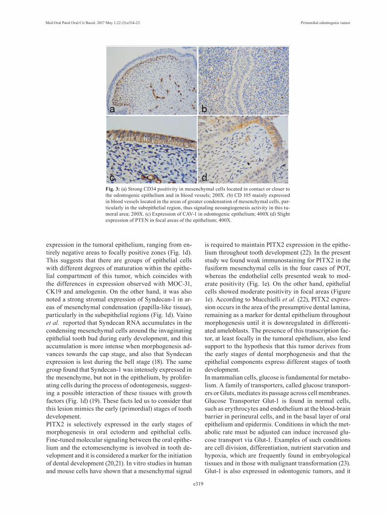
Fig. 3: (a) Strong CD34 positivity in mesenchymal cells located in contact or closer to the odontogenic epithelium and in blood vessels; 200X. (b) CD 105 mainly expressed in blood vessels located in the areas of greater condensation of mesenchymal cells, par-ticularly in the subepithelial region, thus signaling neoangiogenesis activity in this tu-moral area; 200X. (c) Expression of CAV-1 in odontogenic epithelium; 400X (d) Slight expression of PTEN in focal areas of the epithelium; 400X.
expression in the tumoral epithelium, ranging from en-tirely negative areas to focally positive zones (Fig. 1d). This suggests that there are groups of epithelial cells with different degrees of maturation within the epithe-lial compartment of this tumor, which coincides with the differences in expression observed with MOC-31, CK19 and amelogenin. On the other hand, it was also noted a strong stromal expression of Syndecan-1 in ar-eas of mesenchymal condensation (papilla-like tissue), particularly in the subepithelial regions (Fig. 1d). Vaino et al. reported that Syndecan RNA accumulates in the condensing mesenchymal cells around the invaginating epithelial tooth bud during early development, and this accumulation is more intense when morphogenesis ad-vances towards the cap stage, and also that Syndecan expression is lost during the bell stage (18). The same group found that Syndecan-1 was intensely expressed in the mesenchyme, but not in the epithelium, by prolifer-ating cells during the process of odontogenesis, suggest-ing a possible interaction of these tissues with growth factors (Fig. 1d) (19). These facts led us to consider that this lesion mimics the early (primordial) stages of tooth development.PITX2 is selectively expressed in the early stages of morphogenesis in oral ectoderm and epithelial cells. Fine-tuned molecular signaling between the oral epithe-lium and the ectomesenchyme is involved in tooth de-velopment and it is considered a marker for the initiation of dental development (20,21). In vitro studies in human and mouse cells have shown that a mesenchymal signal
is required to maintain PITX2 expression in the epithe-lium throughout tooth development (22). In the present study we found weak immunostaining for PITX2 in the fusiform mesenchymal cells in the four cases of POT, whereas the endothelial cells presented weak to mod-erate positivity (Fig. 1e). On the other hand, epithelial cells showed moderate positivity in focal areas (Figure 1e). According to Mucchielli et al. (22), PITX2 expres-sion occurs in the area of the presumptive dental lamina, remaining as a marker for dental epithelium throughout morphogenesis until it is downregulated in differenti-ated ameloblasts. The presence of this transcription fac-tor, at least focally in the tumoral epithelium, also lend support to the hypothesis that this tumor derives from the early stages of dental morphogenesis and that the epithelial components express different stages of tooth development.In mammalian cells, glucose is fundamental for metabo-lism. A family of transporters, called glucose transport-ers or Gluts, mediates its passage across cell membranes. Glucose Transporter Glut-1 is found in normal cells, such as erythrocytes and endothelium at the blood-brain barrier in perineural cells, and in the basal layer of oral epithelium and epidermis. Conditions in which the met-abolic rate must be adjusted can induce increased glu-cose transport via Glut-1. Examples of such conditions are cell division, differentiation, nutrient starvation and hypoxia, which are frequently found in embryological tissues and in those with malignant transformation (23). Glut-1 is also expressed in odontogenic tumors, and it

Med Oral Patol Oral Cir Bucal. 2017 May 1;22 (3):e314-23. Primordial odontogenic tumor
e320
has been observed increased in ameloblastomas and ameloblastic carcinomas as compared to tooth germs, which suggests its overexpression may play an impor-tant role in the aggressive behavior of these neoplasms (24). In the present study we found a strong expression of this glucose transporter in odontogenic epithelium, showing both cytoplasmic and membranal localization (Figure 1f), which correlates well with the fact that the pattern of Glut-1 expression changes from strong cyto-plasmic expression to membranous expression during maturation of the enamel organ in the late bell stage (24). The strong expression of this protein in the epi-thelium of POT indicates the existence of high glucose transport requirements in this pathological condition. On the other hand, the cytoplasmic and membranous expression also suggests the co-existence of different stages of maturation in this epithelium. In order to quantify the proliferative activity of the tu-mor we used markers associated to cell proliferation and factors related to cell –cycles, such as Ki-67, MCM7 and Cyclin D1, which revealed that the tumor presents a low rate of proliferation (< 5%) in both epithelial and mesenchymal cells (Fig. 2a,b), similar to that reported for other benign odontogenic tumors, such as the odon-togenic myxoma (25). Additionally, it is important to highlight the fact that there was a significantly higher proliferative index among the cells located within the subepithelial mesenchymal condensation as compared to that found in other mesenchymal areas of the tumor (Fig. 2b). This may imply that the tumor shows more active growth in this specific area, and perhaps it may be related to the inductive ability of that part of the mes-enchyme to modify the overlying epithelium. Galectin-3, a lectin involved in diverse biological events including embryogenesis, cell adhesion, proliferation, apoptosis, mRNA splicing and regulation of the im-mune system (26), is widely present in human tissues. Changes in Galectin-3 expression and its subcellular and intercellular localization are commonly observed in cancer and precancerous conditions (27). While a general shift of galectin-3 localization from nucleus to cytoplasm occurs in cancer development from adeno-ma to carcinoma and also this protein plays an impor-tant pivotal role within the nucleus in the regulation of cancer-related gene expression (27), including Cyclin D1, its expression in POT in this study was exclusively found in the epithelial component, mainly in the cyto-plasm (yet some cells showed immunopositivity in the nucleus) (Figs. 2c,h), suggesting a possible role in cell proliferation. As occurred with other immunomarkers, there were areas in the epithelium that were completely negative and some which showed clear expression (Fig. 2c). Nakahara et al. (28) have suggested that overex-pression of intracellular galectin-3 favors neoplastic transformation, cell cycle progression and inhibition of
apoptosis, but the exact significance of our findings re-mains to be elucidated. The range of survival or cellular death is determined by the relationship between Bcl-2 (an apoptosis inhibi-tor) and Bax (an apoptosis promoter). In addition, Sur-vivin is an antiapoptotic protein that leads to negative regulation of apoptosis by inhibiting caspase activation (29). When Bax predominates over Bcl-2, the activity of Bcl-2 is repressed and the apoptotic pathway of Bax is activated (30). In this study Bcl-2, Survivin and Bax manifested strong positivity in both mesenchymal and epithelial cells of POT (Figs. 2d,e,f). The expression of Bax, Survivin and Bcl-2 was similar, with slightly greater presence of the antiapoptotic proteins; however, the results of this study does not allow us to conclude that the overexpression of antiapoptotic proteins play an important role as growth mechanism of POT, as it has been suggested for other odontogenic neoplasms (31). It is interesting to note that nuclear immunoexpression of Survivin was detected in some areas of the epithelium (Fig. 2f), which according to Stasikowska-Kanicka et al. (32), fosters cell proliferation, while its cytoplasmic immunoexpression is involved in the mechanisms of apoptosis regulation. On the other hand, both Bcl-2 and Bax participate as transcriptional targets for p53, a tu-mor suppressor protein, which brings about cell cycle arrest or apoptosis in response to DNA damage. The coordinate activity of these molecules is vital in order to control life and death of a cell (33). A weak to moderate expression of p53 was found both in the mesenchymal component and in the surrounding epithelium in our cases (Fig. 2g), which is evidence that this protein par-ticipates as part of the regulation of this tissue.Blood supply is required for tumor growth, to provide the oxygen, metabolites and growth factors necessary for cell proliferation. Blood vessels feeding the tu-mor were identified by means of CD34 and Endoglin (CD-105) expression, demonstrating strong positivity for CD34 in all the endothelial cells, whereas CD 105 marked a smaller number of blood vessels (Figs. 3a,b). This situation may be explained by the fact that CD-105 expression, contrary to CD34, is a characteristic prop-erty of newly formed blood vessels and not of previous-ly-existing blood and lymphatic vessels (34). CD 105 in POT was found mainly in blood vessels located in the areas of greater condensation of mesenchymal cells, particularly in the subepithelial region, thus signaling neoangiogenesis activity in this tumoral area (Fig. 3b), which agrees with our previous suggestion that the tu-mor has a greater growth rate in this area. In this recently recognized tumoral entity we found that CD34 showed immunopositive areas not only in the mesenchymal ves-sels, but also in the mesenchymal cells located in con-tact or closer to the odontogenic epithelium (Fig. 3a). CD34 is known to be a marker for hematopoietic stem

Med Oral Patol Oral Cir Bucal. 2017 May 1;22 (3):e314-23. Primordial odontogenic tumor
e321
cells, and hematopoietic progenitor cells (35), but also it is regarded as a marker of several other nonhematopoi-etic cell types, including vascular endothelial progeni-tors and embryonic fibroblasts (36,37), and therefore we consider that its positivity in this area could indicate the presence of embryonic fibroblasts (38,39).CD90 has been established as a marker for a wide vari-ety of human cell-stem types, and particularly mesen-chymal stem cells. It is considered to be a vital regulator, critical at cell-cell and cell-matrix interactions. It takes part in cellular migration and fibrosis, both of which are important events for tissue regeneration and oncogen-esis (40). Its expression in tumor stroma, especially in endothelial and mesenchymal stem cells, including tu-mor-associated fibroblasts, appears to be an influential agent in disease progression (41). Stromal expression of CD90 in our POT cases was found in blood vessels and in some mesenchymal cells, particularly in those in close proximity to epithelial odontogenic cells, which led us to suggest that CD90 expression in POT could play an important role in cellular adhesion and migra-tion, serving as an important regulator of cell-matrix interactions for determining the biological behavior of this tumor. Expression of Caveolin-1 (Cav-1) was reported by Schwab et al. (42) in epithelial and mesenchymal cells of tooth germ in various stages of development, particularly in the inner enamel epithelium and ameloblastic cells in de-veloping tooth germ (42), and it has also been established that Cav-1 participates in oncogenic transformation and differentiation in oral squamous cell carcinoma and pros-tate cancer (43,44). Regarding its tumor promoting func-tion, higher expression of Cav-1 drives into tumorgenesis by inhibiting apoptosis (44). The variable expression that we found within the epithelial component of POT (Fig. 3c) could suggest different stages of cellular differen-tiation within the odontogenic epithelium covering the lesion, implying that positive cells are the transformed cells of this particular type of tumor.PTEN gene, which in humans encodes the protein phos-phatase and tensin homolog, (PTEN), was identified as a tumor suppressor gene that mutates with a high frequen-cy in a large number of cancerous condition. Reduced expression of PTEN has also been reported in some oral cancers, in studies that suggest PTEN plays an important role at signaling pathways in the carcinogenesis of head and neck squamous cell carcinoma (45). In our study, we observed only a slight expression of PTEN in focal areas of the epithelium (Fig. 3d), reinforcing the hypothesis of the existence of cells with different stages of develop-ment within the epithelial compartment. Recently, dysregulation of the MAPK pathway signal-ing have been proved as a critical step in the pathogen-esis of ameloblastoma. The mutation most frequently identified is BRAF V600E. (46). To date, BRAF V600E
mutations have been identified also in ameloblastic fi-bromas (2/2) and fibrodentinomas (1/1), as well as in ameloblastic carcinomas (3/8) and clear cell odontogen-ic carcinomas (1/1), but not in other odontogenic tumors (47,48). In our POT cases, we did not find the presence of the mutant protein BRAFV600E, and the absence of this mutation positions POT in a different category with respect to the ameloblastic lesions.According to the variable extent of expression of the diverse proteins tested in this study, we may conclude that POT has a low proliferative index, which defines it as a slowly growing lesion with variable production of loose fibrous tissue in which cell proliferation does not seem to be the only growth mechanism implicated in its expansion. POT is a moderately vascularized tumor, in which there is greater vascular density and vascular neoformation in the mesenchyme located just below the epithelium, an area that also shows greater expression of Syndecan 1, CD 34, Ki-67 and Glut-1, indicating that this is the area with the greatest proliferative activity and in which the necessary elements for the develop-ment of the tumor converge. In addition, there is some evidence of an increase of antiapoptotic activity that may participate in the growth of this tumoral entity. The uneven expression of CK19, Vimentin, Amelogenin, Syndecan-1, CAV 1, Glut-1 1, MOC-31 and PITX2 in the surrounding epithelium could be interpreted as that there are groups of cells in different stages of develop-ment within an apparently unique epithelial structure. This situation leads us to speculate that the epithelial compartment of POT is not a uniform or static structure in odontogenesis, but that it expresses the transition be-tween the earliest stages of tooth development and those in which ameloblastic maturation is present, but before having the potential for induction of odontoblastic dif-ferentiation or the production of mineralized tissues, supporting the denomination of “primordial” for this unique entity. The absence of identifiable BRAF V600E mutant protein suggest that mutation of BRAF gene is not implicated in the pathogenesis of POT, and may confirm that this tumor does not belong to the category of those BRAF-mutant positive ameloblastic tumors.Considering and putting together all these immunohis-tochemical findings, we interpret that the lesion studied is an odontogenic tumor of benign behavior that pos-sibly originates from the mesenchyme of an abortive tooth germ that fails to produce a dental organ. Given the rarity of this tumor and the small number of cases reported to date, it is necessary to collect new cases in order to obtain a larger sample, towards a better under-standing of the histogenesis and tumoral behavior of this condition.
References 1. Mosqueda-Taylor A. New findings and controversies in odon-togenic tumors. Med Oral Patol Oral Cir Bucal. 2008;13:555-8.

Med Oral Patol Oral Cir Bucal. 2017 May 1;22 (3):e314-23. Primordial odontogenic tumor
e322
2. Mosqueda-Taylor A, Pires FR, Aguirre-Urízar JM, Carlos-Breg-ni R, de la Piedra-Garza JM, Martínez-Conde R, et al. Primordial odontogenic tumour: clinicopathological analysis of six cases of a previously undescribed entity. Histopathology. 2014;65:606-12.3. Slater LJ, Eftimie LF, Herford AS. Primordial Odontogenic Tu-mor: Report of a Case. J Oral Maxillofac Surg. 2016;74:547-51.4. Bologna-Molina R, Damián-Matsumura P, Molina-Frechero N. An easy cell counting method for immunohistochemistry that does not use an image analysis program. Histopathology. 2011;59:801-3.5. Moll R, Franke WW, Schiller DL, Geiger B, Krepler R. The cata-log of human cytokeratins: patterns of expression in normal epithe-lia, tumors and cultured cells. Cell. 1982;31:11-24.6. Pelissier A, Ouhayoun JP, Sawaf MH, Forest N. Evolution of cy-tokeratin expression in developing human tooth germ. J Biolog Buc-cale. 1990;18:99-108.7. Heikinheimo K, Hormia M, Stenman G, Virtanen I, Happonen RP. Patterns of expression of intermediate filaments in ameloblastoma and human fetal tooth germ. J Oral Pathol Med. 1989;18:264-73.8. Crivelini MM, de Araújo VC, de Sousa SOM, de Araújo NS. Cytok-eratins in epithelia of odontogenic neoplasms. Oral Dis. 2003;9:1-6.9. Kumamoto H, Yoshida M, Ooya K. Immunohistochemical detec-tion of amelogenin and cytokeratin 19 in epithelial odontogenic tu-mors. Oral Dis. 2001;7:171-6.10. Mori M, Yamada K, Kasai T, Yamada T, Shimokawa H, Sasaki S. Immunohistochemical expression of amelogenins in odontogenic epithelial tumours and cysts. Virchows Arch. 1991;418:319-25.11. Zalzal SF, Smith CE, Nanci A. Ameloblastin and amelogenin share a common secretory pathway and are co-secreted during enamel formation. Matrix Biol. 2008;27:352-9.12. Litvinov SV, Balzar M, Winter MJ, Bakker HA, Briaire-de Bruijn IH, Prins F, et al. Epithelial cell adhesion molecule (ep-cam) modulates cell-cell interactions mediated by classic cadherins. J Cell Biol. 1997;139:1337-48.13. Ordóñez NG. Value of the MOC-31 monoclonal antibody in dif-ferentiating epithelial pleural mesothelioma from lung adenocarci-noma. Hum Pathol. 1998;29:166-9.14. Wang A, Ramjeesingh R, Chen CH, Hurlbut D, Hammad N, Mulligan LM, et. al. Reduction in membranous immunohistochemi-cal staining for the intracellular domain of epithelial cell adhesion molecule correlates with poor patient outcome in primary colorectal adenocarcinoma. Curr Oncol. 2016;23:171-8.15. Bologna-Molina R, González-González R, Mosqueda-Taylor A, Molina-Frechero N, Damián-Matsumura P, Dominguez-Malagón H. Expression of Syndecan-1 in Papillary Carcinoma of the Thyroid with Extracapsular Invasion . Arch Med Res. 2010;401:33-7.16. Trautman MS, Kimelman J, Bernfield M. Developmental expres-sion of syndecan, an integral membrane proteoglycan, correlates with cell differentiation. Develop. 1991;111:213-20.17. Hayashi K, Hayashi M, Jalkanen M, Firestone JH, Trelstad RL, Bernfield M. Immunocytochemistry of cell surface heparan sulfate proteoglycan in mouse tissues. A light and electron microscopic study. J Histochem Cytochem. 1987;35:1079-88.18. Vainio S, Jalkanen M, Vaahtokari A, Sahlberg C, Mali M, Bern-field M, et al. Expression of syndecan gene is induced early, is tran-sient, and correlates with changes in mesenchymal cell proliferation during tooth organogenesis. Dev Biol. 1991;147:322-33.19. Elenius K, Salmivirta M, Inki P, Mali M, Jalkanen M. Binding of human syndecan to extracellular matrix proteins. J Biol Chem. 1990;265:17837-43.20. Kioussi C, Briata P, Baek SH, Rose DW, Hamblet NS, Herman T, et al. Identification of a Wnt/Dvl/beta-catenin PITX2 pathway mediating cell-type-specific proliferation during development. Cell. 2002;111:673-85.21. Wang Y, Li L, Zheng Y, Yuan G, Yang G, He F, et al. BMP activ-ity is required for tooth development from the lamina to bud stage. J Dent Res. 2012;91:690-5. 22. Mucchielli ML, Mitsiadis TA, Raffo S, Brunet JF, Proust JP, Goridis C. Mouse Otlx2/RIEG expression in the odontogenic epi-
thelium precedes tooth initiation and requires mesenchyme-derived signals for its maintenance. Dev Biol. 1997;189:275-84.23. Gordon N, Newton RW. Glucose transporter type1 (GLUT-1) de-ficiency. Brain Dev. 2003;25:477-80.24. Sánchez-Romero C, Bologna-Molina R, Mosqueda-Taylor A, Al-meida OP. Immunohistochemical Expression of GLUT-1 and HIF-1α in Tooth Germ, Ameloblastoma, and Ameloblastic Carcinoma. Int J Surg Pathol. 2016;24:410-8.25. Martínez-Mata G, Mosqueda-Taylor A, Carlos-Bregni R, de Almeida OP, Contreras-Vidaurre E, Vargas PA, et al. Odontogenic myxoma: clinico-pathological, immunohistochemical and ultrastruc-tural findings of a multicentric series. Oral Oncol. 2008;44:601-7.26. Newlaczyl AU, Yu LG. Galectin-3–a jack-of-all-trades in cancer. Cancer Lett. 2011;313:123-8.27. Tuğçe C. Immunhistochemical expression of galectin-3 in cancer: a review of the literature. Türk Patoloji Dergisi. 2012:28:1-10.28. Nakahara S, Raz A. Regulation of cancer-related gene expres-sion by galectin-3 and the molecular mechanism of its nuclear import pathway. Cancer Met Rev. 2007;26:605-10.29. Fukuda S, Pelus LM. Survivin, a cancer target with an emerging role in normal adult tissues. Mol Cancer Ther. 2006;5:1087-98.30. Reed JC. Dysregulation of apoptosis in cancer. J Clin Oncol. 1999;17:2941-53.31. Bast BT, Pogrel MA, Regezi JA. The expression of apoptotic pro-teins and matrix metalloproteinases in odontogenic myxomas. J Oral Maxillofac Surg. 2003;61:1463-6.32. Stasikowska-Kanicka O, Wagrowska-Danilewicz M, Danilewicz M. Immunohistochemical study on survivin in sinonasal tumors and its relationship with the immunoexpression of Ki67 and Bcl-2. Folia Histochem Cytobiol. 2013;51:225-31.33. Basu A, Haldar S. The relationship between Bcl-2, Bax and p53: consequences for cell cycle progression and cell death. Mol Hum Re-prod. 1998;4:1099-109.34. Del Carmen González-Galván M, Aguirre-Urizar JM, Bologna-Molina R, Farfán-Morales JE, Gainza-Cirauqui ML, Marichalar-Mendia X, et al. Assessment of CD-105 as an Angiogenic Modulator in Odontogenic Myxomas and Dental Follicles. Int J Surg Pathol. 2016;24:315-9.35. Fina L, Molgaard HV, Robertson D, Bradley NJ, Monaghan P, Delia D, et al. Expression of the CD34 gene in vascular endothelial cells. Blood. 1990;75:2417-26.36. Sidney LE, Branch MJ, Dunphy SE, Dua HS, Hopkinson A. Con-cise review: evidence for CD34 as a common marker for diverse pro-genitors. Stem Cells. 2014;32:1380-9.37. Brown J, Greaves MF, Molgaard HV. The gene encoding the stem cell antigen, CD34, is conserved in mouse and expressed in haemo-poietic progenitor cell lines, brain, and embryonic fibroblasts. Int Immunol. 1991;3:175-84.38. Nielsen JS, McNagny KM. Novel functions of the CD34 family. J Cell Sci. 2008;121:3683-92.39. Lin CS, Ning H, Lin G. Is CD34 truly a negative marker for mes-enchymal stromal cells? Cytotherapy. 2012;14:1159-63.40. Rege TA, Hagood JS. Thy-1 as a regulator of cell-cell and cell-matrix interactions in axon regeneration, apoptosis, adhesion, migra-tion, cancer, and fibrosis. FASEB J. 2006;20:1045-54.41. Silva FP, Dias A, Coelho CA, Guerra EN, Marques AE, Decur-cio DA, et. al. Expression of CD90 and P75NTR stem cell markers in ameloblastomas: a possible role in their biological behavior. Braz Oral Res. 2016;30:109.42. Schwab W, Harada H, Goetz W, Nowicki M, Witt M, Kasper M, et al. Immunocytochemical and biochemical detection of EMMPRIN in the rat tooth germ: Differentiation-dependent co-expression with MMPs and co-localization with caveolin-1 in membrane rafts of den-tal epithelial cells. Histochem Cell Biol. 2007;128:195-203.43. Williams TM, Lisanti MP. Caveolin-1 in oncogenic transforma-tion, cancer and metastasis. Am J Phisiol. 2005;288:494-506.44. Yang SF, Yang JY, Huang CH, Wang SN, Lu CP, Tsai CJ, et al. Increased caveolin-1 expression associated with prolonged overall survival rate in hepatocellular carcinoma. Pathol. 2010;42:438-45.

Med Oral Patol Oral Cir Bucal. 2017 May 1;22 (3):e314-23. Primordial odontogenic tumor
e323
45. Bian Y, Hall B, Sun ZJ, Molinolo A, Chen W, Gutkind JS, et al. Loss of TGF-ß signaling and PTEN promotes head and neck squa-mous cell carcinoma through cellular senescence evasion and can-cer-related inflammation. Oncogene 2012;31:3322-32.46. Sweeney RT, McClary AC, Myers BR, Biscocho J, Neahring L, Kwei KA. Identification of recurrent SMO and BRAF mutations in ameloblastomas. Nat Genet. 2014;46:722-5.47. Brown NA, Rolland D, McHugh JB, Weigelin HC, Zhao L, Lim MS, et al. Activating FGFR2-RAS-BRAF mutations in ameloblas-toma. Clin Cancer Res. 2014;20:5517-26.48. Diniz MG, Gomes CC, Guimarães BV, Castro WH, Lacerda JC, Cardoso SV, et al. Assessment of BRAFV600E and SMOF412E muta-tions in epithelial odontogenic tumours. Tumour Biol. 2015;36:5649-53.
Conflict of InterestThe authors have declared that no conflict of interest exist.