Primate embryogenesis predicts the hallmarks of human...
12
HYPOTHESIS Primate embryogenesis predicts the hallmarks of human naïve pluripotency Thorsten Boroviak 1, * and Jennifer Nichols 1,2 ABSTRACT Naïve pluripotent mouse embryonic stem cells (ESCs) resemble the preimplantation epiblast and efficiently contribute to chimaeras. Primate ESCs correspond to the postimplantation embryo and fail to resume development in chimaeric assays. Recent data suggest that human ESCs can be ‘reset’ to an earlier developmental stage, but their functional capacity remains ill defined. Here, we discuss how the naïve state is inherently linked to preimplantation epiblast identity in the embryo. We hypothesise that distinctive features of primate development provide stringent criteria to evaluate naïve pluripotency in human and other primate cells. Based on our hypothesis, we define 12 key hallmarks of naïve pluripotency, five of which are specific to primates. These hallmarks may serve as a functional framework to assess human naïve ESCs. KEY WORDS: Amnion, Epiblast, Extraembryonic, Naïve pluripotency, Postimplantation, Primate Introduction Embryonic stem cells (ESCs) have been derived from preimplantation embryos of a variety of non-rodent mammals, including rabbit (Graves and Moreadith, 1993), cow (Gjørret and Maddox-Hyttel, 2005), pig (Notarianni et al., 1991), sheep (Notarianni et al., 1991), marmoset monkey (Sasaki et al., 2005; Thomson et al., 1996), rhesus monkey (Thomson et al., 1995) and human (Ludwig et al., 2006; Thomson et al., 1998). However, in contrast to mouse and rat ESCs, none of the cell lines from non- rodent species has convincingly demonstrated contribution to chimaeras when injected into a host embryo. Conventional human ESCs have a dramatically different transcriptome and methylome compared with the inner cell mass (ICM) of the human blastocyst from which they derive (Guo et al., 2014; Yan et al., 2013). This suggests that the conditions in which the cells are cultured fail to capture the transient developmental programme of the embryo. Instead, human and non-human primate ESCs share distinctive features with cells derived from the mouse postimplantation epiblast, which has led to the proposition that they represent a later stage of development (Brons et al., 2007; Nichols and Smith, 2009; Tesar et al., 2007). These findings have sparked efforts to reset conventional primate, and in particular human, ESCs to an earlier developmental state, more akin to mouse ESCs. These approaches were initially dependent upon overexpression of potent pluripotency factors (Buecker et al., 2010; Hanna et al., 2010; Li et al., 2009; Wang et al., 2011), but recently several culture conditions were reported in which it is possible to convert conventional human ESCs from ʻprimed’ ( postimplantation) to ʻnaïve’ ( preimplantation) pluripotency in the absence of continuous transgene expression (Chan et al., 2013; Chen et al., 2015a,b; Duggal et al., 2015; Gafni et al., 2013; Takashima et al., 2014; Theunissen et al., 2014; Valamehr et al., 2014; Ware et al., 2014). Since ethical considerations prohibit the functional evaluation of these putatively naïve pluripotent human ESCs in germline chimaera assays, stringent criteria are needed to define naïve pluripotency in human and other primates. In this Hypothesis article, we advocate that preimplantation epiblast identity is imperative for the naïve state in human and non- human primates. We discuss the fundamental differences between primate and rodent development and hypothesise that these differences might provide stringent criteria to evaluate naïve pluripotency in human and other primate cells. Based on this hypothesis, we extract 12 hallmarks of naïve pluripotency from early histological studies and recent discoveries in primate embryology. Seven of these equally apply to mouse ESCs; the remaining five reflect the primate-specific adaptations of early development. Our hypothesis provides a testable framework to assess naïve pluripotency in primates – a timely requirement in the light of recent achievements in resetting human ESCs. Capturing pluripotent states from the embryo Mammalian embryos establish an unrestricted state of embryonic potential in the epiblast prior to implantation. After fertilisation, the unicellular zygote undergoes several rounds of cleavage divisions, resulting in a progressively greater number of increasingly smaller cells. These cells are called blastomeres and subsequently go through compaction. During this process, the outer cells establish apical-basal polarity and are directed towards the trophoblast lineage, a prerequisite for blastocyst formation. Interior cells become ICM and gradually diverge into pluripotent epiblast and extraembryonic hypoblast (also called primitive endoderm) (Chazaud et al., 2006; Frankenberg et al., 2011; Plusa et al., 2008; Ralston and Rossant, 2008; Rossant and Tam, 2009; Schrode et al., 2014; Strumpf et al., 2005). At the mid-to-late blastocyst stage, cleavage ceases (Aiken et al., 2004) as cells gain the capacity to replenish cytosol and organelles before division and become autopoietic (ʻself-creating’). The ICM lineages segregate irreversibly (Grabarek et al., 2012) and the founding cell population of the foetus is established in the preimplantation epiblast. In mouse, this stage of development can be captured in the form of ESCs (Brook and Gardner, 1997; Evans and Kaufman, 1981; Martin, 1981). ESCs cultured with mitogen-activated protein kinase kinase (Mek) and Gsk3β inhibition plus leukaemia inhibitory factor 1 Wellcome Trust-Medical Research Council Cambridge Stem Cell Institute, University of Cambridge, Tennis Court Road, Cambridge CB2 1QR, UK. 2 Department of Physiology, Development and Neuroscience, University of Cambridge, Downing Street, Cambridge CB2 4BG, UK. *Author for correspondence ([email protected]) T.B., 0000-0001-8703-8949 This is an Open Access article distributed under the terms of the Creative Commons Attribution License (http://creativecommons.org/licenses/by/3.0), which permits unrestricted use, distribution and reproduction in any medium provided that the original work is properly attributed. 175 © 2017. Published by The Company of Biologists Ltd | Development (2017) 144, 175-186 doi:10.1242/dev.145177 DEVELOPMENT
-
Upload
nguyenthuan -
Category
Documents
-
view
222 -
download
4
Transcript of Primate embryogenesis predicts the hallmarks of human...

HYPOTHESIS
Primate embryogenesis predicts the hallmarks of human naïvepluripotencyThorsten Boroviak1,* and Jennifer Nichols1,2
ABSTRACTNaïve pluripotent mouse embryonic stem cells (ESCs) resemble thepreimplantation epiblast and efficiently contribute to chimaeras.Primate ESCs correspond to the postimplantation embryo and failto resume development in chimaeric assays. Recent data suggestthat human ESCs can be ‘reset’ to an earlier developmental stage,but their functional capacity remains ill defined. Here, we discuss howthe naïve state is inherently linked to preimplantation epiblast identityin the embryo. We hypothesise that distinctive features of primatedevelopment provide stringent criteria to evaluate naïve pluripotencyin human and other primate cells. Based on our hypothesis, we define12 key hallmarks of naïve pluripotency, five of which are specific toprimates. These hallmarks may serve as a functional framework toassess human naïve ESCs.
KEY WORDS: Amnion, Epiblast, Extraembryonic, Naïvepluripotency, Postimplantation, Primate
IntroductionEmbryonic stem cells (ESCs) have been derived frompreimplantation embryos of a variety of non-rodent mammals,including rabbit (Graves and Moreadith, 1993), cow (Gjørret andMaddox-Hyttel, 2005), pig (Notarianni et al., 1991), sheep(Notarianni et al., 1991), marmoset monkey (Sasaki et al., 2005;Thomson et al., 1996), rhesus monkey (Thomson et al., 1995) andhuman (Ludwig et al., 2006; Thomson et al., 1998). However, incontrast to mouse and rat ESCs, none of the cell lines from non-rodent species has convincingly demonstrated contribution tochimaeras when injected into a host embryo. Conventional humanESCs have a dramatically different transcriptome and methylomecompared with the inner cell mass (ICM) of the human blastocystfrom which they derive (Guo et al., 2014; Yan et al., 2013). Thissuggests that the conditions in which the cells are cultured fail tocapture the transient developmental programme of the embryo.Instead, human and non-human primate ESCs share distinctivefeatures with cells derived from the mouse postimplantationepiblast, which has led to the proposition that they represent alater stage of development (Brons et al., 2007; Nichols and Smith,2009; Tesar et al., 2007). These findings have sparked efforts toreset conventional primate, and in particular human, ESCs to anearlier developmental state, more akin to mouse ESCs. These
approaches were initially dependent upon overexpression of potentpluripotency factors (Buecker et al., 2010; Hanna et al., 2010; Liet al., 2009; Wang et al., 2011), but recently several cultureconditions were reported in which it is possible to convertconventional human ESCs from ʻprimed’ (postimplantation) toʻnaïve’ (preimplantation) pluripotency in the absence of continuoustransgene expression (Chan et al., 2013; Chen et al., 2015a,b;Duggal et al., 2015; Gafni et al., 2013; Takashima et al., 2014;Theunissen et al., 2014; Valamehr et al., 2014; Ware et al., 2014).Since ethical considerations prohibit the functional evaluation ofthese putatively naïve pluripotent human ESCs in germlinechimaera assays, stringent criteria are needed to define naïvepluripotency in human and other primates.
In this Hypothesis article, we advocate that preimplantationepiblast identity is imperative for the naïve state in human and non-human primates. We discuss the fundamental differences betweenprimate and rodent development and hypothesise that thesedifferences might provide stringent criteria to evaluate naïvepluripotency in human and other primate cells. Based on thishypothesis, we extract 12 hallmarks of naïve pluripotency fromearly histological studies and recent discoveries in primateembryology. Seven of these equally apply to mouse ESCs; theremaining five reflect the primate-specific adaptations of earlydevelopment. Our hypothesis provides a testable framework toassess naïve pluripotency in primates – a timely requirement in thelight of recent achievements in resetting human ESCs.
Capturing pluripotent states from the embryoMammalian embryos establish an unrestricted state of embryonicpotential in the epiblast prior to implantation. After fertilisation,the unicellular zygote undergoes several rounds of cleavagedivisions, resulting in a progressively greater number ofincreasingly smaller cells. These cells are called blastomeres andsubsequently go through compaction. During this process, theouter cells establish apical-basal polarity and are directed towardsthe trophoblast lineage, a prerequisite for blastocyst formation.Interior cells become ICM and gradually diverge into pluripotentepiblast and extraembryonic hypoblast (also called primitiveendoderm) (Chazaud et al., 2006; Frankenberg et al., 2011; Plusaet al., 2008; Ralston and Rossant, 2008; Rossant and Tam, 2009;Schrode et al., 2014; Strumpf et al., 2005). At the mid-to-lateblastocyst stage, cleavage ceases (Aiken et al., 2004) as cells gainthe capacity to replenish cytosol and organelles before divisionand become autopoietic (ʻself-creating’). The ICM lineagessegregate irreversibly (Grabarek et al., 2012) and the foundingcell population of the foetus is established in the preimplantationepiblast.
In mouse, this stage of development can be captured in the formof ESCs (Brook and Gardner, 1997; Evans and Kaufman, 1981;Martin, 1981). ESCs cultured with mitogen-activated protein kinasekinase (Mek) and Gsk3β inhibition plus leukaemia inhibitory factor
1Wellcome Trust-Medical Research Council Cambridge Stem Cell Institute,University of Cambridge, Tennis Court Road, Cambridge CB2 1QR, UK.2Department of Physiology, Development and Neuroscience, University ofCambridge, Downing Street, Cambridge CB2 4BG, UK.
*Author for correspondence ([email protected])
T.B., 0000-0001-8703-8949
This is an Open Access article distributed under the terms of the Creative Commons AttributionLicense (http://creativecommons.org/licenses/by/3.0), which permits unrestricted use,distribution and reproduction in any medium provided that the original work is properly attributed.
175
© 2017. Published by The Company of Biologists Ltd | Development (2017) 144, 175-186 doi:10.1242/dev.145177
DEVELO
PM
ENT
(2i/LIF) (Ying et al., 2008) correspond to the preimplantationepiblast in terms of gene expression (Boroviak et al., 2014) andfunctionally contribute to chimaeras upon injection into a hostblastocyst (Bradley et al., 1984; Ying et al., 2008). The unrestrictedpotential of preimplantation epiblast and ESCs to give rise robustlyto all somatic lineages and the germline has been termed ʻnaïve’pluripotency (Nichols and Smith, 2009). By contrast, cell linesderived from the mouse postimplantation epiblast are called epiblaststem cells (EpiSCs). Although EpiSCs express several pluripotencyfactors and differentiate into the three germ layers in vitro as well asin teratoma assays, they have lost their ability to re-enter embryonicpreimplantation development consistently in blastocyst chimaeraassays (Brons et al., 2007; Tesar et al., 2007). However, they docontribute to somatic lineages when introduced into thepostimplantation embryo in vitro (Huang et al., 2012) and expressearly markers of lineage specification (Brons et al., 2007; Tesaret al., 2007). EpiSCs share features, including gene expression, withanterior primitive streak cells of the late gastrula, a cell populationheterogeneously ʻprimed’ for successive lineage commitment(Kojima et al., 2014). This renders EpiSCs predisposed todifferentiate into germ layer derivatives to a variable degree(Bernemann et al., 2011; Kojima et al., 2014). Therefore, thisstage of pluripotency is referred to as ʻprimed’ (Nichols and Smith,2009).Primate ESCs in conventional culture conditions containing
knockout serum replacement (KSR) and basic fibroblast growthfactor (bFGF; also known as FGF2) have consistently failed toproduce chimaeras (Okano et al., 2012) and share distinctivefeatures with primed mouse EpiSCs, despite their blastocyst origin(Brons et al., 2007; Tesar et al., 2007). Conventional primate ESCsrely on FGF and activin/Nodal signalling for self-renewal andexhibit a flat colony morphology, low clonogenicity, repressiveepigenetic marks, and differentiation bias (Bernemann et al., 2011;Brons et al., 2007; Han et al., 2010; Nichols and Smith, 2009; Tesaret al., 2007). Recent transcriptome analysis of primate pre- andpostimplantation embryos revealed that human and monkey ESCsshow highest similarity to the late postimplantation epiblast(Nakamura et al., 2016). This confirms the proposition thatprimate ESCs in conventional culture represent a laterdevelopmental state than mouse ESCs (Brons et al., 2007;Nichols and Smith, 2009; Rossant, 2008; Tesar et al., 2007).In rodents, primed cells can be reverted to a naïve state from
EpiSCs (Festuccia et al., 2012; Guo et al., 2009; Martello et al.,2013; Yang et al., 2010) and from the in vivo postimplantationepiblast (Bao et al., 2009). A recent flurry of reports described thederivation of so-called naïve pluripotent human ESCs (Chan et al.,2013; Chen et al., 2015a,b; Duggal et al., 2015; Gafni et al., 2013;Takashima et al., 2014; Theunissen et al., 2014; Valamehr et al.,2014; Ware et al., 2014; reviewed by Ávila-González et al., 2016).All of these conditions are modifications of the 2i/LIF cultureregime developed for efficient mouse ESC derivation and culture.The majority contain additional cytokines, such as activin A orbFGF and generally require feeder cells. Transcriptome comparisonof naïve human ESCs with early embryos suggests that 5i/L/FA (2i/LIF plus inhibitors of BRAF, ROCK and SRC plus activin A andFGF) cells (Theunissen et al., 2014) and t2iL+Gö (2i/LIF withlower, titrated levels of Gsk3β inhibitor plus aPKC inhibitor) resetcells (Takashima et al., 2014) exhibit distinct features of in vivopreimplantation development (Huang et al., 2014; Pastor et al.,2016). Chimaeric foetuses have been generated with non-humanprimate ESCs (Chen et al., 2015b), but low chimaerism and a lack oflineage marker analysis after morula injection prevent definitive
conclusions at present. Human ESCs cannot be tested for their fulldevelopmental potential to make germline chimaeras for ethicalreasons. Analysis of mid-gestation chimaeras for contribution fromhuman ESCs has been met with inconsistent success, marking thiscontroversial technique as an unreliable readout for determininghuman pluripotency (Gafni et al., 2013; Theunissen et al., 2016).This further highlights the need for alternative functional assays todiscriminate between human primed and naïve pluripotent states.We hypothesise that such distinguishing features can be gleanedfrom early primate development.
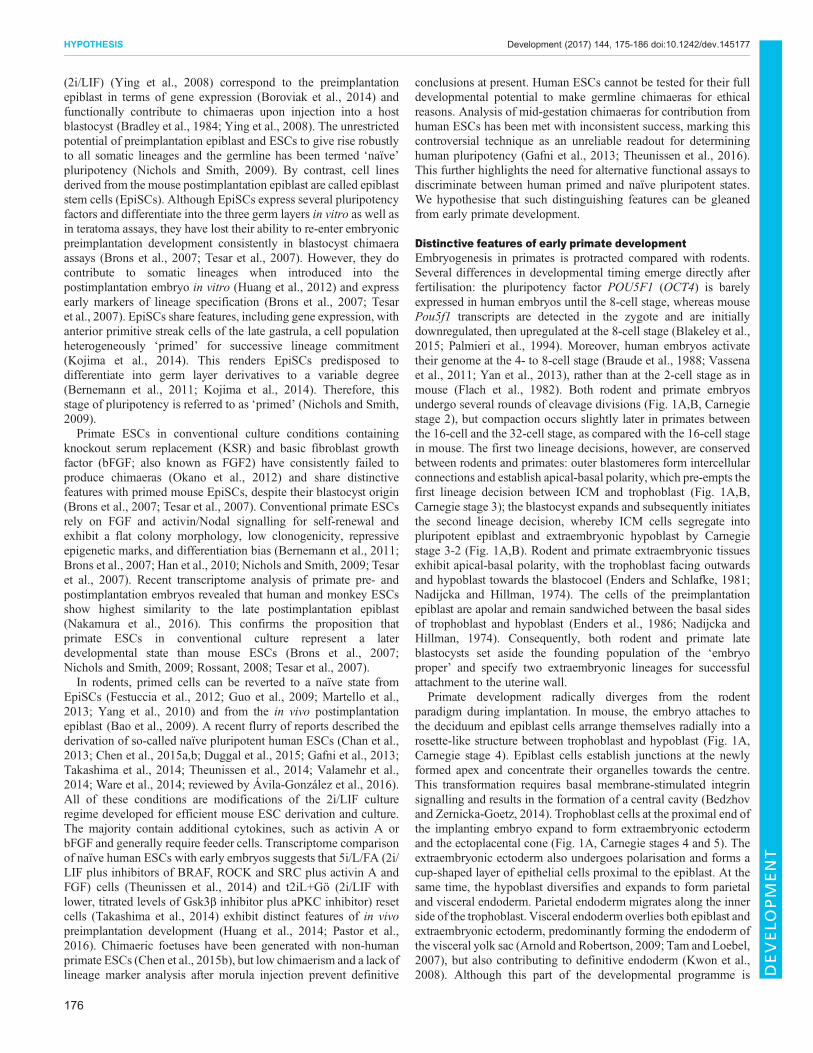
Distinctive features of early primate developmentEmbryogenesis in primates is protracted compared with rodents.Several differences in developmental timing emerge directly afterfertilisation: the pluripotency factor POU5F1 (OCT4) is barelyexpressed in human embryos until the 8-cell stage, whereas mousePou5f1 transcripts are detected in the zygote and are initiallydownregulated, then upregulated at the 8-cell stage (Blakeley et al.,2015; Palmieri et al., 1994). Moreover, human embryos activatetheir genome at the 4- to 8-cell stage (Braude et al., 1988; Vassenaet al., 2011; Yan et al., 2013), rather than at the 2-cell stage as inmouse (Flach et al., 1982). Both rodent and primate embryosundergo several rounds of cleavage divisions (Fig. 1A,B, Carnegiestage 2), but compaction occurs slightly later in primates betweenthe 16-cell and the 32-cell stage, as compared with the 16-cell stagein mouse. The first two lineage decisions, however, are conservedbetween rodents and primates: outer blastomeres form intercellularconnections and establish apical-basal polarity, which pre-empts thefirst lineage decision between ICM and trophoblast (Fig. 1A,B,Carnegie stage 3); the blastocyst expands and subsequently initiatesthe second lineage decision, whereby ICM cells segregate intopluripotent epiblast and extraembryonic hypoblast by Carnegiestage 3-2 (Fig. 1A,B). Rodent and primate extraembryonic tissuesexhibit apical-basal polarity, with the trophoblast facing outwardsand hypoblast towards the blastocoel (Enders and Schlafke, 1981;Nadijcka and Hillman, 1974). The cells of the preimplantationepiblast are apolar and remain sandwiched between the basal sidesof trophoblast and hypoblast (Enders et al., 1986; Nadijcka andHillman, 1974). Consequently, both rodent and primate lateblastocysts set aside the founding population of the ‘embryoproper’ and specify two extraembryonic lineages for successfulattachment to the uterine wall.
Primate development radically diverges from the rodentparadigm during implantation. In mouse, the embryo attaches tothe deciduum and epiblast cells arrange themselves radially into arosette-like structure between trophoblast and hypoblast (Fig. 1A,Carnegie stage 4). Epiblast cells establish junctions at the newlyformed apex and concentrate their organelles towards the centre.This transformation requires basal membrane-stimulated integrinsignalling and results in the formation of a central cavity (Bedzhovand Zernicka-Goetz, 2014). Trophoblast cells at the proximal end ofthe implanting embryo expand to form extraembryonic ectodermand the ectoplacental cone (Fig. 1A, Carnegie stages 4 and 5). Theextraembryonic ectoderm also undergoes polarisation and forms acup-shaped layer of epithelial cells proximal to the epiblast. At thesame time, the hypoblast diversifies and expands to form parietaland visceral endoderm. Parietal endoderm migrates along the innerside of the trophoblast. Visceral endoderm overlies both epiblast andextraembryonic ectoderm, predominantly forming the endoderm ofthe visceral yolk sac (Arnold and Robertson, 2009; Tam and Loebel,2007), but also contributing to definitive endoderm (Kwon et al.,2008). Although this part of the developmental programme is
176
HYPOTHESIS Development (2017) 144, 175-186 doi:10.1242/dev.145177
DEVELO
PM
ENT
Carnegie stage 3-1 Carnegie stage 3-2 Carnegie stage 4
Carnegie stage 5a/b Carnegie stage 6
Trophoblast
Inner cellmass
Carnegie stage 1 Carnegie stage 2
Blastomere
Hypoblast(primitive endoderm)
Preimplantationepiblast
Trophoblast
Parietal endodermVisceral
endoderm Epiblast
Endometrium *
Endometrium
Visceral endoderm
Postimplantationepiblast
Amnioticepithelial cells
Extraembryonicmesoderm
Trophoblast Parietal endoderm
Primary yolk sac
Amnioticcavity
Secondary yolk sac
Amnioticcavity
Endometrium Endometrium
Amniotic epithelial cells
Embryonic disc
Trophoblast Parietal endoderm
Extraembryonicmesoderm Visceral
endoderm
Extraembryonicmesoderm
SyncytiotrophoblastSyncytiotrophoblast
Parietal endoderm
Visceral endoderm
Syncytiotrophoblast
Cytotrophoblast
Trophoblast
Extraembryonicmesoderm Amniotic epithelial
cells
Primitive streak(initiation of gastrulation)
Carnegie stage 5c
Pro
-am
niot
ic c
avity
Trophoblast
BlastomereTrophoblast
Inner cellmass
Hypoblast(primitive endoderm)
Preimplantationepiblast
Postimplantationepiblast
Extraembryonic ectoderm
Endometrium
Endometrium
Endometrium
Pro-amniotic cavityVisceral
endoderm
Parietal endoderm
Yolk sac
Trophoblast
Postimplantationepiblast
Visceral endoderm
Extraembryonic ectoderm
Parietalendoderm
Yolk
sac
Carnegie stage 1 Carnegie stage 2 Carnegie stage 3-1
Carnegie stage 3-2 Carnegie stage 4 Carnegie stage 5 Carnegie stage 6
ExtraembryonicmesodermR
oden
t dev
elop
men
tPr
imat
e de
velo
pmen
t
A
B
Proximal
Distal
Anterior Posterior
Primitive streak(initiation of gastrulation)
Fig. 1. Schematic overview of rodent and primate development from fertilisation to gastrulation. Developmental time of rodent development (A) andprimate development (B) is given in Carnegie stages to facilitate comparison between species. Embryonic lineages are represented in blue shades,extraembryonic lineages in red shades. Cartoons for primate development were drawn based on histological sections of common marmoset (Moore et al., 1985),rhesus macaque (Enders and King, 1988; Enders et al., 1986) and early human stages of the Carnegie collection (Hertig and Rock, 1941, 1946, 1949; Rock andHertig, 1948). Note that extraembryonic mesoderm specification from visceral endoderm is exclusively based on electron micrographs of early rhesus macaqueimplantation stages.
177
HYPOTHESIS Development (2017) 144, 175-186 doi:10.1242/dev.145177
DEVELO
PM
ENT

shared between rodent and primate, there is a clear and crucialexception: the primate embryo establishes two additionalextraembryonic lineages at this stage – the amniotic epithelialcells and the extraembryonic mesoderm (Fig. 1B, highlighted inred).Primates segregate amniotic epithelial cells directly from the peri-
implantation epiblast. During implantation, the primate epiblastforms a rosette-like structure, similar to mouse, with epiblast cellsunderlying the trophoblast sharing desmosomal junctions withtrophoblast cells (Enders et al., 1986). In addition, primate epiblastcells adjacent to the visceral endoderm increase in size and displacethe centre of the rosette (Fig. 1B, Carnegie stage 4). Lumenformation in the centre of the implanting rosette gives rise to theamniotic cavity. These rearrangements yield two morphologicallydistinctive cell types: amniotic epithelial cells, which are theprecursors of the amniotic sac, on the cytotrophoblast side; andpostimplantation epiblast cells, destined to form the embryonic disc,which reside adjacent to visceral endoderm (Fig. 1B, Carnegiestages 5 to 6). The amnion is a smooth epithelium consisting of lowcuboidal cells linked by apical junctional complexes. It iscontiguous with the taller, columnar epiblast, reflecting theircommon origin. Recent progress in the culture of human embryosto early postimplantation stages in vitro has allowed the directobservation of amniotic cavity formation (Deglincerti et al., 2016;Shahbazi et al., 2016). Human epiblast cells acquire apical-basalpolarity, undergo lumen formation and establish columnar andsquamous POU5F1-positive populations, representative ofembryonic disc and prospective amniotic epithelium, respectively
(Deglincerti et al., 2016; Shahbazi et al., 2016). This direct mode ofamnion formation from the preimplantation epiblast beforegastrulation is described in marmoset, rhesus macaque and human(Fig. 2), suggesting a conserved feature of primate development. Arecent report showing that primate germ cells are specified fromamniotic epithelial cells further underlines the major importance ofthis lineage decision (Sasaki et al., 2016). In mouse, amnionformation is initiated later, at the onset of gastrulation, whenextraembryonic mesoderm is specified from the posterior epiblast(Fig. 1A, Carnegie stage 6). This leads to formation of theamniochorionic fold [formerly called the ʻposterior amniotic fold’(Kaufman, 1992)], which gives rise to both amnion and chorion(described by Pereira et al., 2011).
The second fundamental difference between rodent and primatedevelopment is extraembryonic mesoderm specification (Fig. 1B,Carnegie stage 4). In rodents, gastrulation initiates in the primitivestreak, which is induced at the proximal posterior extremity of thepostimplantation epiblast at Carnegie stage 6 (Fig. 1A). Distinctmesodermal cell lineages become allocated according to the timeand site of ingression through the streak (Arnold and Robertson,2009; Lawson, 1999). The earliest population of mouse epiblastcells to undergo epithelial-to-mesenchymal transition and migratethrough the streak gives rise to extraembryonic mesoderm,including the mesodermal layer of the chorion, visceral yolk sacmesoderm and blood islands (Arnold and Robertson, 2009). Thus,in rodents, extraembryonic mesoderm formation occurs duringgastrulation. By contrast, primates specify extraembryonicmesoderm at implantation, long before gastrulation (Fig. 1B,
Common marmoset(Callithrix jacchus)
Car
negi
e st
age
5C
arne
gie
stag
e 6
A B C
D E F
Rhesus macaque(Macaca mulatta)
Human(Homo sapiens)
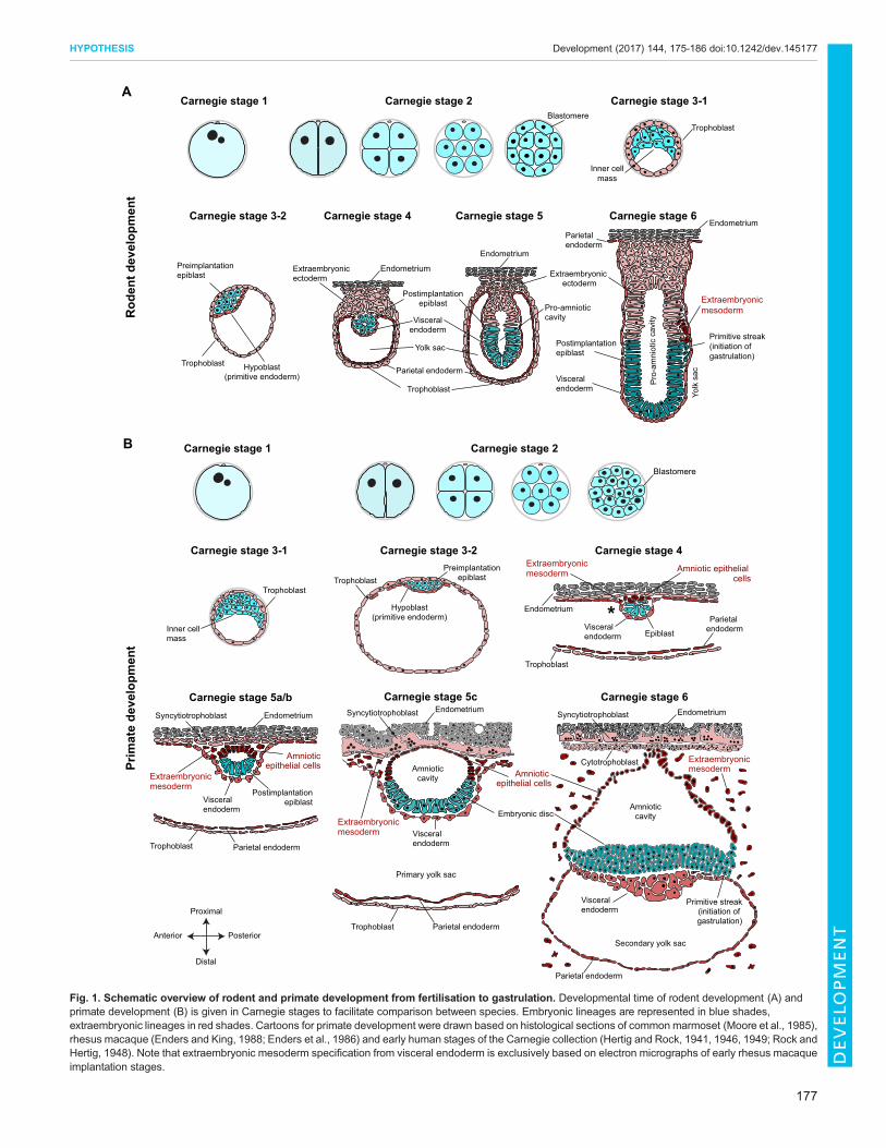
Fig. 2. Images of early primate implantation stages. Carnegie stages 5 and 6 are shown from (A,D) common marmoset (Enders and Lopata, 1999),(B,E) rhesus macaque (Enders et al., 1986) and (C,F) human stages of the Carnegie collection (Hertig and Rock, 1941; O’Rahilly and Muller, 1987). Bluearrowheads indicate postimplantation epiblast (A-C) or embryonic disc (D-F), red arrowheads indicate amniotic epithelial cells. Images reproduced withpermission from John Wiley and Sons (A,B,D,E) and the Carnegie Institution of Washington (C,F).
178
HYPOTHESIS Development (2017) 144, 175-186 doi:10.1242/dev.145177
DEVELO
PM
ENT
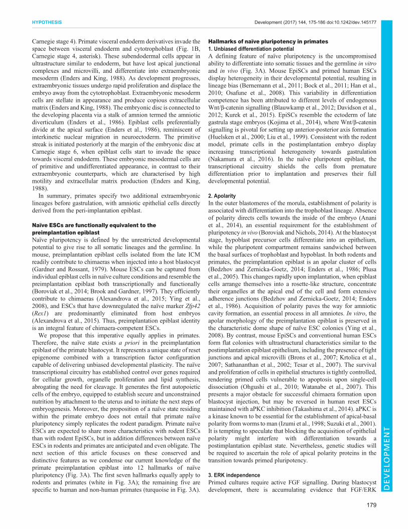
Carnegie stage 4). Primate visceral endoderm derivatives invade thespace between visceral endoderm and cytotrophoblast (Fig. 1B,Carnegie stage 4, asterisk). These subendodermal cells appear inultrastructure similar to endoderm, but have lost apical junctionalcomplexes and microvilli, and differentiate into extraembryonicmesoderm (Enders and King, 1988). As development progresses,extraembryonic tissues undergo rapid proliferation and displace theembryo away from the cytotrophoblast. Extraembryonic mesodermcells are stellate in appearance and produce copious extracellularmatrix (Enders and King, 1988). The embryonic disc is connected tothe developing placenta via a stalk of amnion termed the amnioticdiverticulum (Enders et al., 1986). Epiblast cells preferentiallydivide at the apical surface (Enders et al., 1986), reminiscent ofinterkinetic nuclear migration in neuroectoderm. The primitivestreak is initiated posteriorly at the margin of the embryonic disc atCarnegie stage 6, when epiblast cells start to invade the spacetowards visceral endoderm. These embryonic mesodermal cells areof primitive and undifferentiated appearance, in contrast to theirextraembryonic counterparts, which are characterised by highmotility and extracellular matrix production (Enders and King,1988).In summary, primates specify two additional extraembryonic
lineages before gastrulation, with amniotic epithelial cells directlyderived from the peri-implantation epiblast.
Naïve ESCs are functionally equivalent to thepreimplantation epiblastNaïve pluripotency is defined by the unrestricted developmentalpotential to give rise to all somatic lineages and the germline. Inmouse, preimplantation epiblast cells isolated from the late ICMreadily contribute to chimaeras when injected into a host blastocyst(Gardner and Rossant, 1979). Mouse ESCs can be captured fromindividual epiblast cells in naïve culture conditions and resemble thepreimplantation epiblast both transcriptionally and functionally(Boroviak et al., 2014; Brook and Gardner, 1997). They efficientlycontribute to chimaeras (Alexandrova et al., 2015; Ying et al.,2008), and ESCs that have downregulated the naïve marker Zfp42(Rex1) are predominantly eliminated from host embryos(Alexandrova et al., 2015). Thus, preimplantation epiblast identityis an integral feature of chimaera-competent ESCs.We propose that this imperative equally applies in primates.
Therefore, the naïve state exists a priori in the preimplantationepiblast of the primate blastocyst. It represents a unique state of resetepigenome combined with a transcription factor configurationcapable of delivering unbiased developmental plasticity. The naïvetranscriptional circuitry has established control over genes requiredfor cellular growth, organelle proliferation and lipid synthesis,abrogating the need for cleavage. It generates the first autopoieticcells of the embryo, equipped to establish secure and unconstrainednutrition by attachment to the uterus and to initiate the next steps ofembryogenesis. Moreover, the proposition of a naïve state residingwithin the primate embryo does not entail that primate naïvepluripotency simply replicates the rodent paradigm. Primate naïveESCs are expected to share more characteristics with rodent ESCsthan with rodent EpiSCs, but in addition differences between naïveESCs in rodents and primates are anticipated and even obligate. Thenext section of this article focuses on these conserved anddistinctive features as we condense our current knowledge of theprimate preimplantation epiblast into 12 hallmarks of naïvepluripotency (Fig. 3A). The first seven hallmarks equally apply torodents and primates (white in Fig. 3A); the remaining five arespecific to human and non-human primates (turquoise in Fig. 3A).
Hallmarks of naïve pluripotency in primates1. Unbiased differentiation potentialA defining feature of naïve pluripotency is the uncompromisedability to differentiate into somatic tissues and the germline in vitroand in vivo (Fig. 3A). Mouse EpiSCs and primed human ESCsdisplay heterogeneity in their developmental potential, resulting inlineage bias (Bernemann et al., 2011; Bock et al., 2011; Han et al.,2010; Osafune et al., 2008). This variability in differentiationcompetence has been attributed to different levels of endogenousWnt/β-catenin signalling (Blauwkamp et al., 2012; Davidson et al.,2012; Kurek et al., 2015). EpiSCs resemble the ectoderm of lategastrula stage embryos (Kojima et al., 2014), where Wnt/β-cateninsignalling is pivotal for setting up anterior-posterior axis formation(Huelsken et al., 2000; Liu et al., 1999). Consistent with the rodentmodel, primate cells in the postimplantation embryo displayincreasing transcriptional heterogeneity towards gastrulation(Nakamura et al., 2016). In the naïve pluripotent epiblast, thetranscriptional circuitry shields the cells from prematuredifferentiation prior to implantation and preserves their fulldevelopmental potential.
2. ApolarityIn the outer blastomeres of the morula, establishment of polarity isassociated with differentiation into the trophoblast lineage. Absenceof polarity directs cells towards the inside of the embryo (Ananiet al., 2014), an essential requirement for the establishment ofpluripotency in vivo (Boroviak and Nichols, 2014). At the blastocyststage, hypoblast precursor cells differentiate into an epithelium,while the pluripotent compartment remains sandwiched betweenthe basal surfaces of trophoblast and hypoblast. In both rodents andprimates, the preimplantation epiblast is an apolar cluster of cells(Bedzhov and Zernicka-Goetz, 2014; Enders et al., 1986; Plusaet al., 2005). This changes rapidly upon implantation, when epiblastcells arrange themselves into a rosette-like structure, concentratetheir organelles at the apical end of the cell and form extensiveadherence junctions (Bedzhov and Zernicka-Goetz, 2014; Enderset al., 1986). Acquisition of polarity paves the way for amnioticcavity formation, an essential process in all amniotes. In vitro, theapolar morphology of the preimplantation epiblast is preserved inthe characteristic dome shape of naïve ESC colonies (Ying et al.,2008). By contrast, mouse EpiSCs and conventional human ESCsform flat colonies with ultrastructural characteristics similar to thepostimplantation epiblast epithelium, including the presence of tightjunctions and apical microvilli (Brons et al., 2007; Krtolica et al.,2007; Sathananthan et al., 2002; Tesar et al., 2007). The survivaland proliferation of cells in epithelial structures is tightly controlled,rendering primed cells vulnerable to apoptosis upon single-celldissociation (Ohgushi et al., 2010; Watanabe et al., 2007). Thispresents a major obstacle for successful chimaera formation uponblastocyst injection, but may be reversed in human reset ESCsmaintained with aPKC inhibition (Takashima et al., 2014). aPKC isa kinase known to be essential for the establishment of apical-basalpolarity from worms to man (Izumi et al., 1998; Suzuki et al., 2001).It is tempting to speculate that blocking the acquisition of epithelialpolarity might interfere with differentiation towards apostimplantation epiblast state. Nevertheless, genetic studies willbe required to ascertain the role of apical polarity proteins in thetransition towards primed pluripotency.
3. ERK independencePrimed cultures require active FGF signalling. During blastocystdevelopment, there is accumulating evidence that FGF/ERK
179
HYPOTHESIS Development (2017) 144, 175-186 doi:10.1242/dev.145177
DEVELO
PM
ENT

inhibition promotes epiblast formation across species. In mouse, theFGF/ERK cascade is the predominant driver of hypoblastspecification (Nichols et al., 2009; Yamanaka et al., 2010),whereas primates rely on additional signalling pathways(Boroviak et al., 2015; Kuijk et al., 2012; Roode et al., 2012).However, robust expression of NANOG in the absence of FGF/ERK signalling is reported in mouse, rat, bovine, marmoset andhuman blastocysts (Boroviak et al., 2015; Kuijk et al., 2012; Nicholset al., 2009; Roode et al., 2012). A recent study demonstrates similarfindings in the zebra finch blastoderm (Mak et al., 2015), suggestinghigh conservation of the inverse correlation between naïvepluripotency and FGF/ERK signalling among amniotes.
4. AutopoiesisThe totipotent zygote undergoes cleavage divisions in the absenceof cellular growth or increase in embryo mass. Cleavage occursuntil the mid-blastocyst stage, when epiblast and hypoblast arespecified (Aiken et al., 2004). The birth of naïve pluripotency inthe epiblast is tightly linked to the establishment of autopoiesis(from the Greek meaning ʻself-creating’). In biology, autopoiesisrefers to the ability of a cell to fully reproduce and maintain itself,that is, to ʻself-produce’ all the same organelles, membranes andcytosolic components of which it is composed. This differs fromthe concept of self-renewal, which relates to the renewal ofdevelopmental potential and not necessarily the full self-renewalof cellular components per se. In mouse, embryonic cells gainautopoiesis just before implantation, when a safe and continuous
nutrient supply is within reach (Aiken et al., 2004; Boroviak andNichols, 2014). Cleavage stage and early ICM cells are unable toreplenish their cytosolic compartment before cell division,precluding them from continuous and stable self-renewal(Boroviak et al., 2014; Boroviak and Nichols, 2014). In theprimate embryo, precise measurements of nucleocytoplasmicratios throughout preimplantation development are not yetavailable. However, measurements of cell sizes from histologicalsections (The Virtual Human Embryo, www.ehd.org/virtual-human-embryo/) suggest a decrease from morula and early ICMto the late blastocyst stage and constant or larger sizes at earlypostimplantation stages. Thus, it seems plausible that human andnon-human primate embryos equally acquire autopoiesis aroundimplantation. We suggest that autopoiesis is a hallmark of naïvepluripotency, distinguishing it from totipotent cleavage stages.
5. Core pluripotencyPluripotency is conferred by a unique array of transcriptionfactors. At the core of this network are POU5F1, SOX2 andNANOG, which are evolutionarily conserved in mammals andseveral vertebrates (Dixon et al., 2010; Lavial et al., 2007; Tapiaet al., 2012; Theunissen et al., 2011) and robustly expressed inboth rodent and primate preimplantation epiblasts (Blakeley et al.,2015; Boroviak et al., 2015; Petropoulos et al., 2016).Interestingly, the core circuitry is shared between naïve andprimed pluripotent cells, suggesting a context-dependent role intranscriptional regulation (Boiani and Scholer, 2005; Buecker
2Apolarity
8XIST
expression
XIST
1Unbiased
differentiation
3ERK
independence
ERK
MEK
4Autopoiesis
7Active
X chromosomes
11 Slower
proliferation
5 Core pluripotency
POU5F1
SOX2 NANOG
6DNA
hypomethlyation
12 Extraembryonic
potential
ARGFXKLF17
DNMT3L
NODALSOX15
9Primate-specific
naïve network
SVA-DLTR5_Hs
10 Primate-specificTE expression
Hallmarksof naïve pluripotency
POU5F1distal enhancer
Pol-IIDE
PE
POU5F1 expression1 2 3 4
STAT3
LIF
LIF/STAT3 requirement
SMAD2/3
ALK4/5/7ActRII
NODAL/TGFββrequirement
Oxidativephosphorylation
A B
Fig. 3. Hallmarks of naïve pluripotency in primates. (A) White circles symbolise hallmarks of naïve pluripotency in both rodents and primates, turquoise circlesare specific to primates. (B) Grey circles encompass known features of the rodent preimplantation epiblast, which have not yet been analysed in primate embryos.TE, transposable element.
180
HYPOTHESIS Development (2017) 144, 175-186 doi:10.1242/dev.145177
DEVELO
PM
ENT

et al., 2014). In mouse epiblast and ESCs, the framework isprovided by naïve pluripotency factors such as Klf2, Klf4, Klf5,Esrrb, Tfcp2l1, Tbx3 and Zfp42. This naïve circuitry is specificallyexpressed in pre- but not postimplantation development (Boroviaket al., 2014). Upon implantation in rodents, the wider pluripotencynetwork is replaced with a different suite of transcription factors,including Otx2, Pou3f1 (Oct6), Sox3, Tead2 and Bex1 (Acamporaet al., 2013; Boroviak et al., 2015) to prepare the epiblast forgastrulation. Nanog is downregulated at early postimplantationstages in mouse. It has been proposed that during this formativephase, in the absence of both naïve factors and lineage specifiers,cells become receptive to differentiation stimuli (Kalkan andSmith, 2014; Smith, 2017). Subsequently, Nanog becomes re-expressed in the mouse posterior epiblast; localised expression ofWnt, Nodal and Bmp initiate primitive streak formation andestablishment of the primary germ layers commences. Recenttranscriptome profiling of non-human primate postimplantationstages revealed uninterrupted expression of POU5F1, SOX2 andNANOG until gastrulation (Nakamura et al., 2016). This lendssupport to the crucial role of the core pluripotency network acrossdevelopmental states.
6. DNA hypomethylationThe naïve character of the epiblast extends to epigenetic marks.DNA methylation carries important regulatory information andundergoes global resetting during germ cell and preimplantationdevelopment (Seisenberger et al., 2013a,b). In mouse and human,the preimplantation epiblast has a distinctive epigenetic signatureconsisting of genome-wide DNA hypomethylation with only theimprinted regions spared (Guo et al., 2014; Smallwood et al., 2011;Smith et al., 2014, 2012). This epigenetic status is preserved inmouse ESCs cultured in 2i/LIF, but not in serum-based conditions(Ficz et al., 2013; Habibi et al., 2013; Leitch et al., 2013).Conventional human ESCs exhibit high DNA methylation levelscomparable to those of mouse EpiSCs, ESCs cultured in serum/LIF,or human somatic cells (Pastor et al., 2016). Resetting human ESCswith either 5i/L/FA (Theunissen et al., 2014) or t2iL+Gö(Takashima et al., 2014) induces hypomethylation at levelssimilar to the human ICM, but at the expense of DNAmethylation of primary imprints (Pastor et al., 2016; Theunissenet al., 2016). This is problematic, since erroneous imprinting isimplicated in a variety of human diseases and syndromes (Butler,2009). Also, prolonged culture of human ESCs in 5i/L/FA leads tokaryotypic abnormalities (Pastor et al., 2016; Theunissen et al.,2014), but whether loss of DNAmethylation is the underlying causeremains to be elucidated.
7. Active X chromosomesMost mammals exhibit random X-chromosome inactivation (XCI)in females to compensate X-linked gene expression (Escamilla-Del-Arenal et al., 2011; van den Berg et al., 2011). In mouse, thepaternal X chromosome is silenced at the 4-cell stage (Huynh andLee, 2003) and remains inactive in extraembryonic tissues.However, the paternal X chromosome is reactivated in the epiblast(Mak et al., 2004; Okamoto et al., 2004), tightly linked to theestablishment of naïve pluripotency (Silva et al., 2009). In humanblastocysts, transcription occurs from both X chromosomes in thepreimplantation epiblast, as in mouse (Petropoulos et al., 2016).XCI ensues upon implantation and is associated with theestablishment of primed pluripotency (Kobayashi et al., 2016).Thus, the presence of dually active X chromosomes is a hallmark ofnaïve pluripotency.
8. XIST expressionIn mouse, XCI is mediated by the cis-acting, non-coding RNA Xist,which is downregulated in the epiblast. Re-expression occurs in theearly postimplantation epiblast from either the maternal or paternalX chromosome to induce random XCI. By contrast, rabbit embryosinitially upregulate Xist on both X chromosomes and, via anintermediate phase of biallelic XCI, induce random, monoallelicXCI at the late blastocyst stage before gastrulation (Okamoto et al.,2011). Consequently, biallelic X chromosome expression is not ahallmark of naïve pluripotency in the rabbit embryo.
Human embryos also lack paternal imprints for XIST expression,similar to rabbits, resulting in random XCI in both embryonic andextraembryonic lineages. However, despite biallelic XISTexpression in more than half of the cells examined, both Xchromosomes remain transcriptionally active in human blastocysts(Okamoto et al., 2011). Random XCI presumably occurs uponimplantation, similar to in mice. Recent single-cell transcriptomedata of human ICM cells confirm XIST expression at the blastocyststage (Blakeley et al., 2015; Petropoulos et al., 2016; Yan et al.,2013). However, in contrast to mouse, dosage compensation occursgradually in all three lineages of the human blastocyst, with both Xchromosomes being actively transcribed throughout this process(Petropoulos et al., 2016). The mechanisms of dosage compensationin the presence of biallelic XIST expression remain unknown.Nevertheless, the embryo transcriptome data show that femalehuman naïve pluripotent cells are expected to express XIST withboth X chromosomes being active. This has been demonstratedrecently for 5i/L/FA and t2iL+Gö reset cells (Sahakyan et al., 2016)and is in contrast to female naïve pluripotent cells in rodents, whichdo not express XIST.
9. Primate-specific naïve networkNaïve pluripotency factors are exclusive to preimplantation stagesand sharply downregulated upon implantation and epiblastepithelialisation. Therefore, their identification relies ontranscriptional analysis of both pre- and postimplantation samples.Naïve pluripotency factors in mouse include Klf2, Klf4, Klf5, Stat3,Nr0b1, Esrrb, Tfcp2l1, Tbx3 and Zfp42 (Boroviak et al., 2014,2015; Peng et al., 2016; Scialdone et al., 2016). A subset of naïvefactors, including Stat3 (Yang et al., 2010), Nr5a2 (Guo and Smith,2010), Klf2 (Hall et al., 2009), Esrrb (Festuccia et al., 2012), Klf4(Guo et al., 2009) and Tfcp2l1 (Martello et al., 2013) can single-handedly drive naïve conversion from EpiSCs, and combinations ofNANOG plus KLF2 or KLF4 have been used to reset human ESCs(Takashima et al., 2014; Theunissen et al., 2014).
The advent of single-cell profiling has allowed detailed molecularmapping of primate preimplantation development, and RNAsequencing (RNA-seq) datasets have become available inmarmoset (Boroviak et al., 2015) and human (Blakeley et al.,2015; Petropoulos et al., 2016; Xue et al., 2013; Yan et al., 2013)showing that the majority of pluripotency-associated genes,including POU5F1, SOX2, NANOG, SALL4, KLF4, TFCP2L1and TDGF1 are expressed in the primate epiblast. TFCP2L1, KLF4and NANOG proteins colocalise in a subset of ICM cells inmarmoset (Boroviak et al., 2015) and human (Takashima et al.,2014) blastocysts, suggesting partial conservation of the naïvecircuitry. However, absence of KLF2, ESRRB, NR0B1, FBXO15and BMP4, and increased levels of GDF3, NODAL, LEFTY1,KLF17 and ARGFX, demonstrate extensive primate-specificadaptation of the naïve pluripotency network (Blakeley et al.,2015; Boroviak et al., 2015; Petropoulos et al., 2016).Postimplantation stages in human are impossible to obtain for
181
HYPOTHESIS Development (2017) 144, 175-186 doi:10.1242/dev.145177
DEVELO
PM
ENT

ethical reasons, but a recent report in cynomologus monkeyprovided a transcriptional blueprint from ICM to the late gastrula(Nakamura et al., 2016). Naïve markers expressed in thepreimplantation epiblast but not in postimplantation stagesincluded TFCP2L1, KLF5, KLF17, NODAL and SOX15(Nakamura et al., 2016). KLF4 and DNMT3L were drasticallydownregulated upon implantation, but still expressed in the earlypostimplantation epiblast. The generation of chimaera-competentprimate ESCswill rely on the complete re-establishment of the naïvecircuitry that is operative in the preimplantation epiblast, free fromexpression of the mouse-specific KLF2, ESRRB and NR0B1.
10. Primate-specific transposable element (TE) expressionGlobal resetting of the epigenome during early developmentimpacts on the expression of TEs, which make up half of themammalian genome. Liberation from repressive DNA methylationin early developmental stages results in highly stage-specific TEexpression (Göke et al., 2015). This transposcriptome has beenproposed as an alternative measure to assess the correspondencebetween cultured pluripotent stem cells and the embryo (Theunissenet al., 2016). Human 5i/L/FA (Theunissen et al., 2014) and t2iL+Gö(Takashima et al., 2014) reset cells resemble human morula andblastocyst stages, respectively, showing elevated expression of theSINE-VNTR-Alu D subgroup (SVA-D) and LTR5_Hs (Theunissenet al., 2016). The close correlation to results from gene-basedmethods supports the overall conclusion of this new TE signature-based approach. However, while the transposcriptome may providea more sensitive measure of the cell state in terms of transcriptnumber, the functional relevance of similarities and divergencesremains to be explored.
11. Slower proliferationAn important divergence between rodents and primates is the rate ofproliferation. The mouse late blastocyst consists of ∼150 cells atembryonic day (E) 4.5 (Plusa et al., 2008), reflecting a cell cyclelength of ∼15 h. Human embryos reach this stage after 7 days,having generated ∼250 cells (Niakan and Eggan, 2013). Thus,human embryonic cells have an increase in cell cycle length of atleast 6 h, from 15 h to 21 h. Mouse ESCs exhibit comparablegeneration times (14-16 h) to their embryonic counterpart (Jovicet al., 2013), largely as a result of elevated and cell cycle-independent cyclin-dependent kinase 2 (Cdk2) expression (Steadet al., 2002). Cdk2 promotes the G1–S-phase transition by initiatingDNA replication. By contrast, human (Blakeley et al., 2015; Yanet al., 2013) and marmoset (Boroviak et al., 2015) ICM cells lackconstitutive CDK2 expression, but show higher levels of WEE1, akey cell cycle inhibitor. This demonstrates major differences in thecell cycle machinery between rodents and primates. Thus, authenticprimate ESCs are not expected to typify their rodent counterpartswith regard to proliferation rates.
12. Extraembryonic potentialThe divergence of rodent and primate postimplantationdevelopment transforms the concept and prospects of naïvepluripotency in primates. In contrast to mouse epiblasts, primatessegregate an additional lineage before gastrulation, whereby theproximal epiblast differentiates into amniotic epithelium (Endersand Lopata, 1999; Enders et al., 1986). We therefore hypothesisethat authentic human naïve pluripotent cultures should have anexpanded capacity to produce both postimplantation epiblast andamniotic epithelial cells. This means that naïve primate ESCsshould be able to differentiate into either cell type within a short
time window. However, currently there are two key pieces ofinformation missing: (1) the signalling pathways that control thislineage decision; and (2) the transcriptional and epigenetic signatureof amniotic epithelial cells. A clear understanding of thedevelopmental cues that determine amnion differentiation will berequired to specify this extraembryonic lineage efficiently fromnaïve primate ESCs in vitro. Moreover, this experiment demands adetailed knowledge of the molecular signature of amniotic epithelialcells in vivo for meaningful endpoint analysis. Future studies of non-human primate postimplantation development including samples ofamniotic epithelial cells and tracking of spatial identity within theembryo might be able to tackle these questions.
The 12 hallmarks: a testable framework for human naïveESCsWe propose that the 12 hallmarks of naïve pluripotency outlinedabove can constitute a powerful system to assess human naïvepluripotency in vitro. Primate cells in a naïve state are expected totolerate long-termMEK inhibition via PD0325901 (hallmark 3) andto grow more slowly than mouse ESCs (hallmark 11) in apolar,dome-shaped colonies (hallmark 2). Absence of epithelial charactercan be further examined by antibody staining for apical polarity andtight junction proteins. The autopoietic nature of the cells allowsstable long-term propagation (hallmark 4), distinguishing themfrom totipotent cells, which cannot be propagated indefinitely.Hypomethylation can be evaluated by bisulphite sequencing(hallmark 6). Genome-wide transcriptional profiling by RNA-seqpermits testing for core pluripotency (hallmark 5), XIST expression(hallmark 8), the primate-specific naïve network (hallmark 9) andTE expression (hallmark 10). Read lengths of more than 100 bp arefavourable to facilitate mapping of highly repetitive TEs. Moreover,exploring the wider naïve transcriptional circuitry and the TEsignature are powerful ways to discriminate between primateepiblast identity and artificial mouse ESC-like states. Absence orlow-level expression of mouse-specific pluripotency factors,including KLF2, ESRRB, NR0B1 and FBXO15, are importantindicators for successful resetting towards an authentic humanepiblast state. High-quality RNA-seq datasets may also be used todetect SNPs and assess biallelic expression from the X chromosome(hallmark 7). Alternatively, the X-chromosome activation status canbe determined by fluorescence in situ hybridisation (hallmark 7).
In addition to descriptive analysis, it is pivotal to test functionallyunbiased differentiation potential (hallmark 1) and extraembryoniccapacity for amnion formation (hallmark 12). Human germlinechimaera contribution assays are prohibited on ethical grounds.However, unbiased differentiation can be gauged in vitro and byteratoma formation in vivo. Froma developmental point of view, naïveESCs are expected to differentiate into somatic lineages via successiveformative and primed pluripotent states (Smith, 2017). This needs tobe considered when applying stepwise protocols for directeddifferentiation. Epigenetic resetting to the naïve state may eradicatesomeof the lineagebias observed in conventional humanESCs.Theseexperiments demand careful quantification of various differentiationassays and would only become meaningful after comparing multipleindependent lines. Moreover, it is difficult to discern genetic diversityfrom epigenetic lineage bias. In practice, quantitative differentiationmight not be suitable for routine assessment of naïve pluripotency.
The specific ability of the primate peri-implantation epiblast togive rise to nascent amnion (hallmark 12) might provide a moreexplicit functional assay to discriminate between naïve and primedstates. Conventional human ESCs correspond to the pregastrulaembryonic disc (Nakamura et al., 2016), 7 days after amnion
182
HYPOTHESIS Development (2017) 144, 175-186 doi:10.1242/dev.145177
DEVELO
PM
ENT

segregation. Naïve pluripotency is established in the epiblast justbefore this decision point. Thus, naïve human ESCs should becompetent to replicate amnion segregation and amniotic cavityformation of postimplantation stages. Stimuli from the extracellularmatrix and/or adjacent extraembryonic tissues might be essential forthis transition. The recent reports on amniotic cavity formation ofhuman embryos cultured to postimplantation stages in vitro(Deglincerti et al., 2016; Shahbazi et al., 2016) lend support tothe feasibility of this undertaking. An in vitro system to obtain andstudy human embryonic and extraembryonic lineages from culturedcells would be highly desirable to unravel the continuum ofpluripotent states in the primate embryo.
Unresolved issues in primate developmentSeveral features of naïve pluripotency remain uncertain in primates(Fig. 3B). Mouse ESCs are bivalent in their energy production, usingboth oxidative phosphorylation and glycolysis, whereas EpiSCs shifttheir metabolism to high glycolysis, phenotypically akin to rapidlyproliferating cancer cells (Zhou et al., 2012). A number of recentstudies have characterised metabolic dynamics in different pluripotentstates in vitro (reviewed byTeslaa andTeitell, 2015); however,whetherthis paradigm applies to bona fide primate embryonic developmentremains unclear. While quantitative measurements of metabolites oroxygen consumption rates are difficult to obtain in vivo, results from invitro derived cells might not reflect the situation in the embryo. Forexample, it has been suggested that nicotinamide N-methyltransferase(NNMT) regulates a metabolic switch between human primed andputative naïve ESCs cultured in 2i/FGF (Sperber et al., 2015).However, NNMT is not expressed in the human preimplantationepiblast (Blakeley et al., 2015; Petropoulos et al., 2016; Yan et al.,2013). Another contentious subject is NODAL/TGFβ signalling in theprimate blastocyst. Human embryos cultured in the presence ofthe NODAL/TGFβ inhibitor SB431542 are reported to increase thenumber of NANOG-positive ICM cells (Van der Jeught et al., 2014).However, similar experiments using higher concentrations showed adramatic reduction of NANOG expression (Blakeley et al., 2015). Inmarmoset, NODAL/TGFβ inhibition with A83-01 did not modulateNANOG expression (Boroviak et al., 2015). The question of whetherNODAL/TGFβ signalling is functionally required for primate naïvepluripotency is interesting and deserves further attention. Equallyunclear is the role of LIF/STAT3 signalling or whether POU5F1expression in the embryo primarily relies on its distal enhancer. So far,specificPOU5F1 distal enhancer operation has not been demonstratedin the primate epiblast. Further refinements of ChIP-seq and advancedchromosome configuration capture approaches for single-cell analysiswill help to address some of these questions.
Future perspectives of naïve pluripotency in primatesThe capture of authentic developmental states forms an integral partof both basic and applied research. Naïve ESCs provide a tool tofunctionally assess the factors that control in vivo development. Thisis of particular importance in primates, where embryonic material isprecious and scarce. Second, robust differentiation of pluripotentcells relies on a precise spatiotemporal sequence of specificationevents. A defined developmental starting point is essential to mimicembryonic patterning in vitro. Preimplantation epiblast identitydelivers an exact developmental stage with well-definedcharacteristics (Fig. 3A), in addition to favourable cell biologyfeatures such as apolarity for efficient single-cell cloning. Despitethe remarkable success of designer nucleases (Liu et al., 2014; Satoet al., 2016) and Cas9/RNA-mediated gene targeting (Niu et al.,2014) in non-human primate zygotes, it is technically and
economically challenging to obtain sufficient numbers of primateembryos for knock-in strategies. Currently, this limits gene-editingapproaches to simple gene disruption.
Chimaera-competent ESCs in non-human primates might open upavenues for sophisticated genetic engineering to create versatilemodels for basic and preclinical research. This is important in areaswhere rodent models are insufficient, including infectious diseases,neurodegenerative disorders, aging and reproductive medicine(Carrion and Patterson, 2012; Mansfield, 2003; Okano et al., 2012;Shedlock et al., 2009). Another emerging application for naïve ESCsin biomedical research is organ farming. Rat ESCs are capable offilling the developmental niche of mouse Pdx1 (pancreatogenesis-disabled) null host embryos (Kobayashi et al., 2010), a procedurereferred to as interspecies chimaeric complementation (reviewed byWu and Izpisua Belmonte, 2015). This concept might be exploited togrow human organs in pigs for xenotransplantation. The recentgeneration of apancreatic pigs provides another key step towardsclinical application (Matsunari et al., 2013). However, the lack ofchimaera-competent primate ESCs currently presents a bottleneck forthe generation of primate organs in farm animals. In addition, there areethical concernswith regard to unwanted tissue contribution of humancells to the pig central nervous system or gametes. The use of naïvenon-human primate ESCs in interspecies chimaeric complementationwill be pivotal to resolve these issues and turn the xenomedical visioninto reality.
AcknowledgementsWe thank Dr Maria Rostovskaya and Professor Austin Smith for helpful commentson the manuscript.
Competing interestsThe authors declare no competing or financial interests.
FundingResearch in the authors’ laboratories is supported by the Wellcome Trust, MedicalResearch Council (MRC) and Biotechnology and Biological Sciences ResearchCouncil, and a core support grant from the Wellcome Trust and MRC to theWellcome Trust – Medical Research Council Cambridge Stem Cell Institute.
ReferencesAcampora, D., Di Giovannantonio, L. G. and Simeone, A. (2013). Otx2 is an
intrinsic determinant of the embryonic stem cell state and is required for transitionto a stable epiblast stem cell condition. Development 140, 43-55.
Aiken, C. E. M., Swoboda, P. P. L., Skepper, J. N. and Johnson, M. H. (2004). Thedirect measurement of embryogenic volume and nucleo-cytoplasmic ratio duringmouse pre-implantation development. Reproduction 128, 527-535.
Alexandrova, S., Kalkan, T., Humphreys, P., Riddell, A., Scognamiglio, R.,Trumpp, A. and Nichols, J. (2015). Selection and dynamics of embryonic stemcell integration into early mouse embryos. Development 143, 24-34.
Anani, S., Bhat, S., Honma-Yamanaka, N., Krawchuk, D. and Yamanaka, Y.(2014). Initiation of Hippo signaling is linked to polarity rather than to cell position inthe pre-implantation mouse embryo. Development 141, 2813-2824.
Arnold, S. J. and Robertson, E. J. (2009). Making a commitment: cell lineageallocation and axis patterning in the early mouse embryo. Nat. Rev. Mol. Cell Biol.10, 91-103.
Ávila-Gonzalez, D., Garcıa-Lopez, G., Garcıa-Castro, I. L., Flores-Herrera, H.,Molina-Hernandez, A., Portillo, W. and Dıaz, N. F. (2016). Capturing theephemeral human pluripotent state. Dev. Dyn. 245, 762-773.
Bao, S., Tang, F., Li, X., Hayashi, K., Gillich, A., Lao, K. and Surani, M. A. (2009).Epigenetic reversion of post-implantation epiblast to pluripotent embryonic stemcells. Nature 461, 1292-1295.
Bedzhov, I. and Zernicka-Goetz, M. (2014). Self-organizing properties of mousepluripotent cells initiate morphogenesis upon implantation. Cell 156, 1032-1044.
Bernemann,C.,Greber,B.,Ko,K.,Sterneckert, J.,Han,D.W.,Arauzo-Bravo,M.J.and Scholer, H. R. (2011). Distinct developmental ground states of epiblast stemcell lines determine different pluripotency features. Stem Cells 29, 1496-1503.
Blakeley, P., Fogarty, N. M. E., del Valle, I., Wamaitha, S. E., Hu, T. X., Elder, K.,Snell, P., Christie, L., Robson, P. and Niakan, K. K. (2015). Defining the threecell lineages of the human blastocyst by single-cell RNA-seq. Development 142,3151-3165.
183
HYPOTHESIS Development (2017) 144, 175-186 doi:10.1242/dev.145177
DEVELO
PM
ENT

Blauwkamp, T. A., Nigam, S., Ardehali, R., Weissman, I. L. andNusse, R. (2012).Endogenous Wnt signalling in human embryonic stem cells generates anequilibrium of distinct lineage-specified progenitors. Nat. Commun. 3, 1070.
Bock, C., Kiskinis, E., Verstappen, G., Gu, H., Boulting, G., Smith, Z. D., Ziller,M., Croft, G. F., Amoroso, M. W., Oakley, D. H. et al. (2011). Reference maps ofhuman ES and iPS cell variation enable high-throughput characterization ofpluripotent cell lines. Cell 144, 439-452.
Boiani, M. and Scholer, H. R. (2005). Regulatory networks in embryo-derivedpluripotent stem cells. Nat. Rev. Mol. Cell Biol. 6, 872-884.
Boroviak, T. and Nichols, J. (2014). The birth of embryonic pluripotency. Philos.Trans. R. Soc. Lond. B Biol. Sci. 369.
Boroviak, T., Loos, R., Bertone, P., Smith, A. and Nichols, J. (2014). The ability ofinner-cell-mass cells to self-renew as embryonic stem cells is acquired followingepiblast specification. Nat. Cell Biol. 16, 516-528.
Boroviak, T., Loos, R., Lombard, P., Okahara, J., Behr, R., Sasaki, E., Nichols,J., Smith, A. and Bertone, P. (2015). Lineage-specific profiling delineates theemergence and progression of naive pluripotency in mammalian embryogenesis.Dev. Cell 35, 366-382.
Bradley, A., Evans, M., Kaufman, M. H. and Robertson, E. (1984). Formation ofgerm-line chimaeras from embryo-derived teratocarcinoma cell lines. Nature 309,255-256.
Braude, P., Bolton, V. and Moore, S. (1988). Human gene expression first occursbetween the four- and eight-cell stages of preimplantation development. Nature332, 459-461.
Brons, I. G. M., Smithers, L. E., Trotter, M. W. B., Rugg-Gunn, P., Sun, B., Chuvade Sousa Lopes, S. M., Howlett, S. K., Clarkson, A., Ahrlund-Richter, L.,Pedersen, R. A. et al. (2007). Derivation of pluripotent epiblast stem cells frommammalian embryos. Nature 448, 191-195.
Brook, F. A. and Gardner, R. L. (1997). The origin and efficient derivation ofembryonic stem cells in the mouse. Proc. Natl. Acad. Sci. USA 94, 5709-5712.
Buecker, C., Chen, H.-H., Polo, J. M., Daheron, L., Bu, L., Barakat, T. S.,Okwieka, P., Porter, A., Gribnau, J., Hochedlinger, K. et al. (2010). A murineESC-like state facilitates transgenesis and homologous recombination in humanpluripotent stem cells. Cell Stem Cell 6, 535-546.
Buecker, C., Srinivasan, R., Wu, Z., Calo, E., Acampora, D., Faial, T., Simeone,A., Tan, M., Swigut, T. and Wysocka, J. (2014). Reorganization of enhancerpatterns in transition from naive to primed pluripotency. Cell Stem Cell 14,838-853.
Butler, M. G. (2009). Genomic imprinting disorders in humans: a mini-review.J. Assist. Reprod. Genet. 26, 477-486.
Carrion, R., Jr and Patterson, J. L. (2012). An animal model that reflects humandisease: the common marmoset (Callithrix jacchus).Curr. Opin. Virol. 2, 357-362.
Chan, Y.-S., Goke, J., Ng, J.-H., Lu, X., Gonzales, K. A. U., Tan, C.-P., Tng,W.-Q.,Hong, Z.-Z., Lim, Y.-S. and Ng, H.-H. (2013). Induction of a human pluripotentstate with distinct regulatory circuitry that resembles preimplantation epiblast. CellStem Cell 13, 663-675.
Chazaud, C., Yamanaka, Y., Pawson, T. and Rossant, J. (2006). Early lineagesegregation between epiblast and primitive endoderm in mouse blastocyststhrough the Grb2-MAPK pathway. Dev. Cell 10, 615-624.
Chen, H., Aksoy, I., Gonnot, F., Osteil, P., Aubry, M., Hamela, C., Rognard, C.,Hochard, A., Voisin, S., Fontaine, E. et al. (2015a). Reinforcement of STAT3activity reprogrammes human embryonic stem cells to naive-like pluripotency.Nat. Commun. 6, 7095.
Chen, Y., Niu, Y., Li, Y., Ai, Z., Kang, Y., Shi, H., Xiang, Z., Yang, Z., Tan, T., Si, W.et al. (2015b). Generation of cynomolgus monkey chimeric fetuses usingembryonic stem cells. Cell Stem Cell 17, 116-124.
Davidson, K. C., Adams, A. M., Goodson, J. M., McDonald, C. E., Potter, J. C.,Berndt, J. D., Biechele, T. L., Taylor, R. J. and Moon, R. T. (2012). Wnt/beta-catenin signaling promotes differentiation, not self-renewal, of human embryonicstem cells and is repressed by Oct4. Proc. Natl. Acad. Sci. USA 109, 4485-4490.
Deglincerti, A., Croft, G. F., Pietila, L. N., Zernicka-Goetz, M., Siggia, E. D. andBrivanlou, A. H. (2016). Self-organization of the in vitro attached human embryo.Nature 533, 251-254.
Dixon, J. E., Allegrucci, C., Redwood,C., Kump, K., Bian, Y., Chatfield, J., Chen,Y.-H., Sottile, V., Voss, S. R., Alberio, R. et al. (2010). Axolotl Nanog activity inmouse embryonic stem cells demonstrates that ground state pluripotency isconserved from urodele amphibians to mammals. Development 137, 2973-2980.
Duggal, G., Warrier, S., Ghimire, S., Broekaert, D., Van der Jeught, M., Lierman,S., Deroo, T., Peelman, L., Van Soom, A., Cornelissen, R. et al. (2015).Alternative routes to induce naive pluripotency in human embryonic stem cells.Stem Cells 33, 2686-2698.
Enders, A. C. and King, B. F. (1988). Formation and differentiation ofextraembryonic mesoderm in the rhesus monkey. Am. J. Anat. 181, 327-340.
Enders, A. C. and Lopata, A. (1999). Implantation in the marmoset monkey:expansion of the early implantation site. Anat. Rec. 256, 279-299.
Enders, A. C. and Schlafke, S. (1981). Differentiation of the blastocyst of the rhesusmonkey. Am. J. Anat. 162, 1-21.
Enders, A. C., Schlafke, S. and Hendrickx, A. G. (1986). Differentiation of theembryonic disc, amnion, and yolk sac in the rhesus monkey. Am. J. Anat. 177,161-185.
Escamilla-Del-Arenal, M., da Rocha, S. T. and Heard, E. (2011). Evolutionarydiversity and developmental regulation of X-chromosome inactivation. Hum.Genet. 130, 307-327.
Evans, M. J. and Kaufman, M. H. (1981). Establishment in culture of pluripotentialcells from mouse embryos. Nature 292, 154-156.
Festuccia, N., Osorno, R., Halbritter, F., Karwacki-Neisius, V., Navarro, P.,Colby, D.,Wong, F., Yates, A., Tomlinson, S. R. andChambers, I. (2012). Esrrbis a direct Nanog target gene that can substitute for Nanog function in pluripotentcells. Cell Stem Cell 11, 477-490.
Ficz, G., Hore, T. A., Santos, F., Lee, H. J., Dean, W., Arand, J., Krueger, F.,Oxley, D., Paul, Y.-L., Walter, J. et al. (2013). FGF signaling inhibition in ESCsdrives rapid genome-wide demethylation to the epigenetic ground state ofpluripotency. Cell Stem Cell 13, 351-359.
Flach, G., Johnson, M. H., Braude, P. R., Taylor, R. A. and Bolton, V. N. (1982).The transition from maternal to embryonic control in the 2-cell mouse embryo.EMBO J. 1, 681-686.
Frankenberg, S., Gerbe, F., Bessonnard, S., Belville, C., Pouchin, P., Bardot, O.and Chazaud, C. (2011). Primitive endoderm differentiates via a three-stepmechanism involving Nanog and RTK signaling. Dev. Cell 21, 1005-1013.
Gafni, O., Weinberger, L., Mansour, A. A. F., Manor, Y. S., Chomsky, E., Ben-Yosef, D., Kalma, Y., Viukov, S., Maza, I., Zviran, A. et al. (2013). Derivation ofnovel human ground state naive pluripotent stem cells. Nature 504, 282-286.
Gardner, R. L. and Rossant, J. (1979). Investigation of the fate of 4-5 day post-coitum mouse inner cell mass cells by blastocyst injection. J. Embryol. Exp.Morphol. 52, 141-152.
Gjørret, J. O. and Maddox-Hyttel, P. (2005). Attempts towards derivation andestablishment of bovine embryonic stem cell-like cultures. Reprod. Fertil. Dev. 17,113-124.
Goke, J., Lu, X., Chan, Y.-S., Ng, H.-H., Ly, L.-H., Sachs, F. and Szczerbinska, I.(2015). Dynamic transcription of distinct classes of endogenous retroviralelements marks specific populations of early human embryonic cells. Cell StemCell 16, 135-141.
Grabarek, J. B., Zyzynska, K., Saiz, N., Piliszek, A., Frankenberg, S., Nichols, J.,Hadjantonakis, A. K. and Plusa, B. (2012). Differential plasticity of epiblast andprimitive endoderm precursors within the ICM of the early mouse embryo.Development 139, 129-139.
Graves, K. H. and Moreadith, R. W. (1993). Derivation and characterization ofputative pluripotential embryonic stem cells from preimplantation rabbit embryos.Mol. Reprod. Dev. 36, 424-433.
Guo, G. and Smith, A. (2010). A genome-wide screen in EpiSCs identifies Nr5anuclear receptors as potent inducers of ground state pluripotency. Development137, 3185-3192.
Guo, G., Yang, J., Nichols, J., Hall, J. S., Eyres, I., Mansfield, W. and Smith, A.(2009). Klf4 reverts developmentally programmed restriction of ground statepluripotency. Development 136, 1063-1069.
Guo, H., Zhu, P., Yan, L., Li, R., Hu, B., Lian, Y., Yan, J., Ren, X., Lin, S., Li, J. et al.(2014). The DNA methylation landscape of human early embryos. Nature 511,606-610.
Habibi, E., Brinkman, A. B., Arand, J., Kroeze, L. I., Kerstens, H. H. D., Matarese,F., Lepikhov, K., Gut, M., Brun-Heath, I., Hubner, N. C. et al. (2013). Whole-genome bisulfite sequencing of two distinct interconvertible DNA methylomes ofmouse embryonic stem cells. Cell Stem Cell 13, 360-369.
Hall, J., Guo, G., Wray, J., Eyres, I., Nichols, J., Grotewold, L., Morfopoulou, S.,Humphreys, P., Mansfield, W., Walker, R. et al. (2009). Oct4 and LIF/Stat3additively induce Kruppel factors to sustain embryonic stem cell self-renewal. CellStem Cell 5, 597-609.
Han, D.W., Tapia, N., Joo, J. Y., Greber, B., Arauzo-Bravo, M. J., Bernemann, C.,Ko, K., Wu, G., Stehling, M., Do, J. T. et al. (2010). Epiblast stem cellsubpopulations represent mouse embryos of distinct pregastrulation stages. Cell143, 617-627.
Hanna, J., Cheng, A. W., Saha, K., Kim, J., Lengner, C. J., Soldner, F., Cassady,J. P., Muffat, J., Carey, B. W. and Jaenisch, R. (2010). Human embryonic stemcells with biological and epigenetic characteristics similar to those of mouseESCs. Proc. Natl. Acad. Sci. USA 107, 9222-9227.
Hertig, A. T. andRock, J. (1941). Two human ova of the pre-villous stage, having anovulation age of about eleven and twelve days respectively. Contrib. Embryol. 29,127-156.
Hertig, A. T. and Rock, J. (1946). On a human blastula recovered from the uterinecavity 4 days after ovulation. Anat. Rec. 94, 469.
Hertig, A. T. and Rock, J. (1949). A series of potentially abortive ova recovered fromfertile women prior to the first missed menstrual period. Am. J. Obstet. Gynecol.58, 968-993, illust.
Huang, Y., Osorno, R., Tsakiridis, A. and Wilson, V. (2012). In vivo differentiationpotential of epiblast stem cells revealed by chimeric embryo formation. Cell Rep.2, 1571-1578.
Huang, K., Maruyama, T. and Fan, G. (2014). The naive state of human pluripotentstem cells: a synthesis of stem cell and preimplantation embryo transcriptomeanalyses. Cell Stem Cell 15, 410-415.
184
HYPOTHESIS Development (2017) 144, 175-186 doi:10.1242/dev.145177
DEVELO
PM
ENT

Huelsken, J., Vogel, R., Brinkmann, V., Erdmann, B., Birchmeier, C. andBirchmeier, W. (2000). Requirement for beta-catenin in anterior-posterior axisformation in mice. J. Cell Biol. 148, 567-578.
Huynh, K. D. and Lee, J. T. (2003). Inheritance of a pre-inactivated paternal Xchromosome in early mouse embryos. Nature 426, 857-862.
Izumi, Y., Hirose, T., Tamai, Y., Hirai, S., Nagashima, Y., Fujimoto, T., Tabuse, Y.,Kemphues, K. J. and Ohno, S. (1998). An atypical PKC directly associates andcolocalizes at the epithelial tight junction with ASIP, a mammalian homologue ofCaenorhabditis elegans polarity protein PAR-3. J. Cell Biol. 143, 95-106.
Jovic, D., Sakaue-Sawano, A., Abe, T., Cho, C. S., Nagaoka, M., Miyawaki, A.and Akaike, T. (2013). Direct observation of cell cycle progression in living mouseembryonic stem cells on an extracellular matrix of E-cadherin. Springerplus 2,585.
Kalkan, T. and Smith, A. (2014). Mapping the route from naive pluripotency tolineage specification. Philos. Trans. R. Soc. Lond. B Biol. Sci. 369.
Kaufman, M. H. (1992). The Atlas of Mouse Development. San Diego: AcademicPress.
Kobayashi, T., Yamaguchi, T., Hamanaka, S., Kato-Itoh, M., Yamazaki, Y., Ibata,M., Sato, H., Lee, Y. S., Usui, J., Knisely, A. S. et al. (2010). Generation of ratpancreas in mouse by interspecific blastocyst injection of pluripotent stem cells.Cell 142, 787-799.
Kobayashi, S., Hosoi, Y., Shiura, H., Yamagata, K., Takahashi, S., Fujihara, Y.,Kohda, T., Okabe, M. and Ishino, F. (2016). Live imaging of X chromosomereactivation dynamics in early mouse development can discriminate naïve fromprimed pluripotent stem cells. Development 143, 2958-2964.
Kojima, Y., Kaufman-Francis, K., Studdert, J. B., Steiner, K. A., Power, M. D.,Loebel, D. A. F., Jones, V., Hor, A., de Alencastro, G., Logan, G. J. et al.(2014). The transcriptional and functional properties of mouse epiblast stem cellsresemble the anterior primitive streak. Cell Stem Cell 14, 107-120.
Krtolica, A., Genbacev, O., Escobedo, C., Zdravkovic, T., Nordstrom, A.,Vabuena, D., Nath, A., Simon, C., Mostov, K. and Fisher, S. J. (2007).Disruption of apical-basal polarity of human embryonic stem cells enhanceshematoendothelial differentiation. Stem Cells 25, 2215-2223.
Kuijk, E. W., van Tol, L. T., Van de Velde, H., Wubbolts, R., Welling, M., Geijsen,N. and Roelen, B. A. (2012). The roles of FGF and MAP kinase signaling in thesegregation of the epiblast and hypoblast cell lineages in bovine and humanembryos. Development 139, 871-882.
Kurek, D., Neagu, A., Tastemel, M., Tuysuz, N., Lehmann, J., van de Werken,H. J., Philipsen, S., van der Linden, R., Maas, A., van IJcken, I. W. F. et al.(2015). Endogenous WNT signals mediate BMP-induced and spontaneousdifferentiation of epiblast stem cells and human embryonic stem cells. Stem CellRep. 4, 114-128.
Kwon, G. S., Viotti, M. and Hadjantonakis, A.-K. (2008). The endoderm of themouse embryo arises by dynamic widespread intercalation of embryonic andextraembryonic lineages. Dev. Cell 15, 509-520.
Lavial, F., Acloque, H., Bertocchini, F., MacLeod, D. J., Boast, S., Bachelard, E.,Montillet, G., Thenot, S., Sang, H. M., Stern, C. D. et al. (2007). The Oct4homologue PouV and Nanog regulate pluripotency in chicken embryonic stemcells. Development 134, 3549-3563.
Lawson, K. A. (1999). Fate mapping the mouse embryo. Int. J. Dev. Biol. 43,773-775.
Leitch, H. G., McEwen, K. R., Turp, A., Encheva, V., Carroll, T., Grabole, N.,Mansfield, W., Nashun, B., Knezovich, J. G., Smith, A. et al. (2013). Naivepluripotency is associated with global DNA hypomethylation. Nat. Struct. Mol.Biol. 20, 311-316.
Li, W., Wei, W., Zhu, S., Zhu, J., Shi, Y., Lin, T., Hao, E., Hayek, A., Deng, H. andDing, S. (2009). Generation of rat and human induced pluripotent stem cells bycombining genetic reprogramming and chemical inhibitors. Cell Stem Cell 4,16-19.
Liu, P., Wakamiya, M., Shea, M. J., Albrecht, U., Behringer, R. R. and Bradley, A.(1999). Requirement for Wnt3 in vertebrate axis formation. Nat. Genet. 22,361-365.
Liu, H., Chen, Y., Niu, Y., Zhang, K., Kang, Y., Ge, W., Liu, X., Zhao, E., Wang, C.,Lin, S. et al. (2014). TALEN-mediated gene mutagenesis in rhesus andcynomolgus monkeys. Cell Stem Cell 14, 323-328.
Ludwig, T. E., Levenstein, M. E., Jones, J. M., Berggren, W. T., Mitchen, E. R.,Frane, J. L., Crandall, L. J., Daigh, C. A., Conard, K. R., Piekarczyk, M. S. et al.(2006). Derivation of human embryonic stem cells in defined conditions. Nat.Biotechnol. 24, 185-187.
Mak, S. S., Alev, C., Nagai, H., Wrabel, A., Matsuoka, Y., Honda, A., Sheng, G.and Ladher, R. K. (2015). Characterization of the finch embryo supportsevolutionary conservation of the naive stage of development in amniotes. Elife 4,e07178.
Mak, W., Nesterova, T. B., de Napoles, M., Appanah, R., Yamanaka, S., Otte,A. P. and Brockdorff, N. (2004). Reactivation of the paternal X chromosome inearly mouse embryos. Science 303, 666-669.
Mansfield, K. (2003). Marmoset models commonly used in biomedical research.Comp. Med. 53, 383-392.
Martello, G., Bertone, P. and Smith, A. (2013). Identification of the missingpluripotency factor downstream of leukaemia inhibitory factor. EMBO J.
Martin, G. R. (1981). Isolation of a pluripotent cell line from early mouse embryoscultured in medium conditioned by teratocarcinoma stem cells. Proc. Natl. Acad.Sci. USA 78, 7634-7638.
Matsunari, H., Nagashima, H., Watanabe, M., Umeyama, K., Nakano, K.,Nagaya, M., Kobayashi, T., Yamaguchi, T., Sumazaki, R., Herzenberg, L. A.et al. (2013). Blastocyst complementation generates exogenic pancreas in vivo inapancreatic cloned pigs. Proc. Natl. Acad. Sci. USA 110, 4557-4562.
Moore, H. D. M., Gems, S. and Hearn, J. P. (1985). Early implantation stages in themarmoset monkey (Callithrix jacchus). Am. J. Anat. 172, 265-278.
Nadijcka, M. andHillman, N. (1974). Ultrastructural studies of themouse blastocystsubstages. J. Embryol. Exp. Morphol. 32, 675-695.
Nakamura, T., Okamoto, I., Sasaki, K., Yabuta, Y., Iwatani, C., Tsuchiya, H.,Seita, Y., Nakamura, S., Yamamoto, T. and Saitou, M. (2016). A developmentalcoordinate of pluripotency among mice, monkeys and humans. Nature 537,57-62.
Niakan, K. K. and Eggan, K. (2013). Analysis of human embryos from zygote toblastocyst reveals distinct gene expression patterns relative to the mouse. Dev.Biol. 375, 54-64.
Nichols, J. and Smith, A. (2009). Naive and primed pluripotent states. Cell StemCell 4, 487-492.
Nichols, J., Silva, J., Roode, M. and Smith, A. (2009). Suppression of Erksignalling promotes ground state pluripotency in themouse embryo.Development136, 3215-3222.
Niu, Y., Shen, B., Cui, Y., Chen, Y., Wang, J., Wang, L., Kang, Y., Zhao, X., Si, W.,Li, W. et al. (2014). Generation of gene-modified cynomolgus monkey via Cas9/RNA-mediated gene targeting in one-cell embryos. Cell 156, 836-843.
Notarianni, E., Galli, C., Laurie, S., Moor, R. M. and Evans, M. J. (1991).Derivation of pluripotent, embryonic cell lines from the pig and sheep. J. Reprod.Fertil. Suppl. 43, 255-260.
Ohgushi, M., Matsumura, M., Eiraku, M., Murakami, K., Aramaki, T., Nishiyama,A., Muguruma, K., Nakano, T., Suga, H., Ueno, M. et al. (2010). Molecularpathway and cell state responsible for dissociation-induced apoptosis in humanpluripotent stem cells. Cell Stem Cell 7, 225-239.
Okamoto, I., Otte, A. P., Allis, C. D., Reinberg, D. and Heard, E. (2004).Epigenetic dynamics of imprinted X inactivation during early mouse development.Science 303, 644-649.
Okamoto, I., Patrat, C., Thepot, D., Peynot, N., Fauque, P., Daniel, N.,Diabangouaya, P., Wolf, J.-P., Renard, J.-P., Duranthon, V. et al. (2011).Eutherian mammals use diverse strategies to initiate X-chromosome inactivationduring development. Nature 472, 370-374.
Okano, H., Hikishima, K., Iriki, A. and Sasaki, E. (2012). The common marmosetas a novel animal model system for biomedical and neuroscience researchapplications. Semin. Fetal. Neonatal. Med. 17, 336-340.
O’Rahilly, R. and Muller, F. (1987). Developmental Stages in Human Embryos.Washington: Carnegie Institution of Washington.
Osafune, K., Caron, L., Borowiak, M., Martinez, R. J., Fitz-Gerald, C. S., Sato, Y.,Cowan, C. A., Chien, K. R. and Melton, D. A. (2008). Marked differences indifferentiation propensity among human embryonic stem cell lines. Nat.Biotechnol. 26, 313-315.
Palmieri, S. L., Peter, W., Hess, H. and Scholer, H. R. (1994). Oct-4 transcriptionfactor is differentially expressed in the mouse embryo during establishment of thefirst two extraembryonic cell lineages involved in implantation. Dev. Biol. 166,259-267.
Pastor, W. A., Chen, D., Liu, W., Kim, R., Sahakyan, A., Lukianchikov, A., Plath,K., Jacobsen, S. E. andClark, A. T. (2016). Naive human pluripotent cells featurea methylation landscape devoid of blastocyst or germline memory. Cell Stem Cell18, 323-329.
Peng, G., Suo, S., Chen, J., Chen, W., Liu, C., Yu, F., Wang, R., Chen, S., Sun, N.,Cui, G. et al. (2016). Spatial transcriptome for the molecular annotation of lineagefates and cell identity in mid-gastrula mouse embryo. Dev. Cell 36, 681-697.
Pereira, P. N., Dobreva, M. P., Graham, L., Huylebroeck, D., Lawson, K. A. andZwijsen, A. N. (2011). Amnion formation in the mouse embryo: the singleamniochorionic fold model. BMC Dev. Biol. 11, 48.
Petropoulos, S., Edsgard, D., Reinius, B., Deng, Q., Panula, S. P., Codeluppi, S.,Plaza Reyes, A., Linnarsson, S., Sandberg, R. and Lanner, F. (2016). Single-cell RNA-seq reveals lineage and X chromosome dynamics in humanpreimplantation embryos. Cell 165, 1012-1026.
Plusa, B., Frankenberg, S., Chalmers, A., Hadjantonakis, A. K., Moore, C. A.,Papalopulu, N., Papaioannou, V. E., Glover, D. M. and Zernicka-Goetz, M.(2005). Downregulation of Par3 and aPKC function directs cells towards the ICMin the preimplantation mouse embryo. J. Cell Sci. 118, 505-515.
Plusa, B., Piliszek, A., Frankenberg, S., Artus, J. and Hadjantonakis, A.-K.(2008). Distinct sequential cell behaviours direct primitive endoderm formation inthe mouse blastocyst. Development 135, 3081-3091.
Ralston, A. and Rossant, J. (2008). Cdx2 acts downstream of cell polarization tocell-autonomously promote trophectoderm fate in the early mouse embryo. Dev.Biol. 313, 614-629.
Rock, J. and Hertig, A. T. (1948). The human conceptus during the first two weeksof gestation. Am. J. Obstet. Gynecol. 55, 6-17.
185
HYPOTHESIS Development (2017) 144, 175-186 doi:10.1242/dev.145177
DEVELO
PM
ENT

Roode, M., Blair, K., Snell, P., Elder, K., Marchant, S., Smith, A. and Nichols, J.(2012). Human hypoblast formation is not dependent on FGF signalling.Dev. Biol.361, 358-363.
Rossant, J. (2008). Stem cells and early lineage development. Cell 132, 527-531.Rossant, J. and Tam, P. P. L. (2009). Blastocyst lineage formation, early embryonicasymmetries and axis patterning in the mouse. Development 136, 701-713.
Sahakyan, A., Kim, R., Chronis, C., Sabri, S., Bonora, G., Theunissen, T. W.,Kuoy, E., Langerman, J., Clark, A. T., Jaenisch, R. and Plath, K. (2016).Human naive pluripotent stem cells model X chromosome dampening and Xinactivation. Cell Stem Cell doi:10.1016/j.stem.2016.10.006.
Sasaki, E., Hanazawa, K., Kurita, R., Akatsuka, A., Yoshizaki, T., Ishii, H.,Tanioka, Y., Ohnishi, Y., Suemizu, H., Sugawara, A. et al. (2005).Establishment of novel embryonic stem cell lines derived from the commonmarmoset (Callithrix jacchus). Stem Cells 23, 1304-1313.
Sasaki, K., Nakamura, T., Okamoto, I., Yabuta, Y., Iwatani, C., Tsuchiya, H.,Seita, Y., Nakamura, S., Shiraki, N., Takakuwa, T. et al. (2016). The germ cellfate of cynomolgus monkeys is specified in the nascent amnion. Dev. Cell 39,169-185.
Sathananthan, H., Pera, M. and Trounson, A. (2002). The fine structure of humanembryonic stem cells. Reprod. Biomed. Online 4, 56-61.
Sato, K., Oiwa, R., Kumita, W., Henry, R., Sakuma, T., Ito, R., Nozu, R., Inoue, T.,Katano, I., Sato, K. et al. (2016). Generation of a nonhuman primate model ofsevere combined immunodeficiency using highly efficient genome editing. CellStem Cell 19, 127-138.
Schrode, N., Saiz, N., Di Talia, S. and Hadjantonakis, A.-K. (2014). GATA6 levelsmodulate primitive endoderm cell fate choice and timing in the mouse blastocyst.Dev. Cell 29, 454-467.
Scialdone, A., Tanaka, Y., Jawaid, W., Moignard, V., Wilson, N. K., Macaulay,I. C., Marioni, J. C. and Gottgens, B. (2016). Resolving early mesodermdiversification through single-cell expression profiling. Nature 535, 289-293.
Seisenberger, S., Peat, J. R., Hore, T. A., Santos, F., Dean, W. and Reik, W.(2013a). Reprogramming DNA methylation in the mammalian life cycle: buildingand breaking epigenetic barriers. Philos. Trans. R. Soc. Lond. B Biol. Sci. 368,20110330.
Seisenberger, S., Peat, J. R. andReik,W. (2013b). Conceptual links betweenDNAmethylation reprogramming in the early embryo and primordial germ cells. Curr.Opin. Cell Biol. 25, 281-288.
Shahbazi, M. N., Jedrusik, A., Vuoristo, S., Recher, G., Hupalowska, A., Bolton,V., Fogarty, N. M., Campbell, A., Devito, L. G., Ilic, D. et al. (2016). Self-organization of the human embryo in the absence of maternal tissues. Nat. CellBiol. 18, 700-708.
Shedlock, D. J., Silvestri, G. andWeiner, D. B. (2009). Monkeying aroundwith HIVvaccines: using rhesus macaques to define ‘gatekeepers’ for clinical trials. Nat.Rev. Immunol. 9, 717-728.
Silva, J., Nichols, J., Theunissen, T. W., Guo, G., van Oosten, A. L., Barrandon,O., Wray, J., Yamanaka, S., Chambers, I. and Smith, A. (2009). Nanog is thegateway to the pluripotent ground state. Cell 138, 722-737.
Smallwood, S. A., Tomizawa, S., Krueger, F., Ruf, N., Carli, N., Segonds-Pichon, A., Sato, S., Hata, K., Andrews, S. R. and Kelsey, G. (2011). DynamicCpG island methylation landscape in oocytes and preimplantation embryos. Nat.Genet. 43, 811-814.
Smith, A. (2017). Formative pluripotency: the executive phase in a developmentalcontinuum. Development 144 (in press).
Smith, Z. D., Chan, M. M., Mikkelsen, T. S., Gu, H., Gnirke, A., Regev, A. andMeissner, A. (2012). A unique regulatory phase of DNA methylation in the earlymammalian embryo. Nature 484, 339-344.
Smith, Z. D., Chan, M. M., Humm, K. C., Karnik, R., Mekhoubad, S., Regev, A.,Eggan, K. and Meissner, A. (2014). DNA methylation dynamics of the humanpreimplantation embryo. Nature 511, 611-615.
Sperber, H., Mathieu, J., Wang, Y., Ferreccio, A., Hesson, J., Xu, Z., Fischer,K. A., Devi, A., Detraux, D., Gu, H. et al. (2015). The metabolome regulates theepigenetic landscape during naive-to-primed human embryonic stem celltransition. Nat. Cell Biol. 17, 1523-1535.
Stead, E., White, J., Faast, R., Conn, S., Goldstone, S., Rathjen, J., Dhingra, U.,Rathjen, P., Walker, D. and Dalton, S. (2002). Pluripotent cell division cycles aredriven by ectopic Cdk2, cyclin A/E and E2F activities. Oncogene 21, 8320-8333.
Strumpf, D., Mao, C. A., Yamanaka, Y., Ralston, A., Chawengsaksophak, K.,Beck, F. and Rossant, J. (2005). Cdx2 is required for correct cell fatespecification and differentiation of trophectoderm in the mouse blastocyst.Development 132, 2093-2102.
Suzuki, A., Yamanaka, T., Hirose, T., Manabe, N., Mizuno, K., Shimizu, M.,Akimoto, K., Izumi, Y., Ohnishi, T. and Ohno, S. (2001). Atypical protein kinaseC is involved in the evolutionarily conserved par protein complex and plays acritical role in establishing epithelia-specific junctional structures. J. Cell Biol. 152,1183-1196.
Takashima, Y., Guo, G., Loos, R., Nichols, J., Ficz, G., Krueger, F., Oxley, D.,Santos, F., Clarke, J., Mansfield, W. et al. (2014). Resetting transcription factorcontrol circuitry toward ground-state pluripotency in human. Cell 158, 1254-1269.
Tam, P. P. L. and Loebel, D. A. F. (2007). Gene function in mouse embryogenesis:get set for gastrulation. Nat. Rev. Genet. 8, 368-381.
Tapia, N., Reinhardt, P., Duemmler, A., Wu, G., Arauzo-Bravo, M. J., Esch, D.,Greber, B., Cojocaru, V., Rascon, C. A., Tazaki, A. et al. (2012).Reprogramming to pluripotency is an ancient trait of vertebrate Oct4 and Pou2proteins. Nat. Commun. 3, 1279.
Tesar, P. J., Chenoweth, J. G., Brook, F. A., Davies, T. J., Evans, E. P., Mack,D. L., Gardner, R. L. andMcKay, R. D. (2007). Newcell lines frommouse epiblastshare defining features with human embryonic stem cells. Nature 448, 196-199.
Teslaa, T. and Teitell, M. A. (2015). Pluripotent stem cell energy metabolism: anupdate. EMBO J. 34, 138-153.
Theunissen, T. W., Costa, Y., Radzisheuskaya, A., van Oosten, A. L., Lavial, F.,Pain, B., Castro, L. F. and Silva, J. C. (2011). Reprogramming capacity of Nanogis functionally conserved in vertebrates and resides in a unique homeodomain.Development 138, 4853-4865.
Theunissen, T.W., Powell, B. E., Wang, H., Mitalipova, M., Faddah, D. A., Reddy,J., Fan, Z. P., Maetzel, D., Ganz, K., Shi, L. et al. (2014). Systematic identificationof culture conditions for induction and maintenance of naive human pluripotency.Cell Stem Cell 15, 471-487.
Theunissen, T. W., Friedli, M., He, Y., Planet, E., O’Neil, R. C., Markoulaki, S.,Pontis, J., Wang, H., Iouranova, A., Imbeault, M. et al. (2016). Molecular criteriafor defining the naive human pluripotent state. Cell Stem Cell 19, 502-151
Thomson, J. A., Kalishman, J., Golos, T. G., Durning, M., Harris, C. P., Becker,R. A. andHearn, J. P. (1995). Isolation of a primate embryonic stem cell line.Proc.Natl. Acad. Sci. USA 92, 7844-7848.
Thomson, J. A., Kalishman, J., Golos, T. G., Durning, M., Harris, C. P. andHearn, J. P. (1996). Pluripotent cell lines derived from common marmoset(Callithrix jacchus) blastocysts. Biol. Reprod. 55, 254-259.
Thomson, J. A., Itskovitz-Eldor, J., Shapiro, S. S., Waknitz, M. A., Swiergiel,J. J., Marshall, V. S. and Jones, J. M. (1998). Embryonic stem cell lines derivedfrom human blastocysts. Science 282, 1145-1147.
Valamehr, B., Robinson, M., Abujarour, R., Rezner, B., Vranceanu, F., Le, T.,Medcalf, A., Lee, T. T., Fitch, M., Robbins, D. et al. (2014). Platform for inductionand maintenance of transgene-free hiPSCs resembling ground state pluripotentstem cells. Stem Cell Rep. 2, 366-381.
van den Berg, I. M., Galjaard, R. J., Laven, J. S. and van Doorninck, J. H. (2011).XCI in preimplantation mouse and human embryos: first there is remodelling.Hum. Genet. 130, 203-215.
Van der Jeught, M., Heindryckx, B., O’Leary, T., Duggal, G., Ghimire, S.,Lierman, S., Van Roy, N., Chuva de Sousa Lopes, S. M., Deroo, T., Deforce, D.et al. (2014). Treatment of human embryos with the TGFbeta inhibitor SB431542increases epiblast proliferation and permits successful human embryonic stemcell derivation. Hum. Reprod. 29, 41-48.
Vassena, R., Boue, S., Gonzalez-Roca, E., Aran, B., Auer, H., Veiga, A. andIzpisua Belmonte, J. C. (2011). Waves of early transcriptional activation andpluripotency program initiation during human preimplantation development.Development 138, 3699-3709.
Wang,W., Yang, J., Liu, H., Lu, D., Chen, X., Zenonos, Z., Campos, L. S., Rad, R.,Guo, G., Zhang, S. et al. (2011). Rapid and efficient reprogramming of somaticcells to induced pluripotent stem cells by retinoic acid receptor gamma and liverreceptor homolog 1. Proc. Natl. Acad. Sci. USA 108, 18283-18288.
Ware, C. B., Nelson, A. M., Mecham, B., Hesson, J., Zhou, W., Jonlin, E. C.,Jimenez-Caliani, A. J., Deng, X., Cavanaugh, C., Cook, S. et al. (2014).Derivation of naive human embryonic stem cells. Proc. Natl. Acad. Sci. USA 111,4484-4489.
Watanabe, K., Ueno, M., Kamiya, D., Nishiyama, A., Matsumura, M., Wataya, T.,Takahashi, J. B., Nishikawa, S., Nishikawa, S., Muguruma, K. et al. (2007). AROCK inhibitor permits survival of dissociated human embryonic stem cells. Nat.Biotechnol. 25, 681-686.
Wu, J. and Izpisua Belmonte, J. C. (2015). Dynamic pluripotent stem cell statesand their applications. Cell Stem Cell 17, 509-525.
Xue, Z., Huang, K., Cai, C., Cai, L., Jiang, C. Y., Feng, Y., Liu, Z., Zeng, Q., Cheng,L., Sun, Y. E. et al. (2013). Genetic programs in human and mouse early embryosrevealed by single-cell RNA sequencing. Nature 500, 593-597.
Yamanaka, Y., Lanner, F. and Rossant, J. (2010). FGF signal-dependentsegregation of primitive endoderm and epiblast in the mouse blastocyst.Development 137, 715-724.
Yan, L., Yang, M., Guo, H., Yang, L., Wu, J., Li, R., Liu, P., Lian, Y., Zheng, X.,Yan, J. et al. (2013). Single-cell RNA-Seq profiling of human preimplantationembryos and embryonic stem cells. Nat. Struct. Mol. Biol. 20, 1131-1139.
Yang, J., vanOosten, A. L., Theunissen, T.W., Guo, G., Silva, J. C. R. and Smith,A. (2010). Stat3 activation is limiting for reprogramming to ground statepluripotency. Cell Stem Cell 7, 319-328.
Ying, Q.-L., Wray, J., Nichols, J., Batlle-Morera, L., Doble, B., Woodgett, J.,Cohen, P. and Smith, A. (2008). The ground state of embryonic stem cell self-renewal. Nature 453, 519-523.
Zhou,W., Choi, M., Margineantu, D., Margaretha, L., Hesson, J., Cavanaugh, C.,Blau, C. A., Horwitz, M. S., Hockenbery, D., Ware, C. et al. (2012). HIF1alphainduced switch from bivalent to exclusively glycolytic metabolism during ESC-to-EpiSC/hESC transition. EMBO J. 31, 2103-2116.
186
HYPOTHESIS Development (2017) 144, 175-186 doi:10.1242/dev.145177
DEVELO
PM
ENT