
Primary Trauma Care
48
Primary Trauma Care Dr KABERA René Family Physician Kabutare Hospital Oct 2013
-
Upload
kabera-rene -
Category
Documents
-
view
21 -
download
0
description
A presentation done at Kabutare Hospital by KABERA Rene 2013 in October
Transcript of Primary Trauma Care
Primary Trauma Care
Dr KABERA RenéFamily Physician Kabutare HospitalOct 2013
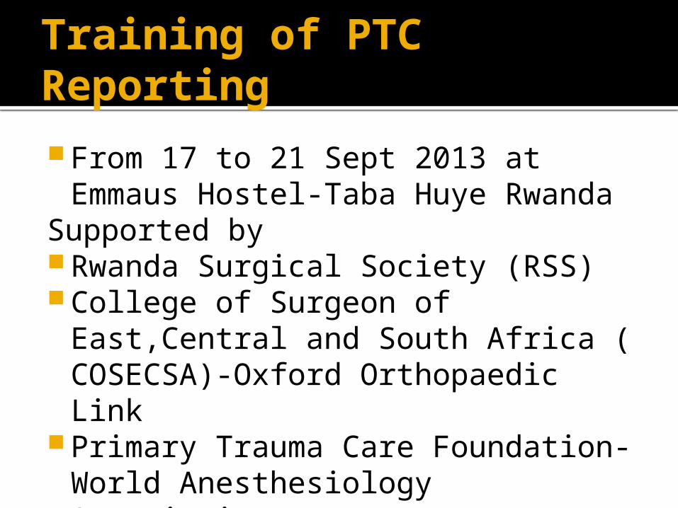
Training of PTC Reporting
From 17 to 21 Sept 2013 at Emmaus Hostel-Taba Huye Rwanda
Supported by Rwanda Surgical Society (RSS) College of Surgeon of East,Central
and South Africa ( COSECSA)-Oxford Orthopaedic Link
Primary Trauma Care Foundation-World Anesthesiology Association
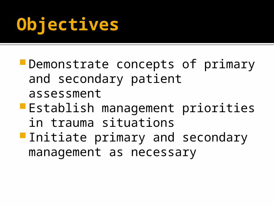
Objectives
Demonstrate concepts of primary and secondary patient assessment
Establish management priorities in trauma situations
Initiate primary and secondary management as necessary
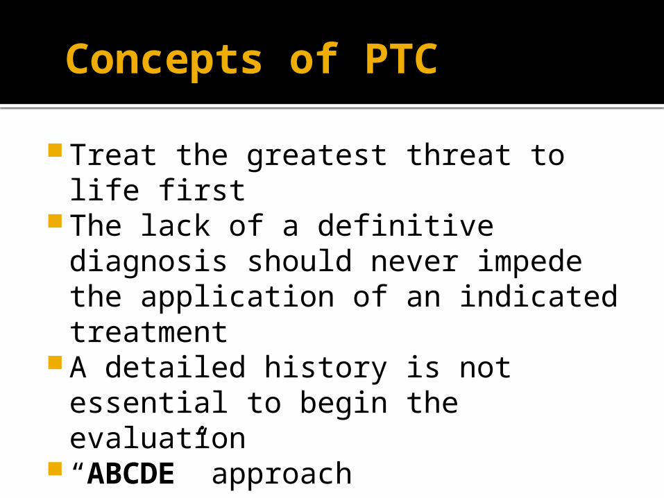
Concepts of PTC
Treat the greatest threat to life first The lack of a definitive diagnosis
should never impede the application of an indicated treatment
A detailed history is not essential to begin the evaluation
“ABCDE” approach
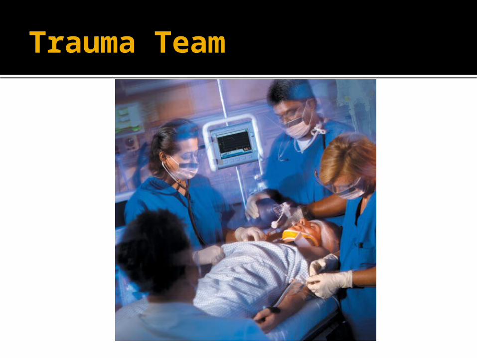
Trauma Team
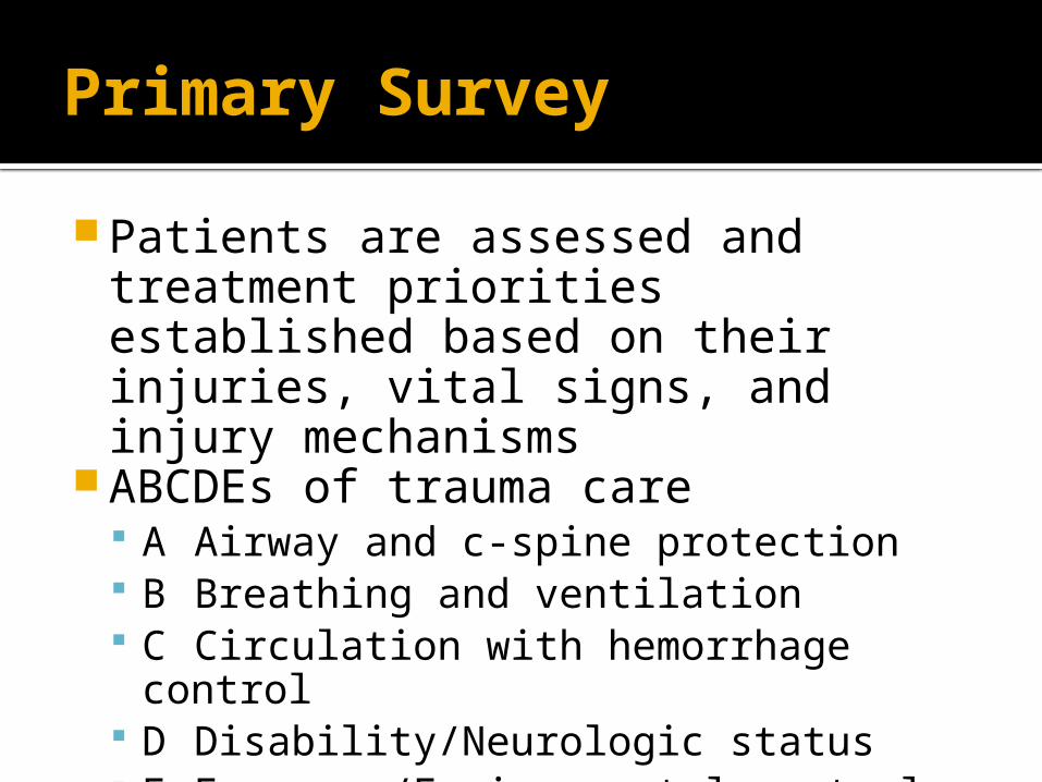
Primary Survey
Patients are assessed and treatment priorities established based on their injuries, vital signs, and injury mechanisms
ABCDEs of trauma care A Airway and c-spine protection B Breathing and ventilation C Circulation with hemorrhage control D Disability/Neurologic status E Exposure/Environmental control
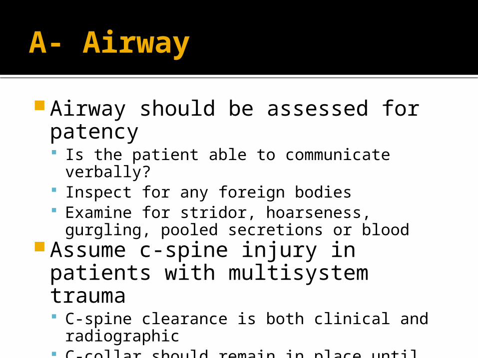
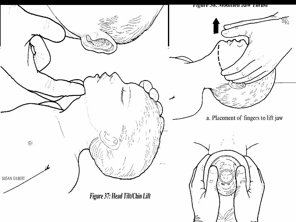
A- Airway
Airway should be assessed for patency Is the patient able to communicate verbally? Inspect for any foreign bodies Examine for stridor, hoarseness, gurgling,
pooled secretions or blood Assume c-spine injury in patients
with multisystem trauma C-spine clearance is both clinical and
radiographic C-collar should remain in place until patient can
cooperate with clinical exam
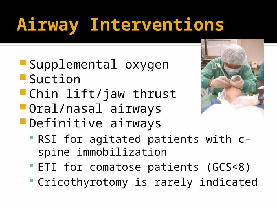
Airway Interventions
Supplemental oxygen Suction Chin lift/jaw thrust Oral/nasal airways Definitive airways
RSI for agitated patients with c-spine immobilization
ETI for comatose patients (GCS<8) Cricothyrotomy is rarely indicated
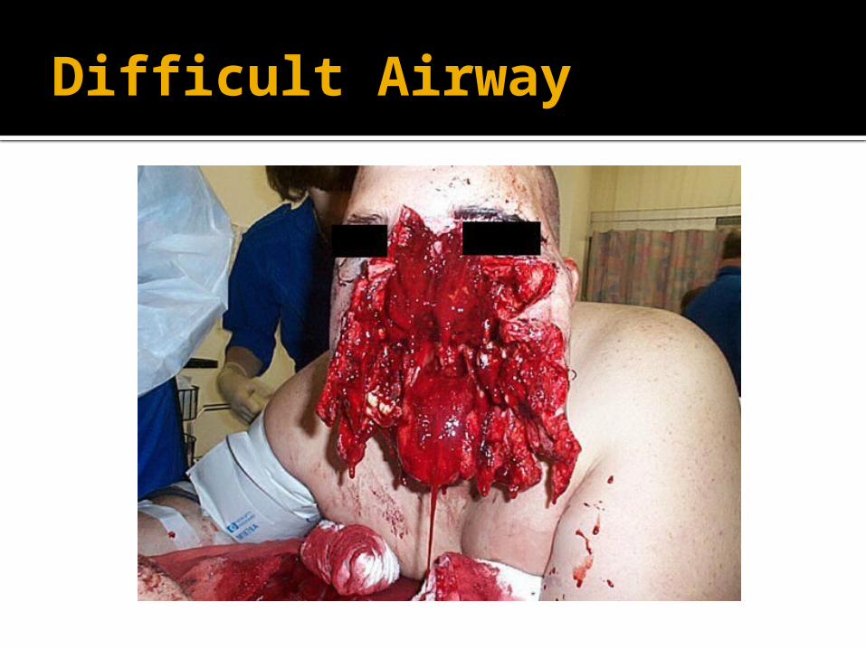
Difficult Airway
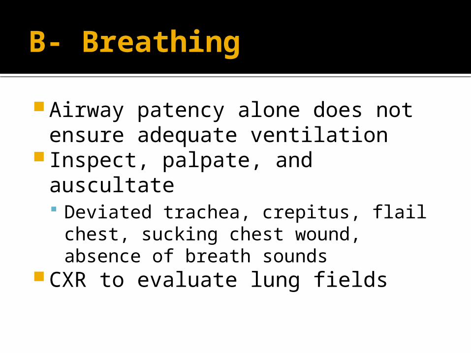
B- Breathing
Airway patency alone does not ensure adequate ventilation
Inspect, palpate, and auscultate Deviated trachea, crepitus, flail chest,
sucking chest wound, absence of breath sounds
CXR to evaluate lung fields
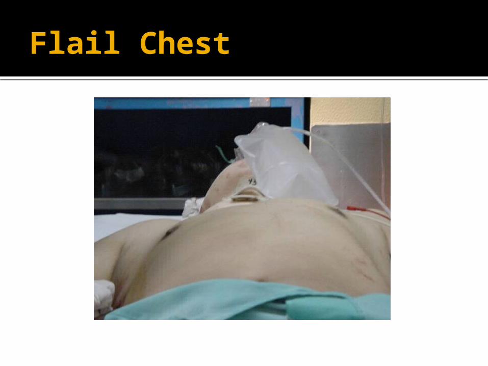
Flail Chest
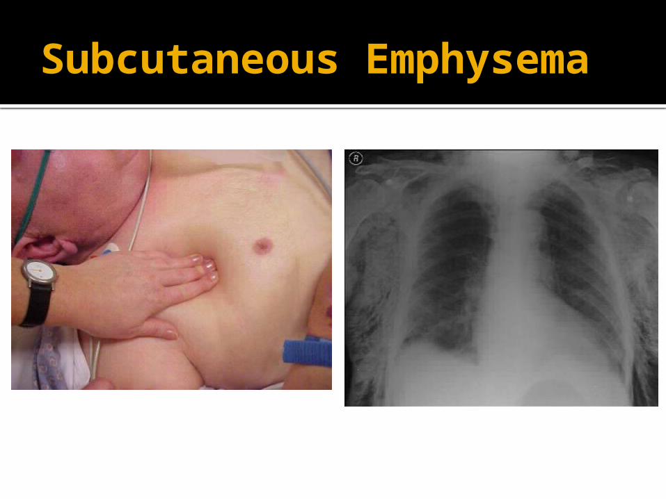
Subcutaneous Emphysema
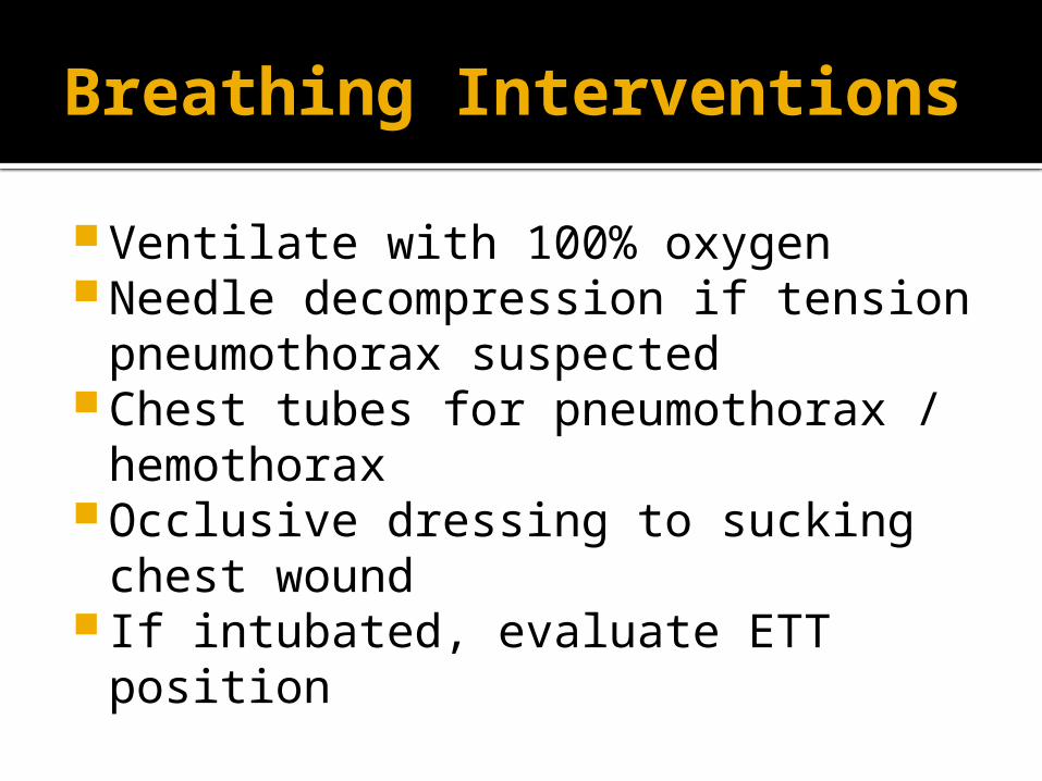
Breathing Interventions
Ventilate with 100% oxygen Needle decompression if tension
pneumothorax suspected Chest tubes for pneumothorax /
hemothorax Occlusive dressing to sucking chest
wound If intubated, evaluate ETT position
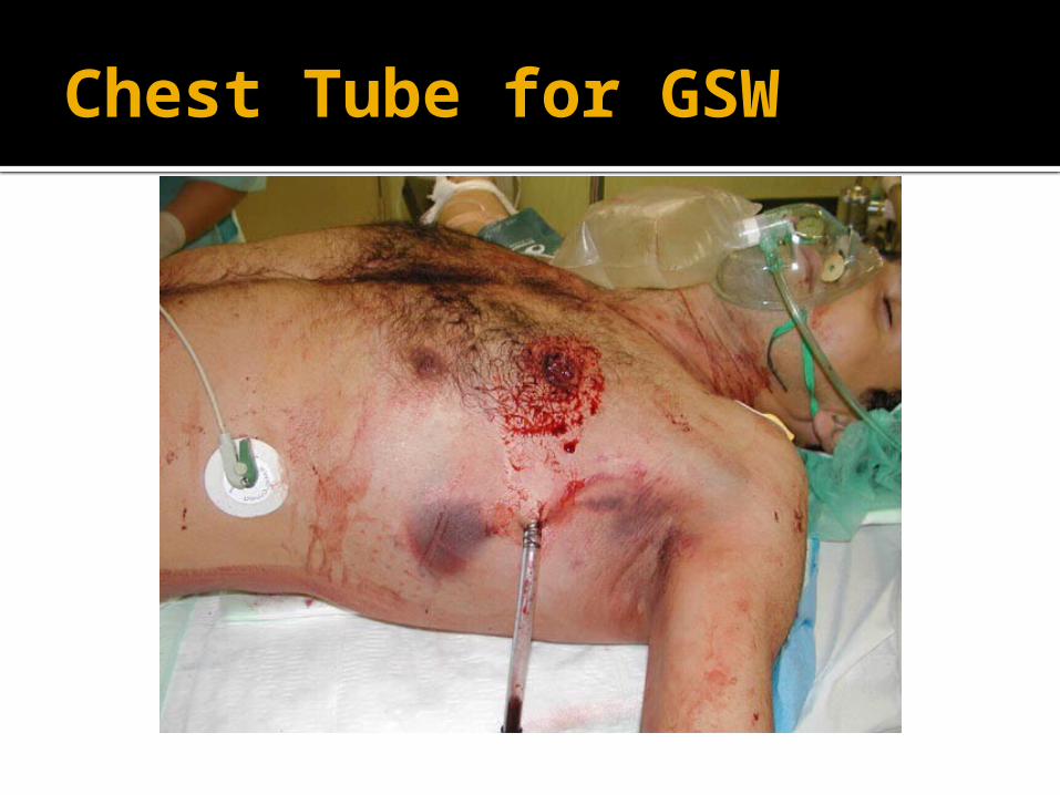
Chest Tube for GSW
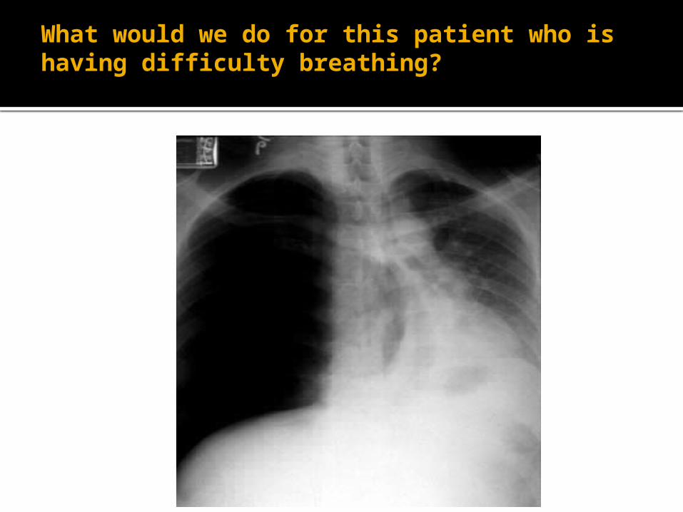
What would we do for this patient who is having difficulty breathing?
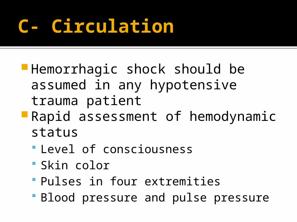
C- Circulation
Hemorrhagic shock should be assumed in any hypotensive trauma patient
Rapid assessment of hemodynamic status Level of consciousness Skin color Pulses in four extremities Blood pressure and pulse pressure
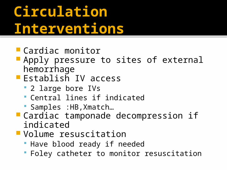
Circulation Interventions
Cardiac monitor Apply pressure to sites of external
hemorrhage Establish IV access
2 large bore IVs Central lines if indicated Samples :HB,Xmatch…
Cardiac tamponade decompression if indicated
Volume resuscitation Have blood ready if needed Foley catheter to monitor resuscitation
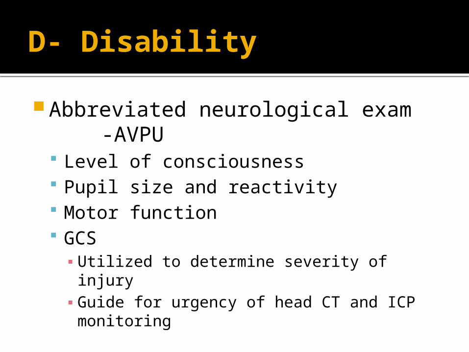
D- Disability
Abbreviated neurological exam -AVPU
Level of consciousness Pupil size and reactivity Motor function GCS ▪ Utilized to determine severity of injury▪ Guide for urgency of head CT and ICP
monitoring
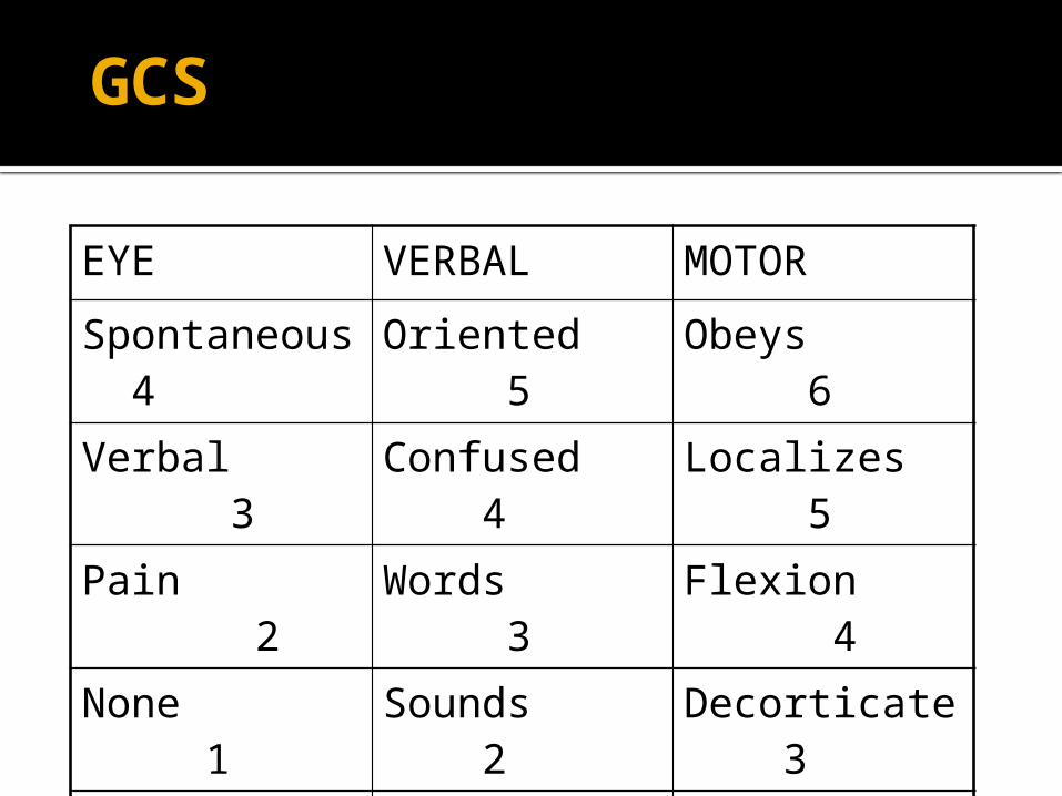
GCS
EYE VERBAL MOTOR
Spontaneous 4 Oriented 5 Obeys 6
Verbal 3 Confused 4 Localizes 5
Pain 2 Words 3 Flexion 4
None 1 Sounds 2 Decorticate 3
None 1 Decerebrate 2
None 1
Disability Interventions
Spinal cord injury High dose steroids if within 8 hours
ICP monitor- Neurosurgical consultation
Elevated ICP Head of bed elevated Mannitol Hyperventilation Emergent decompression
E- Exposure
Complete disrobing of patient Logroll to inspect back Rectal temperature Warm blankets/external warming
device to prevent hypothermia
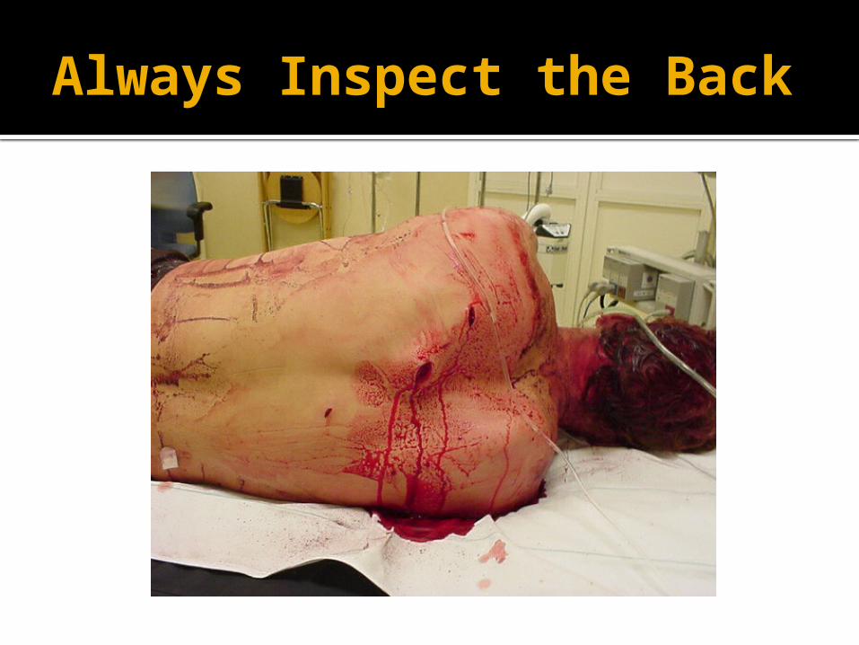
Always Inspect the Back
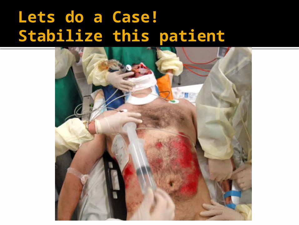
Lets do a Case!Stabilize this patient
Case
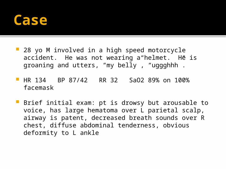
28 yo M involved in a high speed motorcycle accident. He was not wearing a helmet. He is groaning and utters, “my belly”, “uggghhh”.
HR 134 BP 87/42 RR 32 SaO2 89% on 100% facemask
Brief initial exam: pt is drowsy but arousable to voice, has large hematoma over L parietal scalp, airway is patent, decreased breath sounds over R chest, diffuse abdominal tenderness, obvious deformity to L ankle
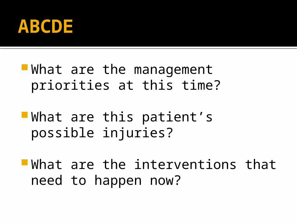
ABCDE
What are the management priorities at this time?
What are this patient’s possible injuries?
What are the interventions that need to happen now?
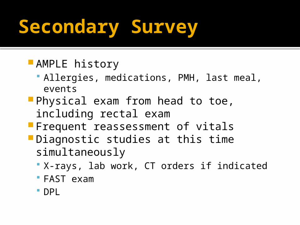
Secondary Survey
AMPLE history Allergies, medications, PMH, last meal, events
Physical exam from head to toe, including rectal exam
Frequent reassessment of vitals Diagnostic studies at this time
simultaneously X-rays, lab work, CT orders if indicated FAST exam DPL
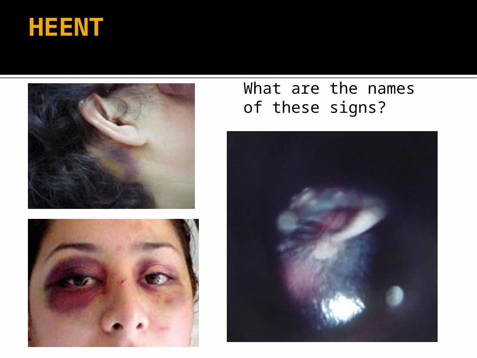
HEENT
What are the names of these signs?
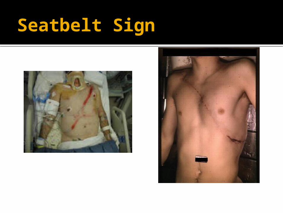
Seatbelt Sign
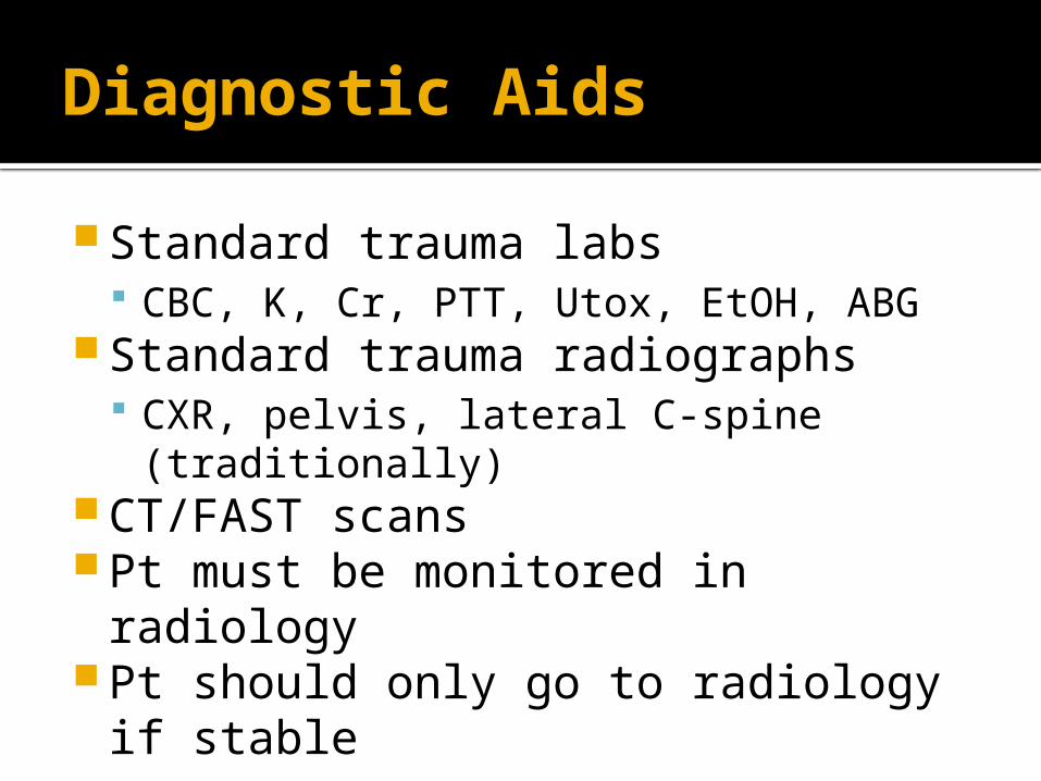
Diagnostic Aids
Standard trauma labs CBC, K, Cr, PTT, Utox, EtOH, ABG
Standard trauma radiographs CXR, pelvis, lateral C-spine (traditionally)
CT/FAST scans Pt must be monitored in radiology Pt should only go to radiology if
stable
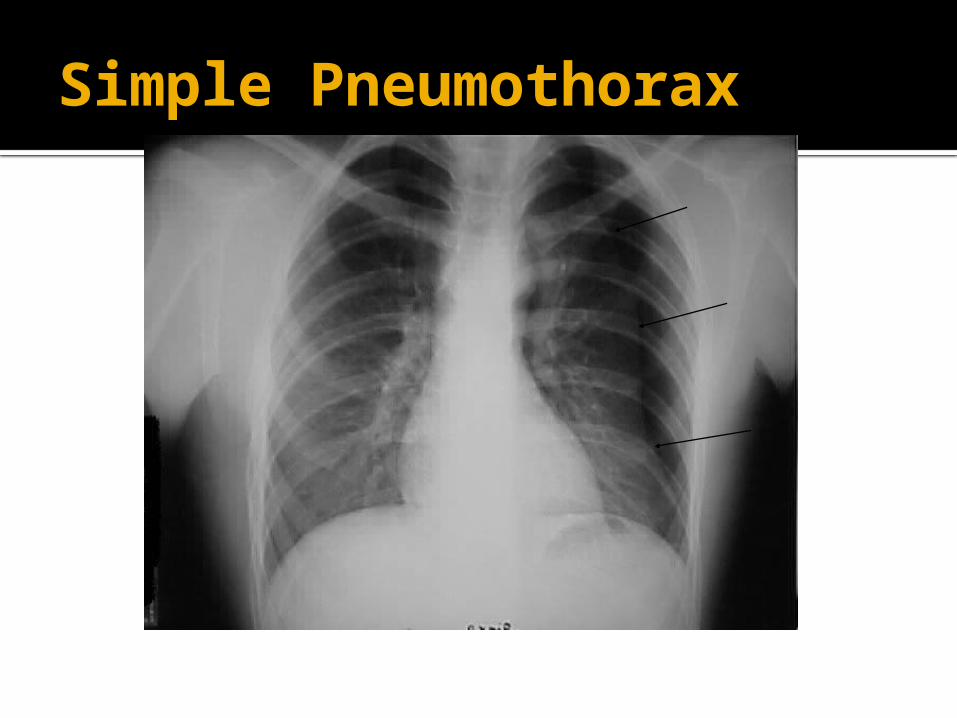
Simple Pneumothorax
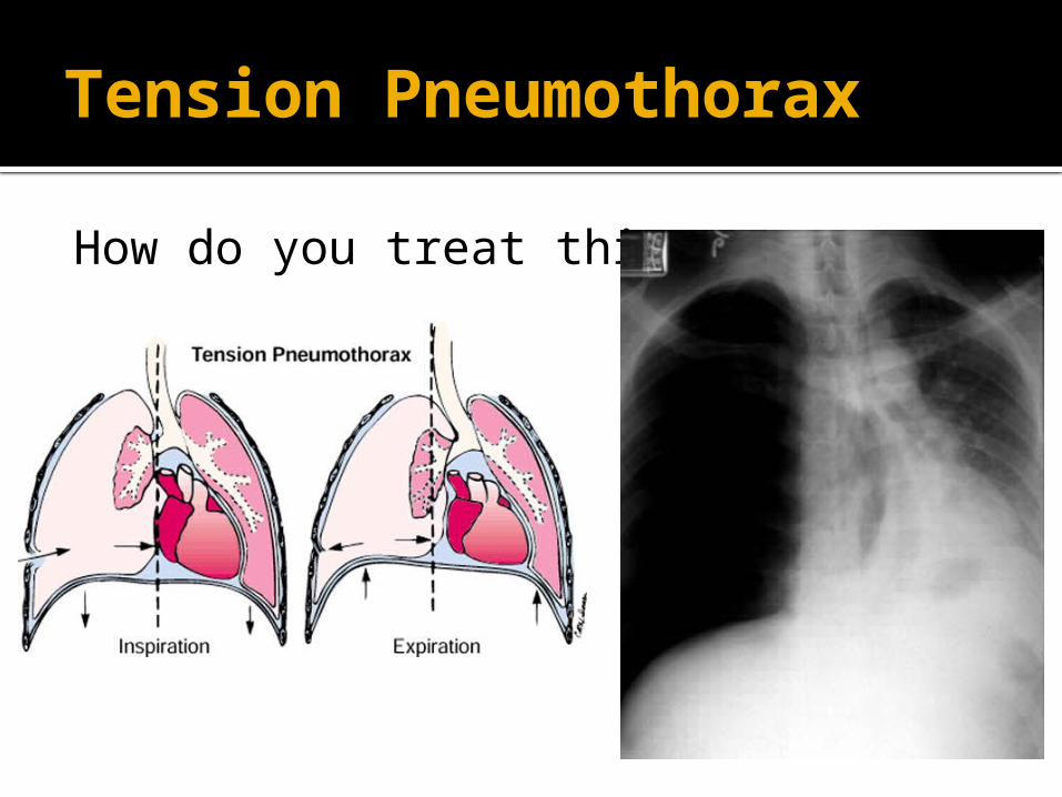
Tension Pneumothorax
How do you treat this?
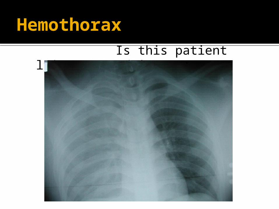
Hemothorax Is this patient lying or
upright?
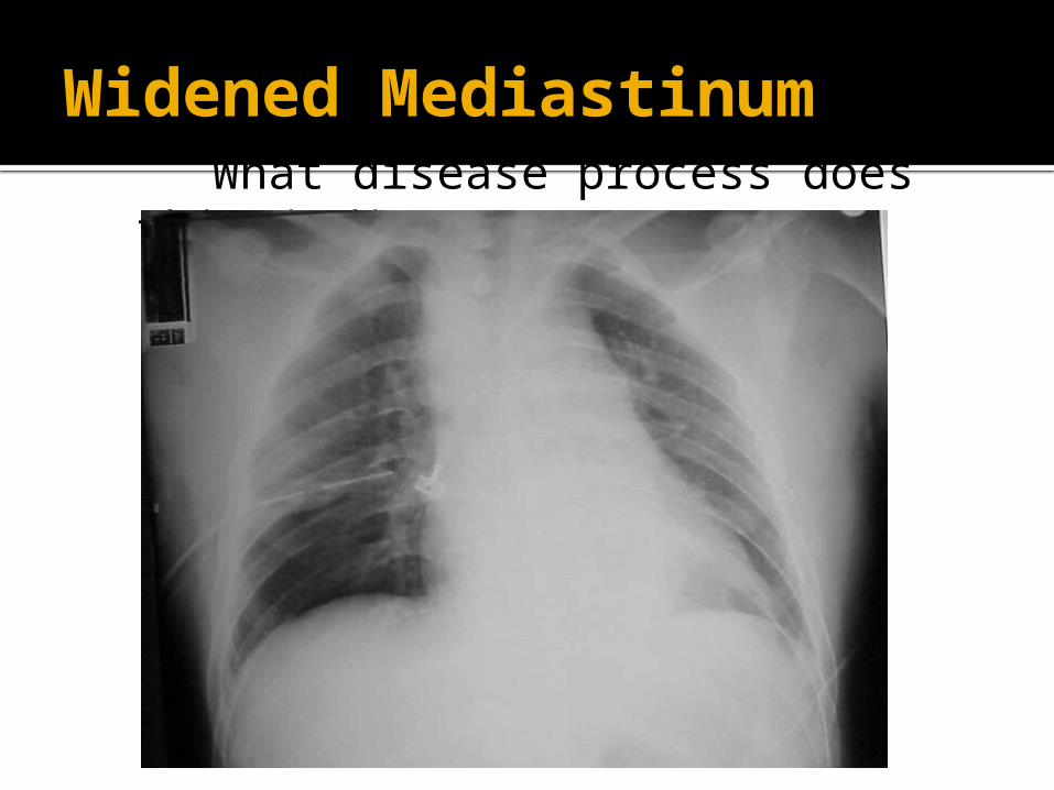
Widened Mediastinum What disease process does this
indicate?
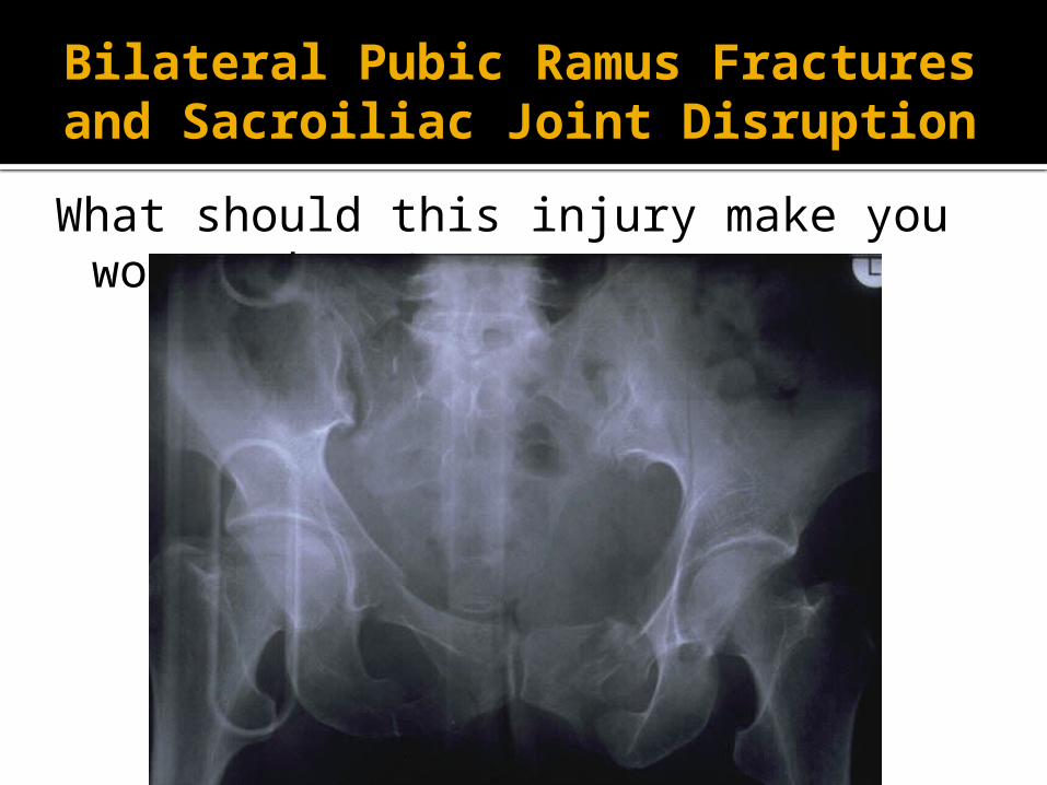
Bilateral Pubic Ramus Fractures and Sacroiliac Joint Disruption
What should this injury make you worry about?
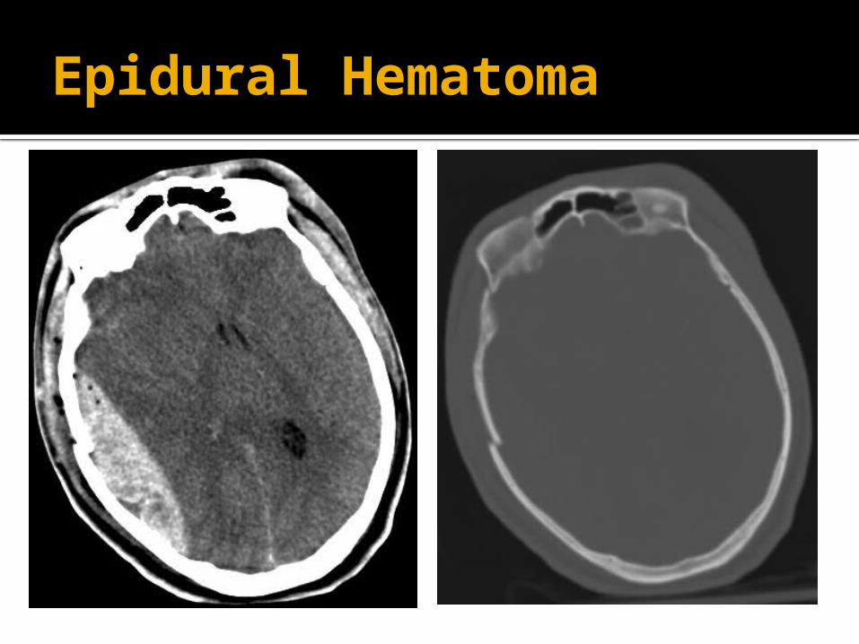
Epidural Hematoma
Abdominal Trauma
Common source of traumatic injury Mechanism is important
Bike accident over the handlebars MVC with steering wheel trauma
High suspicion with tachycardia, hypotension, and abdominal tenderness
Can be asymptomatic early on FAST exam can be early screening
tool
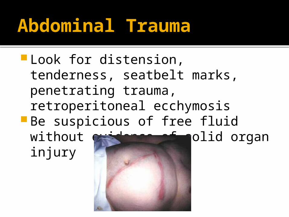
Abdominal Trauma
Look for distension, tenderness, seatbelt marks, penetrating trauma, retroperitoneal ecchymosis
Be suspicious of free fluid without evidence of solid organ injury
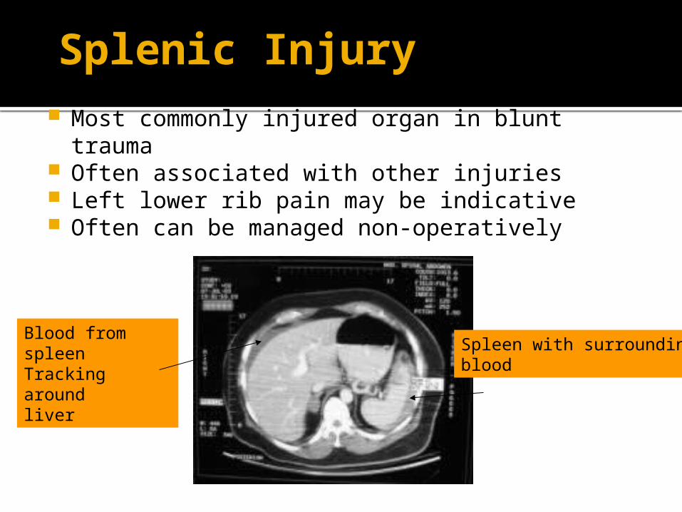
Splenic Injury Most commonly injured organ in blunt trauma Often associated with other injuries Left lower rib pain may be indicative Often can be managed non-operatively
Spleen with surroundingblood
Blood from spleenTracking aroundliver
Liver injury
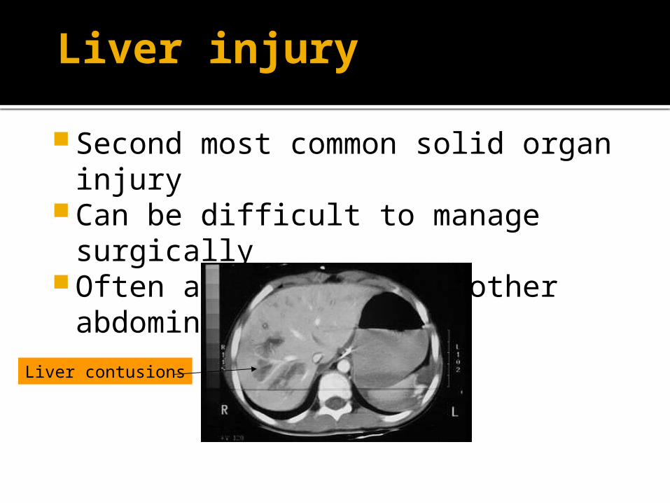
Second most common solid organ injury
Can be difficult to manage surgically Often associated with other
abdominal injuries
Liver contusions
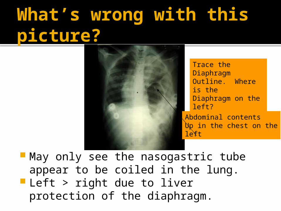
What’s wrong with this picture?
May only see the nasogastric tube appear to be coiled in the lung.
Left > right due to liver protection of the diaphragm.
Trace the Diaphragm Outline. Where is theDiaphragm on the left?
Abdominal contentsUp in the chest on theleft
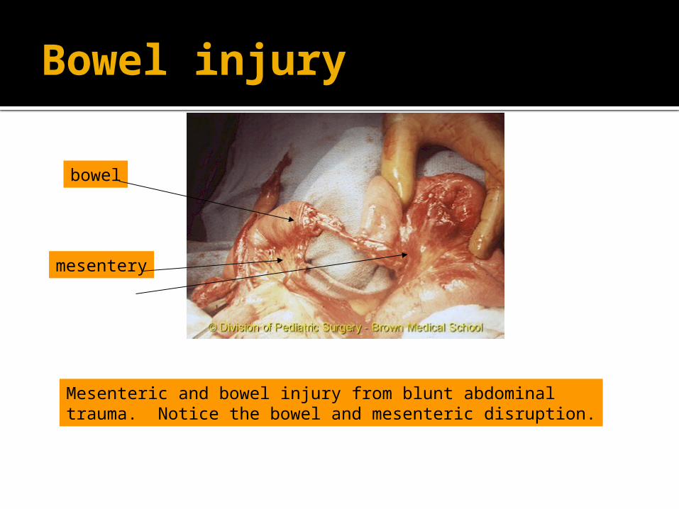
Bowel injury
Mesenteric and bowel injury from blunt abdominaltrauma. Notice the bowel and mesenteric disruption.
bowel
mesentery
CT Scan in Trauma
Abdominal CT scan visualizes solid organs and vessels well
CT does NOT see hollow viscus, duodenum, diaphram, or omentum well
Some recent surgery literature advocates whole body scans on all trauma Keep in mind that there is an increase in
mortality related to cancer from CT scans
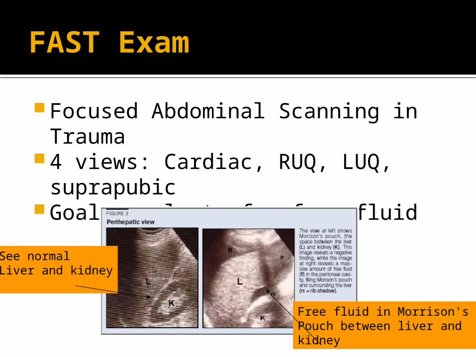
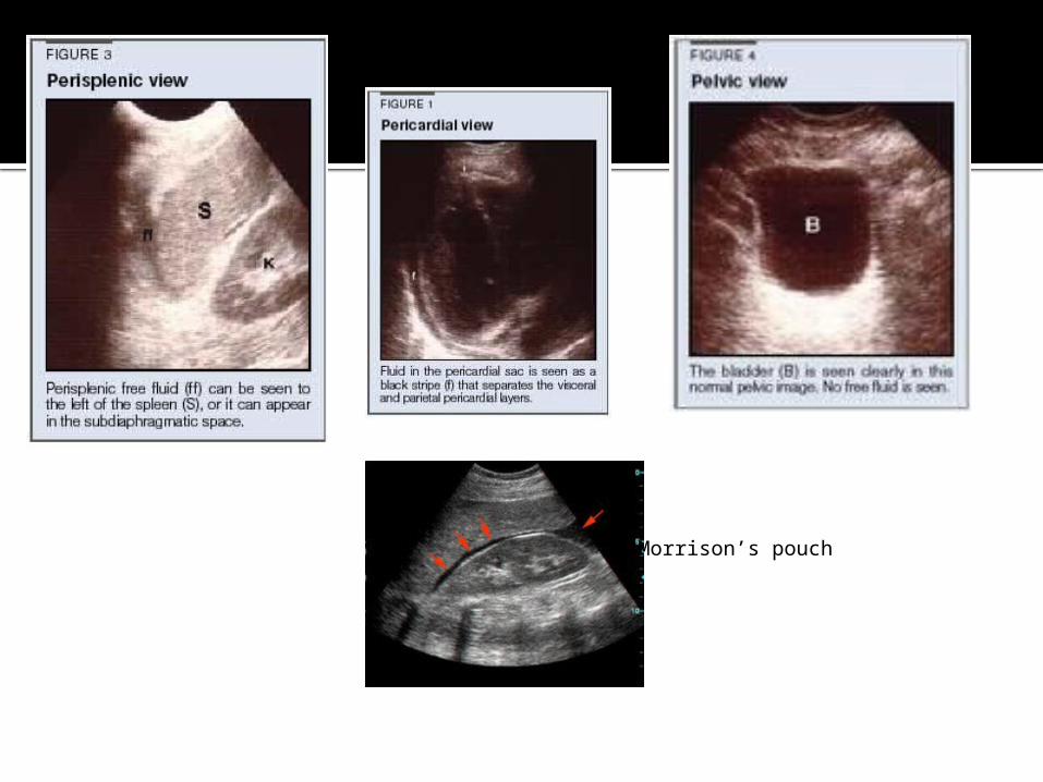
FAST Exam
Focused Abdominal Scanning in Trauma
4 views: Cardiac, RUQ, LUQ, suprapubic
Goal: evaluate for free fluid
See normalLiver and kidney
Free fluid in Morrison's Pouch between liver andkidney
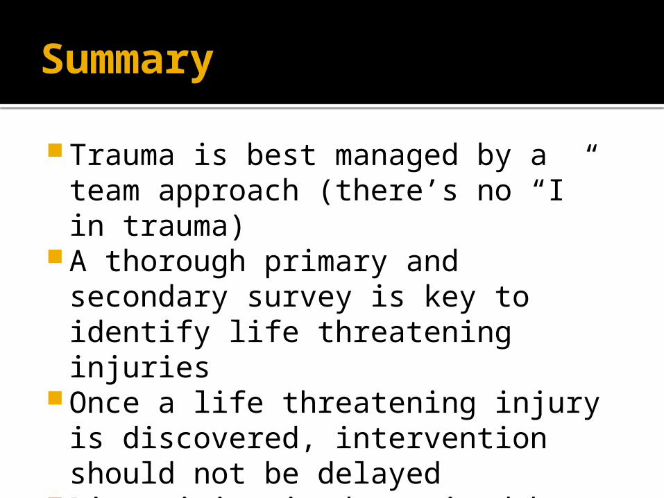
Summary
Trauma is best managed by a team approach (there’s no “I” in trauma)
A thorough primary and secondary survey is key to identify life threatening injuries
Once a life threatening injury is discovered, intervention should not be delayed
Disposition is determined by the patient’s condition as well as available resources.


Thank you