Primary structure of the aspartic proteinase A from ... res. commun. vol. 51, p. 27-41, 1986 primary...
15
Carlsberg Res. Commun. Vol. 51, p. 27-41, 1986 PRIMARY STRUCTURE OF THE ASPARTIC PROTEINASE A FROM SACCHAROMYCES CEREVlSlAE by THOMAS DREYER, BARBARA HALKIER, IB SVENDSEN and MARTIN OTTESEN Department of Chemistry, Carlsberg Laboratory, Gamle Cadsbergvej 10, DK-2500 Copenhagen Valby Keywords: Proteinase A, Saccharomyces cerevisiae, Saccharomyces carlsbergensis, amino acid sequence, immunology, N-linked carbohydrate Proteinase A was purified by an improved large scale procedure and split into fragments by means of trypsin, cyanogen bromide, hydroxylamine, and o-iodosobenzoic acid. On the basis of high degrees of homology with cathepsin D and pepsin its amino acid sequence was determined. Proteinase A contains 329 amino acid residues, and in addition 8.5% neutral sugar and 1%glucosamine, attached to asparagines in positions 67 and 267. Proteinase A contains two disulfide bonds, as opposed to three in mammalian aspartic proteinases. Comparison with the tertiary structure of pepsin indicates, that the two catalytically essential aspartic acid residues, and the residues corresponding to their surroundings, are conserved. The sequence shows 46% identity with porcine cathepsin D and 40% with porcine pepsin. An aspartic proteinase from Saccharomyces carlsbergensis had the same N-terminal 40 amino acid sequence as proteinase A. Immunological cross-reactivity between proteinase A and calfchymosin was demonstrated by immune blotting assay. 1. INTRODUCTION The occurrence ofproteolytic enzyme activity in an acid yeast lysate was recognized as early as 1889 (41), and the enzyme responsible for this activity, proteinase A, was isolated in 1967 (19, 30). The, enzyme is classified as an aspartic proteinase, due to its proteolytic activity at low pH and inhibition by 1,2-epoxy-3-(p-nitrophe- noxy)propane (EPNP) and diazoacetyl-D,L-nor- leucine methyl ester (DAN) (32, 49). Proteinase A is located in the lysosome-like vacuoles of the yeast, together with the serine proteases, protein- ase B and carhoxypeptidase Y (31). The three vacuolar enzymes have been implicated with unspecific proteolytic functions, e.g. protein degradation during nitrogen starvation (51). Proteinase A is a glycoprotein with a molecular weight of approx. 41,500, and the amino acid composition shows similarities with other aspar- tic proteinases: pepsin, penicillopepsin, cathep- sin D and chymosin (35). Cathepsin D, which is the only other intracellular aspartic proleinase that has been sequenced, exhibits structural features which are thought to be significant for processing and targeting of a lysosomal enzyme (12, 44). The primary structure ofproteinase A may generate some insight into the correspond- ing processes for vacuolar enzymes (11, 26). Abbreviations: DAN = diazoacetyl-norieucine methyl ester; DPCC = diphenyl carbamyl chloride; EPNP: 1,2-epoxy-3-(p-nitrophenoxy) propane; FPLC = fast protein liquid chromatography; HPLC = high performance liquid chromatography; PMSF = phenyl methylsulfonyl fluoride; SDS-PAGE = sodium dodecylsulphate-polyacryl- amide gel electrophoresis; TEA = triethylamine; TFA = trifluoroacefic acid. Springer-Verlag 0105-1938/86/0051/0027/$03.00
Transcript of Primary structure of the aspartic proteinase A from ... res. commun. vol. 51, p. 27-41, 1986 primary...

Carlsberg Res. Commun. Vol. 51, p. 27-41, 1986
PRIMARY STRUCTURE OF THE ASPARTIC PROTEINASE A
FROM SACCHAROMYCES CEREVlSlAE by
T H O M A S D R E Y E R , BARBARA H A L K I E R , IB S V E N D S E N and M A R T I N O T T E S E N
Department of Chemistry, Carlsberg Laboratory, Gamle Cadsbergvej 10, DK-2500 Copenhagen Valby
Keywords: Proteinase A, Saccharomyces cerevisiae, Saccharomyces carlsbergensis, amino acid sequence, immunology, N-linked carbohydrate
Proteinase A was purified by an improved large scale procedure and split into fragments by means of trypsin, cyanogen bromide, hydroxylamine, and o-iodosobenzoic acid. On the basis of high degrees of homology with cathepsin D and pepsin its amino acid sequence was determined. Proteinase A contains 329 amino acid residues, and in addition 8.5% neutral sugar and 1% glucosamine, attached to asparagines in positions 67 and 267. Proteinase A contains two disulfide bonds, as opposed to three in mammalian aspartic proteinases. Comparison with the tertiary structure of pepsin indicates, that the two catalytically essential aspartic acid residues, and the residues corresponding to their surroundings, are conserved. The sequence shows 46% identity with porcine cathepsin D and 40% with porcine pepsin. An aspartic proteinase from Saccharomyces carlsbergensis had the same N-terminal 40 amino acid sequence as proteinase A. Immunological cross-reactivity between proteinase A and calfchymosin was demonstrated by immune blotting assay.
1. I N T R O D U C T I O N The occurrence ofproteolytic enzyme activity
in an acid yeast lysate was recognized as early as 1889 (41), and the enzyme responsible for this activity, proteinase A, was isolated in 1967 (19, 30). The, enzyme is classified as an aspartic proteinase, due to its proteolytic activity at low pH and inhibition by 1,2-epoxy-3-(p-nitrophe- noxy)propane (EPNP) and diazoacetyl-D,L-nor- leucine methyl ester (DAN) (32, 49). Proteinase A is located in the lysosome-like vacuoles of the yeast, together with the serine proteases, protein- ase B and carhoxypeptidase Y (31). The three vacuolar enzymes have been implicated with
unspecific proteolytic functions, e.g. protein degradation during nitrogen starvation (51). Proteinase A is a glycoprotein with a molecular weight of approx. 41,500, and the amino acid composition shows similarities with other aspar- tic proteinases: pepsin, penicillopepsin, cathep- sin D and chymosin (35). Cathepsin D, which is the only other intracellular aspartic proleinase that has been sequenced, exhibits structural features which are thought to be significant for processing and targeting of a lysosomal enzyme (12, 44). The primary structure ofproteinase A may generate some insight into the correspond- ing processes for vacuolar enzymes (11, 26).
Abbreviations: DAN = diazoacetyl-norieucine methyl ester; DPCC = diphenyl carbamyl chloride; EPNP: 1,2-epoxy-3-(p-nitrophenoxy) propane; FPLC = fast protein liquid chromatography; HPLC = high performance liquid chromatography; PMSF = phenyl methylsulfonyl fluoride; SDS-PAGE = sodium dodecylsulphate-polyacryl- amide gel electrophoresis; TEA = triethylamine; TFA = trifluoroacefic acid.
Springer-Verlag 0105-1938/86/0051/0027/$03.00
T. DREYER et al.: Aspartic proteinase A
The present paper describes an improved purification procedure of proteinase A from Saccharomyces cerevisiae, based on anion-ex- change and hydrophobic-interaction chromato- graphy, and the complete sequence of 329 amino acid residues has been determined by automated Edman degradation of fragments obtained by chemical and enzymatic cleavages. The sequence ofproteinase A is compared to the known primary structures of porcine cathepsin D (44), human renin (21), porcine pepsin (43), bovine chymosin (13) and penicillopepsin (22), with emphasis on residues corresponding to catalytically and structurally important residues in the enzymes with known three-dimensional structure.
An aspartic proteinase was isolated from Sac- charomyces carlsbergensis, which had the same N-terminal amino acid sequence as proteinase A, and immunological studies revealed com- plete cross-reactivity between these two en- zymes.
2. MATERIALS Baker's yeast (Saccharomyces cerevisiae) was
obtained from De Danske Spritfabrikker, Den- mark, and Brewer's yeast (Saccharomyces cads- bergensis) was obtained from the Carlsberg Breweries, Denmark. Sephadex G100, Sepha- dex G200 (superfine), Phenyl-Sepharose and a "Mono Q" anion-exchange column were ob- tained from Pharmacia, Sweden. Bio-Gel P4, P6 and P60 were from Bio-Rad Lab., USA and DE-52 anion-exchange cellulose from What- mann, U.K. An affinity resin for carboxypepti- dase Y, ((N-e-aminocaproyl)-p-aminobenzyl) ~uccinyl-Sepharose (CABS-Sepharose) (24) and carboxypeptidase II from malt was kindly do- nated by K. BREDDAM. Hog pepsin, DPCC- treated trypsin, carboxypeptidase A and B, PMSF, sperm whale myoglobin, pepstatin, DAN and EPNP were purchased from Sigma, USA, and 2-vinylpyridine was from Janssen, Belgium. Extracts of calf chymosin, porcine pepsin and the aspartic proteinases from Mucor miehei, Mucor pusillus and Endothia parasitica, as well as the antisera against the first four of these enzymes, were a kind gift from Christian Hansens Laboratories, Denmark. Peroxidase
conjugated goat anti-rabbit antibodies were ob- tained from Dako, Denmark. All other che- micals were analytical grade.
3. METHODS 3.1. Isolation and characterization of aspartic
proteinases from yeast 3.1.1. Aspartic proteinase from Saccharomyces
carlsbergensis An aspartic proteinase from Saccharomyces
carlsbergensis was purified as earlier described (5), by a modification of MEUSSDOERvVeR et al.'s procedure (35). The enzyme was stored in solution, 5 mg/ml in 0.05 M-ammonium acetate pH 5.0, at -23 ~
3.1.2. Proteinase A from Saccharomyces cerevisiae
Proteinase A from Saccharomyces cerevisiae was obtained by a simplified procedure, omit- ting the ammonium sulfate precipitation and addition of mercuric chloride in the earlier procedure (5). The initial steps followed the procedure for isolating carboxypeptidase Y (CPD-Y) (24): 15 kg of baker's yeast was crushed and mixed with 1.5 1 ether, which liquefied the yeast, 15 1 water was added, pH was adjusted to 7.4, and cell autolysis was allowed to proceed for 21 hours at 20 ~ After centrifugation at 7500xg for 30 min, pH of the supernatant was adjusted to 5.0 with 30% (v/v) acetic acid, and the solution was kept at 30 ~ for 20 hours with stirring. This treatment activated proteinase A and after clarification by centrifugation, CPD-Y was removed from the solution by passing it through a 20 ml CABS-Sepharose column, equilibrated with 10 mM-sodium acetate, pH 5.0. This and all subsequent steps were per- formed at 4 ~ The run-off (25 1) was dialysed against 10 raM-sodium acetate, pH 5.0, and concentrated to 10 1 in a Millipore Pellicon Ultrafiltration system, with a PTGC membrane with cut-off level 10,000 dalton. Two liters of anion-exchange cellulose DE 52, was added and after stirring for 20 min the gel was transferred to a column (10x25 cm), and washed with 10 1 of 10 mM-sodium acetate pH 5.0. Enzymatic activity was eluted in one step with 0.3 M-KCI in the
28 Carlsberg Res. Commun. Vol. 51, p. 27-41, 1986

T. DREYER et al.: Aspartic proteinase A
same buffer. Fractions containing enzymes were diaconcentrated to 1250 ml against 10 mM-SO- dium acetate, pH 5.0, and applied to a second DE 52 column (10x 12.5 cm), equilibrated with the same buffer. The column was eluted with a 10 1 linear gradient from 0-0.3 M-KC1 in the buffer, flow 400 ml/h. Fractions containing acid proteolytic activity were pooled and applied.at 1000 ml/h directly to a Phenyl-Sepharose col- umn (10x 12.5 cm), which had been equilibrated with 50 mM-sodium acetate, pH 5.0. The gel was washed with 5 1 of 50 mM-sodium acetate, pH 5.0, and eluted with a 3 1 linear gradient from 0-90% (v/v) ethylene glycol in the same buffer, flow 200 ml/h. Fractions corresponding to the UV-absorbing peak eluted with 85% (v/v) ethylene glycol were pooled and diaconcen- trated against 10 mM-sodium acetate, pH 5.0, to 50 ml on a high pressure Amicon cell with a YM 10 membrane. Finally the concentrate was ap- plied to a Sephadex Gl00 column (2.5x90 cm) in the same buffer, 9 ml/h. Fractious with acid proteolytic activity were pooled and concen- trated before storage in solution at -18 ~
3.1.3. Molecular weight and isoelectric point determination
The molecular weights ofproteinase A and the Saccharomyces carlsbergensis proteinase were estimated by gel filtration on a Sephadex G 200 (superfine) column (1.6• cm) in 0.1 M-am- monium acetate pH 5.5, and by electrophoresis in a 10% (w/v) SDS-polyacrylamide gel (29).
Isoelectric focusing was performed in a 0.5 m m 5% (w/v) polyacrylamide slab gel contain- ing a pH gradient from pH 2.5 to 5.0 created by ampholytes. Polymerization of the acrylamide in this pH interval was made possible by the addition of 0.3% (w/v) silver nitrate. Marker proteins were used to standardize the methods.
3.1.4. Amino acM analysis Samples were hydrolyzed with 6 N-HCI at 110
~ in evacuated tubes for 24 hours, in some cases also for 48 and 72 hours. Hydrolysates were analyzed by a Durrum D500 amino acid ana- lyzer. Tryptophan was determined by the meth-
od of EDELHOCH (10) and halfcystine as cysteic acid after performic acid oxidation according to HIRS (20).
3.1.5. Carbohydrate analysis An estimation of the total content of neutral
carbohydrate in proteinase A and the Saccharo- myces carlsbergensis proteinase was obtained by means of the phenol-sulphuric acid procedure (7), using mannose as standard. Glucosamine was determined on the Durrum amino acid analyzer after hydrolysis in 6 N-hydrochloric acid at 110 ~ in vacuo for 2, 4 and 6 hours, using glucosamine as standard.
3.1.6. Activity measurements The proteolytic activities of proteinase A and
the aspartic proteinase from Saccharomyces carlsbergensis were measured with acid dena- tured tritium-labeled bovine hemoglobin, as described by DREVER et al. (5). A 125 gtl sample, diluted in 0.1 M-glycine-HCl buffer, pH 3.5, 37 ~ was added to 125 gtl 2% (w/v) tritium labelled hemoglobin in this buffer. After 30 rain at 37 ~ 250 ~tl 10% (v/v) trichloroacetic acid, 4 ~ was added. The mixture was kept at 4 ~ for 45 min to complete precipitation. After centrifugation 400 gtl supernatant was mixed with 5 ml Dimi- lume 30 and counted in a liquid scintillation counter. Since different batches of hemoglobin showed variable digestability a sample of pu- rified homogenous proteinase A was used to standardize each assay, and the activity is ex- pressed as gtg proteinase A.
3.1.7. Immunological procedures Rabbit antiserum raised against purified pro-
teinase A from S. cerevisiae, was produced and isolated as described by HARBOE and 1NGJLD (17). The antiserum was specific for proleinase A, as judged from the occurrence of a single immune stained band on a nitrocellulose filter with yeast extract, electroblotted from SDS- PAGE. This antiserum was used to examine proteinase A and the Saccharomyces carIsberg- ensis proteinase for immunological homology by tandem crossed immunoelectrophoresis (28),
Carlsberg Res. Commun. Vol. 51, p. 27-41, 1986 29
T. DREYER et al.: Aspartie proteinase A
in 1% (w]v) agarose gel in Tris-veronal buffer pH 8.6, containing antiserum, 0.6 ~tl per cm 2. Elec- trophoretic blotting and immune assay was performed by means of SDS-PAGE according to LAEMMLI (29). Immediately ~fter completion of the electrophoretic run, the polypeptides were transferred to a nitrocellulose filter, as described by TOwmN et al. (50). The nitrocellulose filter was cut into strips, and incubated with antibo- dies at pH 10.2, according to BJERRUM et at. (2). Bands were visualized using peroxidase conju- gated goat anti-rabbit antibodies and 3-amino- 9-ethylcarbazole as described by GRAHAM et al. (15).
3.2. Amino acid sequence 3.2.1. HPLC-separation of peptides
Peptides from enzymatic and chemical cleav- ages of proteinase A were purified by reverse- phase HPLC applying a 7.8• mm steel column with Synchropak RP-P wide pore C- 18 material, packed according to the dynamic slurry packing technique described by KELLER el al. (27). 2-10 mg peptide mixtures were separated employing Waters HPLC equipment: two Model 600A pumps, a WISP sample injec- tor, a Model 660 solvent programmer, a Model 450 var. wavelength UV detector and a Model 420 fluorescence detector together with an LKB 2210 2-channel recorder and an LKB 2211 "Superrac" fraction collector. Gradient elution was achieved with continuously increasing con- centrations of acetonitril in 0.05% (v/v) TFA, 0.05% (v/v) TEA. The eluent was monitored at 220 nm, supplemented with fluorescence detec- tion at 375 nm after excitation at 280 nm, to detect tryptophan containing peptides. Peaks conttaining more than one peptide were rechro- mat0graphed either in the same system with a flatter gradient, by using a Novapak column with gradients of acetonitril in 0.1% TFA, or by employing the Pharmacia FPLC system with Mono Q anion-exchanger and 20-30 min gra- dients from 0.05 - 0.4 M-ammonium bicar- bonate, pH 7.9, flow I ml/min.
3.2.2. Reduction and 2-pyridylethylation Proteinase A, 200 mg in 10 ml 7 M-guanidi-
nium-HC1, 0.2 M-Tris, pH 7.6, 5 mM-EDTA,
was flushed with nitrogen and reduced by adding 2 ml 0.23 M-dithiothreitol. After 30 min the cysteine residues were alkylated with 3x70 ~tl 2-vinylpyridine (14). After 2 hours the pH of the solution was adjusted to 4.0 with acetic acid, and it was dialysed against 2% acetic acid with 5% ethanol. A precipitate was formed, and the suspension was lyophilized.
3.2.3. Chemical cleavages Cyanogen bromide cleavages of reduced and
alkylated proteinase A, or tryptic peptides, were performed in 70% (v/v) trifluoroacetic acid at 21 ~ for 20 hours with a 250 fold molar excess of CNBr over methionine. 2 M-hydroxylamine was used to cleave Asn-Gly bonds (3) in 6 M-guanidi- nium hydrochloride, adjusted and kept at pH 9.0 with LiOH, during 4 hours reaction at 45 ~ After desalting on Bio-Gel P4 in 0.05 M-ammo- nium bicarbonate the fragments were separated by reverse-phase HPLC using Synchropak RP-P and gradients of acetonitril in 0.1% (v/v) TFA.
Cleavage after tryptophans was performed using o-iodosobenzoic acid (33), 20 mg/ml, and tyramine hydrochloride, 4 mg/ml, in 4 M-guani- dinium hydrochioride and 80% (v/v) acetic acid. After reaction of the enzyme, 3.5 mg/ml, with this mixture at 21 ~ for 22 hours in the dark, the solution was desalted on Bio-Gel P6 in 30% (v/v) acetic acid, and the fragments were separated on Bio-Gel P60 (lx90 cm) in 0.2 M-ammonium bicarbonate.
3.2.4. C-terminal determination The C-terminal sequence was established af-
ter addition of carboxypeptidase A (3 ~tM) to proteinase A (500 pM) in 0.05 M-N-ethylmor- pholine buffer, pH 8.5. Aliquots were withdrawn after 2, 6, 20 and 60 min, 5 ~tl 1 M-HC1 was added to each aliquot to stop the hydrolysis. Carboxy- peptidase B was then added (3 pM) and aliquots withdrawn after further 2, 6, 20 and 60 min incubation time for inactivation with 5 pl 1 M-HC1. Liberated amino acids were determined in samples applied directly to the amino acid analyzer.
A tryptic peptide T5 (410 ~tM) was digested with malt carboxypeptidase II (1.6 ~tM) in 0.05
30 Carlsberg Res. Commun. Vol. 51, p. 27-41, 1986
T. DREYER et al.: Aspartic proteinase A
M-sodium acetate pH 4.0, and 30 ~tl aliquots were withdrawn after 5, 25 and 120 min. To each of these 5 ~tl 1 M-HCI was added before direct injection in the amino acid analyzer.
3.2.5. Trypsin digestions Two procedures were used. In the first 100 mg
reduced and 2-pyridylethylated proteinase A was dissolved in 20 ml 0.1 M-sodium bicar- bonate, pH 8.0, and digested with 1 mg DPCC- trypsin at 37 ~ for 3 hours. In the second the buffer used was 0.02 M-N-ethylmorpholine-ace- tic acid, pH 8.0, DPCC-trypsin was added in the ratio 1:50 and digestion proceeded for 30 rain at 12 ~ (16). In both cases the reaction was terminated by the addition of PMSF in 50% excess.
Modification of the primary amino groups of lysine residues in reduced and alkylated protein- ase A with citraconic anhydride was performed according to the procedure of DIXON and PERHAM (4), and the subsequent tryptic diges- tion took place in 0. l M-sodium bicarbonate, pH 8.0, 37 ~ for 3 hours with an enzyme to substrate ratio of 1:100.
3.2.6. Isolation of tryptic peptides Peptides from tryptic digestion of reduced,
2-pyridylethylated proteinase A were separated on a column (2.6• cm) ofBio-Gel P60 in 0.2 M-sodium bicarbonate pH 7.6. Four resolved peptide pools were obtained, three of which were largely soluble after lyophilization in 0.1% (v/v) TFA, 5% (v/v) acetic acid, whereas a small high molecular weight pool was insoluble even in 30% (v/v}. acetic acid. Subsequent isolation of peptides from the three pools were performed with semi-preparative reverse-phase HPLC (see section 3.2.1). All peptides were lyophilized immediately after the chromatographic runs.
3.2. 7. Sequencing Amino acid sequences were determined either
with a Beckman 890C spinning-cup sequencer, (9, 25), or with an Applied Biosystems gas phase sequencer Model 470A, using the programme provided by the company. Phenylthiohyd-
antoins were identified by reverse-phase HPLC, as described by SVENIBSEN et al. (48).
4. RESULTS 4.1. Isolation of proteinase A from
Saccharomyces cerevisiae The purification procedure outlined in sec-
tion 3.1.2, is a simplification and improvement of the purification procedure previously used for the aspartic proteinase from Saccharomyces carlsbergensis (5). Ammonium sulfate precipita- tion is replaced by a diafiltration step after the ether plasmolysis. Hydrophobic-interaction chromatography preceeds anion-exchange in the earlier procedure, while the order is reversed in the present procedure, to take advantage of the higher binding capacity of the DE cellulose.
The purification procedure of proteinase A includes an activation step where the specific proteinase A inhibitor I ~ is digested by other proteolytic enzymes in the yeast (40). In order to obtain high yields, it is important to separate proteinase A from these other proteolytic en- zymes immediately after the activation. Carbo- xypeptidase Y is removed with the CABS-Se- pharose, and the second anion-exchange on DE 52, separates proteinase A from an earlier dub ing peak containing proteinase B, as judged by the ability to release a 520 nm absorbing trichlo- roacetic acid soluble dye from the substrate azocoll at pH 7.0 (39). Hydrophobic-interaction chromatography on Phenyl-Sepharose removes most of a dark brown pigment which does not bind to the column under the conditions de- scribed. The final gel filtration isolates protein- ase A from a high molecular weight brown contaminant, which is presumably an aggregate ofproteinase A and pigments, as judged from the amino acid composition, which is similar to proteinase A. Lyophilization of proteinase A is avoided, as this frequently leads to inactivation of the enzyme. The purification procedure is summarized in Table I.
The stability ofproteinase A in frozen solution at 5-25 mg/ml is high, the specific activity remains almost constant for 6 months.
During later purifications, run-offs from an affinity column used for commercial production of carboxypeptidase Y, were kindly donated by
Carlsberg Res. Commun. Vol. 51, p. 27-41, 1986 31
T. DREYER et al.: Aspartic proteinase A
Table I. Purification of proteinase A from Saccharomyces cerevisiae
Volume Protein ml mg
Spec. act. Yield IJg prot.A/ % mg protein
Activated extract, pH 5.0 25,000 307,500 Run-off from CABS-Sepharose 25,000 307,500 After ultrafiltration 10,000 84,300 DE 52 anion-exchange, one step elution 1,250 9,000 DE 52 anion-exchange, gradient elution 2,100 4,120 Phenyl-Sepharose 50 680 Sephadex G 100 41 430
16 100 16 100 19 30 86 14
150 11 691 9
1000 8
Pharmacia, Denmark. This run-offwas equiva- lent to the above described in all subsequent purification steps, and resulted in a high yield of pure proteinase A.
4.2. Isolation of aspartic proteinase from S. carlsbergensis
The amount ofcarlsbergensis enzyme isolated from each kg of yeast was approx, half o f the amount ofproteinase A isolated from Saccharo- myces cerevisiae. This was partially due to a very high content of dark pigments that bound irre- versibly to chromatographic columns and filters leading to reduced capacity, prolonged separa- tion times, and increased losses, and partially to a lower content of aspartic proteinase in the S. carlsbergensis. The specific activity of the earls- bergensis enzyme, based on amino acid analysis, was approx. 20% lower than the activity of proteinase A, probably because the preparation r some denatured or damaged enzyme.
found after acid hydrolysis. These values for proteinase A are in close agreement with those reported for this enzyme by MEUSSDOERFFER et al. (35).
In the isoelectric focusing gel, proteinase A and the aspartic proteinase from Saccharomyces carlsbergensis both displayed 3-4 distinct bands in a narrow range between pH 4.0 and pH 4.1, which is approx. 0.4 pH units lower than the earlier reported isoelectric point ofproteinase A (35).
~! ~i ~
4.3. Physical and chemical properties SDS-PAGE indicated molecular weights of
proteinase A and the aspartic proteinase from Saccharomyces carlsbergensis of 41,000 and 43,000, respectively, while gel filtration resulted in 43,000 and 45,000, respectively. The content of neutral carbohydrate was estimated to 10% (w/w) in the cerevisiae enzyme and 8.5% (w/w) in the carlsbergensis enzyme while glucosamine contents of 1.0% and 1.7%, respectively, were
I II
Figure 1. Tandem crossed immunoelectrophoresis of proteinase A from Saccharomyces cerevisiae and as- partic proteinase from Saccharomyces carlsbergensis, using antiserum against proteinase A. Well I contained 23 lag proteinase A from Saccharomyces cerevisiae and Well II contained 7.5 ~tg Saccharomyces carlsbergensis proteinase.
32 Carlsberg Res. Commun. Vol. 51, p. 27-41, 1986
T. DREYER et al.: Aspartic proteinase A
prot.A
1 2 3 4 5 Figure 2. Cross-reactivity between proteinase A and rabbit antisera raised against other aspartic protein- ases. l: Amidoblack staining ofproteinase A on nitro- cellulose filter. 2: Immuno-staining of proteinase A after incubation with antiserum against the aspartic proteinases from Mucor miehei (2) and Mucor pusillus (3), bovine chymosin (4) and porcine pepsin (5).
The amino acid compositions ofproteinase A (not shown) was similar to the earlier reported composition (35). The proteolytic activity of proteinase A towards acid denatured hemoglo- bin was inhibited by DAN, EPNP, pepstatin and I~, as previously observed (35). The aspartic proteinase from Saccharomyces carlsbergensis revealed similar behaviour in inhibition assays (results not shown). The pH optimum towards acid denatured hemoglobin was 3.2 for both enzymes, and they retained more than 80% of the initial activity after incubation for 48 hours at pH between 3.5 and 7.0 (results not shown).
E r o r
p 0 . 6
i.iJ Z 0 0 . 4
m w- 0 0 . 2 m I• I I I
1 0 0 3 0 0 V O L U M E (ml)
5 0 0
Figure 3. Elution profile oftryptic digest of reduced and alkylated proteinase A (37 ~ 3 hours) on Bio Gel P60 (2.5x90 cm) in 0.2 M-sodium bicarbonate. Flow 9 ml/h.
4.4. Immunological properties Proteinase A and the Saccharomyces cads-
bergensis proteinase formed, in tandem crossed immunoelectrophoresis with the proteinase A antiserum, immune precipitates which comple- tely fused, and thus suggested identity between the two enzymes (see Figure 1).
The immunological relationship between proteinase A and other aspartic proteinases was investigated by immune blotting. Proteinase A was allowed to react with antisera against calf chymosin, porcine pepsin and aspartic protein- ases from Mucor miehei and Mucor pusillus. Only the anti-chymosin antiserum cross- reacted, indicating common antigenic deter- minants on proteinase A and chymosin (see Figure 2). However, antiserum raised against proteinase A did not cross-react with calf chy- mosin, porcine pepsin or the aspartic protein- ases from Mucor miehei, Mucor pusillus and Endothia parasitica.
4.5. Amino acid sequences Reduced and vinylpyridinylated proteinase A
was sequenced through residue 44 on the Beck- man spinning-cup sequencer, with the sequence shown in Figure 6. The N-terminal sequence of aspartic proteinase from Saccharomyces cads-
Carlsberg Res. Commun. Vol. 51, p. 27-41, 1986 33

T. DREYER et al.: Aspartic proteinase A
T, T i A 0.05
18 22 26 30 34 38 TIME (MINUTES)
=E
P=
T6 T7 T8 B
,OFS IIII
T5
T4
12 20 28 32 40 TIME ( MINUTES)
E= 0 Od Od
== o m
Tll T14 TO.05 "1-12 T15
T10 IAUFS 11 i i12
; ' 1 ; 2;= ' 3'2
CI
T15 ~ TI 1411
NI
TIME (MINUTES) Figure 4. Fractionation of peptide pools 2 - 4 on a reverse-phase widepore HPLC column, Synchropak RP-P (7.8x300 ram). The gradients were acetonitril, A: 15%-60% (v/v), B: 10%-40% (v/v) and C: 5%-40% (v/v) in 0.05% (v/v) TFA plus 0.05% (v/v) TEA. The flow rate was 4 ml/min, and the eluent was monitored at 220 am.
bergensis from residue 1 to residue 40, was identical to the sequence of proteinase A.
Digestion of reduced and alkylated proteinase A with carboxypeptidase A and carboxypeptid-
i a il I = I I
0 4 8 12 1 6 2 0 2 4 T IME ( M I N U T E S )
Figure 5. Rechromatography of the first eluting peak in Figure 4A, on a Mono Q anion-exchange column. The gradient was ammonium bicarbonate, 0.05 M - 0.40 M, in 20 min, flow rate 1 ml/min. The first three peaks had identical amino acid composition, corresponding to peptide T l, and they all contained carbohydrate.
ase B, suggested the same C-terminal sequence, -Ala-Lys-Ala-Ile-OH, as in chymosin (13) (see Figure 7).
Proteinase A contains 2 methionines, which should give 3 peptides after cyanogen bromide cleavage. However, the reaction mixture was partially insoluble, and only 2 fragments were isolated: One was the N-terminal fragment, and the other a fragment, CB2, of which 27 residues were sequenced, see Figure 6. Gel filtration of fragments from o-iodosobenzoic acid cleavage of proteinase A yielded a complex pool of unresolved high molecular weight peptides, and a low molecular weight pool, from which frag- ment IBA 1 was sequenced.
After tryptic digestion and gel filtration the chromatogram shown in Figure 3 was obtained. 3 peptide pools, 2, 3 and 4 were soluble in 0.1% (v/v) TFA, 5% (v/v) acetic acid after lyophiliz- ation, and further separated by reverse-phase HPLC. Figure 4A shows the elution profile of peptide pool 2, in which the large fragments T 1, T2 and T3 were separated. Since peptide TI was poorly soluble at acid pH, it was dissolved in 20 mM-ammonium bicarbonate, pH 7.8, and pu- rified by anion-exchange on a Mono Q column as shown in Figure 5. From peptide pool 3 reverse-phase HPLC separated tryptic peptides
34 Carlsberg Res. Commun. Vol. 51, p. 27-41, 1986

T. DREYER et al.: Aspartic proteinase A
T4-T9 (Figure 4B), of which T4, T7 and T9 needed no further purification before sequenc- ing. T5, T6 and T8 had to be rechromatographed as described in section 3.2.1. Peptide pool 4 was separated into the smaller tryptic peptides T 10- T15 (Figure 4C).
Using this fractionation scheme the two diges- tion procedures (section 3.2.5) gave different yields of the tryptic peptides: T 1, T7 and T 10 were preferentially obtained from 30 min, 12 ~ digestion, while T5, T6 and T8 were obtained in higher yields from the 3 hours, 37 ~ digestion.
The reaction mixture after tryptic digestion of citraconylated proteinase A was insoluble in 0.2 M-sodium bicarbonate, but partially soluble in 30% (v/v) acetic acid. Gel filtration on Sephadex G100 (1.5• cm) led to isolation of three peptides TC1 (identical to T3), TC2 (N-ter- minus equal to T13) and TC3.
The tryptic peptide T1 was glycosylated in position 2, as judged by carbohydrate deter- mination and the absence of detectable PTH amino acid after Edman degradation. An aspar- agine placed in this position is justified from the amino acid composition ofT 1, and the presence of a threonine in position 4. T3 contained asparagine linked carbohydrate in position 14, as judged by the same criteria. T4 had the N-terminal sequence Asp-Thr-Glu-Asn- and a total of 17 residues, but the amount of released amino acid was abruptly reduced after step 3, with only traces of Asn in step 4, which indicated an Asn-Gly peptide bond. Hence, the peptide was treated with hydroxylamine, and this yielded the peptide NGT4, sequencing of which completed T4. The four C-terminal residues of T5 were -Ala-Phe-Gly-Lys, according to the digestion,.with carboxypeptidase II from malt, which successively released Lys, Gly, Phe, Ala, Phe and Thr. T6 - T10 were sequenced to the C-terminal basic residues. T l 1 was a tryptic fragment identical with position 2 - 3 in TC3, from citraconylated proteinase A. T 13 and TC2 had the same N-terminal sequences, and the arginine prior to the N-terminal phenylalanine was inferred from the tryptic cleavage, also when lysines were blocked by citraconic anhydride.
Sequences of the fragments accounted for 327 residues, and overlap ofT 15 with the C-terminal sequence -Ala-Lys-Ala-Ile establishes a total of
329 residues. The N-terminal sequence of pro- teinase A linked residues 1 to 56 (T3 and T7), while cyanogen bromide fragment CB2, o-iodo- sobenzoic acid fragment IBA1 and the tryptic fragment of citraconylated proteinase A, TC3, linked residues 179-310 (TI4, IBAI, TC3, T2, CB2, TI2, and T3).
The assembly of the tryptic fragments, a cyanogen bromide peptide and an o-iodosoben- zoic acid peptide shown in Figure 6, was based on sequence homologies of 8 tryptic peptides, 10 to 33 residues in length, with porcine cathepsin D (44) and porcine pepsin (43).
While the present sequence determination was near completion, we learned from E.W. JONES and T.H. STEVENS (personal communi- cations), that two groups, independently, had isolated and sequenced two genes from Saccha- romyces cerevisiae, coding for a protein, which presumably was an aspartic proteinase (46, 52). Comparison of the amino acid sequences derived from these gene sequences with the present chemically determined proteinase A sequence, revealed identity.
5. DISCUSSION The proteinases isolated from Saccharomyces
cerevisiae and Saccharomyces carlsbergensis ex- hibited optimum pH, stability and physical properties similar to the previously described proteinase A from baker's yeast (19, 30, 35). The inhibition by EPNP, DAN and pepstatin con- firm that the two proteinases are aspartic pro- teinases, with a catalytical apparatus consisting of two aspartic acid residues.
The primary structure of proteinase A from Saccharomyces cerevisiae, in Figure 6, consists of 329 residues with a molecular weight of 35,770, calculated from the amino acid compo- sition. When the carbohydrate content of 10% is taken into account, this agrees reasonably well with the molecular weight estimate of 41,000- 43,000 by gel chromatography and SDS-PAGE.
The primary structure of proteinase A is, in Figure 7, compared with the primary structures of porcine cathepsin D, human renin, porcine pepsin, bovine chymosin and penicillopepsin. The amino acid sequences are aligned to maxi- mize sequence homologies, using the number-
Carlsberg Res. Commun. Vol. 51, p. 27-41, 1986 35

T. DREYER et al.: Aspartic proteinase A
I 10 20 G•y-G•y-His-AsP•Va••Pr••Leu•Thr-Asn-Tyr-Leu-Asn-A•a•G•n•Tyr-Tyr-Thr-Asp-I•e-Thr•Leu-G•y-Thr•Pr•-Pr•-G•n-Asn-Phe-Ly•- I N-terminal
I T8 I
30 40 50 Va•-I•e-Leu-As•-Thr-G•y-Ser-Ser-Asn-Leu-Tr•-Va•-Pr•-Ser-Asn-G•u-Cys-G•y-Ser-Leu-A•a-Cys-Phe-Leu-His-Ser-Lys-Tyr-Asp-
N-terminal ~ [ TI0,, I T7
60 CRO 70 80 His-G•u-A•a-Ser-Ser-Ser-Tyr-Lys-Ala-Asn-G•y-Thr-G•u-Phe-A•a-Ile-Gln-Tyr-G•y-Thr-G•y-Ser-Leu-Glu-Gly-Tyr-Ile-Ser-G•n-
TI0 I I TI
90 100 110 A s p - T h r - L e u - S e r - I ~ e - G ~ y - A s ~ - L e u - T h r - I ~ e - P r ~ - L y s - G ~ n - A s ~ - P h e - A ~ a - G ~ u - A ~ a - T h r - S e r - G ~ u - P r ~ - G ~ y - L e u - T h r - P h e - A ~ a - P h e - G ~ y -
T5 I
120 130 140 L y s - P h e - A s p - G ~ y - I l e - L e u - G ~ y - L e u - G ~ y - ~ y r - A s p - T h r - I ~ e - S e r - ~ a ~ - A s ~ - L y s - v a ~ - V a ~ - P r ~ - P r ~ - P h e - T y r - A s n - A ~ a - I ~ e - G ~ n - G ~ n - A s ~ -
--~ T6 I T9
150 160 170 Leu -Leu -As • -G•u -Lys -A rg -Phe-A1a-Phe-Ty r -Leu -G•y -As • •Th r -Se r -Lys -As • -Th r -G•u -Asn -G•y -G•y -G1u-A1a-Th r -Phe -G•y -G•y • I •e -
T9 T4 I T13 I NGT4
I T c 2 I I
180 190 200 A s ~ G l u ~ S e r ~ L y s ~ P h e ~ L y s - G ~ y ~ A s p ~ I ~ e ~ T h r ~ T r ~ L e u ~ P r ~ v a ~ A r g ~ A r g ~ L y s ~ A ~ a ~ T y r ~ T r p ~ G ~ u ~ V a ~ L y s ~ P h e - G ~ u ~ G ~ y ~ I ~ e ~ G ~ y ~ L e u -
T4 -- 41 T14 1 l TC3 NGT4 4 ~ I B A I " q I T2
rc2 q
210 220 230 G•y -Asp -G•u -Ty r -A •a -G•u -Leu -G•u -Se r -H i s -G•y -A •a -A •a - I •e -As • -Th r -G•y -Th r -Se r -Leu - I •e -Th r -Leu -P r • -Se r -G•y -Leu -A•a -G•u -
TC3 T2
240 2 5 0 260 Met-Ile-Asn-Ala-G~u-Ile-G~y-A~a-Lys-Lys-G~y-Tr~-Thr-G~y-Gln-Tyr-Thr-Leu-As~-cys-Asn-Thr-Arg-As~-Asn-Leu-Pr~-Asp -Leu-
TC3 T3 TCI T12 q l r 2 q~ cB2
CHO 270 280 290 I•e-Phe-Asn-Phe-Asn-G•y-Tyr-Asn-Phe-Thr-I•e-G•y-Pr•-Tyr-As•-Tyr-Thr-Leu-G•u-Va•-Ser-G•y-Ser-Cys-I•e-Ser-A•a-I•e-Thr-
T3 TC I CB2
3 0 0 3 t 0 P r ~ - M e t - A s ~ - P h e - P r ~ - G ~ u - P r ~ - V a ~ - G ~ y - ~ r O - L e u - A ~ a - I ~ e - V a ~ - G ~ y - A s P - A ~ a - P h e - L e u - A r g - L y s - ~ r - ~ r - S e r - I ~ e - T y r - A s p - L e u - G ~ y -
T3 TCI T15 r _ c ~ 41 CBT3 |
320 Asn-Asn-Ala-Val-Gly-Leu-Ala-Lys-Ala-Ile
T15
Figure 6. Amino acid sequence ofproteinase A from Saccharomyces cerevisiae. Tryptic peptides are assigned Tn, cyanogen bromide peptides CBn and o-iodosobenzoic acid peptides IBAn. Peptides from hydroxylamine and cyanogen bromide cleavage of tryptic peptides are assigned NGTn and CBTn, respectively. Solid lines indicate sequenced sections of peptides, dashed lines indicate non-sequenced sections. Arrows indicate residues determined by C-terminal digestion. CHO indicates N-linked carbohydrate. The residues are numbered continuously.
36 Carlsberg Res. Commun. Vol. 51, p. 27-41, 1986
T. DREYER et al.: Aspartic proteinase A
ing of pepsin (43), which is also used in the following.
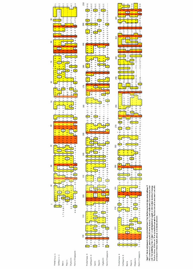
Proteinase A shows 46% sequence identity with porcine cathepsin D, 40% with porcine pepsin and 38% with human renin. The homol- ogy with cathepsin D is increased to 63% if conservative replacements among acid, basic, hydrophobic and hydroxy-amino acids are dis- regarded. Proteinase A and bovine chymosin, which are immunologically related (see Figure 2), show 37% sequence identity. The sequence homology with penicillopepsin and endothia- pepsin (38) is 25% and 26%, respectively. It is striking, that the enzyme with closest homology with proteinase A, cathepsin D, is the only other intracellular enzyme on the list, suggesting that specific functional requirements on intracellular aspartic proteinases restrict possibilities for structural diversifications. The amino acid se- quence of proteinase A does not, however, include the four-residue insertions found in cathepsin D at positions 91A-91D and 280A- 280D, which have been proposed to have a processing function for the lysosomal enzyme (11). Cleavage occurs in the insertion 91A-91D in porcine cathepsin D during conversion from the one-chain to two-chain form of the enzyme (44). The gene sequence of human cathepsin D also reveals an insertion in position 91A-91K (12) and mouse submandibulary renin is cleaved in an insertion 280A-280D (36). The signi- ficance of this proteolytic processing in cathep- sin D and renin is not established, and there is no evidence for a corresponding two-chain form of the intravacuolar yeast proteinase A.
In position 159 a three residue insertion (in reference to pepsin) was necessary. This inser- tion occurs in a position corresponding to a beta-hairpin loop, from residue 157-159, in the penicillopepsin, endothiapepsin and pepsin structure. Similar insertions occur at position 159 in mouse submandibulary renin (one resi- due), cathepsin D (two residues) and human renin (four residues). It was furthermore neces- sary to introduce a deletion at position 209, in a region where the homology between the aspartic proteinases is low.
One of the three disulphide bridges in mam- malian aspartic proteinases, Cys206-Cys210, is lacking in proteinase A, which in this regard
resembles the fungal aspartic proteinases. The other halfcystines in proteinase A are in con- served positions, suggesting conservation of the disulfide bridges Cys45-CysS0 and Cys250- Cys283, found in pepsin (43) etc. These two disulfide bridges are also retained in an aspartic proteinase from Mucor miehei (1), while endo- thiapepsin and penicillopepsin possess only cystine (Cys250-Cys283) (22, 38).
In aspartic proteinases from Endothia parasi- tica and Penicillium janthinellum (23, 37) the two catalytically essential aspartic acid residues, Asp32 and Asp215, are arranged in a symmet- rical hydrogen bonded network involving Asp32, Thr33, Gly34, Ser35 and Asp215, Thr216, Gly217 and Thr218. The conservation of these residues in proteinase A, as well as in all other sequenced aspartic proteinases, strongly suggests a common catalytic mechanism and active site structure of these enzymes. Asp304, which is hydrogen bonded to the peptide oxygen of Thr 216 in the fungal proteinases, is also conserved, like Tyr75 in the flap region, which projects above the active site. JAMES and SIELECKI (23) have emphasized a hydrophobic patch in the penicillopepsin binding cleft, adja- cent to Asp32 and Asp215, which might be essential for interactions with the hydrophobic residues in substrates cleaved by aspartic pro- teinases (6, 8). The corresponding residues in proteinase A comprise Tyr275, I1e284, Leu220, Thr222, Gly297, Leu299, Ile301, Ala213, Tyr189, and lle128, which also are predomi- nantly hydrophobic.
Another structural relationship between pro- teinase A and e.g. pepsin, is the remarkable conservation of hydrophobic residues corre- sponding to the molecular core (22, 45, 47). These residues are in positions 4,6,14,18,20, 27,29,31,38-42,45,56,71,73,80,84,89,91,94,101, 119-125, 137, 145, 151-155, 165,170, 175, 179, 194, 197, 199,212-214, 22 l, 223,228,232,236, 238,246,248,250, 256,259, 26 l, 268,270,275, 277,283,292,300,307,311,314, and 321-324. The conservation of glycines in positions 2 l, 34,76,78,82,92,119,122,168,217,237,297,303, and 322 indicates a common structural restric- tion on the. size of the side chain in these positions, which are predominantly at beta-hair- pin loops in the structure of e.g. penicillopepsin.
Carlsberg Res. Commun. Vol. 51, p. 27-41, 1986 37

T. DREYER et al.:
In contrast to this, the distribution of charged residues in proteinase A differs considerably from the distribution in other aspartic protein- ases. Glu81, Asp93, Aspl00, Asp126, Argl50, Glu172, Asp 178, Arg185, Glul91, Asp201, Arg253, and Glu294 correspond to uncharged residues in other aspartic proteinases. Residues Ala60, Tyr138, and Leu316 are hydrophobic like in penicillopepsin, while residues in these positions are charged in all mammalian aspartic proteinases. Such alterations in the charge distri- bution might influence substrate binding, and hence the specificity of proteinase A relative to the other aspartic proteinases.
The high number of paired or tripled basic residues in proteinase A (149-150, 185-187, 239-240 and 308-309) is remarkable, and in this respect proteinase A resembles human renin, which also possesses four such combinations (239-242, 280D-281,308-309, and 316-317).
There are two glycosylated asparagines in proteinase A. One is in position 67, which is also glycosylated in cathepsin D and human renin (21, 44), the other is in position 267, which is a glycosylation site thus far unique to proteinase A. Both residues correspond to surface regions on penicillopepsin, where the two sites are facing away from the active site. The role of the carbohydrate moiety is not well established. In mammalian cells the glycosylation oflysosomal hydrolases has been shown to direct the tran- sport from the Golgi body to the lysosomes (18), but results of experiments with yeast treated to inhibit glycosylation with tunicamycin, indicate that this targeting mechanism is not essential in yeast (42). Furthermore, correct processing of the precursor of proteinase A is apparently independent ofglycosylation (34). However, the conservation ofglycosylation site 67 in intracel- lular aspartic proteinases from species as distantly related as an unicellular yeast and mammals, suggests a critical role of this carbohy- drate moiety.
The occurrence of 3 peaks containing glycosy- lated tryptic peptide T 1 after ion-exchange (Fig- ure 5), and 3-4 close bands ofproteinase A on the isoelectric focusing gel between pH 4.0 and 4.1 might be explained from possible inhomogene- ity of the carbohydrate moiety.
The aspartic proteinase isolated from Saccha-
Aspartic proteinase A
romyces carlsbergensis did not reveal significant differences from proteinase A with respect to molecular weight, isoelectric point, proteolytic activity and stability, and the identical 40 amino acid N-terminal sequences and complete immu- nological cross-reactivity, suggest that these en- zymes have similar structures.
ACKNOWLEDGEMENTS The authors wish to acknowledge Mss. SIDSEL
EHLERS and Mr. THORKILD BEENFELD for excellent technical assistence with preparation and characterization of enzymes and peptides, and Mss. LONE SORENSEN, PIA BREDDAM, and BODIL CORNELIUSSEN for the amino acid ana- lysis and sequencing. Cand.scient. LARS PETER JEPSEN is thanked for performing immune blot- ting assays.
REFERENCES 1. BECH, A. -M. & B. FOLTMANN: Partial primary
structure of Mucor miehei protease. Neth. Milk Dairy J. 35, 275-280 (1981)
2. BJERRUM, O. J., K. P. LARSEN & M. WILKEN: Some recent developments of the electroimmunocbe- mical analysis of membrane proteins. In: Modern Methods in Protein Chemistry, Tschesche, H. Ed., Walter de Gruyter, Berlin, New York 79-124 (1983)
3, BORNSTEIN, P. & G. BALIAN: Cleavage at Asn-Gly bonds with hydroxylamine. Meth. Enzymol. 47, 132-145 (1977)
4. DIXON, H. B. F. & R. N. PERHAM: Reversible block- ing of amino groups with citraconic anhydride. Biochem. J. 109, 312-314 (1968)
5. DREYER, T., K. BIEDERMANN & M. OTTESEN." Yeast proteinase in beer. Carlsberg Res. Commun. 48, 249-253 (1983)
6. DREYER, T.. I. SVENDSEN & M. OTTESEN: Partial primary structure and substrate specificity of pro- teinase A from Saccharomyces cerevisiae. BiD- chem. Soc. Transac. 13, 1142-1143 (1985)
7. DUBOlS, M., K. A. GILLES, J. K. HAMILTON. P. A. REBERS & F. SMITH: Colorimetric method for de- termination of sugar and related substances. Anal. Chem. 28, 350-356 (1956)
8. DUNN, B. M., B. KAMMERMANN & K. R. Mc- CURRY: The synthesis, purification and evalu- ation of a chromophoric substrate for pepsin and other aspartyl proteases. Anal. Biochem. 138, 68- 73 (1984)
38 Carlsberg Res. Commun. Vol. 51, p. 27-41, 1986

T. DREYER et at.: Aspartic proteinase A
9. EDMAN, P. & A. BEGG: A protein sequenator. Eur. J. Biochem. 1, 80-91 (1967)
10. EDELHOCH, H.: Spectroscopic determination of tryptophan and tyrosin in proteins. Biochemistry 6, 1948-1954 (1967)
11. ERICKSON, A. H., G. E. CONNER & G. BLOBEL: Biosynthesis ofa lysosomal enzyme. J. Biol. Chem. 256, 11224-11231 (1981)
12. FAUST, P. L., S. KORNFELD & J. M. CHIRGWIN: Cloning and sequence analysis of cDNA for hu- man cathepsin D. Proc. Natl. Acad. Sci. USA 82, 4910-4914 (1985)
13. FOLTMANN, B., V. B PEDERSEN, H. JACOBSEN, D. KAUFFMAN & G. WYBRANDT: The complete amino acid sequence of prochymosin. Proc. Nat. Acad. Sci. USA 74, 2321-2324 (1977)
14. FRIEDMAN, M., J. C. ZAHNLEY & J. R. WAGNER: Estimation of the disulfide content of trypsin inhibitors as S-13-(2-pyridylethyl)-L-cysteine. Anal. Biochem. 106, 27-34 (1980)
15. GRAHAM, R. J, JR., U. LUNDHOLM & M. J. KAR- NOVSKY: Cytochemical demonstration of peroxi- dase activity with 3-amino-9-ethyl-carbazole. J. Histochem. Cytochem. 13, 150-153 (1965)
16. HAPNER, K. D. & P. E. WILCOX: Fragmentation of bovine chymotry0sinogen A and chymotrypsin Act. Specific cleavage at arginine and methionine residues and separation of peptides including B and C chains of chymotrypsin. Biochemistry 9, 4470-4480 (1970)
17. HARBOE, N.&A.INGILD: Immunization, isolation ofimmunoglobulins, estimation of antibody titre. In: A Manual of Quantitative Immunoelectropho- resis. Methods and Applications, eds. : Axelsen, N. H., J. Kroll & B. Weeke. Universitetsforlaget, Oslo, pp. 161-164 (1973)
18. HASILIK, A. & E NEUFELD: Biosynthesis of lyso- somal enzymes in fibroblasts: Phosphorylation of mannose residues. J. Biol. Chem. 255, 4946-4950 (1980)
19. HATA, T., R. HAYASHI & E. DOI: Purification of yeast pmteinases I. Fractionation and some prop- erties of these proteinases. Agr. Biol. Chem. 31, 150-159 (1967)
20. HIRS, C. H. W.: Determination ofcystine as cysteic acid. Methods Enzymol. 11, 59-62 (1967)
21. HOBART, P. M., M. FOGLIANO, B. A. O'CONNOR, I. M. SCHAEFER & J. M. CHIRGWIN; Human lenin gene: Structure and sequence analysis. Proc. Nat. Acad. Sci. USA 81, 5026-5030 (1984)
22. Hsu. I. -N, L. T. J DALBAERE, M. N. G. JAMES & T. HOFMANN: Penicillopepsin from Penieillium jan- thinellum. Crystal structure at 2.8 ~, and sequence homology with porcine pepsin. Nature 266, 140- 145 (1977)
23. JAMES, M. N. G. & A. R. SIELECKI: Stereochemical analysis of peptide bond hydrolysis catalyzed by the aspartic proteinase penicillopepsin. Biochemi- stry 24, 3701-3713 (1985)
24. JOHANSEN, J. T., K. BREDDAM & M. OTTESEN: Isolation of carboxypeptidase Y by affinity chro- matography. Carlsberg Res. Commun. 41, 1-14 (1976)
25. JOHANSEN, J. T., C. OVERBALLE-PETERSEN, B. MARTIN, V. HASEMANN & I. SVENDSEN: The com- plete amino acid sequence of copper, zinc super- oxide dismutase from Saccharomyccs cerevisiae. Carlsberg Res. Commun. 44, 201-217 (1979)
26. JONES, E. W." Genetic approaches to the study of protease function and proteolysis in Saccharo- myces cerevisiae. In: Yeast Genetics, Funda- mental and Applied Aspects, Spencer, J. F. T., D. M. Spencer & A. R. W. Smith Eds., Springer Vedag, New York 167-203 (1983)
27. KELLER, H. P., F. ERNI, H. R. LINDER & R.. W. FREI: Dynamic slurry-packing technique for liquid chro- matography columns. Anal. Chem. 49, 1958-1963 (1977)
28. KROLLJ.: Tandem crossed immunoelectropho- resis. In: Handbook of lmmunoprecipitation-in- gel-techniques, Axelsen, N. H. Ed., Blackwell, pp. 135-139 (1983)
29. LAEMMLI, U. K.: Cleavage of structural proteins during the assembly of the head of bacteriophag T4. Nature 227, 324-331 (1980)
30. LENNEY, J. F. & J. M. DALBEC: Purification and properties of two proteinases from Saccharomyces cerevisiae. Arch. Biochem. Biophys. 120, 42-48 (1967)
31. LENNEY, J. F., P. MATILE, A. WlEMKEN, M. SCHEL- LENBERG &J. MEYER: Activities and cellular local- ization of yeast proteases and their inhibitors. Biochem. Biophys. Res. Commun. 60, 1378-1383 (1974)
32. LUNDBLAD, R. L. & W. H. STEIN: On the reaction of diazoacetyl compounds with pepsin. J. Biol. Chem. 244, 154-160 (1969)
33. MAHONEY, W. C., P. K. SMITH & M. A. HERMOD- SON: Fragmentation ofproteins with o-iodosoben- zoic acid and a reactive contaminant that modify tyrosyl residues. Biochemistry 20, 443-448 ( 1981)
34. MECHLER, B., M.MULLER, F. MEUSSDOERFFER & D H. WOL~. In vivo biosynthesis of the vacuolar proteinases A and B in the yeast Saceharomyces cerevisiae. J. Biol. Chem. 257, 11203-11206 ( 1982)
35. MEUSSDOERFFER, F., P. TORTORA & H. HOLZER" Purification and properties of proteinase A from yeast. J. Biol. Chem. 255, 12087-12093 (1980)
36. MISONO, K. S., J. -J. CHANG & T. INAGAMI: Amino acid sequence of mouse submaxillary gland renin.
Carlsberg Res. Commun. Vol. 51, p. 27-41, 1986 39
T. DREYER et at.: Aspartic proteinase A
Proc. Natl. Acad. Sci. USA 79, 4858-4862 (1982) 37. PEARL, L. & T. BLUNDELL: The active site of aspar-
tic proteinases. FEBS Lett. 174, 96-101 (1984) 38. PEDERSEN~ V. B.: (Personal communication) 39. SAHEKI, T. & H. HOLZER: Comparisons of the
tryptophan synthase inactivating enzymes with proteinases from yeast. Eur. J. Biochem, 42, 621 - 626 (1974)
40, SAHEKI, T. & H. HOLZER: Proteolytic activities in yeast. Biochim. Biophys. Acta 384,203-214 (1975)
41. SALKOWSKI, E.: Ueber Zuckerbildung und andere Fermentationen in der Hefe. Z. Phys. Chem. 13, 506-538 (1889)
42. SCHWAIGER, H., A, HASILIK, K. YON FIGURA, A. WIEMKEN & W. TANNER: Carbohydrate-free car- boxypeptidase Y is transferred into the lysosome- like yeast vacuole. Biochem. Biophys. Res. Com- mun. 104, 950-956 (1982)
43. SEPULVEDA, P., J. MARCINISZYN, D. LIU & J. TANG: Primary structure of porcine pepsin. J. Biol. Chem. 250, 5082-5088 (1975)
44. SHEWALE, J. G & J. TANG: Amino acid sequence of porcine spleen cathepsin D. Proc. Nat. Acad. Sci. USA 81, 3703-3707 (1984)
45. SIBANDA, B. L., T. BLUNDELL, P. M. HOBART, M. FOGLIANO, J. S. BINDRA, B. W. DOMINY & J. M. CHIRGWIN" Computer graphics modelling of hu- man renin. FEBS Lett. 174, 102-111 (1984)
46. STEVENS, T. H.: (Personal communication) 47. SUBRAMANIAN, E., I. D. A. SWAN, M. LIU, D. R.
DAVIES, J. A. JENKINS, I. J. TICKLE & T. L BLUN- DELL: Homology among acid proteases: Compari- son of crystal structures at 3J~ resolution of acid proteases from Rhizopus chinensis and Endothia parasitica. Proc. Natl. Acad. Sci. USA 74, 556-559 (1977)
48. SVENDSEN, I., B. MARTIN & I. JONASSEN: Charac- teristics ofhiproly barley III. Amino acid sequence of two-lysine rich proteins. Carlsberg Res. Com- mun. 45, 79-85 (1980)
49. TANG, J.: Specific and irreversible inactivation of pepsin by substrate like epoxides. J. Biol. Chem. 246, 4510-4517 (1971)
50. TOWBIN, H., T. STAEHELIN & J. GORDON: Electro- phoretic transfer of proteins from polyacrylamide gels to nitrocellulose sheets: Procedure and some applications. Proc. Natl. Acad. Sci. USA 76, 4350- 4354 (1979)
51. WOLF, D. H. & H. HOLZER: Proteolysis in yeast. In: Microorganisms and Nitrogen Sources, Payne, J. W. Ed., John Wiley and Sons Ltd., 431-458 (1980)
52. WOOLEORD, C. A., L. B. DANIELS, F. J. PARK, E. W. JONES, J. V. ARSDELL & M. A. INNIS: Vacuolar hydrolase muturase ofSaccharomyces cerevisiae is an aspartyl protease encoded by the PEP4 gene. (in preparation)
Accepted by H. KLENOW
40 Carlsberg Res. Commun. Vol. 51, p. 27-41, 1986
o
- r a - + , 7 -
~ ~ S ~ S S ,
, , . , ~ ~ l = ~ r ' - ~ l
i ~ ~ 1 ~ ~ ~
o
I ~ = '
s = = X a 3
o
I Z Z Z Z Z l ~
i ~ ~ ~ ~ ~ 1 ~
o i ~ . - ~ = i > >
[ks: ck] ~ :
< = }
o r ~ - ~ r ~ - e - ~
I ~ ~ ~ ~ i '
I . . . . = = I = '
= [ s = = I X
o 1:~::12, = ~ 2 , 2
i ~ ~ ~ - ~
S % X . S 2 X
i ~ ~ 1 ~
. ~ ' a ~ . ~
~ g - z ~
~ g N 8 E ~