
Primary mucinous adenocarcinoma of the scalp: A case ... · diagnosis of a primary eccrine mucinous...
4
CASE REPORT – OPEN ACCESS International Journal of Surgery Case Reports 10 (2015) 241–244 Contents lists available at ScienceDirect International Journal of Surgery Case Reports journal homepage: www.casereports.com Primary mucinous adenocarcinoma of the scalp: A case report and literature review Osama S. Al Beteddini a,∗ , Salwa Sheikh b , Faisal Shareefi a , Rana Shahab c a Surgery Department, Johns Hopkins Aramco Healthcare/Dhahran Health Center, Saudi Aramco, P.O. Box 76, Dhahran 31311, Eastern Province, Saudi Arabia b Pathology Department, Johns Hopkins Aramco Healthcare/Dhahran Health Center, Saudi Aramco, P.O. Box 76, Dhahran 31311, Eastern Province, Saudi Arabia c Dermatology Department, Johns Hopkins Aramco Healthcare/Dhahran Health Center, Saudi Aramco, P.O. Box 76, Dhahran 31311, Eastern Province, Saudi Arabia article info Article history: Received 2 December 2014 Received in revised form 31 January 2015 Accepted 2 February 2015 Available online 7 February 2015 Keywords: Skin Scalp Eccrine Mucinous Adenocarcinoma abstract INTRODUCTION: Primary mucinous adenocarcinoma or mucinous eccrine carcinoma of the skin is a rare malignant neoplasm showing predilection to the head and neck. Distinguishing between these primary neoplasms and the more frequent metastatic mucinous deposits on the skin from primaries in the breast and gastrointestinal tract constitutes a diagnostic dilemma. PRESENTATION OF CASE: We report a case of a 61-year-old lady who presented with a slow-growing, pain- less scalp nodule. Upon excision, it was diagnosed as “primary mucinous adenocarcinoma”. An extensive work-up in search for another primary tumour failed to show a primary malignancy elsewhere and the diagnosis of a primary eccrine mucinous adenocarcinoma of the skin was rendered. DISCUSSION: A review of the literature on this entity is presented, discussing diagnostic challenges and therapeutic options that of interest to surgeons, pathologists and dermatologists. CONCLUSION: These tumours are indolent and low-grade, with a tendency for local, sometimes multiple, recurrences. Proper patient counselling and follow-up are important in treatment. Sound collaboration between clinicians and pathologists, for good therapeutic results, is of utmost importance. © 2015 The Authors. Published by Elsevier Ltd. on behalf of Surgical Associates Ltd. This is an open access article under the CC BY-NC-ND license (http://creativecommons.org/licenses/by-nc-nd/4.0/). 1. Introduction Mucinous sweat gland carcinoma is a rare malignant tumour of the skin. We report herein the case of a 61-year-old lady who presented with a scalp mass. After excision, histopathologi- cal examination of the mass revealed mucinous adenocarcinoma. Extensive metastatic work-up, including a total body computed tomographic and positron emission scan (PET/CT scan), showed the neoplasm to be a primary mucinous adenocarcinoma of the skin. Distinguishing this primary neoplasm from metastatic mucinous adenocarcinoma originating from other primary sites in particu- lar from the breast presents a diagnostic dilemma. A review of the literature of this rare, yet challenging, entity follows. 2. Report of a case A 61-year-old lady, of Caucasian origin, presented to our care with a slow growing, painless, spherical, scalp nodule overlying the right temporal region, measuring 1.5 × 1 cm. The mass was ∗ Corresponding author. Tel.: +966 502512572; fax: +966 138773695. E-mail address: [email protected] (O.S.A. Beteddini). subcutaneous, being tethered to the skin, yet mobile with respect to the underlying skull. It was firm, non-compressible, non-pulsatile and showed no central punctum. The nodule was present for the last 5 years with no history of pain, secretions or overlying redness. Upon examination, no regional lymphadenopathy or abnormal findings in the head or neck region could be noticed. The patient had been medically followed up in the past with regular screening for breast cancer and gastrointestinal disease, namely colorectal cancer. An excisional biopsy of the nodule was done under local anaes- thesia with no adverse events and the skin defect was primarily closed. Histopathological examination of the mass showed hair- bearing skin with underlying lakes of mucin separated by delicate fibrous septae. Floating in between the mucinous lakes were nests, clusters, and cords of predominantly uniform round to cuboidal epithelial cells that exhibit mild to moderate nuclear variability. Focal areas show duct formation. Occasional tumour nests show prominent proliferation of cells with cribriforming. The tumour showed no connection to the overlying skin or adnexal structures (See Fig. 1). The neoplastic cells were strongly positive for cytok- eratin 7 (CK7) and oestrogen and progesterone receptors. The cells were negative for cytokeratin 20 (CK20) and Her-2-neu (See Fig. 2). http://dx.doi.org/10.1016/j.ijscr.2015.02.006 2210-2612/© 2015 The Authors. Published by Elsevier Ltd. on behalf of Surgical Associates Ltd. This is an open access article under the CC BY-NC-ND license (http:// creativecommons.org/licenses/by-nc-nd/4.0/).
Transcript of Primary mucinous adenocarcinoma of the scalp: A case ... · diagnosis of a primary eccrine mucinous...
CASE REPORT – OPEN ACCESSInternational Journal of Surgery Case Reports 10 (2015) 241–244
Contents lists available at ScienceDirect
International Journal of Surgery Case Reports
journa l homepage: www.caserepor ts .com
Primary mucinous adenocarcinoma of the scalp: A case reportand literature review
Osama S. Al Beteddinia,∗, Salwa Sheikhb, Faisal Shareefia, Rana Shahabc
a Surgery Department, Johns Hopkins Aramco Healthcare/Dhahran Health Center, Saudi Aramco, P.O. Box 76,Dhahran 31311, Eastern Province, Saudi Arabiab Pathology Department, Johns Hopkins Aramco Healthcare/Dhahran Health Center, Saudi Aramco, P.O. Box 76,Dhahran 31311, Eastern Province, Saudi Arabiac Dermatology Department, Johns Hopkins Aramco Healthcare/Dhahran Health Center, Saudi Aramco, P.O. Box 76,Dhahran 31311, Eastern Province, Saudi Arabia
a r t i c l e i n f o
Article history:Received 2 December 2014Received in revised form 31 January 2015Accepted 2 February 2015Available online 7 February 2015
Keywords:SkinScalpEccrineMucinousAdenocarcinoma
a b s t r a c t
INTRODUCTION: Primary mucinous adenocarcinoma or mucinous eccrine carcinoma of the skin is a raremalignant neoplasm showing predilection to the head and neck. Distinguishing between these primaryneoplasms and the more frequent metastatic mucinous deposits on the skin from primaries in the breastand gastrointestinal tract constitutes a diagnostic dilemma.PRESENTATION OF CASE: We report a case of a 61-year-old lady who presented with a slow-growing, pain-less scalp nodule. Upon excision, it was diagnosed as “primary mucinous adenocarcinoma”. An extensivework-up in search for another primary tumour failed to show a primary malignancy elsewhere and thediagnosis of a primary eccrine mucinous adenocarcinoma of the skin was rendered.DISCUSSION: A review of the literature on this entity is presented, discussing diagnostic challenges andtherapeutic options that of interest to surgeons, pathologists and dermatologists.CONCLUSION: These tumours are indolent and low-grade, with a tendency for local, sometimes multiple,recurrences. Proper patient counselling and follow-up are important in treatment. Sound collaborationbetween clinicians and pathologists, for good therapeutic results, is of utmost importance.
© 2015 The Authors. Published by Elsevier Ltd. on behalf of Surgical Associates Ltd. This is an openaccess article under the CC BY-NC-ND license (http://creativecommons.org/licenses/by-nc-nd/4.0/).
1. Introduction
Mucinous sweat gland carcinoma is a rare malignant tumourof the skin. We report herein the case of a 61-year-old ladywho presented with a scalp mass. After excision, histopathologi-cal examination of the mass revealed mucinous adenocarcinoma.Extensive metastatic work-up, including a total body computedtomographic and positron emission scan (PET/CT scan), showed theneoplasm to be a primary mucinous adenocarcinoma of the skin.Distinguishing this primary neoplasm from metastatic mucinousadenocarcinoma originating from other primary sites in particu-lar from the breast presents a diagnostic dilemma. A review of theliterature of this rare, yet challenging, entity follows.
2. Report of a case
A 61-year-old lady, of Caucasian origin, presented to our carewith a slow growing, painless, spherical, scalp nodule overlyingthe right temporal region, measuring 1.5 × 1 cm. The mass was
∗ Corresponding author. Tel.: +966 502512572; fax: +966 138773695.E-mail address: [email protected] (O.S.A. Beteddini).
subcutaneous, being tethered to the skin, yet mobile with respect tothe underlying skull. It was firm, non-compressible, non-pulsatileand showed no central punctum. The nodule was present for thelast 5 years with no history of pain, secretions or overlying redness.Upon examination, no regional lymphadenopathy or abnormalfindings in the head or neck region could be noticed. The patienthad been medically followed up in the past with regular screeningfor breast cancer and gastrointestinal disease, namely colorectalcancer.
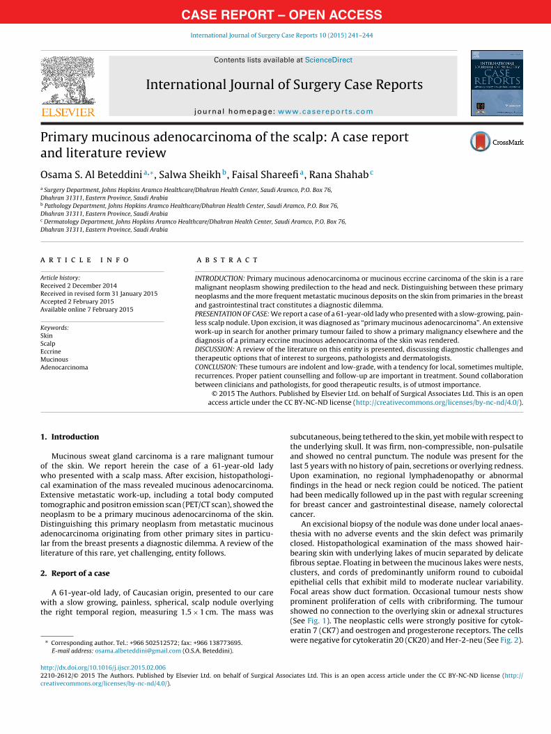
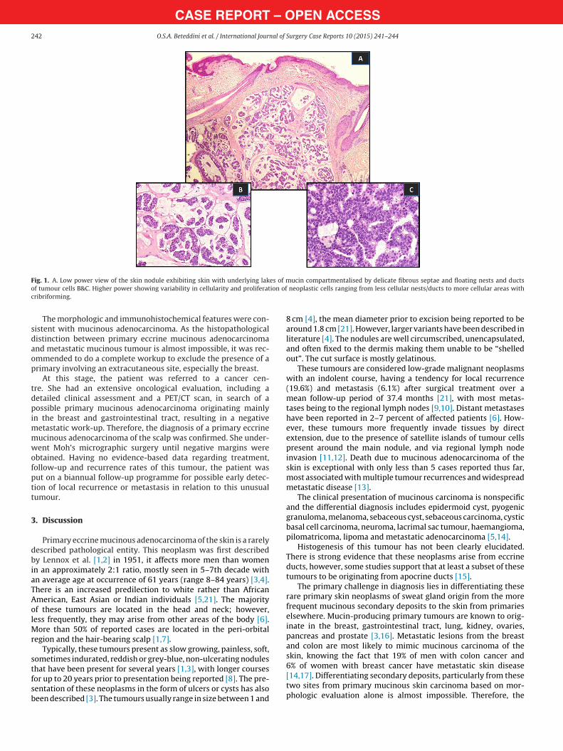
An excisional biopsy of the nodule was done under local anaes-thesia with no adverse events and the skin defect was primarilyclosed. Histopathological examination of the mass showed hair-bearing skin with underlying lakes of mucin separated by delicatefibrous septae. Floating in between the mucinous lakes were nests,clusters, and cords of predominantly uniform round to cuboidalepithelial cells that exhibit mild to moderate nuclear variability.Focal areas show duct formation. Occasional tumour nests showprominent proliferation of cells with cribriforming. The tumourshowed no connection to the overlying skin or adnexal structures(See Fig. 1). The neoplastic cells were strongly positive for cytok-eratin 7 (CK7) and oestrogen and progesterone receptors. The cellswere negative for cytokeratin 20 (CK20) and Her-2-neu (See Fig. 2).
http://dx.doi.org/10.1016/j.ijscr.2015.02.0062210-2612/© 2015 The Authors. Published by Elsevier Ltd. on behalf of Surgical Associates Ltd. This is an open access article under the CC BY-NC-ND license (http://creativecommons.org/licenses/by-nc-nd/4.0/).
CASE REPORT – OPEN ACCESS242 O.S.A. Beteddini et al. / International Journal of Surgery Case Reports 10 (2015) 241–244
Fig. 1. A. Low power view of the skin nodule exhibiting skin with underlying lakes of mucin compartmentalised by delicate fibrous septae and floating nests and ductsof tumour cells B&C. Higher power showing variability in cellularity and proliferation of neoplastic cells ranging from less cellular nests/ducts to more cellular areas withcribriforming.
The morphologic and immunohistochemical features were con-sistent with mucinous adenocarcinoma. As the histopathologicaldistinction between primary eccrine mucinous adenocarcinomaand metastatic mucinous tumour is almost impossible, it was rec-ommended to do a complete workup to exclude the presence of aprimary involving an extracutaneous site, especially the breast.
At this stage, the patient was referred to a cancer cen-tre. She had an extensive oncological evaluation, including adetailed clinical assessment and a PET/CT scan, in search of apossible primary mucinous adenocarcinoma originating mainlyin the breast and gastrointestinal tract, resulting in a negativemetastatic work-up. Therefore, the diagnosis of a primary eccrinemucinous adenocarcinoma of the scalp was confirmed. She under-went Moh’s micrographic surgery until negative margins wereobtained. Having no evidence-based data regarding treatment,follow-up and recurrence rates of this tumour, the patient wasput on a biannual follow-up programme for possible early detec-tion of local recurrence or metastasis in relation to this unusualtumour.
3. Discussion
Primary eccrine mucinous adenocarcinoma of the skin is a rarelydescribed pathological entity. This neoplasm was first describedby Lennox et al. [1,2] in 1951, it affects more men than womenin an approximately 2:1 ratio, mostly seen in 5–7th decade withan average age at occurrence of 61 years (range 8–84 years) [3,4].There is an increased predilection to white rather than AfricanAmerican, East Asian or Indian individuals [5,21]. The majorityof these tumours are located in the head and neck; however,less frequently, they may arise from other areas of the body [6].More than 50% of reported cases are located in the peri-orbitalregion and the hair-bearing scalp [1,7].
Typically, these tumours present as slow growing, painless, soft,sometimes indurated, reddish or grey-blue, non-ulcerating nodulesthat have been present for several years [1,3], with longer coursesfor up to 20 years prior to presentation being reported [8]. The pre-sentation of these neoplasms in the form of ulcers or cysts has alsobeen described [3]. The tumours usually range in size between 1 and
8 cm [4], the mean diameter prior to excision being reported to bearound 1.8 cm [21]. However, larger variants have been described inliterature [4]. The nodules are well circumscribed, unencapsulated,and often fixed to the dermis making them unable to be “shelledout”. The cut surface is mostly gelatinous.
These tumours are considered low-grade malignant neoplasmswith an indolent course, having a tendency for local recurrence(19.6%) and metastasis (6.1%) after surgical treatment over amean follow-up period of 37.4 months [21], with most metas-tases being to the regional lymph nodes [9,10]. Distant metastaseshave been reported in 2–7 percent of affected patients [6]. How-ever, these tumours more frequently invade tissues by directextension, due to the presence of satellite islands of tumour cellspresent around the main nodule, and via regional lymph nodeinvasion [11,12]. Death due to mucinous adenocarcinoma of theskin is exceptional with only less than 5 cases reported thus far,most associated with multiple tumour recurrences and widespreadmetastatic disease [13].
The clinical presentation of mucinous carcinoma is nonspecificand the differential diagnosis includes epidermoid cyst, pyogenicgranuloma, melanoma, sebaceous cyst, sebaceous carcinoma, cysticbasal cell carcinoma, neuroma, lacrimal sac tumour, haemangioma,pilomatricoma, lipoma and metastatic adenocarcinoma [5,14].
Histogenesis of this tumour has not been clearly elucidated.There is strong evidence that these neoplasms arise from eccrineducts, however, some studies support that at least a subset of thesetumours to be originating from apocrine ducts [15].
The primary challenge in diagnosis lies in differentiating theserare primary skin neoplasms of sweat gland origin from the morefrequent mucinous secondary deposits to the skin from primarieselsewhere. Mucin-producing primary tumours are known to orig-inate in the breast, gastrointestinal tract, lung, kidney, ovaries,pancreas and prostate [3,16]. Metastatic lesions from the breastand colon are most likely to mimic mucinous carcinoma of theskin, knowing the fact that 19% of men with colon cancer and6% of women with breast cancer have metastatic skin disease[14,17]. Differentiating secondary deposits, particularly from thesetwo sites from primary mucinous skin carcinoma based on mor-phologic evaluation alone is almost impossible. Therefore, the

CASE REPORT – OPEN ACCESSO.S.A. Beteddini et al. / International Journal of Surgery Case Reports 10 (2015) 241–244 243
Fig. 2. Immunohistochemistry shows tumour cells to be strongly positive for CK7, oestrogen receptors (ER), and progesterone receptors (PR) and negative for CK 20.
differentiation is mainly based on firstly, ruling out the pres-ence of another primary malignant site through a full oncologicalevaluation and, secondly, on certain histological and pathologicalcharacteristics of the mucinous lesion.
Histologically, these tumours are generally well circumscribed,unencapsulated, asymmetric, dermal nodules that may extend toinvolve subcutis and even deeper tissue planes. Most tumours showno connection to the overlying epidermis or skin adnexae. Charac-teristically, there are pools of basophilic mucin compartmentalisedby delicate thin fibrous septae creating a honey comb pattern.Within these mucinous lakes are small islands, clusters, and/ortubules of floating epithelial cells (Fig. 1A). The neoplastic cellsare round to cuboidal with abundant eosinophilic to clear cyto-plasm, small central nuclei, minimal nuclear pleomorphism, andonly rare mitotic figures (Fig. 1B&C). These tumour cells are oftengrouped in nests and duct-like structures reminiscent of eccrineand apocrine sweat glands. Some glandular structures show promi-nent proliferation and may form a cribriforming pattern. Smallertumour satellites may occur around the primary nodule. Epider-motism by these neoplastic cells is an unusual feature [3,14]. Thepresence of copious amounts of mucin has been hypothised to serveas a physical barrier to spread, compressing the tumour stroma andrestricting growth, as well as inhibiting DNA synthesis, decreasingthe rate of angiogenesis [3,16], and consequently metastatic disease[9].
Histologic distinction between primary and secondary tumoursmay be impossible, albeit the latter may show certain features sup-porting metastasis such as less abundance of mucin, larger tumourclusters and sheets with predominance of malignant epithelial cellsover mucin, and lack of honey combing due to the absence of fibrousseptae in-between the mucinous lakes [18].
Histochemical and immunohistochemical staining can assist indifferentiating tumour types. The tumour cells express cytokeratinAE1/AE3, epithelial membrane antigen (EMA), and carcinoembry-onic antigen (CEA) in keeping with its derivation from secretorylobule. There is strong nuclear staining for oestrogen receptorsand variable but mostly positive staining for progesterone recep-tors (Fig. 2). The cells are also often positive for alpha-lactalbumin,salivary amylase, beta-2-microglobulin, cytokeratin CAM 5.2, and
gross cystic disease fluid protein -15 (GCDFP-15). There is variableexpression of S-100 protein. The tumour shows a low prolifera-tion index by Ki-67. In addition, the cells are negative for CK20,a helpful feature in distinguishing it from metastasis from pri-mary gastrointestinal tumours that are often CK20 positive (Fig. 2).Primary tumours may occasionally show focal neuroendocrine dif-ferentiation as evidenced by positive reaction by Grimilius, andpositive staining by chromogranin, synaptophysin, and neuronspecific enolase (NSE) [3,15,18]. Alcian blue staining of primaryskin lesions is positive at a pH of 2.4 but is negative at a pH of0.4, differentiating them from other sweat gland tumours [14].The mucin in primary skin lesions is a sialomucin which is resis-tant to digestion with diastase and hyaluronidase but sensitive tosialidase [3,16], unlike mucinous lesions from gastrointestinal pri-maries that contain sulphomucin [14,19]. Moreover, cytokeratin20 (CK20) is commonly expressed in gastrointestinal neoplasmsbut absent in skin primaries. Additionally, the expression of cytok-eratin 5, 6 and 7 (CK5/6/7) (Fig. 2) and p63 in primary eccrinecarcinoma of the skin (indicating the presence of myoepithelialcells) can help exclude metastatic mucinous breast carcinoma[20]. In summary, although helpful, histopathological featurescannot exclude metastatic disease with 100% certainty; there-fore, the importance of a full metastatic work-up cannot beoveremphasised.
As for treatment, surgical excision of primary eccrine muci-nous adenocarcinoma of the skin is the therapeutic mainstay inmost cases. Because of the recurring nature of the tumour, ade-quate excision with wide margins (at least 1 cm) is advocated.Moh’s micrographic surgery can be a particularly advantageoustreatment modality in this setting. These tumours are generallyresistant to radiotherapy and chemotherapy and these tools arenot usually employed [3,14]. Finally, patients are to be coun-selled about the importance of frequent follow-up to rule outlocal tumour recurrence or the development of regional lym-phadenopathy. Knowing that younger patient age at presentation,Asian background, location of the tumour on the trunk, tumourslarger than 1.5 cm and longer follow-up periods are factors thatcould be associated with poor outcomes, namely, recurrence andmetastasis [21].

CASE REPORT – OPEN ACCESS244 O.S.A. Beteddini et al. / International Journal of Surgery Case Reports 10 (2015) 241–244
4. Conclusion
Before a diagnosis of a primary mucinous adenocarcinoma isestablished, a primary mucinous carcinoma of breast, gastroin-testinal tract, or other organs must be excluded as the majorityof cutaneous mucinous carcinoma cases, in fact, represent metas-tasis. Primary mucinous carcinoma of the skin, in contrast toother sweat gland carcinomas, is an indolent low-grade malignanttumour with tendency for local, sometimes multiple, recurrences.Distant metastasis is rare and is often limited to regional lymphnodes. Primary eccrine mucinous adenocarcinoma of the skin poseschallenges in relation to its diagnosis and differentiation frommetastatic adenocarcinoma to the skin and with regard to itstreatment. Its rarity precludes comparative evaluation of therapeu-tic options. However, sound collaboration between clinicians andpathologists, for a good therapeutic result, is of utmost importance.Proper patient counselling and follow-up are important in treatingthis low-grade, indolent and frequently recurring tumour.
Conflicts of interest
Nothing to declare.
Sources of funding
Nothing to declare.
Patient’s consent
Informed consent has been obtained from the patient and a copyof this document is available when requested. The figures related tothe article are pathological slides and contain no information thatmay affect patient’s privacy in any way.
Authors contribution
Osama S. Al Beteddini: The corresponding author. General sur-geon. He has written the case report and collected relevant data forthe literature review.
Salwa Sheikh: Author, pathologist. She has provided all infor-mation related to the histopathological properties of the tumour.She has further provided the pathological slides. She has reviewedthe article and rewritten the section on pathology of the article.
Faisal Shareefi: Author, plastic and reconstructive surgeon. He isthe attending physician of the patient. He has reviewed the article.
Rana Shahab: Author, dermatologist. She has referred thepatient to surgical care. She has reviewed the article.
References
[1] I.M. Scholz, W. Hartschuh, Primary mucinous eccrine carcinoma of the skin-arare clinical tumor with many differential diagnoses, J. Deutschen Dermatol.Gesellschaft 8 (6) (2010) 446–448.
[2] B. Lennox, A.G. Pearse, H.G. Richards, Mucin-secreting tumours of the skinwith special reference to the so-called mixed-salivary tumour of the skin andits relation to hidranoma, J. Pathol. Bacteriol. 64 (4) (1952) 865–880.
[3] B.C. Kelly, J. Koay, M.S. Driscoll, S.S. Raimer, M.I. Colome-Grimmer, Report of acase: primary mucinous carcinoma of the skin, Dermatol. Online J. 14 (6)(2008) 4.
[4] T. Brenn, P. McKee, Tumors of sweat glands, in: P. McKee, E. Calonje, S.Granker (Eds.), Pathology of the Skin with Clinical Correlations, 3rd Edition,Elsevier Mosby, 2005.
[5] D.J. Karimipour, T.M. Johnson, S. Kang, T.S. Wang, L. Lowe, Mucinouscarcinoma of the skin, J. Am. Acad. Dermatol. 36 (2015) 323–326.
[6] T.V. Ajithkumar, N. Nileena, E.K. Abraham, F.V. James, M.K. Nair, Bone marrowrelapse in primary mucinous carcinoma of skin, Am. J. Clin. Oncol. 22 (3)(1999) 303–304.
[7] G. Bellezza, A. Sidoni, E. Bucciarelli, Primary mucinous carcinoma of the skin,Am. J. Dermatopathol. 22 (2) (2000) 166–170.
[8] T.M. Andrews, J.L. Gluckman, M.A. Weiss, Primary mucinous adenocarcinomaof the eyelid, Head Neck 14 (4) (1992) 303–307.
[9] A. Chauhan, M. Ganguly, P. Takkar, V. Dutta, Primary mucinous carcinoma ofeyelid: a rare clinical entity, Indian J. Ophthalmol. 57 (2) (2009)150–152.
[10] C.E. Cabell, K.F. Helm, P.J. Sakol, E.M. Billingsley, Primary mucinous carcinomain a 54-year-old man, J. Am. Acad. Dermatol. 49 (5) (2003) 941–943.
[11] A. Latorre, L. Alghothani, D. Lambert, K. Jatana, S. Peters, J. Foster, R. Hill,Mucin-producing malignant tumor of lower eyelid presenting in a14-year-old patient, J. Clin. Aesthet. Dermatol. 5 (4) (2012)44–47.
[12] M.R. Wick, J.R. Goellner, J.T. Wolfe, W.P. Su, Adnexal carcinoma of the skin. I.Eccrine carcinomas, Cancer 56 (5) (1985) 1147–1162.
[13] V.D. Durairaj, E.M. Hink, M.Y. Kahook, M.J. Hawes, P.U. Paniker, B. Esmaeli,Mucinous eccrine adenocarcinoma of the periocular region, Ophthalmol.Plast. Reconstruct. Surg. 22 (2006) 30–35.
[14] E. Coan, A. Doan, A. Calliope, Mucinous eccrine carcinoma: a rare case ofrecurrence with lacrimal gland extension, Ophthalmol. Plast. Reconstruct.Surg. 28 (5) (2012) e109–e111.
[15] L. Requena, H. Kutzner, A.B. Ackerman, Neoplasms with ApocrineDifferentiation, Lippincott Williams & Wilkins, Philadelphia, 1998.
[16] C. Urso, R. Bondi, M. Plaglierani, A. Salvadori, C. Anichini, A. Giannini,Carcinoma of sweat glands: report of 60 cases, Arch. Pathol. Lab. 125 (2001)498–505.
[17] T. Ohnishi, H. Takizawa, S. Watanabe, Immunohistochemical analysis ofcytokeratin and human milk fat globulin expression in mucinous carcinomaof the skin, J. Cutaneous Pathol. 29 (2002) 38–43.
[18] L. Requena, Y. Mengeesha, H. Kutzner, et al., Appendageal tumors, in: P.LeBoit, G. Burg, D. Weedon, A. Sarasin (Eds.), Pathology and Genetics, SkinTumors. World Health Organization Classification of Tumors, InternationalAgency for Research on Cancer (IARC), Lyon, 2006.
[19] S. Martinez, S. Young, Primary mucinous carcinoma of the skin: a review,Internet J. Oncol. 2 (2005) 2, http://dx.doi.org/10.5580/13e7.
[20] J.S. Qureshi, M.E. Salama, D. Chitale, I. Banasl, C.K. Ma, U. Raju, A. Ormsby,M.W. Lee, Primary cutaneous mucinous carcinoma: presence of myoepithelialcells as a clue to the cutaneous origin, Am. J. Dermatopathol. 26 (5) (2004)353–358.
[21] L. Kamalpur, R.T. Brindise, M. Nodzenski, D.Q. Bach, E. Veledar, M. Alam,Primary cutaneous mucinous carcinoma: a systemic review and meta-analysisof outcomes after surgery, JAMA Dermatol. 150 (4) (2014) 380–384.
Open AccessThis article is published Open Access at sciencedirect.com. It is distributed under the IJSCR Supplemental terms and conditions, whichpermits unrestricted non commercial use, distribution, and reproduction in any medium, provided the original authors and source arecredited.


![Primary Mucinous Adenocarcinoma of the Ovary with ... · PDF fileseeding and lymphatic spread [14]. Most stage I invasive mucinous carcinomas of the intestinal type with expansile](https://static.fdocuments.in/doc/165x107/5ab668757f8b9a86428d9b6b/primary-mucinous-adenocarcinoma-of-the-ovary-with-and-lymphatic-spread-14.jpg)
![Surgical Management of Primary Cutaneous Mucinous Carcinoma · represents 0.005% of all malignant epithelial neoplasms [1]. These adnexal tumours have been thought to be of eccrine](https://static.fdocuments.in/doc/165x107/5f0b6f0f7e708231d4307f6a/surgical-management-of-primary-cutaneous-mucinous-represents-0005-of-all-malignant.jpg)