PRIMARY FAILURE OF ERUPTION: FURTHER …
44
PRIMARY FAILURE OF ERUPTION: FURTHER CHARACTERIZATION OF A RARE ERUPTION DISORDER Karen E. Koehler, DDS A thesis submitted to the faculty of the University of North Carolina at Chapel Hill in partial fulfillment of the requirements for the degree of Master of Science in the School of Dentistry (Orthodontics). Chapel Hill 2006 Approved by Advisor: Sylvia A. Frazier-Bowers, DDS, PhD Reader: James L. Ackerman, DDS Reader: Eric T. Everett, DDS, MS, PhD Reader: William R. Proffit, DDS, MS, PhD
Transcript of PRIMARY FAILURE OF ERUPTION: FURTHER …
PRIMARY FAILURE OF ERUPTION: FURTHER CHARACTERIZATION OF A RARE ERUPTION DISORDER
Karen E. Koehler, DDS A thesis submitted to the faculty of the University of North Carolina at Chapel Hill in partial fulfillment of the requirements for the degree of Master of Science in the School of Dentistry (Orthodontics).
Chapel Hill
2006
Approved by
Advisor: Sylvia A. Frazier-Bowers, DDS, PhD Reader: James L. Ackerman, DDS Reader: Eric T. Everett, DDS, MS, PhD Reader: William R. Proffit, DDS, MS, PhD

ii
ABSTRACT
KAREN E. KOEHLER: Primary Failure of Eruption: Further Characterization of a Rare Eruption Disorder
(Under the direction of Dr. Sylvia A. Frazier-Bowers)
Primary failure of eruption (PFE) is a rare condition that leads to spectacular posterior
open bites and does not respond to orthodontic treatment. Records from 97 patients with
posterior open bite were analyzed. Based on key characteristics, subjects were classified as
having PFE, indeterminate failure of eruption (IFE), and mechanical failure of eruption
(MFE). Results showed that PFE affects mostly posterior teeth, affects all teeth distal to the
first affected tooth, often presents with a cleared eruption path that the tooth fails to follow,
and appears to have two forms. Type I exhibits a similar lack of eruption potential of affected
teeth, and Type II has a varied eruption potential. This study also supports the genetic
etiology of PFE which is likely due to a defect in a tooth-specific gene product.
Differentiation between PFE and ankylosis is key to determining prognosis for orthodontic
treatment and requires adequate longitudinal data.
iii
ACKNOWLEDGEMENTS
This study would not have been possible without the many concerned orthodontists
who sent cases one or two at a time for evaluation. Special thanks go to Drs. Peter Shapiro,
Thomas Ahman, Richard Boyd and Loring Ross for submission of multiple cases and
continued support of this research.
I would like to thank my committee members, Drs. Frazier-Bowers, Ackerman, Everett
and Proffit, for their service, guidance and support. I would also like to thank Melody Torain
for her efforts in working with the families. Finally, I would like to extend my sincerest
appreciation to Dr. Jim Ackerman for the many hours he spent reviewing records and sharing
his vast knowledge and experience.

iv
TABLE OF CONTENTS
Page
LIST OF TABLES…………..………………………..…………………………………….... v
LIST OF FIGURES………….…………………..……………………………………….….. vi
LIST OF ABBREVIATIONS……………………………..………………………………....vii
SECTION
I. LITERATURE REVIEW…………………………………………………………. 1
A. Spectrum of Eruption Disturbances……………………………………...... 1
B. Primary Failure of Eruption...……………………………………………... 7
C. Familial Aspect of Eruption Failures……………..……………................. 10
D. Limitations of Previous Studies…………………………………………... 13
E. Purpose of Present Study…………………………………………………. 14
F. References………………………………………………………………… 15
II. MANUSCRIPT ………...………………………………………………………… 18
A. Introduction…..…………………………………………………………… 18
B. Materials and Methods……………………………………………………. 19
C. Results…………………………………………………………………….. 22
D. Discussion………………………………………………………………… 29
E. Conclusions……………………………………………………………….. 34
F. References………………………………………………………………… 36

v
LIST OF TABLES
Table Page
1. Typology of sample classification………..……………………………………………….. 21
2. Angle classification of sample…………………………………………………………….. 27

vi
LIST OF FIGURES
Figure Page
1. Number of subjects in each category……………………………………………………… 22
2. Representative sample of IFE……………………………………………………………... 23
3. Subject exhibiting ankylosis…………………………..…………………………………... 23
4. Subject demonstrating mild lateral open bite……………………………………………… 24
5. Distribution of affected teeth in PFE group………………………………………………. 25
6. PFE Type I………………………………………………………………………………… 26
7. PFE Type II………………………………………………………………………………... 26
8. PFE in a mother and daughter…………………………………………………………...... 28
9. Pedigree of PFE-001………………………………………………………………………. 29
10. Orthodontic intrusion of normal teeth…………………………………………………… 32

vii
LIST OF ABBREVIATIONS
Description Abbreviation
Indeterminate Failure of Eruption………………………………….………………………. IFE
Mechanical Failure of Eruption…………………………………………………………... MFE
Posterior Open Bite…………………………………………………..…………………… POB
Primary Failure of Eruption……….……………….……………………………………… PFE
SECTION I
LITERATURE REVIEW
Primary failure of eruption is a rare condition that can lead to spectacular posterior
open bites. It is difficult to diagnose and even more difficult to treat due to the lack of
response to orthodontic forces. Adding to the complexity of diagnosis is the myriad of
confusing and sometimes conflicting terms used in the literature to describe eruption
problems. Proper diagnosis is paramount and can save the patient and the orthodontist years
of frustration and disappointment. The role of heredity as an etiology has been questioned
and evidence of familial occurrence warrants further study. Understanding the genetic
component of failure of eruption can aid in differential diagnosis of PFE, help in the early
identification of affected family members and may eventually lead to new treatment
modalities.
The scientific literature presented will discuss failure to erupt and the terminology used
in the description of eruption problems. Possible causes will be identified with emphasis on
impaction, ankylosis and PFE. Current literature on PFE and potentially related conditions
will be reviewed, and case studies on the familial component of this condition will be
presented. Limitations of previous studies will also be discussed.
Spectrum of Eruption Disturbances
Tooth eruption has been defined as the movement of a tooth in an axial and occlusal
direction from its developmental position within the jaw to its final functional position in the

2
occlusal plane.1 In spite of concentrated research, the precise mechanisms that control
eruption are still not well-understood, thus compounding the difficulty in studying the
pathogenesis of abnormal eruption. Although eruption proceeds without incident in most
individuals, sometimes eruption failures can occur because of a variety of environmental and
genetic factors.
When a tooth with normal development and adequate root length fails to erupt
significantly behind its appropriate schedule many options must be considered before an
adequate differential diagnosis can be made. Possible etiologies fall into two broad
categories: systemic and local factors. Some of the systemic conditions that can lead to
delayed or failed eruption are genetic disorders like cleidocranial dysplasia, Gardner
syndrome and osteopetrosis.2,3 Endocrine derangements like hypothyroidism can also cause
generalized underdevelopment and delayed eruption of the dentition.4,5 In general, systemic
causes lead to widespread impact on most of the dentition, as opposed to local factors that
tend to affect a smaller number of teeth. Local causes are varied and range from physical
barriers to local metabolic disturbances, trauma and infection.6,7 Probably the most common
local factor is mechanical obstruction either integral or peripheral to the tooth, as in ankylosis
or impaction. Barriers can also be of soft tissue origin, as in tumors and cysts.8 Events that
shift the equilibrium environment of the tooth can affect the tooth’s ability to erupt, such as
an unfavorable tongue posture or digit habit.5,9,10 Failure of the eruption mechanism itself is
another possibility, as in primary failure of eruption.
The study of eruption, like the process itself, is complicated. Many conflicting terms
have been used in the literature to describe eruption problems. Although unerupted
permanent teeth are rare “in reviewing the scant literature, the terminology alone can be
3
confusing.”5 Different authors define and apply the same terms to describe distinctly
different problems, thus adding uncertainty to an already difficult topic. For instance, the
terms submerged, depressed, reimpaction, reinclusion and ankylosis may be used to describe
secondary retention.11,12 Rasmussen uses the term “primary failure of eruption” to describe
any tooth which fails to erupt regardless of whether the failure is due to a mechanical
obstacle in the eruption path or a failure of the eruption mechanism itself. He includes local
barriers such as supernumerary teeth and cysts, as well as, impaction due to lack of space and
cleidocranial dysplasia as examples of primary failure of eruption. Rasmussen also makes a
distinction between the terms “late” and “retarded.” The term late applies when the
coordination between tooth development and eruption is normal although delayed by more
than 2 SD. He uses the term retarded to describe an eruption pattern that has an interruption
in the coordination between tooth development and eruption.13 Other authors use do not
apply the same criteria and use the term late in the general sense of simply behind schedule,
regardless of the developmental stage.14
Prevalence of Eruption Failures in Permanent Teeth
Failure of permanent teeth to erupt, with the exception of third molars, is very rare,
particularly when looking at first and second permanent molars.15 Only two studies could be
found evaluating the prevalence of delayed or unerupted permanent teeth, and local factors
were the predominant cause. In Grover’s study of 5000 Army recruits, only 8 out of over
10,000 unerupted teeth were first or second molars. The etiology of at least two of these
cases could be attributed to impaction or an odontoma.15 In Johnsen’s study only 5 out of
1000 cases involved first or second molars. In all five cases, the delayed eruption was

4
attributed to impactions, cysts or ankyloses.16 Failure of permanent molars to erupt without
known cause is extremely uncommon.17
Impaction
Impacted teeth are those prevented from erupting by some physical barrier in their
path.6,11 Impactions can occur as the result of malpositioning of the tooth bud, inadequate
space in the dental arch, or obstruction in the path of eruption.12,15 According to Kokich,
impacted teeth are diverted from their normal eruption path or angulated aberrantly and
eventually lose their potential to erupt. However, these teeth are not hopeless because
orthodontics alone or in combination with surgery to remove the barrier can be employed to
move these teeth into the arch.18 The most commonly impacted teeth are the third molars and
maxillary canines. Impaction of first and second molars is rare.15,16 Raghoebar suggests that
impaction may be diagnosed by examining the angulation of the tooth relative to its
neighbors. If the long axis is not parallel to the normal eruption path, the tooth is diagnosed
as impacted.12
Ankylosis
Ankylosis is defined as a fusion of dentin or cementum with the alveolar bone.19 The
affected teeth are fixed in position and cannot continue to erupt or be moved orthodontically.
Ankylosis can occur at any time during the lifespan of a tooth and is often a major
contributing factor in dental malocclusion.11 The cause is essentially unknown but is often
attributed to local disturbances in metabolism or trauma. Given that certain teeth such as
primary molars are frequently involved and that a familial tendency has been demonstrated
by Kurol,20 evidence points to the idea of genetic predisposition, at least in the case of
primary molars.11,19

5
Diagnosis of ankylosis can be difficult. Andersson, et al, demonstrated in their study of
reimplanted incisor teeth in monkeys that more than 20% of the root surface must be affixed
to bone before an accurate clinical diagnosis can be made. Percussion and mobility tests were
more sensitive and accurate than radiographic examination of the periodontal ligament space.
In one case, 79% of the root surface was involved yet the radiograph was not indicative of
ankylosis. In three cases, radiographic examination yielded a diagnosis of ankylosis, yet
histologic examination proved normal.19 Therefore, the diagnosis of ankylosis may not be
supported by radiographic analysis alone.11
Incidence of ankylosis in primary teeth is fairly common and ranges from about 1.5%
to 10%, depending on the study.11 Occurrence of ankylosis in permanent teeth is largely
unexamined, but frequency is estimated at only 10% of that of primary teeth.21 The maxillary
canine is believed to be most frequently affected.11
Primary and Secondary Retention
Primary retention has been described by Raghoebar and others as the cessation of
eruption of a normally placed and developed tooth germ before emergence for which no
physical barrier can be identified.7,22,23 The term “primary retention” is synonymous with
embedded and unerupted. When a tooth is at least two years behind its scheduled eruption
primary retention should be suspected.12 The etiology is unknown; however, a disturbance in
the resorption of overlying bone similar to that found in cleidocranial dysplasia has been
proposed. Primary retention is not believed to be related to an abnormality of the periodontal
ligament but may be due to a disturbance of the dental follicle which fails to initiate the
metabolic events necessary for resorption.12

6
Secondary retention refers to the cessation of eruption of a tooth after emergence
without the evidence of a physical barrier in its path or as a result of an abnormal position.7
Causative factors may be ankylosis, trauma, infection, disturbed local metabolism and
genetic factors.7 Illuminating studies by Raghoebar et al have provided histologic evidence of
ankylosis as a primary etiologic agent. In their studies of secondarily retained molars, all
retained teeth showed focal areas of ankylosis, mostly in the bifurcation and interradicular
surfaces.7,24,25 The proposed mechanism was the replacement of cementoblasts by osteoblasts
due to a local disturbance in the periodontal ligament during the repair process of local
physiologic resorption. In addition to histologic studies, focal ankylosis of secondarily
retained molars has been shown using scanning electron and light microscopy. In the
examination of 12 secondarily retained molars, the ankylosis involved 10-60% of the root
surfaces.26 Some of these were well below the clinically detectable level of 20%.19
Due to reports of a definite familial tendency, the cause of the developmental
disturbance of the PDL may be inheritable.7,27 Raghoebar demonstrated that this condition
has a familial component in about 10% of the cases with the suggestion of an autosomal
dominant inheritance pattern.25 During one of his studies, six new cases of secondarily
retained permanent molars occurred in the same sample population over four years.24
Many of the characteristics of secondary retention and the radiographs of index cases
seem similar to primary failure of eruption. The fact that ankylosis was put forth as the
causative factor of secondary retention does not preclude a failed eruption mechanism.
Whether the mechanism of eruption is disturbed before or after ankylosis occurs is a question
that remains unanswered, even by Raghoebars’ studies. “It is apparent that some defect,

7
failure, or alteration of the periodontal ligament must precede ankylosis, because no
ankylosis will occur in case of a normal periodontal membrane.”7
Primary Failure of Eruption
When teeth do not erupt normally another possible etiology that must be considered is
primary failure of eruption, or PFE. The term “primary failure of eruption” was first used by
Proffit in a paper discussing equilibrium theory and the factors that affect the position of
teeth.28 The condition was further defined in the landmark paper published by Proffit and Vig
in which PFE was described as a condition in which “nonankylosed teeth fail to erupt fully or
partially because of malfunction of the eruption mechanism.” In their study of 16 cases,
several key characteristics were identified:9
1. Posterior teeth were more frequently involved, and the teeth distal to the first affected
tooth were also affected to some degree.
2. Capacity for eruption of affected teeth varied.
a. Involved teeth may have erupted partially and then ceased to erupt, relatively
submerging although not ankylosed.
b. Involved teeth may have completely failed to erupt, with an uncoupling of the
eruption and resorption mechanisms. In these cases the resorption appeared to
be normal, but the tooth failed to follow the path created.
3. Deciduous molars were likely to be involved.
4. The condition was rarely symmetric, frequently unilateral, but could be bilateral.
5. Involved permanent teeth tended to become ankylosed at some point.
6. Orthodontic forces led to ankylosis rather than normal tooth movement.
7. Patients did not seem to have similarly affected close relatives.

8
Proffit and Vig also noted that involved teeth may erupt slightly but at rates that are far
below the normal eruption rate. A cleared eruption pathway or enlarged follicle is sometimes
seen radiographically lending evidence to the idea of aberrant eruption due to a failed
eruptive force. Affected teeth that have been surgically exposed are generally reported to be
easily movable within the crypt and not ankylosed. Although these teeth may have some
response to orthodontic forces (at best 1-2mm), the response is abnormal and the teeth
invariably become ankylosed before reaching occlusion.
The point is made that the permanent molars develop from a distal extension of the
dental lamina. A gradient of eruption could explain why posterior teeth are affected more
often than anterior teeth. The best evidence of a failure in the eruption mechanism is the lack
of eruption in spite of the absence of any obstruction. Bone resorption without tooth
movement is another indication. When posterior teeth erupt partially into the oral cavity and
then stop, an asymmetrical pattern may indicate a problem with the eruption mechanism, as
opposed to a bilaterally symmetric pattern which may indicate lip or tongue interference.
Failure of these teeth to respond to orthodontic treatment such as vertical elastics is a strong
indication that the eruption mechanism may be impaired.9 Case studies illustrate that not only
do affected teeth fail to respond to treatment, but also adjacent normal teeth are adversely
affected by intrusion to the level of the affected teeth.9
Although an abnormal periodontal ligament seems to be the cause of PFE, “a precise
definition of the problem in these patients will have to await elucidation of the eruption
mechanism in normal persons.” Proffit and Vig concluded that “the problem in primary
failure of eruption not only differs from the problem when there is mechanical obstruction to
postemergent eruption but also is significantly different from the eruption failure owing to

9
lack of bone resorption which is observed in patients with cleidocranial dysplasia and related
syndromes.”9 Distinguishing between failure due to obstruction and failure due to an absent
or abnormal eruption mechanism is key to determining the prognosis for the affected teeth
because those that are mechanically blocked can presumably be treated orthodontically with
some hope for success.
Prior to the Proffit and Vig paper, very few studies had been published on the subject
and essentially all of the literature was in the form of case reports27,29,30 describing either a
failure of vertical adaptation31 or reinclusion of molars. The same is true today.
Case Studies of Eruption Failure
Case studies offer powerful testimony to support the beliefs held and observations
made concerning failure of eruption. Nashed’s case report on a patient with a severe posterior
open bite suspected of PFE or mechanical obstruction illustrates the value of therapeutic
diagnosis in some cases. The case involved one 13 year old boy with a Class III skeletal
relationship and bilateral posterior open bites extending from the lateral incisors. The patient
reported that his first molars had been extracted at age 8 (no reason given). After taking the
patient’s history and completing the clinical and radiographic examinations, the differential
diagnosis of PFE or mechanical obstruction was concluded. Nashed decided mechanical
obstruction was the preferred diagnosis because of the bilateral, symmetrical pattern of the
open bite. Mechanical obstruction was confirmed by successful orthodontic correction.10
Spieker reports a male patient with a maxillary right first molar which initially erupted
into occlusion before age 9 and subsequently began to submerge. Although retrospective
examination of radiographs revealed signs of submergence by the age of 10, the problem did
not become clinically evident until one year later. Further observation at six months revealed

10
that the condition had worsened and the tooth was extracted. Although histologic
examination was not performed, the oral surgeon who extracted the tooth noted no
macroscopic ankylosis. As of nine months following the extraction, the adjacent second
molar showed normal signs of eruption. Spieker comments that general dentists are in the
best position to report on this phenomenon since they have the longitudinal radiographic data
as well as access to family histories.5
Patients are not always aware of their malocclusions. Nagpal et al write about a 21 year
old patient who reported to their clinic for a routine dental check up. The patient had no chief
complaint related to difficulty in chewing and his medical history was unremarkable. On
clinic examination, a significant bilateral posterior open bite was discovered involving all
posterior teeth. All affected teeth showed radiographic signs of ankylosis. Since the patient
had no functional problems, he declined treatment.17
Familial Aspect of Eruption Failures
Although none of the cases examined by Proffit and Vig had similarly affected
relatives, they did suppose that a genetic disturbance of varying penetrance and expressivity
was the likely etiology, possibly leading to a local disturbance in metabolic activity or altered
blood flow. They recommended further study of the families of affected individuals to
examine the idea of autosomal dominant inheritance put forth by Bosker et al.27
Case Studies of Familial Occurrence
Familial reinclusion of permanent molars was described by Bosker et al in their 1978
study of 55 individuals from 9 families affected by this condition. In a substantial number of
cases, histologic autopsy of extracted teeth showed no evidence of ankylosis. Examination of
pedigrees showed an autosomal dominant inheritance pattern with vertical transmission,

11
instances of male-to-male inheritance and no skipping of generations. Linkage studies were
also done in which the possibility of close linkage between the “reinclusion gene” and blood
group P could not be eliminated. They also proposed that the incidence of this failure of
eruption may be more prevalent than previously reported due to the frequent overlooking of
familial occurrence.27
Cases presented by Brady, Ireland and DiBiase highlight the difficulties in both
diagnosis and treatment of affected patients as well as familial occurrence of PFE. Brady
described a mother and son affected by primary failure of eruption. The son was followed for
six years during which eruption was examined; teeth were exposed to no avail and finally
extracted. Ultimately, prosthetic reconstruction was determined to be the best treatment. The
mother also presented with symptoms suggestive of PFE. At an early age, her delayed
eruption of permanent teeth led to a provisional diagnosis of cleidocranial dysplasia although
the abnormalities were confined only to the dentition. Ireland reported on two sisters affected
by abnormal eruption of posterior teeth. With the first sister he attempted to orthodontically
extrude her posterior teeth using various techniques and eventually abandoned treatment after
three years. The only occlusal contact attained was on the central incisors. When the younger
sister presented with a similar although seemingly milder form of the problem, the decision
was made to forego attempts for orthodontic extrusion. DiBiase also described PFE involving
two sisters in which exposure of unerupted molars and extrusive mechanics failed to correct
the open bites. The first sister presented with an asymmetrical bilateral posterior open bite
that did not respond to orthodontic treatment of 4 ½ years. The younger sister also presented
with an asymmetrical bilateral posterior open bite. Orthodontic correction was attempted, but
again treatment was abandoned after 3 years leaving the patient with a posterior open bite.

12
Teeth were extracted atraumatically and were not thought to be ankylosed. In both cases,
however, third molars erupted into occlusion.30,32,33
Winter et al determined their own definition of primary failure of eruption to be any
tooth buried deep in the jaw bone covered by an intact mucosa. They studied severe
infraocclusion of deciduous molars associated with eruptive disturbances of the permanent
dentition. Their sample consisted of 28 children from 26 families with a mean observation
time of 3 years and 7 months. In this sample 12 of the children had eruption disturbances of
their permanent teeth, of which half were thought to be due to impaction and two of those
confirmed. Winter also reported hypodontia in 17% along with a high incidence of
taurodontism in his sample (68%). In the four families affected inheritance patterns were
inconsistent. Three of the families showed an autosomal dominance pattern with complete
penetrance and variable expressivity while the fourth showed an autosomal recessive pattern.
Winter questions the role of ankylosis in the failed eruptive process as a possible secondary
rather than initiating process and reiterates that orthodontic procedures designed to improve
eruption are doomed to failure.34
Autosomal dominant inheritance has also been implicated in the etiology of a rare
condition similar to PFE but involving all the permanent teeth. Inherited retarded eruption, so
named by Rasmussen and Kotsaki, is a condition in which all permanent teeth fail to erupt
well beyond the normal schedule. The eruption is called “retarded” because tooth formation
and tooth eruption are uncoordinated. The typical presentation is severe retardation in
eruption of the entire permanent dentition (most often considerably beyond 3 SD) with
possible involvement of some primary teeth, eventual eruption of the teeth unless prevented
by impaction, no other recognized somatic abnormalities, and familial inheritance following

13
an autosomal dominant pattern. Because the teeth eventually erupted, Rasmussen proposed
that the “gene for tooth eruption” was present but demonstrated a “delayed onset.” This
phenomenon is known to occur in human genetics as seen in Huntington’s disease.13,35
Rasmussen and Kotsaki studied 14 cases from 10 families over 15 years. The only
etiological factor that could be identified was inheritance. The entire permanent dentition was
affected; however, eruption of permanent teeth eventually occurred in the normal sequence
although severely delayed (8 SD or more in some cases).35
Shokeir described a family with inherited retarded eruption in three individuals. Not
only were the permanent teeth completely unerupted, but the primary teeth were considerably
delayed with the first tooth emerging between 1-2 years of age. The father was 46 years old
with no permanent teeth clinically present. Two of his five children displayed the same
disorder. One had a history of dentigerous cysts.36
Limitations of Previous Studies
Distinguishing between the many causes of delayed or failed eruption is challenging
and the wide variety of terminology used in literature today is complicated. Thorough family
histories and information from patient interviews were often missing or incomplete. The
most critical step in the differential diagnosis of abnormal eruption is a thorough history.14
Many of the systemic and local causes can only be eliminated as potential etiologies through
the patient interview. The problem-oriented approach to diagnosis advocated by Proffit was
not always in evidence.
“Unfortunately, the low prevalence of impaction of the first and second permanent
molars and the difficulty of distinguishing between primary and secondary retention and
impaction have been major factors underlying the lack of uniformity in the management of
14
these eruption disturbances.”22 Since PFE is so rare, finding a sample size large enough to
study the characteristics of the condition has been a difficult problem. Practically all of the
current literature is in the form of isolated case studies.
Purpose of Present Study
In spite of many years of research, the precise mechanisms that govern eruption are still
not well-defined, although advances have been made in the field of molecular biology.
Because of the gaps in knowledge, treatment decisions are often made via therapeutic
diagnosis based on the practitioner’s judgment and clinical experience. Misdiagnosis can
lead to years of unsuccessful treatment, deleterious effects on adjacent teeth, and frustrated
patients and practitioners.
The purpose of this research is to provide critical information to the scientific and
orthodontic communities that will aid in the diagnosis and characterization of primary failure
of eruption. The specific aims are to define PFE in light of the new cases that have been
collected, distinguish PFE from other causes of posterior open bite such as ankylosis and
examine the hereditary nature of this condition. This is an observational, retrospective study
of a series of cases sent between 1985 and 2005 by practicing orthodontists to the University
of North Carolina for consultation regarding patients with suspected PFE.

15
REFERENCES
1. Daskalogiannakis J. Glossary of Orthodontic Terms. First ed. van der Linden, F., Miethke RR, McNamara Jr. JA, editors. Berlin: Quintessence Publishing Co, Inc; 2000.
2. Wise GE, Frazier-Bowers S, D'Souza RN. Cellular, molecular, and genetic determinants of tooth eruption. Crit Rev Oral Biol Med. 2002;13(4):323-34.
3. O'Connell AC, Torske KR. Primary failure of tooth eruption: a unique case. Oral Surg Oral Med Oral Pathol Oral Radiol Endod. 1999 Jun;87(6):714-20.
4. Loevy HT, Aduss H, Rosenthal IM. Tooth eruption and craniofacial development in congenital hypothyroidism: report of case. J Am Dent Assoc. 1987 Sep;115(3):429-31.
5. Spieker RD. Submerged permanent teeth: literature review and case report. Gen Dent. 2001 Jan-Feb;49(1):64,8; quiz 69-70.
6. Suri L, Gagari E, Vastardis H. Delayed tooth eruption: pathogenesis, diagnosis, and treatment. A literature review. Am J Orthod Dentofacial Orthop. 2004 Oct;126(4):432-45.
7. Raghoebar GM, Boering G, Jansen HW, Vissink A. Secondary retention of permanent molars: a histologic study. J Oral Pathol Med. 1989 Sep;18(8):427-31.
8. Baccetti T. Tooth anomalies associated with failure of eruption of first and second permanent molars. Am J Orthod Dentofacial Orthop. 2000 Dec;118(6):608-10.
9. Proffit WR, Vig KW. Primary failure of eruption: a possible cause of posterior open-bite. Am J Orthod. 1981 Aug;80(2):173-90.
10. Nashed RR, Holmes A. A posterior open bite. Br J Orthod. 1990 Feb;17(1):47-53.
11. Albers DD. Ankylosis of teeth in the developing dentition. Quintessence Int. 1986 May;17(5):303-8.
12. Raghoebar GM, Boering G, Vissink A, Stegenga B. Eruption disturbances of permanent molars: a review. J Oral Pathol Med. 1991 Apr;20(4):159-66.
13. Rasmussen P, Kotsaki A. Inherited retarded eruption in the permanent dentition. J Clin Pediatr Dent. 1997 Spring;21(3):205-11.
14. Kracke RR. Delayed tooth eruption versus impaction. ASDC J Dent Child. 1975 Sep-Oct;42(5):371-4.
15. Grover PS, Lorton L. The incidence of unerupted permanent teeth and related clinical cases. Oral Surg Oral Med Oral Pathol. 1985 Apr;59(4):420-5.

16
16. Johnsen DC. Prevalence of delayed emergence of permanent teeth as a result of local factors. J Am Dent Assoc. 1977 Jan;94(1):100-6.
17. Nagpal A, Sharma G, Sarkar A, Pai KM. Eruption disturbances: an aetiological-cum-management perspective. Dentomaxillofac Radiol. 2005 Jan;34(1):59-63.
18. Kokich VG, Mathews DP. Surgical and orthodontic management of impacted teeth. Dent Clin North Am. 1993 Apr;37(2):181-204.
19. Andersson L, Blomlof L, Lindskog S, Feiglin B, Hammarstrom L. Tooth ankylosis. Clinical, radiographic and histological assessments. Int J Oral Surg. 1984 Oct;13(5):423-31.
20. Kurol J. Infraocclusion of primary molars: an epidemiologic and familial study. Community Dent Oral Epidemiol. 1981 Apr;9(2):94-102.
21. Biederman W. The incidence and etiology of tooth ankylosis. Am J Orthod. 1956(42):921-6.
22. Valmaseda-Castellon E, De-la-Rosa-Gay C, Gay-Escoda C. Eruption disturbances of the first and second permanent molars: results of treatment in 43 cases. Am J Orthod Dentofacial Orthop. 1999 Dec;116(6):651-8.
23. Verma DK, Nair PN, Luder HU. Quantitative histological and ultrastructural features of opercula of normally erupting human teeth. Microsc Res Tech. 2005 Aug 15;67(6):279-85.
24. Raghoebar GM, Boering G, Vissink A. Clinical, radiographic and histological characteristics of secondary retention of permanent molars. J Dent. 1991 Jun;19(3):164-70.
25. Raghoebar GM, Ten Kate LP, Hazenberg CA, Boering G, Vissink A. Secondary retention of permanent molars: a report of five families. J Dent. 1992 Oct;20(5):277-82.
26. Raghoebar GM, Jansen HW, Jongebloed WL, Boering G, Vissink A. Secondary retention of permanent molars: an assessment of ankylosis by scanning electron and light microscopy. Br J Oral Maxillofac Surg. 1992 Feb;30(1):50-5.
27. Bosker H, ten Kate LP, Nijenhuis LE. Familial reinclusion of permanent molars. Clin Genet. 1978 Mar;13(3):314-20.
28. Proffit WR. Equilibrium theory revisited: factors influencing position of the teeth. Angle Orthod. 1978 Jul;48(3):175-86.
29. Kaban LB, Needleman HL, Hertzberg J. Idiopathic failure of eruption of permanent molar teeth. Oral Surg Oral Med Oral Pathol. 1976 Aug;42(2):155-63.

17
30. Brady J. Familial primary failure of eruption of permanent teeth. Br J Orthod. 1990 May;17(2):109-13.
31. Ballard CF. The adaptive alveolar processes. Ann R Coll Surg Engl. 1966 Nov;39(5):299-311.
32. Ireland AJ. Familial posterior open bite: a primary failure of eruption. Br J Orthod. 1991 Aug;18(3):233-7.
33. Dibiase AT, Leggat TG. Primary failure of eruption in the permanent dentition of siblings. Int J Paediatr Dent. 2000 Jun;10(2):153-7.
34. Winter GB, Gelbier MJ, Goodman JR. Severe Infra-occlusion and failed eruption of deciduous molars associated with eruptive and developmental disturbances in the permanent dentition: a report of 28 selected cases. Br J Orthod. 1997 May;24(2):149-57.
35. Rasmussen P, Kotsaki A. Inherited primary failure of eruption in the primary dentition: report of five cases. ASDC J Dent Child. 1997 Jan-Feb;64(1):43-7.
36. Shokeir MH. Complete failure of eruption of all permanent teeth: an autosomal dominant disorder. Clin Genet. 1974;5(4):322-6.

SECTION II
MANUSCRIPT
Normal eruption of teeth is of fundamental importance to dentists and orthodontists.
The normal eruptive process involves navigation through bone and oral epithelium in a
precise, bilaterally-timed sequence that must be coordinated with the growth of the jaws in
all three planes of space. It is incorrect to think that an erupting tooth forces its way through
the overlying tissues. Instead, the controlling element is resorption of overlying bone, tooth
roots and the alveolar mucosa. Experiments in dogs, and inadvertent experiments in humans,
have shown clearly that an eruption path is cleared, and then the tooth moves along the path
that has been created for it.1,2
Eruption failure can be attributed to a variety of environmental and/or genetic factors,
of which mechanical obstruction is the most common.3 Obstacles can be any of a great
variety of objects peripheral to the tooth, including cysts, other teeth, bone, unfavorable
tongue posture, or digit habit. The obstruction can also be integral to the tooth in the form of
fusion of cementum to bone. The resulting ankylosis prevents further eruption. Eruption
failure due to mechanical obstruction may be thought of as a “secondary” failure, because the
eruption mechanism is normal. If the obstruction is removed, eruption usually resumes; if
not, the previously-obstructed tooth or teeth can be moved orthodontically. Because an area
of the periodontal ligament is abnormal or even absent when ankylosis occurs, permanently
removing this type of obstruction is impossible. If a small area of ankylosis is broken by
19
manipulating the tooth, it may be possible to move it for a short time, but re-ankylosis is
inevitable.
The term “primary failure of eruption” (PFE) was coined by Proffit and Vig4 to
describe a condition in which nonankylosed teeth fail to erupt along an eruption path that has
been cleared by normal resorption, because of a malfunction of the eruption mechanism.
Only posterior teeth are affected, so the result is a posterior open bite. A key characteristic is
an abnormal or complete lack of response to orthodontic force, except that a tooth that was
not ankylosed when force was applied eventually became ankylosed. Although the cause of
PFE remains unknown, it has been presumed that a genetic disturbance with varying
penetrance and expressivity is the most likely explanation,5 and subsequent reports of PFE
have described a familial component.6-10
Since the original publication, orthodontists from around the country have been sending
patient records with unusual eruption problems to Dr. Proffit for evaluation and consultation.
Using this collection of clinical records, we seek to define PFE more clearly in light of the
new cases that have been collected, distinguish it from other causes of posterior open bite
(especially molar ankylosis with which it easily can be confused), and examine its hereditary
nature.
MATERIALS AND METHODS
Subjects
The initial sample consisted of records of 112 individuals with posterior open bite,
almost all of which were provided by orthodontists who sought consultation about diagnosis
and treatment possibilities. In most cases, individual clinical examinations and patient
interviews were not possible. The minimum record was a clear panoramic radiograph, but

20
additional photographs and cephalometric radiographs were available for many of the
patients. Any information provided by the referring orthodontist such as patient
demographics, significant medical and dental history, family history, treatment approaches
and responses to treatment was recorded. From the initial sample, fifteen subjects were
excluded due to a missing panoramic radiograph, suspected syndrome, successful orthodontic
correction, or surgery in a location which may have interfered with eruption. The small
sample of excluded patients who had successful orthodontic correction (n=2) may be
explained by a tendency to refer only those patients who did not respond to orthodontics. Of
the 97subjects who were entered into the study, there were 50 males, 46 females and 1
gender unknown (data not provided). The sample population represented 24 states and
Ireland, with ages ranging from 7 to 29 years. Dental age was established according to the
method devised by Demirjian.11 Dental age could not be compared with chronologic age
because this information was not consistently available. Observation periods ranged from a
single point in time to 9 years, with an average of 3 years. In 15 of these cases (9 families)
there was a reported family history of eruption disturbance.
Differentiation of Types of Eruption Problems
Panoramic radiographs, and intraoral photos when available, were analyzed to confirm
the presence and extent of a posterior open bite. In each case the posterior open bite was
classified as unilateral or bilateral, and the location of the open bite and teeth involved was
noted. In addition, any dental anomalies or unusual characteristics were recorded. In cases
where a lateral cephalogram was provided, the skeletal relationship was determined using the
ANB angle. This angular measurement was used because most radiographs were taken on
different cephalostats with unknown magnification factors. Finally, each subject was

21
classified into one of four categories: mechanical obstruction of eruption (MFE), primary
failure (PFE), indeterminate failure (IFE), or “other”. The parameters used to distinguish
these groups were the number of affected teeth, impact on neighboring teeth, visible
obstruction to eruption, teeth visible intraorally, and type of treatment response. The
typology shown in Table I summarizes this information.
Classification Number of affected
teeth
Impact on neighboring
teeth
Observable obstruction to eruption
Affected teeth visible intraorally
Typical treatment response
Proposed cause of failure
MFE A few at most
Adjacent teeth normal
Yes Maybe Positive- may require
luxation
Obstruction, impaction, ankylosis
PFE Unilateral or bilateral,
may involve whole
quadrants
Distal teeth also affected
No Usually some portion
of at least one tooth
Negative Failed eruption mechanism
IFE Too early to determine
Unknown at this stage
No Maybe N/A Unknown
Other Any Unknown No Yes May respond but tends to
relapse
Possible tongue or soft tissue interference
Table I. Typology of sample classification. Subjects were classified as MFE when the eruption problem was due to an apparent mechanical interference; as PFE when the posterior open bite involved all teeth distal to the first affected tooth, and no mechanical obstruction was identified; and as IFE when there was insufficient evidence for classification. Subjects who did not fit into the above categories were classified as “other” to be evaluated separately.
Pedigree Analysis
Subjects who indicated a family history of eruption problems were interviewed when
accessible, and their families were recruited to participate in this study. This study was
22
reviewed and approved by the University of North Carolina Institutional Review Board.
Consent was obtained for each individual who participated in the study and by parents in the
case of a minor. When possible, family members were also interviewed and dental records
obtained. Participants were categorized as affected or unaffected. Based on these diagnoses,
pedigrees were constructed and analyzed by inspection to determine the pattern of
inheritance.
RESULTS
Quantification of the Sample by Category
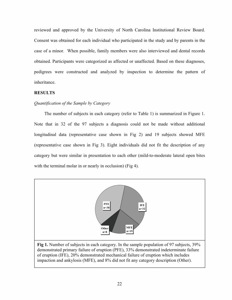
The number of subjects in each category (refer to Table 1) is summarized in Figure 1.
Note that in 32 of the 97 subjects a diagnosis could not be made without additional
longitudinal data (representative case shown in Fig 2) and 19 subjects showed MFE
(representative case shown in Fig 3). Eight individuals did not fit the description of any
category but were similar in presentation to each other (mild-to-moderate lateral open bites
with the terminal molar in or nearly in occlusion) (Fig 4).
MFEn=19
Othern=8
IFEn=32
PFEn=38
Fig 1. Number of subjects in each category. In the sample population of 97 subjects, 39% demonstrated primary failure of eruption (PFE), 33% demonstrated indeterminate failure of eruption (IFE), 20% demonstrated mechanical failure of eruption which includes impaction and ankylosis (MFE), and 8% did not fit any category description (Other).
23
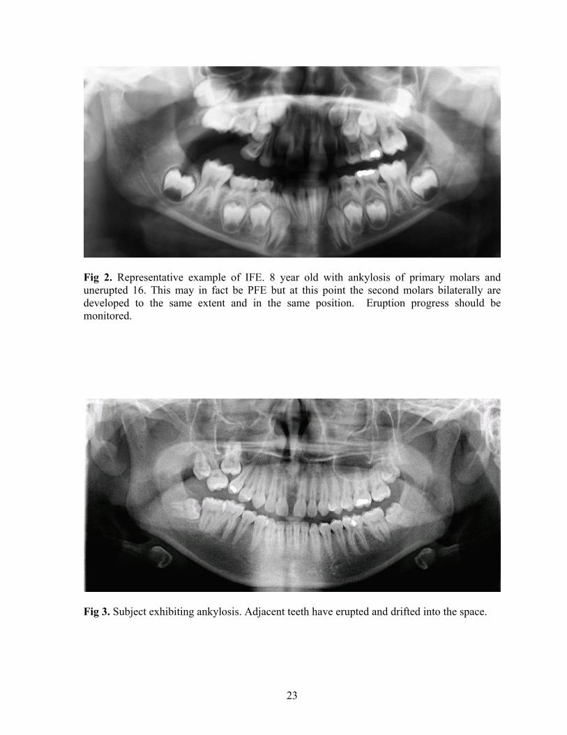
Fig 2. Representative example of IFE. 8 year old with ankylosis of primary molars and unerupted 16. This may in fact be PFE but at this point the second molars bilaterally are developed to the same extent and in the same position. Eruption progress should be monitored.
Fig 3. Subject exhibiting ankylosis. Adjacent teeth have erupted and drifted into the space.

24
Fig 4. Subject demonstrating mild lateral open bite on the right side and moderate lateral open bite on the left. There is no indication of a failed eruption mechanism.

25
Characterization of PFE
The location of affected teeth in the PFE group is shown in Figure 5. Subjects had
affected teeth as far forward as the first premolars with increasing frequency toward the first
and second molars. In most cases subjects were too young to evaluate the third molars, and
only those third molars that were obviously affected were counted in the distribution.
The PFE group showed two distinguishable forms. One group (17 of 38) had a similar
lack of eruption potential of all affected teeth with a progressive open bite from anterior to
posterior (Fig 6). The second group (11 of 38) had a distal tooth with greater although
inadequate eruption; therefore, the eruption potential varied among the affected teeth (Fig 7).
Finally, ten of the cases showed a coexistence of the two types in different quadrants within
the same patient. Sometimes MFE was also present in yet another quadrant.
-30
-20
-10
0
10
20
30
8 7 6 5 4 3 2 1 1 2 3 4 5 6 7 8
Teeth
Num
ber
of A
ffec
ted
Tee
th
MaxillaMandible
Fig 5. Distribution of affected teeth in the PFE group. Overall distribution among the four quadrants and between maxillary and mandibular teeth was fairly equal, although individual cases were rarely symmetric.
26
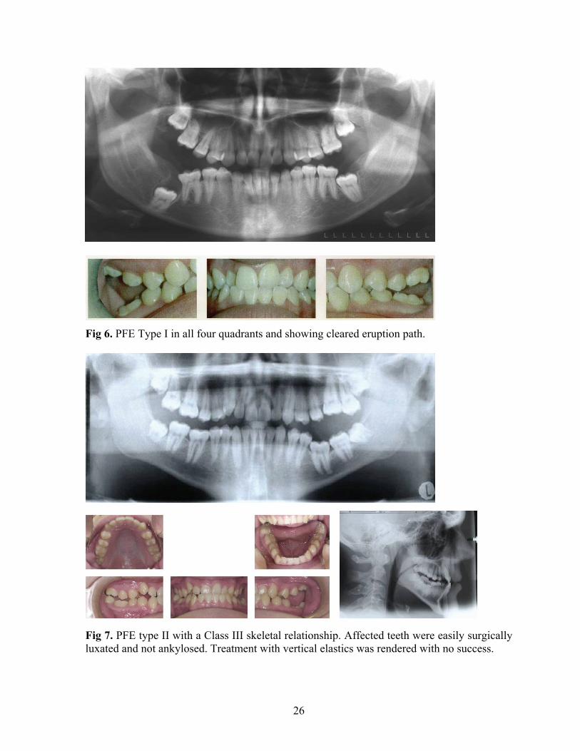
Fig 6. PFE Type I in all four quadrants and showing cleared eruption path.
Fig 7. PFE type II with a Class III skeletal relationship. Affected teeth were easily surgically luxated and not ankylosed. Treatment with vertical elastics was rendered with no success.

27
Further characterization of PFE was carried out for the 29 subjects who had
cephalometric records available. The Angle classifications for the sample population, the
PFE group and the familial group are shown in Table II. Note the relative high percentage of
Class III skeletal pattern. On further examination of the subgroups of PFE, there appears to
be no difference, other than the varied eruption potential of affected teeth, between subjects
with PFE Types I and II and those in the familial group.
Overall PFE Familial
Class I 62% 55% 38%
Class II 10% 10% 38%
Class III 28% 35% 25%
Table II. Angle classification of sample.
Other Radiographic Findings
At least one ankylosed primary tooth was noted in 24 of the 97 subjects (PFE = 8, IFE
= 12, MFE = 2, other = 2). Dental anomalies were fairly rare and did not appear to be
associated with PFE. In the entire sample of 97, four subjects showed hypodontia (IFE = 3,
MFE = 1), five subjects showed hyperdontia (PFE = 2, IFE = 2, MFE = 1) and three subjects
showed taurodontism (IFE = 3).
Description of the Familial Subjects
Twenty-six percent of the PFE cases in this sample were familial (10 out of 38). There
was no obvious difference in the types of PFE expressed by family members versus the
isolated cases. Figure 8 shows PFE in a mother and daughter. Five other subjects within the
sample population of 97 reported familial eruption problems. Two subjects who were

28
brothers were classified as IFE because they were too young for diagnosis, and the other
three (classified as either IFE or MFE) were related to PFE subjects. Other than a high
prevalence of ankylosed primary molars (5 out 15 or 33%), no other dental anomalies were
found in the familial group.
Fig 8. PFE in a mother (A) and daughter (B). Mother is affected in all four quadrants and has been treated with multiple extractions. Daughter has ankylosed primary teeth and is bilaterally affected although more severe on the right.
Pattern of Inheritance
Of the nine families who had a reported familial history of eruption problems four
pedigrees were constructed. One of these pedigrees is shown in Figure 9. Pedigree analysis
by inspection strongly suggests an autosomal dominant inheritance pattern in that both sexes
B
B
B
A
A

29
were affected without preference, about half the members in the kindred were affected, and
the trait did not skip generations. The possibility of an X-linked autosomal dominant
inheritance pattern cannot be excluded; however, this mode of inheritance is extremely rare
and therefore a less likely candidate.
Fig 9. Pedigree of PFE-001. Analysis by inspection shows autosomal dominance with complete penetrance.
DISCUSSION
Characteristics of PFE
The original characteristics of PFE identified by Proffit and Vig in 1981 are still valid
today. (1)Posterior teeth are more frequently involved; (2) the teeth distal to the first affected
tooth are also affected to some degree; (3) involved teeth may erupt partially and then cease
to erupt, relatively submerging although not ankylosed; (4) deciduous molars are likely to be
involved; (5) the condition is rarely symmetric, frequently unilateral, but could be bilateral;
(6) involved permanent teeth tend to become ankylosed at some point; (7) orthodontic forces
lead to ankylosis rather than normal tooth movement.5
II:1 II:2
III:1 III:2 III:3 III:4 III:5
I:1 I:2
II:3 II:4 II:5

30
Because permanent molars develop from a distal extension of the same dental lamina
from which the primary teeth are formed, a gradient of eruption could explain why posterior
teeth are affected more often than anterior teeth.5 The condition can affect any quadrant, may
be unilateral or bilateral and is rarely symmetric. Involved teeth do not respond normally to
orthodontic forces.
New findings from this study indicate two distinguishable types of PFE which may be
related to the timing of onset. In Type I, which is the classic form described initially, loss of
eruption potential appears to strike at a certain chronologic time leading to a similar lack of
eruption potential of all the affected teeth. In Type II, the timing of onset may be related to
the stage of root development, leading to a varied eruption potential among affected teeth. In
a significant number of cases a combination of the two types was found, and a few cases
showed PFE in one quadrant coupled with a single ankylosed tooth in a different quadrant.
Therefore, PFE and ankylosis may be closely related, as the studies by Raghoebar seem to
show.12,13 Perhaps an abnormal periodontal ligament can lead to either condition.
Association between Eruption Failure and Skeletal Relationship
Within the small subset of this sample population for which a lateral cephalogram was
available, a high percentage of subjects demonstrated a skeletal Class III relationship.2 This
finding is previously unreported. Of the other publications on PFE, only a few account for
the skeletal relationships of some of their subjects. Proffit and Vig reported on one subject in
eight who had a Class III relationship.5 Ireland had two Class I subjects.8 Brady reported one
out of two with a Class II pattern.7 Dibiase and Leggat reported that both of their subjects
were Class II.10 Since failure of permanent molars to erupt is so rare,14-16 finding a sample
size large enough to study the characteristics of the condition has been a difficult problem.

31
Role of Heredity in PFE
Reports of a definite familial tendency associated with PFE indicate the cause of the
developmental disturbance in the periodontal ligament may be inheritable.6,12 In this study
26% of the PFE cases were familial. Raghoebar reported a heritable component to eruption
failure in 10% of his cases, while other individual case reports provide studies of a few single
families.7-10,17
Pedigree analysis by inspection of the familial cases in this study is highly suggestive
of an autosomal dominant inheritance pattern with complete penetrance and variable
expressivity. Most of the familial studies in the literature also report an autosomal dominant
inheritance pattern;6,7,17 however, Winter reported one family as autosomal recessive.17
Etiology of PFE
Although none of the cases examined by Proffit and Vig in the original study had
similarly affected relatives, they did suppose that a genetic disturbance of varying penetrance
and expressivity was the likely etiology. The current reports of affected families support this
hypothesis,6-10 and suggest that spontaneous mutation(s) may account for the cases with no
previous family history. Perhaps this leads to a local disturbance in metabolic activity or
altered blood flow which then hinders the eruption mechanism. Raghoebar et al, based on
histologic examination of 26 molars from 20 patients, suggest that the mechanism is
replacement of cementoblasts by osteoblasts due to a local disturbance in the PDL during the
repair process of local physiologic resorption.12
The best evidence of a failure in the eruption mechanism is bone resorption without
tooth movement. Affected teeth that have been surgically exposed are generally reported to
be easily movable within the crypt and not ankylosed. Although these teeth may have some

32
slight response to orthodontic forces, the response is abnormal and the teeth invariably
become ankylosed before reaching occlusion. Case studies illustrate that not only do affected
teeth fail to respond to treatment, but also adjacent normal teeth are adversely affected by
intrusion to the level of the affected teeth (Fig 10). Winter and Raghoebar also concluded that
ankylosis in the failed eruptive process may be a secondary rather than initiating process and
reiterate that orthodontic procedures designed to improve eruption are doomed to failure in
individuals with PFE.12,17
Fig 10. Orthodontic intrusion of normal teeth. Attempt at orthodontic treatment led to intrusion of normal teeth mesial to the affected teeth.
Differential Diagnosis
In the diagnosis of eruption failures, the first step is to rule out local, systemic and
endocrine factors. Endocrine abnormalities have not been identified in PFE or ankylosis

33
patients to our knowledge. Ultimately, the principal differential diagnosis is mechanical
obstruction versus a failed eruption mechanism. Distinguishing between the two is key to
determining the prognosis for the affected teeth. Unfortunately, MFE and PFE have very
similar presentations in the early stages and definitive diagnosis cannot be made without
sufficient longitudinal data and therapeutic diagnosis (an attempt at orthodontically erupting
the tooth or teeth that may or may not be affected).
The first encounter with these patients often occurs around age 8 or 9 when an
asymmetry in the eruption pattern of the first permanent molars is noticed. The conservative
approach is to take a panoramic radiograph with the patient’s teeth together and recall in 6-12
months to determine eruption progress. Evaluation at recall will show progress, no change, or
relative submergence. If there is eruption progress, PFE and ankylosis can be ruled out.
Ultimately assessing the eruption capacity of the neighboring teeth is the only way to
distinguish PFE from ankylosis. The number of teeth affected, a positive family history and a
skeletal Class III relationship may provide valuable clues. Differentiation between the two
types of PFE cannot be done until at least age 14 or 15 when the second molar either
completely fails to erupt or erupts partially and then stops.
Clinical Application: Current View on Patient Management
Once PFE has been diagnosed, treatment options are disappointing and limited. Patients
and orthodontists must often either accept premolar occlusion or opt for more invasive
techniques. In the mildest of cases, teeth may be restored with onlays and crowns;18 however,
definitive restorations should not be placed prior to completion of vertical growth. For
moderate cases, extraction of teeth with placement of implants may be an option. Another
option may be a small segmental osteotomy to surgically reposition teeth into occlusion.5 In

34
the severest of cases, a significant deficit in alveolar bone height precludes implant
restorations as well as subapical osteotomy. One report of distraction osteogenesis to correct
an extreme posterior open bite provides an interesting potential treatment alternative.19
Sometimes the only feasible option is a removable prosthesis.20
CONCLUSIONS
Primary failure of eruption is a rare condition that can lead to spectacular posterior
open bites. It is difficult to diagnose and even more difficult to treat due to the lack of
response to orthodontic forces. Proper diagnosis can save the patient and the orthodontist
years of frustration and disappointment. The developmental disturbance that leads to PFE
may be inheritable. Future studies to determine the genetic etiology of PFE can aid in
differential diagnosis, allow early identification of affected family members and may
eventually lead to new treatment modalities.
Findings
The original characteristics of PFE identified 1981are still valid today
Two distinguishable forms of PFE were identified. Type I had a similar lack of eruption
potential of all affected teeth, and Type II had a varied eruption potential.
About a third of the cases showed a coexistence of the two types in different quadrants.
Sometimes a mechanical failure was also present in yet another quadrant.
26% of the PFE cases were familial; analysis by inspection revealed an autosomal
dominant inheritance pattern of complete penetrance and variable expressivity.
No difference other than eruption capacity of affected teeth was identified between
subjects with PFE Types I and II and those with familial PFE.

35
Differential diagnosis requires adequate longitudinal data and sometimes therapeutic
diagnosis (orthodontic traction); family history and skeletal pattern may provide valuable
information.
Although there are many reasons why a tooth may fail to erupt, determining the origin
is difficult yet vital to the success of orthodontic treatment. Eruption failure cases are among
the most challenging that an orthodontist will encounter. Not only does proper eruption
critically affect the success of the occlusal outcome, but it also greatly affects the efficiency
of treatment.
36
REFERENCES
1. Cahill DR. Eruption pathway formation in the presence of experimental tooth impaction in puppies. Anat Rec. 1969 May;164(1):67-77.
2. Proffit WR, Fields HW. Contemporary Orthodontics. 3rd ed. St. Louis: Mosby; c2000.
3. O'Connell AC, Torske KR. Primary failure of tooth eruption: a unique case. Oral Surg Oral Med Oral Pathol Oral Radiol Endod. 1999 Jun;87(6):714-20.
4. Proffit WR. Equilibrium theory revisited: factors influencing position of the teeth. Angle Orthod. 1978 Jul;48(3):175-86.
5. Proffit WR, Vig KW. Primary failure of eruption: a possible cause of posterior open-bite. Am J Orthod. 1981 Aug;80(2):173-90.
6. Bosker H, ten Kate LP, Nijenhuis LE. Familial reinclusion of permanent molars. Clin Genet. 1978 Mar;13(3):314-20.
7. Brady J. Familial primary failure of eruption of permanent teeth. Br J Orthod. 1990 May;17(2):109-13.
8. Ireland AJ. Familial posterior open bite: a primary failure of eruption. Br J Orthod. 1991 Aug;18(3):233-7.
9. Raghoebar GM, Ten Kate LP, Hazenberg CA, Boering G, Vissink A. Secondary retention of permanent molars: a report of five families. J Dent. 1992 Oct;20(5):277-82.
10. Dibiase AT, Leggat TG. Primary failure of eruption in the permanent dentition of siblings. Int J Paediatr Dent. 2000 Jun;10(2):153-7.
11. Demirjian A, Goldstein H, Tanner JM. A new system of dental age assessment. Hum Biol. 1973 May;45(2):211-27.
12. Raghoebar GM, Boering G, Jansen HW, Vissink A. Secondary retention of permanent molars: a histologic study. J Oral Pathol Med. 1989 Sep;18(8):427-31.
13. Raghoebar GM, Boering G, Vissink A, Stegenga B. Eruption disturbances of permanent molars: a review. J Oral Pathol Med. 1991 Apr;20(4):159-66.
14. Grover PS, Lorton L. The incidence of unerupted permanent teeth and related clinical cases. Oral Surg Oral Med Oral Pathol. 1985 Apr;59(4):420-5.
15. Johnsen DC. Prevalence of delayed emergence of permanent teeth as a result of local factors. J Am Dent Assoc. 1977 Jan;94(1):100-6.
16. Nagpal A, Sharma G, Sarkar A, Pai KM. Eruption disturbances: an aetiological-cum-management perspective. Dentomaxillofac Radiol. 2005 Jan;34(1):59-63.
37
17. Winter GB, Gelbier MJ, Goodman JR. Severe Infra-occlusion and failed eruption of deciduous molars associated with eruptive and developmental disturbances in the permanent dentition: a report of 28 selected cases. Br J Orthod. 1997 May;24(2):149-57.
18. Yatani H, Watanabe EK, Kaneshima T, Yamashita A, Suzuki K. Etched-porcelain resin-bonded onlay technique for posterior teeth. J Esthet Dent. 1998;10(6):325-32.
19. Kater WM, Kawa D, Schafer D, Toll D. Treatment of posterior open bite using distraction osteogenesis. J Clin Orthod. 2004 Sep;38(9):501,4; quiz 487-8.
20. Siegel SC, O'Connell A. Oral rehabilitation of a child with primary failure of tooth eruption. J Prosthodont. 1999 Sep;8(3):201-7.