Primary Extrauterine Endometrial Stromal Sarcoma in the ...€¦ · Primary Extrauterine...
6
pISSN 2287-9714 eISSN 2287-9722 www.coloproctol.org Annals of Coloproctology www.coloproctol.org 68 Primary Extrauterine Endometrial Stromal Sarcoma in the Sigmoid Colon Hyun-Jin Son, Joo-Heon Kim, Dong-Wook Kang, Hye-Kyung Lee, Mee-Ja Park, Seung Yun Lee Department of Pathology, Eulji University School of Medicine, Daejeon, Korea Case Report Ann Coloproctol 2015;31(2):68-73 http://dx.doi.org/10.3393/ac.2015.31.2.68 An endometrial stromal sarcoma (ESS) is an uncommon uterine neoplasm, and its primary occurrence in the intestine as an extrauterine ESS (EESS) is exceedingly rare. We hereby report a primary EESS arising in the sigmoid colon with a review of the literature. A 52-year-old woman presented with bloody stool and underwent a colon fiberscopy, which revealed a fungating mass obstructing the lumen at the distal sigmoid. A laparoscopic low anterior resection was performed, and an umbilicated polypoid mass was identified; on section, it had infiltrated the mesocolic fat and measured 3.8 cm × 2.5 cm. The tumor showed geographic sheets or nests composed of relatively monotonous stromal cells, expansion or infiltration to the proper muscle and mesocolic fat, and extensive lymphovascular invasion and metastasis to regional lymph nodes and the pelvic peritoneum. The tumor cells were strongly and diffusely immunoreactive for CD10, but negative for c-kit, CD34, and Dog1. Two months later, a hysterectomy with a bilateral salpingo-oophorectomy was performed, and no evidence of an ESS was found in the uterus. Keywords: Endometrial stromal sarcoma; Extrauterine; Sigmoid colon; CD10 INTRODUCTION An endometrial stromal sarcoma (ESS), an uncommon uterine mesenchymal neoplasm, accounts for approximately 0.2% of all uterine malignancies [1]. Its occurrence at extrauterine sites is very rare, but it has been reported to occur at the ovary, bowel wall, ab- domen, peritoneum, pelvis, and vagina [2]. Primary gastrointesti- nal involvement is rare [3-13]. We hereby present a case of a pri- mary low-grade extrauterine ESS (EESS) in the sigmoid colon with a review of the literature. No evidence of a primary uterine ESS was found. CASE REPORT A 52-year-old woman was transferred from an outside clinic in order to evaluate the bloody stool and a pedunculated mass in the sigmoid colon that had been detected in a rigid sigmoid-scope ex- amination. She was suffering from constipation, unspecified ab- dominal pain and hematochezia for a month. Her past and family histories were all unremarkable. Laboratory findings were unre- markable, except for mild anemia. Colon fiberscopy was performed and revealed a fungating mass obstructing the lumen at the distal sigmoid colon (20 cm from the anal verge) (Fig. 1A). Under the impression of colon cancer, a bi- opsy was taken and suggested the possibility of a gastrointestinal stromal tumor (GIST) showing uncertain malignant potential. Abdominal computed tomography showed an eccentric and irreg- ular wall thickening with enhancement measuring about 5 cm and perirectal lymph node enlargement (T3 or T4aN1M0, stage IIIB) (Fig. 1B). A laparoscopic low anterior resection was carried out, and an umbilicated polypoid mass measuring about 3.8 cm × 2.5 cm was identified in the distal sigmoid colon; on serial sections, it was found to have infiltrated the mesocolic fat (Fig. 1C, D). No di- lation of the proximal bowel side to the mass was identified. On histopathologic findings, the tumor showed geographic sheets or nests expanding to or infiltrating into the proper muscle and mesocolic fat (Fig. 2A), extensive lymphovascular invasion (Fig. 2B), and metastasis to regional lymph nodes (Fig. 2C) and to the pelvic peritoneum. In spite of extensive sampling, no focus of endometriosis was found in the resected colon specimen. Uni- Received: December 11, 2014 • Accepted: February 6, 2015 Correspondence to: Hyun-Jin Son, M.D. Department of Pathology, Eulji University Hospital, 95 Dunsanseo-ro, Seo-gu, Daejeon 302-799, Korea Tel: +82-42-611-3451, Fax: +82-42-611-3459 E-mail: [email protected] © 2015 The Korean Society of Coloproctology This is an open-access article distributed under the terms of the Creative Commons Attribution Non- Commercial License (http://creativecommons.org/licenses/by-nc/3.0) which permits unrestricted non- commercial use, distribution, and reproduction in any medium, provided the original work is properly cited.
Transcript of Primary Extrauterine Endometrial Stromal Sarcoma in the ...€¦ · Primary Extrauterine...
pISSN 2287-9714 eISSN 2287-9722www.coloproctol.org
Annals of
Coloproctology
www.coloproctol.org68
Primary Extrauterine Endometrial Stromal Sarcoma in the Sigmoid Colon
Hyun-Jin Son, Joo-Heon Kim, Dong-Wook Kang, Hye-Kyung Lee, Mee-Ja Park, Seung Yun LeeDepartment of Pathology, Eulji University School of Medicine, Daejeon, Korea
Case Report
Ann Coloproctol 2015;31(2):68-73http://dx.doi.org/10.3393/ac.2015.31.2.68
An endometrial stromal sarcoma (ESS) is an uncommon uterine neoplasm, and its primary occurrence in the intestine as an extrauterine ESS (EESS) is exceedingly rare. We hereby report a primary EESS arising in the sigmoid colon with a review of the literature. A 52-year-old woman presented with bloody stool and underwent a colon fiberscopy, which revealed a fungating mass obstructing the lumen at the distal sigmoid. A laparoscopic low anterior resection was performed, and an umbilicated polypoid mass was identified; on section, it had infiltrated the mesocolic fat and measured 3.8 cm × 2.5 cm. The tumor showed geographic sheets or nests composed of relatively monotonous stromal cells, expansion or infiltration to the proper muscle and mesocolic fat, and extensive lymphovascular invasion and metastasis to regional lymph nodes and the pelvic peritoneum. The tumor cells were strongly and diffusely immunoreactive for CD10, but negative for c-kit, CD34, and Dog1. Two months later, a hysterectomy with a bilateral salpingo-oophorectomy was performed, and no evidence of an ESS was found in the uterus.
Keywords: Endometrial stromal sarcoma; Extrauterine; Sigmoid colon; CD10
INTRODUCTION
An endometrial stromal sarcoma (ESS), an uncommon uterine mesenchymal neoplasm, accounts for approximately 0.2% of all uterine malignancies [1]. Its occurrence at extrauterine sites is very rare, but it has been reported to occur at the ovary, bowel wall, ab-domen, peritoneum, pelvis, and vagina [2]. Primary gastrointesti-nal involvement is rare [3-13]. We hereby present a case of a pri-mary low-grade extrauterine ESS (EESS) in the sigmoid colon with a review of the literature. No evidence of a primary uterine ESS was found.
CASE REPORT
A 52-year-old woman was transferred from an outside clinic in
order to evaluate the bloody stool and a pedunculated mass in the sigmoid colon that had been detected in a rigid sigmoid-scope ex-amination. She was suffering from constipation, unspecified ab-dominal pain and hematochezia for a month. Her past and family histories were all unremarkable. Laboratory findings were unre-markable, except for mild anemia.
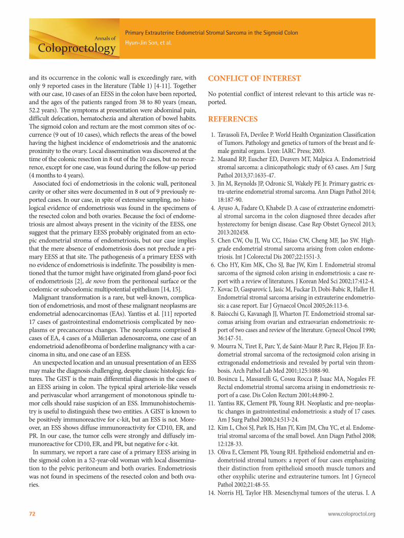
Colon fiberscopy was performed and revealed a fungating mass obstructing the lumen at the distal sigmoid colon (20 cm from the anal verge) (Fig. 1A). Under the impression of colon cancer, a bi-opsy was taken and suggested the possibility of a gastrointestinal stromal tumor (GIST) showing uncertain malignant potential. Abdominal computed tomography showed an eccentric and irreg-ular wall thickening with enhancement measuring about 5 cm and perirectal lymph node enlargement (T3 or T4aN1M0, stage IIIB) (Fig. 1B). A laparoscopic low anterior resection was carried out, and an umbilicated polypoid mass measuring about 3.8 cm × 2.5 cm was identified in the distal sigmoid colon; on serial sections, it was found to have infiltrated the mesocolic fat (Fig. 1C, D). No di-lation of the proximal bowel side to the mass was identified.
On histopathologic findings, the tumor showed geographic sheets or nests expanding to or infiltrating into the proper muscle and mesocolic fat (Fig. 2A), extensive lymphovascular invasion (Fig. 2B), and metastasis to regional lymph nodes (Fig. 2C) and to the pelvic peritoneum. In spite of extensive sampling, no focus of endometriosis was found in the resected colon specimen. Uni-
Received: December 11, 2014 • Accepted: February 6, 2015Correspondence to: Hyun-Jin Son, M.D.Department of Pathology, Eulji University Hospital, 95 Dunsanseo-ro, Seo-gu, Daejeon 302-799, KoreaTel: +82-42-611-3451, Fax: +82-42-611-3459E-mail: [email protected]
© 2015 The Korean Society of ColoproctologyThis is an open-access article distributed under the terms of the Creative Commons Attribution Non-Commercial License (http://creativecommons.org/licenses/by-nc/3.0) which permits unrestricted non-commercial use, distribution, and reproduction in any medium, provided the original work is properly cited.

Annals of
Coloproctology
www.coloproctol.org 69
Volume 31, Number 2, 2015
Ann Coloproctol 2015;31(2):68-73
formly small, bland tumor cells with rounded-ovoid nuclei, evenly dispersed chromatin, no or inconspicuous nucleoli, and scanty cytoplasm with indistinct cell borders were noticed (Fig. 2D). Small, prominent arterioles were scattered among the stromal tu-mor cells. No mitosis or necrosis was identified. The tumor cells were strongly and diffusely immunoreactive for CD10 (1:50, No-vocastra, Newcastle, UK) (Fig. 2E), estrogen receptor (ER; 1:40, Novocastra), and progesterone receptor (PR; 1:200, Novocastra),
but negative for c-kit (1:600, Dako, Carpinteria, CA, USA), CD34 (predilution, Novocastra), Dog1 (1:500, Dako), α-smooth muscle actin (1:100, Novocastra), S100 protein (1:200, Novocastra), des-min (1:50, Dako) and pan-cytokeratin (1:200, Novocastra). The Ki-67 (1:100, Dako) labeling index was 8% (Fig. 2F).
Endometrial curettage was conducted postoperatively and showed secretory hyperplasia and no evidence of ESS. Two months later, a total abdominal hysterectomy with a bilateral sal-
Fig. 1. (A) Colon fiberscopy reveals a fungating mass obstructing the lumen at the distal sigmoid colon (20 cm from the anal verge). (B) Con-trast-enhanced axial abdomen computed tomography shows an eccentric and irregular wall thickening with enhancement in the distal sig-moid colon measuring about 5 cm and a pericolic lymph node enlargement. (C) Grossly, an umbilicated polypoid mass measuring about 3.8 cm × 2.5 cm is identified in the distal sigmoid colon, and (D) on serial sections, it was found to have infiltrated to the mesocolic fat and to in-volve a regional lymph node.
A
C
B
D

Annals of
Coloproctology
www.coloproctol.org70
Primary Extrauterine Endometrial Stromal Sarcoma in the Sigmoid Colon
Hyun-Jin Son, et al.
pingo-oophorectomy was performed. The uterus measured 6.0 cm × 10.0 cm × 4.5 cm, and upon cross section, the uterine wall was thickened up to 3.2 cm and revealed an ill-defined trabecu-lated area with several hemorrhagic foci and a myomatous mass in the low uterine segment (Fig. 3A, B). Microscopically, it was di-agnosed as adenomyosis, an intramural leiomyoma, and a rela-tively atrophic endometrium (Fig. 3C). Grossly, the bilateral ad-
nexae were unremarkable, but microscopic foci of the endome-trial stromal nodule were identified in the right and left ovaries and measured 0.6 cm and 0.8 cm, respectively (Fig. 3D). However, no focus of endometriosis was found in the uterus or bilateral ad-nexae. The patient was alive four months after the resection with no evidence of recurrence.
Fig. 2. (A) The tumor shows geographic sheets or nests expanding to or infiltrating into the proper muscle and mesocolic fat (H&E, ×12.5). (B) Extensive lymphovascular invasion (H&E, ×40) and (C) metastasis to regional lymph nodes (H&E, ×40) are seen. (D) Uniformly small, bland tumor cells have rounded-ovoid nuclei, evenly dispersed chromatin, no or inconspicuous nucleoli, and scanty cytoplasm with indistinct cell borders. Small, prominent arterioles are scattered among the stromal tumor cells (H&E, ×200). (E) The tumor cells are strongly and diffusely immunoreactive for CD10 (×12.5). (F) The Ki-67 labeling index is 8% (×100).
A
D
B
E
C
F
Fig. 3. (A) The uterus measures 6.0 cm × 10.0 cm × 4.5 cm, and bilateral adnexae are grossly unremarkable. (B) Upon cross sectioning, the uterine wall was found to have thickened by 3.2 cm, and an ill-defined trabeculated area with several hemorrhagic foci was revealed. (C) Mi-croscopically, the endometrium is relatively atrophic, and the uterine wall is thickened due to adenomyosis (H&E, ×40). (D) A microscopic fo-cus of the endometrial stromal nodule is identified in the ovary (H&E, ×40) (Inset: high power view of the endometrial stromal nodule [H&E, ×200]).
DCBA

Annals of
Coloproctology
www.coloproctol.org 71
Volume 31, Number 2, 2015
Ann Coloproctol 2015;31(2):68-73
Table 1. Clinical and pathological features of an EESS arising in the colon
Age(yr)
Pasthistory
Symptomsat presentation
Involvingcolon site(s)
Gross findings, colon
Foci ofendometriosis
Treatment Follow-up Dissemination Ref.
80 TAH, BSO Rectal bleeding and chronic discharge
Sigmoid colon 5-cm Mass involving mucosa, muscularis, and peritoneum
Ovary Low anterior colon resection
NED, 4 yr
Local [4]
42 Lung cancer, chemotherapy
Difficult defecation, rectal bleeding
Sigmoid colon Multiple 1- to 3-cm nodular masses involving mucosa to pericolic fat
Sigmoid colon
Rectosigmoidectomy, total hysterectomy, BSO
NED, 1 yr
Local (omentum, left adnexa)
[5]
48 Subtotal hysterectomy -> myoma, LSO -> endometriosis
Difficult defecation and tenesmus
Sigmoid colon Multinodular masses of 1–3 cm involving the whole layer of the intestine and extending to the urinary bladder and ureter
Left ovary sigmoid colon
Segmental resection
NED, 4 mo
Local (mesentery, urinary bladder, ureter)
[6]
46 TAH, RSO -> leiomyomas, normal adnexa
Stenosing process Rectosigmoid colon
A tumor 6 cm in diameter
Rectosigmoid colon
Oophorectomy, tumorectomy, omentectomy, resection of colon
NED, 11 mo
Local (ovary, omentum)
[7]
38 Ovarian cystectomy -> endometriosis, -> TAH, BSO
Abdominal pain and pressure
Ascending and transverse colon, terminal ileum
A large multilobular mass involving the transverse, ascending colon and the terminal ileum
Ovary, colon Partial ileal resection, resection of the transverse and ascending colon -> chemotherapy
NED Local (falciform, gastrocolic ligament, mesentery, pelvis)
[8]
50 TAH, RSO -> endometriosis
Abdominal pain Transverse colon, junction of descending and sigmoid colon
A large grapelike tumor, with individual nodular areas 2 cm × 2.5 cm
Ovary LSO, radical omentectomy -> hormonal therapy
NED Local (omentum)
[8]
61 None Epigastric pain Rectosigmoid colon
An 2.7-cm polypoid mass with stenosis of the lumen involving all layers of the rectal wall
Posterior wall of right broad ligament
Resection of rectosigmoid colon
NED, 30 mo
None [9]
42 None Fever and abdominal pain
Rectum Massive transmural infiltration involving mucosa, which was polypoid and ulcerated
Adventitial rectal layer
TAH, BSO, omentectomy, colorectal resection -> chemotherapy
NED, 20 mo
Local (serosal uterine, parametrial, peritoneal lymphatics)
[10]
63 NA NA Rectum 2-cm Polypoid mass NA Partial colectomy -> radiation therapy
Recurrent, 3 yr
NA [11]
52 None Constipation, unspecified abdominal pain, hematochezia
Sigmoid colon An umbilicated polypoid mass measuring about 3.8 cm × 2.5 cm involving the whole layer of the intestine to pericolic fat
Not identified Laparoscopic low anterior resection TAH, BSO
NED, 4 mo
Local (pelvic peritoneum, both ovaries)
Present case
EESS, extrauterine endometrial stromal sarcoma; TAH, total abdominal hysterectomy; BSO, bilateral salpingo-oophorectomy; NED, no evidence of disease; LSO, left sal-pingo-oophorectomy; RSO, right salpingo-oophorectomy; NA, not available.
DISCUSSION
An ESS arising outside the uterus at extraovarian sites in the ab-
sence of a primary uterine lesion is extremely rare, and only sev-eral case reports are present in the English literature. The abdo-men and the pelvis are the most common locations for an EESS,
Annals of
Coloproctology
www.coloproctol.org72
Primary Extrauterine Endometrial Stromal Sarcoma in the Sigmoid Colon
Hyun-Jin Son, et al.
and its occurrence in the colonic wall is exceedingly rare, with only 9 reported cases in the literature (Table 1) [4-11]. Together with our case, 10 cases of an EESS in the colon have been reported, and the ages of the patients ranged from 38 to 80 years (mean, 52.2 years). The symptoms at presentation were abdominal pain, difficult defecation, hematochezia and alteration of bowel habits. The sigmoid colon and rectum are the most common sites of oc-currence (9 out of 10 cases), which reflects the areas of the bowel having the highest incidence of endometriosis and the anatomic proximity to the ovary. Local dissemination was discovered at the time of the colonic resection in 8 out of the 10 cases, but no recur-rence, except for one case, was found during the follow-up period (4 months to 4 years).
Associated foci of endometriosis in the colonic wall, peritoneal cavity or other sites were documented in 8 out of 9 previously re-ported cases. In our case, in spite of extensive sampling, no histo-logical evidence of endometriosis was found in the specimens of the resected colon and both ovaries. Because the foci of endome-triosis are almost always present in the vicinity of the EESS, one suggest that the primary EESS probably originated from an ecto-pic endometrial stroma of endometriosis, but our case implies that the mere absence of endometriosis does not preclude a pri-mary EESS at that site. The pathogenesis of a primary EESS with no evidence of endometriosis is indefinite. The possibility is men-tioned that the tumor might have originated from gland-poor foci of endometriosis [2], de novo from the peritoneal surface or the coelomic or subcoelomic multipotential epithelium [14, 15].
Malignant transformation is a rare, but well-known, complica-tion of endometriosis, and most of these malignant neoplasms are endometrial adenocarcinomas (EAs). Yantiss et al. [11] reported 17 cases of gastrointestinal endometriosis complicated by neo-plasms or precancerous changes. The neoplasms comprised 8 cases of EA, 4 cases of a Müllerian adenosarcoma, one case of an endometrioid adenofibroma of borderline malignancy with a car-cinoma in situ, and one case of an EESS.
An unexpected location and an unusual presentation of an EESS may make the diagnosis challenging, despite classic histologic fea-tures. The GIST is the main differential diagnosis in the cases of an EESS arising in colon. The typical spiral arteriole-like vessels and perivascular whorl arrangement of monotonous spindle tu-mor cells should raise suspicion of an ESS. Immunohistochemis-try is useful to distinguish these two entities. A GIST is known to be positively immunoreactive for c-kit, but an ESS is not. More-over, an ESS shows diffuse immunoreactivity for CD10, ER, and PR. In our case, the tumor cells were strongly and diffusely im-munoreactive for CD10, ER, and PR, but negative for c-kit.
In summary, we report a rare case of a primary EESS arising in the sigmoid colon in a 52-year-old woman with local dissemina-tion to the pelvic peritoneum and both ovaries. Endometriosis was not found in specimens of the resected colon and both ova-ries.
CONFLICT OF INTEREST
No potential conflict of interest relevant to this article was re-ported.
REFERENCES
1. Tavassoli FA, Devilee P. World Health Organization Classification of Tumors. Pathology and genetics of tumors of the breast and fe-male genital organs. Lyon: IARC Press; 2003.
2. Masand RP, Euscher ED, Deavers MT, Malpica A. Endometrioid stromal sarcoma: a clinicopathologic study of 63 cases. Am J Surg Pathol 2013;37:1635-47.
3. Jin M, Reynolds JP, Odronic SI, Wakely PE Jr. Primary gastric ex-tra-uterine endometrial stromal sarcoma. Ann Diagn Pathol 2014; 18:187-90.
4. Ayuso A, Fadare O, Khabele D. A case of extrauterine endometri-al stromal sarcoma in the colon diagnosed three decades after hysterectomy for benign disease. Case Rep Obstet Gynecol 2013; 2013:202458.
5. Chen CW, Ou JJ, Wu CC, Hsiao CW, Cheng MF, Jao SW. High-grade endometrial stromal sarcoma arising from colon endome-triosis. Int J Colorectal Dis 2007;22:1551-3.
6. Cho HY, Kim MK, Cho SJ, Bae JW, Kim I. Endometrial stromal sarcoma of the sigmoid colon arising in endometriosis: a case re-port with a review of literatures. J Korean Med Sci 2002;17:412-4.
7. Kovac D, Gasparovic I, Jasic M, Fuckar D, Dobi-Babic R, Haller H. Endometrial stromal sarcoma arising in extrauterine endometrio-sis: a case report. Eur J Gynaecol Oncol 2005;26:113-6.
8. Baiocchi G, Kavanagh JJ, Wharton JT. Endometrioid stromal sar-comas arising from ovarian and extraovarian endometriosis: re-port of two cases and review of the literature. Gynecol Oncol 1990; 36:147-51.
9. Mourra N, Tiret E, Parc Y, de Saint-Maur P, Parc R, Flejou JF. En-dometrial stromal sarcoma of the rectosigmoid colon arising in extragonadal endometriosis and revealed by portal vein throm-bosis. Arch Pathol Lab Med 2001;125:1088-90.
10. Bosincu L, Massarelli G, Cossu Rocca P, Isaac MA, Nogales FF. Rectal endometrial stromal sarcoma arising in endometriosis: re-port of a case. Dis Colon Rectum 2001;44:890-2.
11. Yantiss RK, Clement PB, Young RH. Neoplastic and pre-neoplas-tic changes in gastrointestinal endometriosis: a study of 17 cases. Am J Surg Pathol 2000;24:513-24.
12. Kim L, Choi SJ, Park IS, Han JY, Kim JM, Chu YC, et al. Endome-trial stromal sarcoma of the small bowel. Ann Diagn Pathol 2008; 12:128-33.
13. Oliva E, Clement PB, Young RH. Epithelioid endometrial and en-dometrioid stromal tumors: a report of four cases emphasizing their distinction from epithelioid smooth muscle tumors and other oxyphilic uterine and extrauterine tumors. Int J Gynecol Pathol 2002;21:48-55.
14. Norris HJ, Taylor HB. Mesenchymal tumors of the uterus. I. A

Annals of
Coloproctology
www.coloproctol.org 73
Volume 31, Number 2, 2015
Ann Coloproctol 2015;31(2):68-73
clinical and pathological study of 53 endometrial stromal tumors. Cancer 1966;19:755-66.
15. Lauchlan SC. The secondary Müllerian system. Obstet Gynecol Surv 1972;27:133-46.