Primary Care Orthopaedics Conference The Foot Panelcmetracker.net/EH/Files/EventMaterials/18026/FOOT...
61
1 Primary Care Orthopaedics Conference The Foot Panel Sheree Christian, PT Xan Courville, MD Boni Jo Silbernagel, DPM K jiS d h MD Kenji Sudoh, MD Laura Trombino, MD Michael Watson, CPO
Transcript of Primary Care Orthopaedics Conference The Foot Panelcmetracker.net/EH/Files/EventMaterials/18026/FOOT...

1
Primary Care Orthopaedics ConferenceThe Foot Panel
Sheree Christian, PTXan Courville, MDBoni Jo Silbernagel, DPMK ji S d h MDKenji Sudoh, MDLaura Trombino, MDMichael Watson, CPO
2
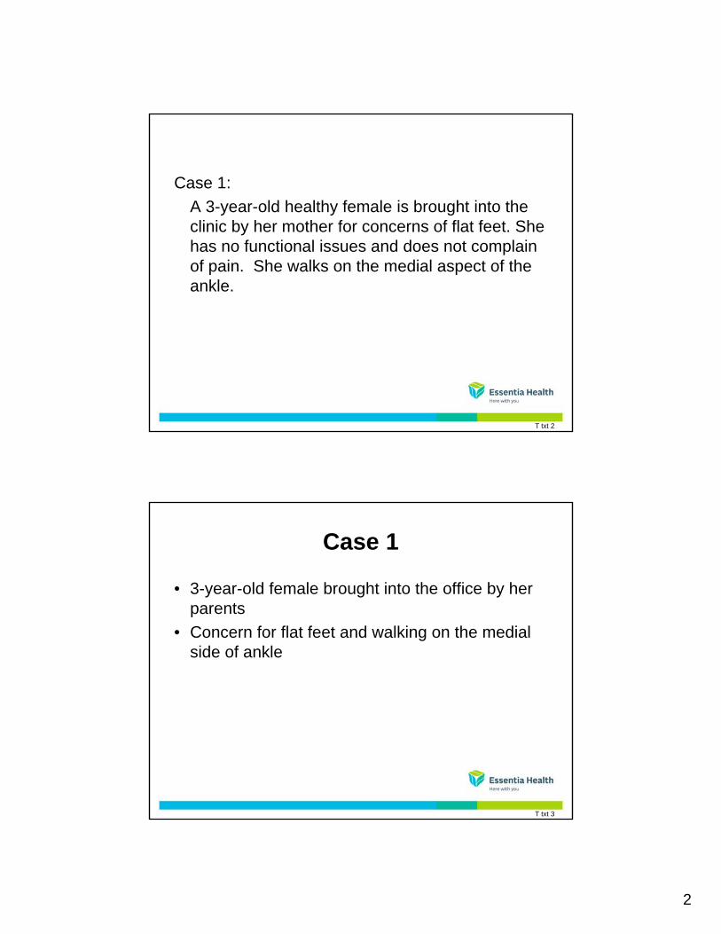
Case 1:
A 3-year-old healthy female is brought into the clinic by her mother for concerns of flat feet. She has no functional issues and does not complain of pain. She walks on the medial aspect of the ankle.
T txt 2
Case 1
• 3-year-old female brought into the office by her parentsparents
• Concern for flat feet and walking on the medial side of ankle
T txt 3
3
History
• No complaints of pain
P d t f NSVD t d li• Product of NSVD vertex delivery
• Normal developmental milestones
• Walking independently at 12 months
• No functional problems (runs, jumps, and climbs)
T txt 4
Physical Examination
• Spine—straight, no scoliosis or kyphosis
N l di li h i t h• No sacral dimpling or hairy patches
• Hips and knee full range of motion (FROM)
T txt 5
4
Physical Examination
• Spine—straight, no scoliosis or kyphosis
N l di li h i t h• No sacral dimpling or hairy patches
• Hips and knee full range of motion (FROM)
T txt 6
Foot and Ankle Alignment
• Pes planus
Mild hi df t kl l• Mild hindfoot ankle valgus
• Ankle range of motion (ROM) is full
• Dorsiflexion 20 degrees, plantarflexion full
• Excellent subtalar motion
• Heel goes from vagus to varusg g
T txt 7
5
Foot and Ankle Alignment
• Pes planus
Mild hi df t kl l• Mild hindfoot ankle valgus
• Ankle range of motion (ROM) is full
• Dorsiflexion 20 degrees, plantarflexion full
• Excellent subtalar motion
• Heel goes from vagus to varusg g
T txt 8
Evaluating Flat Feet in Children
• Flexible versus nonflexible
• Painful versus non painful
Sullivan, J A et al Journal of AAOS, 7:1:44-53
T txt 9
6
T txt 10
T txt 11
7
Natural History
• Studies from countries where most people don’t wear shoeswear shoes
• Patients with flexible flat feet are generally asymptomatic as adults
Rao et al, J Bone Joint Surg Br 1992, 74:525-27
Sachithananadam, V. J Bone Joint Surg Br 1995;77:254-257
T txt 12
Key Elements of Exam
• Arch developed until age 5
F tt ti i h• Fatty tissue in arch
• Tip-toe reconstitutes arch
• Walks on toes, heels, inner, and outer border of feet
• Arch when on toes
T txt 13
8
Best Prospective Study
• Wenger et al.
C t l• Control group
• Corrective shoes
• Helfet heel cups
• UCBL
No statistical difference in arch at 3-year follow-up.
T txt 14
Treatment
• Observation
S ft th ti• Soft orthotic
• Molded orthotic
• Heelcord tight– Stretching
– Casting
– Lengthening
– Surgery—rarely indicated
T txt 15
9
Case 2:
A 10 ld i l t ith i f l i htA 10-year-old girl presents with a painful right foot and ankle. She has multiple episodes of giving way during her favorite sport, soccer. No noted swelling. Limited subtalar motion on exam.
T txt 16
Case 2
• Ten-year-old girl presents with painful right foot and ankleand ankle
• Favorite sport is soccer
• Multiple episodes of ankle giving way
• No swelling noted at ankle
T txt 17
10
Examination
• Gait—antalgic
F t iti hi df t i li ht l• Foot position—hindfoot in slight valgus
• Patient can walk on toes and heels
• Does not develop good arch formation when walking on toes
T txt 18
Examination (continued)
• Heel does not go into varus with walking on toes
N kl i t bilit• No ankle instability
• Ankle range of motion (ROM) normal
• Subtalar joint limited
T txt 19
11
Tarsal Coalition
• Incidence probably 1 in 1000 but not known definitivelydefinitively
• Most common
– Calcaneonavicular
– Talocalcaneal
– Others possible
T txt 20
Examination
• Pes planovalgus
Li it d bt l ti• Limited subtalar motion
• Everted—pain with inversion
• Generally presents
• Calcaneonavicular—preteen
• Talocalcaneal—later teens
T txt 21
12
Imaging
• Radiographs
B t f l i l• Best for calcaneonavicular
• Standing AP lateral and oblique
• 45-degree oblique important
– Bony coalition—synostosis
– Cartilaginous—synchondrosisg y
– Fibrous
– Syndesmosis
T txt 22
Imaging
• Radiographs
B t f l i l• Best for calcaneonavicular
• Standing AP lateral and oblique
• 45-degree oblique important
– Bony coalition—synostosis
– Cartilaginous—synchondrosisg y
– Fibrous
– Syndesmosis
T txt 23
13
Further Imaging
• Talocalcaneal difficult to diagnose
Will ti h i l i• Will sometimes show up on axial view
• CT scan best
• MRI sometimes helpful
T txt 24
Further Imaging
• Talocalcaneal difficult to diagnose
Will ti h i l i• Will sometimes show up on axial view
• CT scan best
• MRI sometimes helpful
T txt 25
14
Further Imaging
• Talocalcaneal difficult to diagnose
Will ti h i l i• Will sometimes show up on axial view
• CT scan best
• MRI sometimes helpful
T txt 26
Treatment
• Conservative
O th ti• Orthotics
• Casting
• Activity modification
T txt 27
15
Foot Orthosis
• Support medial longitudinal (M-L) arch
R d bt l j i t ti d t• Reduce subtalar joint motion and stresses
• Reduce midtarsal joint motion
• Provide hindfoot control
W txt 1
Downey, M. Vol.24, Issue 10, Oct. 2011, Keys to Treating Tarsal Conditions Podiatry TodayVu, Louis, Feb 9, 2010, Tarsal Coalition Treatment &Management, Medscape
UCBL
• University of California Biomechanics Lab
I d di l l it di l (M L) t l• Increased medial longitudinal (M-L) control
• Increased control of calcaneus
W txt 2
16
Surgical Treatment
• Calcaneonavicular
E i i f b ith i t iti f t– Excision of bar with interposition fat or extensor digitorum brevis
– Generally good results—80%
Gonzalez et al J Bone Joint Surg 72:71-77
Mosca V. AAOS Instructional Course Lectures 1996;45:347-354
T txt 28
Surgical Treatment
• Talocalcaneal—usually middle facet
C id if l th 50% f j i t• Consider if less than 50% of joint
• Excision with interposition of fat
• Results not as predictable
• Osteotomies
• Fusion
McCormack, T J Pediatr Orthop 1997;17:13-15
T txt 29
17
Case 3:
A 12-year-old boy presents with concerns for flat feet It seems to be getting orse o er time Hefeet. It seems to be getting worse over time. He also notes intermittent arch pain with activity. The family recalls the patient being a toe-walker until age 5. He has tried shelf inserts without relief. He did not go out for sports this year because of his symptoms.because of his symptoms.
T txt 30
Case 3
• 15-year-old boy presents with what the family feels is progressive flatfootfeels is progressive flatfoot
• Intermittent pain with activities in arch
• History remarkable for patient being toe-walker until age 5
• Tried off-the-shelf orthotics without relief
• Patient has significantly decreased activities
T txt 31
18
Physical Examination
• Spine straight with anomalies
• Mild to moderate external tibial torsionMild to moderate external tibial torsion
• Gait: toe heel to foot flat with some midfoot break
• Patient can walk on toes and hindfoot goes from valgus to varus on toes
Ankle ROM• Ankle ROM– Plantarflexion full
– Dorsiflexion limited to -5 degrees at subtalar neutral
T txt 32
Physical Examination
• Spine straight with anomalies
• Mild to moderate external tibial torsionMild to moderate external tibial torsion
• Gait: toe heel to foot flat with some midfoot break
• Patient can walk on toes and hindfoot goes from valgus to varus on toes
Ankle ROM• Ankle ROM– Plantarflexion full
– Dorsiflexion limited to -5 degrees at subtalar neutral
T txt 33

19
Physical Examination
• Spine straight with anomalies
• Mild to moderate external tibial torsionMild to moderate external tibial torsion
• Gait: toe heel to foot flat with some midfoot break
• Patient can walk on toes and hindfoot goes from valgus to varus on toes
Ankle ROM• Ankle ROM– Plantarflexion full
– Dorsiflexion limited to -5 degrees at subtalar neutral
T txt 34
Physical Examination
• Spine straight with anomalies
• Mild to moderate external tibial torsionMild to moderate external tibial torsion
• Gait: toe heel to foot flat with some midfoot break
• Patient can walk on toes and hindfoot goes from valgus to varus on toes
Ankle ROM• Ankle ROM– Plantarflexion full
– Dorsiflexion limited to -5 degrees at subtalar neutral
T txt 35
20
Physical Examination
• Spine straight with anomalies
• Mild to moderate external tibial torsionMild to moderate external tibial torsion
• Gait: toe heel to foot flat with some midfoot break
• Patient can walk on toes and hindfoot goes from valgus to varus on toes
Ankle ROM• Ankle ROM– Plantarflexion full
– Dorsiflexion limited to -5 degrees at subtalar neutral
T txt 36
T txt 37
21
Pes Planovalgus
• Stretching
Ph i l th• Physical therapy
• Orthotics
• Casts
• Botox
T txt 38
Pes Planovalgus
• Stretching
Ph i l th• Physical therapy
• Orthotics
• Casts
• Botox
T txt 39
22
Foot Orthosis
• Support medial longitudinal (M-L) arch
R d bt l j i t ti d t• Reduce subtalar joint motion and stresses
• Reduce midtarsal joint motion
• Provide hindfoot control
W txt 3
Off the Shelf vs. Custom
• Cost
A il bilit• Availability
• Pressure = Force/Area (P=F/A)
• Correct placement of support
• Break-in schedule
W txt 4
23
UCBL
• University of California Biomechanics Lab
I d di l l it di l (M L) t l• Increased medial longitudinal (M-L) control
• Increased control of calcaneus
W txt 5
Custom Night Splint
• Cast in corrected position
Hi h t i li• High trimlines
• Maintain range of motion (ROM)
W txt 6
24
Treatment Options Pediatric Flexible Flatfoot
• Heel cord stretching• Pelvic stability exercises• Shoe wear instruction• Temporary inserts (Powerstep or
accommodative)
SC-2
Treatment Options Pediatric Flexible Flatfoot
• Heel cord stretching• Pelvic stability exercises• Pelvic stability exercises• Shoe wear instruction• Temporary inserts (Powerstep or
accommodative)
SC-3
25
Treatment Options Pediatric Flexible Flatfoot
• Heel cord stretchingg• Pelvic stability exercises• Shoe wear instruction• Temporary inserts (Powerstep or
accommodative)
SC-4
Surgical Treatment
• Gastrocnemius lengthening
C l l t t• Calcaneal osteotomy
• Sliding varus
• Lengthening osteotomy with bone graft
• Cuneiform open-wedge osteotomy with bone graft
• Possible peroneus brevis lengthening
T txt 40
26
Surgical Treatment
• Gastrocnemius lengthening
C l l t t• Calcaneal osteotomy
• Sliding varus
• Lengthening osteotomy with bone graft
• Cuneiform open-wedge osteotomy with bone graft
• Possible peroneus brevis lengthening
T txt 41
Surgical Treatment
• Gastrocnemius lengthening
C l l t t• Calcaneal osteotomy
• Sliding varus
• Lengthening osteotomy with bone graft
• Cuneiform open-wedge osteotomy with bone graft
• Possible peroneus brevis lengthening
T txt 42
27
Surgical Treatment
• Gastrocnemius lengthening
C l l t t• Calcaneal osteotomy
• Sliding varus
• Lengthening osteotomy with bone graft
• Cuneiform open-wedge osteotomy with bone graft
• Possible peroneus brevis lengthening
T txt 43
Case 4:
A 16-year-old female presents with a history of recurrent ankle sprains. She injured her leftrecurrent ankle sprains. She injured her left ankle 4 months ago during basketball. X-rays were negative. She wants to run track, but her medial ankle and arch bother her when she runs. She had gone to PT once and tried orthotics, but the orthotics hurt her arch. Last year, she had to sit out due to knee pain.
ARS txt 4
28
16 Year Old with Ongoing Ankle Pain
• 16-year-old female with a history of recurrent ankle sprainsp
• She injured her left ankle 4 months ago during basketball
• X-rays were negative• She wants to run track, but her medial ankle and
arch bother her when she runs. She has gone t PT d t i d th ti b t th th tito PT once and tried orthotics, but the orthotics hurt her arch. Last year, she had to sit out due to knee pain.
KS txt 1
Differential Diagnosis
• Posterior tibial tendon strain• Anterior tibial tendon strain• Anterior tibial tendon strain• Flexor hallucis longus strain• Os navicula• Spring ligament sprain• Stress fracture
Pl t f i l i j• Plantar facial injury• Deltoid ligament injury
KS txt 2

29
Exam
• Tender medial ankle
P t i t ll l• Posterior to malleolus
• Medial foot tenderness
• Gait:
– Pes planus
– Over pronationp
• Step down
• Internal rotation of femur
KS txt 3
Exam
• Tender medial ankle
P t i t ll l• Posterior to malleolus
• Medial foot tenderness
• Gait:
– Pes planus
– Over pronation
KS txt 4
p
• Step down
• Internal rotation of femur
30
Exam
• Tender medial ankle
P t i t ll l• Posterior to malleolus
• Medial foot tenderness
• Gait: – Pes planus
– Over pronation
KS txt 5
• Step down
• Internal rotation of femur
Exam
• Tender medial ankle
P t i t ll l• Posterior to malleolus
• Medial foot tenderness
• Gait: – Pes planus
– Over pronation
KS txt 6
• Step down
• Internal rotation of femur
31
Imaging
• X-ray
MRI• MRI
• CT
• Ultrasound
KS txt 7
Diagnosis
• Os NaviculareA N i l i S d– Accessory Navicularis Syndrome
– Os Tibiale Externum
– Accessory Navicular Ossicle
• 3 Types
KS txt 8
32
Treatment
• Orthotics• Physical therapy• Physical therapy• Activity modification• NSAIDs• Ice• Brace
T• Tape• Surgery
KS txt 9
Off the Shelf vs. Custom
• Total contact: P = F/A (Pressure = Force/Area)
O ti h i ht/ ti d l t• Optimum height/correction and placement
• Break-in schedule
W txt 7
33
Foot Orthosis
• Support/raise medial longitudinal (M-L) arch
R d bt l d idt l j i t ti• Reduce subtalar and midtarsal joint motion
• Provide hindfoot control
W txt 8
UCBL
• University of California Biomechanics Lab
I d M L t l• Increased M-L control
• Increased control of calcaneus
W txt 9

34
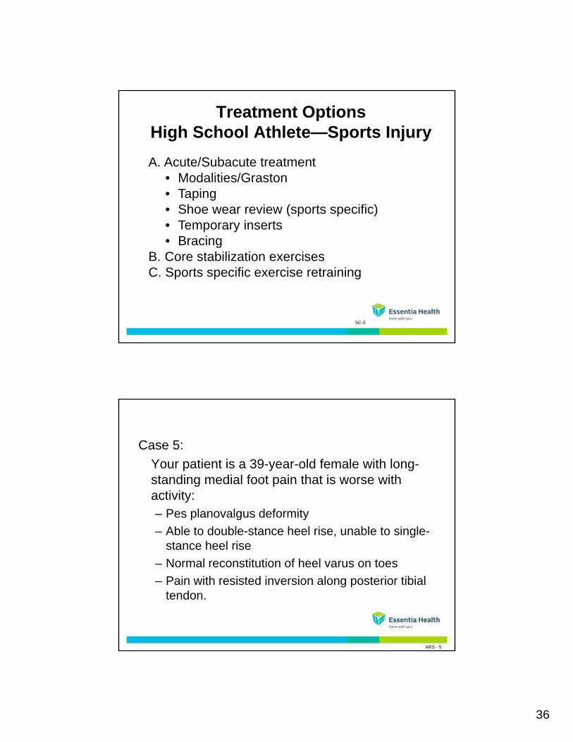
Treatment Options High School Athlete—Sports Injury
A. Acute/Subacute treatmentM d liti /G t• Modalities/Graston
• Taping• Shoe wear review (sports specific)• Temporary inserts• Bracing
B Core stabilization exercisesB. Core stabilization exercisesC. Sports specific exercise retraining
SC-5
Treatment Options High School Athlete—Sports Injury
A. Acute/Subacute treatmentM d liti /G t• Modalities/Graston
• Taping• Shoe wear review (sports specific)• Temporary inserts• Bracing
B Core stabilization exercisesB. Core stabilization exercisesC. Sports specific exercise retraining
SC-5
35
Treatment Options High School Athlete—Sports Injury
A. Acute/Subacute treatmentM d liti /G t• Modalities/Graston
• Taping• Shoe wear review (sports specific)• Temporary inserts• Bracing
B Core stabilization exercisesB. Core stabilization exercisesC. Sports specific exercise retraining
SC-5
Treatment Options High School Athlete—Sports Injury
A. Acute/Subacute treatmentM d liti /G t• Modalities/Graston
• Taping• Shoe wear review (sports specific)• Temporary inserts• Bracing
B Core stabilization exercisesB. Core stabilization exercisesC. Sports specific exercise retraining
SC-5
36
Treatment Options High School Athlete—Sports Injury
A. Acute/Subacute treatmentM d liti /G t• Modalities/Graston
• Taping• Shoe wear review (sports specific)• Temporary inserts• Bracing
B Core stabilization exercisesB. Core stabilization exercisesC. Sports specific exercise retraining
SC-5
Case 5:
Your patient is a 39-year-old female with long-standing medial foot pain that is worse withstanding medial foot pain that is worse with activity:– Pes planovalgus deformity
– Able to double-stance heel rise, unable to single-stance heel rise
– Normal reconstitution of heel varus on toesNormal reconstitution of heel varus on toes
– Pain with resisted inversion along posterior tibial tendon.
ARS - 5
37
Progressive Flat Foot
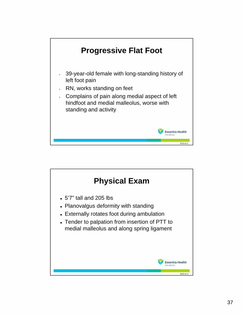
39-year-old female with long-standing history of• 39-year-old female with long-standing history of left foot pain
• RN, works standing on feet
• Complains of pain along medial aspect of left hindfoot and medial malleolus, worse with standing and activitystanding and activity
BJS txt 2
Physical Exam
5’7” tall and 205 lbs
Pl l d f it ith t di Planovalgus deformity with standing
Externally rotates foot during ambulation
Tender to palpation from insertion of PTT to medial malleolus and along spring ligament
BJS txt 3
38
Physical Exam
• More normal appearing arch when sitting
C ll f h i t l l f t h• Collapse of arch into planovalgus foot when standing
BJS txt 4
Physical Exam
• More normal appearing arch when sitting
Pl l f t h t di• Planovalgus foot when standing
BJS txt 5
39
Physical Exam
• More normal appearing arch when sitting
Pl l f t h t di• Planovalgus foot when standing
BJS txt 6
Physical Exam: Appearance
• More normal appearing arch when sitting
Pl l f t h t di• Planovalgus foot when standing
BJS txt 7
40
Physical Exam: Heel Angle
• Posterior examination of patient’s heels
BJS txt 8
Physical Exam: Heel Angle
• Line depicting normal 7 degrees of valgus
BJS txt 9
41
Physical Exam: Heel Angle
• Second line depicting this patient’s hindfoot alignmentalignment– Angle between axis of tibia and posterior axis of
calcaneus
BJS txt 10
Physical Exam: Posterior Standing Observations
• Asymmetric heel valgus
F f t bd ti• Forefoot abduction
• From incompetent medial support structures (spring ligament, deltoid ligament, talonavicular joint capsule)
• “too many toes” sign
• Ability to single limb heel rise
• Painful heel rise
BJS txt 11

42
Physical Exam: Double-stance Hindfoot Raise
• Important to distinguish between flexible and rigid hindfoot valgus to select appropriate treatment
• Flexible valgus hindfoot corrects to varus with double-stance hindfoot raise
BJS txt 12
Physical Exam: Silverskjold Test
• Reduce subtalar and talonavicular joints prior to measurementmeasurement
• Ankle dorsiflexion with knee extended and knee bent
• Tightness only in extension from gastrocnemius contracture
Ti ht b th i t i d fl i f• Tightness both in extension and flexion from Achilles tendon contracture
BJS txt 13
43
Physical Exam: Forefoot Varus
• Check for fixed forefoot varus with hindfoot reducedreduced
BJS txt 14
Imaging Studies – 3/7/11
• 40% uncovering talar head on AP xray
“ i ” ith fl tt i f l l it h l• “c sign” with flattening of calcaneal pitch angle on lateral xray
• No early arthritic changes
BJS txt 15
44
Imaging Studies – 3/7/11
• Valgus angulation of ankle on AP ankle x-ray
Hi df t l hi df t li t i• Hindfoot valgus on hindfoot alignment view
• No ankle joint space narrowing or osteophytes
BJS txt 16
Impression
BJS txt 17
45
Treatment Options
BJS txt 18
Conservative Options
Cast
W lki b t• Walking boots
• Functional shoe gear
• Modified Low dye foot strapping
• NSAIDS
BJS txt 19
46
Conservative Options
• F-scan/gait analysisSt l f th ti /b i d i– Style of orthotic/bracing device
– Materials of products
• Physical therapy– Stretching
– Strengthening
BJS txt 20
Treatment Options30 to 40-Year-Old Bunion Patient
Subacute/Chronic Treatment 1 Symptom presentation:1. Symptom presentation:
• Posterior tibialis tendonitis• Plantar Fasciitis• Flexor Hallucis Longus
tendonitis
2. Symptomatic Treatment2. Symptomatic Treatment• Modalities• Graston• Taping
SC-9
47
3. Temporary Support
Treatment Options30 to 40-Year-Old Bunion Patient
• Powerstep inserts with posting• Motion control shoe wear• Rocker bottom shoe wear
4 Stretching/Strengthening4. Stretching/Strengthening
SC-10
Articulated Ankle Foot Orthosis
• Ankle-foot orthosis (AFO) with joints
S th it l• Smoother gait cycle
• May facilitate muscle activation
Lake, Trexler, and Barringer, 1999 Vol. 11, Num. 1, Posterior Tibial Tendo Dysfunction: A Review of Pain and Activity Levels of Twenty-one Patients, Journal of Prosthetics and Orthotics
Groner, C. , Dec. 2010, AFO Choices for PTTD Grow Clearer, Journal of Foot and Ankle Research
W txt 10

48
Surgical Options
1 M di li i l l t t1. Medializing calcaneal osteotomy
2. Resection of degenerative posterior tibial tendon
3. FDL transfer
4. Gastrocnemius recession
XC txt 1
Medial Incision
• Medial incision
Id tifi ti t i tibi l t d• Identification posterior tibial tendon
• Resection of diseased portion of PT tendon
• FDL identification and transfer into navicular
• Repair of spring ligament
XC txt 2
49
FDL Transfer
• Attaching FDL to navicular in place of posterior tibial tendontibial tendon
XC txt 3
Calcaneal Osteotomy
• Additional lateral calcaneus incision
L bli t t f l• Long oblique osteotomy of calcaneus
• Fixation with one or two screws
XC txt 4
50
Follow-up
• Patients NWB x6-8 weeks for bone and tendon healinghealing
• Physical therapy to start around 4-6 weeks for ROM and strengthening
• Boot or cast for 3 months total
• Wearing regular shoes at 3 months if minimal i i f tpain in foot
XC txt 5
Adult Acquired Flatfoot Deformity
• Posterior tibial tendon dysfunction = most common cause of adult acquired flatfootcommon cause of adult acquired flatfoot deformity
• 10-25% incidence
• Most asymptomatic, medial foot/ankle discomfort
U ll iddl d• Usually middle aged women
• Progressive deformity– Collapse medial arch
– Hindfoot valgus
– Abduction hindfootXC txt 6
51
Case 6:
Your patient is a 61-year-old female with bilateral increasing pain and progressive collapse of archincreasing pain and progressive collapse of arch for several years. She is currently able to ambulate only very short distances. Physical exam reveals severe planovalgus deformities, unable to heel rise, and fixed deformities that are not correctable.
XC txt 7A
Case 6:
Your patient is a 61-year-old female with bilateral increasing pain and progressive collapse of archincreasing pain and progressive collapse of arch for several years. She is currently able to ambulate only very short distances. Physical exam reveals severe planovalgus deformities, unable to heel rise, and fixed deformities that are not correctable.
XC txt 7B
52
History and Physical
• 61-year-old female with increasing pain and deformity bilateral feet for several yearsdeformity bilateral feet for several years
• Progressive collapse arch, crepitus with walking, severe pain
• Pain worse with activity
XC txt 8
Physical Exam
• PE: 5’1” 220 lbs
Bil t l l l d f iti R L• Bilateral severe planovalgus deformities R>L
• Unable walk
• Unable heel rise
• Pain along PT tendon bilaterally and lateral at distal fibula
XC txt 9

53
Physical Exam
• Hindfoot deformities stiff and not correctable
I d fl ibilit f f f t d idf t• Increased flexibility of forefoot and midfoot
• ROM talonavicular joint there is audible and palpable clunk
• Gastrocnemius tightness
• Weakness of posterior tibial tendon
XC txt 10
Radiographs Preoperatively
• Subluxation of talonavicular and subtalar joints on AP and lateral radiographson AP and lateral radiographs
• Irregular calcaneocuboid joint
• Talus no longer in line with 1st MT
• Negative calcaneal pitch angle
XC txt 11
54
Radiographs Preoperatively
• Hindfoot valgus
P ti ll l fib l• Proportionally long fibula
• Medial prominence
XC txt 12
Impression
XC txt 13
55
Conservative Treatment Options
• Activity modification and rest
S l ti i j ti• Selective injections
• AFO or Arizona braces
XC txt 14
Solid Ankle Foot Orthosis
• AFO without joints
• Increased ankle stability in all planes
• Preservation of bony and ligamentous structures
Olszewski, H., Nov 2011, Evidence-based Orthotic Management of PTTD, Journal of Foot and Ankle Research
Houk, H., et all, Nov 2009, Kinematics of PTTD Dictate Management, Journal of Foot and Ankle Research
W txt 11

56
Rocker Bottom Shoe
• Simulate lost motion
D k l t• Decrease peak plantar pressures
• Address bilaterally to prevent LLD
W txt 12
Surgical Options: Selective Fusions
• Clear joints of remaining cartilage
R li f t d f iti• Realign foot deformities
• Hold joints in place with compression screws
• Triple arthrodesis– Talonavicular
– Subtalar
– Calcaneocuboid
XC txt 15
57
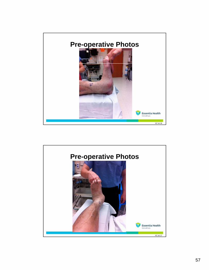
Pre-operative Photos
XC txt 16
Pre-operative Photos
XC txt 17
58
Post-operative Care
• 2 months non-weight bearing
3 th i t b t• 3 months in cast or boot
• At 3 months, minimal pain and wearing normal shoes
XC txt 18
3-Month Follow-Up
XC txt 19

59
Outcomes Triple Arthrodesis
• 5-10% nonunion
• 90 96% patients satisfied with surgery• 90-96% patients satisfied with surgery
• One-third walk with limp following surgery
• Three-fourths have progressive arthritis in surrounding joints, however many asymptomatic or mild pain
Double arthrodesis for PTTD. CORR 365:74-80, 1999
Triple arthrodesis in adults with non-paralytic disease: A minimum ten-year follow-up study. Smith RW, Shen W, DeWitt S, Reischl S. JBJS 86-A:2707-13, 2004
Triple arthrodesis. Fortia PT and Walling AK. CORR 365:91-99, 1999
XC txt 20
Treatment of PTT DysfunctionDependent Upon Stages
• Early stages, flexible
First treated conservative (rest ice NSAID’s PT– First treated conservative (rest, ice, NSAID s, PT, orthotics)
– Foot reconstruction and tendon transfers if conservative treatment fails
• Later stages, rigid
– Bracing cortisone injections activity modification– Bracing, cortisone injections, activity modification
– Hindfoot arthrodesis/ fusion
– Joints to be fused determined by deformity and severity of arthritis
XC txt 21
60
ReferencesKrause F, Bosshard A, Lehmann O, Weber M. Shell brace for stage II posterior tibial tendon
insufficiency. Foot Ankle Int 2008;29(11):1095-1100.
Augustin JF, Lin SS, Berberian WS, Johnson JE. Nonoperative treatment of adult acquired flat foot withthe Arizona brace Foot Ankle Clin 2003;8(3):491 502the Arizona brace. Foot Ankle Clin 2003;8(3):491-502.
Lin JL, Balbas J, Richardson EG. Results of non-surgical treatment of stage II posterior tibial tendondysfunction: a 7-10 year follow-up. Foot Ankle Int 2008;29(8):781-786.
Kulig K, Reischl SF, Pomrantz AB, et al. Nonsurgical management of posterior tibial tendon dysfunctionwith orthoses and resistive exercise: a randomized controlled trial. Phys Ther 2009;89(1):26-37.
Alvarez RG, Marini A, Schmitt C, Saltzman CL. Stage I and II posterior tibial tendon dysfunction treatedby a structured non-operative management protocol: an orthosis and exercise program. Foot AnkleInt 2006;27(1):2-8.
Lester JJ, Todd WF. Abnormal Biomechanics of Flatfoot Deformities and Related Theories ofBiomechanical Development. Clinic in Podiatric Medicine and Surgery - Vol. 6, No. 3, July 1989.
Van Boerum DH, Sangeorzan. Foot Ankle Clin N Am 8 (2003) 419-430.
References (Continued)
Treatment of stage II PTTD with FDL tendon transfer and calcaneal osteotomy. Myerson MS, Badekas A, Schon LC. FAI, 25:445-450, 2004.
Double arthrodesis for PTTD. CORR 365:74-80, 1999
Triple arthrodesis in adults with non-paralytic disease: A minimum ten-year follow-up study. Smith RW, Shen W, DeWitt S, Reischl S. JBJS 86-A:2707-13, 2004
Triple arthrodesis. Fortia PT and Walling AK. CORR 365:91-99, 1999
Downey, M. Vol.24, Issue 10, Oct. 2011, Keys to Treating Tarsal Conditions Podiatry Today
Vu, Louis, Feb 9, 2010, Tarsal Coalition Treatment & Management, Medscape
Lake, Trexler, and Barringer, 1999 Vol. 11, Num. 1, Posterior Tibial Tendo Dysfunction: A Review of Pain and Activity Levels of Twenty-one Patients, Journal of Prosthetics and Orthotics

61
Groner, C. , Dec. 2010, AFO Choices for PTTD Grow Clearer, Journal of Foot and Ankle Research
References (Continued)
Olszewski, H., Nov 2011, Evidence-based Orthotic Management of PTTD, Journal of Foot and Ankle Research
Houk, H., et all, Nov 2009, Kinematics of PTTD Dictate Management, Journal of Foot and Ankle Research
http://www.lowerextremityreview.com
Kawartha Regional Orthopedic Specialists (http://www.kros.ca/UCBL.php)
Shoe Modification and the Use of Orthoses in the Treatment of Foot and Ankle PathologyShoe Modification and the Use of Orthoses in the Treatment of Foot and Ankle PathologyDennis J. Janisse and Erick Janisse , J Am Acad Orthop Surg March 2008 ; 16:152-158.
http://www.wheatonbrace.com
http://www.orthopedicinserts.org
References (Continued)
http://www.actionbp.com
http://www.podiatrytoday.com
http://www.orthoticshop.com