Primary amelanotic malignant melanoma of the small ... · Malignant melanoma develops primarily in...
6
CASE REPORT Primary amelanotic malignant melanoma of the small intestine diagnosed by esophagogastroduodenoscopy before surgical resection Takanori Suganuma • Junko Fujisaki • Toshiaki Hirasawa • Akiyosi Ishiyama • Yorimasa Yamamoto • Tomohiro Tsuchida • Masahiro Igarashi Received: 29 November 2012 / Accepted: 25 March 2013 / Published online: 13 April 2013 Ó The Author(s) 2013. This article is published with open access at Springerlink.com Abstract A 67-year-old man, presenting with anemia and suspected gastric cancer, was referred to our hospital, where he underwent esophagogastroduodenoscopy (EGD). Biopsy revealed densely populated semi-circular cells with abundant cytoplasm that were positive for S-100 protein, melanoma antigen, and HMB-45, resulting in a diagnosis of malignant melanoma. A gastrointestinal barium study for further exploration demonstrated a filling defect 6 cm in size at the ligament of Treitz. Follow-up EGD of this finding revealed an ulcerated, half-circumferential lesion with a distinct ulcer mound extending from the ascending part of the duodenum to the jejunum, and additional biopsy also indicated malignant melanoma. Computed tomogra- phy scans showed wall thickening from the ascending part of duodenum to the proximal jejunum, whereas positron emission tomography revealed accumulation at the upper gastric body, the duodenum to the jejunum, and the left adrenal gland. Systemic exploration of the patient, including the skin, anus, and eyeballs, revealed no other lesions, and primary small intestinal malignant melanoma with metastasis to the stomach and adrenal gland was diagnosed. Partial duodenojejunectomy, partial gastrec- tomy, and left adrenalectomy were performed, and adju- vant chemotherapy with dacarbazine, nimustine hydrochloride, and vincristine sulfate was administered. No postoperative recurrence has been observed in the past 3 years. Keywords AMM Á Melanoma Á DAV Á Small intestine Introduction Malignant melanoma develops primarily in the skin, the transitional zone between the skin and mucosa, and the eyeballs. Primary gastrointestinal malignant melanoma (PGIM) is rare (occurring in approximately 1.0 % of cases) [1], progresses quickly, and most often occurs in the anus, rectum, and esophagus. PGIM rarely occurs in the small intestine, and a diagnosis is usually determined by post- operative resected specimen pathology. Here, we report a case of primary amelanotic malignant melanoma (AMM), which developed in the small intestine and was identified by preoperative tissue biopsy. Case report A 67-year-old man, who first felt dyspnea upon exercise and fatigue in October, 2006, was found to have anemia (hemoglobin 5.6 g/dl) in April 2007 and was referred to our hospital on May 8, 2006. Patient characteristics and initial laboratory results are presented in Table 1. Eso- phagogastroduodenoscopy (EGD) was performed, and a 2-cm submucosal tumor-like elevated lesion with a depression was observed at the posterior wall of the middle gastric body. The adjacent mucosa showed no abnormality, and endoscopic ultrasonography showed a low echoic mass with a distinct margin located in the second to third layer (Fig. 1a, b, c). Biopsy of the gastric tumor determined densely populated semi-circular cells, which were positive by immunohistochemical staining for S-100 protein and melanoma antigen (Melan A) and weakly positive for HMB-45 (Fig. 2a, b, c, d, e). A gastrointestinal barium study was performed for fur- ther exploration of the gastric lesion that showed a filling defect 6 cm in size adjacent to the ligament of Treitz T. Suganuma (&) Á J. Fujisaki Á T. Hirasawa Á A. Ishiyama Á Y. Yamamoto Á T. Tsuchida Á M. Igarashi Department of Gastroenterology, Cancer Institute Ariake Hospital, 3-8-31, Ariake, Koto-ku, Tokyo 135-8550, Japan e-mail: [email protected] 123 Clin J Gastroenterol (2013) 6:211–216 DOI 10.1007/s12328-013-0380-3
Transcript of Primary amelanotic malignant melanoma of the small ... · Malignant melanoma develops primarily in...

CASE REPORT
Primary amelanotic malignant melanoma of the small intestinediagnosed by esophagogastroduodenoscopy before surgicalresection
Takanori Suganuma • Junko Fujisaki • Toshiaki Hirasawa •
Akiyosi Ishiyama • Yorimasa Yamamoto • Tomohiro Tsuchida •
Masahiro Igarashi
Received: 29 November 2012 / Accepted: 25 March 2013 / Published online: 13 April 2013
� The Author(s) 2013. This article is published with open access at Springerlink.com
Abstract A 67-year-old man, presenting with anemia and
suspected gastric cancer, was referred to our hospital,
where he underwent esophagogastroduodenoscopy (EGD).
Biopsy revealed densely populated semi-circular cells with
abundant cytoplasm that were positive for S-100 protein,
melanoma antigen, and HMB-45, resulting in a diagnosis
of malignant melanoma. A gastrointestinal barium study
for further exploration demonstrated a filling defect 6 cm
in size at the ligament of Treitz. Follow-up EGD of this
finding revealed an ulcerated, half-circumferential lesion
with a distinct ulcer mound extending from the ascending
part of the duodenum to the jejunum, and additional biopsy
also indicated malignant melanoma. Computed tomogra-
phy scans showed wall thickening from the ascending part
of duodenum to the proximal jejunum, whereas positron
emission tomography revealed accumulation at the upper
gastric body, the duodenum to the jejunum, and the left
adrenal gland. Systemic exploration of the patient,
including the skin, anus, and eyeballs, revealed no other
lesions, and primary small intestinal malignant melanoma
with metastasis to the stomach and adrenal gland was
diagnosed. Partial duodenojejunectomy, partial gastrec-
tomy, and left adrenalectomy were performed, and adju-
vant chemotherapy with dacarbazine, nimustine
hydrochloride, and vincristine sulfate was administered. No
postoperative recurrence has been observed in the past
3 years.
Keywords AMM � Melanoma � DAV � Small intestine
Introduction
Malignant melanoma develops primarily in the skin, the
transitional zone between the skin and mucosa, and the
eyeballs. Primary gastrointestinal malignant melanoma
(PGIM) is rare (occurring in approximately 1.0 % of cases)
[1], progresses quickly, and most often occurs in the anus,
rectum, and esophagus. PGIM rarely occurs in the small
intestine, and a diagnosis is usually determined by post-
operative resected specimen pathology. Here, we report a
case of primary amelanotic malignant melanoma (AMM),
which developed in the small intestine and was identified
by preoperative tissue biopsy.
Case report
A 67-year-old man, who first felt dyspnea upon exercise
and fatigue in October, 2006, was found to have anemia
(hemoglobin 5.6 g/dl) in April 2007 and was referred to
our hospital on May 8, 2006. Patient characteristics and
initial laboratory results are presented in Table 1. Eso-
phagogastroduodenoscopy (EGD) was performed, and a
2-cm submucosal tumor-like elevated lesion with a
depression was observed at the posterior wall of the middle
gastric body. The adjacent mucosa showed no abnormality,
and endoscopic ultrasonography showed a low echoic mass
with a distinct margin located in the second to third layer
(Fig. 1a, b, c). Biopsy of the gastric tumor determined
densely populated semi-circular cells, which were positive
by immunohistochemical staining for S-100 protein and
melanoma antigen (Melan A) and weakly positive for
HMB-45 (Fig. 2a, b, c, d, e).
A gastrointestinal barium study was performed for fur-
ther exploration of the gastric lesion that showed a filling
defect 6 cm in size adjacent to the ligament of Treitz
T. Suganuma (&) � J. Fujisaki � T. Hirasawa � A. Ishiyama �Y. Yamamoto � T. Tsuchida � M. Igarashi
Department of Gastroenterology, Cancer Institute Ariake
Hospital, 3-8-31, Ariake, Koto-ku, Tokyo 135-8550, Japan
e-mail: [email protected]
123
Clin J Gastroenterol (2013) 6:211–216
DOI 10.1007/s12328-013-0380-3

(Fig. 3a, b). Follow-up EGD was performed and the
intestinal lesion was determined to be an ulcerated, half-
circumferential lesion with a distinct ulcer mound
extending from the ascending part of the duodenum to the
jejunum. Biopsy of the intestinal lesion indicated mela-
noma, as previously identified in the stomach (Fig. 4a, b).
Wall thickening was observed by computed tomography
scanning from the ascending part of duodenum to the
proximal jejunum, and a 2-cm lobular tumor was observed
at the lateral surface of the left adrenal gland (Fig. 5a, b).
Positron emission tomography showed accumulation in the
upper gastric body, the duodenum, and the left adrenal
gland (Fig. 6a, b).
Systemic exploration was performed, and the patient’s
body surface and eyeballs were examined, with no abnormal
findings observed. These results suggested that the primary
lesion was in the small intestine, and partial duodenojejun-
ectomy, partial gastrectomy, and left adrenalectomy were
performed. The identified small intestinal tumor was
62 9 46 mm in size and had no recognized lymphatic
metastasis. In the resected specimens, blackish changes
were not observed (Fig. 7a, b, c). Hematoxylin and eosin
(H&E) staining revealed stranded proliferation of spindle-
shaped tumor cells, and bright cytoplasm and nuclear atypia
were recognized by high power magnification field obser-
vation. Deposition of pigmented granules, which would be
typical in melanotic melanoma, was not observed. Immu-
nohistochemical staining revealed that the primary intestinal
tumor was negative for HMB-45 but positive for S-100
protein and Melan A, which led to the diagnosis of AMM
(Fig. 8a, b, c, d, e). Given the high risk for recurrence of the
disease, adjuvant chemotherapy with dacarbazine (DTIC),
nimustine hydrochloride, and vincristine sulfate (DAV) was
administered for 5 cycles, and the patient has been recur-
rence free for 3 years.
Discussion
Malignant melanoma occurs at an approximate rate of 10
per 100,000 in Western countries, but is a markedly rare
malignant tumor in Japan, occurring at a rate of 1–1.5 per
100,000. In Japan, approximately 80 % of diagnosed
malignant melanomas occur in their primary form on the
skin (32.7 % in the transitional zone between the skin and
mucosa, 27.1 % in the nasal and oral cavities, and 21.4 % in
the eyeballs). This is because neuroectodermal melanocytes
are abundantly present in the skin; the mucosal epithelium
of the nasopharynx, oral cavity, and rectum; the cranial pia
mater; and the choroid of the eyeballs. It is extremely rare
for malignant melanomas to occur at other primary sites in
the body [1]. A study on malignant melanomas originating
from the gastrointestinal tract showed that the primary sites
of the cancer were the oropharynx and nasopharynx
Table 1 Laboratory results at the first visit. Iron deficiency anemia
(Hb 7.6 g/dl; MCV 70.7; and Fe 26 lg/dl) was observed, but bio-
chemistry and tumors markers showed no abnormality
Peripheral blood Chemistry
WBC 6400/ll TP 7.0 mg/dl Ca 8.8 mEq/l
RBC 379 9 104/ll ALB 4.0 mg/dl CRP 0.1 mg/dl
Hb 7.6 g/dl T-Bil 0.3 mg/dl Glu 93 md/dl
MCV 70.7 AST 18 IU/l Ferritin 6.9 ng/ml
MCH 20.1 ALT 30 IU/l Fe 26 l/dl
PLT 53.5 9 104/ll LDH 139 IU/l Serology
Coagulation cGTP 16 IU/l HBsAg (-)
PT/INR 1.09 BUN 19.0 mg/dl HCVAb (-)
APTT 33.3 s Cr 0.85 mg/dl CEA 1.0 ng/ml
Na 141 mEq/l CA19-9 2.0 ng/dl
K 4.2 mEq/l AFP 3.7 ng/ml
Cl 103 mEq/l
Fig. 1 Esophagogastroduodenoscopy. a A 2-cm SMT-like elevated
lesion with a depression was observed at the posterior wall of the
middle gastric body. b Indigo carmine chromoendoscopy: the surface
of the tumor was lobular. c EUS revealed a low echoic mass located at
the first to third layers
212 Clin J Gastroenterol (2013) 6:211–216
123
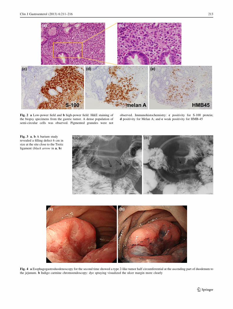
Fig. 2 a Low-power field and b high-power field: H&E staining of
the biopsy specimens from the gastric tumor. A dense population of
semi-circular cells was observed. Pigmented granules were not
observed. Immunohistochemistry: c positivity for S-100 protein;
d positivity for Melan A; and e weak positivity for HMB-45
Fig. 3 a, b A barium study
revealed a filling defect 6 cm in
size at the site close to the Treitz
ligament (black arrow in a, b)
Fig. 4 a Esophagogastroduodenoscopy for the second time showed a type 2-like tumor half circumferential at the ascending part of duodenum to
the jejunum. b Indigo carmine chromoendoscopy: dye spraying visualized the ulcer margin more clearly
Clin J Gastroenterol (2013) 6:211–216 213
123

Fig. 7 a Diameter of the small
intestinal tumor, 62 9 46 mm;
b diameter of the left adrenal
tumor, 20 9 15 mm; and
c diameter of the gastric tumor,
27 9 22 mm
Fig. 6 a Frontal view, b lateral
view: PET showed
accumulations at the upper
gastric body, duodenum and left
adrenal (white circle in a, b)
Fig. 5 a Wall thickening was
observed from the ascending
part of duodenum to the
proximal jejunum (blue arrowin a). b A lobular tumor was
detected at the lateral side of the
left adrenal (blue circle in b)
214 Clin J Gastroenterol (2013) 6:211–216
123

(32.8 %), anal canal (31.4 %), rectum (22.2 %), esophagus
(5.9 %), stomach (2.7 %), small intestine (2.3 %), gall-
bladder (1.4 %), and colon (0.9 %) [2].
In the present case, malignant melanoma was observed in
the small intestine, stomach, and left adrenal gland, with the
largest tumor detected in the small intestine. A diagnosis of
malignant melanoma originating in the small intestine was
confirmed because the largest lesion was in the small
intestine; no changes were seen in the skin, eyeballs,
esophagus, rectum, and transitional zone from the rectum to
the anus, and no lymphatic metastasis was seen in the small
intestinal lymph nodes either. In this case, the metastases to
the stomach and adrenal gland were thought to occur
through local hematogenous metastasis and not by direct
invasion. The direct invasion for other organs was absent in
the resected specimen. The small intestinal lesion was
confirmed in the second EGD, and an iteration biopsy was
necessary for differentiation with other disease. There has
long been a concern that biopsy of primary melanoma could
lead to an increase rate of micrometastases and an increased
risk of recurrence. However, in a report of cutaneous
malignant melanoma, there was no significant difference
found between the 5-year overall survival (OS) rate, local
recurrence rate, rate of melanoma-associated death, and
metastasis rate for sentinel lymph nodes between a total
resection biopsy group and a partial biopsy group [3–6].
AMM is a rare disease that accounts for approximately
2 % of all malignant melanomas and is often diagnosed
during the follow-up and treatment of a different disease.
While the 5-year OS rate of malignant melanoma is
50–60 %, AMM has an extremely poor prognosis, with a
5-year OS rate as low as 25 % [7, 8]. This poor prognosis
may be due to delayed diagnosis due to macroscopic
problems and abnormal melanin synthesis in addition to
undifferentiated tumor characteristics [6]. In AMM, mela-
nin granules cannot be confirmed by H&E staining or may
be present at extremely low levels. Macroscopically, the
tumor is white or grayish white and is difficult to differ-
entiate from malignant tumors such as undifferentiated
cancer and gastrointestinal stromal tumor [9, 10]. For
definitive diagnosis, immunohistochemical staining using
anti-S-100, anti-Melan A, and anti-HMB-45 antibodies is
indispensable. In the present case, the gastric lesion was
weakly positive for HMB-45, while the primary lesion in
the small intestine was negative for HMB-45.
Three years have passed in the present case, with no
observable relapse. As regional lymph nodes account for
55 % of the first metastatic sites in malignant melanoma
cases at the advanced stage [11], the observed long-term
survival in this case may be attributable to the lack of
lymph node involvement in the resected specimens,
effective resection of the primary lesion, and a positive
Fig. 8 H&E staining revealed stranded proliferation of spindle-
shaped cells and higher magnification views showed bright cytoplasm
and nuclear atypia (a lower magnification; b higher magnification).
Immunohistochemistry: c negativity for HMB-45; d positivity for
S-100 protein; and e positivity for Melan A. Deposition of brownish
black pigmented granules, usually recognized in melanotic mela-
noma, was not observed
Clin J Gastroenterol (2013) 6:211–216 215
123

response to the postoperative DAV chemotherapy. DITC
was first introduced in Japan in 1977, and tripartite DAV
therapy has been used in many institutions. In cases of
primary cutaneous malignant melanoma, DTIC alone has
an approximate response rate of 20 %, whereas DAV
therapy has a reported response rate of 37.5 % [12],
although the effect on prolongation of OS of DAV therapy
over DTIC alone has not been ascertained [13]. There are
currently no reports of the use of this regimen for primary
gastrointestinal malignant melanoma. In malignant mela-
noma of the skin, postoperative DAV-feron therapy
(incorporation of interferon-beta with DAV) was reported
to possibly improve the OS rate [13]. As this case showed
no skin involvement, DAV therapy alone was chosen.
Conclusions
In conclusion, it is difficult to diagnose primary small
intestinal malignant melanoma before surgical resection,
and thus, AMM is often detected at advanced stages. The
present case of malignant melanoma diagnosed by biopsy of
a gastric lesion before surgical resection is rare, and this is
the first study to report a diagnosis of AMM before surgery.
Conflict of interest The authors declare that they have no conflict
of interest.
Open Access This article is distributed under the terms of the
Creative Commons Attribution License which permits any use, dis-
tribution, and reproduction in any medium, provided the original
author(s) and the source are credited.
References
1. Chang AE, Karnell LH, Menck HR. The National Cancer Data
Base report on cutaneous and noncutaneous melanoma: a sum-
mary of 84,836 cases from the past decade: The American Col-
lege of Surgeons Commission on Cancer and the American
Cancer Society. Cancer. 1996;83:1664–78.
2. Cheung MC, Perez EA, Molina MA, Jin X, Gutierrez JC,
Franceschi D, et al. Defining the role of surgery for primary
gastrointestinal tract melanoma. J Gastrointest Surg. 2008;12:
731–73.
3. Lederman JS, Sober AJ. Does biopsy type influence survival in
clinical stage I cutaneous melanoma? J Am Acad Dermatol.
1985;13:983–7.
4. Lees VC, Briggs JC. Effect of initial biopsy procedure on prog-
nosis in Stage 1 invasive cutaneous malignant melanoma: review
of 1086 patients. Br J Surg. 1991;78:1108–10.
5. Bong JL, Herd RM, Hunter JA. Incisional biopsy and melanoma
prognosis. J Am Acad Dermatol. 2002;46:690–4.
6. Martin RC 2nd, Scoggins CR, Ross MI, Reintgen DS, Noyes RD,
Edwards MJ, et al. Is incisional biopsy of melanoma harmful?
Am J Surg. 2005;190:913–7.
7. Giuliano AE, Cochran AJ, Morton DL. Melanoma from unknown
primary site and amelanotic melanoma. Semin Oncol. 1982;9:442–7.
8. Bhawan J. Amelanotic melanoma or poorly differentiated mela-
noma? J Cutan Pathol. 1980;7:55–6.
9. Huvos AG, Shah JP, Goldsmith HS. A clinicopathologic study of
amelanotic melanoma. Surg Gynecol Obstet. 1972;135:917–20.
10. Ariel IM. Amelanotic melanomas: an analysis of 77 patients. Curr
Surg. 1981;38:151–5.
11. Reintgen DS, Thompson W, Garbutt J, Seigler HF. Radiologic,
endoscopic, and surgical considerations of melanoma metastatic
to the gastrointestinal tract. Surgery. 1984;95:635–9.
12. Ikeda S, Ishihara K. Clinical study of Dacarbazine (DTIC) for the
malignant melanoma. Clin Derm. 1982;36:183–8 (in Japanese).
13. Bajetta E, Del Vecchio M, Bernard-Marty C, Vitali M, Buzzoni
R, Rixe O, et al. Metastatic melanoma: chemotherapy. Semin
Oncol. 2002;29:427–45.
216 Clin J Gastroenterol (2013) 6:211–216
123