Prevention Screening and Prenatal Diagnostic Approaches · PDF file2 Thalassaemia This...
80
Thalassemia Prevention : Screening and Prenatal Diagnostic Approaches Distance Learning Course From Research to practice: Training course in Sexual and Reproductive Health Research Community Genetics Marina Kleanthous Molecular Genetics Thalassaemia Department The Cyprus Institute of Neurology & Genetics
Transcript of Prevention Screening and Prenatal Diagnostic Approaches · PDF file2 Thalassaemia This...
11
Thalassemia Prevention :Screening and PrenatalDiagnostic Approaches
Distance Learning CourseFrom Research to practice: Training course in
Sexual and Reproductive Health ResearchCommunity Genetics
Marina KleanthousMolecular Genetics Thalassaemia DepartmentThe Cyprus Institute of Neurology & Genetics

2
Thalassaemia
This presentation includes:IntroductionThalassaemia control programsStrategy for the prevention of the diseasePrenatal diagnostic approaches

3
Haemoglobinopathies
Structure of globin chainRate of synthesis of globin chains(Thalassaemias)Hereditary Persistence of Fetal Haemoglobin(HPFH)

4
thalassaemia
Thalassaemias (350)
thalassaemia
Hb S
Abnormal Haemoglobins (887) Hb D
Hb E
Haemoglobinopathies

Thalassaemia
Reduction or absence of one of the globinpolypeptides making up haemoglobinHaemoglobin is a tetramer composed of 2type globin chains and 2 type globin chains
5

6
Human Haemoglobins and Globin Genes
Thalassaemias are hereditary blood disorders caused by areduced synthesis of one or more of the globin chains
Chrom. 16 Chrom. 11
2 1 2 1 1 G
2 2(Gower - I)
2 2(Portland)
2 2(Gower-II)
2 2(Hb-F)
2 2(Hb- 2)
2 2(Hb- )
Embryo Fetus Adult

Haematopoiesis
Gower-I 2 2
Gower-II 2 2
Hb-F 2 2
Hb-A 2 2
Hb-A2 2 2
Portland 2 2
Embryonic haemoglobins
Foetal Haemoglobin
Adult haemoglobin
7

8
>200 globin genemutations
thalassaemia
Common globin gene mutations

9
Globin chain imbalance
Accumulation of excess globin chains inerythroid precursors (ineffectiveerythropoiesis) and RBC (haemolyticanaemia)
thalassaemia

10
Common Deletional and non DeletionalThalassaemia Mutations
InterHVR
2 1 2 1InterHVR
- 3.7- -MEDI- -MEDII
- 20.5
Non deletional thal mutations
2 IVSI Donor site GA[GGTGA]GG GAGG�….(5nt deletion)
2 Poly(A) signal AATAAA AATGAA (PA-2)

11
World Distribution of Haemoglobinopathies
One of the most common inherited blood disorder in the world
250 million people (4.5%) are carriers of a potentially pathologic gene
300, 000 infants are born with a major haemoglobinopathy

Severe anaemiaRegular blood transfusionIron chelation therapyBonemarrow transplantation (BMT)Gene therapyDrug therapy
Thalassaemia
12

13
Thalassaemia control programs
National program effective strategy
Infrastructure
Patient Treatment
Prevention of the disease

14
Thalassaemia control programsNational Program �– Effective Strategy
Help fromWHO and TIF and experts in the field
Extend of the problem
Community priorities
Economic situation
Distribution
Ethical (therapeutic abortion option)
National financial support of the program

15
Thalassaemia control programsInfrastructure �– Thalassaemia Center
Clinics
Haematology Lab
Molecular BiologyLab
Clinic
Screening Lab
Molecular Biology
Peripheral Center
Peripheral Center
Peripheral Center
Peripheral Center
Peripheral Center
Peripheral Center
Reference Center

16
Thalassaemia control programsPrevention
Public education
Carrier screening
Genetic counseling
Prenatal Diagnosis

17
Prevention Programs
Euro Mediterranean countries (Italy, Greece, Cyprus)
Middle East countries (Iran 1997)
SE-Asia countries (Asian Network for the control of thalassaemia was established on 2004)

18
Prevention ProgramsPublic education
Schools
Leaflets
Media
Conferences/Seminars
Professionals
To informNOT to stigmatize

Prevention ProgramsCarrier Screening
Population screening
High risk groups
Pregnant women
19

20
Carrier Detection
Haematology
Hbs electrophoresis
Biosynthesis
Family study
Molecular diagnosis
-thal carrier<27>3.5
A+(F)+A2
NORMAL MCH (pgt) >27HbA2(%) <3.3Hb A+A2

21
Thalassaemia Carrier ScreeningFlow Chart
SCREENINGFOR COMMON
-THALMUTATIONS
UNDEFINED-THAL
MUTATIONDGGE
DIRECTSEQUENCING
IRON STUDIES-GLOBIN GENE
BY PCR
-THAL NORMAL-GENES
GLOBIN CHAINSYNTHESIS AND/OR
-GENE ANALYSIS
+ -THAL, THAL
OTHER NORMAL HbA2 -THAL
<27<3
A+F+A2
HbF
/ RATIO ANALYSIS
-THAL
HPFH
<27<3.5
A+A2
<27>3.5
A+(F)+A2
MCH (pg) >27HbA2(%) <3.3Hb A+A2
NORMAL -THAL

22
Prevention ProgramsGenetic counseling
Risks
Clinical features
Patient treatment
Options
Procedures to follow

23
Prevention ProgramsPrenatal Diagnosis
Blood samples from family members
CVS biopsy/Amniocentesis
Molecular analysis (ARMS, Sequencing etc)
Diagnosis

Prenatal DiagnosisCyprus example
Amniocentesis (2nd Trimester)
CVS (1st Trimester)
PGD (Pre Implantation)
Non invasive prenatal diagnosis (EC FP6 Network of Excellence �“SAFE�”)
24
25
Prenatal Diagnosis forhaemoglobinopathies
thalassaemia/Hb variants
Hydrops Fetalis ( thalassaemia)
Severe haemoglobin disease

26
Steps followed up for prenatal diagnosis by CVSCyprus experience
Thalassaemia trait testingCard and Premarital certificateGenetic counselingPregnancyBlood samples and family tree
DNA extractionDNA analysis of family members
Ultrasound
CVS biopsy at 11th week of gestation (obstetrician)CVS cleaning under microscopeDNA extractionMolecular analysisDiagnosis
Thalassaemia center
CING Molecular Biology Lab
Obstetrician
CINGMolecular Biology Lab
MATERNAL TISSUE CHORIONIC VILLI
BLOOD CLOT(Maternal origin)

27
Steps followed up for prenatal diagnosisby CVS Cyprus experience
Thalassaemia trait testingCard and Premarital certificateGenetic counselingPregnancyBlood samples and family tree
DNA extractionDNA analysis of family members
UltrasoundCVS biopsy at 11th week of gestation
CVS cleaning under microscopeDNA extractionMolecular analysisDiagnosis

28
Typical couple at risk for thalassaemia
Hb 13.3MCV 71.8MCH 22.9HbA2 5.9
Hb 13.4MCV 82.1MCH 26.3HbA2 2.9
Hb 14.2MCV 71.7MCH 22.9HbA2 6.2
Hb 12.1MCV 83.1MCH 28.1HbA2 3.0
Hb 11.2MCV 73.1MCH 23.1HbA2 5.2
Hb 13.3MCV 71.8MCH 22.9HbA2 6.3
CVS

29
N/IVSI-110 N/IVSI-110
CVS
IVSI-110/IVSI-110
Typical couple at risk for thalassaemia

30
Typical couple at risk for thalassaemia
N/IVS-I-110 N/IVS-I-110
CVS
IVS-I-110/IVS-I-110
NN+ ++ +
+ ++ + NN
+ ++ +
-- --NN
--thalthal+ + --
+ + --NNNN
--thalthal/Father/Father--thalthal/Mother/Mother } --thalthal majormajor
+ ++ +
-- --NN
--thalthal
+ ++ +
-- --NN
--thalthal
+ + --
-- --NN
--thalthal
-- --
-- --

31
Atypical couple
One parent is a typical -thalassaemia carrier while the other partner has abnormal haematological indices and normal HbA2
-thal carrier Hb 13.4MCV 68.5MCH 22.2HbA2 2.5
?-thalassaemiaand thal comp. heter.
Silent -thalassaemia, and thal comp. heter.-thal with low HbA2
-thal
32
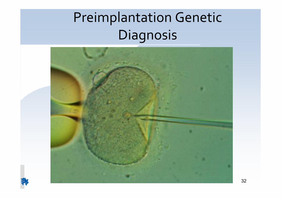
Preimplantation GeneticDiagnosis
33
Preimplantation Genetic Diagnosis
Preimplantation Genetic Diagnosis (PGD)uses in vitro fertilisation (IVF) to createembryosTests one or two cells from each embryo for aspecific genetic abnormalityIdentifies unaffected embryos for transfer tothe uterusThe approach through PGD assists couples atrisk of an inherited disorder to avoid the birthof an affected child.

34
STAGES
CounselingInduction of ovulationOocyte collectionFertilization by ICSIEmbryo biopsyGenetic diagnosisImplantation of 1 2 suitable embryosConfirmation of pregnancyPrenatal diagnosis (ESHRE guidelines)

35
Disorders tested by PGD
FISHChromosomal Disorders
PCR basedSingle gene defects
ThalassaemiaCystic FibrosisHaemophiliaMuscular dystrophies
etc
36
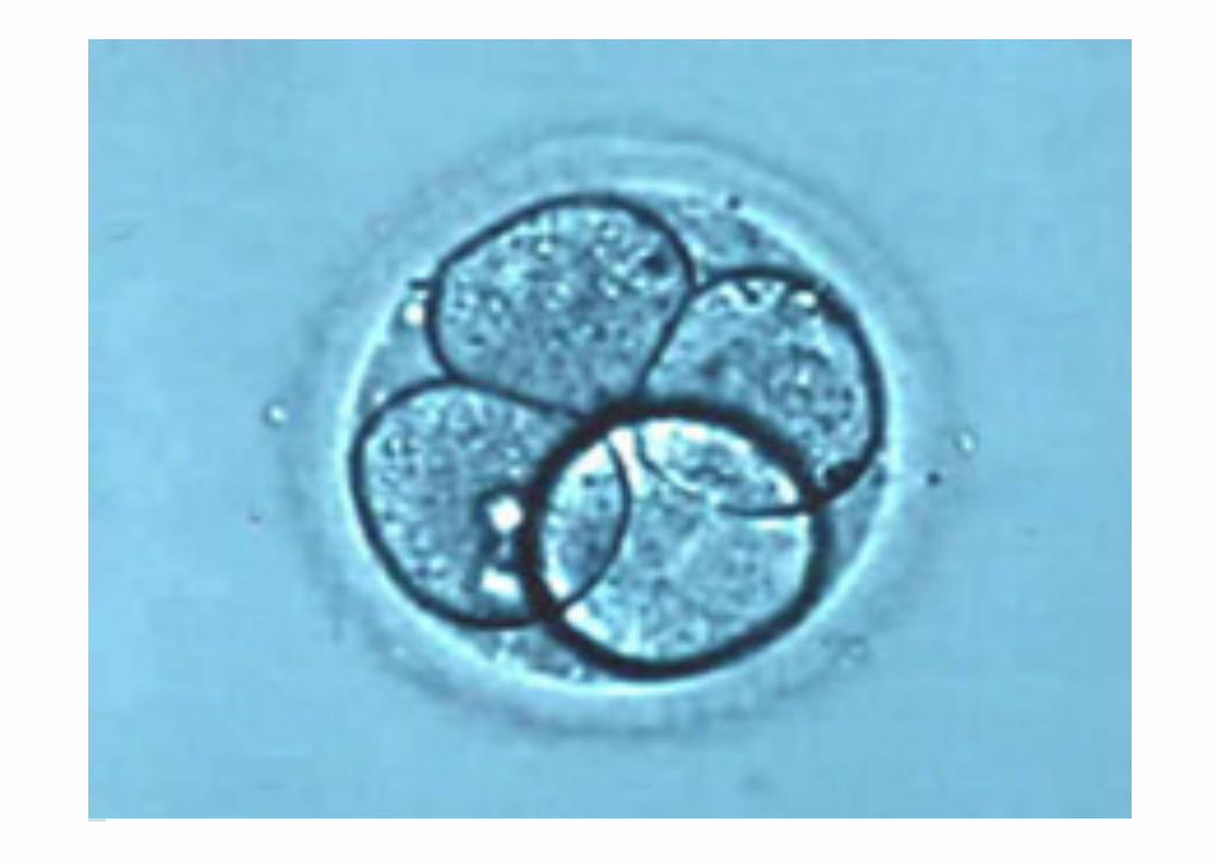
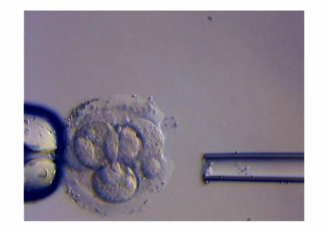
PGD approaches
Polar body analysis
Blastomere biopsy analysis

37
Induction of ovulation
In order to obtain a large number of oocytes,the patients undergo controlled ovarianstimulation (COH), with the use of FSH.
Ultrasound guided trans vaginal oocyteretrieval

38
Intracytoplasmic sperm injection (ICSI)Pronuclear formation (+ 2nd polar body)Pronuclear fusionZygote
Fertilization

PGDPGD
39

40
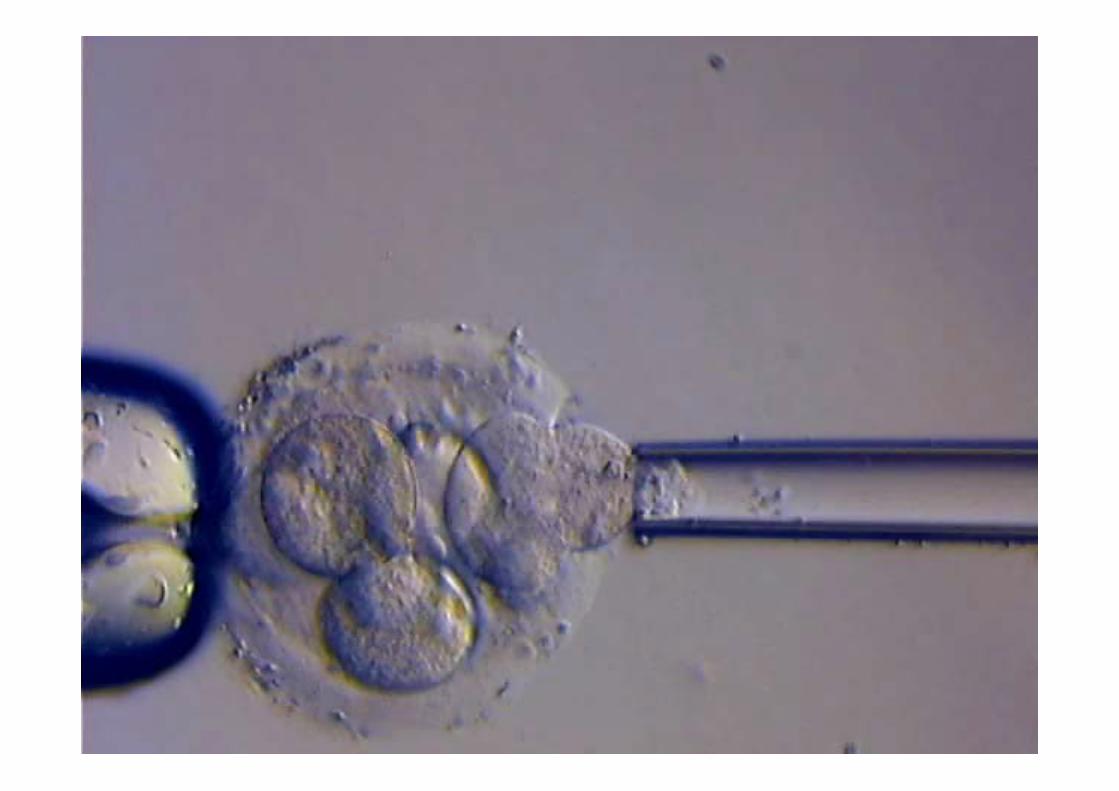
Micromanipulator

41

42
Second polar body extrusion and pronuclear formationfollowing ICSI in a zona free human oocyte

43
44

45

46

47
48
49

50

51

52

53
Lab
Lysis / PK digestion
1st round PCR (external primers)
Freeze (-200C >30 min)
2nd round PCR for DGGE analysis
LightCycler analysis (real-time PCR)
BlastomereBiopsy
PCR based PGD analysis
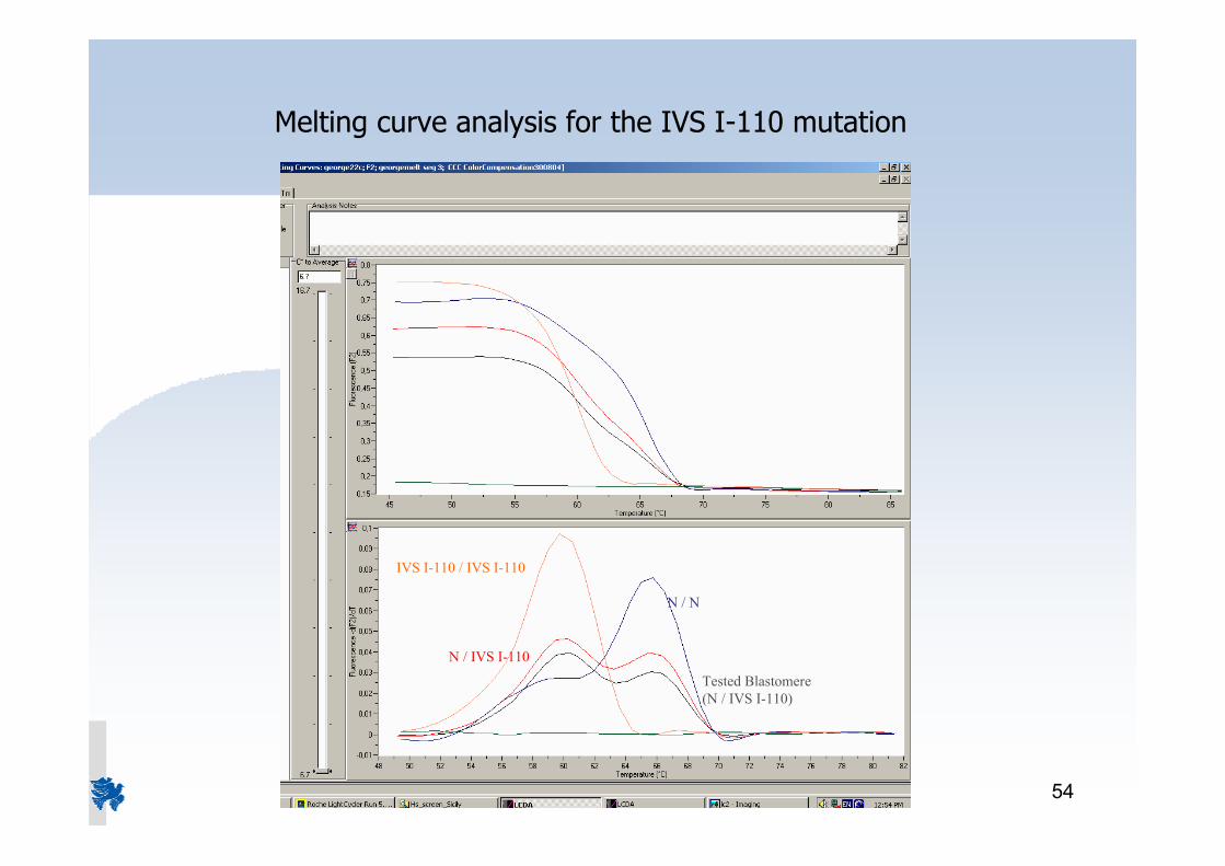
54
N / IVS I-110
IVS I-110 / IVS I-110
N / N
Melting curve analysis for the IVS I-110 mutation
Tested Blastomere(N / IVS I-110)

55
DGGE analysis of 6 blastomeres during PGD

56
Preparation workup
StrategyTrainingSetup of techniques on genomic DNATests on single cells (lymphocytes) >200Maximize amplification efficiency (>90%)Minimize allele dropout (<10%)Eliminate contamination factorsBlastomere test from unused embryos

57
Determining factors for successful PGD
Adequate number of ovaNot all will be fertilized successfully
Adequate number of embryosNot all will survive biopsySomemay fail to develop normallyAfter analysis, ~25% expected not be suitable for transfer (affected)A fewmay fail to amplify (5 10%) �– no result
Laboratory proceduresBiopsy techniquesContamination controlSuccessful amplification of biopsy DNA

58
Sources of error
Contamination
Biopsy material (blastomere) actually notdeposited in sample tube
Cell fragmentation (bad quality embryos)
Amplification efficiency

59
Contamination
EmbryomanipulationHandlingBiopsy
Biopsy manipulationTransferPK digestFirst PCR amplification

60
Contamination Control
BlanksCulture Medium BlanksBiopsy Medium BlanksReagent Blanks
Polymorphic MarkersD6S1056 (tetra )D15S652 (tri )

6161

6262
Non InvasivePrenatal Diagnosis

63
Fetal Cells in Maternal Circulation
A very small amount of fetal cells are presentin the maternal circulationMethods for separating FNRBCs failed torecover a pure fetal cell populationNew technologies are now tested
Non contact laser capture microdisectionSeparation by electric field

64
Circulating Nucleic Acids
First report 1948 (Mandel and Metais)Studies on Circulatory DNA focused on autoimmune diseases
Diagnosis and prognosis of cancer 1977Discovery of fetal DNA in maternal plasma (Lo et al, 1997)NIPD offered for RHD and fetal sex for X-linked disordersNIPD under development for other single gene andchromosomal disorders
65
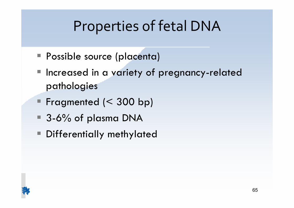
Properties of fetal DNA
Possible source (placenta)Increased in a variety of pregnancy-related pathologies Fragmented (< 300 bp)3-6% of plasma DNADifferentially methylated

66
Development of NIPDmethods Limitations
Low quantity of fetal DNABad quality of fetal DNAThe isolated DNA is mainly maternal(3-6% is fetal)Parents have the samemutations

67
NIPDMethodology
10ml peripheral blood
4 ml Plasma
Plasma DNA(Maternal + Fetal)
PCR based methods

68
NIPD for X linked Disorders
Test for the presence Y chromosome sequences in the
maternal plasma
Used for severe X linked disorders:
Duchene/Becker muscular dystrophy
linked agammaglobulinaemia
Hemophilia
Norrie disease (Episcopi blindness)
X linked severe combined immunodeficiency (SCID)

69
Hemolytic Diseaseof the Newborn (HDN)
Mother: RHDFather: RHD+
RhD-negative woman with Rh-positive fetus
RhD-negative woman and RhD-positive man conceive a child
Cells from RhD-positive fetus enter woman�’s bloodstream
Woman becomes sensitized
Antibodies form to fight RhD-positive blood cells
In the next pregnancy RhD-positive pregnancy, maternal antibodies attack fetal red blood cells

70
NIPD for the RhD of the fetus
For RhD negativewomen
Blood sample fromthemother after the16 week of gestation
Analysis of the plasmaDNA
Diagnosis70

71
Thalassaemia Non Invasive PrenatalDiagnosis by Cell Free Fetal DNA
Themethod is based on the detection
of the paternally inherited fetal alleles

72
Selection/Analysis of SNPs for NIPD
High degree of heterozygosityInformative SNPs
Mother A/A, Father A/B (determination ofallele)Mother A/A, Father B/B (confirmation ofpaternal allele)

73
Selection of SNPs with high degree ofheterozygosity
SNP genotyping analysis130 SNPs located on the globin gene cluster(http://www.ncbi.nlm.nih.gov)75 random samples (Cyprus Population)Sequenom®MALDI TOFFMass Array

74
Analysis on 67 families at risk forthalassaemia for 42 SNPs
NIPD methodusing SNPs canbe performedon 84% offamilies

75
ThalassochipAPEX (Arrayed Primer Extension)
54 beta thalassaemia mutations6 Hb Variants6 delta thalassaemia mutations10 SNPs
It was validated as a diagnostic tool for haemoglobinopathies (EC MedGeNet project)
L. Cremonesi et al Hemoglobin 31:1-23, 2007

76
Sensitivity/Specificity of APEX genDNA SNP rs7480526 (g/t)
1:1000 (1.5 ng/rxn)
1:10000(0.15 ng/rxn)
Sense
Antisense
A C G T
Genotype 100% g/g
(15ng/rxn)
Genotype
95% g/g + 5% g/t
Sensitivity
Specificity
Gen DNA: t/t

77
APEX analysis on maternal plasmars10837631(a/t) mo: a/a, fa:a/t
1
2
3
4
5
6
7
8
9
10
11
a/t a/t
a/a a/a
a/t a/t
a/t a/t
a/t a/t
a/a a/a
a/t a/a
a/t a/t
DNW
a/t a/t
a/t a/t
Mat. A C G T CVS Plasma
X

78
NIPD on Family 22
rs 10768683 c/gmother c/cfetus c/c
rs 10837631 a/tmother ǩ/ǩfetusǩ/t
IVSII-745 c/gmother c/cfetus c/c
A C G T
79
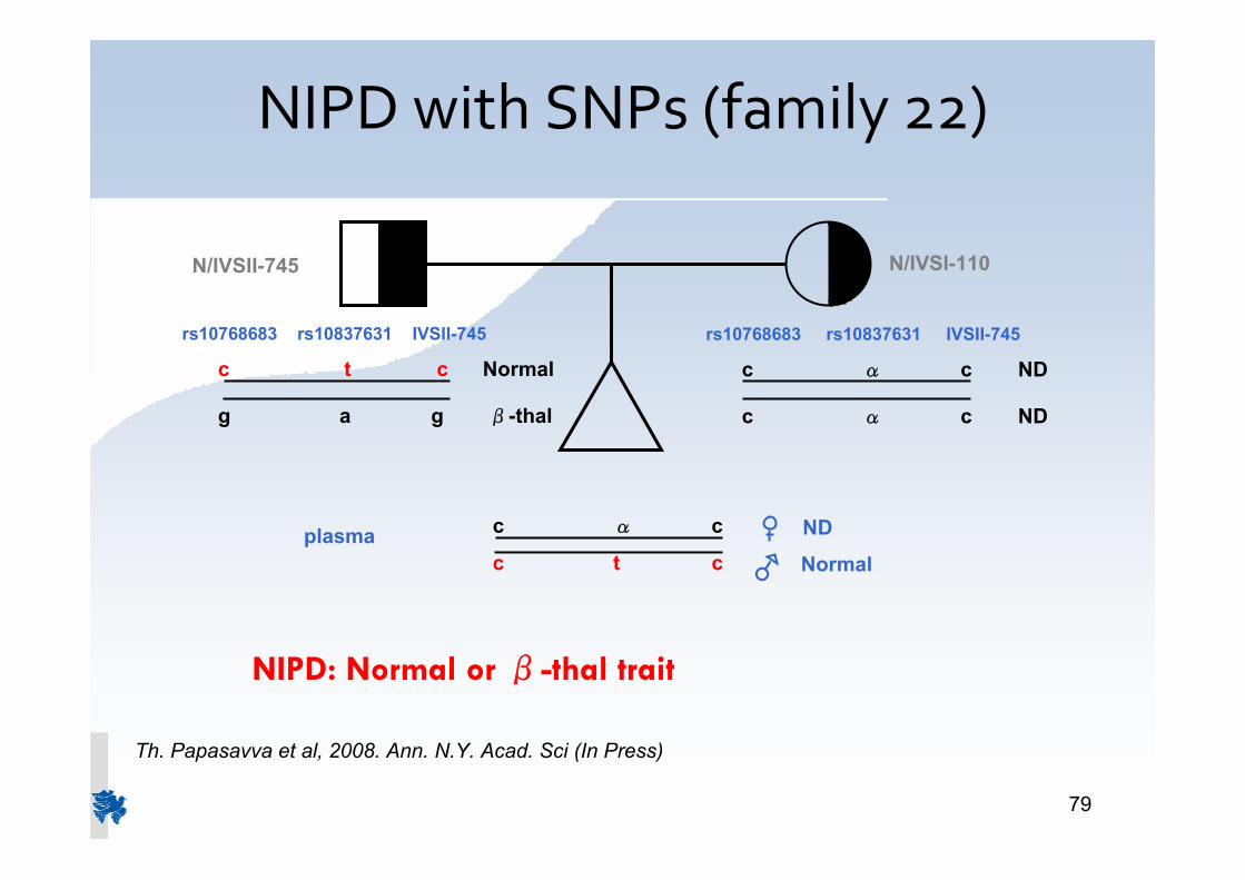
NIPD with SNPs (family 22)
NIPD: Normal or Ǫ-thal trait
rs10768683 rs10837631 IVSII-745
c t c Normal
g a g Ǫ-thal
rs10768683 rs10837631 IVSII-745
c ǩ c ND
c ǩ c ND
N/IVSII-745 N/IVSI-110
c ǩ c
c t cplasma
Normal
ND
Th. Papasavva et al, 2008. Ann. N.Y. Acad. Sci (In Press)

80
Conclusions
NIPD using SNPs analysis is possible
Risk of error reduces to acceptable levels using three or moreSNPs ( The higher the number of SNPs the moreefficient/reliable NIPD.
APEX promising technique, needs improvement
Paternal allele of the fetus non invasively detected