Prevalence Rate of Facial Pain - CDDS · 2017-04-14 · Prevalence Rate of Facial Pain Per 100,000...
49
1 Orofacial Pain: Diagnosis & Management Clarifying the issues Orofacial Pain: Diagnosis & Management Clarifying the issues Henry A. Gremillion, D.D.S., M.A.G.D. Louisiana State University School of Dentistry February 3, 2017 – An unpleasant sensory and emotional experience. – Associated with actual or potential tissue damage. – Described in terms of such damage. UNC Pain Center – Primary Pain Complaints Head, face, and neck 43% Back, lower extremities 23% Other 34% Body Region Prevalence Rate of Facial Pain Per 100,000 Toothache 12,361 Oral Ulcer 8,392 TM Joint 5,289 Face Pain 1,415 Burning Mouth 707 45,711 Households Interviewed Lipton, Ship, Larach-Robinson JADA 124:115, 1993 45,711 Households Interviewed 22% of population suffered from orofacial pain more than once in the previous 6 months. Orofacial Pain Lipton, Shipp, Larach-Robinson JADA 124:115, 1993 Categories of Common Orofacial Pain Conditions Somatic (nociceptive pain) -local (oral/perioral) tissue injury / inflammation Musculoskeletal -TMD Neuropathic orofacial pain -neuralgias -deafferentation -dysesthesia Headache -migraine -tension-type
Transcript of Prevalence Rate of Facial Pain - CDDS · 2017-04-14 · Prevalence Rate of Facial Pain Per 100,000...
1
Orofacial Pain: Diagnosis & ManagementClarifying the issues
Orofacial Pain: Diagnosis & ManagementClarifying the issues
Henry A. Gremillion, D.D.S., M.A.G.D.Louisiana State University School of Dentistry
February 3, 2017
– An unpleasant sensory and
emotional experience.
– Associated with actual or potential
tissue damage.
– Described in terms of such damage.
UNC Pain Center – Primary Pain Complaints
Head, face, and neck 43%
Back, lower extremities 23%
Other 34%
Body Region
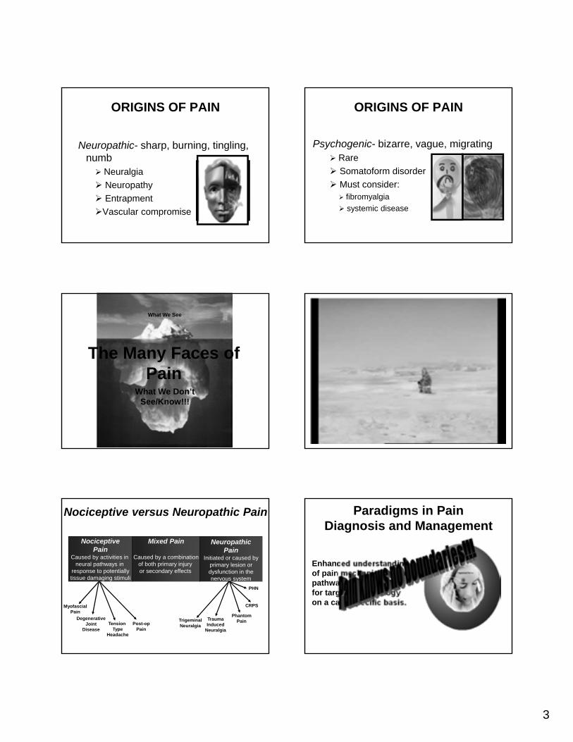
Prevalence Rate of Facial Pain
Per 100,000
Toothache 12,361
Oral Ulcer 8,392
TM Joint 5,289
Face Pain 1,415
Burning Mouth 707
45,711 Households Interviewed
Lipton, Ship, Larach-Robinson JADA 124:115, 1993
45,711 Households Interviewed
22% of population suffered from
orofacial pain more than once in the
previous 6 months.
Orofacial Pain
Lipton, Shipp, Larach-Robinson JADA 124:115, 1993
Categories of CommonOrofacial Pain Conditions
Somatic (nociceptive pain)-local (oral/perioral) tissue injury / inflammation
Musculoskeletal-TMD
Neuropathic orofacial pain-neuralgias-deafferentation-dysesthesia
Headache-migraine-tension-type

2
Chief concern-bitemporal headache (frequent)-clicking and pain with jaw function-severe throbbing headache (occasional)-fatigue
Should I treat this patient?
What is/are the diagnosis(es)
How should I treat this patient?
What factors are important in this case?
Patient Evaluation
Chief concern(s)
History of chief concern(s)
Past medical/dental history
Review of systems
Physical examination
Additional studies if indicated
Differential diagnosis
Data collectionData collection
Acute Pain Characteristics
Protective mechanism
Sudden onset
Limited duration
Patients usually show anxiety
No persisting psychologic reactions
Responds well to traditional therapy
Quality
may suggest mechanism(s)
ORIGINS OF PAIN
Musculoskeletal- dull, aching stiff, sore Myofascial pain
Myalgia
Tension-type headache
Arthrosis/arthritis
ORIGINS OF PAIN
Vascular- throbbing, poundingMigraine
Temporal arteritis
Inflammation
Hypertension
3
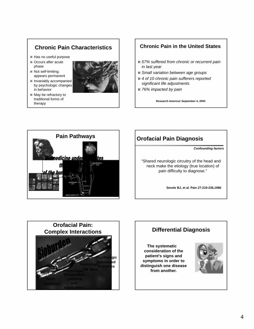
ORIGINS OF PAIN
Neuropathic- sharp, burning, tingling, numb Neuralgia
Neuropathy
Entrapment
Vascular compromise
ORIGINS OF PAIN
Psychogenic- bizarre, vague, migrating Rare
Somatoform disorder
Must consider: fibromyalgia
systemic disease
What We See
What We Don’t See/Know!!!
The Many Faces of Pain
NociceptivePain
Caused by activities in neural pathways in
response to potentiallytissue damaging stimuli
Mixed Pain
Caused by a combinationof both primary injury or secondary effects
Neuropathic Pain
Initiated or caused byprimary lesion ordysfunction in thenervous system
Nociceptive versus Neuropathic Pain
MyofascialPain
DegenerativeJoint
DiseaseTension
TypeHeadache
Post-opPain
TrigeminalNeuralgia
TraumaInduced
Neuralgia
PhantomPain
CRPS
PHN
Paradigms in PainDiagnosis and Management
Enhanced understanding of pain mechanisms and pathways has provided for targeting pathology on a case specific basis.
4
Chronic Pain Characteristics
Has no useful purpose
Occurs after acute phase
Not self-limiting; appears permanent
Invariably accompanied by psychologic changes in behavior
May be refractory to traditional forms of therapy
Chronic Pain in the United States
57% suffered from chronic or recurrent pain in last year
Small variation between age groups
4 of 10 chronic pain sufferers reported significant life adjustments
76% impacted by pain
Research America! September 4, 2003
Pain Pathways Orofacial Pain Diagnosis
“Shared neurologic circuitry of the head and neck make the etiology (true location) of
pain difficulty to diagnose.”
Sessle BJ, et al. Pain 27:219-236,1986
Confounding factors
Orofacial Pain: Complex Interactions
Anatomic
Physiologic
Neurologic
Psychologic
TeethMusculature
(cervicaland
masticatory)
TM Joint
Associatedstructures
Differential Diagnosis
The systematic consideration of the patient’s signs and
symptoms in order to distinguish one disease
from another.

5
Differential Diagnosis
Teeth
Glandular
Vascular
Neurogenous
Myogenous
Arthrogenous
Paranasal sinuses
Otologic
Individualization of care:• Investigate all possible facets to include:
-etiology-perpetuation-recurrence-co-morbid conditions

6
NEOPLASM
Neoplasms can be the cause of refractory chronic pain
Mass in posterior fossa as a trigger for trigeminovascular system and upper cervical afferents resulting in secondary chronic headaches
Bigal ME, Rapoport AM, Camel M. Cephalalgia 2002 Mar; 23(2): 124-8
EVALUATION STRATEGIES
Red Flags”
“Investigate
the
Atypical
and the
WORRISOME HEADACHE RED FLAGS
“SNOOP”
Older: new onset and progressive headache, especially in middle-age >50 (giant cell arteritis)
Systemic symptoms (fever, weight loss) or
Secondary risk factors (HIV, systemic cancer)
Neurologic deficits lateralizing to side of pain or abnormal signs (confusion, impaired alertness, or consciousness)
Onset: sudden, abrupt, or split-second
Previous headache history: first headache or different (change in attack frequency, severity, or clinical features)
Differential Diagnosis
Teeth
Glandular
Vascular
Neurogenous
Myogenous
Arthrogenous
Paranasal sinuses
Otologic
7
Patient: Betty
51 year old Caucasian female
Medical history significant for:– left temporomandibular
surgery X2
– hypothyroidism
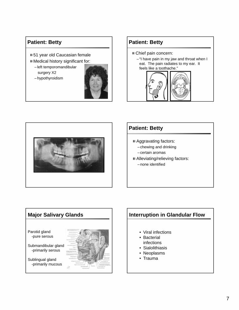
Patient: Betty
Chief pain concern:– “I have pain in my jaw and throat when I
eat. The pain radiates to my ear. It feels like a toothache.”
Patient: Betty
Aggravating factors:– chewing and drinking
– certain aromas
Alleviating/relieving factors:– none identified
Major Salivary Glands
Parotid gland-pure serous
Submandibular gland-primarily serous
Sublingual gland-primarily mucous
Interruption in Glandular Flow
• Viral infections• Bacterial
infections• Sialolithiasis• Neoplasms• Trauma

8
Obstructions
• Mucous plug• Stones
– hydroxyapatite– trace magnesium carbonate– trace ammonia– organic matrix (amino acids /
carbohydrates)
Sialolithiasis
• Most common obstruction
• Primarily affects submandibular gland
SialolithiasisSialolithiasisDiagnosis
• History– pain with salivation
• Inspection• Palpation
SialolithiasisSialolithiasisDiagnosis
• Imaging– occlusal– lateral jaw– panoramic– sialogram
Patient: Juan
28 year old Hispanic male
Medical history:– unexplained intermittent
facial swelling and lymphadenopathy
previously treated with Pen VK 500 mg

9
Patient: Juan
Chief pain concern(s):– “pain on the right side of my face;
headaches in the temples; clicking in my right jaw; face feels numb and tingles on the right side; throbbing when I eat”
Patient: Juan
Aggravating factors:– eating
– opening wide
– yawning
Alleviating/relieving factors:– antibiotics (Pen VK 500)
– analgesics (Ibuprofen)-- “takes the edge off”
Parotido-Masseteric HypertrophyTraumatic Occlusion Syndrome
Parotid swelling– duct obstruction– pain
Sialdochitis– bacterial infection due to retrograde
travel of organisms from the oral cavity
Traumatic occlusion
Parotid duct
Superficial masseter muscle
Buccinator

10
Parotido-Masseteric HypertrophyTraumatic Occlusion Syndrome
Treatment
• Antibiotic therapy• Analgesics• Occlusal therapy• Control parafunctional habits
Differential Diagnosis
Teeth
Glandular
Vascular
Neurogenous
Myogenous
Arthrogenous
Paranasal sinuses
Otologic
Headache And Dental Pain Migraine: Demographics
28 million Americans; 1/4 householdsUp to 90 % have family historyOne-year prevalence (one attack) - 12.6 %– 6% men– 15 - 18% women
Increasing incidence ?Preventive therapy only used by 3 - 5 %
In children the incidence in males is only slightly lower than in females
Migraine PrevalenceAge and gender perspectives
Differential Diagnosis of Headache
Headache Classification Subcommittee of the International Headache Society. Cephalalgia. 2004;24(suppl 1):9-160.
Pulsating quality
Moderate/severepain intensity
Aggravation byroutine physical activity
MigraineMigraine
11
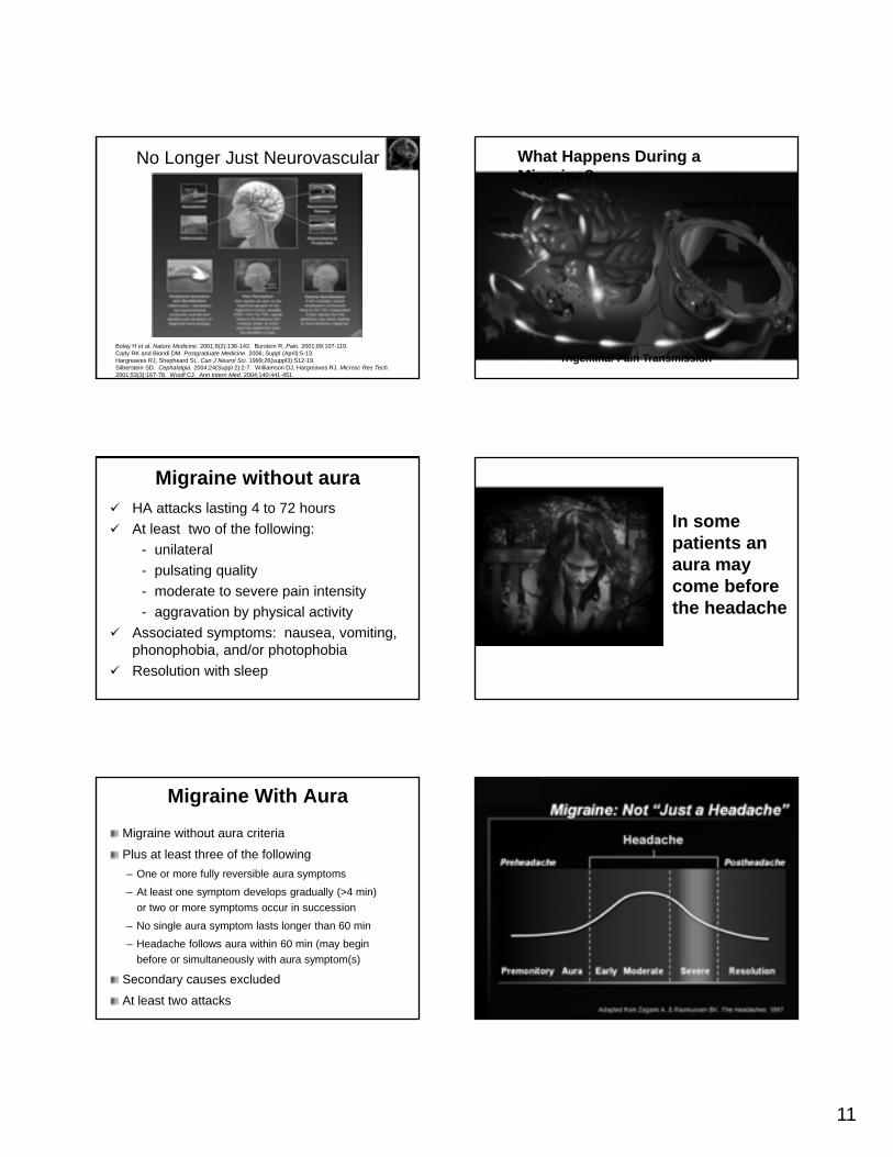
No Longer Just Neurovascular
Bolay H et al. Nature Medicine. 2001;8(2):136-142. Burstein R. Pain. 2001;89:107-110. Cady RK and Biondi DM. Postgraduate Medicine. 2006; Suppl (April):5-13. Hargreaves RJ, Shepheard SL. Can J Neurol Sci. 1999;26(suppl3):S12-19. Silberstein SD. Cephalalgia. 2004;24(Suppl 2):2-7. Williamson DJ, Hargreaves RJ. Microsc Res Tech.2001;53(3):167-78. Woolf CJ. Ann Intern Med. 2004;140:441-451.
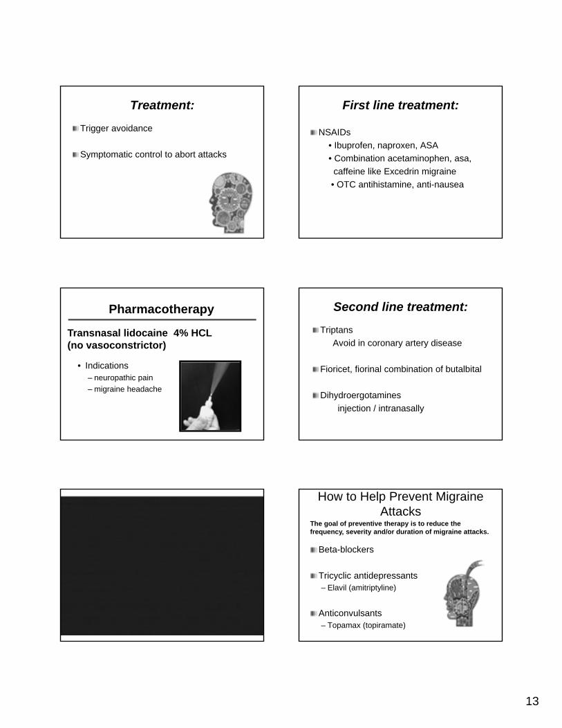
What Happens During a Migraine?
Trigeminal Pain Transmission
Pain
Vasodilation
Neuropeptide release
Migraine without aura
HA attacks lasting 4 to 72 hours
At least two of the following:
- unilateral
- pulsating quality
- moderate to severe pain intensity
- aggravation by physical activity
Associated symptoms: nausea, vomiting, phonophobia, and/or photophobia
Resolution with sleep
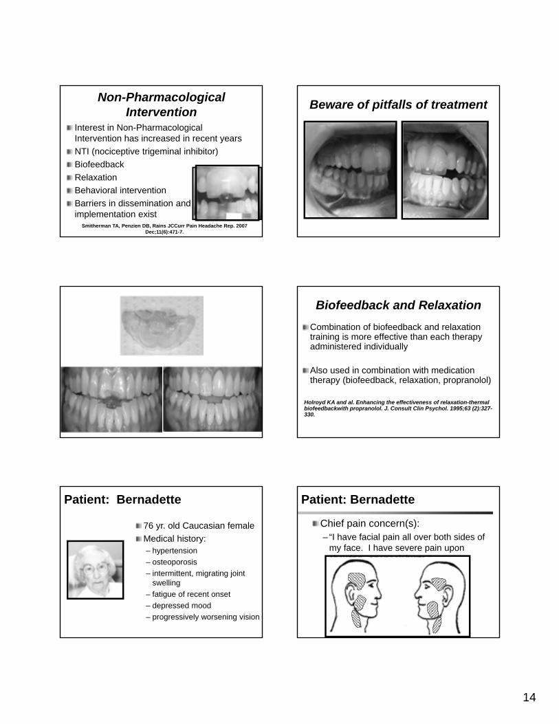
In some patients an aura may come before the headache
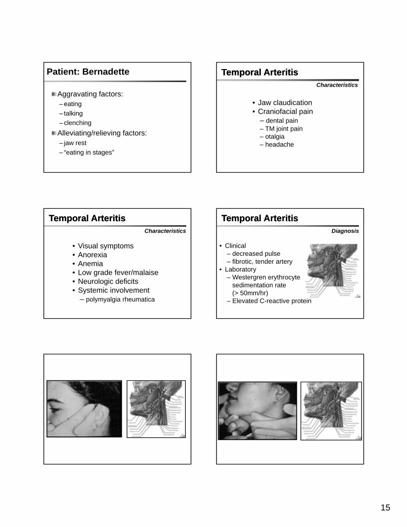
Migraine With Aura
Migraine without aura criteria
Plus at least three of the following
– One or more fully reversible aura symptoms
– At least one symptom develops gradually (>4 min)
or two or more symptoms occur in succession
– No single aura symptom lasts longer than 60 min
– Headache follows aura within 60 min (may begin
before or simultaneously with aura symptom(s)
Secondary causes excluded
At least two attacks

12
Visual
Flashes (photopsia)
Zigzag lines (teichopsia)
Blurred / cloudy vision
Tunnel vision
How Do Patients Describe the Impact of Migraine on Their Lives?
• “I can’t do anything. All I want to dois hide in a dark room.”
• “Any sound bothers me. I can’t eventalk to my children.”
• “I can’t control the pain. I’ve tried[almost] everything.”
• “When I have a severe migraineheadache, I am completely unableto function, unable to drive or towork.”
Migraine Disability Assessment Study, September 2000. Language patients used to describe migraine as stated by physicians.Durham C.F. et al. Headache 1998;38:427-435
Stress/Relief of stress
Hormonal Changes– Oral contraceptives– Menstruation
Lack of or too much sleep
Missed meals
Certain foods, drinks and
ingredients– For example, red wine, chocolate, and cheese
What Can Trigger a Migraine Attack?
13
Treatment:
Trigger avoidance
Symptomatic control to abort attacks
First line treatment:
NSAIDs
• Ibuprofen, naproxen, ASA
• Combination acetaminophen, asa,
caffeine like Excedrin migraine
• OTC antihistamine, anti-nausea
Pharmacotherapy
Transnasal lidocaine 4% HCL (no vasoconstrictor)
• Indications– neuropathic pain
– migraine headache
Second line treatment:
Triptans
Avoid in coronary artery disease
Fioricet, fiorinal combination of butalbital
Dihydroergotamines
injection / intranasally
Beta-blockers
Tricyclic antidepressants– Elavil (amitriptyline)
Anticonvulsants– Topamax (topiramate)
How to Help Prevent Migraine Attacks
The goal of preventive therapy is to reduce the frequency, severity and/or duration of migraine attacks.
14
Non-Pharmacological Intervention
Interest in Non-Pharmacological Intervention has increased in recent years
NTI (nociceptive trigeminal inhibitor)
Biofeedback
Relaxation
Behavioral intervention
Barriers in dissemination and implementation exist
Smitherman TA, Penzien DB, Rains JCCurr Pain Headache Rep. 2007 Dec;11(6):471-7.
Beware of pitfalls of treatment
Biofeedback and Relaxation
Combination of biofeedback and relaxation training is more effective than each therapy administered individually
Also used in combination with medication therapy (biofeedback, relaxation, propranolol)
Holroyd KA and al. Enhancing the effectiveness of relaxation-thermal biofeedbackwith propranolol. J. Consult Clin Psychol. 1995;63 (2):327-330.
Patient: Bernadette
76 yr. old Caucasian female
Medical history:– hypertension
– osteoporosis
– intermittent, migrating joint swelling
– fatigue of recent onset
– depressed mood
– progressively worsening vision
Patient: Bernadette
Chief pain concern(s):– “I have facial pain all over both sides of
my face. I have severe pain upon chewing. My neck hurts.”
15
Patient: Bernadette
Aggravating factors:– eating
– talking
– clenching
Alleviating/relieving factors:– jaw rest
– “eating in stages”
Temporal ArteritisTemporal ArteritisCharacteristics
• Jaw claudication• Craniofacial pain
– dental pain– TM joint pain– otalgia– headache
Temporal ArteritisTemporal ArteritisCharacteristics
• Visual symptoms• Anorexia• Anemia• Low grade fever/malaise• Neurologic deficits• Systemic involvement
– polymyalgia rheumatica
Temporal ArteritisTemporal ArteritisDiagnosis
• Clinical– decreased pulse– fibrotic, tender artery
• Laboratory– Westergren erythrocyte
sedimentation rate (> 50mm/hr)
– Elevated C-reactive protein
16
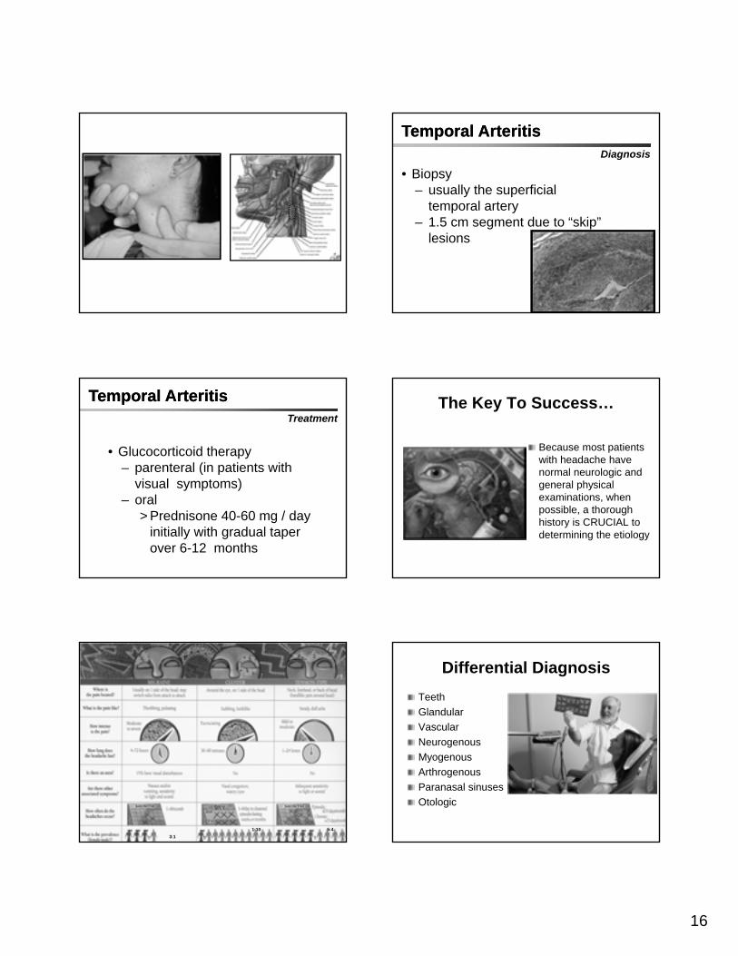
Temporal ArteritisTemporal ArteritisDiagnosis
• Biopsy– usually the superficial
temporal artery– 1.5 cm segment due to “skip”
lesions
Temporal ArteritisTemporal ArteritisTreatment
• Glucocorticoid therapy– parenteral (in patients with
visual symptoms)– oral
> Prednisone 40-60 mg / day initially with gradual taper over 6-12 months
The Key To Success…
Because most patients with headache have normal neurologic and general physical examinations, when possible, a thorough history is CRUCIAL to determining the etiology
3:1
5:41:10
Differential Diagnosis
Teeth
Glandular
Vascular
Neurogenous
Myogenous
Arthrogenous
Paranasal sinuses
Otologic

17
NeuropathicPain“Pain initiated or caused by a primary lesion or dysfunction
in the nervous system”
International Association for the Study of Pain (IASP)
Neuropathic Pain
Common-25-30% of Facial Pain Center patients
Under/misdiagnosed and undertreated
Interpatient variability regardingpresentation and response to treatment
Complex pathophysiology
Practitioner doubt
Key issues and challenges
Differential Diagnosis
Allodynia - pain from stimulus that does not normally cause pain
Hyperalgesia - increased response to a painful stimuli
Sympathetic hyperfunction - swelling, redness, sweating
Physical Examination
Neuropathic Orofacial Pain
Trigeminal NeuralgiaOnset
“Facial Pain II. A Prospective Survey of 1052 Patients with a View
of: Character of Attacks, Onset, Course and Character of Pain”
Rasmussen P. Acta Neurochirurgica, 1990;107:121-128
Atypical trigeminal neuralgia (ATN)
Characterized by brief pain paroxysmswith interval pain or attacks of several
minutes duration
Rasmussen P. Acta Neurochirurgica, 1990;107:121-128
Trigeminal NeuralgiaOnset

18
Patient: Charles
Chief pain concern:– “hurts when touched- electric like shock;
almost constant aching”
Patient: Charles
Aggravating factors:– touching area (occasionally)
– blowing nose, sneezing
– occasionally when smiling
Alleviating/relieving factors:– Tegretol (200 mg bid)- several hours
Pre-trigeminal Neuralgia
Historical perspectives
“…avoid the useless and unnecessary extraction of entire rows of healthy
teeth.”
Fothergill J. London. 1769;3:400-418Pujol M. Paris: Theophile Barrois, 1787
Pre-trigeminal Neuralgia
Historical perspectives
“…prodromal sensations experienced in the upper or lower jaws at the onset of
their illness.”
Symons C. Ann R Coll Surg Engl 1949;4:206-212Mitchell PG. Br Dent J 1980;149:167-170
Teeth were extracted 10 years prior to pain onset.
No osseous pathology is evident radiographically.
The soft tissues overlying the area is of normal color and texture.

19
Pre-trigeminal NeuralgiaPre-trigeminal Neuralgia
1. dull, aching pain (toothache/sinus-like pain)2. spontaneous onset3. no specific trigger zone4. duration- minutes to hours5. pain may spread
Pre-trigeminal Neuralgia
6. sporadic sharp, lancinating pain7. triggered by chewing, drinking hot/cold
liquids brushing teeth, yawning, talking8. pain decreases with somatic blocks9. precedes trigeminal neuralgia
Pre-trigeminal NeuralgiaPre-trigeminal Neuralgia
Differential diagnostic considerations
•neoplasm•atypical odontalgia•lower half headache•odontogenic pain
•sinusitis•myofascial pain•TM joint dysfunction•osseous pathology
Neuropathic Facial PainClassification
Classic Trigeminal NeuralgiaType 1 (TN 1)
Facial pain of spontaneous onset >50% limited to duration of an episode
of pain (temporary pain)
Neuropathic Facial PainClassification
Trigeminal NeuralgiaType 2 (TN 2)
Facial pain of spontaneous onset with greater than 50% presenting as a constant pain.
20
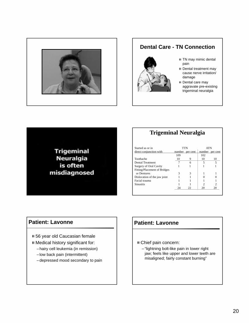
Dental Care - TN Connection
TN may mimic dental pain
Dental treatment may cause nerve irritation/ damage
Dental care may aggravate pre-existing trigeminal neuralgia
Trigeminal Neuralgia
Started as or in TTN ATN direct conjunction with number per cent number per cent
109 102Toothache 10 9 10 10Dental Treatment 7 6 5 5Surgery of Oral Cavity 1 1 1 1Fitting/Placement of Bridges
or Dentures 3 3 1 1Dislocation of the jaw joint 1 1 0 0Facial trauma 1 1 1 1Sinusitis 1 1 2 2
24 22 20 20
Patient: Lavonne
56 year old Caucasian female
Medical history significant for:– hairy cell leukemia (in remission)
– low back pain (intermittent)
– depressed mood secondary to pain
Patient: Lavonne
Chief pain concern:– “lightning bolt-like pain in lower right
jaw; feels like upper and lower teeth are misaligned; fairly constant burning”
21
Patient: Lavonne
Aggravating factors:– chewing, yawning, and talking
– cool/cold breeze on face
Alleviating/relieving factors:– occlusal appliance therapy
– Tegretol
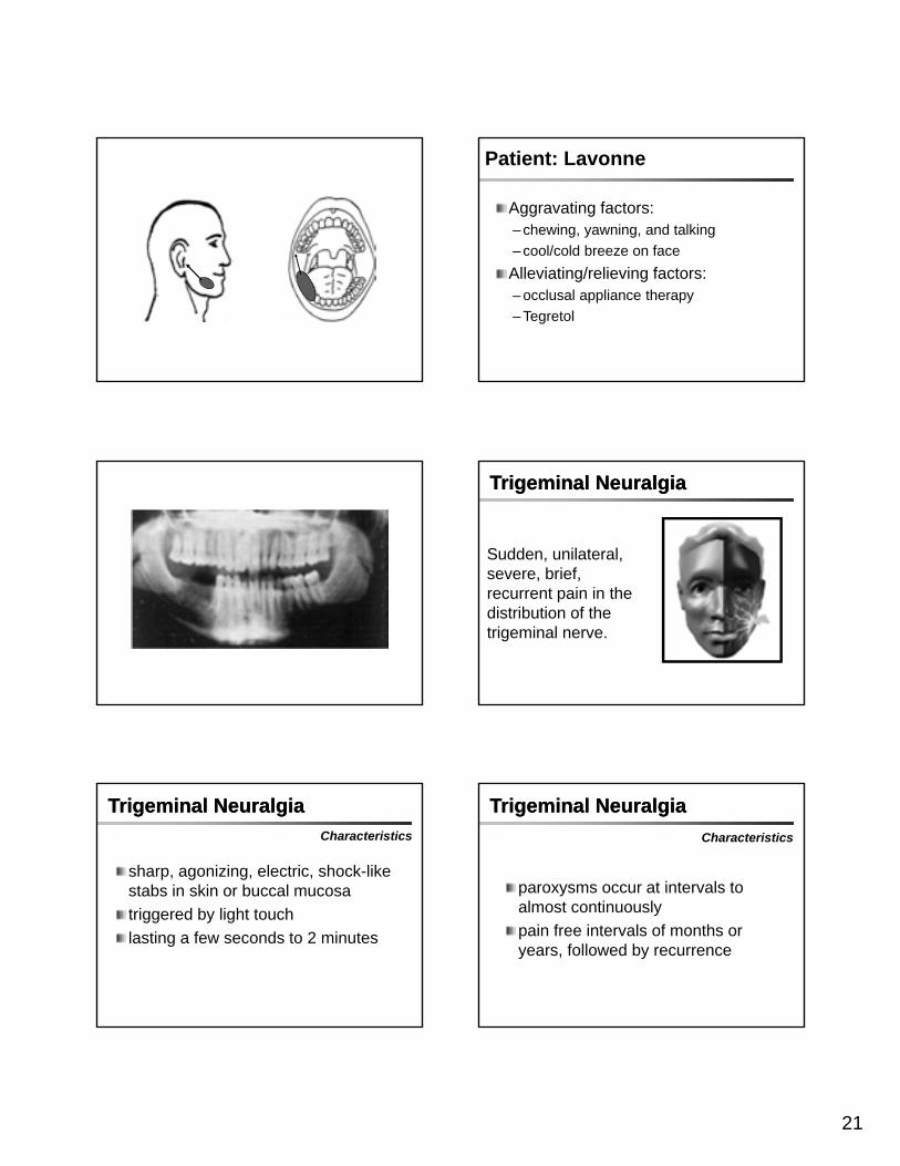
Trigeminal NeuralgiaTrigeminal Neuralgia
Sudden, unilateral, severe, brief, recurrent pain in the distribution of the trigeminal nerve.
sharp, agonizing, electric, shock-like stabs in skin or buccal mucosa
triggered by light touch
lasting a few seconds to 2 minutes
Trigeminal NeuralgiaTrigeminal NeuralgiaCharacteristics
paroxysms occur at intervals to almost continuously
pain free intervals of months or years, followed by recurrence
Trigeminal NeuralgiaTrigeminal Neuralgia
Characteristics
22
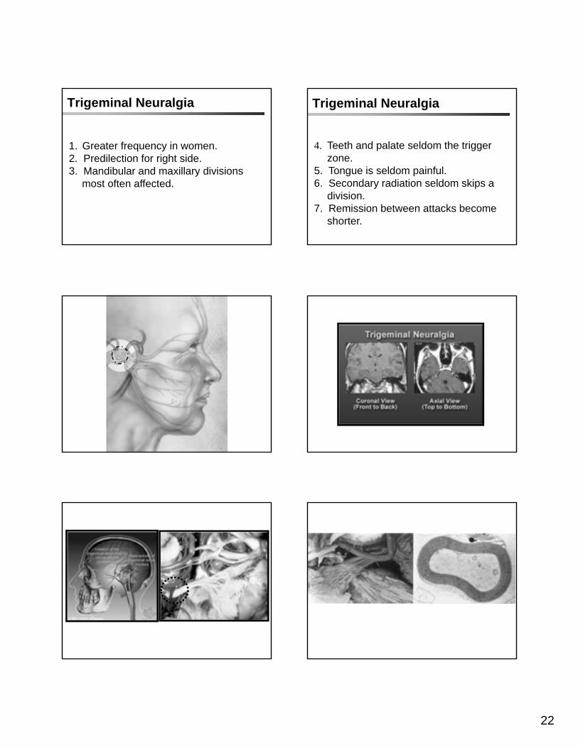
Trigeminal Neuralgia
1. Greater frequency in women.2. Predilection for right side.3. Mandibular and maxillary divisions
most often affected.
Trigeminal Neuralgia
4. Teeth and palate seldom the trigger zone.
5. Tongue is seldom painful.6. Secondary radiation seldom skips a
division.7. Remission between attacks become
shorter.

23
Pharmacotherapeutic
Surgical
Trigeminal NeuralgiaPrimary Treatment ModalitiesPrimary Treatment Modalities
Antidepressants
Anticonvulsants
GABA agonists
Local anesthetics
Neuroleptics
Muscle relaxants
Miscellaneous
PharmacotherapyPharmacotherapyAdjuvant analgesics
137
Trigeminal Neuralgia
Proposed etiologies:• vascular compression of trigeminal ganglion
• traumatic or auto-immune demyelination (MS)
• central / peripheral neural injury
• intracranial mass (tumor, aneurysm, cyst)
• unknown
Consider AGE and SYMPTOMS: idiopathic
versus secondary
Neuropathic Facial PainClassification
Secondary Symptomatic Trigeminal Neuralgia(STN)
Facial pain resulting from multiple sclerosis

24
Trigeminal NeuralgiaTrigeminal Neuralgia
Age of onset:Idiopathic / classic
-typically after age 30 (50-75 years)
Multiple sclerosis-related-20-40 years of age
Secondary Trigeminal Neuralgia• Multiple sclerosis affects approximately 1:700
people, with an estimated US prevalence of 250,000-500,000.• ~1-2% of patients with MS develop TN (~ 10
new cases per year, and cumulative total of approximately 4,000-5,000 people).
• Only about 3% of patients with TN have MS.
• TN due to an intracranial mass such as a tumor or aneurysm (excluding vascular compression from cerebellar arteries) is rare, probably accounting for no more than 5% of cases.
Multiple Sclerosis - Craniofacial Pain
1. Pain may be first symptom.2. Identical to trigeminal neuralgia.3. Begins between age 20 and 40.4. Associated with leg weakness.5. Sclerotic plaque in rootlets of V.
Multiple Sclerosis - Craniofacial PainMultiple Sclerosis - Craniofacial Pain
Herpes Zoster (shingles) is an acute infectious disease caused by herpes zoster virus. It primarily affects the posterior spinal root ganglion of the spinal nerves.
Herpes Zoster
25
Overall incidence of HZ: 131 per 100,000.
No gender difference.
Directly related to age; older > younger.
More common and severe in immunosuppressed patients– lymphoma
– chronic lymphocytic leukemia
– radiation therapy
– chemotherapy
– lupus erythematosus
Herpes Zoster: IncidenceHerpes Zoster: Incidence Distribution of herpes zoster
Region Cases (%)
Cranial 15
Cervical 12
Thoracic 55
Lumbar 14
Sacral 3
Generalized 1
All 100
Herpes Zoster of V
1. Pain may appear before vesicles.2. Ophthalmic division most often affected.3. Nerve affected unilaterally.4. Pathologic changes in V ganglion and rootlets.5. Chronic postherpetic pain rare but incurable.
Herpes Zoster
Pain/dysesthesia preceeds vesicles by 24-72 hours.
Evidenced in the distribution of the nerve affected.
Herpes Zoster
May occur at any age
Incidence highest in the 6th – 8th
decade
Recurs in 6% of cases
– usually at the same site as the initial lesion
Herpes Zoster
Immunosuppressive therapy
Stress/anxiety
Malignancy
Local irradiation
Trauma
Factors associated with reactivation
26
Neuropathic Facial PainClassification
Post-Herpetic Neuralgia(PHN)
Pain resulting from herpes zoster outbreak (shingles) along the trigeminal nerve
Herpes Zoster
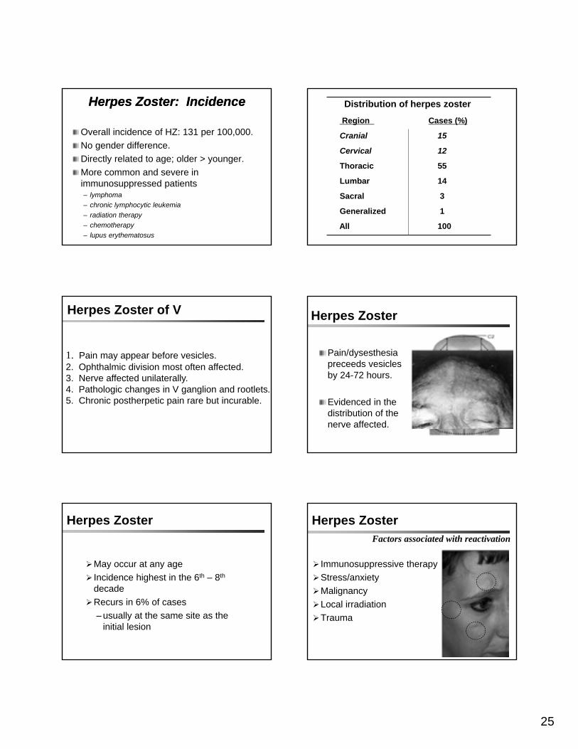
Pain recurring or continuing at the site of shingles 1 or more months after the
rash.
Postherpetic neuralgia
Herpes Zoster
Age dependent
50-70% depending on population studied
Dramatic increase after the age of 50
Postherpetic neuralgia
Incidence:Antiviral agent
Analgesic
Corticosteroid???
Local anesthetic– Peripheral
– Sympathetic
– Intravenous
Topical agents– Capsaicin
– Local anesthetic
– Aspirin/chloroform
– clonidine
Tricyclic antidepressants
Neruontin (gabapentin)
Herpes ZosterPostherpetic neuralgia
Treatment:
Herpes ZosterPrevention
Oxman N, Levin M. J Infect Dis. Mar 1, 2008; 197(Suppl 2): S228-S236
Herpes ZosterPrevention
Oxman N, Levin M. J Infect Dis. Mar 1, 2008; 197(Suppl 2): S228-S236
27
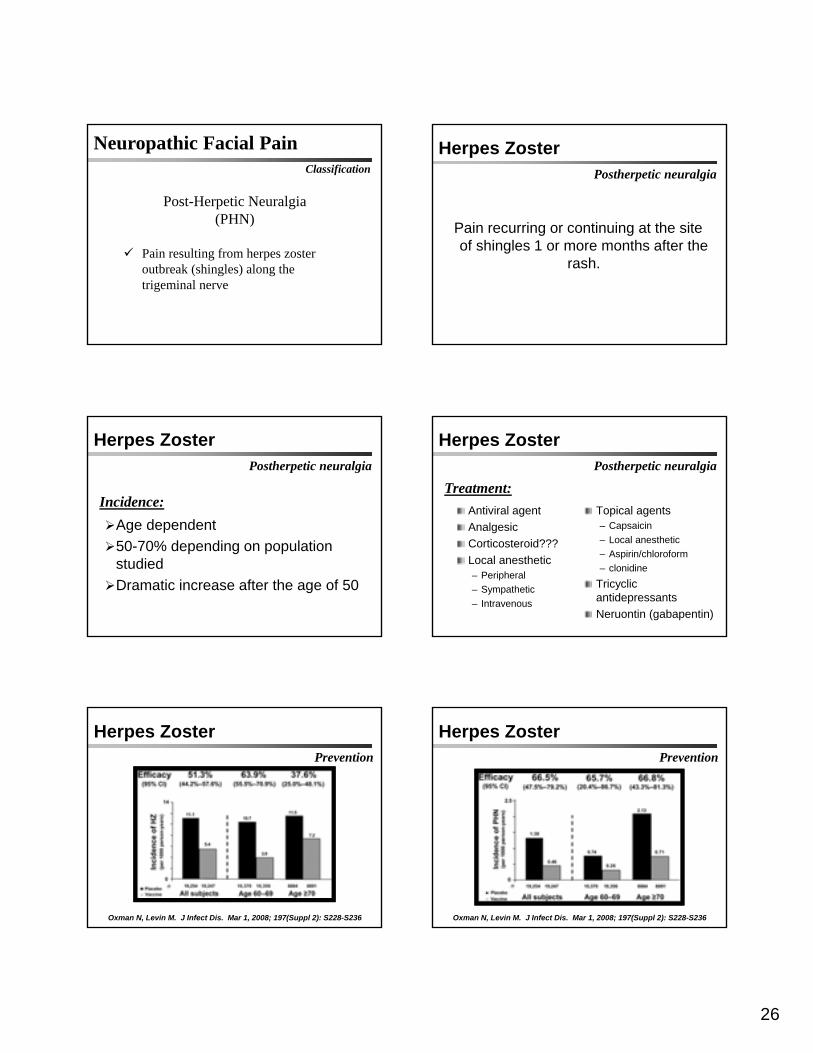
Herpes ZosterPrevention
Harvard Men’s Health Review June 2014
ANATOMIC SOURCESOF
OROFACIAL PAIN
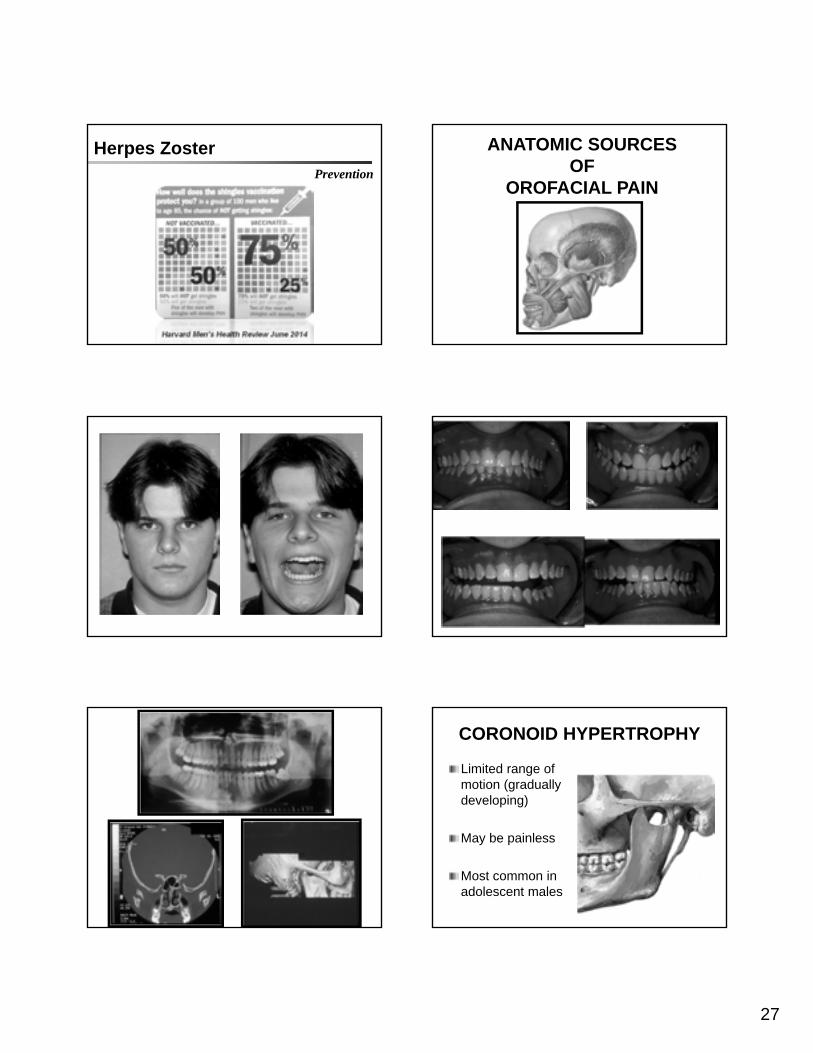
CORONOID HYPERTROPHY
Limited range of motion (gradually developing)
May be painless
Most common in adolescent males
28
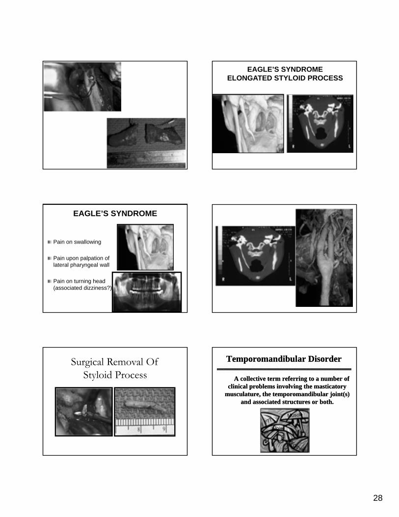
EAGLE’S SYNDROMEELONGATED STYLOID PROCESS
EAGLE’S SYNDROME
Pain on swallowing
Pain upon palpation of lateral pharyngeal wall
Pain on turning head (associated dizziness?)
Surgical Removal Of Styloid Process
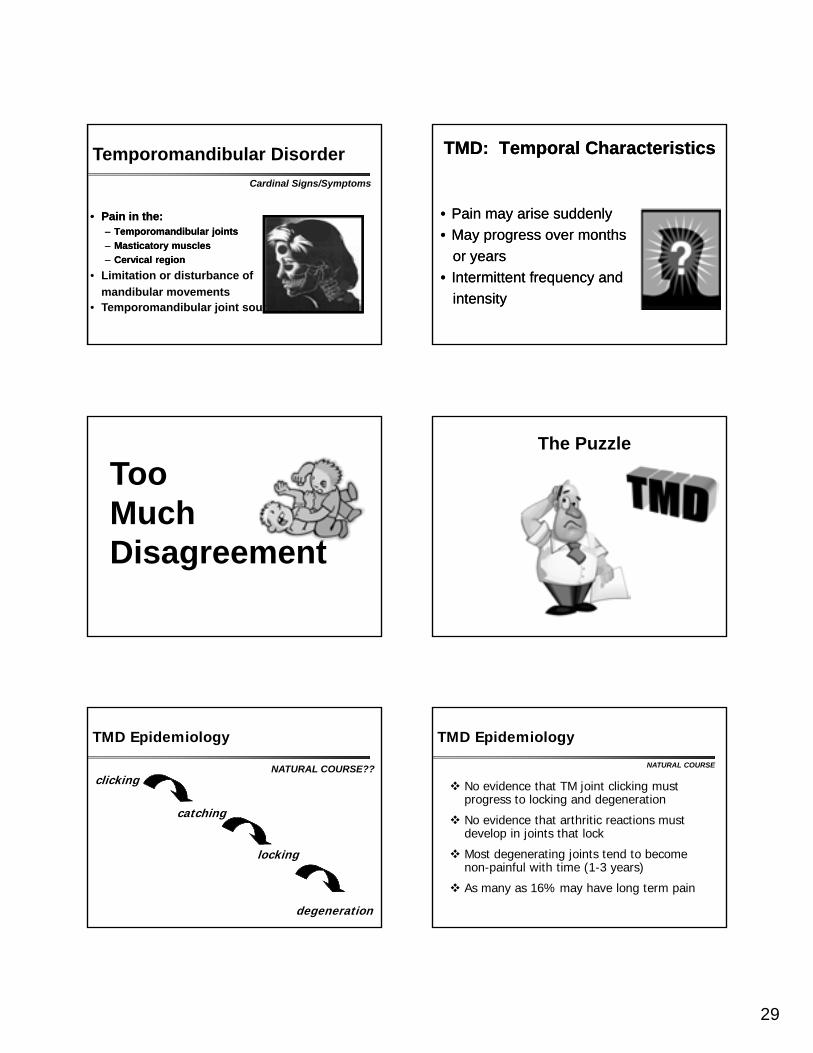
Temporomandibular Disorder Temporomandibular Disorder
A collective term referring to a number of clinical problems involving the masticatory
musculature, the temporomandibular joint(s) and associated structures or both.
A collective term referring to a number of clinical problems involving the masticatory
musculature, the temporomandibular joint(s) and associated structures or both.
29
• Pain in the:– Temporomandibular joints
– Masticatory muscles
– Cervical region
• Pain in the:– Temporomandibular joints
– Masticatory muscles
– Cervical region
• Limitation or disturbance of
mandibular movements• Temporomandibular joint sounds
Cardinal Signs/Symptoms
Temporomandibular Disorder TMD: Temporal CharacteristicsTMD: Temporal Characteristics
• Pain may arise suddenly
• May progress over months
or years
• Intermittent frequency and
intensity
• Pain may arise suddenly
• May progress over months
or years
• Intermittent frequency and
intensity
TooMuchDisagreement
The Puzzle
TMD Epidemiology
clicking
catching
locking
degeneration
NATURAL COURSE??
TMD Epidemiology
No evidence that TM joint clicking must progress to locking and degeneration
No evidence that arthritic reactions must develop in joints that lock
Most degenerating joints tend to become non-painful with time (1-3 years)
As many as 16% may have long term pain
NATURAL COURSE

30
TMD: Complex InteractionsTMD: Complex Interactions
Anatomic
Physiologic
Neurologic
Psychologic
Teeth
Musculature(cervical
and masticatory)
TM Joint
Associatedstructures
PATHOGENESISPATHOGENESIS
The cellular events and reactions and other pathologic mechanisms occurring
in the development of disease.
The cellular events and reactions and other pathologic mechanisms occurring
in the development of disease.
Temporomandibular DisorderTemporomandibular Disorder
Many things can light the fuse…many things can keep it burning!
HomeostaticBalance
HomeostaticBalance
Pathofunction
Anatomy Stress Nutrition Parafunction Trauma Gender Sleep Disorders Pain Depression Occlusion Coping Posture
Pathofunction
TMD: Etiologic VariablesTMD: Etiologic Variables
Differential Diagnosis
Teeth
Glandular
Vascular
Neurogenous
Myogenous
Arthrogenous
Paranasal sinuses
Otologic
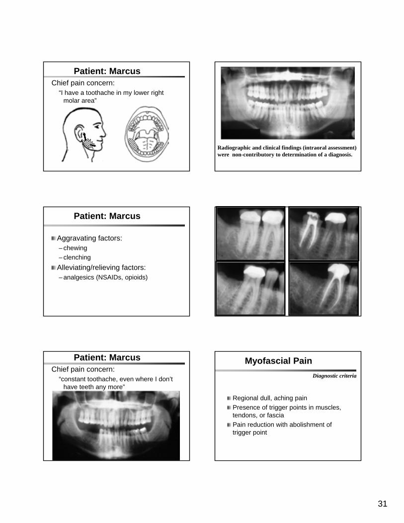
Patient: Marcus
28 year old Caucasian male
Medical history:– non-contributory
31
Patient: MarcusChief pain concern:
“I have a toothache in my lower right molar area”
Radiographic and clinical findings (intraoral assessment)were non-contributory to determination of a diagnosis.
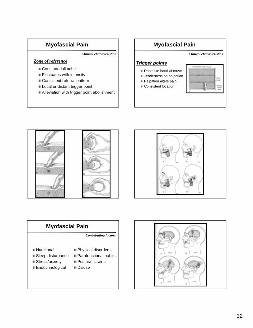
Patient: Marcus
Aggravating factors:– chewing
– clenching
Alleviating/relieving factors:– analgesics (NSAIDs, opioids)
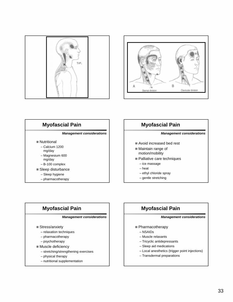
Patient: MarcusChief pain concern:
“constant toothache, even where I don’t have teeth any more”
Regional dull, aching pain
Presence of trigger points in muscles, tendons, or fascia
Pain reduction with abolishment of trigger point
Myofascial Pain
Diagnostic criteria
32
Constant dull ache
Fluctuates with intensity
Consistent referral pattern
Local or distant trigger point
Alleviation with trigger point abolishment
Myofascial Pain
Clinical characteristics
Zone of reference
Rope-like band of muscle
Tenderness on palpation
Palpation alters pain
Consistent location
Myofascial Pain
Clinical characteristics
Trigger points
Nutritional
Sleep disturbance
Stress/anxiety
Endocrinological
Physical disorders
Parafunctional habits
Postural strains
Disuse
Myofascial Pain
Contributing factors
33
Nutritional– Calcium 1200
mg/day
– Magnesium 600 mg/day
– B-100 complex
Sleep disturbance– Sleep hygiene
– pharmacotherapy
Myofascial Pain
Management considerations
Avoid increased bed rest
Maintain range of motion/mobility
Palliative care techniques– ice massage
– heat
– ethyl chloride spray
– gentle stretching
Myofascial Pain
Management considerations
Stress/anxiety– relaxation techniques
– pharmacotherapy
– psychotherapy
Muscle deficiency– stretching/strengthening exercises
– physical therapy
– nutritional supplementation
Myofascial Pain
Management considerations
Pharmacotherapy– NSAIDs
– Muscle relaxants
– Tricyclic antidepressants
– Sleep aid medications
– Local anesthetics (trigger point injections)
– Transdermal preparations
Myofascial Pain
Management considerations

34
Differential Diagnosis
Teeth
Glandular
Vascular
Neurogenous
Myogenous
Arthrogenous
Paranasal sinuses
Otologic
TM Joint Inflammatory Conditions
TM Joint Inflammatory Conditions
an inflammation of the synovial lining, capsular, or retrodiscal tissues of the
temporomandibular joint that can be due to infection, an immunologic condition
secondary to articular surface degeneration, or trauma.
Capsulitis, Synovitis, RetrodiscitisCapsulitis, Synovitis, Retrodiscitis
Capsulitis, Synovitis, RetrodiscitisCapsulitis, Synovitis, Retrodiscitis
• Patient education• Restrict mandibular function• Control parafunctional activity• Pharmacotherapy
– Analgesic/anti-inflammatory– Muscle relaxant (?)
• Stabilization orthotic• Physical therapy
• Patient education• Restrict mandibular function• Control parafunctional activity• Pharmacotherapy
– Analgesic/anti-inflammatory– Muscle relaxant (?)
• Stabilization orthotic• Physical therapy
Management Considerations:

35
TM Joint: Normal BiomechanicsTM Joint: Normal Biomechanics
Disc Displacement With ReductionDisc Displacement With Reduction Articular Disc DisplacementWith Reduction
36
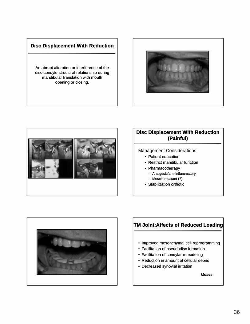
Disc Displacement With ReductionDisc Displacement With Reduction
An abrupt alteration or interference of the disc-condyle structural relationship during
mandibular translation with mouth opening or closing.
An abrupt alteration or interference of the disc-condyle structural relationship during
mandibular translation with mouth opening or closing.
Disc Displacement With Reduction (Painful)
Disc Displacement With Reduction (Painful)
• Patient education
• Restrict mandibular function
• Pharmacotherapy– Analgesic/anti-inflammatory
– Muscle relaxant (?)
• Stabilization orthotic
• Patient education
• Restrict mandibular function
• Pharmacotherapy– Analgesic/anti-inflammatory
– Muscle relaxant (?)
• Stabilization orthotic
Management Considerations:
TM Joint:Affects of Reduced LoadingTM Joint:Affects of Reduced Loading
• Improved mesenchymal cell reprogramming
• Facilitation of pseudodisc formation
• Facilitation of condylar remodeling
• Reduction in amount of cellular debris
• Decreased synovial irritation
• Improved mesenchymal cell reprogramming
• Facilitation of pseudodisc formation
• Facilitation of condylar remodeling
• Reduction in amount of cellular debris
• Decreased synovial irritation
Moses

37
Pseudodisc HypothesisPseudodisc Hypothesis
When subjected to constant repetitive compressive forces and loading, the
retrodiscal tissue may transform into a disc-like tissue.
When subjected to constant repetitive compressive forces and loading, the
retrodiscal tissue may transform into a disc-like tissue.
Scapino. OS,OM,OP 1983 (April):382-97Baustein, Scapino. Plas Recon Surg 1986 (December):756-64
Pseudodisc Hypothesis
Many TM joints display an adaptive capacity to remodel themselves and continue to function
without ideal disc position.
Solberg, Hansson. J Oral Rehab 1985, 12:303-321Westesson, Rohlin. OS,OM,OP 1984;4:17-22
Disc Displacement with Reduction(Painless)
Sudden Onset Closed Lock with no prior history of clicking
Anchored-disc phenomenonversus
Acute Closed Lock(Disk displacement without reduction)
Sudden Onset Closed Lock with no prior history of clicking
Anchored-disc phenomenonversus
Acute Closed Lock(Disk displacement without reduction)

38
TM Joint DistractionTM Joint Distraction
Arthrosimplicity-Outpatient ArthroscopyArthrosimplicity-Outpatient Arthroscopy
1.1mm scope
Normal AnatomyNormal Anatomy

39
Arthroscopy: Diagnostic findingsa. Normal findings
b. Synovitis
c. Disk displacement
d. Fibrillation
e. Adhesions
Arthroscopy: Diagnostic findingsa. Normal findings
b. Synovitis
c. Disk displacement
d. Fibrillation
e. Adhesions
TMJ ARTHROSCOPY Procedures AdhesionsAdhesions
Adhesions Adhesions Upper Joint SpaceAdhesions Upper Joint Space

40
1. Aggressive ROM exercises
2. NSAIDs
3. Reduce joint loadinga. Medications
b. Occlusal orthosis
1. Aggressive ROM exercises
2. NSAIDs
3. Reduce joint loadinga. Medications
b. Occlusal orthosis
TMJ ARTHROSCOPY
Post-op Management
Degenerative Joint Disease
A chronic inflammatory or non-inflammatory disease resulting in joint
deformity caused by degenerative changes in the articular cartilage, fibrous
connective tissue, and/or the articular disc within the temporomandibular joint.
Degenerative temporomandibular jointdisease is the result of maladaptation
to increased joint loading.
Westesson, Rohlin 1984Axelson, et al. 1992, 1993
Stegenga, et al. 1992deBont, Stegenga 1993
Macrotrauma
• Impact injury
• Extension-flexion injury
• Prolonged / excessive
mouth opening
• Intubation
41
TMD DJD: Trauma-Related
• 400 patients
• 25.5% reported an identifiable specific event
Macrotrauma
deBouver JA, Keersmakers K. J Oral Rehab 1996;23(2):91-96
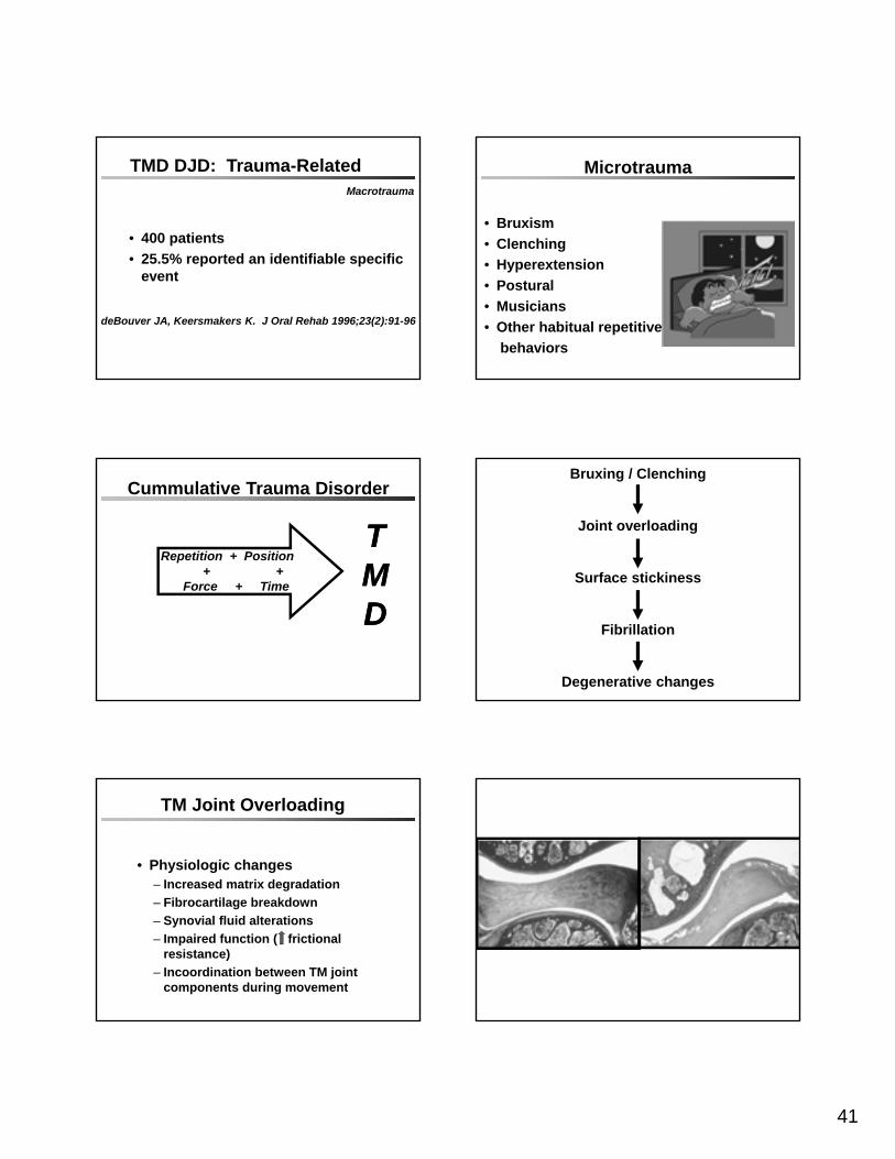
Microtrauma
• Bruxism
• Clenching
• Hyperextension
• Postural
• Musicians
• Other habitual repetitive
behaviors
Cummulative Trauma Disorder
Repetition + Position+ +
Force + Time
TMD
TMD
Bruxing / Clenching
Joint overloading
Surface stickiness
Fibrillation
Degenerative changes
TM Joint Overloading
• Physiologic changes– Increased matrix degradation
– Fibrocartilage breakdown
– Synovial fluid alterations
– Impaired function ( frictional resistance)
– Incoordination between TM joint components during movement

42
52 year old female
Chief concern: bilateral pre-auricular pain (severe) with swelling

43
Clinical Findings
TM joint– Severe pain at lateral and medial aspects
on palpation bilaterally
– Severe pain on loading bilaterally
– Maximum painless opening 15mm
– Course crepitus
Masticatory musculature– Generalized moderate pain on palpation
TM Joint Degenerative Joint Disease
Patient education
Restricted function
Pharmacotherapy– analgesic/anti-inflammatory
– muscle relaxant ???
Control parafunctional activities
Occlusal orthosis therapy
Physical therapy
Management considerations
4) Antioxidants
Vitamin C (sustained release)
1000 mg/d
Vitamin E
400 I.U./d
Betacarotene
2500 I.U./d (am)
Pharmacotherapya. NSAIDs
b. Muscle relaxantsc. Supportive
1) Glygoaminoglycan
1200-1500 mg/d
1) Chondroitin Sulfate
1500 mg/d
3) MSM
Degenerative Joint Disease Treatment
44
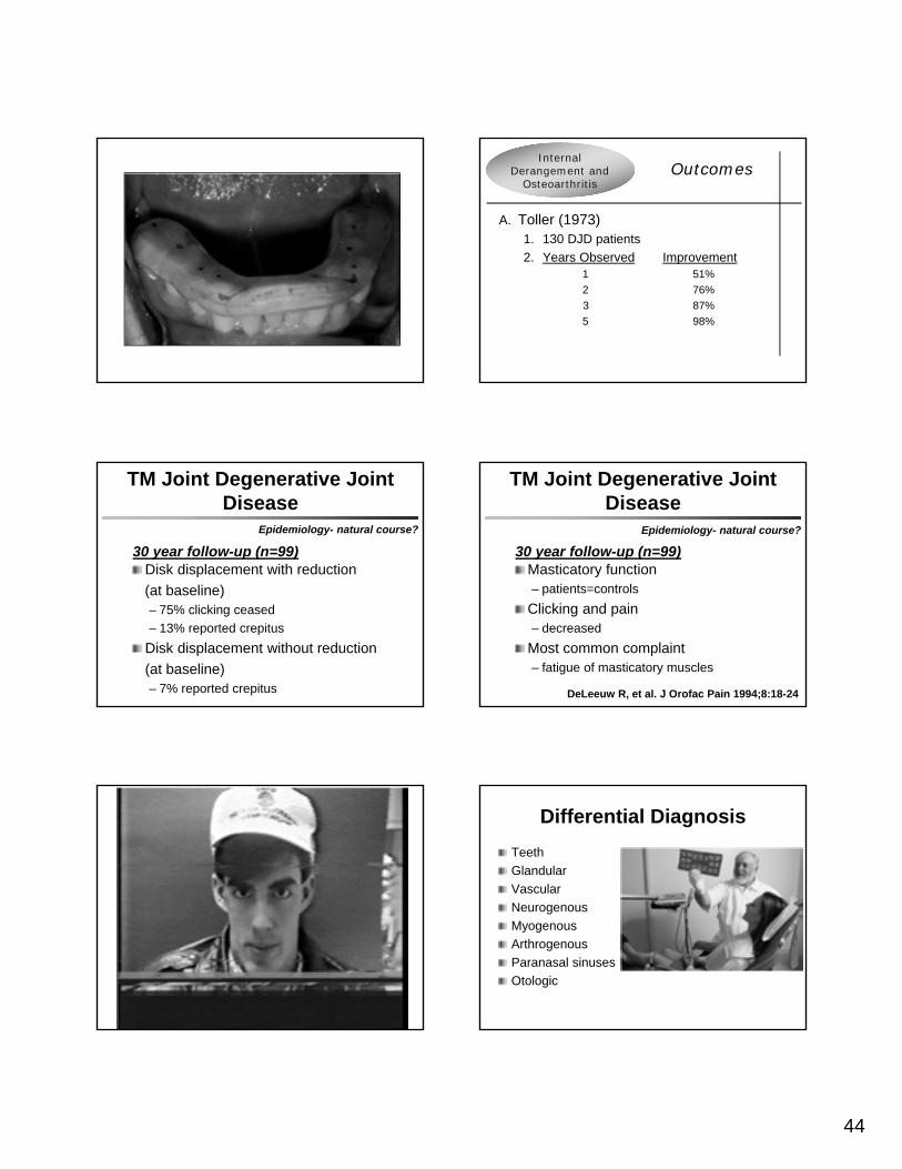
A. Toller (1973)1. 130 DJD patients
2. Years Observed Improvement1 51%
2 76%
3 87%
5 98%
Internal Derangement and
OsteoarthritisOutcomes
TM Joint Degenerative Joint Disease
Disk displacement with reduction
(at baseline)– 75% clicking ceased
– 13% reported crepitus
Disk displacement without reduction
(at baseline)– 7% reported crepitus
Epidemiology- natural course?
30 year follow-up (n=99)
TM Joint Degenerative Joint Disease
Masticatory function– patients=controls
Clicking and pain– decreased
Most common complaint– fatigue of masticatory muscles
Epidemiology- natural course?
30 year follow-up (n=99)
DeLeeuw R, et al. J Orofac Pain 1994;8:18-24
Differential Diagnosis
Teeth
Glandular
Vascular
Neurogenous
Myogenous
Arthrogenous
Paranasal sinuses
Otologic
45
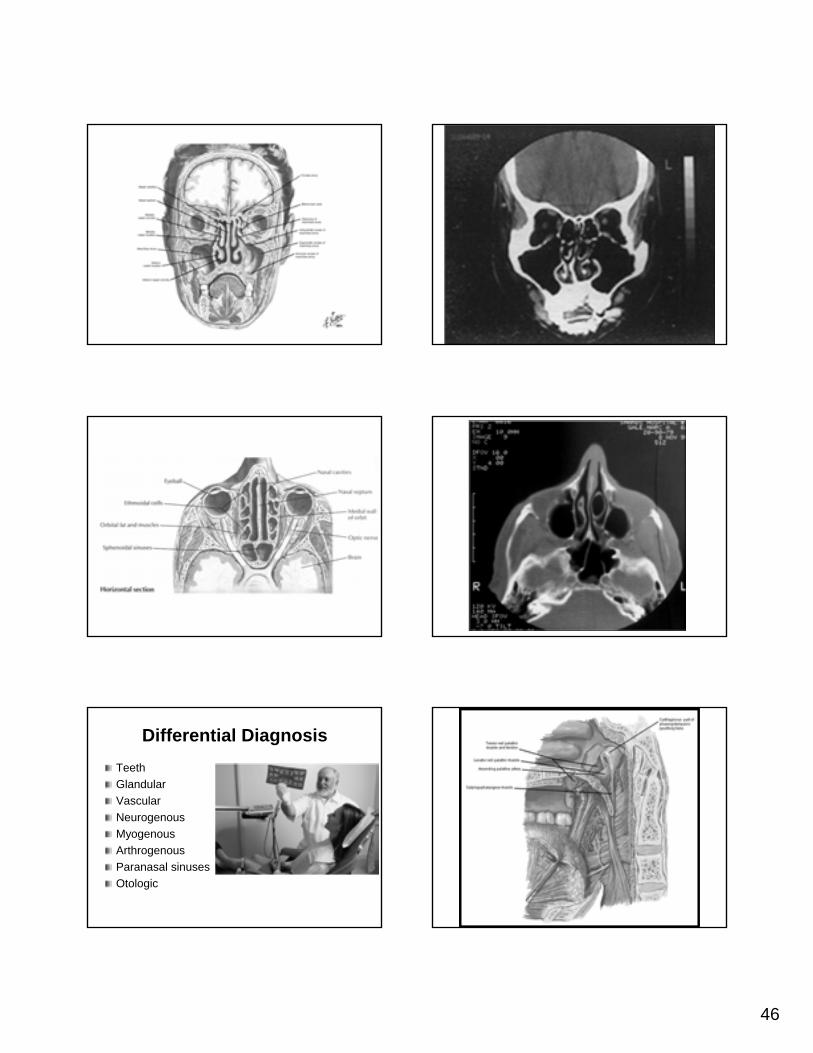
Paranasal SinusesParanasal Sinuses
Headache and facial pain are commonly related to infection,
inflammation, and/or obstruction of the outflow of the tracts of the
paranasal sinuses.
Acute / Chronic Sinusitis:PAINFUL COMPLICATIONS
Mucosal inflammation and thickening in cases of acute sinusitis
Partial or complete obstruction of sinus ostia
Pressure sensation
Maxillary mucoceles
Osteomyelitis
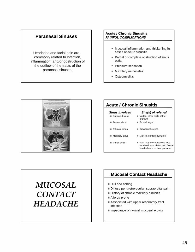
Sphenoid sinus
Frontal sinus
Ethmoid sinus
Maxillary sinus
Pansinusitis
Vertex, other parts of the cranium
Frontal region
Between the eyes
Maxilla, dental structures
Pain may be coalescent, less localized, associated with frontal headaches, constant pressure
Acute / Chronic SinusitisAcute / Chronic Sinusitis
Sinus involved Site(s) of referral
MUCOSALCONTACTHEADACHE
Mucosal Contact HeadacheMucosal Contact Headache
Dull and aching
Diffuse peri-/retro-ocular, supraorbital pain
History of chronic maxillary sinusitis
Allergy prone
Associated with upper respiratory tract infection
Impedance of normal mucosal activity
46
Differential Diagnosis
Teeth
Glandular
Vascular
Neurogenous
Myogenous
Arthrogenous
Paranasal sinuses
Otologic

47
Tinnitus: Differential DiagnosisTinnitus: Differential Diagnosis
Noise-inducedMetabolic diseaseEndocrine diseaseAutoimmune disordersStructural abnormalitiesMedication-inducedOccluso-muscle
Noise-inducedMetabolic diseaseEndocrine diseaseAutoimmune disordersStructural abnormalitiesMedication-inducedOccluso-muscle
Plate 88
Normal Tympanic MembraneNormal Tympanic Membrane Otitis Media
Otitis MediaOtitis MediaTympanic Membrane
PerforationTympanic Membrane
Perforation

48
Eustachian tube dysfunctionEustachian tube dysfunction
Normal function– Dilatation
– Primarily involves the tensor veli palatini
– Swallowing causes momentary eustachian tube dilitation which equalizes pressure
– Secondarily involves Levator veli palatini
Salpingopharyngeus
Superior constrictor
Plate 89Plate 89
Tonic Tensor Tympani Phenomenon
Tonic Tensor Tympani Phenomenon
Hypertonia of medial pterygoid produces a concomitant reflex hypertonia of the tensor tympani muscle
Tonic tensor tympani cannot initiate the reflex that increases the tonus of the tnsor veli palatini muscle
Failure of the eustachian tube to open during deglutition
Should I treat this patient?
What is/are the diagnosis(es)?
What factors are important in this case?
How should I treat this case?
49
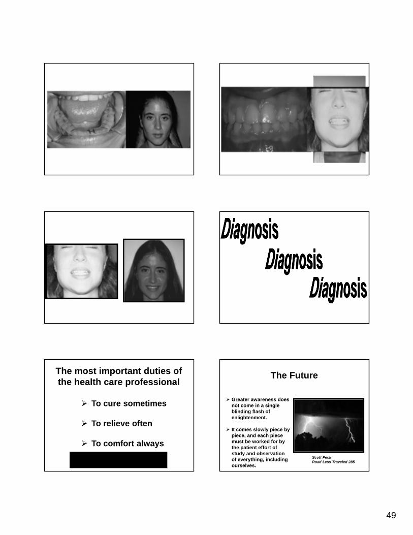
The most important duties ofthe health care professional
To cure sometimes
To relieve often
To comfort always
ABOVE ALL…..DO NO HARM!!!
Greater awareness does not come in a single blinding flash of enlightenment.
It comes slowly piece by piece, and each piece must be worked for by the patient effort of study and observation of everything, including ourselves.
Scott PeckRoad Less Traveled 285
The Future