Prevalence of radiographic primary hip and knee osteoarthritis in a representative Central European...
5
ORIGINAL PAPER Prevalence of radiographic primary hip and knee osteoarthritis in a representative Central European population Gábor Horváth & Gabriella Koroknai & Barnabás Ács & Péter Than & Árpád Bellyei & Tamás Illés Received: 9 May 2010 / Revised: 24 May 2010 / Accepted: 26 May 2010 / Published online: 16 June 2010 # Springer-Verlag 2010 Abstract The aim of our study was to determine the radiographic prevalence of hip and knee osteoarthritis and compare our results with prevalence data reported by other studies, as no similar study had been performed in Hungary previously. Our aim was also to investigate the usefulness of the different radiological scoring methods for the definition of osteoarthritis. Patients who earlier reported complaints and gave written consent were asked to participate in a clinical follow-up. In the 682 participants Harris hip score, visual analogue pain scale values for both joints, Knee Society score and knee functional score were calculated. Weight-bearing radiographs were taken of both joints. Kellgren-Lawrence radiological evaluation was performed and osteoarthritis prevalence was defined. Hip osteoarthritis was found in 109 cases (16.49%), and knee osteoarthritis was found in 111 cases (16.54%). Harris hip score, Knee Society score, functional score and visual analogue scale values were significantly worse in people with radiographically proven osteoarthritis compared to the control group (p <0.05). Significantly higher osteoarthritis prevalence of both joints was found in those with increased body mass index values. Age also plays a significant role in the development of both hip and knee osteoarthritis. No significant difference was observed between male and female participants regarding osteoarthritis prevalence. The Kellgren-Lawrence score with a cut-off value of 2 or more is a useful evaluation method for the detection of osteoarthritis prevalence in epidemiological studies; accord- ing to our observations, in clinical practice a cut-off value of three or more is more relevant. Introduction Osteoarthritis is one of the most frequent causes of musculoskeletal pain, and one of the leading causes of work absenteeism in the general adult population in the developed countries. The disease most frequently affects the weight-bearing joints, primarily the knee joint followed by the hip joint [1]. Apart from known aetiology leading to secondary osteoarthritis, in the vast majority of the cases the cause remains unknown, leaving primary osteoarthritis the most frequent form of degeneration in both joints. Although the symptoms and presentation of the disease vary widely, with progression surgical management includ- ing total hip and knee arthroplasty remains the main treatment option for these patients. For decision makers in the health care systems it is important to know at what rate the number of candidates for joint replacement surgery is increasing. On the basis of population and health trends, and only partially relying on epidemiological data, the American Academy of Orthopaedic Surgeons (AAOS) made predictions regarding the need for primary hip and knee arthroplasty by 2030. These calculations proved to be G. Horváth (*) : P. Than : Á. Bellyei : T. Illés Department of Orthopedics, Faculty of Medicine, University of Pécs, Ifjúság Street 13, 7643 Pécs, Hungary e-mail: [email protected] G. Koroknai Department of Physiotherapy, Faculty of Health Sciences, University of Pécs, Berek Street 15, 7632 Pécs, Hungary B. Ács Department of Applied Studies, Faculty of Business and Economics, University of Pécs, Rákóczi Street 80, 7622 Pécs, Hungary International Orthopaedics (SICOT) (2011) 35:971–975 DOI 10.1007/s00264-010-1069-6
-
Upload
gabor-horvath -
Category
Documents
-
view
212 -
download
0
Transcript of Prevalence of radiographic primary hip and knee osteoarthritis in a representative Central European...

ORIGINAL PAPER
Prevalence of radiographic primary hip and kneeosteoarthritis in a representative Central Europeanpopulation
Gábor Horváth & Gabriella Koroknai & Barnabás Ács &
Péter Than & Árpád Bellyei & Tamás Illés
Received: 9 May 2010 /Revised: 24 May 2010 /Accepted: 26 May 2010 /Published online: 16 June 2010# Springer-Verlag 2010
Abstract The aim of our study was to determine theradiographic prevalence of hip and knee osteoarthritis andcompare our results with prevalence data reported by otherstudies, as no similar study had been performed in Hungarypreviously. Our aim was also to investigate the usefulnessof the different radiological scoring methods for thedefinition of osteoarthritis. Patients who earlier reportedcomplaints and gave written consent were asked toparticipate in a clinical follow-up. In the 682 participantsHarris hip score, visual analogue pain scale values for bothjoints, Knee Society score and knee functional score werecalculated. Weight-bearing radiographs were taken of bothjoints. Kellgren-Lawrence radiological evaluation wasperformed and osteoarthritis prevalence was defined. Hiposteoarthritis was found in 109 cases (16.49%), and kneeosteoarthritis was found in 111 cases (16.54%). Harris hipscore, Knee Society score, functional score and visualanalogue scale values were significantly worse in peoplewith radiographically proven osteoarthritis compared to the
control group (p<0.05). Significantly higher osteoarthritisprevalence of both joints was found in those with increasedbody mass index values. Age also plays a significant role inthe development of both hip and knee osteoarthritis. Nosignificant difference was observed between male andfemale participants regarding osteoarthritis prevalence.The Kellgren-Lawrence score with a cut-off value of 2 ormore is a useful evaluation method for the detection ofosteoarthritis prevalence in epidemiological studies; accord-ing to our observations, in clinical practice a cut-off valueof three or more is more relevant.
Introduction
Osteoarthritis is one of the most frequent causes ofmusculoskeletal pain, and one of the leading causes ofwork absenteeism in the general adult population in thedeveloped countries. The disease most frequently affectsthe weight-bearing joints, primarily the knee joint followedby the hip joint [1]. Apart from known aetiology leading tosecondary osteoarthritis, in the vast majority of the casesthe cause remains unknown, leaving primary osteoarthritisthe most frequent form of degeneration in both joints.
Although the symptoms and presentation of the diseasevary widely, with progression surgical management includ-ing total hip and knee arthroplasty remains the maintreatment option for these patients. For decision makers inthe health care systems it is important to know at what ratethe number of candidates for joint replacement surgery isincreasing. On the basis of population and health trends,and only partially relying on epidemiological data, theAmerican Academy of Orthopaedic Surgeons (AAOS)made predictions regarding the need for primary hip andknee arthroplasty by 2030. These calculations proved to be
G. Horváth (*) : P. Than :Á. Bellyei : T. IllésDepartment of Orthopedics, Faculty of Medicine,University of Pécs,Ifjúság Street 13,7643 Pécs, Hungarye-mail: [email protected]
G. KoroknaiDepartment of Physiotherapy, Faculty of Health Sciences,University of Pécs,Berek Street 15,7632 Pécs, Hungary
B. ÁcsDepartment of Applied Studies,Faculty of Business and Economics, University of Pécs,Rákóczi Street 80,7622 Pécs, Hungary
International Orthopaedics (SICOT) (2011) 35:971–975DOI 10.1007/s00264-010-1069-6
gross underestimations by 2005 [2]. To promote moreprecise estimations, epidemiological reports on osteoarthri-tis prevalence can be very helpful.
Although the relationship between radiographic hiposteoarthritis and the need for total hip arthroplasty(THA) is not fully established, according to Ingvarssonsevere osteoarthritis and range of motion (ROM) restrictioneventually necessitate surgical intervention [3]. A recentreview article investigated reports regarding hip osteoar-thritis prevalence published in previous decades. Pointprevalence of radiographic hip osteoarthritis varied from0.9 to 27% depending on the selection criteria and methodof evaluation [4]. Several methods are used for radiographicevaluation of both joints, most widely the scoring system ofKellgren and Lawrence, and for good intra- and inter-observer reliability the joint space width narrowing [5].Neither in the hip nor in the knee joints is there isconsensus regarding the epidemiological definition ofosteoarthritis [6]; however, it has been suggested thatlongstanding pain of both joints shows the closest correla-tion with radiographic structural changes [7].
Prevalence data of major degenerative joint complaintshave already been reported in our earlier publication with22.2% of the surveyed having hip and 30.3% having kneepain [8]. In this study our aim was to give radiographicprevalence data—using the Kellgren-Lawrence classifica-tion—on primary hip and knee osteoarthritis for the firsttime in a Hungarian population. The functional status of thepatients was also evaluated using the Harris hip score andthe Knee Society score and cross-matched with theradiographic findings [9, 10]. The influence of body massindex (BMI), age and gender on the development ofosteoarthritis was also investigated.
Materials and methods
From the participants of our previous representative question-naire survey—carried out in the south-western part of
Hungary—those who reported either hip or knee pain andhad given written consent were invited for a clinical andradiological follow-up in the search for primary osteoarthritis.From this patient pool of 2,241 people, 682 (244 men and 438women) with an average age 52.31 years (20–67) came for afollow-up to our outpatient department. Detailed informationregarding general health and musculoskeletal condition wasobtained; patients’ anthropometric parameters were recorded.Participants were asked to describe the severity of their painon a visual analogue scale and to localise it on a pain drawingtest for both hip and knee joints. This was followed by amusculoskeletal physical examination performed by one oftwo experienced orthopaedic surgeons. ROM of both joints,alignment of the knee and ligament balance were defined.Possible contractures, Trendelenburg’s sign and lower limbdiscrepancy were also recorded. Standardised weight-bearinganteroposterior (AP) pelvis and bilateral weight-bearing APand lateral view radiographs of both knee joints were taken in676 cases.
Using anamnestic data and our records, joint functionwas evaluated by calculating the Harris hip score and KneeSociety scores for the hip and the knee joints, respectively.
Radiographic evaluation was performed using theKellgren-Lawrence osteoarthritis score. The diagnosis ofosteoarthritis was established at Kellgren-Lawrence≥2points and severe osteoarthritis at Kellgren-Lawrence≥3points in our study. Joint space widths of the hip and kneejoints were measured with 0.5 mm accuracy on the APpelvis and AP knee radiographs. The narrowest part wasrecorded as joint space width.
Participants were categorised according to their BMIvalues: (1) under 25 normal, (2) 26–30 overweight, (3) 31–35 obese and (4) above 35 extremely obese.
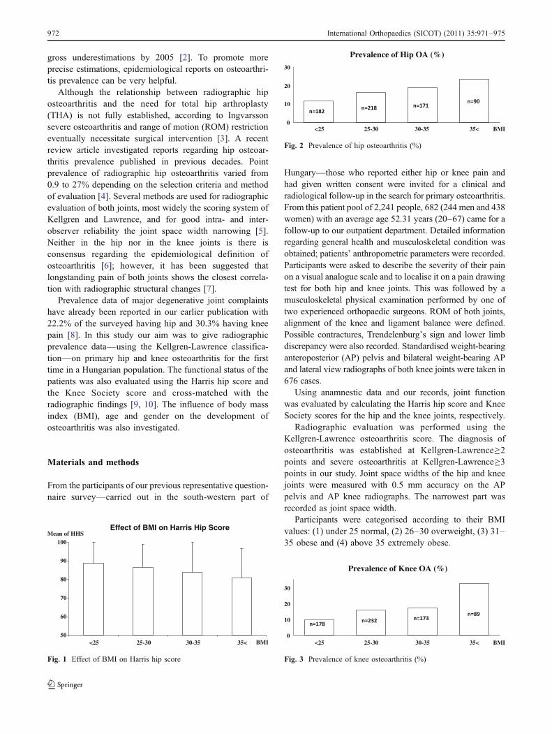
Effect of BMI on Harris Hip Score
50
60
70
80
90
100
<25 25-30 30-35 35< BMI
Mean of HHS
Fig. 1 Effect of BMI on Harris hip score
Prevalence of Hip OA (%)
0
10
20
30
<25 25-30 30-35 35< BMI
Fig. 2 Prevalence of hip osteoarthritis (%)
Prevalence of Knee OA (%)
0
10
20
30
<25 25-30 30-35 35< BMI
Fig. 3 Prevalence of knee osteoarthritis (%)
972 International Orthopaedics (SICOT) (2011) 35:971–975
Associations between functional scores and radiographicfindings were analysed. The influence of BMI, age andgender on symptoms and osteoarthritis prevalence wasfurther investigated.
Our results were statistically analysed. Continuousvariables were analysed in terms of mean ± standarddeviation. The χ2 test was used to establish data correlation.Standard Student’s t test for paired samples or one-wayanalysis of variance (ANOVA) were performed for com-paring data, as needed. A p value<0.05 was consideredsignificant. All analyses were carried out with SPSSsoftware for Windows version 13.0.
Results
Of the patients, 15 had undergone THA, three of them totalknee arthroplasty and two of them unicondylar kneearthroplasty, leaving 661 radiographs for hip osteoarthritisand 672 radiographs for knee osteoarthritis evaluation.
Regarding hip osteoarthritis prevalence, 101 hips of 63women and 69 hips of 46 men had Kellgren-Lawrence≥2, ofwhich 18 had Kellgren-Lawrence≥3. Overall 16.49% had hiposteoarthritis with 2.7% having severe osteoarthritis.
Harris hip score was calculated in 682 cases andaveraged 85.72 points. Patients with osteoarthritis had anaverage value significantly lower (77.37) in comparison
with the negative reference group (87.75) (p≥0.05). Avisual analogue scale for hip pain was completed by 661patients who averaged 2.9 points. These values showed asignificant negative correlation with the Harris hip scorevalues (p≥0.05).
The associations between BMI and Harris hip scorevalues are summarised in Fig. 1. The prevalence of hiposteoarthritis with respect to the BMI values is presented inFig. 2. The average joint space width was 3.56 for the rightand 3.52 for the left hip joints.
We investigated whether age influences the different hipparameters and BMI. BMI and visual analogue scale scoreshowed a significant positive correlation and joint spacewidth and Harris hip score a significant negative correlation(p≥0.05).
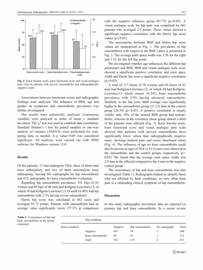
A total of 117 knees of 70 women and 68 knees of 41men had Kellgren-Lawrence≥2, of which 20 had Kellgren-Lawrence≥3 which means 16.54% knee osteoarthritisprevalence with 2.9% having advanced osteoarthritis.Similarly in the hip joint, BMI average was significantlyhigher in the osteoarthritic group (31.13) than in the controlgroup (28.39) (p<0.05). A positive correlation is clearlyvisible: only 10% of the normal BMI group had osteoar-thritis, whereas in the extremely obese group almost a thirdof the patients were affected (Fig. 3). Knee Society score,knee functional score and visual analogue pain scaleshowed that patients with proven osteoarthritis havesignificantly lower values than radiographically negativecases, showing marked pain and worse functional status(Fig. 4). The influence of age on knee osteoarthritis couldalso be proven as ages of 56.8 vs 51.4 years were observed inthe osteoarthritic and the control groups, respectively (p<0.05). We found that the average joint space width was2.9 mm in the affected compared to the 4 mm in the negativecontrol group.
The coexistence of hip and knee osteoarthritis was alsoinvestigated (Table 1). Radiographs helped us identify thosewho are affected by both conditions, as very often kneepain is a misleading clinical symptom of hip osteoarthritis.
Discussion
In this study radiographic prevalence data are reported onprimary hip and knee osteoarthritis. In a recent review
5.7
69.864.1
4.2
76.074.1
0
25
50
75
100
Knee society score Knee functional score Visual analogue painscale
OsteoarthritisNegative
10
7,5
5
2,5
Kne
e sc
ores V
AS
Fig. 4 Knee Society score, knee functional score and visual analoguepain scale in patients with proven osteoarthritis and radiographicallynegative cases
Hip condition
Knee condition Negative Hip osteoarthritis No radiograph Total
Negative 483 74 3 560
Knee osteoarthritis 69 41 1 111
Total 552 115 4 671
Table 1 Coexistence of hip andknee osteoarthritis in the groupexamined
International Orthopaedics (SICOT) (2011) 35:971–975 973

article our previous study was rightfully criticised for notpresenting prevalence data based on radiographic evidence,despite the fact that the aim of our previous work was togive data based on self-reported pain of the major weight-bearing joints [4, 8]. Although osteoarthritis is the leadingcause of joint pain, in several cases patients with severe,radiographically proven hip osteoarthritis are free of clinicalsymptoms as reported by Birrell et al. [11]. As far as weknow, with the exception of Cvijetić et al., who publishedCroatian prevalence data on hip osteoarthritis recently norepresentative study has been carried out in the CentralEuropean region [12].
Upon reviewing the literature we have found that mostpublications reporting osteoarthritis prevalence of the kneefocus on an elderly population. Although most of theprevious studies used the Kellgren-Lawrence classificationsystem for radiographic evaluation of the weight-bearingjoints, no clear recommendation is given regarding the cut-off value for the definition of osteoarthritis [3, 13–15]. Dueto this fact we differentiated two cut-off values for mild andmore advanced osteoarthritis using 2 and 3 as cut-offvalues, respectively.
In their study Lawrence and Sebo reported 12–25% hiposteoarthritis prevalence in the Caucasian race dependingon the country surveyed [14]. In our study a similar valuewas found for hip osteoarthritis prevalence at 16.5%. Eventhough the reproducibility of the Kellgren-Lawrence scor-ing is reported to be 75% by the authors in the originalarticle, several publications use joint space width narrowingto establish the diagnosis of hip osteoarthritis. Cut-offvalues are between 1.5 and 4 mm resulting in hugedifferences in prevalence data. Ingvarsson used 2.5 mm asa cut-off value in his survey of 1,517 people in Iceland andfound a high 8% prevalence based on coronagraphs [3].Danielsson and Lindbergh screening a Swedish populationfound 1.2% [16]. Even though we also used the Kellgren-Lawrence classification system, in the advanced cases(2.7%) joint space width was less than 2 mm.
Regarding the knee joint most studies in the literatureuse the Kellgren-Lawrence classification for its clinicalusefulness [17]. Scandinavian authors using the sameclassification method found 1.5% knee osteoarthritisprevalence in a group aged 35–54, although anthropomet-ric parameters of the subjects examined are not reported[18]. In a recent Asian population study over 30%radiographic prevalence is reported in a group of 598people aged above 65 (17.7% men and 36.5% women)[19]. According to their observations higher age, BMI,female sex and higher bone density play a significant rolein the development of knee osteoarthritis. Depending onthe Kellgren-Lawrence cut-off value prevalence data were16.5 and 2.9% in our study with a higher, yet notsignificant prevalence in male patients.
Due to these large prevalence differences observed inboth the hip and knee joints when using differentKellgren-Lawrence cut-off values, we think it is desirableto differentiate according to study design. For epidemi-ological purposes only, Kellgren-Lawrence 2 is a goodalternative, whereas in studies with the aim of screeningthose with more clinical relevance Kellgren-Lawrence 3is a better choice. In this study joint space widthnarrowing in the diagnosis of osteoarthritis directly wasnot investigated, yet substantial functional decline wasobserved at 2 mm in both the hip and knee joints. Thelimitation of our study of osteoarthritis is that it couldonly assess prevalence within the group examined. Thusthe sample is only representative regarding age, munic-ipal structure and employment.
We found that higher BMI and age also show apositive correlation with both hip and knee osteoarthri-tis. Felson and Zhang in their review article showed thatin both joints the higher the BMI value, the higher theprevalence of symptoms and radiographic osteoarthritis.They found that the correlation was stronger in the kneejoint [20]. Several publications discuss the role of higherBMI as a risk factor in degenerative conditions [21–23].Other authors emphasise rather the body composition asBMI values may incorporate the mass of the skeletalmuscle which plays a protective role as opposed toadipose tissue [24].
According to our observations, patients with hip painand lower Harris hip score had a higher prevalence ofknee osteoarthritis and lower Knee Society and func-tional scores. Whether this is caused by radiating painor the greater age causing multiple joint involvementsneeds further investigation.
Conflict of interest The authors declare that they have no conflict ofinterest.
References
1. Zhang W, Doherty M (2006) EULAR recommendations for kneeand hip osteoarthritis: a critique of the methodology. Br J SportsMed 40:664–669
2. Iorio R, Robb WJ, Healy WL et al (2008) Orthopaedic surgeonworkforce and volume assessment for total hip and kneereplacement in the United States: preparing for an epidemic. JBone Joint Surg Am 90:1598–1605
3. Ingvarsson T (2000) Prevalence and inheritance of hip osteoar-thritis in Iceland. Acta Orthop Scand Suppl 298:1–46
4. Dagenais S, Garbedian S, Wai EK (2009) Systematic review ofthe prevalence of radiographic primary hip osteoarthritis. ClinOrthop Relat Res 467:623–637
5. Kellgren J, Lawrence JS (1957) Radiological assessment of osteo-arthrosis. Ann Rheum Dis 16:494–502
974 International Orthopaedics (SICOT) (2011) 35:971–975

6. Schiphof D, de Klerk BM, Koes BW et al (2008) Good reliability,questionable validity of 25 different classification criteria of kneeosteoarthritis: a systematic appraisal. J Clin Epidemiol 61:1205–1215
7. O’Reilly S, Doherty M (1998) Signs, symptoms and laboratorytests. In: Brandt KD, Doherty M, Lohmander LS (eds) Osteoar-thritis. Oxford University Press, Oxford, pp 197–217
8. Horváth G, Than P, Bellyei Á et al (2006) Prevalence ofdegenerative joint complaints of the lower extremity: a represen-tative study. Int Orthop 30:118–122
9. Harris WH (1969) Traumatic arthritis of the hip after dislocationand acetabular fractures: treatment by mold arthroplasty. An end-result study using a new method of result evaluation. J Bone JointSurg Am 51:737–755
10. Insall JN, Dorr LD, Scott RD et al (1989) Rationale of the KneeSociety clinical rating system. Clin Orthop Relat Res 248:13–14
11. Birrell F, Lunt M, Macfarlane G et al (2005) Association betweenpain in the hip region and radiographic changes of osteoarthritis:results from a population-based study. Rheumatology (Oxford)44:337–341
12. Cvijetić S, Campbell L, Cooper C et al (2000) Radiographicosteoarthritis in the elderly population of Zagreb: distribution,correlates, and the pattern of joint involvement. Croat Med J41:58–63
13. Hoaglund FT, Yau MC, Wong WL (1973) Osteoarthritis of the hipand other joints in southern Chinese in Hong Kong. J Bone JointSurg Am 55:545–557
14. Lawrence JS, Sebo M (1980) The geography of osteoarthritis. In:Nuki G (ed) The aetiopathogenesis of osteoarthritis. Pitman,London, pp 155–183
15. Odding E, Valkenburg HA, Algra D et al (1998) Associations ofradiological osteoarthritis of the hip and knee with locomotordisability in the Rotterdam Study. Ann Rheum Dis 57:203–208
16. Danielsson L, Lindbergh H (1997) Prevalence of coxarthrosis inan urban population during four decades. Clin Orthop Relat Res342:106–110
17. Kessler S, Guenther KP, Puhl W (1998) Scoring prevalence andseverity in gonarthritis: the suitability of the Kellgren & Lawrencescale. Clin Rheumatol 17:205–209
18. Petersson IF, Boegård T, Saxne T et al (1997) Radiographicosteoarthritis of the knee classified by the Ahlbäck andKellgren & Lawrence systems for the tibiofemoral joint inpeople aged 35–54 years with chronic knee pain. Ann RheumDis 56:493–496
19. Sudo A, Miyamoto N, Horikawa K et al (2008) Prevalence andrisk factors for knee osteoarthritis in elderly Japanese men andwomen. J Orthop Sci 13:413–418
20. Felson DT, Zhang Y (1998) An update on the epidemiology ofknee and hip osteoarthritis with a view to prevention. ArthritisRheum 41:1343–1355
21. Anderson JJ, Felson DT (1988) Factors associated with osteoar-thritis of the knee in the first national Health and NutritionExamination Survey (HANES I). Evidence for an association withoverweight, race, and physical demands of work. Am J Epidemiol128:179–189
22. Hochberg MC, Lethbridge-Cejku M, Scott WW et al (1995) Theassociation of body weight, body fatness and body fat distributionwith osteoarthritis of the knee: data from the Baltimore Longitu-dinal Study of Aging. J Rheumatol 22:488–493
23. Lachance L, Sowers MF, Jamadar D et al (2001) The experienceof pain and emergent osteoarthritis of the knee. OsteoarthritisCartilage 9:527–532
24. Sowers M, Yosef M, Jamadar D et al (2008) BMI vs. bodycomposition and radiographically defined osteoarthritis of theknee in women: a 4-year follow-up study. Osteoarthritis Cartilage16:367–372
International Orthopaedics (SICOT) (2011) 35:971–975 975