Prevalence of Malaria and Typhoid Fever Co-infection and...
8
AASCIT Journal of Bioscience 2017; 3(6): 79-86 http://www.aascit.org/journal/bioscience ISSN: 2381-1250 (Print); ISSN: 2381-1269 (Online) Keywords Malaria, Typhoid Fever, Co-infection, Haematological Parameters, Patients, Wukari Received: July 25, 2017 Accepted: November 22, 2017 Published: December 23, 2017 Prevalence of Malaria and Typhoid Fever Co-infection and the Haematological Profile of Patients Attending Hospitals in Wukari Taraba State, Nigeria Ubandoma Andefiki * , Awache Ibrahim, Ebuara Francis Ushie Department of Microbiology, Faculty of Pure and Applied Sciences, Federal University Wukari, Wukari, Nigeria Email address [email protected] (U. Andefiki), [email protected] (U. Andefiki) * Corresponding author Citation Ubandoma Andefiki, Awache Ibrahim, Ebuara Francis Ushie. Prevalence of Malaria and Typhoid Fever Co-infection and the Haematological Profile of Patients Attending Hospitals in Wukari Taraba State, Nigeria. AASCIT Journal of Bioscience. Vol. 3, No. 6, 2017, pp. 79-86. Abstract This study on the prevalence of malaria and typhoid fever co-infection and the haematological profile of patients attending hospitals in Wukari Taraba State was concluded in June, 2017. The aim was to determine the prevalence of malaria and typhoid fever co-infection and their effects on blood parameters. It goes without saying, that both malaria and typhoid fever are endemic in the tropical regions in which Nigeria is no exception. Veinous blood was used for the various analyses. Of the 100 patients examined, 88(88%) patients were positive for malaria and 64(64%) patients were positive for typhoid fever. Females were more infected with both malaria and typhoid fever (91% and 64.2% respectively) than males (84% and 63.6% respectively). However, the difference in the prevalence of infections between the genders were statistically insignificant (P >0.05). In the prevalence of co-infection, 56(56%) patients were co- infected. Of the 56 patients, 23(52.3%) were males and 33(58.9%) were females. Gender and age wise, males between age group 31 – 40years had the highest co-infection (75%) while females between age group 41 – 50years had the highest prevalence of co- morbidity. In the haematological analyses, this study showed that a reasonable percentage of malaria and typhoid fever infected patients were anaemic (25%), 5.4% had higher than normal leucocyte count, 21.4% with lymphocyte count lower than normal and 8.9% of the co-infected patients had monocyte count higher than the normal range. 1. Introduction Malaria and Typhoid infections have been associated with increasing poverty, deterioration in sanitation, poor public health services, compounded by increasing drug resistance of the two etiologic agents [1]. Owing to this high poverty rate, a large number of Wukari residents live in houses where there are no clean, safe drinking water and poor or no drainage system. Also, because both malaria and typhoid fever share similar predisposing factors (such as poverty, poor sanitation and public health services etc), and present in the same way, individuals in areas endemic for these infections are being put at a substantial risk of contracting both concurrently. The concurrent occurrence of both malaria and typhoid fever (malaria – typhoid
Transcript of Prevalence of Malaria and Typhoid Fever Co-infection and...
AASCIT Journal of Bioscience
2017; 3(6): 79-86
http://www.aascit.org/journal/bioscience
ISSN: 2381-1250 (Print); ISSN: 2381-1269 (Online)
Keywords Malaria,
Typhoid Fever,
Co-infection,
Haematological Parameters,
Patients,
Wukari
Received: July 25, 2017
Accepted: November 22, 2017
Published: December 23, 2017
Prevalence of Malaria and Typhoid Fever Co-infection and the Haematological Profile of Patients Attending Hospitals in Wukari Taraba State, Nigeria
Ubandoma Andefiki*, Awache Ibrahim, Ebuara Francis Ushie
Department of Microbiology, Faculty of Pure and Applied Sciences, Federal University Wukari,
Wukari, Nigeria
Email address [email protected] (U. Andefiki), [email protected] (U. Andefiki) *Corresponding author
Citation Ubandoma Andefiki, Awache Ibrahim, Ebuara Francis Ushie. Prevalence of Malaria and Typhoid
Fever Co-infection and the Haematological Profile of Patients Attending Hospitals in Wukari
Taraba State, Nigeria. AASCIT Journal of Bioscience. Vol. 3, No. 6, 2017, pp. 79-86.
Abstract This study on the prevalence of malaria and typhoid fever co-infection and the
haematological profile of patients attending hospitals in Wukari Taraba State was
concluded in June, 2017. The aim was to determine the prevalence of malaria and
typhoid fever co-infection and their effects on blood parameters. It goes without saying,
that both malaria and typhoid fever are endemic in the tropical regions in which Nigeria
is no exception. Veinous blood was used for the various analyses. Of the 100 patients
examined, 88(88%) patients were positive for malaria and 64(64%) patients were
positive for typhoid fever. Females were more infected with both malaria and typhoid
fever (91% and 64.2% respectively) than males (84% and 63.6% respectively). However,
the difference in the prevalence of infections between the genders were statistically
insignificant (P >0.05). In the prevalence of co-infection, 56(56%) patients were co-
infected. Of the 56 patients, 23(52.3%) were males and 33(58.9%) were females. Gender
and age wise, males between age group 31 – 40years had the highest co-infection (75%)
while females between age group 41 – 50years had the highest prevalence of co-
morbidity. In the haematological analyses, this study showed that a reasonable
percentage of malaria and typhoid fever infected patients were anaemic (25%), 5.4% had
higher than normal leucocyte count, 21.4% with lymphocyte count lower than normal
and 8.9% of the co-infected patients had monocyte count higher than the normal range.
1. Introduction
Malaria and Typhoid infections have been associated with increasing poverty,
deterioration in sanitation, poor public health services, compounded by increasing drug
resistance of the two etiologic agents [1]. Owing to this high poverty rate, a large number
of Wukari residents live in houses where there are no clean, safe drinking water and poor
or no drainage system. Also, because both malaria and typhoid fever share similar
predisposing factors (such as poverty, poor sanitation and public health services etc), and
present in the same way, individuals in areas endemic for these infections are being put
at a substantial risk of contracting both concurrently.
The concurrent occurrence of both malaria and typhoid fever (malaria – typhoid
AASCIT Journal of Bioscience 2017; 3(6): 79-86 80
co-infection) was first described in the Medical Literature in
the middle of the 19th century and was named typhoid-
malarial fever by the United State Army Doctor Joseph J.
Woodward (1833 – 1884) in 1862. Typhoid – malaria fever
was found among young soldiers during the American Civil
War who were suffering from febrile illness that seemed to
be typhoid rather than a new species of disease [2].
Malaria originates from Medieval Italian: mala aria
meaning "bad air"; the disease was formerly called ague or
marsh fever due to its association with swamps and
marshland [3]. The term first appeared in the English
literature about 1829 [4]. It is a mosquito-borne infectious
disease that affects man and other animals and is caused by
parasitic protozoans belonging to the Plasmodium species
[5]. In humans, malaria is caused by P. falciparum, P.
malariae, P. ovale, P. vivax and P. knowlesi [6]. P. falciparum
traditionally accounts for the majority of deaths [7], recent
evidence suggests that P. vivax malaria is associated with
potentially life-threatening conditions about as often as with
a diagnosis of P. falciparum infection [8].
Malaria infection develops via two phases: one that
involves the liver (exoerythrocytic phase), and one that
involves red blood cells, or erythrocytes (erythrocytic phase).
When an infected mosquito pierces a person's skin to take a
blood meal, sporozoites in the mosquito's saliva enter the
bloodstream and migrate to the liver where they infect
hepatocytes, multiplying asexually and asymptomatically for
a period of 8–30 days [9]. Mosquito parasite is therefore
relatively protected from attack by the body's immune system
because for most of its human life cycle it resides within the
liver and blood cells and is relatively invisible to immune
surveillance.
The World Health Organization (WHO) estimates that in
2015 there were 214 million new cases of malaria resulting in
438,000 deaths [5]. Others have estimated the number of
cases at between 350 and 550 million for falciparum malaria
[10]. According to Layne (2007), malaria is presently
endemic in a broad band around the equator, in areas of the
Americas, many parts of Asia, and much of Africa; in Sub-
Saharan Africa, 85–90% of malaria fatalities occur. An
estimate for 2009 reported that countries with the highest
death rate per 100,000 of population were Ivory Coast
(86.15), Angola (56.93) and Burkina Faso (50.66) [11].
Typhoid fever is caused by the gram negative bacterium
Salmonella typhi, also known as Salmonella enterica
serotype Typhi [12]. There are two main types of Typhi
namely the ST1 and ST2 based on MLST subtyping scheme,
which are currently widespread globally [13].
The bacterium that causes typhoid fever may be spread
through poor hygiene habits and public sanitation conditions,
and sometimes also by flying insects feeding on feces. Public
education campaigns encouraging people to wash their hands
after defecating and before handling food are an important
component in controlling spread of the disease. Sanitation
and hygiene are important to prevent typhoid. Typhoid does
not affect animals other than humans. Typhoid can only
spread in environments where human feces or urine are able
to come into contact with food or drinking water. Careful
food preparation and washing of hands are crucial to prevent
typhoid.
Changes in haematological parameters are likely to be
influenced by any disease condition including endemic
diseases, such as malaria and typhoid fever, which can affect
human health with various clinical presentations.
Haematological changes are some of the most common
complications in these infections and they play a major role
in their pathogenesis. These changes involve the major cell
types such as red blood cells, leucocytes and thrombocytes.
The similarities in the predisposing factors to both malaria
and typhoid fever infections and the similar signs and
symptoms presented by both infections have propelled
researchers into investigating the co-infection of these
diseases. Apparently, this present study may be useful to
individuals, medical practitioners, government policy makers
and researchers. The findings from this research hopefully
will form the basis for further research while providing
baseline epidemiologic data for further studies.
2. Materials and Method
2.1. Study Area
The study was carried out in Wukari Local Government
Area of Taraba State. Wukari Local Government Area is in
the Southern Senatorial District of Taraba State. Its
headquarter is in the town of Wukari on the A4 highway. The
Donga River flows through the area and the Benue River
forms a boundary with Nasarawa State to the northwest. The
town is the base of the Wukari Federation, a traditional state.
It has an area of 4,308 km² and a population of 241,546 at the
2006 census.
The occupation of the inhabitants of the area is basically
farming. Although some are civil servants while others are
involved in one form of trade or the other.
2.2. Study Population
A total number of one hundred (100) patients (age ranged
1 to 70 years) consisting of 75 patients from General Hospital
and 25 patients from Bethel Hospital both in Wukari town,
out of which 44 males and 56 females attending these
hospitals were included in the study. These patients were
selected for the study as there were considered to be in a
better position to give accurate and reliable information
required for the study regarding the co-infection of malaria
and typhoid within the metropolis. The researcher was
indifferent as to whether the patients were nationals or non
nationals or has varying social classes or religion.
2.3. Sample Collection
Five milliliters (5ml) of blood sample was collected by
venepuncture from each patient into
ethylenediaminetetracetic acid (EDTA) tubes by trained and
81 Ubandoma Andefiki et al.: Prevalence of Malaria and Typhoid Fever Co-infection and the Haematological
Profile of Patients Attending Hospitals in Wukari Taraba State, Nigeria
licensed medical laboratory technologists from the two
hospitals.
2.4. Examination for Malaria Parasite
Thick blood films were made by using the end of a pipette
to apply a large drop of blood on the slide to produce a thick
smear. An area of about 15 mm × 15 mm was covered by the
film.
The blood films were air-dried and the slide placed on a
horizontal position. Field stains A and B were used for
staining. The slides were placed face downwards on a slide
rack. Immersion oil was added by the edge and it spread to
cover an area of about that is equivalent to the diameter of
the film. Blood films were examined under ×100 objective
and malaria parasites recorded. The trophozoites, schizonts
and gametocytes were looked for. A smear was considered
negative for malaria parasites if no parasites were seen after
examining at least 100 microscopic fields [14].
2.5. Widal Test
This is a serological technique used to detect the presence
of Salmonella antibodies in the patient’s serum. Serum was
obtained from 5ml of the patient’s venous blood. Quality
control was done to ascertain the effectiveness of the Widal
kits.
The agglutination test was performed on all blood samples
by the rapid slide titration method using CJ Smart Widal
commercial antigen suspensions, for the somatic (O) and
flagella (H) antigens by adding one drop of the widal antigen
suspension to the reaction circles containing the patient’s
serum. The content of each circle was uniformly mixed over
the entire circle with separate mixing sticks. The slides were
gently rocked back and forth, and observed for agglutination
for one minute. A positive widal test was considered for any
serum sample with antibody titre ≥ 1:160 to the O and H
antigens of S. typhi.
2.6. Identification
Positive samples were identified on the basis of
microscopy for malaria parasite and agglutination reaction
for widal test. The following plus sign scheme was used to
report parasite numbers [14]:
1 – 10 parasites per 100 high power fields +
11 – 100 parasites per 100 high power fields ++
1 – 10 parasites in every high power field +++
More than 10 parasites in every high power field ++++
The degree of agglutination in widal test was recorded in
titres according to manufacturer’s instruction of CJ Smart
antigen suspension reagents as follows:
No agglutination 1:20
Scanty agglutination 1:40
Slight agglutination 1:80
Heavy agglutination 1:160
Very heavy agglutination 1:320
2.6.1. Estimation of Packed Cell Volume
(PCV) and Haemoglobin (Hb)
The aim of the PCV test was to measure the volume of
packed red cells present in the blood. The principle of the
PCV test is the ability of different blood components (red and
white blood cells, and platelets) to pack together according to
their rate of sedimentation after centrifuging for 5minutes
using the Haematocrit centrifuge. Similarly, Haemoglobin
was also estimated by Sahli's haemoglobinometer (acid
haematin method).
2.6.2. Total White Blood Cell (WBC) Count
The total WBC count was determined by measuring
0.38ml of Turk’s solution using 1ml pipette into a clean
cuvette in which 0.02ml (20µL) of blood sample was
measured using a micropipette and also added, mixed and
incubated for 1hour. WBC count was read microscopically
by counting each of the cells as seen on the Hemocytometer
(Neubaer’s counting chamber).
2.6.3. WBC Differential Count
The identification of the different types of white blood
cells was done. In identifying the numbers of different WBC,
a thin blood film was made, stained with Leishman stain,
observed microscopically using the X100 objective and a
large number of WBC (at least 100) was counted. This gave
the percentage of cells that are of each type. By multiplying
the percentage by the total number of WBC, the absolute
number of each type of WBC was obtained. Five (5) kinds of
WBC were encountered viz; neutrophils, lymphocytes,
monocytes, eosinophils, and basophils.
3. Result
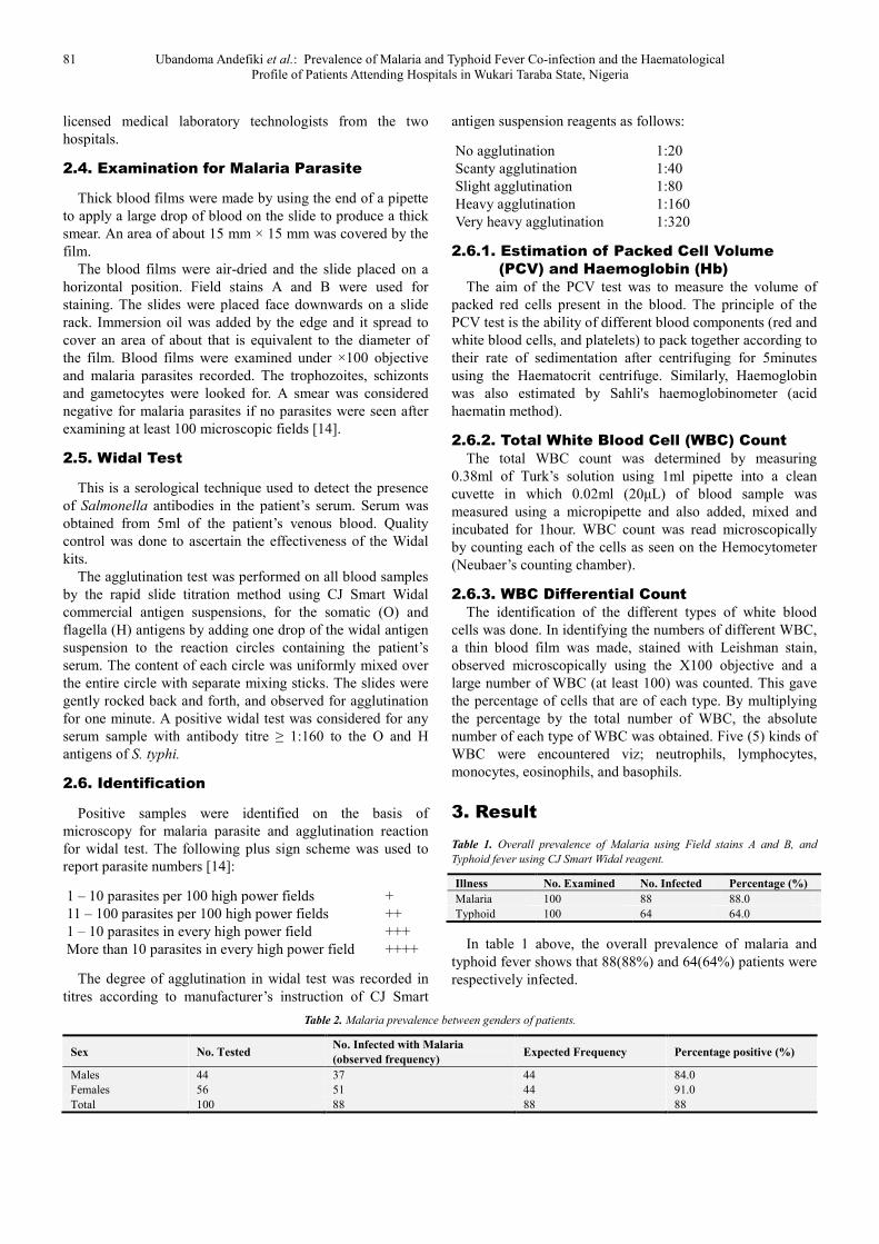
Table 1. Overall prevalence of Malaria using Field stains A and B, and
Typhoid fever using CJ Smart Widal reagent.
Illness No. Examined No. Infected Percentage (%)
Malaria 100 88 88.0
Typhoid 100 64 64.0
In table 1 above, the overall prevalence of malaria and
typhoid fever shows that 88(88%) and 64(64%) patients were
respectively infected.
Table 2. Malaria prevalence between genders of patients.
Sex No. Tested No. Infected with Malaria
(observed frequency) Expected Frequency Percentage positive (%)
Males 44 37 44 84.0
Females 56 51 44 91.0
Total 100 88 88 88
AASCIT Journal of Bioscience 2017; 3(6): 79-86 82
The distribution of malaria (Table 2 above) showed that of the 44 males examined, 37(84%) were positive and 51(91%) of
the 56 females were positive for malaria.
Table 3. Typhoid fever prevalence between genders of patients.
Sex No. Tested No. Infected with Typhoid fever
(observed frequency) Expected Frequency Percentage positive (%)
Males 44 28 32 63.6
Females 56 36 32 64.2
Total 100 64 64 64.0
In table 3 above, 28(63.6%) males out of the 44 tested were positive for typhoid fever while 36(64.2%) female patients were
typhoid positive.
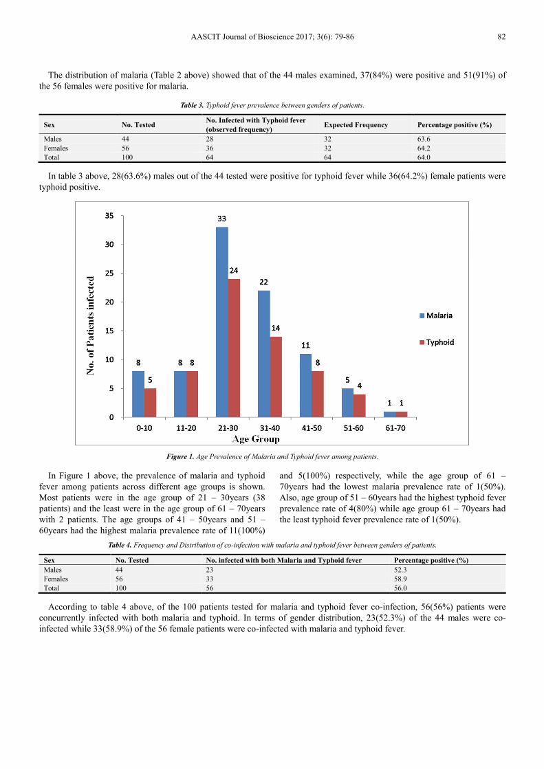
Figure 1. Age Prevalence of Malaria and Typhoid fever among patients.
In Figure 1 above, the prevalence of malaria and typhoid
fever among patients across different age groups is shown.
Most patients were in the age group of 21 – 30years (38
patients) and the least were in the age group of 61 – 70years
with 2 patients. The age groups of 41 – 50years and 51 –
60years had the highest malaria prevalence rate of 11(100%)
and 5(100%) respectively, while the age group of 61 –
70years had the lowest malaria prevalence rate of 1(50%).
Also, age group of 51 – 60years had the highest typhoid fever
prevalence rate of 4(80%) while age group 61 – 70years had
the least typhoid fever prevalence rate of 1(50%).
Table 4. Frequency and Distribution of co-infection with malaria and typhoid fever between genders of patients.
Sex No. Tested No. infected with both Malaria and Typhoid fever Percentage positive (%)
Males 44 23 52.3
Females 56 33 58.9
Total 100 56 56.0
According to table 4 above, of the 100 patients tested for malaria and typhoid fever co-infection, 56(56%) patients were
concurrently infected with both malaria and typhoid. In terms of gender distribution, 23(52.3%) of the 44 males were co-
infected while 33(58.9%) of the 56 female patients were co-infected with malaria and typhoid fever.
83 Ubandoma Andefiki et al.: Prevalence of Malaria and Typhoid Fever Co-infection and the Haematological
Profile of Patients Attending Hospitals in Wukari Taraba State, Nigeria
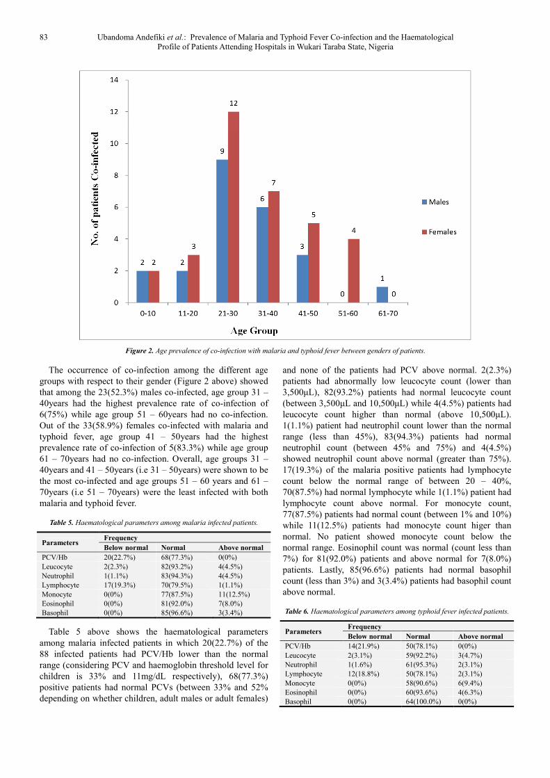
Figure 2. Age prevalence of co-infection with malaria and typhoid fever between genders of patients.
The occurrence of co-infection among the different age
groups with respect to their gender (Figure 2 above) showed
that among the 23(52.3%) males co-infected, age group 31 –
40years had the highest prevalence rate of co-infection of
6(75%) while age group 51 – 60years had no co-infection.
Out of the 33(58.9%) females co-infected with malaria and
typhoid fever, age group 41 – 50years had the highest
prevalence rate of co-infection of 5(83.3%) while age group
61 – 70years had no co-infection. Overall, age groups 31 –
40years and 41 – 50years (i.e 31 – 50years) were shown to be
the most co-infected and age groups 51 – 60 years and 61 –
70years (i.e 51 – 70years) were the least infected with both
malaria and typhoid fever.
Table 5. Haematological parameters among malaria infected patients.
Parameters Frequency
Below normal Normal Above normal
PCV/Hb 20(22.7%) 68(77.3%) 0(0%)
Leucocyte 2(2.3%) 82(93.2%) 4(4.5%)
Neutrophil 1(1.1%) 83(94.3%) 4(4.5%)
Lymphocyte 17(19.3%) 70(79.5%) 1(1.1%)
Monocyte 0(0%) 77(87.5%) 11(12.5%)
Eosinophil 0(0%) 81(92.0%) 7(8.0%)
Basophil 0(0%) 85(96.6%) 3(3.4%)
Table 5 above shows the haematological parameters
among malaria infected patients in which 20(22.7%) of the
88 infected patients had PCV/Hb lower than the normal
range (considering PCV and haemoglobin threshold level for
children is 33% and 11mg/dL respectively), 68(77.3%)
positive patients had normal PCVs (between 33% and 52%
depending on whether children, adult males or adult females)
and none of the patients had PCV above normal. 2(2.3%)
patients had abnormally low leucocyte count (lower than
3,500µL), 82(93.2%) patients had normal leucocyte count
(between 3,500µL and 10,500µL) while 4(4.5%) patients had
leucocyte count higher than normal (above 10,500µL).
1(1.1%) patient had neutrophil count lower than the normal
range (less than 45%), 83(94.3%) patients had normal
neutrophil count (between 45% and 75%) and 4(4.5%)
showed neutrophil count above normal (greater than 75%).
17(19.3%) of the malaria positive patients had lymphocyte
count below the normal range of between 20 – 40%,
70(87.5%) had normal lymphocyte while 1(1.1%) patient had
lymphocyte count above normal. For monocyte count,
77(87.5%) patients had normal count (between 1% and 10%)
while 11(12.5%) patients had monocyte count higer than
normal. No patient showed monocyte count below the
normal range. Eosinophil count was normal (count less than
7%) for 81(92.0%) patients and above normal for 7(8.0%)
patients. Lastly, 85(96.6%) patients had normal basophil
count (less than 3%) and 3(3.4%) patients had basophil count
above normal.
Table 6. Haematological parameters among typhoid fever infected patients.
Parameters Frequency
Below normal Normal Above normal
PCV/Hb 14(21.9%) 50(78.1%) 0(0%)
Leucocyte 2(3.1%) 59(92.2%) 3(4.7%)
Neutrophil 1(1.6%) 61(95.3%) 2(3.1%)
Lymphocyte 12(18.8%) 50(78.1%) 2(3.1%)
Monocyte 0(0%) 58(90.6%) 6(9.4%)
Eosinophil 0(0%) 60(93.6%) 4(6.3%)
Basophil 0(0%) 64(100.0%) 0(0%)
AASCIT Journal of Bioscience 2017; 3(6): 79-86 84
In table 6 above, the haematological parameters among
typhoid fever positive patients show that 14(21.9%) of the 64
infected patients had PCV/Hb lower than normal, 50(78.1%)
positive patients had normal PCVs and none of the patients
had PCV above the normal range. 2(3.1%) patients had
abnormally low leucocyte count, 59(92.2%) patients had
normal leucocyte count while 3(4.7%) patients had leucocyte
count higher than normal. 1(1.6%) patient had neutrophil
count lower than normal, 61(95.3%) patients had normal
neutrophil count and 2(3.1%) showed neutrophil count above
normal. 12(18.8%) of the typhoid positive patients had
lymphocyte count below the normal range, 50(78.1%) had
normal lymphocyte while 2(3.1%) patients had lymphocyte
count above normal. For monocyte count, 58(90.6%) patients
had normal count while 6(9.4%) patients had monocyte count
higher than normal. No patient showed monocyte count
below the normal range. Eosinophil count was normal for
60(93.6%) patients and above normal for 4(6.3%) patients.
All typhoid positive patients had normal basophil count.
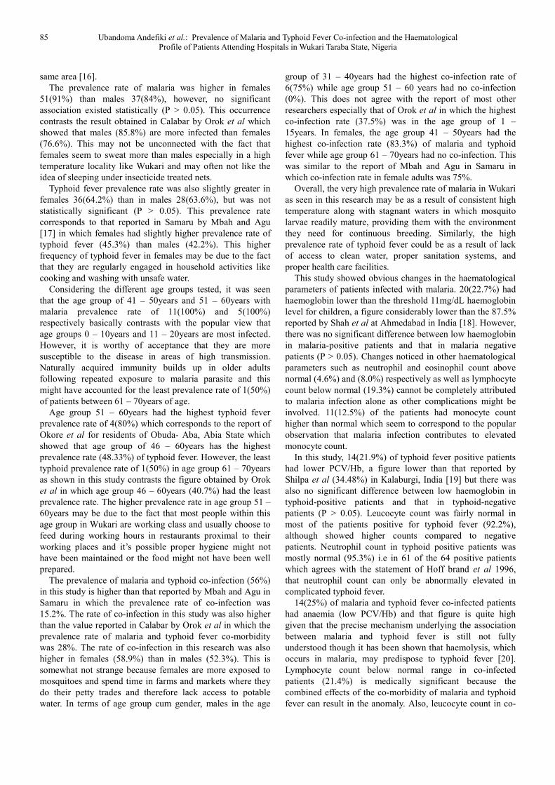
Figure 3. Haematological parameters among malaria and typhoid fever co-infected patients.
Figure 3 shows the haematological parameters among
malaria and typhoid fever co-infected patients. From the
figure, 14(25.0%) of the 56 co-infected patients had PCV/Hb
lower than normal, 42(75.0%) co-infected patients had
normal PCVs and none had PCV/Hb above the normal range.
2(3.6%) patients infected with both malaria and typhoid fever
had abnormally low leucocyte count, 51(90.0%) patients had
normal leucocyte count while 3(5.4%) patients had leucocyte
count higher than normal. 1(1.8%) co-infected patient had
neutrophil count lower than normal, 53(94.6%) patients had
normal neutrophil count and 2(3.6%) had neutrophil count
above normal. 12(21.4%) of the co-infected patients had
lymphocyte count below the normal range, 43(76.8%) had
normal lymphocyte while 1(1.8%) patient had lymphocyte
count above normal. For monocyte count, 52(92.9%) patients
had normal count while 5(8.9%) patients had monocyte count
higher than normal. No patient showed monocyte count
below the normal range. Eosinophil count was normal for
52(92.9%) patients and above normal for 4(7.1%) co-infected
patients. All the patients concurrently infected with both
malaria and typhoid fever had normal basophil count.
4. Discussion
Of the 100 patients attending selected hospitals in Wukari
in which 75 patients from General Hospital and 25 from
Bethel Hospital were included in the study, 56(56%) were
females and 44(44%) males. Included patients were between
the ages of 1 and 70 years with most of the patients being
youths (between the age of 18 and 35years).
This study shows that the overall prevalence (88%) of
malaria in Wukari is high. The prevalence rate (64%) of
typhoid fever as well as the co-morbidity (56%) of malaria
and typhoid fever is also very high. The overall prevalence of
malaria 88(88%) and typhoid 64(64%) were similar to the
figure obtained in Calabar (80.8% and 46.8% respectively)
by Orok et al in 2016 [15]. Malaria prevalence rate in this
study was also similar to the value reported in Sierra Leone
(79.94%) but typhoid prevalence rate 64(64%) in this study
was relatively lower than the figure (83.5%) reported in the
85 Ubandoma Andefiki et al.: Prevalence of Malaria and Typhoid Fever Co-infection and the Haematological
Profile of Patients Attending Hospitals in Wukari Taraba State, Nigeria
same area [16].
The prevalence rate of malaria was higher in females
51(91%) than males 37(84%), however, no significant
association existed statistically (P > 0.05). This occurrence
contrasts the result obtained in Calabar by Orok et al which
showed that males (85.8%) are more infected than females
(76.6%). This may not be unconnected with the fact that
females seem to sweat more than males especially in a high
temperature locality like Wukari and may often not like the
idea of sleeping under insecticide treated nets.
Typhoid fever prevalence rate was also slightly greater in
females 36(64.2%) than in males 28(63.6%), but was not
statistically significant (P > 0.05). This prevalence rate
corresponds to that reported in Samaru by Mbah and Agu
[17] in which females had slightly higher prevalence rate of
typhoid fever (45.3%) than males (42.2%). This higher
frequency of typhoid fever in females may be due to the fact
that they are regularly engaged in household activities like
cooking and washing with unsafe water.
Considering the different age groups tested, it was seen
that the age group of 41 – 50years and 51 – 60years with
malaria prevalence rate of 11(100%) and 5(100%)
respectively basically contrasts with the popular view that
age groups 0 – 10years and 11 – 20years are most infected.
However, it is worthy of acceptance that they are more
susceptible to the disease in areas of high transmission.
Naturally acquired immunity builds up in older adults
following repeated exposure to malaria parasite and this
might have accounted for the least prevalence rate of 1(50%)
of patients between 61 – 70years of age.
Age group 51 – 60years had the highest typhoid fever
prevalence rate of 4(80%) which corresponds to the report of
Okore et al for residents of Obuda- Aba, Abia State which
showed that age group of 46 – 60years has the highest
prevalence rate (48.33%) of typhoid fever. However, the least
typhoid prevalence rate of 1(50%) in age group 61 – 70years
as shown in this study contrasts the figure obtained by Orok
et al in which age group 46 – 60years (40.7%) had the least
prevalence rate. The higher prevalence rate in age group 51 –
60years may be due to the fact that most people within this
age group in Wukari are working class and usually choose to
feed during working hours in restaurants proximal to their
working places and it’s possible proper hygiene might not
have been maintained or the food might not have been well
prepared.
The prevalence of malaria and typhoid co-infection (56%)
in this study is higher than that reported by Mbah and Agu in
Samaru in which the prevalence rate of co-infection was
15.2%. The rate of co-infection in this study was also higher
than the value reported in Calabar by Orok et al in which the
prevalence rate of malaria and typhoid fever co-morbidity
was 28%. The rate of co-infection in this research was also
higher in females (58.9%) than in males (52.3%). This is
somewhat not strange because females are more exposed to
mosquitoes and spend time in farms and markets where they
do their petty trades and therefore lack access to potable
water. In terms of age group cum gender, males in the age
group of 31 – 40years had the highest co-infection rate of
6(75%) while age group 51 – 60 years had no co-infection
(0%). This does not agree with the report of most other
researchers especially that of Orok et al in which the highest
co-infection rate (37.5%) was in the age group of 1 –
15years. In females, the age group 41 – 50years had the
highest co-infection rate (83.3%) of malaria and typhoid
fever while age group 61 – 70years had no co-infection. This
was similar to the report of Mbah and Agu in Samaru in
which co-infection rate in female adults was 75%.
Overall, the very high prevalence rate of malaria in Wukari
as seen in this research may be as a result of consistent high
temperature along with stagnant waters in which mosquito
larvae readily mature, providing them with the environment
they need for continuous breeding. Similarly, the high
prevalence rate of typhoid fever could be as a result of lack
of access to clean water, proper sanitation systems, and
proper health care facilities.
This study showed obvious changes in the haematological
parameters of patients infected with malaria. 20(22.7%) had
haemoglobin lower than the threshold 11mg/dL haemoglobin
level for children, a figure considerably lower than the 87.5%
reported by Shah et al at Ahmedabad in India [18]. However,
there was no significant difference between low haemoglobin
in malaria-positive patients and that in malaria negative
patients (P > 0.05). Changes noticed in other haematological
parameters such as neutrophil and eosinophil count above
normal (4.6%) and (8.0%) respectively as well as lymphocyte
count below normal (19.3%) cannot be completely attributed
to malaria infection alone as other complications might be
involved. 11(12.5%) of the patients had monocyte count
higher than normal which seem to correspond to the popular
observation that malaria infection contributes to elevated
monocyte count.
In this study, 14(21.9%) of typhoid fever positive patients
had lower PCV/Hb, a figure lower than that reported by
Shilpa et al (34.48%) in Kalaburgi, India [19] but there was
also no significant difference between low haemoglobin in
typhoid-positive patients and that in typhoid-negative
patients (P > 0.05). Leucocyte count was fairly normal in
most of the patients positive for typhoid fever (92.2%),
although showed higher counts compared to negative
patients. Neutrophil count in typhoid positive patients was
mostly normal (95.3%) i.e in 61 of the 64 positive patients
which agrees with the statement of Hoff brand et al 1996,
that neutrophil count can only be abnormally elevated in
complicated typhoid fever.
14(25%) of malaria and typhoid fever co-infected patients
had anaemia (low PCV/Hb) and that figure is quite high
given that the precise mechanism underlying the association
between malaria and typhoid fever is still not fully
understood though it has been shown that haemolysis, which
occurs in malaria, may predispose to typhoid fever [20].
Lymphocyte count below normal range in co-infected
patients (21.4%) is medically significant because the
combined effects of the co-morbidity of malaria and typhoid
fever can result in the anomaly. Also, leucocyte count in co-
AASCIT Journal of Bioscience 2017; 3(6): 79-86 86
infected patients was higher than normal in 3(5.4%) patients.
This could be due to the body’s effort to resist infection by
plasmodium and salmonella resulting in continuous
production of leucocytes.
5. Conclusion
There is a high prevalence of malaria and typhoid fever
infections and co-infections in Wukari that require adequate
attention to reduce morbidity.
Infections with malaria and typhoid fever have noticeable
effects on blood parameters, although minute, could serve as
useful indices during diagnosis.
Both malaria and typhoid fever remain life-threatening and
a major health problem in under developed and developing
countries like Nigeria and since both infections cut across all
strata of the society, irrespective of age and sex as shown in
this study, all hands must be on deck in order to turn the tide
around in favour of millions who are affected or are at risk.
Finally, it is important to note that though typhoid and
malaria have effects on blood components, it will be slightly
out of place to attribute the changes in the haematological
profile of patients to malaria and typhoid fever alone.
References
[1] Frimpong, E. H., Feglo, P., Essel-Ahun, M. and Addy P. A. K. (2000). Malaria-typhoid infections in the tropics. West African Journal of Medicine. 19: 34-38
[2] Bynum, B. (2002). Typhoid- malaria infection. Lancet, 360: 1339-1339.
[3] Reiter, P. (1999). "From Shakespeare to Defoe: malaria in England in the Little Ice Age". Emerging Infectious Diseases. 6 (1): 1-11.
[4] Strong and Richard P. (1944). Stitt's Diagnosis, Prevention and Treatment of Tropical Diseases (Seventh ed.). York, PA: The Blakiston Company. p. 3.
[5] World Health Organization (2014). "Malaria Fact sheet N°94". Retrieved 28 August 2014.
[6] Collins, W. E (2012). "Plasmodium knowlesi: A malaria parasite of monkeys and humans". Annual Review of Entomology. 57: 107-21.
[7] Sarkar, P. K., Ahluwalia, G., Vijayan, V. K. and Talwar, A. (2009). "Critical care aspects of malaria". Journal of Intensive Care Medicine. 25 (2): 93-103.
[8] Baird, J. K. (2013). "Evidence and implications of mortality associated with acute Plasmodium vivax malaria". Clinical Microbiology Reviews. 26 (1): 36-57.
[9] Bledsoe, G. H. (2005). "Malaria primer for clinicians in the United States". Southern Medical Journal. 98 (12): 1197–204; quiz 1205, 1230.
[10] Olupot-Olupot, P. and Maitland, K. (2013). "Management of severe malaria: Results from recent trials". Advances in Experimental Medicine and Biology. 764: 241-50.
[11] Provost, C. (2011). "World Malaria Day: Which countries are the hardest hit? Get the full data". The Guardian. Retrieved 2012-05-03.
[12] Centre for Disease Control (CDC) (2013). "Typhoid Fever Information for Health Professionals". Retrieved 20 August 2016.
[13] Yap, K. (2016). "Global MLST of Salmonella typhi Revisited in Post-genomic Era: Genetic Conservation, Population Structure, and Comparative Genomics of Rare Sequence Types." Frontiers in Microbiology. pp. 23-37.
[14] Cheesbrough, M. (2005). Examination of blood for parasites. District Laboratory Practise in Tropical Countries, second edition, part 1, Cambridge University Press, pp 244-250.
[15] Orok, D. A., Usang, A. I., Ikpan, O. O., Duke, E. E., Eyo, E. E, Edadi, U. E., Ati, B. U. and Udida, J. A. (2016). Prevalence of Malaria and Typhoid Fever Co-infection among Febrile Patients Attending College of Health Technology Medical Centre in Calabar, Cross River State, Nigeria. Int.J.Curr.Microbiol.App.Sci. 5 (4): 825-835.
[16] Michaella, S. K., Lamin, D. M., Sallieu, K. S., Xiaojing, M. and Fei, Z. (2014). The relative prevalence of typhoid and malaria in febrile patients in Freetown, Sierra Leone. Open Journal of preventive medicine. 4: 338-346.
[17] Mbah, C. E. and Agu, B. (2014). Prevalence of malaria and typhoid co-infection among patients in some hospitals in Samaru, Zaria. Ann Bioanthropol 2: 43-8.
[18] Shah, U. B., Shah, A. M., Dave, K. K., Sakera, B. L. and Gonsai, R. N. (2007). Comparative study of microscopic detection methods with hematological changes and coagulation profile in malaria. I.M.A.G.S.B. News Bulletin., 2 (7): 37-40.
[19] Shilpa, V. U., Syeda, H. K. and Mandakini B. T. (2017). “Haematological profile in typhoid fever”. Indian Journal of Pathology and Oncology; 4 (2): 263-265.
[20] Kaye, D. and Hook, E. W. (1963). “The influence of haemolysis on susceptibility to salmonella infection: additional observations” Journal of Immunology 91: 65-75.