Prevalence and Implications of Feline Coronavirus Infections of
9
JOURNAL OF VIROLOGY, May 1990, p. 1964-1972 0022-538X/90/051964-09$02.00/0 Prevalence and Implications of Feline Coronavirus Infections of Captive and Free-Ranging Cheetahs (Acinonyx jubatus) J. L. HEENEY,lt J. F. EVERMANN,1'2 A. J. McKEIRNAN,2 L. MARKER-KRAUS, M. E. ROELKE,"4 M. BUSH,5 D. E. WILDT,5 D. G. MELTZER,6 L. COLLY,7 J. LUKAS,8 V. J. MANTON,9 T. CARO,0"t AND S. J. O'BRIENl.S* Laboratory of Viral Carcinogenesis, National Cancer Institute, Frederick, Maryland 21701-10131*; Department of Veterinary Clinical Medicine and Surgery, College of Veterinary Medicine, Washington State University, Pullman, Washington 991642; NOAHS Center,3 National Zoological Park,5 Smithsonian Institution, Washington, D.C. 20008; Laboratory of Wildlife Research, Florida Game and Freshwater Fish Commission, Gainesville, Florida 326014; Department of Physiology, Faculty of Veterinary Science, Onderstepoort 0110, Republic of South Africa6; Johannesburg Zoological Gardens, Johannesburg, Republic of South Africa7; White Oak Plantation, Yulee, Florida 320978; Whipsnade Park, Zoological Society of London, Dunstable Beds LV62LF, London, Englanda; and Evolution and Behavior Program, University of Michigan, Ann Arbor, Michigan 48109-J07010 Received 18 September 1989/Accepted 8 January 1990 The extent and progression of exposure to feline infectious peritonitis (FIP) virus in the cheetah, Acinonyx jubatus, was monitored by a world-wide serological survey with indirect fluorescent antibody titers to coronavirus. The indirect fluorescent antibody assay was validated by Western blots, which showed that all indirect fluorescent antibody-positive cheetah sera detected both domestic cat and cheetah coronavirus structural proteins. There was a poor correlation between indirect fluorescent antibody results and the presence of coronaviruslike particles in cheetah feces, suggesting that electron microscopic detection of shed particles may not be an easily interpreted diagnostic parameter for FIP disease. Low, but verifiable (by Western blots [immunoblots]) antibody titers against coronavirus were detected in eight free-ranging cheetahs from east Africa as well as from captive cheetahs throughout the world. Of 20 North American cheetah facilities screened, 9 had cheetahs with measurable antibodies to feline coronavirus. Five facilities showed patterns of an ongoing epizootic. Retrospective FIP virus titers of an FIP outbreak in a cheetah-breeding facility in Oregon were monitored over a 5-year period and are interpreted here in terms of clinical disease progression. During that outbreak the morbidity was over 90% and the mortality was 60%, far greater than any previously reported epizootic of FIP in any cat species. Age of infection was a significant risk factor in this epizootic, with infants (less than 3 months old) displaying signfficantly higher risk for mortality than subadults or adults. Based upon these observations, empirical generalizations are drawn which address epidemiologic concerns for cheetahs in the context of this lethal infectious agent. Feline infectious peritonitis (FIP) is a fatal immune-medi- ated disease of domestic cats that is a consequence of infection by an immunogenic coronavirus, FIP virus (FIPV), with a positive-stranded RNA genome. The epizootiology and etiology of FIP are not well understood, despite consid- erable study since the original description in 1963 (3, 10, 15, 37, 38). At least three clinical forms of disease are recog- nized: (i) effusive or wet FIP, which is characterized by fibrinous peritonitis or pleuritis and which is always fatal; (ii) the noneffusive or dry form of FIP, which does not have the fluid but which does have the fibrinous peritoneal deposition and is also fatal; and (iii) a subclinical enteritis, which is mild in terms of recognizable symptoms (27; J. E. Barlough and C. A. Stoddart, in C. E. Greene, ed., Infectious Diseases of the Dog and Cat, in press). In domestic cats the fatal form is rare (ca. 1% of an infected colony will die), and the virus seldom affects more than 10% of domestic cats in a group even under the most severe conditions for disease (27). Several FIPV isolates have been described, and they may vary from extreme virulence and pathology to subclinical outcomes (5, 21, 27, 28). The non-FIP forms of feline * Corresponding author. t Present address: TNO Primate Centre, 2280 HV Rijswijk, The Netherlands. t Present address: Department of Wildlife Fisheries Biology, University of California at Davis, Davis, CA 95616. coronavirus have been designated feline enteric coronavirus (FECV) to distinguish them from immunologically related but pathological FIPV isolates (20, 21, 27, 28, 36; Barlough and Stoddart, in press). Before 1982, serological evidence established that chee- tahs (Acinonyx jubatus) were susceptible to infection by feline coronaviruses (FIPV, FECV, and other related strains), but no cases of clinical FIP were reported (1, 16). Beginning in 1982, a devastating epizootic occurred in a cheetah-breeding colony located at Wildlife Safari in Win- ston, Oreg. (2, 6, 9, 31). What initially appeared as an acute anorexia, jaundice, and enteritis in an adult cheetah resulted in death from FIP and rapid viral transmission to other resident cheetahs. Within 6 months, every cheetah in the facility developed immunofluorescent antibodies to feline coronavirus, ostensibly because of exposure to the fatal FIPV that had afflicted the original cheetah (2, 9, 23). Over the next 12 months, clinical signs (intermittent and chronic diarrhea, chronic gingivitis, hepatic and renal disease, weight loss, and depression) and morbidity were apparent in over 90% of the cheetahs. Despite aggressive clinical ther- apy, a total of 27 cheetahs died between 1983 and 1987 from one or more of the following coronavirus-associated dis- eases: fibrinous peritonitis, renal and hepatic disease, enteri- tis and malabsorption syndrome, hemorrhagic gastroenteri- tis, and FIP-associated kitten mortality complex. In the same period, 18 cheetahs were exposed to the FIPV and 1964 Vol. 64, No. 5 Downloaded from https://journals.asm.org/journal/jvi on 21 January 2022 by 42.113.70.208.
Transcript of Prevalence and Implications of Feline Coronavirus Infections of
JOURNAL OF VIROLOGY, May 1990, p. 1964-19720022-538X/90/051964-09$02.00/0
Prevalence and Implications of Feline Coronavirus Infections ofCaptive and Free-Ranging Cheetahs (Acinonyx jubatus)
J. L. HEENEY,lt J. F. EVERMANN,1'2 A. J. McKEIRNAN,2 L. MARKER-KRAUS, M. E. ROELKE,"4 M. BUSH,5D. E. WILDT,5 D. G. MELTZER,6 L. COLLY,7 J. LUKAS,8 V. J. MANTON,9 T. CARO,0"t AND S. J. O'BRIENl.S*
Laboratory of Viral Carcinogenesis, National Cancer Institute, Frederick, Maryland 21701-10131*; Department ofVeterinary Clinical Medicine and Surgery, College of Veterinary Medicine, Washington State University, Pullman,Washington 991642; NOAHS Center,3 National Zoological Park,5 Smithsonian Institution, Washington, D.C. 20008;Laboratory of Wildlife Research, Florida Game and Freshwater Fish Commission, Gainesville, Florida 326014;Department of Physiology, Faculty of Veterinary Science, Onderstepoort 0110, Republic of South Africa6;Johannesburg Zoological Gardens, Johannesburg, Republic of South Africa7; White Oak Plantation, Yulee,Florida 320978; Whipsnade Park, Zoological Society ofLondon, Dunstable Beds LV62LF, London, Englanda;
and Evolution and Behavior Program, University of Michigan, Ann Arbor, Michigan 48109-J07010Received 18 September 1989/Accepted 8 January 1990
The extent and progression of exposure to feline infectious peritonitis (FIP) virus in the cheetah, Acinonyxjubatus, was monitored by a world-wide serological survey with indirect fluorescent antibody titers tocoronavirus. The indirect fluorescent antibody assay was validated by Western blots, which showed that allindirect fluorescent antibody-positive cheetah sera detected both domestic cat and cheetah coronavirusstructural proteins. There was a poor correlation between indirect fluorescent antibody results and thepresence of coronaviruslike particles in cheetah feces, suggesting that electron microscopic detection of shedparticles may not be an easily interpreted diagnostic parameter for FIP disease. Low, but verifiable (byWestern blots [immunoblots]) antibody titers against coronavirus were detected in eight free-ranging cheetahsfrom east Africa as well as from captive cheetahs throughout the world. Of 20 North American cheetah facilitiesscreened, 9 had cheetahs with measurable antibodies to feline coronavirus. Five facilities showed patterns of anongoing epizootic. Retrospective FIP virus titers of an FIP outbreak in a cheetah-breeding facility in Oregonwere monitored over a 5-year period and are interpreted here in terms of clinical disease progression. Duringthat outbreak the morbidity was over 90% and the mortality was 60%, far greater than any previouslyreported epizootic of FIP in any cat species. Age of infection was a significant risk factor in this epizootic, withinfants (less than 3 months old) displaying signfficantly higher risk for mortality than subadults or adults.Based upon these observations, empirical generalizations are drawn which address epidemiologic concerns forcheetahs in the context of this lethal infectious agent.
Feline infectious peritonitis (FIP) is a fatal immune-medi-ated disease of domestic cats that is a consequence ofinfection by an immunogenic coronavirus, FIP virus (FIPV),with a positive-stranded RNA genome. The epizootiologyand etiology of FIP are not well understood, despite consid-erable study since the original description in 1963 (3, 10, 15,37, 38). At least three clinical forms of disease are recog-nized: (i) effusive or wet FIP, which is characterized byfibrinous peritonitis or pleuritis and which is always fatal; (ii)the noneffusive or dry form of FIP, which does not have thefluid but which does have the fibrinous peritoneal depositionand is also fatal; and (iii) a subclinical enteritis, which is mildin terms of recognizable symptoms (27; J. E. Barlough andC. A. Stoddart, in C. E. Greene, ed., Infectious Diseases ofthe Dog and Cat, in press). In domestic cats the fatal form israre (ca. 1% of an infected colony will die), and the virusseldom affects more than 10% of domestic cats in a groupeven under the most severe conditions for disease (27).Several FIPV isolates have been described, and they mayvary from extreme virulence and pathology to subclinicaloutcomes (5, 21, 27, 28). The non-FIP forms of feline
* Corresponding author.t Present address: TNO Primate Centre, 2280 HV Rijswijk, The
Netherlands.t Present address: Department of Wildlife Fisheries Biology,
University of California at Davis, Davis, CA 95616.
coronavirus have been designated feline enteric coronavirus(FECV) to distinguish them from immunologically relatedbut pathological FIPV isolates (20, 21, 27, 28, 36; Barloughand Stoddart, in press).
Before 1982, serological evidence established that chee-tahs (Acinonyx jubatus) were susceptible to infection byfeline coronaviruses (FIPV, FECV, and other relatedstrains), but no cases of clinical FIP were reported (1, 16).Beginning in 1982, a devastating epizootic occurred in acheetah-breeding colony located at Wildlife Safari in Win-ston, Oreg. (2, 6, 9, 31). What initially appeared as an acuteanorexia, jaundice, and enteritis in an adult cheetah resultedin death from FIP and rapid viral transmission to otherresident cheetahs. Within 6 months, every cheetah in thefacility developed immunofluorescent antibodies to felinecoronavirus, ostensibly because of exposure to the fatalFIPV that had afflicted the original cheetah (2, 9, 23). Overthe next 12 months, clinical signs (intermittent and chronicdiarrhea, chronic gingivitis, hepatic and renal disease,weight loss, and depression) and morbidity were apparent inover 90% of the cheetahs. Despite aggressive clinical ther-apy, a total of 27 cheetahs died between 1983 and 1987 fromone or more of the following coronavirus-associated dis-eases: fibrinous peritonitis, renal and hepatic disease, enteri-tis and malabsorption syndrome, hemorrhagic gastroenteri-tis, and FIP-associated kitten mortality complex. In thesame period, 18 cheetahs were exposed to the FIPV and
1964
Vol. 64, No. 5
Dow
nloa
ded
from
http
s://j
ourn
als.
asm
.org
/jour
nal/j
vi o
n 21
Jan
uary
202
2 by
42.
113.
70.2
08.
FELINE CORONAVIRUS INFECTIONS OF CHEETAHS 1965
survived, thereby permitting an overall mortality estimate of60%. Since the Oregon epizootic, and perhaps in some partbecause of it, the understanding of the etiology of FIP incheetahs has become an important priority in discussions ofcaptive breeding and wildlife management strategies for thespecies (8, 19). The inability to develop an effective vaccineor treatment for FIP has made this virus a serious concernthat must be considered in programs designed to stabilizeand protect cheetah populations.We present here an update on the extent and progress of
the Oregon FIP epizootic from 1982 through 1987 whichserves as an important model for interpreting immunologicaldata in other populations. In addition, we present a serologicsurvey of the prevalence of feline coronavirus infection incaptive cheetahs from zoological facilities in North America,Europe, East Africa, and South Africa plus a populationsurvey from free-ranging cheetahs of the Serengeti ecosys-tem. The various feline coronavirus detection techniques,including immunofluorescence, Western blots, and electronmicroscopy, were compared, and the epizootical resultswere interpreted in terms of management recommendationsfor this severely threatened species.
MATERIALS AND METHODS
Serological assays. Serum samples were collected from 45cheetahs at Wildlife Safari in Winston, Oreg., on a yearlybasis since 1982. In addition, serum samples were submittedfrom 132 cheetahs held at 20 zoologic facilities throughoutthe United States and Canada (18). Serum was also collectedfrom 101 cheetahs in Africa and Europe. There were twosites in eastern Africa, one site in southern Africa, and onesite in Great Britain. Antibodies against the feline coronavi-ruses were measured by an indirect fluorescent antibody(IFA) assay with canine coronavirus (1-71 strain) as theantigen substrate (2, 6, 9). Previous studies assessing felinecoronavirus antibody titers in serum from domestic cats andcheetahs have shown that there is considerable antigenicsimilarity when the substrate is prepared with either caninecoronavirus, transmissible gastroenteritis virus of swine, orFIPV (16, 30). The substrate was prepared in Crandell felinekidney (CrFK) cells set on microscope slides with an 8-wellBellco template (Beilco, Vineland, N.J.). The slides werefixed in chilled acetone for 10 min, air dried, and stored at-20°C until serologic tests were run. Cheetahs with IFAtiters in serum of 1:25 and higher were considered seropos-itive to feline coronavirus (6).EM. Fecal samples from cheetahs located in facilities in
North America were processed and observed by electronmicroscopy (EM) as previously described (33). Fecal sam-ples were refrigerated during shipment to the laboratory andstored at 4°C before analysis to avoid freezing, which canalter virus morphology.Western blots. The Western blot (immunoblot) analyses
were performed by the method of Towbin et al. (35). Briefly,virus samples were treated with 2 x phosphate-bufferedsaline and electrophoresed for 60 min at 200 V (constantvoltage) in 12% sodium dodecyl sulfate-polyacrylamide gelsin a Bio-Rad mini-gel apparatus (Bio-Rad Laboratories,Richmond, Calif.) equilibrated for 30 min in 25 mM Tris-192mM glycine-20% methanol (transfer buffer). Proteins weretransferred by electric charge to nitrocellulose paper(Scheller & Schuell, Inc., Keene, N.H.) in a minitransferapparatus (Bio-Rad) in transfer buffer at 30 V (constantvoltage) overnight. Blots were disassembled and brieflywashed in TBS (20 mM Tris, 500 mM NaCl [pH 7.4]) for 10
min, blocked in 3% gelatin-TBS, and reacted with cheetahor domestic cat sera to detect the presence of coronavirusantibodies. After two 10-min washes, blots were immersedin 1:100 dilution of anti-cat whole immunoglobulin G(Kirkegaard and Perry Laboratories, Gaithersburg, Md.) in1% gelatin-TBS for 60 min, washed twice, and reacted withBCIP/NBT alkaline phosphatase substrate system (Kirke-gaard and Perry Laboratories) for 15 min. Blots were scored0 to 3 based on the intensity of bands identified at 205kilodaltons (nucleocapsid) and 24 kilodaltons (membraneproteins).
Viruses. Comparison of antigenic profiles identified byseropositive cheetahs with seropositive domestic cats wasconducted by using four pathologic strains of domestic felinecoronavirus. The strains included FECV WSU 79-1683,FIPV WSU 79-1146, FIPV NOR-15, and FIPV UCD-1 (5,21, 27, 29). The cheetah coronavirus isolate AJUCV-1 (pre-viously designated WSU 83-4497) was maintained in CrFKcells as a persistent nonlytic infection (6, 7). Supernatantsfrom cells infected with the four feline coronavirus strainsand from cells persistently infected with WSU 83-4497 wereharvested and processed by differential centrifugation.Briefly, FC-009 cells (courtesy of N. C. Pedersen) weregrown in Dulbecco minimal essential medium, and 10% fetalbovine serum was used to propagate the four feline corona-virus strains, which were inoculated at a multiplicity ofinfection of approximately 0.1. The supernatant was har-vested from flasks when 80 to 90% cytopathic effect wasobtained and stored at -20°C. After clarification at 800 x gat 4°C for 20 min, the supernatant was carefully removed,and the virus was pelleted at 16,500 rpm 4°C for 45 min.Samples of 100x-concentrated virus were mixed with 2xsample buffer and run on 12% sodium dodecyl sulfate-polyacrylamide gels and stained with Coomassie blue toquantify virus antigens.
RESULTS
An epizootic of FIP in captive cheetahs. The Wildlife Safariin Winston, Oreg., began what was to become a highlysuccessful cheetah-breeding program in 1973. In May 1982,two cheetahs, studbook (SB) numbers 79 and 80 (18), wereimported into the park. Within a few weeks SB79 developedsevere jaundice, depression, fever, and diarrhea and died inlate June of FIP (31). A retrospective survey of cheetahserum collected from the facility's cheetahs before May 1982revealed that none (of 25 tested) had circulating antibodytiters against coronavirus, based upon an IFA assay. Within6 to 8 months, sera from every cheetah in the facility werepositive for antibodies to coronavirus, a result consistentwith dynamic highly contagious infection with FIPV.A summary of the antibody titer progression of 45 chee-
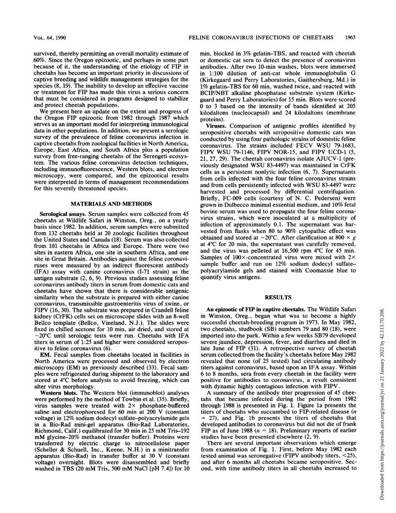
tahs that became infected during the period from 1982through 1988 is presented in Fig. 1. Figure la presents thetiters of cheetahs who succumbed to FIP-related disease (n= 27), and Fig. lb presents the titers of cheetahs thatdeveloped antibodies to coronavirus but did not die of frankFIP as of June 1988 (n = 18). Preliminary reports of earlierstudies have been presented elsewhere (2, 9).There are several important observations which emerge
from examination of Fig. 1. First, before May 1982 eachtested animal was seronegative (FIPV antibody titers, <25),and after 6 months all cheetahs became seropositive. Sec-ond, with time antibody titers in all cheetahs increased to
VOL. 64, 1990
Dow
nloa
ded
from
http
s://j
ourn
als.
asm
.org
/jour
nal/j
vi o
n 21
Jan
uary
202
2 by
42.
113.
70.2
08.

1966 HEENEY ET AL.
1982 1983 1984 1982 1983 1984a F J O F J O F J O f J O F J O F J O
>1
I1
>1I
>1I
>11
1982 1983 1984F J O F J O F J----;-----; - - ; -~~~W r %F w ;w r w;% W-WWw %O-;
1600 _ 35 79 60 258
25_t t/\
600
25 -
<25 195 25 2931 294
1600 _
400<25 L.L ..fiI fI||'''''''' ___ _
16001600
400
100
25
>16001600
400
100
25
_160
>16001600
400
100
25
295
t....
ft I315
ILF J F J O F J 01982 1983 1964
296
1...I310
I316
I
297
311
IX.1L
317
306 388
i
I
I
F J F J F J F JO F J F J F J F J O F J 01982 1983 1964 1962 1963 1964 1962 1963 19
Date of Serum Collection
FIG. 1. Time course of FIP IFA titers in serum from cheetahs at Wildlife Safari, Winston, Oreg. Studbook numbers are from the NorthAmerican cheetah studbook (18). Arrows indicate dates of death. SB79 and SB80 were the two animals that arrived at the park with FIP titers.(a) Animals that have died of FIP-related disease based upon necropsy diagnosis. (b) Animals that have been exposed acutely but recoveredand were without symptoms at the last sampling.
between 1:400 and 1:1,600. Third, there was often a modestdecrease in FIPV antibody titer in dying animals, presum-ably as a consequence of immune suppression during laterdisease stages. Fourth, there was no obvious difference inthe seroprevalence patterns in animals that succumbed toFIPV and those that survived. Fifth, in surviving animals,antibody titers tended to persist for several years, suggestinga chronic infection with the FIPV which continually stimu-lated the immune system of infected cheetahs. There werecertain exceptions to this pattern (e.g., SB319 and SB383),but the more common scenario was persistence of apprecia-ble antibody titers against coronavirus for a period of 4 to 6years.An important risk factor that would influence mortality
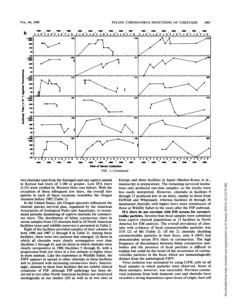
due to FIPV appears to be age at exposure (see reference 18for dates of birth). Of the 45 cheetahs studied, 20 wereexposed as infants (less than 5 months old); of these, only 3(15%) survived. The survival rate of 25 older cheetahs was60%. The overall mortality related to FIPV exposure was60%. The relative survival of infants exposed to FIPV wassignificantly different from that for all cheetahs (X2 = 9.37; P< 0.01).
Among the cheetahs which succumbed to FIP, the mediantime from seroconversion to death was 7 to 12 months (Fig.2). Three animals, SB258, SB259, and SB294, each survivedover 38 months before dying of FIP, but their FIPV antibodytiter patterns are perhaps illuminating. All three cheetahsshowed declining antibody titers in 1983 through 1984 (thetiter of SB259 dropped to c25 for two consecutive samplesin 1984), and thereafter the two tested animals developedelevated titers in 1985 until their deaths in 1986 (Fig. la).Since the three animals were housed in the same pens, theirparallel pattern may indicate an acute secondary infection orpossibly a common environmental change that caused acti-vation of latent FIPV. The other surviving cheetahs havelived for up to 5 years (Fig. 2b) without apparent symptoms.
Seroprevalence in captive and free-ranging cheetah popula-tions. Coronavirus antibody titers from 101 cheetah serumsamples collected from four locations outside of the UnitedStates are presented in Table 1. With the exceptions of seracollected in the Serengeti from 25 free-ranging cheetahs (25),the sera were from captive animals, although several of thesecheetahs in Africa were born in the wild. Low antibody titerswere detected in a few animals from each locale, but only
1982 1983 1984 11985 11986
0 F J O F J O F J O F J O F J O
a
8C3000
C.)
0co
xa-
I,
S
C.4
JO F J O1965 196
312
318
J. VIROL.
l
Dow
nloa
ded
from
http
s://j
ourn
als.
asm
.org
/jour
nal/j
vi o
n 21
Jan
uary
202
2 by
42.
113.
70.2
08.
FELINE CORONAVIRUS INFECTIONS OF CHEETAHS 1967
1982 1983 1984 1985 198 1987II. I % -
1982 1983 1 1984 195 198 1987 1988
F J O F J O F J O F J O
1982 1983 1984 1985F J O F J 0 F J 0 F J 0 F J 0 F J 0
1982 1983 1984 1985 1988 1987
Date of Serum Collection
F J 0 F J 0 F J 0 F J 0 F J 0 F J O F J C
1982 1983 1984 1985 1988 1987 1988
FIG. 1-Continued.
two cheetahs (one from the Serengeti and one captive animalin Kenya) had titers of 1:100 or greater. Low IFA titers(1:25) were verified by Western blots (see below). With theexception of these infrequent low titers, the overall titerpattern in each of these locations resembles the Oregonsituation before 1982 (Table 1).
In the United States, the Oregon epizootic influenced thecheetah species survival plan, organized by the AmericanAssociation of Zoological Parks and Aquariums, to recom-mend periodic monitoring of captive cheetahs for coronavi-rus titers. The distribution of feline coronavirus titers inserum samples from 132 cheetahs held in 20 North Americanfacilities (zoos and wildlife reserves) is presented in Table 2.
Eight of the facilities provided samples of their colonies inboth 1986 and 1987 (1 through 8 in Table 2). Among thesefacilities, there were two patterns that emerged: (i) those inwhich all cheetahs were clearly seronegative over time(facilities 1 through 4); and (ii) those in which cheetahs were
clearly seropositive in 1986 (facilities 5 through 8), with a
progression from low titers in a few animals to higher titersin more animals. Like the experience at Wildlife Safari, theFIPV appears to spread to other cheetahs in these facilitiesand to proceed with increasing coronavirus titers. Interest-ingly, animals in these four facilities did not exhibit clinicalsymptoms of FIP, although FIP pathology has been ob-served in two other North American facilities not monitoredserologically in our studies (19) as well as in two sites in
Europe and three facilities in Japan (Marker-Kraus et al.,manuscript in preparation). The remaining surveyed institu-tions only produced one-time samples, so the results were
less easily interpreted. However, cheetahs in facilities 9through 15 produced low or no titers, similar to those fromDeWildt and Whipsnade, whereas facilities 16 through 20maintained cheetahs with higher titers more reminiscent ofthose at Wildlife Safari in the years after the FIP outbreak.IFA titers do not correlate with EM screens for coronavi-
ruslike particles. Seventy-four fecal samples were submittedfrom captive cheetah populations at 13 facilities in NorthAmerica for EM analysis. The overall prevalence of chee-tahs with evidence of fecal coronaviruslike particles was
31% (21 of 68) (Table 2). Of the 21 cheetahs sheddingcoronaviruslike particles in their feces, only 8 (38%) haddemonstrable serum IFA titers to coronavirus. The highfrequency of discordance between feline coronavirus anti-bodies and the presence of fecal particles is difficult toexplain but could be the result of the occurrence of corona-viruslike particles in the feces which are immunologicallydistinct from the pathological FIPV.
Virus isolation was attempted by using CrFK cells on allfecal samples in which particles were observed. None ofthese attempts, however, was successful. Previous corona-
viral isolations from both domestic cats and cheetahs haverevealed a strong dependence upon tissue of origin, host cell
b
S2
SC<i:
0
00aC*104
xS
F10.0C4E
280
VOL. 64, 1990
Dow
nloa
ded
from
http
s://j
ourn
als.
asm
.org
/jour
nal/j
vi o
n 21
Jan
uary
202
2 by
42.
113.
70.2
08.
1968 HEENEY ET AL.
a
aI.6-4)4)
0
.0E
z
4-6 7-12 13-18 19-24 25-30 31-36 37-42
Time IntervalFirst FIP To Death (months)
5
° 34
03
.0
E
z
Years Survival Since First FIP+FIG. 2. (a) Distribution of the time intervals between infection
with FIPV and death by FIP-related disease in cheetahs at WildlifeSafari, Winston, Oreg. (b) Time of survival since FIPV infection insurviving cheetahs.
system, and the presence of exogenous protease to enhanceviral replication (5-7, 20).Western blot analyses. Serum samples (diluted 1:5, 1:10,
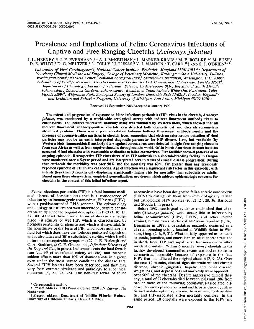
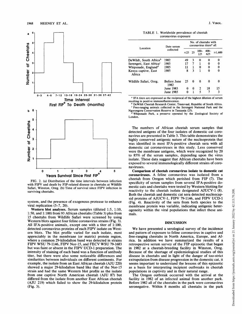
1:50, and 1:100) from 93 African cheetahs (Table 3) plus from15 cheetahs from Wildlife Safari were screened by usingWestern blots against four feline coronavirus strains (Fig. 3).All IFA-positive animals, except one with a titer of 1:100,detected coronavirus proteins of each FIPV isolate on West-ern blots. The blot profile varied for each isolate, mostappreciably in the membrane (or matrix) protein region,where a common 29-kilodalton band was detected in strainsFIPV WSU 79-1146, FIPV Nor-15, and FECV WSU 79-1683but was faint or absent in the FIPV UCD-1 preparation. Theintensity of staining of each band was a function of antibodytiter, but there were also some noticeable differences andsimilarities between individuals on different continents. Forexample, the isolate from an East African cheetah (AJU 220)showed a major 29-kilodalton band like that of the Nor-15strain and had the same Western blot profile as the isolatefrom one captive North American cheetah (AJU 87) butdiffered from the isolate from another East African cheetah(AJU 219) which failed to show the 29-kilodalton protein(Fig. 3).
TABLE 1. Worldwide prevalence of cheetahcoronavirus exposure
No. of cheetahs withDate serum coronavirus titersa of:Locationcoltecollected c25 25 100- 400- 1600~25125 625 -1,0
DeWildt, South Africab 1982 49 3 0 0 0Serengeti, East Africac 1985 17 7 1 0 0Whipsnade, Englandd 1985 11 1 0 0 0Kenya captive, East 1985 8 3 1 0 0
Africa
Wildlife Safari, Oreg. Before June 25 0 0 0 01982
June 1983 0 0 2 18 15June 1985 0 1 5 7 3
a IFA titers are expressed as the reciprocal of the highest dilution of serumresulting in positive immunofluorescence.
b DeWildt Cheetah Research Center, Transvaal, Republic of South Africa.c Free-ranging animals collected in the Serengeti National Park and the
Ngorongora Conservation Reserve in Tanzania (25).d Whipsnade Park, a preserve operated by the Zoological Society of
London.
The numbers of African cheetah serum samples thatdetected antigens of the four isolates of domestic cat coro-navirus are presented in Table 3. This table demonstrates thehighly conserved antigenic nature of the nucleoprotein thatwas identified in most IFA-positive cheetah sera with alldomestic cat coronaviruses in this study. Less conservedwere the membrane antigens, which were recognized by 20to 85% of the serum samples, depending upon the virusisolate. These data suggest that African cheetahs have beenexposed to several immunologically different strains of coro-naviruses.Comparison of cheetah coronavirus isolate to domestic cat
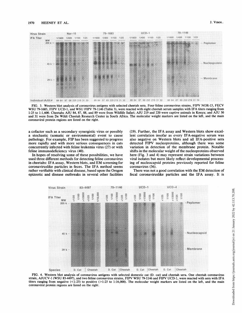
coronaviruses. A feline coronavirus was isolated from acheetah from Oregon which perished from FIP (7). Thespecificity of serum samples from several IFA-positive do-mestic cats and cheetahs were tested by Western blotting forreactivity to the cheetah isolate designated AJUCV-1 (8).Both the cheetah and domestic cat sera detected nucleocap-sid proteins of AJUCV-1, FIPV 79-1146, and FIPV UCD-1(Fig. 4). Reactivity of the sera from both species to themembrane protein was variable, indicating antigenic heter-ogeneity within the viral populations that infect these ani-mals.
DISCUSSION
We have presented a serological survey of the incidenceand pattern of exposure to feline coronavirus in captive andfree-ranging cheetahs in North America, Europe, and Af-rica. In addition we have summarized the results of aretrospective serum survey of the FIP epizootic that beganin 1982 at a cheetah-breeding facility in Winston, Oreg.Because of the shortage of epidemiological studies of thisdisease in cheetahs and in light of the danger of too-strictextrapolation from disease progression in the domestic cat, itseems important to understand the lessons of this epizooticas a basis for interpreting incipient outbreaks in cheetahpopulations in captivity and in their natural range.The Oregon outbreak occurred with the arrival at the
facility in 1982 of an infected animal from another park.Before 1982 all of the cheetahs in the park were coronavirusseronegative. Within 8 months all cheetahs in the park
J. VIROL.
Dow
nloa
ded
from
http
s://j
ourn
als.
asm
.org
/jour
nal/j
vi o
n 21
Jan
uary
202
2 by
42.
113.
70.2
08.

FELINE CORONAVIRUS INFECTIONS OF CHEETAHS 1969
TABLE 2. Feline coronavirus antibody titers and EM results in North American zoologic facilities in 1986 and 1987
No. of cheetahs with the following EM screen of fecesSerum result Total no. of coronavirus IFA titers (X10)
and facility no. Year cheetahs at facility N sti-25 25 100-125 4004625 -1,600 no. tested % Positive
Seronegative1 1986 10 9 0 0 0 0 0/9 0
1987 2 0 0 0 0 NDa2 1986 14 7 0 0 0 0 1/8 13
1987 11 0 0 0 0 ND3 1987 3 3 0 0 0 0 ND4 1986 9 9 0 0 0 0 9/9 100
Seropositive5 1986 4 3 1 0 0 0 3/3 100
1987 1 0 0 5 3 ND6 1986 20 3 1 1 0 0 2/6 33
1987 7 6 2 0 6 ND7 1986 5 2 1 0 1 1 3/5 60
1987 0 0 0 1 4 ND8 1986 22 10 0 5 3 1 1/22 5
1987 39 17 5 5 9 3 ND
Indeterminant9 1986 3 3 0 0 0 010 1986 5 5 0 0 0 011 1986 2 3 1 0 0 0 0/1 012 1987 1 1 0 0 0 013 1986 1 1 1 0 0 0 1/2 5014 1986 17 1 0 0 0 015 1986 4 2 0 0 0 2 1/2 5016 1986 2 0 2 0 0 017 1986 1 0 0 0 0 1 1/1 10018 1986 5 0 0 0 2 319 1986 4 1 2 0 0 120 1986 7 0 0 0 1 0
a ND, Not determined.
seroconverted, and after 4 years 60% had died of FIP-relateddisease. Age of infection was a significant risk factor, withinfants (0 to 5 months of age) being at greater risk. Themedian time from seroconversion (as a marker of exposure)
TABLE 3. Western blot results comparing antigens offour strains of feline coronavirus in reaction
with sera from 20 cheetahsa
No. positive (%) on
Feline Western blot ofb:coronavirus Sequelae inisolonateru domestic cats Membrane Nucleo-
antigen protein(29 kDa) (45 kDa)
WSU 79-1683 Enteritis, non-FIP; 17 (85) 19 (95)(FECV) high morbidity, low
mortalityUCD-1 FIP dose-related viru- 4 (20) 18 (90)(FIPV) lence; high morbid-
ity, high mortalityWSU 79-1146 FIP very virulent, low 13 (65) 20 (100)(FIPV) dose; high morbidity,
high mortalityNOR-15 FIP very virulent, low 14 (70) 18 (90)
(prototype dose; high morbidity,strain) high mortalitya Twenty cheetah sera (Table 1) with coronavirus IFA titers of >25.b Results indicate numbers and percentages of cheetah sera which identified
the nucleoprotein and membrane antigens in Fig. 3.
to death was 6 to 12 months. Interestingly, there was noobvious difference between the temporal pattern of felinecoronavirus serum antibody titers in cheetahs who diedversus those who survived (compare Fig. la and b).The reasons why some cheetahs survived while others
perished are not clear, but, based on precedence from othercoronavirus and retroviral epizootics, there are three possi-ble explanations (22, 24, 36). First, there is genetic variationin virus isolates that produce different clinical results. Thisseems to be the case in the domestic cat viruses, which rangefrom extremely virulent to subclinical (Table 3). Neverthe-less, despite the isolation of clinically distinct domestic catcoronaviruses, it has not been possible to develop type-specific immunological reagents that discriminate betweenthem (11-14). There is currently no direct evidence forfunctional heterogeneity of cheetah coronaviruses; however,the discordance of coronaviruslike particles detected by EMand the results of IFA and Western blot analyses (Table 2) isconsistent with appreciable antigenic diversity among chee-tah coronaviruses. Second, there could be genetic differ-ences in the host cheetahs that influence the pathology. Thisis possible despite our earlier observation of the reducedgenetic diversity of cheetahs (23-26), since those resultsshowed reduced (10 to 100-fold) diversity but not completehomozygosity at all loci. In fact, we have observed limitedheterozygosity in cheetahs by using molecular clones of onegene involved in immune surveillance, the major histocom-patibility complex (40a). Third, FIP pathology could involve
VOL. 64, 1990
Dow
nloa
ded
from
http
s://j
ourn
als.
asm
.org
/jour
nal/j
vi o
n 21
Jan
uary
202
2 by
42.
113.
70.2
08.
1970 HEENEY ET AL.
Virus Strain Nor-15 79-1683 UCD-1 719-1146
IFA Titer 111600 11400 1.100 1 25 111600 1 400 11100 12 11600 1 400 1 10O0 11 1 '600 400 1 l 00 12iMW --
2k
........il~AJU)a)-.61136 - i1 31 5 -~ V
~~~~~~~~~~~~~~~~~~~~~~~~~~~~~~~~~~~~~~~~~~~~~~~~~~~1........... . . 5 ' .: : : , .t 1 .... . .:.~~~~~~~~~~~~~~~~~~~~~~~~~~~~~~~~~~~~~~~~~~~~~~~~~~~~~~~~~~~~~~~~~~~~~~~~* 4:N-:
45 ki J. JuXtS - f '.t i'.;'Sii. .oi ;J-X tl8f<...s ;! 'Lm-tJ|i', l S
FIG. 3. Western blot analysis of coronavirus antigens with selected cheetah sera. Four feline coronavirus strains, FIPV NOR-15, FECVWSU 79-1683, FIPV UCD-1, and WSU FIPV 79-1146 (Table 3), were reacted with eight cheetah serum samples with IFA titers ranging from1:25 to 1:1,600. Cheetahs AJU 84, 87, 88, and 89 were from Wildlife Safari; AJU 219 and 220 were captive animals in Kenya; and AJU 30and 31 were from De Wildt Cheetah Research Center in South Africa. The molecular weight markers are listed on the left, and the maincoronaviral protein regions are listed on the right.
a cofactor such as a secondary synergistic virus or possiblya stochastic (somatic or environmental) event to cause
pathology. For example, FIP has been suggested to progress
more rapidly and with more serious consequences in catsconcurrently infected with feline leukemia virus (27) or withfeline immunodeficiency virus (40).
In hopes of resolving some of these possibilities, we haveused three different methods for detecting feline coronavirusin cheetahs: IFA assay, Western blots, and EM screening forcoronaviruslike particles in feces. The IFA method seems
rather verifiable with clinical disease, based upon the Oregonepizootic and disease outbreaks in several other facilities
Virus Strain
IFA TiterMW
205 k --
45 k -
29 k
Species
83-4497
U) 0c nV4
V A V
10td
Cat Cheetah
79-1146
c)0 0)LO 0 O)0
v A v
(19). Further, the IFA assay and Western blots show excel-lent correlation insofar as every IFA-negative serum wasalso negative on Western blots and all IFA-positive seradetected FIPV nucleoproteins, although there was somevariation in detection of the membrane protein. Notableshifts in the molecular weight of the nucleoproteins observedhere (Fig. 3 and 4) may represent strain variations betweenviral isolates but more likely reflect developmental process-ing of nucleocapsid proteins previously reported for felinecoronavirus (36).There was not a good correlation with the EM detection of
fecal coronaviruslike particles and the IFA assay. It is
UCD-1
a Ul)
YA
UCD-4
0~ 0
Ul) 0~ 0 Ul) 0)
V A
W
i iw
!ef
W ! ........B.
.X |*-t :..
r..>
Peplomer
Nucleocapsid
Membrane
D. Cat Cheetah D. Cat Cheetah D. Cat Cheetah
FIG. 4. Western blot analysis of coronavirus antigens with selected domestic cat (D. cat) and cheetah sera. One cheetah coronavirusstrain, AJUCV-1 (WSU 83-4497), and two feline coronavirus strains, FIPV WSU 79-1146 and FIPV UCD-1, were reacted with sera with IFAtiters ranging from negative (<1:25) to positive (>1:25 to 1:16,000). The molecular weight markers are listed on the left, and the maincoronaviral protein regions are listed on the right.
Peplomtlei
Ntc Ieoprotciu
J. VIROL.
Dow
nloa
ded
from
http
s://j
ourn
als.
asm
.org
/jour
nal/j
vi o
n 21
Jan
uary
202
2 by
42.
113.
70.2
08.
FELINE CORONAVIRUS INFECTIONS OF CHEETAHS 1971
possible that this discordance would result from an acquiredinfection before the development of an immune response.Based upon the well-known existence of antigenic drift inRNA viruses (41) and in coronaviruses (4, 17, 32, 39), it isalso likely that these particles may represent viruses that aremorphologically similar to but antigenically distinct from therecognized strains of FIPV and FECV (20, 36). The discor-dance between IFA serology results versus EM coronavirusscreening in cheetahs makes it difficult to interpret the EMresults as a diagnostic parameter for cheetahs.The observations of the Oregon outbreak plus the results
of the IFA serum survey for free-ranging and captive chee-tahs have suggested the following conclusions, which werecommend for consideration of this pathogen in managedcheetah populations.
(i) Pathological FIPV is highly infectious and spreadsrapidly among cheetahs when physical contact occurs, prob-ably through exchange of excretory material and/or secre-tions (34).
(ii) Pathological FIP is a dynamic disease process that iscorrelated with continuous, measurable, and often increas-ing titers to feline coronavirus.
(iii) Occasional low antibody titers have been observed inseveral populations, including free-ranging animals (Table 1and 2), which could signal an incipient disease epizootic butmay also reflect infection with a nonpathological but anti-genically related coronavirus. The confirmation of a clinicalepizootic would require time points showing increasingnumbers and higher titers (as in facilities 5 through 8) plusmorbidity. Facilities with increasing titers but no disease(facilities 5 through 8 in Table 2) may prove particularlyinteresting, because they raise the possibility of clinicalheterogeneity of virus isolates between different facilities.Long-term monitoring of these facilities to confirm thishypothesis is desirable.The present results indicate that coronavirus infections
are occurring in captive and free-ranging cheetah popula-tions, as has been reported for domestic cats (1, 16, 27). Thefactors that appear to be important with the occurrence ofFIP in a population of cats are a combination of a virulentstrain of virus together with a susceptible group of cats (1,16, 27). The cheetah has been reported as being unusuallyvulnerable to FIPV, based on epizootics that have occurredin the United States, Canada, Ireland, Namibia-SouthwestAfrica, The Netherlands, and Japan (9, 18, 19, 23). Theresults of the study reported herein indicated that the expo-sure rate to feline coronavirus, or a closely related corona-virus, is similar to that reported for domestic cats. Untilfurther information is reported concerning coronavirus in-fections in exotic cats, the management of cheetahs shouldfollow basic guidelines for control of infectious diseases,such as segregation of seropositive animals from seronega-tive animals, quarantine before and after arrival at thezoologic facility, and regular monitoring by serology (8).
ACKNOWLEDGMENTSWe are grateful to the curators and veterinarians who assisted in
the collection of serum samples from cheetahs. Appreciation isextended to M. Briggs for collection of recent serum samples on theWildlife Safari collection. Thanks are also extended to R. Brown, P.Dilbeck, and T. Byington for the processing and observation ofsamples by EM.
LITERATURE CITED1. Barlough, J. E., J. C. Adsit, and F. W. Scott. 1982. The
worldwide occurrence of feline infectious peritonitis. FelinePract. 12:26-30.
2. Briggs, M. B., J. F. Evermann, and A. J. McKeirnan. 1986.Feline infectious peritonitis. An update of a captive cheetahpopulation. Feline Pract. 16:13-16.
3. Colby, E. D., and R. J. Low. 1970. Feline infectious peritonitis.Vet. Med. Small Anim. Clin. 65:783-786.
4. De Groot, R. J., A. C. Andeweg, M. C. Horzinek, and W. J. M.Spaan. 1988. Sequence analysis of the 3' end of the felinecoronavirus FIPV 79-1146 genome; comparison with the ge-nome of porcine coronavirus TGEV reveals large insertions.Virology 167:370-376.
5. Evermann, J. F., L. Baumgartener, R. L. Ott, E. V. Davis, andA. J. McKeirnan. 1981. Characterization of a feline infectiousperitonitis virus isolate. Vet. Pathol. 18:256-265.
6. Evermann, J. F., G. Burns, M. E. Roelke, A. J. McKeirnan, A.Greenlee, A. C. Ward, and M. L. Pfeifer. 1984. Diagnosticfeatures of an epizootic of feline infectious peritonitis in captivecheetahs. Am. Assoc. Vet. Lab. Diag. 26:265-382.
7. Evermann, J. F., J. L. Heeney, A. J. McKeirnan, and S. J.O'Brien. 1989. Comparative features of a coronavirus isolatedfrom a cheetah with feline infectious peritonitis. Virus Res.13:15-28.
8. Evermann, J. F., J. L. Heeney, M. E. Roelke, A. J. McKeirnan,and S. J. O'Brien. 1988. Biological and pathological conse-quences of feline infectious peritonitis virus infection in thecheetah. Arch. Virol. 102:155-171.
9. Evermann, J. F., M. E. Roelke, and M. B. Briggs. 1986. Clinicaland diagnostic features of feline coronavirus infections of chee-tahs. Feline Pract. 26:21-30.
10. Feldmann, B. H., and B. S. Jortner. 1964. Clinico-pathologicconference. J. Am. Vet. Med. Assoc. 144:1409-1418.
11. Fiscus, S. A., B. L. Rivoire, and Y. A. Teramoto. 1987. Epitope-specific antibody responses to virulent and avirulent felineinfectious peritonitis virus isolates. J. Clin. Microbiol. 25:1529-1534.
12. Fiscus, S. A., and Y. A. Teramoto. 1987. Antigenic comparisonof feline coronavirus isolates: evidence for markedly differentpeplomer glycoproteins. J. Virol. 61:2607-2613.
13. Fiscus, S. A., and Y. A. Teramoto. 1987. Functional differencesin the peplomer glycoproteins of feline coronavirus isolates. J.Virol. 61:2655-2657.
14. Fiscus, S. A., Y. A. Teramoto, M. M. Mildbrand, C. V. Kinsley,S. E. Winston, and N. C. Pedersen. 1985. Competitive enzymeimmunoassays for the rapid detection of antibodies to felineinfectious peritonitis virus polypeptides. J. Clin. Microbiol.22:395-401.
15. Holzworth, J. 1963. Some important disorders of cats. CornellVet. 53:157-160.
16. Horzinek, M. C., and A. D. M. E. Osterhaus. 1979. Felineinfectious peritonitis: a worldwide serosurvey. Am. J. Vet. Res.40:1487-1492.
17. Lai, M. M. C. 1988. Replication of coronavirus RNA, p.116-136. In E. Domingo, J. J. Holland, and P. Ahlquist (ed.),RNA genetics, vol. 1. RNA-directed virus replication. CRCPress, Inc., Boca Raton, Fla.
18. Marker, L. 1986. North American regional cheetah studbook.Wildlife Safari, Winston, Oreg.
19. Marker, L., and S. J. O'Brien. 1989. Captive breeding of thecheetah (Acinonyx jubatus) in North American zoos (1971-1986). Zoo Biol. 8:3-16.
20. McKeirnan, A. J., J. F. Evermann, E. V. Davis, and R. L. Ott.1987. Comparative properties of feline coronaviruses in vitro.Can. J. Vet. Res. 51:212-216.
21. McKeirnan, A. J., J. F. Evermann, A. Hargis, L. M. Miller, andR. L. Ott. 1981. Isolation of feline coronaviruses from two catswith diverse disease manifestations. Feline Pract. 11:16-20.
22. O'Brien, S. J., and J. F. Evermann. 1988. Interactive influenceof infectious disease and genetic diversity in natural popula-tions. Trends Ecol. Evol. 3:254-259.
23. O'Brien, S. J., M. E. Roelke, L. Marker, A. Newman, C. A.Winkler, D. Meltzer, L. Colly, J. F. Evermann, M. Bush, andD. E. Wildt. 1985. Genetic basis for species vulnerability in thecheetah. Science 227:1428-1434.
24. O'Brien, S. J., D. E. Wildt, and M. Bush. 1986. The cheetah in
VOL. 64, 1990
Dow
nloa
ded
from
http
s://j
ourn
als.
asm
.org
/jour
nal/j
vi o
n 21
Jan
uary
202
2 by
42.
113.
70.2
08.
1972 HEENEY ET AL.
genetic peril. Sci. Am. 254:84-92.25. O'Brien, S. J., D. E. Wildt, M. Bush, T. M. Caro, C. FitzGib-
bon, I. Aggundey, and R. E. Leakey. 1987. East African chee-tahs: evidence for two population bottlenecks? Proc. Natl.Acad. Sci. USA 84:508-511.
26. O'Brien, S. J., D. E. Wildt, D. Goldman, C. R. Merril, and M.Bush. 1983. The cheetah is depauperate in genetic variation.Science 221:459-462.
27. Pedersen, N. C. 1987. Coronavirus diseases (coronavirus enteri-tis, feline infectious peritonitis), p. 193-214. In J. Holzworth(ed.), Diseases of the cat. The W. B. Saunders Co., Philadel-phia.
28. Pedersen, N. C., J. F. Evermann, A. J. McKeirnan, and R. L.Ott. 1984. Pathogenicity studies of feline coronavirus isolates79-1146 and 79-1683. Am. J. Vet. Res. 45:2580-2585.
29. Pedersen, N. C., and K. Floyd. 1985. Experimental studies withthree new strains of feline infectious peritonitis virus: FIPV-UCD2, FIPV-UCD3 and FIPV-UCD4. Comp. Cont. Educ.Prac. Vet. 7:1001-1011.
30. Pedersen, N. C., J. Ward, and W. L. Mengenling. 1978. Anti-genic relationship of the feline infectious peritonitis virus tocoronaviruses of other species. Arch. Viol. 58:45-53.
31. Pfeifer, M. L., J. F. Evermann, M. E. Roelke, A. M. Gallina,R. L. Ott, and A. J. McKeirnan. 1983. Feline infectious perito-nitis in a captive cheetah. J. Am. Vet. Med. Assoc. 183:1317-1319.
32. Spaan, W., D. Cavanagh, and H. C. Horzinek. 1988. Coronavi-ruses: structure and genome expression. J. Gen. Virol. 69:2939-2952.
33. Stoddart, C. A., J. E. Barlough, and F. W. Scott. 1984. Exper-imental studies of a coronavirus and coronavirus-like agent in abarrier-maintained feline breeding colony. Arch. Virol. 79:85-94.
34. Stoddart, M. E., R. M. Gaskell, D. A. Harbour, and C. J.
Gaskeli. 1988. Virus shedding and immune responses in catsinoculated with cell culture-adapted feline infectious peritonitisvirus. Vet. Microbiol. 16:145-158.
35. Towbin, H., T. Staehelin, and J. Gordon. 1979. Electrophoretictransfer of proteins from polyacrylamide gels to nitrocellulosesheets: procedure and some applications. Proc. Natl. Acad. Sci.USA 76:4350-4354.
36. Tupper, G. T., J. F. Evermann, R. G. Russel, and M. E.Thouless. 1987. Antigenic and biological diversity of felinecoronaviruses: feline infectious peritonitis and feline enteritisvirus. Arch. Virol. 96:29-38.
37. Wolfe, L. G., and R. A. Griesemer. 1966. Feline infectiousperitonitis. Vet. Pathol. 3:255-270.
38. Worley, M. 1987. Feline coronavirus, p. 431-436. In M. J. Appel(ed.), Virus infections of carnivores. Elsevier Science Publish-ers, New York.
39. Yaling, Z., J. Ederveen, H. Egberink, M. Pensaert, and M. C.Horzinek. 1988. Porcine epidemic diarrhea virus (CV 777) andfeline infectious peritonitis virus (FIPV) are antigenically re-lated. Arch. Virol. 102:63-71.
40. Yamamoto, J. K., H. Hansen, E. W. Ho, T. Y. Morishita, T.Okuda, T. R. Sawa, R. M. Nakamura, and N. C. Pedersen. 1989.Epidemiologic and clinical aspects of feline immunodeficiencyvirus infection in cats from the continental United States andCanada and possible mode of transmission. J. Am. Vet. Med.Assoc. 194:213-220.
40a.Yuhki, N., and S. J. O'Brien. 1990. DNA variation of themammalian major histocompatibility complex reflects genomicdiversity and population history. Proc. Natl. Acad. Sci. USA87:836-840.
41. Zimmern, D. 1988. Evolution of RNA viruses, p. 211-240. In E.Domingo, J. J. Holland, and P. Ahlquist (ed.), RNA genetics,vol. 2. Retroviruses, viroids, and RNA recombination. CRCPress, Inc., Boca Raton, Fla.
J. VIROL.
Dow
nloa
ded
from
http
s://j
ourn
als.
asm
.org
/jour
nal/j
vi o
n 21
Jan
uary
202
2 by
42.
113.
70.2
08.