Preterm neonatal cardiovascular instability: does...
12
Proceedings of the Australian Physiological Society (2012) 43: 81-92 http://aups.org.au/Proceedings/43/81-92 © L. Bennet 2012 Preterm neonatal cardiovascular instability: does understanding the fetus help evaluate the newborn? Laura Bennet, 1 Lindsea C. Booth, 2 Paul P. Drury, 1 Josine S.L. Quaedackers 1 and Alistair J. Gunn 1 1 Fetal Physiology and Neuroscience Group, Department of Physiology, Faculty of Medical and Health, The University of Auckland, New Zealand and 2 Neurobiology Division, Florey Neuroscience Institutes, University of Melbourne, Parkville, VIC 3010, Australia. Summary 1. Preterm newborns, particularly very low birth weight newborns, frequently experience intermittent hypotension and/or hypoperfusion. 2. Organ perfusion is largely distinct from systemic hypotension, suggesting that changes in underlying vascular tone are the major determinants of perfusion. 3. Preterm fetuses have a remarkable anaerobic tolerance and ability to survive major insults with no or limited injury, balanced by relative immaturity of key autonomic responses. 4. Exposure to hypoxia-ischaemia and infection trigger complex changes in vascular tone that evolve over many days, and there is evidence that these are centrally controlled, and in part linked with underlying organ metabolism. 5. Hypoperfusion frequently occurs after hypoxia- ischaemia without organ injury occurring. 6. Hypoxia-ischaemia, infection and many clinical interventions such as steroid therapy and ventilation can interact to increase or decrease the risk of brain injury. Background Globally, preterm birth rates are rising, reaching 13% of live births in the USA. 1 Overall, preterm infants are 70% more likely to die and 75% more likely to suffer illness and injury than term infants. 2 Understandably, most complications occur in the smallest, most premature infants, 3 but recent studies show that the risks of injury and disability are increased seven fold even in “late preterm” infants. Since delivery is much more common at later gestations and mortality is low, this group may contribute as much disability in absolute numbers as more preterm births. 4,5 The high level of neurodevelopmental disability associated with prematurity poses a considerable burden on individuals and families, and on healthcare and education resources. 2 Thus, the importance of finding ways to improve the outcomes of preterm infants cannot be overstated, for the individuals, their families and the wider community. Improving the outcome of premature birth will require both the development of new interventions, and more detailed understanding of the complications associated with preterm birth. Cardiovascular instability in early neonatal period in preterm infants is highly associated with adverse outcomes. 6 At present, even in intensive care, there is relatively limited physiological monitoring of babies, considerable variance in the treatment of cardiovascular instability between clinicians and institutions, and unresolved uncertainty over optimal treatment strategies, all of which ultimately contribute to failure to effectively improve outcomes. To further improve the outcomes after premature birth we need a greater understanding of the multifactorial pathogenic mechanisms underlying injury and illness. This includes developing a much fuller appreciation of how adverse events and clinical interventions before birth affect the cardiovascular adaptation of the preterm newborn to life. Hypoperfusion is not necessarily due to hypotension Hypotension and hypoperfusion at birth, particularly during the first days of life, and notably in lower birth weight preterm infants, are widely believed to initiate or exacerbate injury. 6,7 Most current management is based on the apparently obvious concept that poor blood flow must be related to inadequate perfusion pressure. In practice, recent studies suggest that for most infants, particularly in the first few days of life, changes in vascular resistance rather than blood pressure are the primary determinant of blood flow. 8,9 Pathologically low systemic blood flow occurs in one third of infants born before 30 weeks gestation. 10 In 80% of cases it was lowest at 5 to 12 hours of age, and progressively improved with time; less than 5% of infants had low flows by 48 hours. 11 Although some preterm infants did appear to be in ‘compensated shock’, with hypotension and global hypoperfusion, in the majority hypoperfusion was associated with normal blood pressure. 6,7,9 It is possible that the definition of ‘normal blood pressure’ might not be appropriate. Nevertheless, there is increasing evidence that blood pressure support with volume or inotropic agents does not generally improve either total blood flow or neonatal outcomes. 8,12 In the first 24 hours of life, superior vena cava (SVC) blood flow, a measure of perfusion of the upper body, is a better prognostic marker for mortality and adverse neurodevelopmental outcome than blood pressure. 13 Similarly, the recent ELGANS study showed that mean blood pressure and labile blood pressure in the first 24 hours did not correlate with neurological outcomes at 24 months. 12 These findings are highly consistent with the poor correlation between mean arterial blood pressure and systemic perfusion as measured by SVC flow or with left ventricular output. 8 Proceedings of the Australian Physiological Society (2012) 43 81
Transcript of Preterm neonatal cardiovascular instability: does...

Proceedings of the Australian Physiological Society (2012)43: 81-92 http://aups.org.au/Proceedings/43/81-92
© L. Bennet 2012
Preterm neonatal cardiovascular instability: does understandingthe fetus help evaluate the newborn?
Laura Bennet,1 Lindsea C. Booth,2 Paul P. Drury,1 Josine S.L. Quaedackers1 and Alistair J. Gunn1
1Fetal Physiology and Neuroscience Group, Department of Physiology, Faculty of Medical and Health,The University of Auckland, New Zealand and
2Neurobiology Division, Florey Neuroscience Institutes, University of Melbourne, Parkville, VIC 3010, Australia.
Summary
1. Preterm newborns, particularly very low birthweight newborns, frequently experience intermittenthypotension and/or hypoperfusion.
2. Organ perfusion is largely distinct from systemichypotension, suggesting that changes in underlying vasculartone are the major determinants of perfusion.
3. Preterm fetuses have a remarkable anaerobictolerance and ability to survive major insults with no orlimited injury, balanced by relative immaturity of keyautonomic responses.
4. Exposure to hypoxia-ischaemia and infectiontrigger complex changes in vascular tone that evolve overmany days, and there is evidence that these are centrallycontrolled, and in part linked with underlying organmetabolism.
5. Hypoperfusion frequently occurs after hypoxia-ischaemia without organ injury occurring.
6. Hypoxia-ischaemia, infection and many clinicalinterventions such as steroid therapy and ventilation caninteract to increase or decrease the risk of brain injury.
Background
Globally, preterm birth rates are rising, reaching 13%of live births in the USA.1 Overall, preterm infants are 70%more likely to die and 75% more likely to suffer illness andinjury than term infants.2 Understandably, mostcomplications occur in the smallest, most prematureinfants,3 but recent studies show that the risks of injury anddisability are increased seven fold even in “late preterm”infants. Since delivery is much more common at latergestations and mortality is low, this group may contribute asmuch disability in absolute numbers as more pretermbirths.4,5 The high level of neurodevelopmental disabilityassociated with prematurity poses a considerable burden onindividuals and families, and on healthcare and educationresources.2 Thus, the importance of finding ways toimprove the outcomes of preterm infants cannot beoverstated, for the individuals, their families and the widercommunity.
Improving the outcome of premature birth willrequire both the development of new interventions, andmore detailed understanding of the complicationsassociated with preterm birth. Cardiovascular instability inearly neonatal period in preterm infants is highly associatedwith adverse outcomes.6 At present, even in intensive care,
there is relatively limited physiological monitoring ofbabies, considerable variance in the treatment ofcardiovascular instability between clinicians andinstitutions, and unresolved uncertainty over optimaltreatment strategies, all of which ultimately contribute tofailure to effectively improve outcomes. To further improvethe outcomes after premature birth we need a greaterunderstanding of the multifactorial pathogenic mechanismsunderlying injury and illness. This includes developing amuch fuller appreciation of how adverse events and clinicalinterventions before birth affect the cardiovascularadaptation of the preterm newborn to life.
Hypoperfusion is not necessarily due to hypotension
Hypotension and hypoperfusion at birth, particularlyduring the first days of life, and notably in lower birthweight preterm infants, are widely believed to initiate orexacerbate injury.6,7 Most current management is based onthe apparently obvious concept that poor blood flow mustbe related to inadequate perfusion pressure. In practice,recent studies suggest that for most infants, particularly inthe first few days of life, changes in vascular resistancerather than blood pressure are the primary determinant ofblood flow.8,9 Pathologically low systemic blood flowoccurs in one third of infants born before 30 weeksgestation.10 In 80% of cases it was lowest at 5 to 12 hoursof age, and progressively improved with time; less than 5%of infants had low flows by 48 hours.11 Although somepreterm infants did appear to be in ‘compensated shock’,with hypotension and global hypoperfusion, in the majorityhypoperfusion was associated with normal bloodpressure.6,7,9 It is possible that the definition of ‘normalblood pressure’ might not be appropriate. Nevertheless,there is increasing evidence that blood pressure supportwith volume or inotropic agents does not generally improveeither total blood flow or neonatal outcomes.8,12
In the first 24 hours of life, superior vena cava (SVC)blood flow, a measure of perfusion of the upper body, is abetter prognostic marker for mortality and adverseneurodevelopmental outcome than blood pressure.13
Similarly, the recent ELGANS study showed that meanblood pressure and labile blood pressure in the first 24hours did not correlate with neurological outcomes at 24months.12 These findings are highly consistent with thepoor correlation between mean arterial blood pressure andsystemic perfusion as measured by SVC flow or with leftventricular output.8
Proceedings of the Australian Physiological Society (2012)43 81

Fetal origins of neonatal cardiovascular instability
Aetiology: can we disentangle the seed from the soil?
Cardiovascular instability is undoubtedlymultifactorial.6 The key elements that need to be consideredinclude the baby’s stage of maturation, adaptation toneonatal life, and the potential for exposure to injury, and toclinical interventions such as maternal corticosteroids, totrigger evolving changes. Likely because of these factors,the newborn’s cardiovascular state changes rapidly over thefirst few days.9 As previously reviewed, factors such aspoor myocardial function secondary to immature cardiacstructure and patentductus arteriosus(PDA), with arterialduct “steal”, may contribute to relatively poor systemicperfusion.14 However, the extent to which conditions suchas PDA contribute to injury remains highly debated.15 Thefocus of the present review is the how maturation of fetalreflex responses, and fetal adaptation to adverse events suchas asphyxia and infection, may shape the adaptation of thepremature infant to life after birth.
Autonomic Maturation
When does the baroreflex ‘come of age’?
In adults, the arterial baroreflex is critical for short-term maintenance of blood pressure around a set mean.The main efferent pathways of the baroreflex are thebranches of the autonomic nervous system, which alterheart rate (HR) through sympathetic and parasympatheticnervous system activity, and peripheral vascular resistancethrough the sympathetic nerves. Potentially, reducedbaroreflex sensitivity in preterm infants could increaseshort-term cardiovascular instability and thus increase riskof neural injury. Supporting this hypothesis, in preterm (0.7gestation) fetal sheep, the cardiac baroreflex is strikinglyasymmetrical, with a significantly slower increase in HR inresponse to a fall in blood pressure than the reciprocal fallin HR following a rapid rise in pressure.16 Further, althoughbackground renal sympathetic nerve activity (RSNA) waspresent even in these very immature fetal lambs, it was notunder baroreflex control despite rapid changes in arterialblood pressure, strongly inferring that vasomotor responsesto hypotension are immature.
In contrast to preterm fetuses, near-term fetal sheephave relatively mature baroreflexes as shown by effectivesuppression of RSNA during hypertension.17 Nevertheless,some aspects remain immature near-term, as although HRincreased during low voltage sleep (active or rapid eyemovement sleep), there were no changes during highvoltage sleep (quiet or non-rapid eye movement sleep), andthere were no significant changes in RSNA during fetalhypotension in either sleep state.17 These findings supporthuman data showing that baroreflex sensitivity was lower inpreterm neonates.18 Thus, the ‘late’ preterm newbornappears to have an impaired ability to respond rapidly toperiods of hypotension, for example during fluid imbalanceand systemic complications after preterm birth.19
The chemoreflex, or rolling with the punches around birth
Because the fetus relies entirely on the uteroplacentalexchange to obtain oxygen, the risk of hypoxia to the fetusis high. Indeed labour is consistently associated withrecurrent fetal hypoxia.20 Whereas the baroreflex istriggered mainly by short-term changes in pressure, thechemoreflex is the primary early response to an acute fall inoxygen tension. Preterm fetal sheep at 0.6 or 0.7 gestationshow very different and apparently blunted responses tomoderate inhalational hypoxia, haemorrhagic hypotension,and partial umbilical cord occlusion compared withterm.21-25 These dramatic differences are largely related tothe far greater anaerobic reserves and ability of the near-midgestation fetus to survive sev ere asphyxia withoutneural injury compared with term.26 When preterm fetusesare exposed to a more profound challenge, such as asphyxiainduced by complete occlusion of the umbilical cord, theyshow a robust initial chemoreflex-mediated bradycardia andperipheral vasoconstriction that are highly similar to theresponses at term.27,28
There is evidence of subtle maturation of theresponse to such supramaximal insults over the last third ofgestation in the sheep. For example, the rate of femoralvasoconstriction at the start of severe asphyxia issignificantly slower at 0.6 than at 0.85 of gestation.28
Consistent with this, pharmacological blockade studiessuggest that resting sympathetic nerve activity (SNA) ismuch lower in the preterm fetus than at term.29,30 Directrecordings of RSNA confirm that the preterm sheep fetushas bursts of RSNA that are coordinated with the cardiaccycle, but at a much lower frequency than in adults.31 Wespeculate that this relative immaturity is part of an overallreduced sensitivity to homeostatic challenges, such assevere hypoxic stress, during or after birth.However, thereare few direct data, and further research is essential todetermine whether this is a substantial problem, or iscompensated for by a much greater neural tolerance toasphyxial injury in the preterm brain.32
The newborn was a fetus not so long ago
Just as the fetus transitions to become a newborn, theresponses triggered by exposure to hypoxia or infectionbefore birth can still evolve after birth. Thus, it is importantto consider whether injury before birth might beexacerbated by ongoing illness or treatments after birth (the‘multiple hits’ hypothesis), or be confounded with neonatalcomplications. Perinatal hypoxia is more common ininfants born prematurely than at term,33 and frequentlyoccurs before the onset of labour.2,33 Consistent with this,there is evidence that neural injury occurs in two thirds ofinfants before birth and or in the immediate neonatalperiod.34
In the preterm fetal sheep, severe asphyxia isassociated with delayed (secondary) onset ofvasoconstriction in both central and peripheral beds thatlasts for many days.35 This may, at least in part, helpsupport impaired myocardial function.19 Similarly,vasoconstriction after perinatal hypoxia has been postulated
82 Proceedings of the Australian Physiological Society (2012)43

Bennet, Booth, Drury, Quaedackers & Gunn
Time (hours)
CaB
F (
ml/m
in)
CaB
F (
ml/m
in)
SM
BF
(m
l/min
)S
MB
F (
ml/m
in)
A
B
C
D
Transient profound secondary cerebral hypoperfusion
Continuing hypoperfusion at 24h
Moderate transient cerebral hypoperfusion
25 minutes UCO 25 minutes UCO
15 minutes UCO 15 minutes UCO
Normal perfusion by 24h
Continuing hypoperfusion at 24h
Repeated vasoconstriction with seizures
Triphasic hypo-hyper-hypo gut perfusion
Moderate triphasic gut perfusion, earlier onset
Normal perfusion by 24h
No seizures, normal variability
Reperfusion
Reperfusion
Reperfusion
Reperfusion
0
10
20
30
40
50
60
70
-3 0 3 6 12 18 24
0
5
10
15
20
0 6 12 18 24
0
10
20
30
40
50
60
70
0 6 12 18 24
0
5
10
15
20
-3 0 3 6 12 18 24
Figure 1. Carotid blood flow (CaBF) and superior mesenteric artery blood flow (SMBF) from preterm fetal sheep.Thedata are at 0.7 gestation after 15 minutes (panels A and C) or 25 minutes (panels B and D) of asphyxia induced by com-plete umbilical cord occlusion (UCO). 15 minutes of asphyxia caused either no or trivial brain injury, in contrast withsevere subcortical and white matter injury after 25 min of occlusion. Neither insult caused gut or kidney injury. These datademonstrate that hypoperfusion is observed in central and peripheral beds regardless of morbidity, and that the patternsdiffer as a function of the duration of the insult (and thus severity) and the specific vascular bed. Data are one minute aver-ages from individual fetuses. The end of the occlusion period is time zero.
to promote redistribution of cardiac output to vital organs.6
The mechanisms of this delayed hypoperfusion remaincontroversial. Post-natally, there is evidence that changes inendothelial function after ischaemia may play a role inmediating delayed hypoperfusion.36 For example, cytokineand chemokine release in response to inflammation canfacilitate release of adhesion molecules, leading to impairedblood flow.36 However, numerous studies now stronglysuggest that secondary cerebral hypoperfusion, particularlyduring the early recovery phase, actually reflects reducedneural metabolism.37 In older fetuses and newborn lambsafter hypoxia, although oxygen delivery to the brain wasreduced during post-hypoxic secondary hypoperfusion,38
there was no significant change in arterio-venous oxygenextraction.38,39 Further, following severe hypoxia in near-term fetal sheep, delayed post-hypoxic cerebralhypoperfusion was associated with suppression of cerebralmetabolism andincreasedcortical tissue oxygenation, againconsistent with the hypothesis that the reduced level of
perfusion is appropriate for demand.37
In adults, the duration and speed of onset of cerebralhypoperfusion, and to a lesser extent its degree, are broadlyrelated to the severity of the insult.40,41Consistent with this,in preterm fetal sheep the patterns of hypoperfusion arerelated to the duration of severe asphyxia (Figure 1).Further, there may be secondary episodes of superimposedvasoconstriction during seizures (Figure 1, panel C). Forexample, we observed that the gut is particularlysusceptible to seizure mediated flow changes andhypoperfusion was less marked after a shorter insult, withfaster resolution of hypoperfusion. These data, and thoseshown in Figure 2, highlight both marked differences in thepatterns of blood flow between the organ beds afterasphyxia, and the dynamic changes over time, particularlyin the first hours of recovery. These changes occur despitenormal or elevated blood pressure (Figure 3).
Proceedings of the Australian Physiological Society (2012)43 83

Fetal origins of neonatal cardiovascular instability
0
2
4
6
8
10
12
-6 0 3 6 12 18 24
0
10
20
30
40
50
60
70
0
5
10
15
20
25
0
2
4
6
8
10
12
Time (hours)
CaB
F(m
l/min
)S
MB
F(m
l/min
)R
BF
(ml/m
in)
FB
F(m
l/min
)
Brain
Gut
Kidney
Femoral
UCO
Figure 2. Carotid blood flow (CaBF), superior mesenteric artery blood flow (SMBF), renal blood flow (RBF) andfemoral blood flow (FBF) responses to 25 minutes of umbilical cord occlusion.These demonstrate the temporal changesin different vascular beds during the first 24 hours of recovery from asphyxia. Data are one minute averages from a singlefetus. The end of the occlusion period is time zero, occlusion period denoted by grey shaded box.
Sympathetic mediation of hypoperfusion: myocardial andneural support?
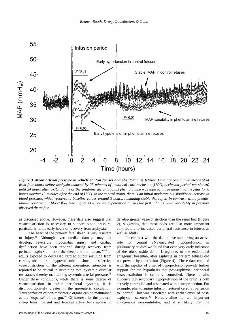
There is increasing evidence that the sympatheticnervous system plays a central role in mediating post-asphyxial changes in central and peripheral blood flow andcontributes to neuroinhibition.42,43 We previously reportedthat post-asphyxial hypoperfusion of the preterm fetal gut,44
was prevented by infusion of the mixed α-adrenergicantagonist phentolamine.42 In control fetuses hypoperfusionwas not associated with hypotension, regardless of thevascular bed being measured (Figure 3). Rather, there was abrief period of mild hypertension during the first 3 hours,
followed by stabilization of blood pressure around baselinevalues (Figure 3). By contrast, hypotension occurred whengut hypoperfusion was prevented by phentolamine,followed by variable pressure fluctuations. Notably, theperiod of hypotension was most pronounced during thecorresponding period of hypertension in control fetuses.These data highlight the need to appreciate the temporalnature of perfusion and blood pressure changes, and theclose relationships with the cessation of an adverse eventlike hypoxia. Interestingly, there was no increase in HR(the primary determinant of combined ventricular output inthe fetus) to compensate for this fall in blood pressure.42
This may reflect the sensitivity of the baroreflex at this age,
84 Proceedings of the Australian Physiological Society (2012)43
Bennet, Booth, Drury, Quaedackers & Gunn
20
25
30
35
40
45
50
55Infusion period
MA
P (
mm
Hg
)
Time (hours)
P<0.01
P<0.05
Early hypertension in control fetuses
Early hypotension in phentolamine fetuses
MAP variability in phentolamine fetusesP<0.05
Stable MAP in control fetuses
-4 -2 0 2 4 6 8 10 12 14 16 18 20 22 24
Figure 3. Mean arterial pressure in vehicle control fetuses and phentolamine fetuses.Data are one minute mean±SEMfrom four hours before asphyxia induced by 25 minutes of umbilical cord occlusion (UCO, occlusion period not shown)until 24 hours after UCO. Saline or theα-adrenergic antagonist phentolamine was infused intravenously to the fetus for 8hours starting 15 minutes after the end of UCO. In the control group, there is an initial moderate but significant increase inblood pressure, which resolves to baseline values around 3 hours, remaining stable thereafter. In contrast, while phento-lamine restored gut blood flow (see Figure 4) it caused hypotension during the first 3 hours, with variability in pressureobserved thereafter.
as discussed above. Howev er, these data also suggest thatvasoconstriction is necessary to support blood pressure,particularly in the early hours of recovery from asphyxia.
The heart of the preterm fetal sheep is very resistantto injury.45 Although overt cardiac damage may notdevelop, reversible myocardial injury and cardiacdysfunction have been reported during recovery fromperinatal asphyxia in both the sheep and the human.46,47 Inadults exposed to decreased cardiac output resulting fromcardiogenic or hypovolaemic shock, selectivevasoconstriction of the afferent mesenteric arterioles isreported to be crucial in sustaining total systemic vascularresistance, thereby maintaining systemic arterial pressure.48
Under these conditions, while there is some degree ofvasoconstriction in other peripheral systems, it isdisproportionately greater in the mesenteric circulation.Thus perfusion of non-mesenteric organs can be maintainedat the ‘expense’ of the gut.48 Of interest, in the pretermsheep fetus, the gut and femoral artery beds appear to
develop greater vasoconstriction than the renal bed (Figure2), suggesting that these beds are also more importantcontributors to increased peripheral resistance in fetuses aswell as adults.
In contrast with the data above supporting an activerole for central SNS-mediated hypoperfusion, inpreliminary studies we found that even very early infusionsof the nitric oxide donor L-arginine, or the endothelialantagonist bosentan, after asphyxia in preterm fetuses didnot prevent hypoperfusion (Figure 4).These data coupledwith the rapidity of onset of hypoperfusion provide furthersupport for the hypothesis that post-asphyxial peripheralvasoconstriction is centrally controlled. There is alsoevidence that secondary hypoperfusion of the brain is bothactively controlled and associated with neuroprotection. Forexample, phentolamine infusion restored cerebral perfusionto ‘normal’, but was associated with earlier onset of post-asphyxial seizures.42 Noradrenaline is an importantendogenous neuroinhibitor, and it is likely that the
Proceedings of the Australian Physiological Society (2012)43 85

Fetal origins of neonatal cardiovascular instability
sympathetic activation after asphyxia contributed to centralprotective suppression of brain activity.
0
5
10
15
20
25
30
-6 0 3 6 12 18 24
0
5
10
15
0
5
10
15
0
5
10
15
Time (hours)
SM
BF
(ml/m
in)
SM
BF
(ml/m
in)
SM
BF
(ml/m
in)
A
B
C
D
Early phentolamine
Delayed phentolamine
L-arginine
Bosentan
SM
BF
(ml/m
in)
Figure 4. The effects of theα-adrenergic blocker phento-lamine. The block was started at 15 minutes (panel A) and30 minutes (panel B) after the end of asphyxia, L-arginine(panel C) and bosentan (panel D). The first arrow denotesthe start of infusions of each agent, and subsequent arrowsrepeated boluses given to attempt to improve blood flow.Data are one minute averages from individual fetuses. Theend of the occlusion period is time zero.
Consistent with this, selective α2-adrenergic receptorblockade was associated with increased epileptiformtransient activity in the latent (early recovery phase) andincreased brain injury after 3 days recovery.43 Conversely,exogenous infusion of anα2-adrenergic receptor agonistwas associated with improved outcomes.49 It is likely thatepileptiform transients are a manifestation of post-hypoxichyperexcitability of the glutamate receptor, as occurs inimmature rodents.50 In adult rodents, peri-infarct
depolarising wav es (spreading depression wav es) developafter cerebral ischaemia, and can contribute to expansion ofinjury by increasing the work load of stressed cells.51 Thisincreasing energy imbalance, in turn, hastens the energyfailure in sick cells, promoting impaired cellularhomeostasis, ATP production, and initiation of cell deathprocesses (secondary energy failure).
Collectively these data further support the conceptthat reduced blood flow is coupled to reduced metabolismduring recovery from hypoxic-ischemic insults and that thisis beneficial for recovery from asphyxia. These data areconsistent with the lack of efficacy of many clinicalinterventions such as a volume expansion, administration ofinotropes, and closure of theductus arteriosusafterbirth.13,52 Thus, despite the differences between fetal andpostnatal life, there is a considerable need for furtherresearch to determine whether hypoperfusion is a cause ofmorbidity or merely a consequence.15
Prolonged effects of exposure to infection/inflammation
There is now considerable evidence that perinatalexposure to infection and inflammation such as clinicalchorioamnionitis (uterine infection), vasculitis(inflammation of blood vessels) in the chorionic plate of theplacenta and/or umbilical cord, and high levels of pro-inflammatory cytokines in amniotic and umbilical blood,are associated with increased mortality and morbidity.53
Some studies have found no association betweenhistological chorioamnionitis and risk of white matterinjury, whereas neonatal sepsis has been consistentlyassociated with increased risk of brain injury.54,55
There is relatively limited information on thehaemodynamic effects of sepsis in preterm newborns,56 andat present there are no agreed treatment guidelines for themanagement of severe sepsis and septic shock for pretermneonates. Thisis complicated by the lack of normativecardiovascular data for preterm infants.57 It is also clear thatthe responses of the newborn are very variable betweeninfants, dynamic over time, and may differ to that of theadult in response to treatment.56,57 In this setting, theimmediate effect of early onset neonatal sepsis appears tobe mainly vasodilation, leading to hyperperfusion and oftenhypotension.54,55 Other studies have reported hypotensionwith peripheral vasoconstriction and increased cardiacoutput, or “normal” blood pressure with eithervasodilatation or vasoconstriction or hypotension witheither state.56,57 This may reflect a continuum from chroniclow key inflammation, secondary to chorioamnionitis,through to acute severe infection.57
Nevertheless, persistent vasodilatation is oftenobserved after exposure to inflammation in adult humansand animal models.58 Indeed, much of the acute morbidityassociated with significant infection or inflammation inadults is associated with endothelial dysfunction, withaltered release and sensitivity to vasodilators andvasoconstrictors, which in turn leads to haemodynamicinstability and organ compromise.58 In fetal experimentalstudies, exposure to gram negative lipopolysaccharide
86 Proceedings of the Australian Physiological Society (2012)43

Bennet, Booth, Drury, Quaedackers & Gunn
(LPS) has been associated with variable haemodynamiceffects, likely reflecting differences in dose and timing ofexposure between studies. Some studies observed nochange in blood pressure or blood flow in fetal sheep,59-61
whereas others reported acute falls in blood pressure,62-65ora transient fall, followed by increased arterial bloodpressure by 48 to 72 hours of recovery.63
Typically, early fetal hypotension in responses to LPShas been associated with cerebral vasodilatation thatmaintained perfusion and oxygen delivery.65,66 By contrast,a study in the late gestation sheep fetus found acutetransient cerebral vasoconstriction after injection of LPS,followed by vasodilatation up to 24 hours after threerepeated injections.66 Vasodilatation was associated withevidence of transiently increased nitric oxide (NO)production,66 consistent with endotoxic shock in adults.67
Similar findings were observed when LPS was given toterm newborn lambs, with chronic loss of sensitivity to thevasodilator bradykinin, possibly secondary to LPS-inducedloss of endothelial cells.68
In 0.7 gestation preterm fetal sheep, exposure tokilled gram positive bacteria (OK-432 or Picabinil), derivedfrom low virulence heat killed Su-strain of type 3 Group Astreptococcus pyogenes,69,70 was associated with acute,transient peripheral and central vasoconstriction withouthypotension.69 This phase was coupled with reduced brainactivity and suppression of fetal body and breathingmovements.70 These data support the hypothesis thatinfection was associated with reduced brain metabolism andan appropriate reduction of blood flow, rather than loss ofregulatory control.
Over the week after exposure to OK-432, there was aprogressive central and peripheral vasodilatation withincreased blood flow (Figure 5).69 Although hypotensiondid not occur, there was loss of the normal secular increasein arterial blood pressure over this time (Figure 5). Fetalgrowth was not affected, suggesting that this relativelymodest vasodilatation reflects a resetting of thebaroreceptor threshold.16 In contrast with previous studiesof LPS,66 there were no significant changes in circulatingnitrite levels, suggesting that these changes were not relatedto altered NO production.69 However, it remains possiblethat there may have been altered responsiveness of thevasculature to vasodilator stimuli, or increased induction ofother vasodilators. Alternatively, there are strong data tosuggest that inflammatory mediators can alter release of, orsensitivity to, vasoconstrictors without changes invasodilator activity.71,72
Notably, although OK-432 was associated with acuteand chronic changes in cerebral blood flow in all fetuses,only one fetus sustained neural injury; a bilateral infarct ofthe hippocampus. This injury occurred despite normalblood gases and a time course of blood flow and bloodpressure changes which was similar to those of the otherfetuses. The early development of seizures suggests thatinjury occurred shortly after exposure. Thus it is unlikelythat injury was directly related to changes in perfusion. Thisfurther supports previous data that neural damageassociated with infection is primarily associated with
neurotoxicity, likely mediated by the cytokine/microglialresponse.73 The fetal hemodynamic changes after exposureto gram positive bacteria, while persistent, were relativelymodest, and it remains unclear if they represent apermanent change in the trajectory of blood pressure, orpersist into neonatal life.Nonetheless, it is reasonable tospeculate that changes in autonomic or endothelial functionmight compromise the ability of the fetus and newborn toadapt to further events such as hypoxia.
Synergy with other insults: sensitization and tolerance
Profound hypoxia or septic shock causing acuteinjury around the time of birth are only seen in a minorityof infants, and are insufficient to account for the majority oflong-term neurodevelopmental disability in preterm infants.However, infants may frequently be exposed to both insults.Intriguingly, experimental data in rodents suggest thatexposure to mild infection or inflammation can sensitise thebrain, so that short or milder periods of hypoxia-ischaemia,which do not normally injure the developing brain, cantrigger severe damage.73 However, the effect is complex andtime dependent. A low dose of LPS given either shortly(four or six hours) or well before (72 hours or more)hypoxia in rat pups was associated withincreasedinjury(‘sensitisation’).73 In mice, fetal exposure to LPS affectedthe responses to hypoxia even in adulthood, with bothreduced and increased injury, in different regions.74 Incontrast, when given at an intermediate time (24 hours)before hypoxia/ischaemia, LPS actuallyreduced injury(‘tolerance’).73
No large animal studies have been undertaken toevaluate key mechanisms or to confirm whether theserelationships hold up in more complex species.Nevertheless, recent data suggest that prenatal exposure toinflammation exacerbates ventilation-mediated brain injury,supporting the concept that multiple insults can contributein complex ways to perinatal brain injury.75 Intriguingly,there is now evidence that exposure of preterm fetal sheepto a clinical course of maternal glucocorticoids is associatedwith acute changes in EEG transient activity andepileptiform events, followed by evidence of neuralmaturation.76 Given that seizures in early life in the rat caninduce neuroprotection through preconditioningmechanisms,77 it is plausible that steroids may also trigger asensitization/preconditioning sequence independently ofpathological events.
Summary
The preterm fetus exhibits a remarkable anaerobictolerance and ability to survive major insults with no orlimited injury.32 At the same time, it is clear that autonomicfunction is still developing over late gestation, and thatresponses to acute arterial hypotension are particularlyimmature. Premature newborns are commonly exposed toperinatal hypoxia and infection around the time of birth,which trigger long-lasting changes in central and peripheraltone, which in combination can sensitise the brain to injury.Finally, there is increasing evidence that ‘standard’ clinical
Proceedings of the Australian Physiological Society (2012)43 87

Fetal origins of neonatal cardiovascular instability
-12 -6
5
10
15
20
25
30
35
6 24 42 60 78 96 114 132 150 168
P<0.05
OK-432 fetuses
Saline fetuses
30
32
34
36
38
40
P<0.05
MA
P (
mm
Hg
)F
BF
(m
l/m
in)
Time (hours)
Normal maturational increase in MAP
Lower MAP due to vasodilatation
Figure 5. Time course changes in mean arterial blood pressure (MAP) and femoral blood flow (FBF) Data are from 0.7gestation fetal sheep who received a bolus intrapleural injection of either saline (open circles) or OK-432 (0.1 mg, closedcircles). Thearrow denotes the point of injection. Data are mean±SEM. Note the failure of blood pressure to increase overtime in the OK-432 group compared to the vehicle control group.
interventions such as antenatal maternal steroids andneonatal ventilation can further modulate the risk ofdamage. The authors believe that to disentangle thesemultiple factors and provide a solid evidence-base to testinnovative paradigms and improve long-term outcomes, weneed to develop an approach of comprehensive monitoringand neurointensive care.
Acknowledgments
The authors’ studies in the present review weresupported by grants from the Health Research Council ofNew Zealand, USPHS grant RO1 HD-32752, March ofDimes, the Auckland Medical Research Foundation, andthe Lottery Grants Board of New Zealand. Mr P. Drury wassupported by the Neurological Foundation.
88 Proceedings of the Australian Physiological Society (2012)43

Bennet, Booth, Drury, Quaedackers & Gunn
References
1. Allen MC, Cristofalo EA, Kim C. Outcomes of preterminfants: morbidity replaces mortality. Clin.Perinatol. 2011;38:441-54.
2. Committee on understanding premature birth andassuring healthy outcomes. In: Behrman RE, ButlerAS, (eds.).Preterm Birth: Causes, Consequences,and Prevention. Institute of Medicine of theNational Academies: Washington DC, USA. 2007.
3. LemonsJA, Bauer CR, Oh W, Korones SB, Papile LA,Stoll BJ, Verter J, Temprosa M, Wright LL,Ehrenkranz RA, Fanaroff AA, Stark A, Carlo W,Tyson JE, Donovan EF, Shankaran S, Stevenson DK.Very low birth weight outcomes of the NationalInstitute of Child health and human developmentneonatal research network, January 1995 throughDecember 1996. NICHD Neonatal ResearchNetwork.Pediatrics2001;107:E1.
4. PetrouS, Eddama O, Mangham L. A structured reviewof the recent literature on the economicconsequences of preterm birth.Arch. Dis. Child.Fetal Neonatal Ed.2011;96:F225-32.
5. MacKayDF, Smith GC, Dobbie R, Pell JP. Gestationalage at delivery and special educational need:retrospective cohort study of 407,503schoolchildren.PLoS Med.2010;7:e1000289.
6. Soleymani S, Borzage M, Seri I. Hemodynamicmonitoring in neonates: advances and challenges.J.Perinatol. 2010;30 Suppl:S38-45.
7. Dempsey EM, Barrington KJ. Evaluation and treatmentof hypotension in the preterm infant. Clin.Perinatol. 2009;36:75-85.
8. Kluckow M. Functional echocardiography in assessmentof the cardiovascular system in asphyxiatedneonates.J. Pediatr.2011;158:e13-8.
9. Groves AM, Kuschel CA, Knight DB, Skinner JR.Relationship between blood pressure and blood flowin newborn preterm infants.Arch. Dis. Child. FetalNeonatal Ed.2008;93:F29-32.
10. OsbornDA, Evans N, Kluckow M. Hemodynamic andantecedent risk factors of early and lateperiventricular/intraventricular hemorrhage inpremature infants.Pediatrics2003;112:33-9.
11. Evans N, Osborn D, Kluckow M. Preterm circulatorysupport is more complex than just blood pressure.Pediatrics2005;115:1114-5.
12. Logan JW, O’Shea TM, Allred EN, Laughon MM,Bose CL, Dammann O, Batton DG, Engelke SC,Leviton A. Early postnatal hypotension anddevelopmental delay at 24 months of age amongextremely low gestational age newborns.Arch. Dis.Child. Fetal Neonatal Ed.2011;96:F321-8.
13. Hunt RW, Evans N, Rieger I, Kluckow M. Lowsuperior vena cava flow and neurodevelopment at 3years in very preterm infants. J. Pediatr. 2004;145:588-92.
14. CapozziG, Santoro G. Patent ductus arteriosus: patho-physiology, hemodynamic effects and clinical
complications. J. Matern. Fetal Neonatal Med.2011;24 Suppl 1:15-6.
15. Sehgal A. Haemodynamically unstable preterm infant:an unresolved management conundrum.Eur. J.Pediatr. 2011.
16. BoothLC, Malpas SC, Barrett CJ, Guild SJ, Gunn AJ,Bennet L. Is baroreflex control of sympatheticactivity and heart rate active in the preterm fetalsheep?Am. J. Physiol. Regul. Integr. Comp. Physiol.2009;296:R603-R9.
17. BoothLC, Gunn AJ, Malpas SC, Barrett CJ, DavidsonJO, Guild SJ, Bennet L.Baroreflex control of renalsympathetic nerve activity and heart rate in near-term fetal sheep.Exp. Physiol.2011;96:736-44.
18. AndriessenP, Oetomo SB, Peters C, Vermeulen B,Wijn PF, Blanco CE. Baroreceptor reflex sensitivityin human neonates: the effect of postmenstrual age.J. Physiol.2005;568:333-41.
19. Bennet L, Booth L, Malpas SC, Quaedackers JS,Jensen E, Dean J, Gunn AJ.Acute systemiccomplications in the preterm fetus after asphyxia:the role of cardiovascular and blood flow responses.Clin. Exp.Pharmacol. Physiol.2006;33:291-9.
20. Westgate JA, Wibbens B, Bennet L, Wassink G, ParerJT, Gunn AJ. The intrapartum deceleration in centerstage: a physiological approach to interpretation offetal heart rate changes in labor. Am. J. Obstet.Gynecol.2007;197:e1-e11.236.
21. BoddyK, Dawes GS, Fisher R, Pinter S, Robinson JS.Foetal respiratory movements, electrocortical andcardiovascular responses to hypoxaemia andhypercapnia in sheep. J. Physiol. 1974;243:599-618.
22. Iwamoto HS, Stucky E, Roman CM. Effect of gradedumbilical cord compression in fetal sheep at 0.6-0.7gestation.Am. J. Physiol. Heart Circ. Physiol.1991;261:H1268-H74.
23. Szymonowicz W, Walker AM, Yu VY, Stewart ML,Cannata J, Cussen L. Regional cerebral blood flowafter hemorrhagic hypotension in the preterm, near-term, and newborn lamb. Pediatr. Res. 1990;28:361-6.
24. GleasonCA, Hamm C, Jones MD, Jr. Effect of acutehypoxemia on brain blood flow and oxygenmetabolism in immature fetal sheep.Am. J. Physiol.Heart Circ. Physiol.1990;258:H1064-H9.
25. Iwamoto HS, Kaufman T, Keil LC, Rudolph AM.Responses to acute hypoxemia in fetal sheep at0.6-0.7 gestation.Am. J. Physiol. Heart Circ.Physiol.1989;256:H613-H20.
26. Shelley HJ. Glycogen reserves and their changes atbirth and in anoxia.Br. Med. Bull. 1961;17:137-43.
27. BennetL, Rossenrode S, Gunning MI, Gluckman PD,Gunn AJ. The cardiovascular and cerebrovascularresponses of the immature fetal sheep to acuteumbilical cord occlusion. J. Physiol. 1999;517:247-57.
28. Wassink G, Bennet L, Booth LC, Jensen EC, WibbensB, Dean JM, Gunn AJ. The ontogeny of
Proceedings of the Australian Physiological Society (2012)43 89

Fetal origins of neonatal cardiovascular instability
hemodynamic responses to prolonged umbilical cordocclusion in fetal sheep.J. Appl. Physiol. 2007;103:1311-7.
29. AssaliNS, Brinkman CR, Woods JR, Jr, Dandavino A,Nuwayhid B. Development of neurohumoral controlof fetal, neonatal, and adult cardiovascular functions.Am. J. Obstet. Gynecol.1977;129:748-59.
30. Nuwayhid B, Brinkman CR, Su C, Bevan JA, AssaliNS. Development of autonomic control of fetalcirculation.Am. J. Physiol.1975;228:337-44.
31. BoothLC, Bennet L, Guild SJ, Barrett CJ, May CN,Gunn AJ, Malpas SC.Maturation-related changes inthe pattern of renal sympathetic nerve activity fromin utero to adulthood.Exp. Physiol.2011;96:85-93.
32. Gunn AJ, Quaedackers JS, Guan J, Heineman E,Bennet L. The premature fetus: not as defenseless aswe thought, but still paradoxically vulnerable?Dev.Neurosci.2001;23:175-9.
33. Low JA. Determining the contribution of asphyxia tobrain damage in the neonate.J. Obstet. Gynaecol.Res.2004;30:276-86.
34. DyetLE, Kennea N, Counsell SJ, Maalouf EF, Ajayi-Obe M, Duggan PJ, Harrison M, Allsop JM, HajnalJ, Herlihy AH, Edwards B, Laroche S, Cowan FM,Rutherford MA, Edwards AD. Natural history ofbrain lesions in extremely preterm infants studiedwith serial magnetic resonance imaging from birthand neurodevelopmental assessment.Pediatrics2006;118:536-48.
35. BennetL, Dean JM, Gunn AJ. The pathogenesis ofpreterm brain injury. In: Stevenson DK, Benitz WE,Sunshine P, Druzin ML, (eds.).Fetal and NeonatalBrain Injury: Mechanisms, Management, and theRisks of Practice. 4th edn. Cambridge UniversityPress: Cambridge, UK. 2009.
36. Amantea D, Nappi G, Bernardi G, Bagetta G,Corasaniti MT. Post-ischemic brain damage:pathophysiology and role of inflammatorymediators.FEBS J.2009;276:13-26.
37. JensenEC, Bennet L, Hunter CJ, Power GG, Gunn AJ.Post-hypoxic hypoperfusion is associated withsuppression of cerebral metabolism and increasedtissue oxygenation in near-term fetal sheep.J.Physiol.2006;572:131-9.
38. Rosenberg AA. Regulation of cerebral blood flow afterasphyxia in neonatal lambs.Stroke1988;19:239-44.
39. Gunn AJ, Gunn TR, de Haan HH, Williams CE,Gluckman PD. Dramatic neuronal rescue withprolonged selective head cooling after ischemia infetal lambs.J. Clin. Invest.1997;99:248-56.
40. HuangJ, Kim LJ, Poisik A, Pinsky DJ, Connolly ES,Jr. Titration of postischemic cerebral hypoperfusionby variation of ischemic severity in a murine modelof stroke.Neurosurgery 1999;45:328-33.
41. KarlssonBR, Grogaard B, Gerdin B, Steen PA. Theseverity of postischemic hypoperfusion increaseswith duration of cerebral ischemia in rats.ActaAnaesthesiol. Scand.1994;38:248-53.
42. Quaedackers JS, Roelfsema V, Heineman E, Gunn AJ,
Bennet L. The role of the sympathetic nervoussystem in post-asphyxial intestinal hypoperfusion inthe preterm sheep fetus.J. Physiol. 2004;557:1033-44.
43. DeanJM, Gunn AJ, Wassink G, George S, Bennet L.Endogenous α2-adrenergic receptor-mediatedneuroprotection after severe hypoxia in preterm fetalsheep.Neuroscience2006;142:615-28.
44. BennetL, Quaedackers JS, Gunn AJ, Rossenrode S,Heineman E. The effect of asphyxia on superiormesenteric artery blood flow in the premature sheepfetus.J. Pediatr. Surg. 2000;35:34-40.
45. BennetL, Kozuma S, McGarrigle HHG, Hanson MA.Temporal changes in fetal cardiovascular,behavioural, metabolic and endocrine responses tomaternally administered dexamethasone in the lategestation fetal sheep.Br. J. Obstet. Gynaecol.1999;106:331-9.
46. Gunn AJ, Maxwell L, De Haan HH, Bennet L,Williams CE, Gluckman PD, Gunn TR.Delayedhypotension and subendocardial injury after repeatedumbilical cord occlusion in near-term fetal lambs.Am. J. Obstet. Gynecol.2000;183:1564-72.
47. LumbersER, Gunn AJ, Zhang DY, Wu JJ, Maxwell L,Bennet L. Nonimmune hydrops fetalis and activationof the renin-angiotensin system after asphyxia inpreterm fetal sheep.Am. J. Physiol. Regul. Integr.Comp. Physiol.2001;280:R1045-R51.
48. Reilly PM, Wilkins KB, Fuh KC, Haglund U, BulkleyGB. The mesenteric hemodynamic response tocirculatory shock: an overview. Shock 2001;15:329-43.
49. DeanJM, George SA, Wassink G, Gunn AJ, Bennet L.Suppression of post hypoxic-ischemic EEGtransients with dizocilpine is associated with partialstriatal protection in the preterm fetal sheep.Neuropharmacology2006;50:491-503.
50. JensenFE, Wang C, Stafstrom CE, Liu Z, Geary C,Stevens MC. Acute and chronic increases inexcitability in rat hippocampal slices after perinatalhypoxia in vivo. J. Neurophysiol.1998;79:73-81.
51. SomjenGG. Mechanisms of spreading depression andhypoxic spreading depression-like depolarization.Physiol. Rev. 2001;81:1065-96.
52. Hunt R, Osborn D. Dopamine for prevention ofmorbidity and mortality in term newborn infantswith suspected perinatal asphyxia. CochraneDatabase Syst. Rev. 2002:CD003484.
53. Mwaniki MK, Atieno M, Lawn JE, Newton CR. Long-term neurodevelopmental outcomes after intrauterineand neonatal insults: a systematic review. Lancet2012;379:445-52.
54. BasuS, Dewangan S, Shukla RC, Anupurva S, KumarA. Cerebral blood flow velocity in early-onsetneonatal sepsis and its clinical significance.Eur. J.Pediatr. 2012;171:901-9.
55. ChauV, Poskitt KJ, McFadden DE, Bowen-Roberts T,Synnes A, Brant R, Sargent MA, Soulikias W, MillerSP. Effect of chorioamnionitis on brain development
90 Proceedings of the Australian Physiological Society (2012)43

Bennet, Booth, Drury, Quaedackers & Gunn
and injury in premature newborns. Ann. Neurol.2009;66:155-64.
56. deWaal K, Evans N. Hemodynamics in preterm infantswith late-onset sepsis.J. Pediatr. 2010;156:918-22,22 e1.
57. Wynn JL, Wong HR. Pathophysiology and treatment ofseptic shock in neonates.Clin. Perinatol. 2010;37:439-79.
58. SesslerCN, Shepherd W. New concepts in sepsis.Curr. Opin. Crit. Care2002;8:465-72.
59. EklindS, Mallard C, Leverin AL, Gilland E, BlomgrenK, Mattsby-Baltzer I, Hagberg H. Bacterialendotoxin sensitizes the immature brain tohypoxic--ischaemic injury. Eur. J. Neurosci. 2001;13:1101-6.
60. Duncan JR, Cock ML, Suzuki K, Scheerlinck JP,Harding R, Rees SM. Chronic endotoxin exposurecauses brain injury in the ovine fetus in the absenceof hypoxemia. J. Soc. Gynecol. Investig. 2006;13:87-96.
61. DuncanJR, Cock ML, Scheerlinck JP, Westcott KT,McLean C, Harding R, Rees SM.White matterinjury after repeated endotoxin exposure in thepreterm ovine fetus.Pediatr. Res. 2002;52:941-9.
62. Dalitz P, Harding R, Rees SM, Cock ML. Prolongedreductions in placental blood flow and cerebraloxygen delivery in preterm fetal sheep exposed toendotoxin: possible factors in white matter injuryafter acute infection.J. Soc. Gynecol. Investig.2003;10:283-90.
63. GarnierY, Frigiola A, Li Volti G, Florio P, Frulio R,Berger R, Alm S, von Duering MU, Coumans AB,Reis FM, Petraglia F, Hasaart TH, Abella R, MufeedH, Gazzolo D. Increased maternal/fetal bloodS100B levels following systemic endotoxinadministration and periventricular white matterinjury in preterm fetal sheep.Reprod. Sci. 2009;16:758-66.
64. BladS, Welin AK, Kjellmer I, Rosen KG, Mallard C.ECG and heart rate variability changes in pretermand near-term fetal lamb following LPS exposure.Reprod. Sci.2008;15:572-83.
65. PeeblesDM, Miller S, Newman JP, Scott R, HansonMA. The effect of systemic administration oflipopolysaccharide on cerebral haemodynamics andoxygenation in the 0.65 gestation ovine fetus inutero.BJOG2003;110:735-43.
66. FengSY, Phillips DJ, Stockx EM, Yu VY, Walker AM.Endotoxin has acute and chronic effects on thecerebral circulation of fetal sheep.Am. J. Physiol.Regul. Integr. Comp. Physiol.2009;296:R640-50.
67. ThiemermannC. Nitric oxide and septic shock.Gen.Pharmacol.1997;29:159-66.
68. Feng SY, Samarasinghe T, Phillips DJ, Alexiou T,Hollis JH, Yu VY, Walker AM. Acute and chroniceffects of endotoxin on cerebral circulation in lambs.Am. J. Physiol. Regul. Integr. Comp. Physiol.2010;298:R760-6.
69. BennetL, Cowie RV, Stone PR, Barrett R, Naylor AS,
Blood AB, Gunn AJ. The neural and vasculareffects of killed Su-Streptococcus pyogenes(OK-432) in preterm fetal sheep.Am. J. Physiol.Regul. Integr. Comp. Physiol.2010;299:R664-72.
70. Cowie RV, Stone PR, Parry E, Jensen EC, Gunn AJ,Bennet L. Acute behavioral effects of intrapleuralOK-432 (Picibanil) administration in preterm fetalsheep.Fetal Diagn. Ther.2009;25:304-13.
71. LangeM, Bröking K, Hucklenbruch C, Ertmer C, VanAken H, Lücke M, Bone HG, Westphal M.Hemodynamic effects of titrated norepinephrine inhealthy versus endotoxemic sheep.J. Endotoxin Res.2007;13:53-7.
72. PleinerJ, Heere-Ress E, Langenberger H, Sieder AE,Bayerle-Eder M, Mittermayer F, Fuchsjäger-MayrlG, Böhm J, Jansen B, Wolzt M. Adrenoceptorhyporeactivity is responsible for Escherichia coliendotoxin-induced acute vascular dysfunction inhumans.Arterioscler. Thromb. Vasc. Biol. 2002;22:95-100.
73. Wang X, Rousset CI, Hagberg H, Mallard C.Lipopolysaccharide-induced inflammation andperinatal brain injury. Semin. Fetal Neonatal Med.2006;11:343-53.
74. Wang X, Hagberg H, Nie C, Zhu C, Ikeda T, MallardC. Dual role of intrauterine immune challenge onneonatal and adult brain vulnerability to hypoxia-ischemia. J. Neuropathol. Exp. Neurol. 2007;66:552-61.
75. PolglaseGR, Nitsos I, Baburamani AA, Crossley KJ,Slater MK, Gill AW, Allison BJ, Moss TJ, Pillow JJ,Hooper SB, Kluckow M. Inflammation in uteroexacerbates ventilation-induced brain injury inpreterm lambs.J. Appl. Physiol.2012;112:481-9.
76. Davidson JO, Quaedackers JS, George SA, Gunn AJ,Bennet L. Maternal dexamethasone and EEGhyperactivity in preterm fetal sheep.J. Physiol.2011;589:3823–35.
77. Saghyan A, LaTorre GN, Keesey R, Sharma A, MehtaV, Rudenko V, Hallas BH, Rafiuddin A, Goldstein B,Friedman LK. Glutamatergic and morphologicalalterations associated with early life seizure-inducedpreconditioning in young rats.Eur. J. Neurosci.2010;32:1897-911.
Received 28 February 2012, in revised form 22 June 2012.Accepted 24 June 2012.© L. Bennet 2012
Proceedings of the Australian Physiological Society (2012)43 91

Fetal origins of neonatal cardiovascular instability
Author for correspondence:
Professor Laura BennetFetal Physiology and Neuroscience GroupDepartment of PhysiologyThe University of AucklandPrivate Bag 92019Auckland Mail CentreAuckland 1142New Zealand
Tel: +64 9 373 7599 ext 84890Fax: +64 9 923 1111E-mail: [email protected]
92 Proceedings of the Australian Physiological Society (2012)43