Presentation - Kim, Youngcmesyllabus.com/wp-content/uploads/2017/09/4-Slides-Kim-Young.pdf · –...
12
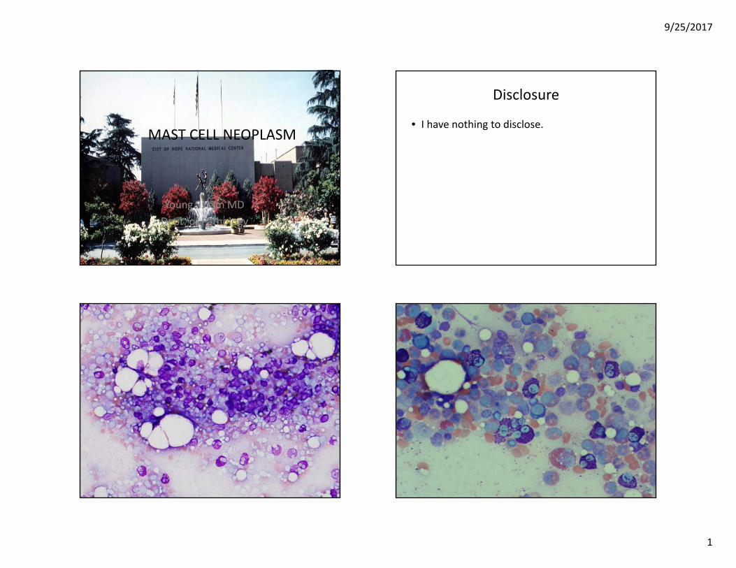
9/25/2017 1 MAST CELL NEOPLASM MAST CELL NEOPLASM Young S. Kim MD Dept. of Pathology Disclosure • I have nothing to disclose.
-
Upload
truonghanh -
Category
Documents
-
view
219 -
download
1
Transcript of Presentation - Kim, Youngcmesyllabus.com/wp-content/uploads/2017/09/4-Slides-Kim-Young.pdf · –...
9/25/2017
1
MAST CELL NEOPLASMMAST CELL NEOPLASM
Young S. Kim MDDept. of Pathology
Disclosure
• I have nothing to disclose.

9/25/2017
2
Objectives
• What is mast cell lineage? • Changes in updated WHO 2016 mastocytosis• Issues of Mastocytosis ‐ CD30 expression, CD2 and CD25 expression, KIT mutation, Mast cell leukemia, and Myelomastocytic leukemia.
• Case presentation Importance of inclusion of• Case presentation ‐ Importance of inclusion of mast cell neoplasm in routine surgical case work up and the unexpected.

9/25/2017
3
• Add lineage diagram of hematopoietic cells
Classical scheme of hematopoiesis
• Article of mast cell development from T‐cell progenitors by GATA

9/25/2017
4
WHO 2001
WHO 2008
Mast cells in mastocytosis: not so “myeloid” and not
WHO classification of mast cell disease – evolving and imperfect
Mast cells in mastocytosis: not so myeloid and not so “proliferative” – Peter Valent, Blood, 2010
Mast cells (SM) show myeloid and lymphoid antigens (CD43, CD117, CD68, CD2, CD25, CD308)
In many SM, neoplastic mast cells do not show signs of significant proliferationof significant proliferation
Recommend‐Mastocytosis, a separate group of hematopoietic neoplasm as classified in 2001 WHO

9/25/2017
5
2016 WHO classification
• Myeloproliferative neoplasms (MPN)– Chronic myeloid leukemia (CML), BCR‐ABL1+Chronic myeloid leukemia (CML), BCR ABL1– Chronic neutrophilic leukemia (CNL)– Polycythemia vera (PV)– Primary myelofibrosis (PMF) prefibrotic stage, overt fibrotic stage– Essential thrombocythemia (ET)– Chronic eosinophilic leukemia, not otherwise specified (NOS)– MPN, unclassifiable
• Mastocytosis
Classification of mastocytosis • 2016 WHO• Cutaneous mastocytosis (CM)
S i i
• 2008 WHO• Cutaneous mastocytosis (CM)
• Systemic mastocytosis– Indolent systemic
mastocytosis (ISM)– Smoldering systemic
mastocytosis (SSM)– Systemic mastocytosis with
an associated hematological neoplasm (SM‐AHN)
– Aggressive systemic
• Indolent systemic mastocytosis (ISM)
• Systemic mastocytosis with associated hematological non‐mast cell lineage disease (SH‐AHNMD)
• Aggressive systemic mastocytosis – Aggressive systemic mastocytosis (ASM)
– Mast cell leukemia (MCL)• Mast cell sarcoma (MCS)
(ASM)• Mast cell leukemia (MCL)• Mast cell sarcoma (MCS)• Extracutaneous mastocytoma
• Smoldering systemic mastocytosis (SSM)– New entity in 2016 WHO– It was a provisional subvariant of ISM.– The prognosis is less favorable compared to ISM but favorable compared with ASM or MCL.
• Extracutaneous mastocytoma (ECM)– Deleted b/c of rarity
Issues in mastocytosis
• CD2/CD25/CD30 expression• KIT mutation‐ D816V is one of miner criteria of SM, how about other KIT mutations?
• What is Mast cell leukemia?• What is Myelomastocytic leukemia?

9/25/2017
6
CD2/CD25/CD30 expression
• Marker Nl BM ISM SSM ASM MCL • CD2 ‐ + + +/‐ ‐/+• CD25 ‐ + + + +/‐• CD30 ‐ ‐‐/+ +/‐ + +
• CD25 is more sensitive marker than CD2• CD30 is expressed in advanced SM
KIT mutation
• KIT D816V detected >80% of SM and >90% in ISM inadultsinadults
• KIT D816V is considered a relatively weak driver that promotes MC differentiation and maturation rather than MC proliferation
• Recently, whole exon sequencing of KIT gene discovered many different KIT mutations in mastocytosis, especially in pediatric mastocytosis
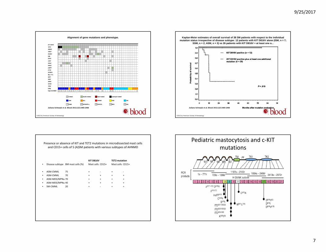
• Pediatric mastocytosis: D816V (only ~42%) and many other KIT gene exons involved, especially exons 8 and 9
Molecular lesions in CM and SM • Molecular• Abnormality Reported in patients with Estimated frequency in patients
with SM
• KIT D816V All SM variants and also in CM >80% (SM) 15‐20% (CM)( ) ( )• KIT D816Y CM, ISM, SM‐AHNMD <5%• KIT D816F CM <5%• KIT D816H MCL, ASM, SM‐AHNMD <5%• KIT D820G ASM <5%• KIT V560G ISM <5%• KIT F522C ISM <5%• KIT E839K CM <5%• KIT V530I SM‐AHNMD <5%• KIT K509I CM, SM (including familial Var) <5%, ( g )• Other KIT mutations CM and/or SM variants <5%
Molecular lesions in CM and SM
• FIP1L1/PDGFRA SM‐CEL <5%• AML1/ETO SM‐AML with t(8;21) <5%( )• JAK2 V617F SM‐PMF <5%• TET2 mutations SM‐AHNMD, ISM, ASM <5%• SRSF2 mutations ASM, SM‐AHNMD <5%• DNMT3A mutations ISM, SM‐AHNMD <5%• ASXL1 mutations SM‐AHNMD <5%• CBL mutations SM‐AHNMD <5%• U2AF1 mutations SM‐AHNMD <5%• EZH2 mutations SM‐AHNMD <5%• RAS mutations ASM, SM‐AHNMD <5%
9/25/2017
7
Alignment of gene mutations and phenotype.
Juliana Schwaab et al. Blood 2013;122:2460-2466
©2013 by American Society of Hematology
Kaplan-Meier estimates of overall survival of 38 SM patients with respect to the individual mutation status irrespective of disease subtype: 12 patients with KIT D816V alone (ISM, n = 7;
SSM, n = 2; ASM, n = 3) vs 26 patients with KIT D816V + at least one a...
Juliana Schwaab et al. Blood 2013;122:2460-2466
©2013 by American Society of Hematology
Presence or absence of KIT and TET2 mutations in microdissected mast cells and CD15+ cells of 5 (A)SM patients with various subtypes of AHNMD
KIT D816V TET2 mutationb ll ( ) ll ll• Disease subtype BM mast cells (%) Mast cells CD15+ Mast cells CD15+
• ASM‐CMML 75 + ‐ + ‐• ASM‐CMML 70 + + + +• ASM‐MDS/MPNu 70 + + + +• ASM‐MDS/MPNu 40 + + + +• SM‐CMML 20 + ‐ + +
Pediatric mastocytosis and c‐KIT mutations

9/25/2017
8
Mast cell leukemia
• Definition by WHO 2016:– Satisfy the criteria of Systemic Mastocytosis – The presence of at least 20% MCs on BM smears– Leukemic infiltration of marrow– If <10% of circulating leukocytes are MCs or no MCs in the blood – aleukemic MCL, which is more frequent (>60%)
Delineation between various forms of mast cell leukemia (MCL)
• Leukemic MCL At least 10% circulating MCsg• Aleukemic MCL <10% of blood leukocytes are MCs• Acute MCL C‐Finding(s) present, mostly
immature MCs• Chronic MCL C‐Findings usually absent, less
aggressive course, mature MCs usually predominate
• Primary MCL No antecedent SM or other myeloid• Primary MCL No antecedent SM or other myeloid neoplasm
• Secondary MCL Transformation of SM (usually ASM) or MC sarcoma
What is Myelomastocytic leukemia
• A term applied to advanced myeloid (non‐MC) l i hi h MC diff ti ti ineoplasms in which MC differentiation is
prominent, but SM criteria are not fulfilled, thereby distinguishing MML from MCL.
• MCs comprise >10% of all cells in blood and/or BM smears./
• Myelomastocytic leukemia (MML) is not recognized as a separate entity.
Temporal Bone Mass
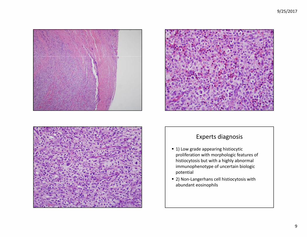
16 yo girl presenting with a 4.2 x 3.5 x 3.2 cm temporal bone mass growing into posteriortemporal bone mass growing into posterior fossa causing mass effect on cerebellum Reported IHC: Positive – CD43, CD68 Negative – CD1a, CD3, CD8, S‐100, Initial Dx: Myeloid neoplasm with minimalInitial Dx: Myeloid neoplasm with minimal mono‐histiocytic maturation
9/25/2017
9
Experts diagnosis
1) Low grade appearing histiocytic lif ti ith h l i f t fproliferation with morphologic features of
histiocytosis but with a highly abnormal immunophenotype of uncertain biologic potential 2) Non‐Langerhans cell histiocytosis with ) g yabundant eosinophils

9/25/2017
10
At COH
Additional IHC study: Positive ‐ CD33, Tryptase, CD25 Negative – CD2, CD30, CD34, MPX, Molecular study for Positive for L799F, not D816V Negative for Birbeck granules by EM Pediatric Mast Cell Sarcoma of Temporal Bone With Novel L799F KIT mutation,
Mimicking Histiocytic Neoplasm – Y.Kim et al., Am J Surg Pathol 37, (3), 2013, 453‐ 457
Pt Courses
Treated as LCH with chemotherapy, but di d ith lti l idisease progressed with multiple spine metastasis Cytogenetic study: del 7q Additional IHC: CD117 positive Multiple chemoradiation treatments without Multiple chemoradiation treatments without improvement Seen at COH for evaluation of transplant
CD1a Langerin
CD117 CD33
9/25/2017
11
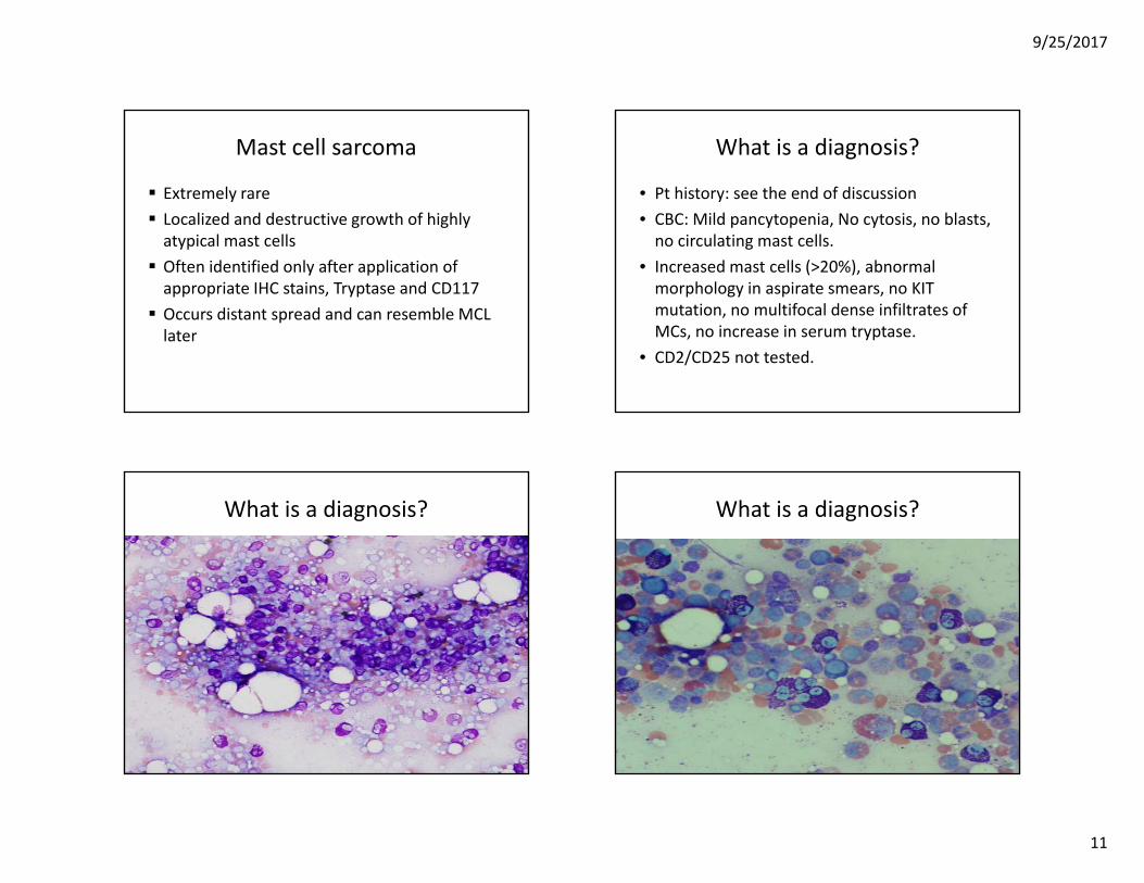
Mast cell sarcoma
Extremely rare Localized and destructive growth of highly atypical mast cells Often identified only after application of appropriate IHC stains, Tryptase and CD117 Occurs distant spread and can resemble MCL Occurs distant spread and can resemble MCL later
What is a diagnosis?
• Pt history: see the end of discussion• CBC: Mild pancytopenia, No cytosis, no blasts, no circulating mast cells.
• Increased mast cells (>20%), abnormal morphology in aspirate smears, no KIT mutation no multifocal dense infiltrates ofmutation, no multifocal dense infiltrates of MCs, no increase in serum tryptase.
• CD2/CD25 not tested.
What is a diagnosis? What is a diagnosis?

9/25/2017
12
Targeted FISH – Tryptase and t(8;21)
• Pt diagnosed with AML t(8;21).• Bone marrow is Day 40 post hematopoietic stem cell transplantation.
• Increased mast cells disappeared on follow up biopsy without treatment.
• These mast cells showed t(8;21) by Targeted• These mast cells showed t(8;21) by Targeted FISH
• P. Valent, et al. Mastocytosis: 2016 updated WHO classification and novel emerging treatment concepts. Blood. 2017; 129:1420‐1427
• J. Schwaab, et al. Comprehensive mutational profiling in advanced systemic mastocytosis. Blood. 2013; 122:2460‐2466
• C. Bodemer, er al. Pediatric mastocytosis is a clonal disease assocaitedD816V and other activating c‐KIT mutations. Journal of Investigative Dermatology 2010 130, 804‐815 gy ,
• P. Valent, et al. Refined diagnostic criteria and classification of mast cell leukemia(MCL) and myelomastocytic leukemia (MML): a consensus proposal. Annals of Oncology 25:1691‐1700,2014
• O. Schmetzer, et al. Murine and human mast cell progenitors. European Journal of Pharmacology. 778(2016)2‐10
• S. Pullarkat, V. Pullarkat, A. Lagoo, R. Brynes, L. Weiss, K.Gaal, D. Weisenburger, and Y. Kim. Characterization of bone marrow mast cells in acute myeloid leukemia with t(8;21)(q22;q22);RUNX1‐RUNX1T1. Leukemia Research, 37, 11, 2013, 1572‐1575
• Y. Kim et al. Pediatric Mast Cell Sarcoma of Temporal Bone With Novel pL799F (2395 C>T) KIT Mutation, Mimicking Histiocytic Neoplasm. Am J Surg Pathol 2013;37:453‐458