
Agar Bacto® Agar . Agar Flake . Agar, Granulated . Agar Noble Agar Bacteriological Technical

Int.J.Curr.Microbiol.App.Sci (2014) 3(8) 1081-1094
1081
Original Research Article
Extra Cellular Enzyme Production by Actinobacillus: A thermophile present in cattle compost
Megha Sharma1*, Kanchan Soni2 and Gagandeep Singh Saggu1
1School of Life Sciences, Jaipur National University, Jaipur, Rajasthan, India 2Department of Biotechnology, SRM University, Chennai, India
*Corresponding author
A B S T R A C T
Introduction
Environment conditions are important factor which impact a lot on the survival of any organism and out of this temperature is the most vital. Moderate environments is near neutral pH, temperature between 20 to 40°C, air pressure 1 atm and adequate levels of
available water, nutrients and salts. Any environmental condition that can be perceived as beyond the normal acceptable range is an extreme condition. However, a variety of microorganisms survive in those conditions also. These organisms not only
ISSN: 2319-7706 Volume 3 Number 8 (2014) pp. 1081-1094 http://www.ijcmas.com
K e y w o r d s
Compost, thermophiles, Actinobacillus, biochemical, extracellular enzymes, molecular analysis, protein profiling.
High temperature exerts a selection pressure which turns a microorganism to a thermophiles and thermo stability is a characteristic of most of the enzymes available from these microorganisms. Thermophilic microorganisms are of special interest as a source of novel thermostable enzymes. Enzymes stable at higher temperature has an immense significance in various industries. A total of 20 bacterial strains, isolated from cattle compost and believed to be thermophiles were screened for the extracellular enzyme production including amylase, lipase, cellulase and protease. The study was designed for characterization of the properties which are responsible for their adequate nature to sustain high temperature. Research finding revealed the production of extracellular enzymes by these isolates. Microorganisms were isolated from cattle compost and initially screened by biochemical tests to characterize till genus level. Confirmation of Actinobacillus spp. was done by fermentation reactions of nine carbohydrates and amplification of 16S rRNA region. Assays were designed to confirm the production of extracellular enzymes. Heat shock protein profiling was also done for the selected isolates to find out their important role in high temperature conditions. This study initially designed to study the diversity of thermophiles present in compost which turned specifically to Actinobacillus. Actinobacillus isolates was shown to be positive for various extracellular enzyme production. Study of heat shock protein and protein profiling suggested that these protein can show a high temperature dependent variation.

Int.J.Curr.Microbiol.App.Sci (2014) 3(8) 1081-1094
1082
tolerate but require such conditions for their growth and survival. Such organisms are called extremophiles (Satyanarayana, 2005). Of these extremophiles, organisms which survive in high temperature areas are called thermophiles. Some species of thermophiles can even survive at the elevated temperature of hot spring. Thermophiles can be further classified as Obligate, which require high temperature for their survival, and moderate, which can thrive at high temperatures but also at lower temperature. For an organism to grow at high temperatures, especially as high as those of the hyperthermophiles discussed here, all cellular components, including proteins, nucleic acids, and lipids, must be heat stable (Brock,1967; Brock et al., 1970). The thermal stabilities of enzymes from various hyperthermophiles are referred as extremozymes (Gomes et al., 2004) and some of such enzymes have been found to remain active up to 140°C (Ladenstein et al., 1998).
Besides temperature, other environmental parameters such as pH, available energy sources, ionic strength and nutrients also influence the population of thermophiles. The structural features that dictate thermal stability in proteins are not well understood but a small number of noncovalent features seem characteristic of thermostable proteins. These include a highly apolar core, which undoubtedly makes the inside of the protein "sticky" and thus more resistant to unfolding, a small surface-to-volume ratio, which confers a compact form on the protein, a reduction in glycine content that tends to remove options for flexibility and thus introduce rigidity to the molecule, and extensive ionic bonding across the protein's surface that helps the compacted protein resist unfolding at high temperature. When thermal stress is applied to any organism the most prominent physiological reactions are the production of a set of novel proteins or
an increase in the quantity of certain types of existing proteins. These proteins can be called as heat shock proteins (HSPs) and they play important roles in the protection of organisms under heat stress [15, 16]. Molecular weight of the protein is major criteria to classify HSPs: According to 1) high molecular size, with molecular size between 39 and 68kDa,) medium molecular between 39 and 68 kDa and 3) low molecular size, with molecular weight below 38 kDa. In addition to these some HSPs also called chaperonins are synthesized by hyperthermophiles, which functions to bind with heat denatured proteins and refolds them into their active form.
Thermophiles can ferment similar carbohydrates, utilize similar nitrogen sources, and have similar oxidative pathways. They can exist as aerobes, anaerobes, or as facultative aerobes. There are also autotrophic and heterotrophic species in thermophiles. The search for extremophilic organism is one of the means for obtaining enzymes with properties suitable for industrial purposes (Ibrahim et al., 2007; Turner et al., 2007). Such enzymes have found their way into the grist of industry in applications as diverse as laundry detergent additives (proteases, lipases) and the genetic identification of criminals. DNA polymerases have been obtained from Thermococcus littoralis, Thermus aquaticus, Thermotoga maritime, Pyrococcus woesii and P. furiosus for application in polymerase chain reaction (PCR). Another important realization that has emerged from the study of extremophiles is that some of these organisms form the cradle of life itself. Many extremophiles, in particular the hyperthermophiles, lie close to the "universal ancestor" of all extant life on Earth. Thus, an understanding of the basic biology of these organisms is an opportunity for biologists to "look backward in time" so

Int.J.Curr.Microbiol.App.Sci (2014) 3(8) 1081-1094
1083
to speak, to a period of early life on Earth. Thermophiles are found in various geothermally heated regions of the earth such as hot springs like those in Yellowstone National Park and deep sea hydrothermal vents, as well as decaying plant matter such as peat bogs and compost. Thermophiles presence can also be seen in compost samples because during composting temperature increase to level where only thermophiles can survive. Composting is the aerobic decomposition of organic materials by microorganisms under controlled conditions into a soil-like substance called compost. During composting, microorganisms such as bacteria and fungi break down complex organic compounds into simpler substances and produce carbon dioxide, water, minerals, and stabilized organic matter (Suler et al., 1977). The composting process at the microbial level involves several interrelated factors, i.e metabolic heat generation, temperature, ventilation, moisture content, and available of nutrients. The temperature both reflects prior microbial activity and current rate of activity. The initial rapid increase of temperature involves a rapid transition from a mesophilic to a thermophilic microflora (Strom, 1985a; Strom, 1985b). The compost ecosystem then tends to limit itself due to inhibitory high temperatures, resulting from excessive heat accumulation (Nakasaki et al., 1985a; Nakasaki et al., 1985b). Aiming isolation of thermophilic microorganism from compost ecosystem will provide a good range of organisms which can survive at the compost temperature (Schulze, 1962).
Materials and Methods
Sample collection
The cattle waste compost samples were collected from (Jaipur, Rajasthan), India. These samples were stored at 60°C and used
for microbiological and analytical study of thermophilic bacteria.
Screening and isolation of thermophilic bacteria
The samples were suspended and diluted serially for 5 times in sterile distilled water. 100µl of each dilution were plated on a nutrient agar plate by using pour plate method and incubated at 60°C for 16-18hr. Morphologically different colonies were picked up, checked for their purity and lyophilized in skim milk. After confirmation, each Actinobacillus isolates were maintained on blood agar plates.
Characterization of colonies
Individual colonies were characterized on the basis of colony morphology (shape, size, texture and colour), gram staining and conventional biochemical tests. Single colonies were obtained by using streak plate method. The isolated colony was streaked on nutrient agar slants and blood agar plates and incubated at 60°C for 16hr to obtain optimum growth. The preliminary characterization was based on colony morphology on blood agar plates after 16-18 hr of incubation at 60°C.
Physiological characterization of the isolates
Growth of the isolates was assessed in Nutrient broth at different temperature i.e. 35, 55, 60 and 70°C and different pH 3, 5, 7, 9 and 11 by incubating at 60°C. Salt tolerance was tested by incorporating 1, 2, 3, 5 and 7% (W/V) sodium chloride in NB.
Biochemical Analysis of the bacterial isolates
Individual colonies were characterized by morphological and conventional

Int.J.Curr.Microbiol.App.Sci (2014) 3(8) 1081-1094
1084
biochemical tests like Gram staining, Endospore staining, Motility test, Catalase activity, Starch hydrolysis, Citrate utilization test, Urease test, Oxidase test, Indole test, MRVP test, Nitrate reductase test and Triple sugar iron agar test. These tests were performed according to Microbiology laboratory manual 4th edition by Cappuccino and Sherman (1999). Tentative identification of all bacterial colonies was done with the above tests (Table 1-5).
Extracellular enzyme Production:
Production of extracellular enzyme was determined by incorporation of test substrate into nutrient medium prepared for thermophiles, inoculated the culture to these medium and microorganisms were allowed to grow. After incubation reagents were added to the plates to detect remnant of test substrate. Colony redial growth rate and substrate clearing zone were measured for each organism on each substrate. Three replicates of each treatment were assayed and non inoculated plates with substrate were used as a negative control (Table 1-5).
Amylase- Amylase activity was assayed by growing the fungi on starch medium (Nutrient agar with 2% Starch) and plates were flooded with 1% aqueous solution of potassium iodide. A yellow zone around the colony on a blue back ground was considered as positive test for starch hydrolysis.
Lipase- Lipase activity was determined by growing the isolates on medium (Nutrient agar with 1% Tween 20). Precipitated form of fatty acid crystals can be observed around the colonies in lipase positive microorganisms.
Protease (Proteolytic activity)- Microorganisms were grown on medium
containing gelatin as the protein substrate (Nutrient agar with 0.4% gelatin). After inoculation and proper incubation plates were flooded with an aqueous saturated solution of ammonium sulphate for protein precipitation. Formation of a clear zone around the colonies shows positive test for protease.
After their confirmation as Actinobacillus, fermentative activity was evaluated in peptone water (Difco) with Andrade's indicator and each of the following carbohydrates (Difco): mannose, arabinose, xylose, sucrose, maltose, lactose, trehalose, mannitol, and salicin.
Molecular Analysis
Genomic DNA isolation
Genomic DNA was extracted from overnight grown culture in Luria broth. 1.5 ml of culture was taken in fresh autoclaved eppendorf tube and centrifuged at 10000 rpm for 5 min. The supernatant was discarded and pellet was resuspended in 500 l of TAE buffer and vortexed. 10 l of lysozyme (20 g/ml) and 40 l of 10% SDS was added and incubated at 4°C for 15 min. Equal volume of Tris saturated phenol was added and tubes were inverted gently 2-3 times. Centrifugation was done at 12000 rpm for 15 min. The upper aqueous layer, containing DNA was transferred to fresh eppendorf. Equal volume of chloroform: isoamyl alcohol (24:1) was added & mixed by inverting the tubes 4-5 times gently. Tubes were centrifuged at 10000 rpm for 10 min. Upper aqueous layer was taken & transferred to fresh tube and chilled absolute alcohol was added. Tubes were inverted gently 2-3 times & centrifuged at 10000 rpm for 10 min. Supernatant was discarded and pellet was washed with 70% alcohol at 10000 rpm for 10 min. After centrifugation

Int.J.Curr.Microbiol.App.Sci (2014) 3(8) 1081-1094
1085
alcohol was removed completely and pellet was allowed to dry for 1 to 2 hrs. Pellet was dissolved in 50 l sterile TE buffer. The isolated DNA was checked by running them on 0.8% agarose gel with DNA marker.
Amplification of 16S rRNA
Amplification of 16S rRNA region was done using PCR with degenerate primers, 25F (5 -AGAGTTTGATCMTGGCTCAG-3 ) and 514R (5- GWATTACCGCGGCKGC TG-3 ) specific to conserved regions. The PCR reaction was done using a BioRad thermal cycler. The volume of reaction mixture was 25 l, containing 8 l of genomic DNA, 2.5 l of 10X PCR buffer (10 mM Tris-HCl; pH: 9.0, 50 mM KCl, 1.5 mM MgCl2), 1 l of Taq DNA polymerase, 1 l of 10mM dNTPs mix and 2 l each of forward and reverse primers (Bangalore Genei, India). The PCR conditions were standardized as follows: initial denaturation at 94°C for 3 min, 30 cycles of denaturation at 94°C for 40s, annealing at 50°C for 45s and primer extension at 72°C for 1min and final extension at 72°C for 7 min. The amplified products were analyzed on a 0.8% agarose gel with ethidium bromide.
Heat Shock proteins profiling
Thermophiles are very well known for heat shock protein expression. Experiment was designed to reveal the heat shock protein expressed by Actinobacillus. Bacterial cultures were grown at optimum condition and further incubated at different temperature in water-bath at 50°C, 60°C and 70°C for one hour. Cells were harvested by centrifugation at 12,000rpm for 25 minutes at 20°C and washed twice with sterilized distilled water. Cell pellets were washed thrice with 10mM ice-cold Tris-HCl (pH = 7.6) and then suspended in 0.5ml of 2X SDS sample loading buffer. All samples were
boiled at 100°C for 10 minutes and directly loaded to SDS PAGE.
Results and Discussion
Morphological and biochemical analysis
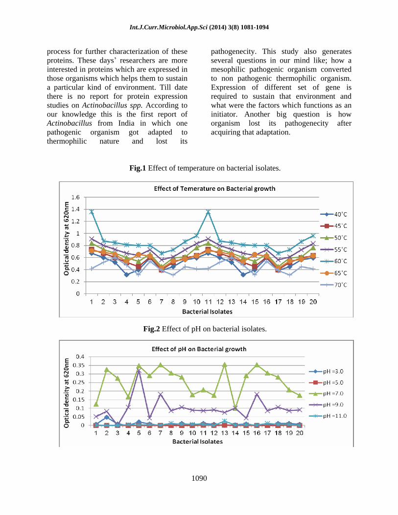
A total of 20 different morphological forms were isolated from the cattle compost samples collected from Jaipur, India. The ability of these isolates to grow at different temperatures, pH and salt concentrations was studied to find their optimal growth condition. All isolates shows maximum growth at 60°C and isolates Jnu1, Jnu6, Jnu11 and Jnu16 shows growth at 55°C but decrease in temperature effect the growth of culture. Isolate Jnu3 and Jnu13 also shows growth at 70°C but the optimum temperature for all isolate is 60°C (Fig 1).
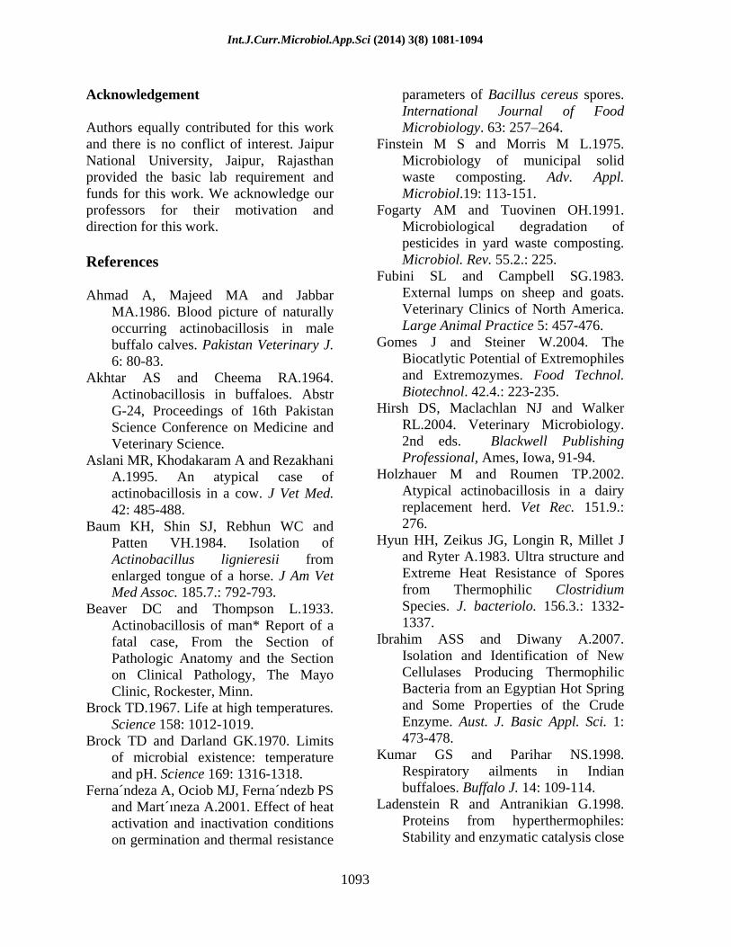
None of the isolates showed growth at 40°C but all strains had growth at 60°C revealing their thermophilic character and can be classified as moderate thermophiles (Gomes et al., 2004). These bacteria are highly active at 60°C and at temperatures above 60°C the degradation process is performed by these microorganisms (Fogarty et al., 1991; Finstein et al., 1975; Nakasaki et al., 1985a; Strom, 1985b; Sharp et al., 1992). Microbial diversity is a prerequisite for a satisfactory composting process. High temperature more than 60°C are considered to reduce dramatically the functional diversity. It is generally assumed that to obtain an efficient and rapid decomposition, temperature should not be allowed to exceed 55 to 60°C (Mckinley et al., 1984; Turner et al., 2007). Isolates growing at 60°C were further used to study the effect of pH and salt concentration. All the isolates showed maximum growth at pH 7 whereas isolates Jnu5, Jnu7 and Jnu16 showed optimal growth at pH 9 (Fig 2).

Int.J.Curr.Microbiol.App.Sci (2014) 3(8) 1081-1094
1086
Table.1 Biochemical analysis for tentative identification of isolated organism
Isolate JNU-1 JNU-2 JNU-3 JNU-4 JNU-5 Gram s staining Gram negative, long
rods, bunches. Gram negative, small rods, highly branched, some also present in chains
Gram negative, small rods, bunches & chain
Gram negative, very long rods, branched.
Gram negative, rods, branched, filamentous, spores present
Endospore staining - - - - - Motility - - - - - Catalase + + but delayed + + + Starch hydrolysis + + + + + Protease + + + + + Lipase + + + + + Citrate - - - - - Urease - - - - - Oxidase + + + - + Indole + + + + + Lysozyme + + + + + Methyl red + - - - - Voges Proskauer - + + + +
Without Zn + + + + + Nitrate reductase With Zn - - - - - Glucose + + + + + Lactose + + + + + Sucrose - + - + - Maltose + + + + +
Oxidative
fermentative test
Fructose + + + + + Triple Sugar Iron Agar - - - - - Tyrosine + + - + - Tentative identification Actinobacillus Actinobacillus Actinobacillus Actinobacillus Actinobacillus Similarity index 0.73596 0.87654 0.95866 0.75467 0.76754
Table.2 Biochemical analyses for tentative identification of isolated organism Isolate JNU-6 JNU-7 JNU-8 JNU-9 JNU-10 Gram s staining Gram negative, long
rods, bunches. Gram negative, small rods, highly branched, some also present in chains
Gram negative, small rods, bunches & chain
Gram negative, very long rods branched.
Gram negative, rods, branched, filamentous, segmented, spores present.
Endospore staining - - - - - Motility - - - - - Catalase + + but delayed + + + Starch hydrolysis + + + + + Protease + + + + + Lipase + + + + + Citrate - - - - - Urease - - - - - Oxidase + + + - + Indole + + + + + Lysozyme + + + + + Methyl red + - - - - Voges Proskauer - + + + +
Without Zn
+ + + + + Nitrate reductase
With Zn - - - - - Glucose + + + + + Lactose - + - + + Sucrose + + + + + Maltose - - - - -
Oxidative
fermentative test
Fructose + + + + + Triple Sugar Iron Agar - - - - Tyrosine - + + - - Tentative identification Actinobacillus Actinobacillus Actinobacillus Actinobacillus Actinobacillus Similarity index 0.97129 0.95467 0.78912 0.75321 0.90122

Int.J.Curr.Microbiol.App.Sci (2014) 3(8) 1081-1094
1087
Table.3 Biochemical analyses for tentative identification of isolated organism
Isolate JNU-11 JNU-12 JNU-13 JNU-14 JNU-15 Gram s staining Gram negative, long rods,
bunches. Gram negative, small rods, highly branched, some also present in
chains
Gram negative, small rods,
bunches & chain
Gram negative, very long rods, branched.
Gram negative, rods, branched, filamentous,
segmented, spores present.
Endospore staining - - - - - Motility - - - - - Catalase + + + + +
Starch hydrolysis + + + + + Protease + + + - - Lipase + + + - - Citrate - - - - - Urease - - - - -
Oxidase + - - + - Indole + + + + +
Lysozyme + + + + + Methyl red + + + - -
Voges Proskauer - - - + + Without Zn + + + + - Nitrate reductase
With Zn - - - - +
Glucose + + + + +
Lactose + + + + + Sucrose - + - + -
Maltose - - - - -
Oxidative
fermentative test
Fructose + + + + + Triple Sugar Iron Agar - - - - -
Tyrosine - - - + + Tentative identification Actinobacillus lignieressi Actinobacillus Actinobacillus Actinobacillus Actinobacillus lignieresii
Similarity index 0.73694 0.86143 0.67120 0.86432 0.75467
Table.4 Biochemical analyses for tentative identification of isolated organism
Isolate JNU-16 JNU-17 JNU-18 JNU-19 JNU-20 Gram s staining Gram negative, short
rods, single & bunches Gram positive, long rods, single & group
Gram negative, long rods, single
& group
Gram positive, small rods,
groups.
Gram negative, small rods, bunch
Endospore staining - - - - - Motility - - - - - Catalase + + + + +
Starch hydrolysis + + + + + Protease + + + + + Lipase - + + - + Citrate - - - - - Urease - - - - -
Oxidase - + + + - Indole + + + + +
Lysozyme + + + + + Methyl red - - - - -
Voges Proskauer + + + + + Without Zn + + - - + Nitrate reductase
With Zn - - + + - Glucose + + + + + Lactose + + + + + Sucrose + + + + + Maltose - - - - -
Oxidative fermentative test
Fructose + + + + + Triple Sugar Iron Agar - - - - -
Tyrosine + + - - - Tentative identification Actinobcillus lignieressi Not identified Not identified Not identified Actinobacillus lignieressi
Similarity index 0.73694 0.94647

Int.J.Curr.Microbiol.App.Sci (2014) 3(8) 1081-1094
1088
Table.5 Fermentative reaction of Actinobacillus lignieresii
Carbohydrate 24 hrs 48hrs Glucose + + Xylose + +
Mannitol + + Lactose - - Sucrose + + Maltose + +
Trehalose - - Salicin - -
Arabinose + +
In the composting process, initially pH goes down because of acid production and then increases by ammonia production. So the isolates are adapted to neutral pH. Four isolates (Jnu4, Jnu9, Jnu17 and Jnu20) showed optimum growth at 1% salt concentration. All isolates except Jnu3 and Jnu5 were able to grow at 2% NaCl concentration compared to higher salt concentration. Isolate Jnu14 was unable to tolerate even 1% salt concentration. Isolate Jnu8 and Jnu17 showed growth up to 7% salt concentration (Fig 3).
These isolates were identified on the basis of morphological, physiological & biochemical characteristics using software for probabilistic identification of bacteria (http://www.som.soton.ac.uk/staff/tnb/pib.html). Colonies were non haemolytic, small, raised, and greyish white. Majority of organisms stained as pleomorphic gram-negative rods. Out of 20 isolates 90% were gram negative and the rest were found to be gram positive. Spore formation was not observed for any of the isolates (Table: 1-4). Heat resistant spores were found among several species, mainly those belonging to the genera Bacillus and Clostridium (Ferna´ndeza et al., 2001; Hyun et al., 1983). Spore formation was not observed in the case of Actinobacillus. In previous research also less attention has been paid to thermophilic heat resistant spores than mesophilic spores which are used in food industry.
Spores from thermophilic bacteria are more heat resistant than spores from mesophilic species (Warth, 1978). Assay was performed to check the extracellular enzyme production by these isolates and most of the isolates found to be positive for protease, lipase and amylase activity. These 20 isolates were identified to belong to Actinobacillus genera and out of these 4 isolates identified to species level as Actinobacillus lignieressi by using the software (http://www.som.soton. ac.uk/staff/ tnb/pib.htm). Isolates which were regarded as a possible Actinobacillus after initial biochemical tests were further evaluated for fermentative activity. The isolates were inoculated into peptone water with Andrade's indicator and each of the following carbohydrates: mannose, arabinose, xylose, sucrose, maltose, lactose, trehalose, mannitol, and salicin. We used the differential identification scheme because Actinobacillus identification is very confusing. Thermophilic nature and ability to secret extracellular enzyme of these isolates indicates their possible role in various industries.
16S rRNA region amplification and sequencing
The DNA was isolated from 10 isolates of Actinobacillus showing optimum growth at 60°C which indicates their non pathogenic nature. The 16S rRNA region of 600bp

Int.J.Curr.Microbiol.App.Sci (2014) 3(8) 1081-1094
1089
amplified from these isolates by using 514R as a reverse primer and 25F as a forward primer (Fig 4).
Sequencing has been done and compared with the sequence already submitted to Genebank of National Central for Biotechnology. Nucleotide sequence data reported in this paper are available in the Genebank, EMBL and DDBJ databases under the accession number: JQ783352
Heat Shock proteins profiling
Thermophiles are eminent source for heat shock protein production. Study has been done to find the heat shock protein expressed by Actinobacillus. In this study both high molecular weight and low molecular weight heat shock proteins were expressed by Actinobacillus. The molecular weight of heat shock proteins ranged from ranged from 18.4 to 100 KDa.
Study was initiated to study the presence of thermophiles in compost samples but after experimental analysis we found that Actinobacillus which generally survive in moderate temperature can sustain higher temperature also. It has been found that Actinobacillus was the most dominant bacteria in cattle compost. A. lignieresii is responsible for actinobacillosis, an infectious, chronic, generally non-fatal disease (Rebhun et al., 1988). In cattle, it typically infects the tongue and hence is also known as the Wooden Tongue (Misra et al., 1981; Mohanty et al., 1970). The involvement of other organs is considered to be atypical (Aslani et al., 1995; Holzhauer et al., 2002; Rebhun et al., 1988; Mallick et al.,1984; Misra et al.,1981). The etiologic agent is found normally in the oropharynx and rumen of cattle and sheep (Smith, 1990; Songer et al., 2005). The organism is an opportunistic pathogen and it causes chronic
pyogranulomatous lesions of the soft tissues of head and neck regions in cows, buffalo, sheep, goats, and horses (Baum et al., 1984; Fubini et al., 1983; Hirsh et al., 2004; Kumar et al., 1998; Songer et al., 2005). Clinically, the lesions appear as nodules, multiple abscesses, ulcers or draining fistulae. A. lignieresii is causative agent for spontaneous actinobacillosis in the buffalo (Bubalus bubalis) which reported as non-fatal (Ahmad et al., 1986; Akhtar et al., 1964). In previous studies A. lignieresii is already described as causative agent of a granulomatous disease of cattle and it was also responsible for the prevalent form of the similar. Report shows that Actinobacillus can also cause infection in human which escort to death (Beaver et al., 1933) previous report on Actinobacillus submitted by our group also shows it presence in cattle compost. Isolation of Actinobacillus has been already done from laboratory rodents, (Lentsch et al., 1980; Simpson et al., 1980) but characterization of these organisms is still mystery; host range for the organism is still incomplete. Optimum growth condition for Actinobacillus is 37°C temperature and acidic pH but in this study we found that it shows adaptation for 60°C temperature also. It was found that increase in temperature can change the protein expression level of the organisms and may be these proteins are responsible for their survival on high temperature. Actinobacillus also shows degradation ability in these high temperature conditions. Amino acid composition of surface proteins in mesophylic and thermophilc bacteria is different, which responsible for the survival in harsh condition, which shows that different set of proteins, are expressed in these conditions. Protein expression and different metabolic activity of Actinobacillus in these conditions was studied and proteins were isolated from Actinobacillus and molecular mass was determined. Peptide mass fingerprinting is in
Int.J.Curr.Microbiol.App.Sci (2014) 3(8) 1081-1094
1090
process for further characterization of these proteins. These days researchers are more interested in proteins which are expressed in those organisms which helps them to sustain a particular kind of environment. Till date there is no report for protein expression studies on Actinobacillus spp. According to our knowledge this is the first report of Actinobacillus from India in which one pathogenic organism got adapted to thermophilic nature and lost its
pathogenecity. This study also generates several questions in our mind like; how a mesophilic pathogenic organism converted to non pathogenic thermophilic organism. Expression of different set of gene is required to sustain that environment and what were the factors which functions as an initiator. Another big question is how organism lost its pathogenecity after acquiring that adaptation.
Fig.1 Effect of temperature on bacterial isolates.
Fig.2 Effect of pH on bacterial isolates.

Int.J.Curr.Microbiol.App.Sci (2014) 3(8) 1081-1094
1091
Fig.3 Effect of salt concentration on bacterial isolates.
Fig.4 DNA isolated from Actinobacillus isolates
.
Fig.5 A. lignieresii DNA amplified by 514R and 25F primer.

Int.J.Curr.Microbiol.App.Sci (2014) 3(8) 1081-1094
1092
Fig.6 Effect of heat shock treatment on the pattern of protein synthesis in Actinobacillus. Bacterial cultures was placed in 40°C (control), 60°C(control) , 50°C, 60°C and 70°C (heat shock) water bath for 1 hour. The position of standard protein
markers and the size of the heat shock proteins are marked. [Lane 1 = Low range protein marker, Lane 2 = 40°C, Lane 3 = 60°C, Lane 4 = 50°C (heat shock), Lane 5 = 60°C (heat shock), Lane 6 = 70°C (heat shock)]
Fig.7 Effect of heat shock treatment on the pattern of protein synthesis in Actinobacillus. Bacterial cultures was placed in 40°C (control), 60°C(control) , 50°C, 60°C and 70°C (heat shock) water bath for 2 hour. The position of standard protein
markers and the size of the heat shock proteins are marked. [Lane 1 = Low range protein marker, Lane 2 = 40°C, Lane 3 = 60°C, Lane 4 = 50°C (heat shock), Lane 5 = 60°C (heat shock), Lane 6 = 70°C (heat shock)]
Int.J.Curr.Microbiol.App.Sci (2014) 3(8) 1081-1094
1093
Acknowledgement
Authors equally contributed for this work and there is no conflict of interest. Jaipur National University, Jaipur, Rajasthan provided the basic lab requirement and funds for this work. We acknowledge our professors for their motivation and direction for this work.
References
Ahmad A, Majeed MA and Jabbar MA.1986. Blood picture of naturally occurring actinobacillosis in male buffalo calves. Pakistan Veterinary J. 6: 80-83.
Akhtar AS and Cheema RA.1964. Actinobacillosis in buffaloes. Abstr G-24, Proceedings of 16th Pakistan Science Conference on Medicine and Veterinary Science.
Aslani MR, Khodakaram A and Rezakhani A.1995. An atypical case of actinobacillosis in a cow. J Vet Med. 42: 485-488.
Baum KH, Shin SJ, Rebhun WC and Patten VH.1984. Isolation of Actinobacillus lignieresii from enlarged tongue of a horse. J Am Vet Med Assoc. 185.7.: 792-793.
Beaver DC and Thompson L.1933. Actinobacillosis of man* Report of a fatal case, From the Section of Pathologic Anatomy and the Section on Clinical Pathology, The Mayo Clinic, Rockester, Minn.
Brock TD.1967. Life at high temperatures. Science 158: 1012-1019.
Brock TD and Darland GK.1970. Limits of microbial existence: temperature and pH. Science 169: 1316-1318.
Ferna´ndeza A, Ociob MJ, Ferna´ndezb PS and Mart´ neza A.2001. Effect of heat activation and inactivation conditions on germination and thermal resistance
parameters of Bacillus cereus spores. International Journal of Food Microbiology. 63: 257 264.
Finstein M S and Morris M L.1975. Microbiology of municipal solid waste composting. Adv. Appl. Microbiol.19: 113-151.
Fogarty AM and Tuovinen OH.1991. Microbiological degradation of pesticides in yard waste composting. Microbiol. Rev. 55.2.: 225.
Fubini SL and Campbell SG.1983. External lumps on sheep and goats. Veterinary Clinics of North America. Large Animal Practice 5: 457-476.
Gomes J and Steiner W.2004. The Biocatlytic Potential of Extremophiles and Extremozymes. Food Technol. Biotechnol. 42.4.: 223-235.
Hirsh DS, Maclachlan NJ and Walker RL.2004. Veterinary Microbiology. 2nd eds. Blackwell Publishing Professional, Ames, Iowa, 91-94.
Holzhauer M and Roumen TP.2002. Atypical actinobacillosis in a dairy replacement herd. Vet Rec. 151.9.: 276.
Hyun HH, Zeikus JG, Longin R, Millet J and Ryter A.1983. Ultra structure and Extreme Heat Resistance of Spores from Thermophilic Clostridium Species. J. bacteriolo. 156.3.: 1332-1337.
Ibrahim ASS and Diwany A.2007. Isolation and Identification of New Cellulases Producing Thermophilic Bacteria from an Egyptian Hot Spring and Some Properties of the Crude Enzyme. Aust. J. Basic Appl. Sci. 1: 473-478.
Kumar GS and Parihar NS.1998. Respiratory ailments in Indian buffaloes. Buffalo J. 14: 109-114.
Ladenstein R and Antranikian G.1998. Proteins from hyperthermophiles: Stability and enzymatic catalysis close

Int.J.Curr.Microbiol.App.Sci (2014) 3(8) 1081-1094
1094
to the boiling point of water. Advances in Biochemical Engineering and Biotechnology 61: 37-85.
Lentsch RH and Wagner JE.1980. Isolation of Actinobacillus lignieresii and Actinobacillus equuli from Laboratory Rodents. J. Clin. Microbio. 351-354.
Mallick KP, Somvansi R and Moga IV.1984. Actinobacillosis in the testicle of a buffalo. Indian J. Anim. health. 23.1.: 95-96.
Mckinley VL and Vestal JR.1984. Biokinetic analyses of adaptation and succession: microbial activity in composting municipal sewage sludge. Appl. Environ. Microbiol. 47: 933-941.
Misra SS and Angelo SJ.1981. Treatment of lingual actinobacillosis in a buffalo. Indian Vet J. 58: 413- 414.
Mohanty GC and Singh CM.1970. Actinobacillotic nephritis in a male buffalo: a case report. Indian Vet J. 47.11.: 938-941.
Nakasaki K, Shoda M and Kubota H.1985a. Effect of temperature on composting of sewage sludge. Appl. Environ. Microbiol. 50: 1526-1530.
Nakasaki K, Sasaki M, Shoda M and Kubota H.1985b. Change in microbial numbers during thermophilic composting sewage sludge with reference to CO2 evolution rate. Appl. Environ. Microbiol. 49: 37-41.
Rebhun WC, King JM and Hillman RB.1988. An atypical actinobacillosis granulomas in cattle. Cornell Vet. 78: 125-130.
Schulze KL.1962 Continuous thermophilic composting. Appl. Microbiol. 10.2.: 108-122.
Sharp RJ, Riley PW and White D.1992. Heterotrophic thermophilic Bacilli. In: Thermophilic Bacteria, In
Kristjansson, J.K. .eds., CRC Press Inc., Boca Raton U.S.A.19-50.
Simpson W and Simmons DJC.1980. Two Actinobacillus species isolated from laboratory rodents. Laboratory Animals 14: 15-16.
Smith BP.1990. Large Animal Internal Medicine. The C.V. Mosby Co. Philadelphia, 719-721.
Satyanarayana T, Raghukumar C and Shivaji S .2005. Extremophilic microbes: Diversity and perspectives. Curr. Sci. 89: 78-90.
Songer JG and Post KW.2005. Veterinary Microbiology - Bacterial and Fungal Agents of Animal Disease. ELSEVIER SAUNDERS, St. Louis, Missouri, 174-180.
Strom PF.1985a. Identification of thermophilic bacteria in solid waste composting. Applied Environ. Microbiol 50: 906-913.
Strom PF.1985b. Effect of temperature on bacterial species diversity in thermophilic solid-waste composting. Appl. Environ. Microbiol. 50: 899-905.
Suler DJ and Finstein MS.1977. Effect of temperature, aeration, and moisture on CO2 formation in bench-scale, continuously thermophilic composting of solid waste. Appl. Environ. Microbiol. 33: 345-350.
Turner P, Mamo G and Karlsson EN.2007. Potential and utilization of thermophiles and thermostable enzymes in biorefining. Microbial Cell Factories 6: 9.
Warth AD.1978. Relationship between the heat resistance of spores and the optimum and maximum growth temperature of Bacillus species. J. Bacteriol. 134.3.: 699.


![Vol. in U.S.A. Effect of Iron and Salt Prodigiosin Synthesismsjuo2011.weebly.com/uploads/2/5/4/7/2547226/s... · culture on TS slants (1.0% ion agar no. 2 [Colab], 3.0% Trypticase](https://static.fdocuments.in/doc/165x107/5f64101668f9546be53be0da/vol-in-usa-effect-of-iron-and-salt-prodigiosin-culture-on-ts-slants-10-ion.jpg)


