Preparation of cell nuclei from fresh tissues for high-quality DNA flow cytometry
12
0 1993 Wiley-Liss, Inc. Cytometry 14:793-804 (1993) Preparation of Cell Nuclei From Fresh Tissues for High-Quality DNA Flow Cytometry' Juan Castro, Thomas Heiden, Naining Wang, and Bernhard Tribukait2 Department of Medical Radiobiology, Karolinska Institute, S-10401 Stockholm, Sweden Received for publication September 23, 1992; accepted March 5, 1992 An easy method for preparation of bare cell nuclei from fresh solid tissues for DNA flow cytometry is described. Pieces of up to 2 x 2 x 2 mm3 size from fresh tissues were fixed in formalin. After removal of formalin by washing with eth- anol and rehydration with tap water, the tissue pieces were incubated with subtili- sin Carlsberg (pronase, Sigma protease XXIV) and then stained directly with DAPI. Staining with ethidium bromide gave unsatisfactory results. Neither me- chanical disaggregation nor centrifuga- tion were used. The resulting cell nucleus suspensions had extremely low frequen- cies of debris particles and of clumped cell nuclei. A good yield, a minimized loss, and a good stainability of cell nuclei were obtained. The applicability of the method was ex- emplified by the analysis of biopsies from the colon-rectum in patients with ulcer- ative colitis and of biopsies from the bladder in patients with bladder cancer and compared to the standard method of this laboratory, which uses mechanical disaggregation, ethanol fixation, pepsin treatment, and staining with ethidium bromide. The formalin-subtilisin Carls- berg technique resulted in good agree- ment of ploidy measurements compared to the standard method, a higher number of evaluable histograms, an improved de- tectability of aneuploid cell populations, and an improved accuracy of the S- and G2-phase analysis, particularly in sam- ples with low proliferation. The method also makes it possible to use long-term storage and to transport samples by post. 0 1993 Wiley-Liss, Inc. Key terms: DNA flow cytometry, solid tissues, formalin fixation, subtilisin Carlsberg DNA flow cytometry of solid tissues is a well-estab- lished technique for the characterization of tumor ploidy and proliferation. Several methods for the dis- sociation of fresh, solid tissues into single-cell or nuclear suspensions are in use and there are two main groups of techniques: enzymatic and detergent-based proce- dures, both of which also use mechanical treatment. A previously published improved Hedley-method for the preparation of paraffin-embedded tissues for flow cytometric DNA analysis has proved useful for the ex- amination of various types of tumor tissues (8). In this preparation, the dewaxed and rehydrated tissue, which had been fixed with formalin, is treated with subtilisin Carlsberg. Since all centrifugation steps are omitted, the loss of material is minimized and the suspensions show extremely low frequencies of clumped cell nuclei and clumped debris particles. In this way the analysis of ploidy and S-phase in archival material can be con- siderably improved. The good results obtained with this method were an encouragement for us to use its essential parts also for the preparation of fresh biopsies; it was an intent to achieve, in such preparations, both low portions of clumpings and low frequencies of debris particles. It had been observed that subtilisin Carlsberg treat- ment of formalin fixed tissue yields suspensions of sta- ble nuclei which remain intact even after prolonged incubation with the enzyme (8) and it has been noticed that the nuclei after this treatment were still highly resistant to mechanical forces such as those imposed by vigorous syringing. Moreover, experiments showed that, besides prolonged incubation time, increased en- 'This investigation has been supported by the Heinrich Warner Foundation. 'Address reprint requests to Professor Bernhard Tribukait, Depart- ment of Medical Radiobiology, Karolinska Institute, Box 60212, S-10401 Stockholm, Sweden.
-
Upload
juan-castro -
Category
Documents
-
view
213 -
download
1
Transcript of Preparation of cell nuclei from fresh tissues for high-quality DNA flow cytometry

0 1993 Wiley-Liss, Inc. Cytometry 14:793-804 (1993)
Preparation of Cell Nuclei From Fresh Tissues for High-Quality DNA Flow Cytometry'
Juan Castro, Thomas Heiden, Naining Wang, and Bernhard Tribukait2 Department of Medical Radiobiology, Karolinska Institute, S-10401 Stockholm, Sweden
Received for publication September 23, 1992; accepted March 5, 1992
An easy method for preparation of bare cell nuclei from fresh solid tissues for DNA flow cytometry is described. Pieces of up to 2 x 2 x 2 mm3 size from fresh tissues were fixed in formalin. After removal of formalin by washing with eth- anol and rehydration with tap water, the tissue pieces were incubated with subtili- sin Carlsberg (pronase, Sigma protease XXIV) and then stained directly with DAPI. Staining with ethidium bromide gave unsatisfactory results. Neither me- chanical disaggregation nor centrifuga- tion were used. The resulting cell nucleus suspensions had extremely low frequen- cies of debris particles and of clumped cell nuclei. A good yield, a minimized loss, and a good stainability of cell nuclei were obtained.
The applicability of the method was ex- emplified by the analysis of biopsies from the colon-rectum in patients with ulcer-
ative colitis and of biopsies from the bladder in patients with bladder cancer and compared to the standard method of this laboratory, which uses mechanical disaggregation, ethanol fixation, pepsin treatment, and staining with ethidium bromide. The formalin-subtilisin Carls- berg technique resulted in good agree- ment of ploidy measurements compared to the standard method, a higher number of evaluable histograms, an improved de- tectability of aneuploid cell populations, and an improved accuracy of the S- and G2-phase analysis, particularly in sam- ples with low proliferation. The method also makes it possible to use long-term storage and to transport samples by post. 0 1993 Wiley-Liss, Inc.
Key terms: DNA flow cytometry, solid tissues, formalin fixation, subtilisin Carlsberg
DNA flow cytometry of solid tissues is a well-estab- lished technique for the characterization of tumor ploidy and proliferation. Several methods for the dis- sociation of fresh, solid tissues into single-cell or nuclear suspensions are in use and there are two main groups of techniques: enzymatic and detergent-based proce- dures, both of which also use mechanical treatment.
A previously published improved Hedley-method for the preparation of paraffin-embedded tissues for flow cytometric DNA analysis has proved useful for the ex- amination of various types of tumor tissues (8). In this preparation, the dewaxed and rehydrated tissue, which had been fixed with formalin, is treated with subtilisin Carlsberg. Since all centrifugation steps are omitted, the loss of material is minimized and the suspensions show extremely low frequencies of clumped cell nuclei and clumped debris particles. In this way the analysis of ploidy and S-phase in archival material can be con- siderably improved.
The good results obtained with this method were an
encouragement for us to use its essential parts also for the preparation of fresh biopsies; it was an intent to achieve, in such preparations, both low portions of clumpings and low frequencies of debris particles.
It had been observed that subtilisin Carlsberg treat- ment of formalin fixed tissue yields suspensions of sta- ble nuclei which remain intact even after prolonged incubation with the enzyme (8) and it has been noticed that the nuclei after this treatment were still highly resistant to mechanical forces such as those imposed by vigorous syringing. Moreover, experiments showed that, besides prolonged incubation time, increased en-
'This investigation has been supported by the Heinrich Warner Foundation.
'Address reprint requests to Professor Bernhard Tribukait, Depart- ment of Medical Radiobiology, Karolinska Institute, Box 60212, S-10401 Stockholm, Sweden.
794 CASTRO ET AL.
zyme concentration and increased incubation temper- ature (see Material and Methods) did not result in in- jured cell nuclei. Because of this high stability of the nuclei, it was thought to be possible to omit any me- chanical tissue disaggregation and to treat, solely en- zymatically, relatively large pieces (up to 2 x 2 x 2 mm3) of formalin-fixed tissues over a prolonged time without injuring cell nuclei. Since these large tissue pieces have a low relative amount of sliced nuclei a t the surfaces, it was expected that the amount of debris par- ticles in the suspensions would be low.
The aim of this study was to establish this method of preparation using formalin fixation and subtilisin Carlsberg treatment of relatively large tissue pieces for the flow cytometric DNA analysis. Fresh mouse colon and bladder tissues and fresh biopsies from bladder and colon in patients with bladder carcinoma and ulcer- ative colitis, respectively, were used, as routinely stud- ied in our laboratory (4,141.
In order to estimate the value of the new method, a comparison was made with the standard method of this laboratory, which uses ethanol fixation and pepsin treatment and which has given good results for many years (13). A series of consecutive biopsies was ana- lyzed using both methods in paired samples. The rep- resentativity and the yield of nuclei were estimated for both methods and histogram features such as the amount of debris, percentage of clumped cell nuclei, and CV as well as the results of ploidy and cell cycle analysis were compared.
MATERIAL AND METHODS Tissues
Bladder tissue and mucosa of colon from mice were used in order to establish the different preparatory steps. Human lymphocytes isolated from peripheral blood using a Ficoll density gradient, as described in (7), were used as an external reference.
Sixty-three consecutive biopsies from colon-rectum in patients with ulcerative colitis taken during routine colonoscopies were analyzed. Since this series con- tained only seven aneuploid biopsies, in a continued, consecutive series a further 11 exclusively aneuploid samples were collected.
The material from bladder cancer patients comprised 148 consecutive biopsies taken at cystoscopy and trans- urethral resection. Fifty-three of these were tumor bi- opsies and 95 were cold-cup biopsies of the mucosa.
The biopsies, transported to the laboratory in 0.9% NaCl solution, varied considerably in size. Each biopsy was divided into two roughly equal pieces with scissors and then further treated using the formalin-subtilisin Carlsberg and the ethanol-pepsin preparation tech- niques.
Formalin-subtilisin Carlsberg Preparation This enzyme-based preparation method comprises
five steps. No mechanical dissociation, centrifugation, or filtration were used.
1. If the size of the biopsies exceeded 2 mm, they were cut with scissors into pieces with sizes of about 2 x 2 x 2 mm3. The NaCl solution was sucked off with a Pasteur pipette and replaced by 2 ml of 4% buffered formalin solution (4% formalin (W/W), 29 mM sodium- dihydrogenphosphate x 2 H,O, 47 mM disodium-hy- drogen-phosphate x 2 H,O, pH 7.0). The biopsies were fixed for about 16 h at room temperature.
2. The formalin solution was sucked off with a Pas- teur pipette and replaced by 3 ml95% ethanol in order to remove the formalin from the tissue. The biopsies were incubated for about 1 h at room temperature. It has been found that residual formalin after insufficient washing inactivates subtilisin Carlsberg. Long-term storage of biopsies in ethanol is possible after this prep- aration step.
3. The ethanol was removed using a Pasteur pipette and the tissues were incubated in 2 ml tap water for about 2 h a t room temperature. In this way, the biop- sies were rehydrated.
4. The tap water was removed using a Pasteur pi- pette and 200 pl subtilisin Carlsberg solution (0.1% Sigma protease XXIV, 0.1 M Tris, 0.07 M NaC1, pH 7.2) was added. The samples were incubated 16 h overnight a t room temperature and were then shaken for 2 h at 37°C in a water-bath. The solid tissue pieces, which usually became like thin and transparent rags, were removed before staining.
5. Staining was performed by directly adding 200 pl DAPI-SR101 solution (8 pM DAPI, 50 pM Sul- forhodamine 101, 0.1 M Tris, 0.07 M NaCl, pH 7.5). It was our impression that additional staining with SRlOl slightly improves the coefficient of variation of the DAPI analysis of bare nuclei as it does for the DAPI analysis of intact ethanol fixed cells (7). No centrifu- gation was carried out. The samples were stained for a t least 30 min and were not filtered before running in the flow cytometer. Extended staining times up to about 24 h did not change the staining intensity.
Ethanol fixed human blood lymphocytes were refixed in formalin and then treated in the same way; the only difference was that centrifugations (400 g, 10 min) were used before preparation steps 1, 2, 3, and 4.
Staining with ethidium bromide or propidium iodide did not give good results and is therefore not recom- mended. The stainability of DNA was reduced in com- parison to samples which had been prepared by the ethanol-pepsin method, and broad peaks were obtained in the DNA histograms. Further studies are necessary to reveal the reasons for this unexpected result.
The time of the protease treatment can be shortened by incubating the biopsies in a water bath for 4 h at 37°C without shaking followed by 10 min with shaking. It is recommended, especially when preparing other kinds of solid tissues, to examine the suspensions mi- croscopically. Only if the cytoplasm of the cells has been completely removed are the suspensions consid- ered ready for analysis. Unspecific fluorescence of re-

FORMALIN FIXATION FOR HIGH-QUALITY DNA FLOW CYTOMETRY 795
sidual cytoplasm results in a broadening at the right side of the basis of peaks.
Further variations of the incubation time with sub- tilisin Carlsberg as well as variations of the incubation temperature and the enzyme concentration were stud- ied. The following combinations were tested: 2 h in- stead of 4 h incubation time (37"C, 0.1% subtilisin Carlsberg), 0.01% subtilisin Carlsberg (6 h, 37°C) or 0.5% (4 h, 37°C) instead of 0.1% subtilisin Carlsberg (4 h, 37"C), and 50°C incubation temperature (2 h, 0.1% subtilisin Carlsberg) instead of incubation a t room temperature (16 h, + 2 h at 37"C, 0.1% subtilisin Carlsberg). The time of formalin-fixation was pro- longed up to 4 weeks. Despite all these variations, the quality of the suspensions, which is described in detail in Results, was essentially unchanged. This indicates that the method is remarkably uncritical concerning such variations and that accurate weighing, pipetting, and timing is obviously not essential.
All results shown in this article were obtained ap- plying 16 h incubation with subtilisin Carlsberg (step 4).
Ethanol-Pepsin Preparation This preparation technique consists of six steps. It
includes mechanical disaggregation, five centrifuga- tions, and filtration of the samples (13).
1. The biopsies were squeezed gently through a ny- lon gauze (40-50 mesh counts/cm, Zuricher Beutel- tuchfabrik, HD140) with isotone Tris EDTA buffer (0.1 Tris, 0.07 M NaC1, 0.005 M EDTA, pH 7.5).
2. After centrifugation (400g, 10 min), the cells were fixed in ice-cold 96% ethanol for a t least 1 day in the refrigerator.
3. The fixed cells were centrifuged (400g, 10 min) and washed in RNAse solution (1 mg Sigma R5125 RNAse A per ml Tris EDTA buffer).
4. Suspensions of cell nuclei were obtained by resus- pending the centrifuged cells in pepsin solution (0.4% Merck 7180 pepsin 1200 E/g in 0.05 n HC1) and by incubating them for 10 min at 37°C.
5. The suspensions of cell nuclei were centrifuged (400g, 10 min) and washed in Tris EDTA buffer.
6. The cell nuclei were centrifuged once more (400g, 10 min) and were resuspended in 1 ml ethidium bro- mide solution (25 pM ethidium bromide in Tris EDTA buffer). The samples were stained for at least 30 min and filtered through nylon gauze before running them in the flow cytometer.
Flow Cytometry The samples were analyzed using a PAS I1 flow cy-
tometer (Partec, Munster, BRD), which was equipped with a 100 W mercury arc lamp HB0100. DAPI was excited a t 365 nm and the fluorescence was measured above 435 nm. For analysis of ethidium bromide, the spectral regions of excitation and measurement of
spectively. The flow rate was usually 18 Fl/min. The measuring rate was usually about 700 nucleilsec for the formalin-subtilisin Carlsberg treated samples and about 100 nuclei/sec for the ethanol-pepsin samples.
Human lymphocytes were used as an external refer- ence in the ploidy analysis for identification of the dip- loid peak. The stainability of cell nuclei from lympho- cytes by DAPI had been found to be about 10% lower compared to diploid bladder cell nuclei when ethanol fixation and pepsin treatment were used (6). Such de- creased stainability of cell nuclei from lymphocytes by DAPI was not seen in comparison to cell nuclei from colon and bladder tissue when the formalin-subtilisin Carlsberg method was applied.
The DNA-index of aneuploid cell populations was calculated by relating the channel number of the ane- uploid G1-peak to the channel number of the G1-peak of the internal and tissue specific diploid cell nuclei. Tetraploid cell populations were assumed when the proportion of cells in the 4c peak exceeded 10% and, in addition, an octaploid G2 peak was present.
The linearity of the flow cytometer was checked with the help of cell nuclei from mouse bladder which com- prises 2c, 4c, and 8c nuclei. A correct G2/Gl-ratio was obtained independent of the type of tissue, the ploidy, and the method used for preparation and staining. Thirty randomly chosen histograms showed a G2/G1- ratio of 2.014 t 0.030 (mean value * SD).
The multicycle program for cell cycle analysis (Phoe- nix Flow Systems, San Diego, CA) was used for histo- gram analysis. The program options for subtraction of exponential background and for clumping correction were used for all histograms when calculating the per- centages of nuclei in S- and G2-phase. The calculations of CV, percentage of aneuploid cell nuclei, and number of cell nuclei per histogram were also made using this program. The percentage of debris particles was deter- mined by calculating the percentage of analyzed parti- cles in the histogram region between channel number 5 and the left side of the diploid G1 peak, which had its maximum in channel number 70.
RESULTS Characteristics of the Formalin-Subtilisin
Carlsberg Preparation Microscopic examination of the suspensions.
Figure 1 shows the effect of 0.1% subtilisin Carlsberg solution on fresh, unfixed mouse bladder tissue and on bladder tissue which had been fixed in ethanol and formalin. The cytoplasm and nuclei of unfixed cells were immediately destroyed, yielding largely swollen nuclei which fell to pieces (Fig. 1, top). Ethanol fixation did not prevent the process of total disintegration of both nuclei and cytoplasm (Fig. 1 center), but it slowed this process down. Fixation in formalin resulted in a suspension of stable, unclumped nuclei with good pres- ervation of nuclear details and completely removed cy- toplasm (Fig. 1, bottom). Moreover, the yield of cell
emission light were 450-490 nm and 590-630 nm, re- nuclei was high and the solid tissue piece usually be-

796 CASTRO ET AL.
came like a thin and transparent rag after enzyme treatment.
Characteristics of the histograms. Figures 2 summarizes the main characteristics of the formalin- subtilisin Carlsberg method as they become visible in the DNA histograms. All histograms show low debris and clumping frequencies and reasonably good CVs. Low level of debris was expected since relatively large tissue pieces were used (2 x 2 x 2 mm3). Apart from the smaller number of preparation steps, this is the main difference in comparison with the preparation of archival material, which uses sections 50 to 100 pm thick containing a high proportion of cut nuclei a t the surfaces.
In Figure 2 (left) the DNA analysis of normal, diploid bladder mucosa from a patient is shown. The cell cycle analyses yielded a S-phase value of 0.6%. Since the level of the exponentially declining debris background is considerably lower than the level of the S-phase nu- clei, the calculated S-phase value is considered correct, although it is low. The G2 value is also considered to be precise since the clumping frequency is low. It has been observed microscopically in formalin-subtilisin Carls- berg preparations that the metaphase chromomosomes stick together, although the cytoplasm has been re- moved. Thus, mitotic cells were obviously not lost in the analysis.
Figure 2 (middle and right) exemplifies the possibil- ity to calculate S-phase values with good precision in heterogeneous cell populations even when the prolifer- ation rate is low. Since the debris originating from the 6 . 6 ~ population is very low (Fig. 2, right), the S phase analysis of the diploid population with low prolifera- tion is not impaired. In both examples, the levels of background debris are considerably lower than the S-phase levels and clumping frequencies are extremely low. Thus, as can be seen in Figure 2 (middle), 6c clumpings of diploid G1 nuclei (<0.2%) do not interfere with the S-phase calculation of the 3 . 6 ~ population, which is only a minor subpopulation.
Long-term storage of samples. The DAPI stain- ing intensity and the CV in cell material from mouse colon and bladder were studied as functions of the time of formalin fixation. Tissue samples were fixed in for- malin a t room temperature for 1 day, 1, 2, 3, and 4 weeks, respectively. The samples of the first four time
FIG. 1. Effect of 0.1% subtilisin Carlsberg solution on unfixed (top), ethanol fixed (center), and formalin fixed (bottom) murine bladder tissue. Solid tissue portions can be seen in the left part of the photos. Staining has been carried out with DAPI-SR101 and both the fluo- rescence of DNA-bound DAPI and of protein-bound SR101, excited with ultraviolet light, were photographed. The unfixed cells were stained and photographed directly. The 95% ethanol fixed tissue was incubated for 30 min a t 37°C and then stained. The formalin fixed sample was prepared according to the description in Material and Methods. All the photo-micrographs were taken at the same magni- fication.
60 pm

FORMALIN FIXATION FOR HIGH-QUALITY DNA FLOW CYTOMETRY 797
2c 2c 3.6c 2C 6.6~
FIG. 2. DNA histograms of cell nuclei from a diploid mucosal biopsy (left) and from two aneuploid bladder tumors (middle and right). The nuclei were prepared with the formalin-subtilisin Carlsberg method. The histograms a t the bottom show the upper histograms with an amplification of the y-scale (left: x 130, middle: x 85, right: x 80). X-axis: ploidy, y-axis: linear number of nuclei.
periods were stored in ethanol and all samples were then prepared and analyzed in one series with un- changed gain setting. The peak position of diploid cell nuclei and the CV did not change as a function of fix- ation time. Even after 4 weeks formalin fixation there was no statistically significant difference of the stain- ing intensity (t test significance P = 0.385) and the CV (t test significance P = 0.363) compared to a fixation time of 1 day.
Thus, specimen transport by post is possible simply by leaving the biopsies in formalin solution. However, if longer preservation times are needed, a transfer of the biopsy from formalin to 95% ethanol might be prefered.
Plainly, for this enzyme-based method, the stainabil- ity is independent of the time of formalin fixation, and in this respect it differs from the non-enzymatic method described by Coon et al. (1) who found a loss of DNA stainability when fixing cells for more than 30 min in formalin. The constancy of the staining inten- sity is a prerequisite for standardisation using forma- lin-fixed human lymphocytes or trout and chicken red blood cells.
Comparison Between the Formalin-Subtilisin Carlsberg and the Ethanol-Pepsin Method
In order to verify its usefulness, the formalin-subtili- sin Carlsberg method was compared with the ethanol- pepsin technique, used in this laboratory in many years. Both methods were applied to consecutive series of human colon-rectum and bladder biopsies from pa- tients with ulcerative colitis and bladder carcinoma.
Figure 3 shows a typical DNA analysis of a bladder mucosa exemplifying the main differences between the
results obtained by the two methods. The formalin-sub- tilisin Carlsberg sample has a higher number of nuclei per histogram, lower CV values, less debris and clump- ing, and S- and G2-phase values which differ from those of the ethanol-pepsin sample. In the following, the two methods are compared in detail.
Comparison of the ploidy analyses. Some biop- sies were excluded from ploidy analysis because of poor histogram quality, defined as <500 nuclei per histo- gram or a CV of >8%. Seventy-four biopsies from pa- tients with ulcerative colitis and 148 biopsies from pa- tients with bladder carcinoma were available. Ploidy could be evaluated in all formalin-subtilisin Carlsberg samples and all ulcerative colitis ethanol-pepsin sam- ples, while six samples from bladder biopsies after eth- anol-pepsin treatment did not fulfill the criteria cho- sen.
Table 1 focuses on biopsies in which differences in the result of ploidy analysis were observed. Forty-four percent of all aneuploid biopsies showed such differ- ences.
The differences in the number of aneuploid cell pop- ulations found in the two parallel samples from the same biopsy are not necessarily due to differences of the preparation techniques but could be a consequence of tumor heterogeneity. However, observing the spe- cific types of differences and their frequencies of occur- rence (Table l), a clear tendency for a higher number of aneuploid cell populations in the formalin-subtilisin Carlsberg samples is obvious.
It is unlikely that differences in chromatin structure led to artefactual, additional DNA peaks in the forma- lin-subtilisin Carlsberg preparation method as de-

Ethanol-P. CASTRO ET AL.
Formalin-S.C. h
h 9834
.- I: ..-.. .._ ?.+. ..
2c
5
Number of Nuclei: 40265 4 .. cv: 1.9
$ %Debris: 0.2
2c FIG. 3. Comparison of typical DNA histograms of cell nuclei from a mucosal bladder biopsy prepared
with the ethanol-pepsin method (left) and the formalin-subtilisin Carlsberg method (right). X-axis: ploidy, y-axis: linear number of nuclei. The histograms at the bottom show the corresponding upper histograms with a logarithmic y-scale.
scribed for some preparation methods (2,6,9). Such ar- tefacts typically result in near-diploid, hyper-diploid extra peaks. Out of our 222 samples only 5 showed cell populations in the DI range between 1.1 and 1.5, three of which were found with both methods. In addition, analyses of stimulated human lymphocytes from pe- ripheral blood, comprising a mixture of GO-phase nu- clei with highly condensed chromatin and considerably enlarged G1-, S-, and G2-phase nuclei with decon- densed chromatin, showed only one narrow peak of dip- loid nuclei (data not shown). Moreover, human lympho- cytes from peripheral blood with highly condensed chromatin, used as external reference, had the same peak position as 2c nuclei from the bladder and colon biopsies.
In summary, we feel that the formalin-subtilisin Carlsberg method enables a higher detectability of true aneuploid cell populations compared to the ethanol- pepsin method.
This is further supported by the results shown in Figure 4 (top), in which the percentages of cell nuclei in aneuploid cell populations of same ploidy are com-
pared. These percentages are generally higher in the formalin-subtilisin Carlsberg samples. The reason for this might be a higher sensitivity of aneuploid cells towards the mechanical and enzymatical treatment used in the ethanol-pepsin method.
The DNA indices of the aneuploid cell populations found using the two methods were, however, in good agreement, although the DNA values in ulcerative colitis seem to be slightly lower in the formalin treated samples (Fig. 4, bottom).
In Figure 5, the extent of clumping and debris for- mation as well as the CV values obtained by the two methods are compared. Obviously, the formalin-sub- tilisin Carlsberg technique resulted for both tissue types in considerably less debris and clumping. The CV was significantly lower for the ulcerative colitis biop- sies and somewhat lower for the bladder cancer biop- sies.
Although the yield of cell nuclei was not directly measured, the number of analyzed nuclei per histo- gram gives some information about this. As a rule, a t least 30,000 cell nuclei were analyzed per histogram.

FORMALIN FIXATION FOR HIGH-QUALITY DNA FLOW CYTOMETRY 799
Table 1 Biopsies From Patients With Ulcerative Colitis and Bladder Cancer in Which the Ethanol-Pepsin and the
Formalin-Subtilisin Carlsberg Methods Gave Different Results in Ploidy Analysis (The total numbers of aneuploid biopsies are piven in parentheses)
Numbers of Formalin-subtilisin biopsies Ethanol-pepsin Carlsberg
Biopsies from 3 Diploid Tetraploid ulcerative colitis 3 Diploid Aneuploid n = 74 1 1 aneuploid population 2 aneuploid populations Total: 7 (18)
Biopsies from 3 No ploidy availablea Aneuploid bladder cancer 3 Diploid Aneuploid n = 148 3 Diploid Tetraploid
3 Tetraploid Tetraploid + aneuploid 1 Aneuploid >1 aneuploid population 1 Aneuploid Tetraploid + aneuploid 2 Aneuploid Aneuploid + polyploid 1 Tetraploid + aneuploid Tetraploid 1 >1 aneuploid population 1 aneuploid population
Total: 18 (39)
T h e histograms had less than 500 cell nuclei per histogram or CVs of more than 8.
In samples with low S-phase portions or a small aneu- ploid population up to 100,000 cell nuclei were ana- lyzed. However, in samples from small biopsies the whole sample often contained less than 30,000 cell nu- clei. Since the biopsies were divided into two pieces of about equal size, the mean values of the numbers of nuclei per histogram roughly reflect the yield of cell nuclei for both methods. As shown in Table 2, the for- malin-subtilisin Carlsberg preparation technique re- sulted in about double the number of cell nuclei per histogram compared to the ethanol-pepsin method in- dicating that the lower level of 30,000 nuclei per his- togram was more often reached.
Comparison of the results of cell cycle analysis. Cell cycle analyses of the specimens were performed when certain minimum requirements were fulfilled. These were more than 5,000 cell nuclei in the diploid or aneuploid population of interest, less than 25% debris, and a clumping portion of less than 33% within the aneuploid population. In Table 3 the frequencies of bi- opsies which were not evaluable according to these cri- teria are shown. The numbers of evaluable histograms are considerably higher for the samples which were prepared with the formalin-subtilisin Carlsberg method.
Two out of twenty formalin-subtilisin Carlsberg samples which did not fulfill the requirements had more than 25% debris and eighteen were excluded solely because the number of cell nuclei per histogram was less than 5,000. Clearly, some of these samples would have had more than 5,000 cell nuclei if the bi- opsies had not been divided. In particular, biopsies from colon-rectum taken at colonoscopy and from blad- der mucosa taken at cystoscopy are generally small and were often very small after division for the com- parison of the two methods. Among the 82 ethanol- pepsin samples, which were excluded from cell cycle
analysis, 73 had histograms with less than 5,000 cell nuclei in the population of interest andior more than 25% debris and 9 had too much clumping within the aneuploid populations.
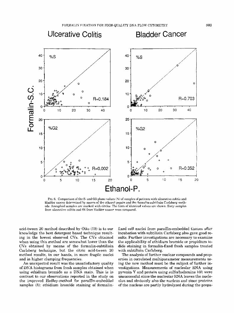
In Figure 6, the paired S- and G2-phase values for the samples, in which both methods gave evaluable histograms, can be seen.
The paired samples from colon-rectum biopsies from patients with ulcerative colitis did not show any corre- lation of the G2 values (r = 0.0021, although clumping correction, which subtracts calculated numbers of clumpings in G2 peaks, was carried out for all the his- tograms. As can be seen in Figure 5, the mean % values of clumped cell nuclei in the samples designated with ulcerative colitis were 0.8 and 14.0 when the prepara- tion was carried out with the formalin-subtilisin Carls- berg and ethanol-pepsin method, respectively. High clumping frequencies obviously do not allow correct calculations of G2 values, at least when using the clumping correction option of the multicycle program.
For the samples from bladder biopsies the correlation was significant (r = 0.352; P < 0.001). Obviously, the reason for the better agreement of the G2 values in biopsies from the bladder is the lower clumping fre- quency in this cell material treated by the ethanol- pepsin method. The clumping frequency was 5.6% com- pared to 14% in the cell material from colon-rectum (see Fig. 5). Using the formalin-subtilisin Carlsberg technique the mean values of clumped cell nuclei were below 1% for both tissue types and the G2 values ob- tained by means of this method are supposed to ap- proach closely to the true values.
In the samples from colon-rectum biopsies from patients with ulcerative colitis, S-phase values ob- tained by means of the two methods showed no corre- lation (r = 0.184) (Fig. 6) , although debris- and clump- ing-corrections were made in all histograms. Mainly

800 CASTRO ET AL.
Ulcerative Colitis Bladder Cancer
%Aneuploid Nuclei c .- 0 c d o 20 40, 60 80 100 - E 5
L L
YOAneuploid Nuclei
0 60 80 100
1 2 3 4 1 2 3 4
Ethanol-P. FIG. 4. Comparison of the percentages of aneuploid cell nuclei and
the DNA indices of aneuploid samples of patients with ulcerative colitis and bladder cancer determined by means of the ethanol-pepsin and the formalin-subtilisin Carlsberg methods. The arrows mark mean values. Diploid cell nuclei of the tumors, identified with the help of the external lymphocyte standard, were used as internal ref- erence for the DNA index calculations. The lines of identical values
the aneuploid samples (round symbols in Fig. 6) showed overestimated S-phase values in ethanol-pep- sin preparations, probably because the S-phases con- tained high frequencies of clumpings which were not correctly calculated by the multicycle program.
In samples from bladder biopsies, as a whole, the paired S-phase values correlated well (r = 0.703; P < 0.001) (Fig. 6). When looking a t various subgroups, aneuploid tumors, diploid tumors, and diploid mucosa, significant correlations can be seen for the aneuploid and diploid tumor groups (r = 0.598; P < 0.05 and r = 0.639; P < 0.001, respectively), but there is no correla-
are shown. Mean values (%) 2 SD of aneuploid cell nuclei of samples from 12 patients with ulcerative colitis were 43.9 2 15.8 for the eth- anol-pepsin and 81.8 * 7.66 for the formalin-subtilisin Carlsberg method. Mean values (%) f SD of aneuploid nuclei of samples from 30 patients with bladder cancer were 41.1 * 28.4 for the ethanol-pepsin and 54.1 ? 31.7 for the formalin-subtilisin Carlsberg method.
tion for the group of diploid mucosa samples. The ob- vious reason for this is that the true S-phase values in the diploid mucosa are too low to be correctly measured by means of the ethanol-pepsin technique due to the influences of clumping and debris.
DISCUSSION An ideal disintergation method for solid tissues for
flow cytometric DNA analysis has the following char- acteristics: release of all nuclei of the tissue to yield suspensions of single, unclumped, and undamaged nu- clei with good quantitative stainability of DNA. The

FORMALIN FIXATION FOR HIGH-QUALITY DNA FLOW CYTOMETRY 801
Ulcerative Colitis Bladder Cancer
0 5 10 , 1 5 20
0 10 15
40 60
/
0
20 1
- 0
10 15 20 "t
0
15
10
5 - 0
CV
+
?++ +
10 15
t 5 0
Ethanol-P. FIG. 5. Comparison of debris (%), clumped nuclei (%I, and CV Val-
ues of samples of patients with ulcerative colitis and bladder cancer determined by means of the ethanol-pepsin and the formalin-subtili- sin Carlsberg methods. The arrows mark mean values. The lines of identical values are shown. For ulcerative colitis mean values t SD were 9.10 f 4.69 debris (%) (n = 51), 13.97 i- 2.48 clumped nuclei (%) (n = 691, and 5.84 ? 1.06 CV (n = 74) for the ethanol-pepsin tech- nique and 2.59 % 2.41 debris (%) (n = 51),0.79 f 1.10 clumped nuclei
(%) (n = 69), and 2.55 f 0.51 CV (n = 74) for the formalin-subtilisin Carlsberg method. For bladder cancer mean values t SD were 20.44 t 17.21 debris (%) (n = 148), 5.64 t 4.05 clumped nuclei (%) (n =
1111, and 4.28 5 1.63 CV (n = 138) for the ethanol-pepsin method and 2.49 -t 4.20 debris (%) (n = 148), 0.66 f 1.10 clumped nuclei (S) (n = 1111, and 2.98 t 0.98 CV (n = 138) for the formalin-subtilisin Carls- berg method.

802 CASTRO ET AL.
Table 2 Numbers of Cell Nuclei in the D N A Histograms of
Colon-Rectum and Bladder Biopsies From Patients With Ulcerative Colitis and Bladder Cancer Analyzed by Means of the Ethanol-Pepsin and the Formalin-Subtilisin Carlsberg
Methods (Mean values t SD.)
Formalin-subtilisin Ethanol-pepsin Carlsberg
Ulcerative colitis, 17,678 t 12,502 56,845 2 26,500 number of cell nuclei (n = 74) (n = 74) per histogram
Bladder cancer, 13,312 t 12,057 28,259 2 20,698 number of cell nuclie (n = 148) (n = 148) Der histoeram
Table 3 Number of Biopsies From Patients With Ulcerative Colitis
and Bladder Cancer in Which Cell Cycle Analysis Was Not Donea
Formalin-subtilisin Ethanol-DeDsin Carlsbere
Ulcerative colitis 13174 (18%) 1174 (1%) Bladder cancer 691148 (47%) 191148 (6%) Total 821222 (37%) 201222 (9%)
"The ethanol-pepsin and the formalin-subtilisin Carlsberg methods are compared. The requirements of histograms cho- sen for cell cycle analysis are described in the text.
obtained results show that the formalin-subtilisin Carlsberg method gives quite a close approach to this ideal. The new method was compared to a standard ethanol-pepsin method and its characteristics and ad- vantages can be summarized as follows:
a) low clumping and debris frequencies; b) reasonably good CV; c) good yield and minimized loss of cell nuclei, result-
ing in high cell nucleus concentrations, short analysis time and high numbers of cell nuclei per histogram; this is particularly advantageous for the analysis of small biopsies;
d) high reliability and a high proportion of evaluable samples;
e) good precision of ploidy measurement as found in the comparison with the ethanol-pepsin method and improved detectibility of aneuploid cell populations;
f ) improved analysis of percentages of G2 cells and better identification of polyploid cell populations;
g) improved S-phase analysis particularly in samples with low numbers of S-phase cells;
h) simplicity, reduced working time, unsusceptibility to pipetting, weighing, and timing errors, low material costs per sample (about 15% compared to the ethanol- pepsin method);
i) possibility to use long-term storage and transport by post.
Potential disadvantages are summarized as follows: long preparation time compared with detergent-based methods due to fixation and protease treatment; inabil- ity to use propidium iodide, which results in difficulties to adapt the technique in argon laser based instru- ments; and loss of at least a part of nuclear RNA and nuclear protein antigens.
A key point in the method is the selective stabiliza- tion of the nuclei by formaldehyde resisting the action of the protease subtilisin Carlsberg (see Fig. 1). Two factors, one is related to fixation the other to enzyme specificity, may explain this selective stabilization.
The cell nucleus contains large amounts of histone proteins with a high portion of the basic amino acids arginine and lysine. Apart from the thiol group of cys- teine, the €-amino group of lysine is plainly one of the two most reactive sites in cells for the cross-link-form- ing formaldehyde (3). Since the lysine content in the nucleus is high, a high number of cross-links, mainly comprising histone-histone and histone-DNA cross- links, is assumed to be formed in the nucleus and the number of cross-links is probably considerably higher in the nucleus than in the cytoplasm.
Details of the primary reaction of the €-amino group of lysine with formaldehyde, which forms the reactive aminomethylol compound, and the various secondary, cross-linking reactions between the aminomethylol group and functional groups in proteins and DNA, which occur under the conditions of fixation of the cells, are reviewed in reference 11.
One can imagine that a protease hydrolizing peptide bonds around the cross-links needs more time to dis- solve tightly cross-linked protein complexes than for those which are less tightly cross-linked.
In addition, because of different substrate specifici- ties, different proteases may have different capabilities to dissolve cross-linked histone-histone and histone- DNA complexes compared to cross-linked non-histone protein complexes. However, subtilisin Carlsberg obvi- ously removes the cytoplasma of formalin-fixed cells efficiently while leaving the nucleus intact.
In the preparation of cell nuclei for flow cytometry, other methods for stabilization of the nuclei are used. Thornthwaite uses bovine serumalbumin and divalent cations to stabilize the nuclear double membrane (12), while Vindelov et al. use sperminetetrahydrochloride to make the chromatin more stable (15). This spermi- netetrahydrochloride treatment prevents the cell nu- clei from complete disintegration, but the nuclei are obviously still fragile since it is recommended that the suspensions should be handled gently as agitation in- creases debris and clumping (16).
The most widely used preparation technique for flow cytometric DNA analysis is probably the enzymatic method of Vindelov et al. (15). Beside the fragility of the nuclei (16), the need of accurate pipetting and weighing, especially of sperminetetrahydrochloride, might be a disadvantage in comparison to the formalin- subtilisin Carlsberg method described here. The citric
40
30
20
10
FORMALIN FIXATION FOR HIGH-QUALITY DNA FLOW CYTOMETRY
Ulcerative Colitis
0 R=O. 1 84 0
0 10 20 30 40 .- t o - a
40
30
20
10
0
Bladder Cancer
I / 0
0 10 20 30 40
803
0 5 10 15 20 0 5 10 15 20
Ethanol-P. FIG. 6 . Comparison of the S- and G2-phase values (%I of samples of patients with ulcerative colitis and
bladder cancer determined by means of the ethanol-pepsin and the formalin-subtilisin Carlsberg meth- ods. Aneuploid samples are marked with circles. The lines of identical values are shown. Sixty samples from ulcerative colitis and 64 from bladder cancer were compared.
acid-tween 20 method described by Otto (10) is to our knowledge the best detergent based technique result- ing in the lowest observed CVs. The CVs obtained when using this method are somewhat lower than the CVs obtained by means of the formalin-subtilisin Carlsberg technique, but the citric acid-tween 20 method results, in our hands, in more fragile nuclei and in higher clumping frequencies.
An unexpected result was the unsatisfactory quality of DNA histograms from fresh samples obtained when using ethidium bromide as a DNA stain. This is in contrast to our observations reported in the study on the improved Hedley-method for paraffin-embedded samples (8): ethidium bromide staining of formalin-
fixed cell nuclei from paraffin-embedded tissues after incubation with subtilisin Carlsberg also gave good re- sults. Further investigations are necessary to examine the applicability of ethidium bromide or propidium io- dide staining in formalin-fixed fresh samples treated with subtilisin Carlsberg.
The analysis of further nuclear compounds and prop- erties in correlated multiparameter measurements us- ing the new method must be the subject of further in- vestigations. Measurements of nucleolar RNA using pyronin Y and protein using sulforhodamine 101 were unsuccessful since the nucleolar RNA leaves the nucle- olus and obviously also the nucleus and since proteins of the nucleus are partly hydrolyzed during the prepa-
804 CASTRO ET AL.
ration (unpublished observations). Scatter- or Coulter- size analyses of the nuclei should of course be possible and are supposed to be useful, since the cytoplasm of the cells is efficiently removed while the nuclei are well preserved.
Formalin fixation in connection with preparation of cell nuclei for flow cytometric DNA analysis of fresh tissues has, to the best of our knowledge, generally not been used. According to Coon et al. (l), a fixation time of more than 30 min resulted in a loss of DNA stain- ability and was therefore not recommended by the au- thors. In archival formalin fixed material, marked dif- ferences in the stainability of various tissue blocks, obviously due to technical variations in fixation, have earlier been observed by Hedley et al. (5). When using the formalin-subtilisin Carlsberg method for fresh tis- sues, described here, fixation times of at least up to 4 weeks a t room temperature did not change the stain- ability of DNA.
Experiences with the formalin-subtilisin Carlsberg method in other tissues such as human carcinoma of the ovary and uterus and various tissues from mouse also showed the characteristic good results.
4.
5.
6.
7.
8.
9.
10.
11.
12.
13.
LITERATURE CITED Coon JS, Deitch AD, de Vere White RW, Koss LG, Melamed MR, Reeder JE, Weinstein RS, Wersto RP, Wheeless LL: Check sam- ples for laboratory self-assessment in DNA flow cytometry. The National Cancer Institute’s flow cytometry network experience. Cancer 63:1592-1599, 1989. 15. Darzynkiewicz A, Traganos F: Unstainable DNA in cell nuclei. Comparison of ten different fluorochromes. Analyt Quant Cytol Histol 10:462-466, 1988. 16. Fox CH, Johnson FB, Whiting J, Roller P P Formaldehyd fixa- tion. J Histochem Cytochem 33345-853, 1985.
14.
Hammarberg C, Slezak P, Tribukait B: Early detection of malig- nancy in ulcerative colitis. Cancer 53:291-295, 1984. Hedley DW, Friedlander ML, Taylor IW, Rugg CA, Musgrove EA: Method for analysis of cellular DNA content of paraffin-embed- ded pathological material using flow cytometry. J Histochem Cy- tochem 31:1333-1335, 1983. Heiden T, Strang P, Stendahl U, Tribukait B The reproducibility of flow cytometric analyses in human tumors. Methodological aspects. Anticancer Res 10:49-54, 1990. Heiden T, Gohde W, Tribukait B: Two-wavelength mercury arc lamp excitation for flow cytometric DNA-protein analyses. Anti- cancer Res 10:1555-1562, 1990. Heiden T, Wang N, Tribukait B: An improved Hedley-method for preparation of paraffin-embedded tissues for flow cytometric analysis of ploidy and S-phase. Cytometry 12:614-621, 1991. Iversen OE, Laerum OD: Trout and salmon erythrocytes and hu- man leukocytes as internal standards for ploidy control in flow cytometry. Cytometry 8:190-196, 1987. Otto F: DAPI staining of fixed cells for high-resolution flow cy- tometry of nuclear DNA. Methods Cell Biol 33:105-110, 1990. Puchtler H, Meloan SN: On the chemistry of formaldehyde fixa- tion and its effects on immunohistochemical reactions. Histochem 82:201-204, 1985. Thornthwaite JT, Sugarbaker EV, Temple W: Preparation of tis- sues for flow cytometric analysis. Cytometry 1:229-237, 1980. Tribukait B, Moberger G, Zetterberg A: Methodological aspects of rapid-flow cytofluorometry for DNA analysis of human urinary bladder cells. In: First International Symposium on Pulse-Cyto- photometry, Haanen CAM, Hillen HFP, Wessels JMC (eds). Eu- ropean Press Medikon, Ghent, Belgium, 1975, pp 50-60. Tribukait B, Gustafson H, Eposti P L Ploidy and proliferation in human bladder tumors as measured by flow-cytofluorometric DNA-analysis and its relation to histopathology and cytology. Cancer 43:1742, 1979. Vindelov LL, Christensen IJ, Nissen NI: A detergent-trypsin method for the preparation of nuclei for flow cytometric DNA analysis. Cytometry 3:323-327, 1983. Vindelov LL, Christensen IJ: An integrated set of methods for routine flow DNA analysis. Methods Cell Biol 33:127-131, 1990.