PREPARATION AND MODIFICATION OF MEDIUM- CHAIN … · absorption spectra for –OH and PO which...
150
PREPARATION AND MODIFICATION OF MEDIUM- CHAIN-LENGTH POLY(3-HYDROXYALKANOATES) AS OSTEOCONDUCTIVE AND AMPHIPHILIC POROUS SCAFFOLD NOR FAEZAH BINTI ANSARI FACULTY OF SCIENCE UNIVERSITY OF MALAYA KUALA LUMPUR 2017
Transcript of PREPARATION AND MODIFICATION OF MEDIUM- CHAIN … · absorption spectra for –OH and PO which...

PREPARATION AND MODIFICATION OF MEDIUM-CHAIN-LENGTH POLY(3-HYDROXYALKANOATES) AS
OSTEOCONDUCTIVE AND AMPHIPHILICPOROUS SCAFFOLD
NOR FAEZAH BINTI ANSARI
FACULTY OF SCIENCEUNIVERSITY OF MALAYA
KUALA LUMPUR
2017
PREPARATION AND MODIFICATION OF MEDIUM-CHAIN- LENGTH POLY(3-HYDROXYALKANOATES)
AS OSTEOCONDUCTIVE AND AMPHIPHILICPOROUS SCAFFOLD
NOR FAEZAH BINTI ANSARI
THESIS SUBMITTED IN FULFILMENT OF THEREQUIREMENTS FOR THE DEGREE OF
DOCTOR OF PHILOSOPHY
INSTITUTE OF BIOLOGICAL SCIENCESFACULTY OF SCIENCE
UNIVERSITY OF MALAYAKUALA LUMPUR
2017

ii
UNIVERSITY OF MALAYA
ORIGINAL LITERARY WORK DECLARATION
Name of Candidate: Nor Faezah Ansari (I.C/Passport No: 860130-38-6926)
Matric No: SHC130090
Name of Degree: Doctor of Philosophy
Title of Project Paper/Research Report/Dissertation/Thesis (“this Work”):
Preparation and modification of medium-chain-length poly(3-hydroxyalkanoates) as
osteoconductive and amphiphilic porous scaffold
Field of Study:
Biotechnology
I do solemnly and sincerely declare that:
(1) I am the sole author/writer of this Work;(2) This Work is original;(3) Any use of any work in which copyright exists was done by way of fair
dealing and for permitted purposes and any excerpt or extract from, orreference to or reproduction of any copyright work has been disclosedexpressly and sufficiently and the title of the Work and its authorship havebeen acknowledged in this Work;
(4) I do not have any actual knowledge nor do I ought reasonably to know thatthe making of this work constitutes an infringement of any copyright work;
(5) I hereby assign all and every rights in the copyright to this Work to theUniversity of Malaya (“UM”), who henceforth shall be owner of thecopyright in this Work and that any reproduction or use in any form or by anymeans whatsoever is prohibited without the written consent of UM havingbeen first had and obtained;
(6) I am fully aware that if in the course of making this Work I have infringedany copyright whether intentionally or otherwise, I may be subject to legalaction or any other action as may be determined by UM.
Candidate’s Signature Date:
Subscribed and solemnly declared before,
Witness’s Signature Date:
Name:
Designation:
Safri
Highlight
iii
ABSTRACT
Polyhydroxyalkanoates (PHA) are hydrophobic biopolymers with huge potential
for biomedical applications due to their biocompatibility, excellent mechanical
properties and biodegradability. A porous composite scaffold made of medium-chain-
length poly(3-hydroxyalkanoates) (mcl-PHA) and hydroxyapatite (HA) was fabricated
using particulate leaching technique and NaCl as porogen. Different percentages of HA
loading was investigated that would support the growth of osteoblast cells. Ultrasonic
irradiation was applied to facilitate the dispersion of HA particles into mcl-PHA matrix.
Different P(3HO-co-3HHX)/HA composites were investigated using Field Emission
Scanning Electron Microscopy (FESEM), X-ray Diffraction (XRD), Fourier Transform
Infrared Spectra (FTIR) and Energy Dispersive X-ray Analysis (EDXA). The scaffolds
were found to be highly porous with interconnecting pore structures and HA particles
were homogeneously dispersed in the polymer matrix. The scaffolds biocompatibility
and osteoconductivity were also assessed following the proliferation and differentiation
of osteoblast cells on them. From the results, it is clear that scaffolds made from
P(3HO-co-3HHX)/HA composites are viable candidate materials for bone tissue
engineering applications. Additionally, glycerol 1,3-diglycerol diacrylate (GDD) was
graft copolymerized onto poly(3-hydroxyoctanoate-co-3-hydroxyhexanoate) P(3HO-co-
3HHX) to render the latter more hydrophilic. Grafting of P(3HO-co-3HHX) backbone
was performed using benzoyl peroxide as free radical initiator in homogenous acetone
solution. The graft copolymer of P(3HO-co-3HHX)-g-GDD was characterized using
spectroscopic and thermal methods. The presence of GDD monomer in the grafted
P(3HO-co-3HHX) materials linked through covalent bond was indicated by
spectroscopic analyses. Different parameters affecting the graft yield viz. monomer
concentration, initiator concentration, temperature and reaction time were also
investigated. Water uptake measurement showed that P(3HO-co-3HHX)-g-GDD
iv
copolymer became more hydrophilic as the GDD concentration in the copolymer
increased. Introduction of hydroxyl groups via grafted GDD monomers improved the
wettability and imparted amphiphilicity to the graft copolymer, thus potentially
improving their facility for cellular interaction. Thermal stability of grafted copolymer
reduced with increased grafting yield. The activation energy, Ea, for the graft
copolymerization was calculated at ~ 51 kJ mol-1. Mechanism of grafting reaction was
also proposed in the study. Scaffolds of P(3HO-co-3HHX)-g-GDD/HA were
successfully fabricated via graft copolymerization and physical blend in order to
improve the hydrophilicity of the mcl-PHA. FTIR analysis showed the presence of new
absorption spectra for –OH and PO which indicated the presence of GDD and HA in
mcl-PHA structure, respectively. EDX analysis was applied to ratify the distribution of
HA particles within the P(3HO-co-3HHX)-g-GDD/HA composite matrix. Toxicity of
the composite was studied against Artemia franciscana in brine shrimp lethality assay
(BSLA). No significant mortality of the test organism was recorded, thus implied that
the novel scaffold poses negligible toxicity risk to the cell. It is concluded that P(3HO-
co-3HHX)-g-GDD/HA composite is potentially useful for biomedical applications.

v
ABSTRAK
Polyhydroxyalkanoates (PHA) adalah biopolimer hidrofobik yang mempunyai
potensi besar untuk aplikasi bioperubatan kerana biokeserasian, ciri-ciri mekanikal yang
sangat baik dan biodegradasi. Kerangka komposit berliang yang diperbuat daripada
rantai sederhana panjang poli(3-hydroxyalkanoates) (mcl-PHA) dan hydroxyapatite
(HA) telah dihasilkan dengan menggunakan teknik zarah larut-lesap dan NaCl sebagai
porogen. Peratusan HA yang berbeza telah disiasat untuk menyokong pertumbuhan sel
tulang. Gelombang ultrabunyi telah diaplikasi untuk memudahkan penyebaran zarah
HA ke dalam matriks mcl-PHA. Perbezaan komposit P(3HO-co-3HHX)/HA telah
disiasat menggunakan mikroskop elektron pengimbas (FESEM), serakan X-ray (XRD),
spektroskopi inframerah transformasi fourier (FTIR) dan tenaga serakan X-ray analisis
(EDXA). Kerangka terhasil didapati sangat berliang dengan struktur liang yang
bersambung dan penyebaran zarah HA yang sekata di dalam matriks polimer.
Biokeserasian kerangka dan osteokonduktiviti juga dinilai berdasarkan proliferasi dan
pembezaan sel-sel osteoblast pada kerangka. Berdasarkan keputusan, kerangka
komposit P(3HO-co-3HHX)/HA yang dihasilkan merupakan material yang sesuai untuk
aplikasi kejuruteraan tisu tulang. Di samping itu, gliserol 1,3-digliserolat diakrilat
(GDD) telah dicantum ke dalam poli(3-hydroxyoctanoate-co-3-hydroxyhexanoate)
P(3HO-co-3HHX) untuk memberi kesan lebih hidrophilic. Cantuman P(3HO-co-
3HHX) dihasilkan dengan menggunakan benzoil peroksida sebagai radikal pemula
bebas di dalam larutan aseton. Cantuman P(3HO-co-3HHX)-g-GDD dicirikan
menggunakan kaedah spektroskopi dan terma. Kehadiran monomer GDD dalam
cantuman P(3HO-co-3HHX) dihubungkan melalui ikatan kovalen telah ditunjukkan
oleh spektroskopi analisis. Parameter berbeza yang mempengaruhi hasil cantuman
seperti kepekatan monomer, kepekatan radikal, suhu dan masa tindak balas telah
disiasat. Pengukuran untuk kebolehan penyerapan air menunjukkan bahawa P(3HO-co-
vi
3HHX)-g-GDD kopolimer menjadi lebih hidrofilik apabila kepekatan GDD dalam
kopolimer meningkat. Pengenalan kumpulan hidroksil melalui percantuman monomer
GDD menambahbaik kebolehbasahan dan memberikan amphiphiliciti untuk
percantuman kopolimer, justeru berpotensi meningkatkan interaksi diantara sel-sel.
Kestabilan terma kopolimer yang dicantumkan menurun dengan peningkatan hasil
cantuman. Tenaga pengaktifan, Ea, untuk cantuman telah dikira pada ~ 51 kJ mol-1.
Mekanisme tindak balas cantuman juga telah dicadangkan di dalam kajian ini. Kerangka
P(3HO-co-3HHX)-g-GDD/HA telah berjaya direka melalui cantuman dan gabungan
fizikal untuk meningkatkan hidrofilik mcl-PHA. Analisis FTIR menunjukkan
kehadiran spektrum penyerapan baru bagi -OH dan PO yang menunjukkan kehadiran
GDD dan HA dalam struktur mcl-PHA. Analisis EDX telah dijalankan untuk
mengesahkan penyebaran zarah HA dalam matriks polimer. Ketoksikan komposit telah
diuji terhadap Artemia franciscana di dalam ujian “brine shrimp lethality assay”
(BSLA). Tiada kematian organisma yang ketara dicatatkan, justeru menunjukkan
bahawa kerangka baru dihasilkan tidak menimbulkan risiko ketoksikan kepada sel.
Kesimpulannya, komposit P(3HO-co-3HHX)-g-GDD/HA berpotensi digunakan untuk
aplikasi bioperubatan.

vii
ACKNOWLEDGEMENTS
Alhamdulillah, all praises to Allah S.W.T for the strengths and His blessing to
complete my PhD research project successfully. I would like to express my sincere
gratitude and deepest appreciation to my supervisor, Prof. Dr. Mohamad Suffian
Mohamad Annuar for his limitless guidance and support throughout the course of my
study. His valuable advice, comments and motivation have guided me in completing my
research successfully. I also appreciate his guidance and scientific views very much.
I would like to express my humble appreciation to Assoc. Prof Ir. Dr. Belinda
Pingguan-Murphy (Department of Biomedical Engineering, Faculty of Engineering,
UM) who was willing to collaborate and extensively revised the scientific paper I have
published.
I would like to express my deepest gratitude goes to my beloved parents Mr.
Ansari b. Abdul Hamid and Mrs. Masrah bt Abdul Rashid, and also to my siblings Dr
Azmah Ansari and Dr Aminuddin Ansari for their boundless support, prayers and love
which kept me motivated throughout. I also acknowledge all my family members Haris,
Salwana, Syifa, Irfan and Aiman for their support. I would also like to thank Mr Amirul
for his moral support.
I would like to thank my fellow labmates in Integrative Bioprocess and Enzyme
Technology research group, especially Dr. Ahmad Gumel, Naziz, Syairah, Nadia,
Haziq, Suhaiyati, Ana, Hindatu, Syed, Rafais and Haziqah for their guidance, assistance
and endless support during the research work.
Finally, I am grateful to International Islamic University Malaysia for awarding
me fellowship SLAB/SLAI to sustain my life throughout the candidature. I also would
like to credit to the grant from IPPP University Malaya (PG043-2014A) for a full
financial support for my project.
viii
TABLE OF CONTENTS
ABSTRACT ...................................................................................................................iii
ABSTRAK .......................................................................................................................v
ACKNOWLEDGEMENTS..........................................................................................vii
TABLE OF CONTENTS.............................................................................................viii
LIST OF FIGURES .....................................................................................................xiv
LIST OF TABLES .......................................................................................................xvi
LIST OF SYMBOLS AND ABBREVIATIONS ......................................................xvii
LIST OF APPENDICES .............................................................................................xxi
CHAPTER 1: INTRODUCTION
1.0 Introduction 1
CHAPTER 2: LITERATURE REVIEW
2.1 Polyhydroxyalkanoates (PHA) 6
2.1.1 Medium-chain-length poly(3-hydroxyalkanoates) 8
2.2 Biosynthetic pathway of PHA 9
2.3 Biosynthesis of PHA 12
2.4 Biodegradability and biocompatibility of PHA 14
2.5 Modification of polyhydroxyalkanoate (PHA) 15
2.6 Modification of PHA via physical blending 20
2.6.1 PHA/hydroxyapatite blending as an osteoconductive scaffold 20
2.7 Functionalization of PHA 24
2.7.1 Graft copolymerization 26
2.7.2 Chemical modification of PHA via graft copolymerization reaction 28
2.7.3 Mechanism and kinetic of free radical polymerization 31

ix
2.8 Mcl-PHA in pharmaceutical and medical application 33
2.8.1 Bone tissue engineering 33
2.8.2 Drug delivery system 34
2.8.3 Cardiovascular system 36
CHAPTER 3: MATERIALS AND METHODS
3.1 Materials 38
3.1.1 Microorganism 38
3.1.2 Media 38
3.1.3 Shaker incubator set-up 39
3.1.4 Stirred tank bioreactor set-up 40
3.1.5 Sterilizer 41
3.1.6 Centrifugation 41
3.1.7 Vacuum evaporation 41
3.1.8 Spectrophotometer 42
3.2 Method 42
3.2.1 Maintenance of culture stock 42
3.2.2 Media preparation 42
3.2.3 Estimation of total biomass 43
3.2.4 Determination of optimum carbon-to-nitrogen (C/N) 45
mol ratio to be used as supplementation solution
in the fed-batch fermentation
3.2.5 Determination of volumetric oxygen mass transfer 45
coefficient (KLa) using static gassing-out method
3.2.6 Batch cultivation of P. putida BET001 in stirred tank bioreactor 46
3.2.7 Biosynthesis of mcl-PHA in fed-batch cultivation 47

x
3.2.8 Cell harvesting 48
3.2.9 PHA extraction and purification 48
3.3 Fabrication of P(3HO-co-3HHX)/HA composite scaffold 48
3.3.1 Material 48
3.3.2 Preparation of composite P(3HO-co-3HHX)/HA scaffold 49
3.3.3 Characterization of polymer composite 51
3.3.3.1 FTIR-ATR spectroscopy 51
3.3.3.2 X-ray diffraction (XRD) analysis 51
3.3.3.3 Differential scanning calorimetry (DSC) 51
3.3.3.4 Surface analysis 52
3.3.3.5 Porosity of the scaffold 52
3.3.3.6 Biocompatibility study 53
3.3.3.6.1 In vitro cell culture 53
3.3.3.6.2 Alamar Blue assay 53
3.3.3.6.3 Alkaline phosphatase (ALP) activity 54
3.4 Functionalization of mcl-PHA by graft copolymerization 54
P(3HO-co-3HHX) with glycerol 1,3-diglycerolate acetate (GDD)
3.4.1 Material 54
3.4.2 Preparation of P(3HO-co-3HHX)-g-GDD copolymer 55
3.4.3 Effects of the initial monomer concentration 57
3.4.4 Effects of reaction time 57
3.4.4.1 Determination of activation energy 57
3.4.5 Effects of reaction temperature 57
3.4.6 Effects of benzoyl peroxide 58
3.4.7 Characterization of P(3HO-co-3HHX)-g-GDD copolymers 58

xi
3.4.7.1 FTIR-ATR Spectroscopy 58
3.4.7.2 Proton (1H) Nuclear Magnetic Resonance (NMR) 58
3.4.7.3 Simultaneous Thermal Analysis (STA) 58
3.4.7.4 Differential Scanning Calorimetry (DSC) 59
3.4.7.5 Gel Permeation Chromatography (GPC) 59
3.4.7.6 Water Uptake Ability 60
3.5 Preparation of P(3HO-co-3HHX)-g-GDD/HA 60
3.5.1 Characterization of the P(3HO-co-3HHX)-g-GDD/HA 61
3.5.1.1 FTIR-ATR Spectroscopy 61
3.5.1.2 Energy Dispersive X-ray Analysis (EDX) 61
3.5.1.3 Toxicity test by Brine shrimp lethality assay (BSLA) 61
CHAPTER 4: RESULTS AND DISCUSSION
4.1 Biosynthesis of medium-chain-length poly(3-hydroxyalkanoates) 63
4.1.1 Determination of optimum carbon-to-nitrogen (C/N) mol 63
ratio to be used as supplementation solution in the
fed-batch fermentation
4.1.2 Determination of volumetric oxygen mass transfer 65
coefficient (KLa) using static gassing-out method
4.1.3 Growth profile of P.putida BET001 from batch 66
cultivation in controlled stirred tank bioreactor
4.1.4 Fed-batch fermentation of P. putida BET001 68
4.2 Blending of P(3HO-co-3HHX) with hydroxyapatite (HA) 70
4.2.1 Characterization of polymer composite 70
4.2.1.1 Fourier transform infrared spectroscopy (FTIR) 70
4.2.1.2 X-ray diffraction (XRD) 72

xii
4.2.1.3 Differential scanning calorimetry (DSC) 74
4.2.1.4 Energy dispersive X-ray analysis (EDX) 76
4.2.1.5 Field emission scanning electron microscope (FESEM) 79
4.2.2 Biological response of osteoblast cells to 81
P(3HO-co-3HHX)/HA composite scaffolds
4.3 Functionalization of mcl-PHA by graft copolymerization 84
P(3HO-co-3HHX) with glycerol 1,3-diglycerolate acetate (GDD)
4.3.1 Authentication of P(3HO-co-3HHX)-g-GDD graft copolymer 84
4.3.1.1 Fourier transform infrared spectroscopy (FTIR) 84
4.3.1.2 Proton (1H) nuclear magnetic resonance (NMR) 86
4.3.2 Mechanism of P(3HO-co-3HHX) grafting with GDD 88
4.3.3 Thermal properties of P(3HO-co-3HHX)-g-GDD 91
graft copolymer
4.3.4 Molecular weight analysis of P(3HO-co-3HHX)-g-GDD 94
graft copolymer
4.3.5 Reaction parameter of graft copolymerization 95
4.3.5.1 Effects of the initial monomer concentration 95
4.3.5.2 Effects of reaction time 96
4.3.5.3 Effects of reaction temperature 99
4.3.5.4 Effects of benzoyl peroxide 101
4.4 Authentication of P(3HO-co-3HHX)-g-GDD/HA 103
4.4.1 Toxicity test of P(3HO-co-3HHX)-g-GDD/HA by 105
Brine shrimp lethality assay (BSLA)

xiii
CHAPTER 5: CONCLUSION
5.1 Summary and conclusions 107
5.2 Future research plan 109
REFERENCES 110
LIST OF PUBLICATIONS AND PAPERS PRESENTED 124
APPENDICES 125

xiv
LIST OF FIGURES
Figure 2.1 General structure of PHA 7
Figure 2.2 Medium-chain-length PHA with different types of monomers 8
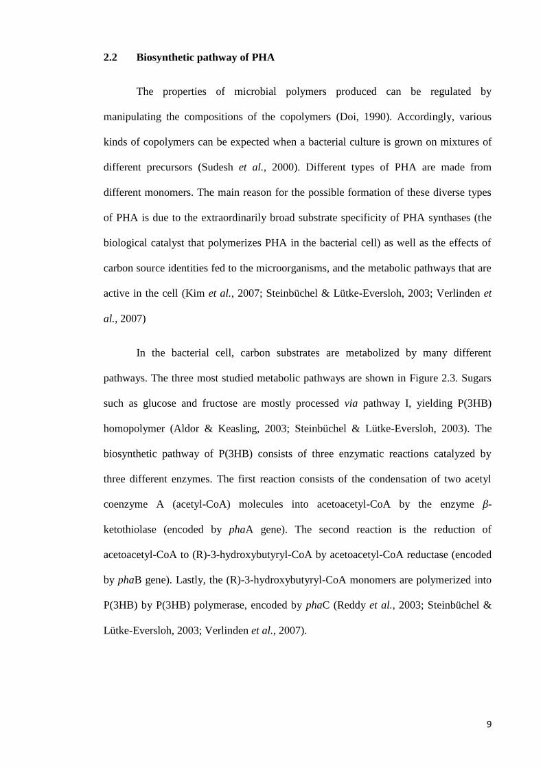
Figure 2.3 Metabolic pathways that supply various hydroxyalkanoate (HA) 10monomers for PHA biosynthesis
Figure 2.4 Schematic representation of the methods of polymer modification 16
Figure 2.5 Typical methods for the chemical modification of PHA to yield 25different types of functionalized polymers
Figure 2.6 Free radical grafting of MMA and HEMA on PHA using 29benzoyl peroxide (BPO) as an initiator
Figure 3.1 Setup of a 2-L stirred tank bioreactor for fed-batch fermentation 41
Figure 3.2 Standard calibration of optical density at 600 nm (OD600 nm) 44to dried total biomass (g L-1)
Figure 3.3 Schematic diagram for preparation of composite 50P(3HO-co-3HHX)/HA scaffold
Figure 3.4 Schematic diagram showing the formation of 56P(3HO-co-3HHX)-g-GDD copolymer
Figure 4.1 Effects of different C/N mol ratios on cell dry weight 64and mcl-PHA content of P. putida BET001
Figure 4.2 Estimation of the KLa value 65
Figure 4.3 Growth profile of P. putida BET001 in batch cultivations 67
Figure 4.4 Growth and biosynthesis of mcl-PHA by P. putida BET001 69in fed-batch fermentation
Figure 4.5 FTIR spectra of (A) P(3HO-co-3HHX); (B) P(3HO-co-3HHX)/ 7110% HA; (C) P(3HO-co-3HHX)/30% HA; and (D) HA powder
Figure 4.6 XRD spectra of (A) P(3HO-co-3HHX); (B) P(3HO-co-3HHX)/ 7310% HA; (C) P(3HO-co-3HHX)/30% HA and (D) HA
Figure 4.7 EDX spectrum obtained at 10 keV on the (A) P(3HO-co-3HHX); 77(B) P(3HO-co-3HHX)/10% HA and (C) P(3HO-co-3HHX)/30% HA

xv
Figure 4.8 FESEM image of the scaffolds (A) P(3HO-co-3HHX) 80(B) cells on scaffold surface P(3HO-co-3HHX) (C) compositeP(3HO-co-3HHX)/10% HA (D) cells on scaffold surfaceP(3HO-co-3HHX)/10% HA (E) composite P(3HO-co-3HHX)/30% HA (F) cells on scaffold surface P(3HO-co-3HHX)/30% HA (magnification 5000)
Figure 4.9 (A) Growth of human osteoblast cells (Alamar Blue Assay) 82(B) ALP activity of human osteoblast cells onP(3HO-co-3HHX) PHO, P(3HO-co-3HHX)/10% HAand P(3HO-co-3HHX)/30% HA scaffolds. (n=6)
Figure 4.10 FTIR spectra of (a) P(3HO-co-3HHX); (b)P(3HO-co-3HHX) 85-g-GDD (0.6 mM); (c) P(3HO-co-3HHX)-g-GDD (0.3 mM);and (d) GDD monomer
Figure 4.11 1H NMR of the P(3HO-co-3HHX)-g-GDD in 87CDCL3-d6i (Graft yield = 30 %)
Figure 4.12 Proposed mechanism for the reaction of GDD monomer 89grafting onto P(3HO-co-3HHX) (m = 1, 2, 3, 4,…..)
Figure 4.13 (A) Derivative weight percentages of neat P(3HO-co-3HHX), 93GDD monomer and P(3HO-co-3HHX)-g-GDD with variousGDD monomer concentrations. (B) TGA curves of neatP(3HO-co-3HHX) and P(3HO-co-3HHX)-g-GDD withvarious GDD monomer concentrations.
Figure 4.14 Graft yield as a function of the GDD monomer concentration. 95Reaction conditions: P(3HO-co-3HHX) 0.2 g; 80 °C;BPO 0.04 mM; 2 h.
Figure 4.15 Regression plot of percentage of graft yield as a function of 97reaction time (h) at different GDD concentrations (mM) anddifferent temperatures (A) 80 °C (B) 95 °C. Reaction conditions:P(3HO-co-3HHX) 0.2 g; BPO 0.04 mM;4 mL acetone.
Figure 4.16 Initial rate of grafting as a function of GDD monomer 98concentration and temperature. Reaction conditions:P(3HO-co-3HHX) 0.2 g; 95 °C; BPO 0.04 mM; 4 mL acetone.
Figure 4.17 Effects of reaction temperature on the graft yield 100copolymerization of P(3HO-co-3HHX)-g-GDD.
Figure 4.18 Effects of radical initiator (BPO) concentration on the graft yield 102
Figure 4.19 FTIR spectra of P(3HO-co-3HHX)-g-GDD/HA 103
Figure 4.20 EDX spectrum of P(3HO-co-3HHX)-g-GDD/HA performed 104at 10keV
xvi
LIST OF TABLES
Table 2.1 Biomedical application of PHA and PHA/inorganic 18phase composites
Table 3.1 Nutrient rich medium (NR) 38
Table 3.2 E2 medium 39
Table 3.3 MT solution 39
Table 4.1 Physical and mechanical properties of P(3HO-co-3HHX) 75and P(3HO-co-3HHX)/HA composites
Table 4.2 Elemental analysis of HA using EDX analysis of 78P(3HO-co-3HHX), P(3HO-co-3HHX)/10 % HA andP(3HO-co-3HHX)/30 % HA scaffolds
Table 4.3 Molecular weight, thermal, water uptake and graft yield data 92of neat P(3HO-co-3HHX) and copolymer P(3HO-co-3HHX)-g-GDD with different concentrations of GDD monomer
Table 4.4 Percentage of elements from EDX analysis of 104P(3HO-co-3HHX)-g-GDD/HA composite
Table 4.5 Mean percentage of mortality of A. franciscana nauplii 106after 24 h exposure to aqueous solutions with differentconcentrations of P(3HO-co-3HHX)-g-GDD/HA composite
xvii
LIST OF SYMBOLS AND ABBREVIATIONS
NH4Cl Ammonium chloride
NH4OH Ammonium hydroxide
ALP Alkaline phosphate
Ea Activation energy
BPO Benzoyl peroxide
β Beta
Ca Calcium
CaCI2.2H20 Calcium chloride dehydrate
CDW Cell dry weight
CoA Coenzyme-A
CoCl2.6H2O Cobalt (II) chloride hexahydrate
CuCl2.2H2O Copper (II) chloride dehydrate
CDCl3 Deuterated chloroform
DCM Dichloromethane
DSC Differential scanning calorimetry
dH2O Distilled water
Na2HPO4 Disodium hydrogen phosphate
Da Dalton
DSC Differential Scanning Calorimeter
ºC Degree Celcius
wt% Dry weight percent
Td Degradation temperature
EDX Energy dispersive X-ray
FESEM Field emission scanning electron microscopy
xviii
FTIR Fourier transform infrared
FID Flame ionization detector
g Gravity
g Gram
GC Gas Chromatography
GDD Glycerol, 1-3 diglycerol diacrylate
g/g Gram per gram
g/L Gram per liter
GPC Gel Permeation Chromatography
Tg Glass transition temperature
HA Hydroxyapatite
h Hour
ΔHm Heat of fusion
FeCl3 Iron (III) chloride
J/g Joule per gram
kDa Kilo Dalton
kg Kilogram
L Liter
μg Microgram
μg/ml Microgram per mililiter
μL Microliter
μm Micrometer
μM Micromolar
Mw Molecular weight
mcl Medium-chain-length
min Minute
xix
mg Miligram
mg/L Miligram per literTm Melting temperature
MgSO4 Magnesium sulphate
MgSO4.7H2O Magnesium sulphate heptahydrate
mL Mililiter
mM Milimolar
Mol % Mole percent
NR Nutrient rich
Mn Number-average molecular weight
NMR Nucleur Magnetic Resonance
C Oxygen concentration
C* Oxygen solubility
KLa Oxygen mass transfer coefficient
OD Optical density
pO2 Oxygen partial pressure
KH2PO4 Potassium dihydrogen phosphate
KOH Potassium hydrogen
% Percentage
Mw/ Mn Polydispersity indexPO Phosphate
P(3HO-co-3HHX) Poly(3-hydroxyoctanoate-co-3-hydroxyhexanoate)
P(3HO-co-3HHX)/HA Poly(3-hydroxyoctanoate-co-3-hydroxyhexanoate)/blendwith hydroxyapatite
P(3HO-co-3HHX)-g-GDD Poly(3-hydroxyoctanoate-co-3-hydroxyhexanoate) graftedwith glycerol, 1,3-diglycerol diacrylate
P(3HO-co-3HHX)-g-GDD/HA Poly(3-hydroxyoctanoate-co-3-hydroxyhexanoate)grafted with glycerol, 1,3-diglycerol diacrylate/ blendwith hydroxyapatite
xx
Pd Polydispersity index
PHA Polyhydroxyalkanoate
PhaA; phaA β-ketothiolase; gene encoding β-ketothiolase
PhaB; phaB NADPH-dependent acetoacetyl-CoA dehydrogenase;gene encoding NADPH-dependent acetoacetyl-CoAdehydrogenase
PhaC; phaC PHA synthase; gene encoding PHA synthase
rpm Revolutions per minute
scl Short-chain-length
sp. Species
NaCl Sodium chloride
Na2CO3 Sodium carbonate
H2SO4 Sulphuric acid
MT Trace element
TCA Tricarboxylic acid
v/v Volume per volume
w/v Weight per volume
w/w Weight per weight
H2O Water
XRD X-ray diffraction
ZnSO4.7H2O Zinc sulfate heptahydrate
3HX 3-hydroxyhexanoate
3HO 3-hydroxyoctanoate

xxi
LIST OF APPENDICES
Appendix 1 DSC thermogram of P(3HO-co-3HHX) 125
Appendix 2 DSC thermogram of P(3HO-co-3HHX)/ 10 % HA 125
Appendix 3 DSC thermogram of P(3HO-co-3HHX)/30 % HA 126
Appendix 4 DSC thermogram of P(3HO-co-3HHX)-g-GDD (0.1 mM) 126
Appendix 5 DSC thermogram of P(3HO-co-3HHX)-g-GDD (0.3 mM) 127
Appendix 6 DSC thermogram of P(3HO-co-3HHX)-g-GDD (0.4 mM) 127
Appendix 7 DSC thermogram of P(3HO-co-3HHX)-g-GDD (0.6 mM) 128
1
CHAPTER 1
INTRODUCTION
The increasing demand on sustainability, eco-efficiency and green chemistry has
generated tremendous search for materials that are renewable and environmental
friendly. Biodegradable polymers offer a sustainable alternative to petroleum-derived
sources. Polyhydroxyalkanoates (PHA) comprised a group of natural biodegradable
polyesters that are synthesized by microorganisms. PHA exhibits a wide range of
physical and mechanical properties owing to the diversity in their chemical structures.
Among its sought after attributes are biodegradability and excellent biocompatibility,
making this class of biopolymer attractive as the potential biomaterial for various
applications, particularly in biomedical field (Ali & Jamil, 2016; Kim et al., 2007).
Medium-chain-length poly(3-hydroxyalkanoates) (mcl-PHA) is structurally
diverse polyester and could be suitably tailored for various biomedical applications.
They are biodegradable, biocompatible and thermoprocessable, hence suitable platform
materials for applications in both conventional medical devices and tissue engineering
(e.g. sutures, cardiovascular application, bone marrow scaffolds, matrices for controlled
drug delivery etc.) (Chen & Wu, 2005; Hazer et al., 2012). However, direct application
of these polyesters, mcl-PHA included, has been hampered by their strong hydrophobic
character and other physical shortcomings (Kim et al., 2008; Rai et al., 2011). Hence,
native mcl-PHA needs to be modified in order to improve its performance in specialized
applications such as environmentally biodegradable polymers and functional materials
for biomedical and industrial applications (Lee et al., 2010; Li et al., 2016).
Synthetic and natural hydroxyapatites (HA) (HCa5O13P3) have similar chemical
composition and crystallographic properties to a human bone (Xi et al., 2008). Their
biocompatibility and osteoconductive behavior are suitable for making bone implants.

2
Studies have shown that incorporation of HA into biomaterials could help to enhance
mechanical performance and osteoblast responses (Baei & Rezvani, 2011; Wang et al.,
2005). Currently, composites of polymers and ceramics are being developed with the
aim to increase the mechanical scaffold stability and to improve tissue interactions. In
addition, efforts have also been invested in developing scaffolds with drug-delivery
capacity. These scaffolds allow for local release of growth factors or antibiotics and
enhance bone in-growth to treat bone defects and even support wound healing (Rezwan
et al., 2006). Pores are necessary for bone tissue formation because they allow
migration and proliferation of osteoblasts and mesenchymal cells as well as
vascularization. In addition, a porous surface improves mechanical interlocking between
the implant biomaterial and the surrounding natural bone thus providing for greater
mechanical stability at this critical interface (Wei & Ma, 2004).
Microbial polyesters can be further diversified via both chemical modification
reaction and genetic engineering of the biosynthetic pathways. Chemical modification is
a promising approach to obtain new types of PHA-composite materials including a wide
range of monomers for graft/block copolymerization with synthetic and other natural
polymers that cannot be obtained by biotechnological processes (Hazer & Steinbüchel,
2007). For instance, chemical modification of PHA could involve grafting reactions
through graft/block copolymerization, chlorination, cross-linking, epoxidation, hydroxyl
and carboxylic acid functionalization. Insertions of an additional different polymer
segment into an existing polymer backbone or at the side chain of an existing polymer
yields block or graft copolymers (Gumel et al., 2014).
Moreover, mcl-PHA has attracted great interest in research due to its potential
wide applicability as biomaterials. Nevertheless, its strong hydrophobicity, slow
degradation under physiological conditions and lack of chemical functionalities hinder
the realization of its potentials. These factors restrict the scope of their applications in

3
biomedical field. In order to expand the range of its versatilities, other properties such as
mechanical strength, surface features, amphiphilicity and degradation rate have to be
modified to match the requirements of specific applications (Kai & Loh, 2013). For
example, intrinsic hydrophobic properties of mcl-PHA restrict their applications as cell
colonizing materials. Therefore, chemical modification with suitable functional groups
or modification of the surface topography of mcl-PHA is needed in order to minimize
hydrophobic interactions with the surrounding tissue. Amphiphilic copolymers could be
produced through chemical modification reactions by inserting the hydrophilic
segments into the hydrophobic PHA (Hazer, 2010).
In this study, a mcl-PHA viz. poly(3-hydroxyoctanoate-co-3-hydroxyhexanoate)
P(3HO-co-3HHX) was investigated as a potential material for bone cells regeneration
scaffold both in its pure form and as P(3HO-co-3HHX)/HA composite. The physical,
thermal and mechanical properties of the composite P(3HO-co-3HHX)/HA scaffold
were investigated. The biocompatibility and osteoconductivity of the porous composite
P(3HO-co-3HHX)/HA scaffold was also studied. In order to enhance hydrophilicity of
the polymer, graft copolymerization of P(3HO-co-3HHX) with the glycerol 1,3-
diglycerolate diacrylate (GDD) was investigated via free radical polymerization
reaction. The P(3HO-co-3HHX)-g-GDD was prepared by thermal treatment of
homogenous solution of P(3HO-co-3HHX), GDD monomer and benzoyl peroxide
(BPO) as a chemical initiator. Differently selected parameters affecting the graft yield
were studied such as monomer concentration, chemical initiator concentration,
temperature and reaction time. In addition, the grafted copolymer P(3HO-co-3HHX)-g-
GDD was characterized and the grafting mechanism was proposed. It is hypothesized
that if scaffolds of P(3HO-co-3HHX)-g-GDD/HA are successfully fabricated via graft
copolymerization and physical blend, the resulting biomaterials will possess the desired
properties as described earlier. The objectives of the investigation includes :

4
Biosynthesis of mcl-polyhydroxyalkanotes (mcl-PHA)
1. To produce mcl-PHA from Pseudomonas putida BET001 in fed-batch fermentation;
Blending of poly(3-hydroxyoctanoate-co-3-hydroxyhexanoate) P(3HO-co-3HHX)
with hydroxyapatite (HA)
1. To study the effects of different concentrations of HA loading onto P(3HO-co-
3HHX);
2. To characterize the polymer before and after blending with HA using FTIR, DSC,
XRD, EDX and FESEM;
3. To determine the osteoblast cell response towards the P(3HO-co-3HHX)/HA blend;
Functionalization of P(3HO-co-3HHX) as amphiphilic material by graft
copolymerization with glycerol 1,3-diglycerol diacrylate (GDD)
1. To study graft copolymerization of P(3HO-co-3HHX) with glycerol 1,3-diglycerolate
diacrylate via free radical polymerization reaction;
2. To determine the effects of monomer concentration, reaction time, initiator
concentration and temperature on graft copolymerization of P(3HO-co-3HHX)-g-GDD;
3. To characterize the P(3HO-co-3HHX)-g-GDD graft copolymer using FTIR, 1H
NMR, STA, DSC, GPC and propose the possible radical polymerization mechanism;
4. To determine the water uptake ability of the grafted copolymer.

5
P(3HO-co-3HHX)-g-GDD/HA composite scaffold
1. To fabricate composite scaffold of P(3HO-co-3HHX)-g-GDD/HA via graft
copolymerization and physical blend;
2. To characterize the newly developed scaffold by using FTIR and EDX;
3. To study the toxicity effect of the P(3HO-co-3HHX)-g-GDD/HA.

6
CHAPTER 2
LITERATURE REVIEW
2.1 Polyhydroxyalkanoates (PHA)
Polyhydroxyalkanoates (PHA) are versatile polyesters produced by a large
number of bacteria as intracellular granules under metabolic stress conditions (Bassas-
Galià et al., 2015). Bacterial-synthesized PHA has attracted attention because they can
be produced from a variety of renewable resources and are truly biodegradable and
highly biocompatible thermoplastic materials (Yu et al., 2006). Microorganisms are
able to accumulate various types of PHA in the form of homopolymer, copolymer and
polymer blends (Bhatt et al., 2008). The properties of PHA copolymers depend strongly
on the type, content and distribution of comonomer units which comprise the polymer
chains, as well as the molecular weight distribution (Chanprateep et al., 2008). In
addition, the nature and proportion of different monomers are also influenced by the
bacterial strains, type and relative quantity and quality of carbon sources supplied to the
growth medium (Shamala et al., 2009).
PHA is a family of optically active biological polyesters which composed of
repeating units of 3-hydroxyalkanoic acids, each carries an aliphatic alkyl side chain
(R). Carbon, oxygen and hydrogen are the main components in the structure of PHA.
The general structure of PHA is shown in Figure 2.1. The carboxyl group of one
monomer forms an ester bond with the hydroxyl group of the adjacent monomer. Each
monomer contains the chiral carbon atom and has the (R) stereochemical configuration
on the hydroxyl-substituted carbon (Madison & Huisman, 1999). According to
Williams and Martin (2005), there are various kind of side chain groups attached
including alkyl, aryl, halogen, aromatic and branched monomers. The variation in side
chain endows the PHA family with excellent properties ranging from rigid and stiff

7
Figure 2.1: General structure of PHA
to flexible and elastomeric. Hence, it significantly expands PHA potential for various
applications, particularly in biomedical field.
Several strains of PHA producing bacteria were studied such as Bacillus sp.,
Alcaligenes sp., Pseudomonas sp., Aeromonas hydrophila, Rhodopseudomonas
palustris, Escherichia coli, Burkholderia sacchari and Halomonas boliviensis
(Verlinden et al., 2007). Over 150 types of PHA have been identified as homopolymers
or as copolymers. The flexibility of PHA biosynthesis makes it possible to design and
produce biopolymers with useful physical properties ranging from stiff and brittle
plastic to rubbery polymers (Bhatt et al., 2008; Sudesh et al., 2000). Examples of PHA
produced at commercial scale under various trademarks including Biomer®, Mirel TM,
Biocycle®, ICI® and Biopol® (Sudesh & Iwata, 2008).
R= H, alkyl group, side chain C1-C13 n= 100-300,000
CO
R
H
CH2 C
O
n

8
2.1.1 Medium-chain-length poly(3-hydroxyalkanoates)
PHA is classified based on the number of carbon atom present in the monomeric
unit. Monomeric unit of short-chain-length PHA (scl-PHA) consisted of 3-5 carbon
atoms, while medium-chain-length PHA (mcl-PHA) consisted of 6-14 carbon atoms.
Scl-PHA like poly(3-hydroxybutyrate), P(3HB) is hard and brittle compared to mcl-
PHA and their copolymers like poly(3-hydroxyhexanoate-co-3-hydroxyoctanoate),
P(3HHx-co-3HO), which are soft and elastomeric. Mcl-PHA and its copolymers exhibit
low crystallinity, low glass transition temperature, low tensile strength and high
elongation-to-break ratio compared to scl-PHA, which is brittle and stiff (Muhr et al.,
2013; Rai et al., 2011; Sudesh et al., 2000).
Mcl-PHA biosynthesis is a general property of the flourescent pseudomonads
belonging to the rRNA homology group I. Most of these bacteria are able to grow on
various carbon sources that can be incorporated into mcl-PHA. Depending on the nature
of the carbon substrate available, the hydroxyacyl monomers are derived from the
intermediates of fatty acid β-oxidation or de novo fatty acid biosynthesis pathways
(Chardron et al., 2010; Zinn et al., 2001). Figure 2.2 shows the structure of the mcl-
PHA with various types of monomers.
Figure 2.2: Medium-chain-length PHA with different types of monomers
9
2.2 Biosynthetic pathway of PHA
The properties of microbial polymers produced can be regulated by
manipulating the compositions of the copolymers (Doi, 1990). Accordingly, various
kinds of copolymers can be expected when a bacterial culture is grown on mixtures of
different precursors (Sudesh et al., 2000). Different types of PHA are made from
different monomers. The main reason for the possible formation of these diverse types
of PHA is due to the extraordinarily broad substrate specificity of PHA synthases (the
biological catalyst that polymerizes PHA in the bacterial cell) as well as the effects of
carbon source identities fed to the microorganisms, and the metabolic pathways that are
active in the cell (Kim et al., 2007; Steinbüchel & Lütke-Eversloh, 2003; Verlinden et
al., 2007)
In the bacterial cell, carbon substrates are metabolized by many different
pathways. The three most studied metabolic pathways are shown in Figure 2.3. Sugars
such as glucose and fructose are mostly processed via pathway I, yielding P(3HB)
homopolymer (Aldor & Keasling, 2003; Steinbüchel & Lütke-Eversloh, 2003). The
biosynthetic pathway of P(3HB) consists of three enzymatic reactions catalyzed by
three different enzymes. The first reaction consists of the condensation of two acetyl
coenzyme A (acetyl-CoA) molecules into acetoacetyl-CoA by the enzyme β-
ketothiolase (encoded by phaA gene). The second reaction is the reduction of
acetoacetyl-CoA to (R)-3-hydroxybutyryl-CoA by acetoacetyl-CoA reductase (encoded
by phaB gene). Lastly, the (R)-3-hydroxybutyryl-CoA monomers are polymerized into
P(3HB) by P(3HB) polymerase, encoded by phaC (Reddy et al., 2003; Steinbüchel &
Lütke-Eversloh, 2003; Verlinden et al., 2007).

10
sugars fatty acids
Krebs cycle acetyl-CoA acyl-CoA
PhaA fatty acidacetoacetyl-CoA 3-ketoacyl-CoA β-oxidation enoyl-CoA
PhaB
(R)-3-hydroxybutyryl-CoA FabG (S)-3-hydroxyacyl-CoA PhaJ
PhaCPhaC (R)-3-hydroxyacyl-CoA
PhaC PhaG
4-hydroxyacyl-CoA (R)-3-hydroxyacyl-CoA
fatty acidcarbon sources 3-ketoacyl-ACP biosynthesis enoyl-ACP
other pathways acyl-ACP
malonyl-ACP
malonyl-CoA
acetyl-CoA
sugars
Figure 2.3: Metabolic pathways that supply various hydroxyalkanoate (HA) monomersfor PHA biosynthesis. PhaA, 3-Ketothiolase; PhaB, NADPH-dependent acetoacetyl-CoA reductase; PhaC, PHA synthase; PhaG, 3-hydroxyacyl-ACP-CoA transferase;PhaJ, (R)-specific enoyl-CoA hydratase; FabG, 3-ketoacyl-ACP reductase (Tsuge,2002; Verlinden et al., 2007).
Pathway IIPathway I
PHA
Pathway III
11
Pathways II and III involved in fatty acid metabolism to generate different HA
monomers based on the different carbon sources utilized in PHA biosynthesis (Tsuge,
2002). Intermediates generated from the fatty acid β-oxidation pathway are usually
inter-related with mcl-PHA biosynthesis in a strain of producer. The carbon sources
used includes alkanes, alkenes and alkanoates. The monomers incorporated depend on
the carbon sources used. The β-oxidation intermediate, trans-2-enoyl-CoA is converted
to (R)-hydroxyacyl-CoA by a (R)-specific enoyl-CoA hydratase (Aldor & Keasling,
2003; Steinbüchel & Lütke-Eversloh, 2003).
The intermediates for the biosynthesis are obtained from the fatty acid
biosynthetic pathway. These pathways are significant of interest because they help
generate monomers for PHA synthesis from structurally unrelated, simple and
inexpensive carbon sources such as glucose, sucrose and fructose. The (R)-3-
hydroxyacyl-ACP (acyl carrier protein) intermediates from the fatty acid biosynthetic
pathway are converted to the (R)-3-hydroxyacyl-CoA by the enzyme acyl-ACP-CoA
transacylase (encoded by phaG gene). This enzyme plays a key role in linking fatty acid
synthesis and PHA biosynthesis (Verlinden et al., 2007).
The β-oxidation intermediate, trans-2-enoyl-CoA is converted to (R)-3-
hydroxyacyl-CoA by (R)-specific enoyl-CoA hydratase (encoded by PhaJ gene).
Several other enzymes have been found to possess the ability to supply the monomers.
The 3-ketoacyl-ACP reductase (encoded by FabG gene) is a constituent of the fatty acid
biosynthesis pathway. It has been demonstrated that the product of FabG could accept
not only acyl-ACP but also acyl-CoA as a substrate and is capable of supplying mcl-
(R)-3HA-CoA from fatty acid β-oxidation in E. coli (Tsuge, 2002).

12
2.3 Biosynthesis of PHA
The choice of operation strategy for the production of bacterial PHA depends on
various factors including carbon source, culture condition, modes of fermentation
(batch, fed-batch, continuous), bioreactor type (air-lift reactor, continuous stirred tank
reactor (CSTR) or sequencing batch system (SBR)) (Amache et al., 2013; Annuar et al.,
2008). The carbon source fed to the bacterial culture may include alkanes, alkenes,
alcohol and carbohydrate instead of fatty acid, and they affect the polymer structure,
quantity and quality (Bassas-Galià et al., 2015). Several mcl-PHA production strategies
in the bioreactor such as batch and continuous (Jung et al., 2001), fed-batch (Jiang et
al., 2013; Poblete-Castro et al., 2014) and high-cell-density process (Le Meur et al.,
2012) under various cultivation conditions have been studied. The imbalance of nutrient
provisions, such as oxygen, nitrogen, phosphorus, sulphur and magnesium forced the
bacteria to accumulate excess carbon intake by polymerization into PHA within the
cells as carbon assimilation for energy reservoir. Thus, the physiological condition can
be regulated in the fermentation process in order to achieve high PHA yields and PHA
productivity (Annuar et al., 2006).
Furthermore, batch fermentation for PHA production is a common process due
to its flexibility, low operation cost and suitable for growth studies and screening of
potential PHA accumulating organisms. However, it is associated with low PHA
productivity since after utilization of the carbon source, bacterial cells degrade the
accumulated PHA resulting in reduced PHA content (Amache et al., 2013). Basically,
the fed-batch culture is a batch culture that is continuously supplemented with selected
nutrients after it enters the late exponential phase. Fed-batch fermentation yields higher
PHA productivity but the overall PHA production is still considered low, when nitrogen
is the limiting nutrient. Thus, batch and fed-batch processes are combined in order to
obtain higher PHA content. The combined process is the most common fermentation

13
strategy used for PHA production. In this strategy, the process is divided into two
stages: in the first stage the microorganism is grown under batch mode until the desired
biomass is achieved and PHA accumulation has started. In the second stage the
fermentation is shifted to fed-batch, where usually one or more essential nutrients (most
common is nitrogen) are maintained in limited concentration and carbon source is
continuously fed into the reactor to further produce and accumulate PHA in the cells
(Zinn et al., 2001).
In addition, the removal of cellular endotoxin from Gram-negative bacteria is
needed for further application especially in biomedical field. Solvent extraction has
undoubted advantages over the other extraction methods of PHA in terms of efficiency.
This method is also able to remove bacterial endotoxin and causes negligible
degradation to the polymers (Chen & Wu, 2005; Wang et al., 2005; Baei & Rezvani,
2011). Most methods to recover intracellular PHA involve the use of organic solvents,
such as acetone, chloroform, methylene chloride or dichloroethane (Furrer et al., 2007;
Verlinden et al., 2007). Lower chain ketone such as acetone is the most prominent
solvent especially for the extraction of mcl-PHA. However, the consumption of large
quantities of solvent makes the procedure economically and environmentally
unattractive (Kunasundari & Sudesh, 2011). For medical applications, the solvent
extraction is a good method as it yields high purity PHA (Chen & Wu, 2005).
14
2.4 Biodegradability and biocompatibility of PHA
PHA has been emerged as potentially useful materials in the biomedical field for
different applications due to their unique properties of being biodegradable and
biocompatible. In vivo implant of PHA have been made possible due to their non-toxic
degradation products, biocompatibility, desired surface modifications, wide range of
physical and chemical properties, cellular growth support, and attachment without
carcinogenic effects. In addition, lower acidity and bioactivity of PHA pose minimal
risk compared to other biopolymers such as poly-lactic acid (PLA) and poly-glycolic
acid (PGA) (Ali & Jamil, 2016; Chen et al., 2013).
For medical applications, materials must be biocompatible, which means that
they cannot cause severe immune reactions when introduced to soft tissues or blood of a
host organism. Moreover, PHA also considered as biocompatible material when the
material does not elicit immune responses during degradation in the body. Generally,
PHA polymers are degraded by the action of non-specific lipases and esterases in
nature. This is presumably how PHA implants and other medical devices are degraded
at the site of implantation in animals. Degradation of PHA matrices in the tissues of the
host organism offers the possibility of coupling this occurrence with the release of
bioactive compounds, such as antibiotic or anti-tumor drug. Kabilan et al. (2012)
reviewed the strategies adapted to make functional polymer from mcl-PHA to be
utilized as drug delivery system. When PHA is impregnated with a compound, the
degradation over time will release the compound, consequently acting as an automatic
dosing agent. The kinetics of dosing of a compound from PHA matrix can be fine-tuned
by altering the polymer properties, including using different types of PHA with different
monomer side chains (Brigham & Sinskey, 2012).

15
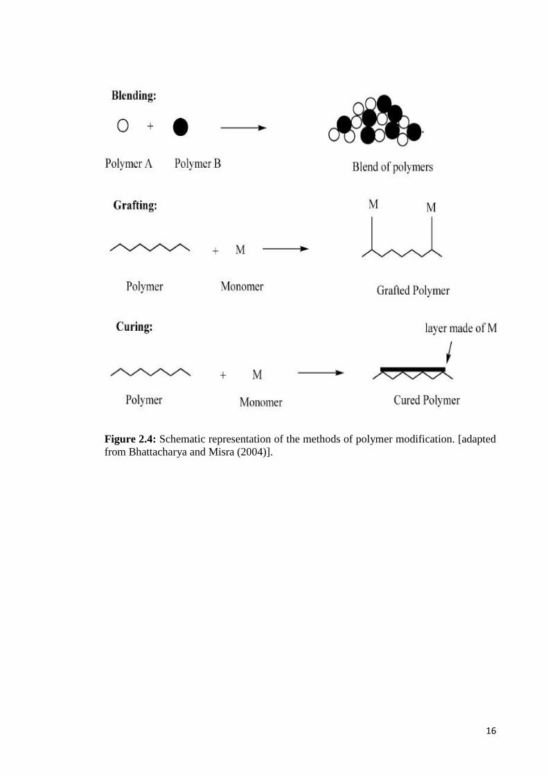
2.5 Modification of PHA
PHA is emerging as a sought-after class of biodegradable polymers for
applications in tissue engineering. Over the years, efforts have been made to extend the
functionalities of PHA and to investigate their uses in numerous biomedical
applications, such as sutures, cardiovascular patches, wound dressings, guided tissue
repair/regeneration devices, and tissue engineering scaffolds (Misra et al., 2006). PHA
is a promising material for tissue engineering and drug delivery system owing to its
properties of being natural, renewable, biodegradable and biocompatible thermoplastics
(Hazer, 2010). However, several limitations constrained its competition with traditional
synthetic plastics or its applications as ideal biomaterials. These include their poor
mechanical properties, high production cost, limited functionalities, incompatibility
with conventional thermal processing techniques and susceptibility to thermal
degradation (Li & Loh, 2015; Rai et al., 2011). Thus, PHA needs to be modified to
ensure improved performance in specific applications. Furthermore, in order for mcl-
PHA to serve as the material of choice in the biomedical field, their hydrophilicity must
be tailored to the requirements of a particular application. Therefore, attempts to modify
the properties of mcl-PHA by chemical and physical methods, such as blending,
crosslinking (curing) and graft copolymerization, have attracted a great deal of interest
(Kim et al., 2007). Blending is the physical mixture of two or more polymers to obtain
the desired properties (Figure 2.4). Grafting is a method where monomers are covalently
bonded onto the polymer chain, whereas in crosslinking (curing), the polymerization of
an oligomer mixture forms a coating which adheres to the substrate by physical forces
(Bhattacharya & Misra, 2004).
16
Figure 2.4: Schematic representation of the methods of polymer modification. [adaptedfrom Bhattacharya and Misra (2004)].
17
Generally, polymers from PHA family are not osteoconductive, thus they are
generally overlooked for bone tissue engineering application. One of the major
limitations is the inability of PHA to form strong interfacial bonding with the
surrounding bone tissue by means of forming biologically active apatite layer on the
implant surface (Misra et al., 2006). Therefore, one of the approaches to overcome this
lack of osteoconductivity and mechanical competence is by combining mcl-PHA with
inorganic bioactive particles or fibres. Incorporation of inorganic phases may lead to
mcl-PHA composites with different mechanical properties suitable for tissue
engineering application. Extensive research is being carried out on the development of
bioactive and biodegradable composite materials in the form of dense and porous
system, where the bioactive inorganic phase incorporated as either filler or coating (or
both) into the biodegradable polymer matrix (Misra et al., 2006; Rai et al., 2011).
With respect to the development of PHA, researchers have looked into the
possibility of designing composites in combination with inorganic phases to further
improve the mechanical properties, rate of degradation, and also impart bioactivity.
Poly(3-hydroxybutyrate), poly(3-hydroxybutyrate-co-3-hydroxyvalerate), and poly(3-
hydroxybutyrate-co-3-hydroxyhexanoate) are some of the polymers that have been
extensively studied to fabricate composites in combination with hydroxyapatite,
bioactive glass, and glass-ceramic fillers or coatings (Misra et al., 2006) (Table 2.1). In
order to improve the properties, PHA is also blended with natural raw materials or other
biodegradable polymers, including starch, cellulose derivatives, lignin, poly(lactic acid),
polycaprolactone and different PHA-type blends (Li et al., 2016) .
Bioceramics are inorganic materials specially developed for use as medical and
dental implants such as alumina and zirconia, bioactive glasses, glass-ceramics,
hydroxyapatite, and resorbable calcium phosphates (Misra et al., 2006). So far, only
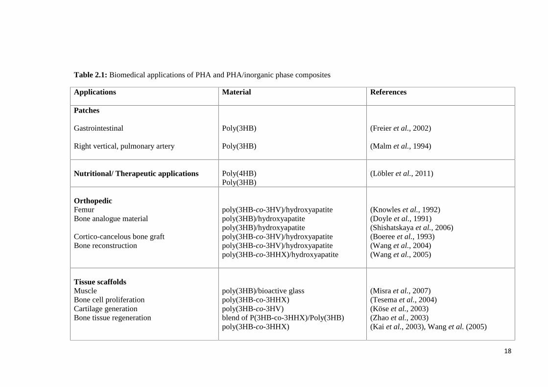
18
Table 2.1: Biomedical applications of PHA and PHA/inorganic phase composites
Applications Material References
Patches
Gastrointestinal
Right vertical, pulmonary artery
Poly(3HB)
Poly(3HB)
(Freier et al., 2002)
(Malm et al., 1994)
Nutritional/ Therapeutic applications Poly(4HB)Poly(3HB)
(Löbler et al., 2011)
OrthopedicFemurBone analogue material
Cortico-cancelous bone graftBone reconstruction
poly(3HB-co-3HV)/hydroxyapatitepoly(3HB)/hydroxyapatitepoly(3HB)/hydroxyapatitepoly(3HB-co-3HV)/hydroxyapatitepoly(3HB-co-3HV)/hydroxyapatitepoly(3HB-co-3HHX)/hydroxyapatite
(Knowles et al., 1992)(Doyle et al., 1991)(Shishatskaya et al., 2006)(Boeree et al., 1993)(Wang et al., 2004)(Wang et al., 2005)
Tissue scaffoldsMuscleBone cell proliferationCartilage generationBone tissue regeneration
poly(3HB)/bioactive glasspoly(3HB-co-3HHX)poly(3HB-co-3HV)blend of P(3HB-co-3HHX)/Poly(3HB)poly(3HB-co-3HHX)
(Misra et al., 2007)(Tesema et al., 2004)(Köse et al., 2003)(Zhao et al., 2003)(Kai et al., 2003), Wang et al. (2005)

19
Applications Material References
Drug releaseTetracyclineSulperazone, Gentamicin
poly(3HB-co-3HV)poly(3HB-co-3HV)poly(3HB-co-3HV)/wollastonite
(Panith et al., 2016)(Gursel et al., 2002)(Li & Chang, 2005)
Suture poly(3HB-co-3HV)poly(3HB-co-4HB)
(Shishatskaya et al., 2004)(Chen et al., 2010)
Conduits poly(3HB-co-3HV) (Mosahebi et al., 2002)
Nerve regeneration Poly(3HB-co-3HHX)poly(3HB-co-3HV)poly(3HB)
(Bian et al., 2009)(Mosahebi et al., 2002)(Novikov et al., 2002)
Wound healing poly(3HB-co-3HV)poly(4HB)/hyaluronic acid
(Leenstra et al., 1998)(Peschel et al., 2008)
Cardiovascular applications Poly(4HB)PHO
(Martin & Williams, 2003)(Sodian et al., 2000)

20
hydroxyapatite, wollastonite and bioactive glasses have been extensively studied in
combination with PHA to form composites (Rai et al., 2011). The mechanical and
biological performances of bioactive ceramic/polymer composites can be controlled
using different particulate bioceramics and also by varying the amount of bioceramic
particles in the composite (Boccaccini & Blaker, 2005). Hydroxyapatite is the major
mineral component of bone, and it is one of the most common biomaterials studied in
bone tissue engineering (Xi et al., 2008). The thermodynamic stability of
hydroxyapatite at physiological pH and its ability to actively take part in bone bonding
by forming strong chemical bonds with surrounding bone make it a suitable bioactive
ceramic for preparing composites (Kokubo et al., 2003).
2.6 Modification of PHA via physical blending
2.6.1 PHA/hydroxyapatite blending as an osteoconductive scaffold
Mcl-PHA are structurally more diverse than scl-PHA such as PHB, and this
imparts a wider and crucial flexibility in determining the physical and mechanical
properties of mcl-PHA in order to meet the requirements of engineered tissue (Muhr et
al., 2013; Rezwan et al., 2006; Zinn et al., 2001). Concomitantly, mcl-PHA such as
poly(3-hydoxyoctanoate), poly(3-hydroxyhexanoate), copolymers like poly(3-
hydroxybutyrate-co-3-hydroxyhexanoate), poly(3-hydroxyoctanoate-co-3-
hydroxyhexanoate) is being increasingly studied to develop osteosynthetic materials,
surgical sutures, stents, scaffolds for tissue engineering and matrices for drug delivery
(Chen & Wu, 2005). Nevertheless, extensive studies on mcl-PHA in general remain
limited because of inavailability of these polymers in testing quantities (Rai et al.,
2011).

21
Synthetic and natural hydroxyapatites (HA) (HCa5O13P3) have similar chemical
composition and crystallographic properties to a human bone (Xi et al., 2008). Their
biocompatibility and osteoconductive behavior are suitable for making bone implants.
Studies have shown that incorporation of HA into biomaterials could help to enhance
mechanical performance and osteoblast responses (Baei & Rezvani, 2011; Wang et al.,
2005). Currently, composites of polymers and ceramics are being developed with the
aim to increase the mechanical scaffold stability and to improve tissue interactions. In
addition, efforts have also been invested in developing scaffolds with drug-delivery
capacity. These scaffolds allow for local release of growth factors or antibiotics and
enhance bone in-growth to treat bone defects and even support wound healing (Rezwan
et al., 2006).
Polymer-based composite scaffold showed great potential in bone tissue
engineering. Efforts have been made to form porous PHB/HA and PHBV/HA
composites for bone tissue repair by utilizing the osteoconductivity property of HA
(Baek et al., 2012; Saadat et al., 2013; Sultana & Khan, 2012; Sultana & Wang, 2008).
For instance, particulate hydroxyapatite (HA) incorporated into poly(3-
hydroxybutyrate) (PHB) formed a bioactive and biodegradable composite for
applications in hard tissue replacement and regeneration (Saadat et al., 2013). Wang et
al. (2005) reported that the presence of hydroxyapatite increased the growth of
osteoblast and cell proliferation compared to neat P(3HB). Studies by them have shown
that the presence of hydroxyapatite particles on the surface helps the formation of
tenacious bonds with osteoblast cells. Moreover, the presence of hydroxyapatite in
P(3HB) matrices helped to increase the strength of the composite along with its
bioactivities. Ni and Wang (2002) demonstrated the formation of apatite crystals on the
surface of their P(3HB) composite containing hydroxyapatite after 1-3 days of
immersion in simulated body fluid (SBF), which is an acellular fluid designed with a
22
composition equivalent to blood plasma. They showed that the quantity of the apatite
crystals formed is directly proportional to the amount of hydroxyapatite used in the
composite. The storage modulus of P(3HB)/HA composites was found to increase with
increasing percentage of hydroxyapatite.
Jack et al. (2009) fabricated PHBV/HA composite scaffolds with high porosity
and controlled pore architectures. They found that incorporation of HA nanoparticles
increased the stiffness and strength, thus improved the in vitro bioactivities of the
scaffolds. Baek et al. (2012) incorporated collagen into PHBV/HA scaffold fabricated
using a hot-press machine and salt leaching method. Their results showed that the
PHBV/HA/Col composite scaffolds allowed for better cell adhesion and significantly
higher proliferation and differentiation than the PHBV/HA composite scaffolds and the
PHBV scaffolds. An ideal biocompatible material should be non-toxic and should not
act as immunostimulant at molecular level (Shabna et al., 2013). Furthermore, various
PHA blends have been developed to improve the performance of scaffold for bone
defect repairs or bone tissue engineering. Several studies on bone tissue engineering
have been conducted using PHA/HA such as poly(3-hydroxybutyrate), poly(3-
hydroxybutyrate-co-3-hydroxyvalerate) and poly(3- hydroxybutyrate-co-3-
hydroxyhexanoate) (Saadat et al., 2013; Sultana & Khan, 2012; Xi et al., 2008). To
date, there are still limited studies on mcl-PHA as a composite scaffold for bone tissue
engineering.
Scaffolds should exhibit high porosity, high interconnectivity and proper pore
sizes in order to facilitate cell adhesion, tissue in-growth and mass transfer. The
appropriate pore characteristics of scaffolds are vital in tissue engineering particularly
during the late stage of implantation when cells need to migrate deep into the scaffold
(Sultana & Wang, 2008). The scaffold should positively interact with cells, enhance cell
adhesion, growth, migration and differentiated function. The basic challenges to the

23
material selection and scaffold design are to achieve the initial strength and stiffness.
For instance, the material for the scaffold must have sufficient inter-atomic and inter-
molecular bonding or physical and chemical structures that allow for hydrolytic
attachment and breakdown. In addition, porosity and proper pore size are important
design parameters for the scaffold design, and high surface area necessary for
mechanical stability (Sabir et al., 2009).
Pores are necessary for bone tissue formation because they allow migration and
proliferation of osteoblasts and mesenchymal cells as well as vascularization. In
addition, a porous surface improves mechanical interlocking between the implant
biomaterial and the surrounding natural bone thus providing for greater mechanical
stability at this critical interface (Wei & Ma, 2004). Porosity of scaffolds for tissue
engineering should be high enough to provide sufficient space for cell adhesion (Chen
et al., 2002). The most common techniques applied to create porosity in a biomaterial
are porogen leaching technique, gas foaming, phase separation, electrospinning, freeze-
drying and sintering depending on the material used to fabricate the scaffold
(Karageorgiou & Kaplan, 2005). Among these methods, particle leaching method has
been identified as a convenient way to fabricate sponge-like scaffold besides being
reproducible (Sodian et al., 2000). Moreover, porogen leaching technique also provides
easy control of pore structure and has been well established in the preparation of porous
scaffolds for tissue engineering. This technique involves the casting of a
polymer/porogen composite followed by aqueous washing out of the incorporated
porogen. The pore size, porosity and pore morphology can be easily controlled by the
properties of porogen (Tan et al., 2011).

24
2.7 Functionalization of PHA
Microbial polyesters can be further diversified via both chemical modification
reaction and genetic engineering of the biosynthetic pathways. Chemical modification is
a promising approach to obtain new types of PHA-composite materials including a wide
range of monomers for graft/block copolymerization with synthetic and other natural
polymers that cannot be obtained by biotechnological processes (Hazer & Steinbüchel,
2007). For instance, chemical modification of PHA could involve grafting reactions
through graft/block copolymerization, chlorination, cross-linking, epoxidation, hydroxyl
and carboxylic acid functionalization (Figure 2.5). Insertion of an additional different
polymer segment into an existing polymer backbone or at the side chain of an existing
polymer yields block or graft copolymers (Gumel et al., 2014).
Moreover, chemical modification of PHA enables easy and precise modulation
of the polymer structure with predictable functionalities (Li et al., 2016). PHA are
predominantly hydrophobic and surface modification of the PHA is necessary to
improve their hydrophilicity, wettability, and surface charge for biomedical applications
(Guzmán et al., 2011). Amphiphilic polymers can be synthesized by introducing
hydrophilic groups such as hydroxyl, carboxyl, amine, glycol, and hydrophilic polymers
such as PEG, poly(vinyl alcohol), polyacryl amide, poly acrylic acids, hydroxy ethyl
methacrylate, poly vinyl pyridine, and poly vinyl pyrrolidone to a hydrophobic moiety.
In grafting reactions, some hydrophilic groups have been attached in the PHA chain to
obtain amphiphilic polymer (Hazer, 2010). Furthermore, improvement on mechanical
and hydrophilic characteristics of the unsaturated microbial polyesters can be performed
by using thiol-ene photo click reaction (Hazer, 2015). Generally, the hydroxylation and
carboxylation modification reactions of the double bonds of the unsaturated PHA
usually lead to dramatic molecular weight decrease. Pendant hydroxyl and carboxyl
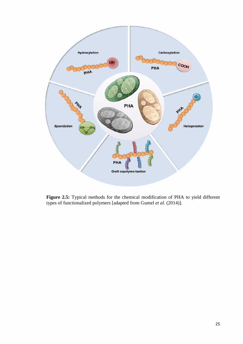
25
Figure 2.5: Typical methods for the chemical modification of PHA to yield differenttypes of functionalized polymers [adapted from Gumel et al. (2014)].

26
groups are also open for further modification reactions in order to prepare novel
modified biodegradable polymers for drug delivery system and industrial applications
(Hazer et al., 1994).
Whilst mcl-PHA has attracted great interest in research due to its potential wide
applicability as biomaterials, its strong hydrophobicity, slow degradation under
physiological conditions and lack chemical functionalities hinder the realization of its
potentials. These factors restrict the scope of their applications in biomedical field. In
order to expand the range of its versatilities, other properties such as mechanical
strength, surface features, amphiphilicity and degradation rate have to be modified to
match the requirements of specific applications (Kai & Loh, 2013). For example,
intrinsic hydrophobic properties of mcl-PHA restrict their applications as cell
colonizing materials. Therefore, chemical modification with suitable functional groups
or modification of the surface topography of mcl-PHA is needed in order to minimize
hydrophobic interactions with the surrounding tissue. Amphiphilic copolymers could be
produced through chemical modification reactions by inserting the hydrophilic
segments into the hydrophobic PHA (Hazer, 2010).
2.7.1 Graft copolymerization
Graft copolymerization is one of the method to modify PHA, which results in
the formation of a modified segmented copolymer with improved properties such as
increased wettability and thermo-mechanical strength (Gumel et al., 2014). Several
studies on graft copolymerization of mcl-PHA have been conducted in order to modify
mcl-PHA properties by chemical and physical methods, such as blending, crosslinking,
and graft copolymerization (Hazer & Steinbüchel, 2007; Meng et al., 2014). Among the
various methods, graft copolymerization is a versatile technique to introduce functional

27
groups on a polymer (Chung et al., 2012). Effective chemical modifications include
changes in chemical group functionality, surface charge, hydrophilicity, and wettability
(Lao et al., 2007). Grafting reaction can be induced by either chemical, radiation or
plasma discharge method (Kim et al., 2007; Nguyen, 2008). There are several
established ways of grafting, focusing on PHA as the polymer of interest namely
“grafting onto”, “grafting from” and “grafting through” or macromonomer method
(Nguyen, 2008).
“Grafting onto” method involved the covalent coupling of reactive sites
distributed along the copolymer main chain with end groups of copolymer segments
(Nguyen, 2008). Examples of grafting onto process include amidation or condensation
reaction between carboxylic group of PHO, PHBV and linoleic acid with amine groups
of chitosan (Arslan et al., 2007a), esterification of PHB and PHBV treated by ozone
treatment with acylic acids (Hu et al., 2003) and also free radical reaction of polystyrene
consisting active peroxide group reaction with poly(β-hydroxynonanoate) (Hazer,
1996). “Grafting from” method is another grafting mechanism of having active sites
along the main polymer chain, with grafting monomer polymerized from the sites. For
instance, the monomer can be copolymerized using the main polymer chain as
macroinitiator with multiple initiation sites along its chain (Nguyen, 2008). Arslan et al.
(2007b) reported that ‘‘grafting from’’ technique led to poly(3-hydroxybutyrate)-g-
poly(methylmethacrylate) (PHB-g-PMMA) brush type graft copolymers following atom
transfer radical polymerization of methyl methacrylate, MMA, in the presence of
cuprous bromide as catalyst.
Furthermore, “grafting through” or macromonomer method can be achieved by
copolymerization of a low molecular weight monomer with a macromonomer, which
can be defined as a polymer or oligomer bearing at least one polymerizable end group.
This method offers more control on branching formation (Nguyen, 2008). The

28
macromonomer is selected and prepared prior to copolymerization, whereas the
macromonomer arrangement can be homopolymer, random or block copolymer. The
size of the side chains is also selected with the length of the macromonomers and their
distribution along the main chain is controlled by the comonomer reactivity ratios.
Nguyen and Marchessault (2004) studied the synthesis of PMMA-g-PHB by
copolymerizing PHB macromonomers with methyl methacrylate. PHB macromonomers
were prepared from the esterification of oligomers with 2-hydroxyethyl methacrylate at
their carboxylic acid end.
Covalent grafting of functional groups is preferred to physical coating
(adsorption) in order to achieve a surface chemistry that would remain stable in
biological environment. In covalent modification, the presence of functional group
facilitates better attachment of bioactive molecules or species onto the surface that leads
to a higher surface stability. Moreover, chemically modified surface offer greater
biocompatibility towards cell growth and flow of body fluids due to their enhanced
wettability (Katti et al., 2008).
2.7.2 Chemical modification of PHA via graft copolymerization reaction
PHA-grafted copolymer can also be produced by radical polymerization of
monomers/oligomers that contain vinyl or methacrylate groups. Several studies on graft
copolymerization of PHA have been conducted by using graft chains of polyethylene
glycol (PEG) (Chung et al., 2003), 2-hydroxyethylmethacrylate (HEMA) (Lao et al.,
2007), poly(methylmethacrylate) (PMMA) (Ilter et al., 2001), poly(styrene peroxide)
(PS-P) (Cakmakli et al., 2001) and poly(N-vinylpyrrolodone) (Wang et al., 2007)
(Figure 2.6). Chung et al. (2003) reported that the presence of PEG chains in the

29
Figure 2.6: Free radical grafting of MMA and HEMA on PHA using benzoyl peroxide(BPO) as an initiator [adapted from Li et al. (2016)].
polymer network helped to increase the hydrophilicity of the final product. Their results
showed significant concentrations of water within the PHO-g-PEG polymers hence
resulting in low interfacial tension with blood. Thus, PHO-g-PEG polymer network
could form an essential component for materials that are employed in blood contacting
devices (Li et al., 2016). Similar study was reported by Kim et al. (2005b) where the
water uptake of the PHO-g-PEO copolymer increases up to 30 % in comparison with
the water uptake of PHO that is only 2 %. These graft copolymers could potentially be
used as blood-contacting devices in a broad range of biomedical applications because of
their excellent blood compatibilities. Kim et al. (2008) grafted glycerol 1,3-
diglycerolate acetate (GDD) onto poly-3-hydroxyoctanoate (PHO) using free radical

30
polymerization reaction. The surface and bulk of GDD-g-PHO copolymers became
increasingly hydrophilic as the grafting density increased. In vitro studies showed the
biocompatibility of Chinese hamster ovary (CHO) cells and adsorption of blood
proteins and platelets towards PHO were enhanced by GDD grafting. Ilter et al. (2001)
studied the graft copolymerization of methyl methacrylate (MMA) onto unsaturated
mcl-PHA. They reported that the presence of double bond opens up potential site for
functionalization of biopolyester in order to improve its mechanical and viscoelastic
properties. For a number of graft copolymers reported, the nature of copolymer and
graft copolymer composition affect properties such as thermal stability, mechanical
resilience, as well as enzymatic degradability, biodegradability, antibacterial activities
and cell compatibility (Nguyen, 2008).
The use of multifunctional monomers in graft copolymerization of polymers is
very effective in producing novel copolymers with high thermal stability and
mechanical strength (Avci & Mathias, 2004). Glycerol 1,3-diglycerolate diacrylate
(GDD) is an excellent example of a multifunctional monomer that could be potentially
grafted onto mcl-PHA. It possesses a number of hydroxyl groups useful for enhancing
the hydrophilicity of its polymer graft. Furthermore, unreacted pendant hydroxyl groups
in grafted polymers provide potential target sites for chemical modifications to further
improve the physical properties of the main polymer. For example, GDD-g-PHO
copolymer is envisaged for a broad range of biomedical applications due to their
improved physical properties, and excellent blood and cell compatibilities (Kim et al.,
2008). Therefore, development of novel mcl-PHA grafts with improved physico-
chemical and biocompatibility properties is needed to extend their range of applications.
31
2.7.3 Mechanism and kinetic of free radical polymerization
The kinetic of a chemical reaction depends on the mechanism involved in the
reaction. It is possible to investigate the mechanism of polymer grafting by considering
its kinetics. The mechanism of polymer networks grafting can be classified depending
on relative dimensions of the reacting species (ex: polymer/polymer mechanism,
polymer/monomer mechanism or monomer/monomer mechanism) or the chemical
nature of the intermediate in the reaction (Bhattacharya et al., 2009). The most
important intermediates for grafting are free radicals. In polymer/monomer
mechanisms, a polymer may be subjected to conditions where the reactive centres are
generated on it from which new polymer chains may grow (“grafting from” reaction), or
to which new growing polymer chains may be attached (“grafting onto” reaction) (Pham
et al., 2000). Radicals can be introduced via thermal or chemical decomposition of an
initiator, by redox reaction between two species, or by excitation of a photoinitiator.
Radical intermediates can also be introduced by exposure of a material to energies
sufficient to cleave chemical bonds homolytically, using γ irradiation, photoirradiation,
thermal treatment, or an electron beam (Bhattacharya & Misra, 2004; Li et al., 2016).
The mechanism for the graft copolymerization generally consists of three major
steps namely initiation, propagation and termination. The mechanism can be described
as an addition reaction of radicalized unit to the reactive site to form higher molecular
mass polymer. As the reaction proceeds, monomer concentration will decrease,
expected to be linked to the polymer backbone (Jenkins & Hudson, 2001). In the
chemical process, free radicals are produced from the initiators and transferred to the
substrate to react with monomer to form the graft copolymers (Bhattacharya & Misra,
2004).

32
Initiation is the first reaction of a chain carrier with a polymer or monomer
molecule to generate a new covalent bond and regenerate the reactive centre at a new
location. This initiation may occur by addition, where the radical adds across a double
bond, or by abstraction, where the radical removes a labile atom from a substrate. The
thermodynamic driving force of these reactions is the replacement of a high energy π
bond by an σ bond (addition) or the replacement of a weaker R−H or R−X bond by a
stronger R′−H or R′−X bond (abstraction) (Bhattacharya et al., 2009). In addition,
initiation step begin with the radicalization of monomer or polymer substrate by the
action of radical initiator such as benzoyl peroxide and eventually generating radicals.
Radicalized monomers undergo chain transfer to the backbone of the polymer.
Therefore, radical formation is the process to enable the monomer to transfer to the
main polymer backbone (Wang et al., 2007).
Propagation is the next step in generating a graft copolymer by a free radical
polymer/monomer mechanism. The overall kinetics of free radical polymerization
reaction will be dominated by this step (Bhattacharya et al., 2009). Moreover, a
particular polymer reactive site grafted with monomer will create propagation of
radicalization, thus radicalizing other site of the backbone. This will allow another chain
transfer reaction, forming successful graft copolymerization. However, radicalized
monomers from the initiation step also have the ability to perform chain transfer among
themselves to undergo unintended homopolymerization process (Wang et al., 2007).
Termination occurs by the pairing of two radicals, stopping both the kinetic of
chain reaction and physical polymer chain extension. Discontinuation of chain
extension in radical polymerization occurs with the pairing of two radicals to form
nonradical species. Termination step will come ultimately, resulting in graft copolymer
and homopolymer as final product due to recombination process. The occurrence is
affected by several factors such as temperature, monomer concentration and initiator

33
concentration, and consequently determines the grafting yield of the copolymer (Lao et
al., 2007; Lao et al., 2010; Lee & Lee, 1997; Wang et al., 2007).
2.8 Mcl-PHA in pharmaceutical and medical applications
2.8.1 Bone tissue engineering
Recently, several bone tissue engineering strategies such as cell transplantation,
acellular scaffolds, gene therapy, stem cell therapy, and growth factor delivery have
been applied to address the challenging requirements of a suitable biomimetic bone
scaffold (Porter et al., 2009). Bone forms the structural framework of the body and is
composed of an inorganic mineral phase of hydroxyapatite (60% by weight) and an
organic phase, which is mainly type I collagen. Tissue engineering has developed in
recent years to overcome the problem of bone loss or defect. Therefore, extensive
studies on various materials for scaffold preparation have been conducted in order to
meet osteocompatibility demand and mechanical properties which are similar to native
bones. Wang et al. (2004) studied the in vitro biocompatibility of 3D scaffold PLA,
P(3HB), and P(3HB-co-3HHx) inoculated on rabbit bone marrow cells. Their results
showed that the cells on the P(3HB-co-3HHx) scaffolds were able to sustain osteoblast
phenotypes, high alkaline phosphatase activity (ALP), round cell shape, fibril collagen
synthesis and strong calcium deposition. The cells also showed best proliferation on the
P(3HB-co-3HHx) scaffolds. They found that after incubation for 10 days, the number of
cells grown on P(3HB-co-3HHx) scaffolds was approximately 40 % higher than on
P(3HB) scaffolds, and 60 % higher than on PLA scaffolds. ALP activity of the cells
grown on P(3HB-co-3HHx) scaffolds increased to about 65 U g-1 of the scaffold, 50 %
higher than P(3HB) and PLA scaffolds, respectively. From SEM analysis, it was
observed that P(3HB-co-3HHx) scaffolds had suitable roughness for osteoblast

34
attachment and proliferation compared to the ones made from P(3HB) and PLA. Thus,
P(3HB-co-3HHx) was determined as an attractive biomaterial for osteoblast attachment,
proliferation and differentiation for bone marrow cells (Hazer et al., 2012).
2.8.2 Drug delivery system
Polymers could play a central role in controlled of drug release systems and
fabrication of drug delivery devices. Controlled drug delivery systems deal with
releasing therapeutic compounds at a desired rate so that the drug level in the body can
be sustained within the therapeutic time frame (Bayram et al., 2008; Nair & Laurencin,
2006). The drug delivery systems are developed for releasing, targeting, uptaking,
retaining, activating, bringing and localizing the drugs at the right time, place, dose and
period. The drug release kinetics can be controlled via manipulating the PHA matrix
parameters to reach desired degradation rates (Hazer et al., 2012). Scl-PHA are
primarily degraded by surface erosion that makes them an attractive candidate to be
applied as drug carriers. However, since scl-PHA are crystalline and hydrophobic, many
pores are formed on the surface. It results in too rapid release of drugs without
significant polymer degradation. On the other hand, mcl-PHA copolymers have low
melting point and low crystallinity, therefore they are more suitable for drug delivery
applications (Rai et al., 2011).
Drug delivery systems can be prepared in different shapes and attributes such as
gels, films, microcapsules, microspheres, nanoparticles, porous matrices, polymeric
micelles and polymer-linked drugs. The physical interactions are generally preferred in
order to bind the drug to the polymer since it will not damage the molecular structure of
the drug (Hazer et al., 2012; Kabilan et al., 2012). Drugs can be entrapped or
microencapsulated in a PHA homopolymer or copolymer. Microsphere or
microcapsule-based delivery systems have been extensively used for the delivery of a

35
number of drugs such as anesthetics, antibiotics, anti-inflammatory agents, anticancer
agents, hormones, steroids, and vaccines. In addition, the introduction of such a delivery
system at the target location also results in site-specific drug delivery (Shrivastav et al.,
2013). The biocompatible and biodegradable properties of PHA serve as a real
advantage to provide excellent matrix for controlled drug delivery (Kabilan et al., 2012;
Pouton & Akhtar, 1996).
Kim et al. (2005a) synthesized monoacrylate-poly(ethylene glycol)-grafted
poly(3-hydroxyoctanoate) (PEGMA-g-PHO) copolymer to develop a swelling
controlled release delivery system for ibuprofen as a model drug. Their results showed
hydrolytic degradation of the copolymer was strongly dependent on the degree of
grafting of the PEGMA group. The in vitro degradation rate of the copolymer films
increased with higher degree of grafting of PEGMA group on the P(3HO) chain. The
copolymer films showed a controlled delivery of ibuprofen to the medium in certain
periods of time depending on the composition, hydrophilic/hydrophobic characteristics,
initial drug loading amount and film thickness of the graft copolymer support. The
research showed that a combination of low degree grafting of PEGMA group in the
P(3HO) chains and low ibuprofen solubility in water led to a long-term constant release
from these matrices in vitro.

36
2.8.3 Cardiovascular system
PHA has been widely used in the cardiovascular area such as artery augments,
cardiologic stents, vascular grafts, heart valves, pericardial patches, implants, dressing
tablets and microparticulate carriers (Philip et al., 2007). Vascular grafting is a
frequently used technique in cardiovascular pathologies. The ideal cardiovascular patch
material should have resistance to degradation and infection, in addition to long
durability, amendable to various size tailoring to suit cardiac and peripheral vascular
reconstructions, lack of immunogenicity and non-toxic (Hazer et al., 2012).
Tissue-engineered heart valves may have the potential to overcome
shortcomings of prosthetic valves and homograft valves that are currently being used in
valve replacements. Many studies have been carried out using several synthetic
absorbable polyesters such as PGA, PLA and scl-PHA viz. P(3HB), as potential
scaffolding materials for heart valves. However, these materials are too stiff to function
as flexible leaflets inside a trileaflet valve and therefore the relatively elastomeric PHA
is considered to be more promising (Rai et al., 2011). One of the early studies using an
elastomeric P(3HO) for the fabrication of a trileaflet heart valve scaffold was carried out
by Sodian et al. (2000). Vascular cells were harvested from ovine carotid arteries,
expanded in vitro and seeded onto the heart valve scaffold. They reported that when the
artificial trileaflet scaffold was incorporated in the animal model, all test subjects
survived the procedure and the valves showed minimal regurgitation (Sodian et al.,
2000).
Biotechnology start-up such as Tepha Inc., based in Cambridge, MA, has been
devoted to manufacturing pericardial patches, artery augments, cardiological stents,
vascular grafts, heart valves, implants and tablets, sutures, dressings, dusting powders,
prodrugs and microparticulate carriers using PHA (Philip et al., 2007). The first PHA-

37
based product approved by the United States Food and Drug Administration (FDA) for
clinical application is the TephaFLEX® absorbable suture prepared from P(4HB). The
most remarkable property of P(4HB) is its very high elasticity that benchmarks closely
to ultrahigh molecular weight polyethylene which can be stretched 10 times than its
original length before breaking (Martin & Williams, 2003). In 2007, the FDA had
cleared its marketing in the US, indicating a bright future for PHA practical applications
in the biomedical field (Wu et al., 2009).

38
CHAPTER 3
MATERIALS AND METHODS
3.1 Materials
3.1.1 Microorganism
The bacterial species used in the studies is a Gram-negative Pseudomonas putida
BET001 able to biosynthesize medium-chain-length polyhydroxyalkanoates (mcl-PHA).
The microorganism was obtained from Bioprocess and Enzyme Technology Laboratory,
Institute of Biological Science, Faculty of Science, University of Malaya. It was isolated
from a palm oil mill effluent (POME) (Gumel et al., 2012).
3.1.2 Media
Two types of media were used for growth and PHA accumulation by P. putida
BET001. Nutrient rich (NR) was used for growing cell biomass and E2 mineral medium
was used for PHA accumulation by the microorganism (Gumel et al., 2012).
Compositions of NR (Table 3.1) and E2 media (Table 3.2) are as follows:
Table 3.1: Nutrient rich medium (NR)
Ingredients Mass (g) in 1.0 L distilled water
Yeast extract
Nutrient broth
10.0
15.0
Ammonium sulphate 5.0

39
Table 3.2: E2 medium
Ingredients Mass (g) in 1.0 L distilled water
NaNH4HPO4.H2O
K2HPO4
3.5
5.7
KH2PO4 3.7
Mineral solutions Volume (mL) in 1.0 L distilled water
MgSO4.7H2O (0.1 M)
Trace elements (MT)
10.0
1.0
The trace elements (MT) were dissolved in 1.0 L of 1 M HCl and its composition is
shown below:
Table 3.3: MT solution
Ingredients Mass (g) in 1.0 L of 1 M HCl
CaCl2.2H2O 1.5
CoSO4.7H2O 2.4
CuCl2.2H2O
FeSO4.7H2O
0.2
2.8
MnCl2.4H2O 2.0
ZnSO4.7H2O 0.3
3.1.3 Shaker incubator set-up
Shake-flask fermentation was carried out in an orbital shaker incubator
(HOTECH, Model 721, Taiwan). The incubation temperature and agitation speed for
the incubator were set at 30 °C and 200 rpm, respectively, throughout the experiments.

40
3.1.4 Stirred tank bioreactor set-up
The fed-batch fermentation for mcl-PHA production was performed in a 2-L
stirred fermenter (Biostat®A, Sartorius, Germany), a culture vessel made of borosilicate
glass equipped with a cooling finger for temperature regulation. The pH, temperature
and partial pressure of oxygen (pO2) of the culture media were measured and regulated
by a digital system. All the reservoir bottles that contained the carbon substrate
(octanoic acid), ammonium solution, antifoam, acid and base solutions were connected
to the bioreactor with autoclavable peroxide-cured silicone tubings. Before autoclaving,
all the tubes and filters were clamped except the exhaust line filter. The filters were
wrapped with non-absorbent wool and aluminium foil to prevent water vapor from
entering into the filters. The temperature sensor, pH probe (Mettler-Toledo) and pO2
probe (Mettler-Toledo) were checked and calibrated prior to the experiments. The pH
probe and temperature sensor were connected to the bioreactor and calibration of pH
probe was carried out using pH 4 and 7 buffer solutions at 25 °C before autoclaving.
After the glass vessel setup was autoclaved at 121 °C and 103 kPa for 15 min and
allowed to cool down to room temperature, the pO2 electrode was connected to the
digital system to polarize the pO2 probe for at least 6 hours. Upon polarization, the pO2
probe was calibrated using industrial grade nitrogen gas followed by industrial grade
oxygen gas. Cool circulating water was allowed to pass through the cooling finger to
help regulate the temperature. The stirrer motor was fitted to the bioreactor and the pH
and temperature of the medium was set at pH 7 and 30 °C, respectively. Agitation of the
liquid culture was achieved using a single impeller (six-bladed Rushton turbine) placed
directly above the ring-shaped gas sparger. Aeration of the culture was provided by
pressurized filtered air supplied at 0.5 vvm. The impeller speed was set at 600 rpm
throughout the fermentation (Annuar, 2004; Chan, 2012). The setup of the bioreactor
for fed-batch fermentation is shown in Figure 3.1.

41
Figure 3.1: Setup of a 2-L stirred tank bioreactor for fed-batch fermentation
3.1.5 Sterilizer
Sterilization was carried out at 121 °C and 103 kPa for 15 minutes using Tomy
SX-500 autoclave (Japan).
3.1.6 Centrifugation
Centrifugation was carried out using Thermo-Line MLX-210 mini centrifuge
(China), Hettich Zentrifugen EBA 20S bench-top centrifuge and Thermo Scientific
Sorvall RC-5C Plus ultracentrifuge (USA) for liquid volumes of 1.0, 10.0 and 50.0 mL,
respectively. The revolution speed and temperature of the ultracentrifuge machine were
fixed at 3,578 × g and 4 °C respectively.
3.1.7 Vacuum evaporation
Rotary evaporator model RE300 from Yamato Scientific Co., Ltd. (Japan) was
used to carry out evaporation of volatile liquid samples.

42
3.1.8 Spectrophotometer
Spectrophotometric analysis was performed using Jasco V-630
spectrophotometer equipped with temperature controller (Model EHC-716 Japan).
3.2 Method
3.2.1 Maintenance of culture stock
P. putida BET001 was maintained on nutrient rich (NR) agar slant and stored at
4 °C. Weekly sub-culture was made on NR agar plate to check for viability and
contamination. For long term maintenance, 40 % glycerol stock was added into the agar
slant in the ratio of 1:1 and then kept at -20 °C.
3.2.2 Media preparation
Nutrient rich (NR) medium was prepared by mixing the components as listed in
Table 3.1 in 1.0 L distilled water. Then, medium was autoclaved at 121 ºC and 103 kPa
for 15 minutes.
The E2 medium was prepared by dissolving ammonium and phosphate salts in
1.0 L distilled water followed by autoclaving at 121 ºC and 103 kPa for 15 minutes.
MgSO4.7H2O, carbon source and trace elements (MT) were autoclaved separately.
Aforementioned step was taken to avoid the possible precipitation of salt ingredients
due to the high heat and pressure during autoclaving. The initial pH of the medium was
7.0 ± 0.2.
43
3.2.3 Estimation of total biomass
The total biomass (residual biomass and PHA) was determined by gravimetry
method. During sampling, 1.0 mL of the culture was pipetted into Eppendorf microfuge
tube. The cells were spun down at 3,578 × g for 5 minutes. The supernatant was
decanted and the pellet was washed twice with saline solution (0.9 % w/v). After that,
the tubes were dried inside an oven at 65 °C until constant weight.
The total biomass was also determined using spectrophotometer. 1.0 mL culture
was centrifuged and washed as described above. Then the cell pellet was diluted with
saline solution and the optical density of the cells was read at 600 nm (OD600 nm). The
calibration of known amount of dried biomass to its corresponding optical density at
600 nm was used for quick estimation of total biomass in the culture during
fermentation run. The calibration was done as follows:
Bacterial culture was grown in 100 mL of NR medium in shake-flasks for 24
hours at 30 °C. Then, 50 mL of culture was harvested and spun down at 3,578 g for 5
minutes at 4 °C. The cell pellet was then washed with saline solution and re-centrifuged.
The washing and centrifugation steps were repeated three times before the cell pellet
was dried in hot air oven at 65 °C until constant weight. Another 50 mL of the liquid
culture was diluted accordingly in saline solution to obtain OD600 nm less than 1.0.
Several calibration points were recorded (Figure 3.2) with a replicate for each. The
difference within the replicate did not exceed ± 0.1 unit OD600 nm.
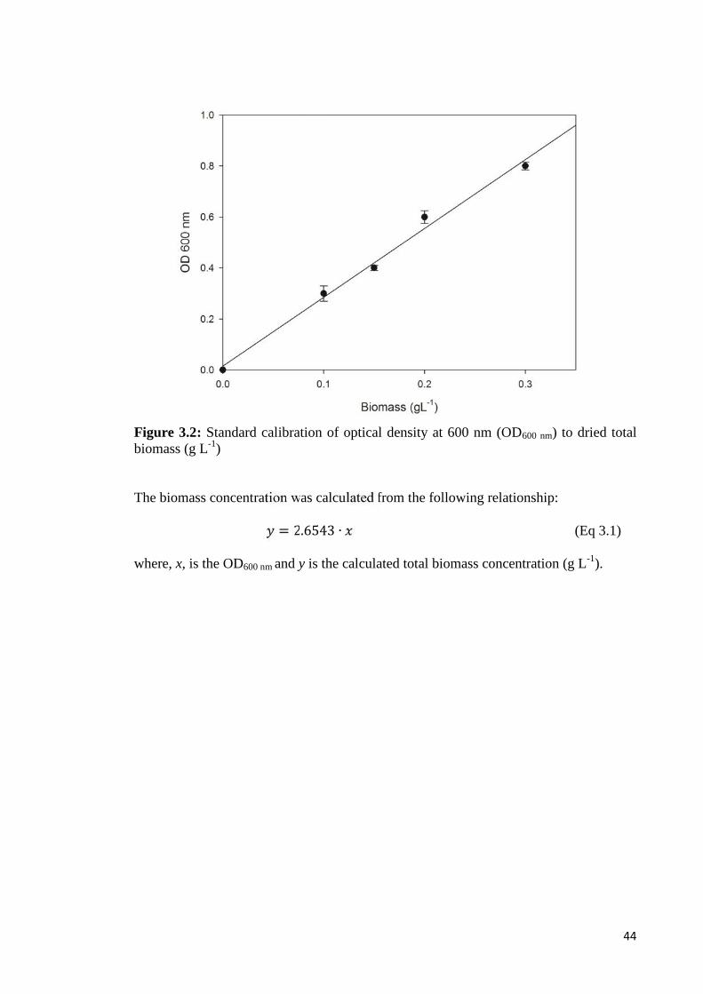
44
Figure 3.2: Standard calibration of optical density at 600 nm (OD600 nm) to dried totalbiomass (g L-1)
The biomass concentration was calculated from the following relationship:= 2.6543 ∙ (Eq 3.1)
where, x, is the OD600 nm and y is the calculated total biomass concentration (g L-1).

45
3.2.4 Determination of optimum carbon-to-nitrogen (C/N) mol ratio to be used as
supplementation solution in the fed-batch fermentation
P. putida Bet001 was used as the producer strain for mcl-PHA accumulation.
Effects of different initial C/N mol ratio on dry cell weight (g L-1) and mcl-PHA content
(wt %) were determined in shake flasks. Different C/N mol ratios of carbon (octanoic
acid, C8H16O2) and nitrogen (sodium ammonium hydrogen phosphate tetrahydrate,
NaNH4HPO4.H2O) sources were prepared at 5, 10, 15, and 20. Sterile NR medium (30
mL per each 100 mL conical flask) was used as medium for growing the bacterial
inoculum. A loopful of culture from NR agar was aseptically introduced into 30 mL NR
liquid medium in shake flasks and cultivated in an aerobic condition at 25 °C and 200
rpm for 24 h. Then, 10 % (v/v) of cell inoculum from NR broth was transferred into E2
medium with different C/N mol ratios (100 mL per each 250 mL conical flask). The
cultures were incubated for 24 hours at 30 ºC with 200 rpm agitation in shake flasks.
After 24 hours cultivation, the culture was centrifuged at 3, 578 × g for 5 minutes at 4
°C. Then, the cells were washed twice with saline solution (0.9 % w/v) followed with n-
hexane before oven dried (60 °C) until constant weight.
3.2.5 Determination of volumetric oxygen mass transfer coefficient (KLa) using
static gassing-out method
The volumetric oxygen mass transfer coefficient, KLa was determined in a
stirred tank bioreactor using static gassing-out method. The experiment was carried out
in order to estimate the oxygen mass transfer efficiency of the bioreactor system. The
conditions were similar to the actual fermentation run and using the same equipment
setup. The aqueous phase consisted of actual composition of the fermentation E2
medium with 3 g L-1 of octanoic acid in 1.0 L of total working volume. The aqueous
medium was first deoxygenated by sparging gaseous nitrogen until all traces of oxygen

46
was stripped away (oxygen partial pressure, pO2 = 0 %). Then pressurized air was
sparged into the bioreactor at 0.6 L min-1 and at 600 rpm agitation rate. The increase in
% pO2 was recorded at regular intervals until readings became constant which indicated
the saturation of the liquid medium with oxygen (Annuar et al., 2007).
The KLa was determined using the relationship:
dCL/dt = KLa (C*L – CL) (Eq. 3.2)
where,CL : % pO2 value at time t;C*L : dissolved oxgen concentration in equilibrium with the gas phase
(constant % pO2);t : time (second);KLa : volumetric oxygen mass transfer coefficient (s-1).
Integration of equation (Eq 3.2) yields
ln (C*L – CL) = - KLa . t + ln C*L (Eq. 3.3)
KLa value was determined directly from the slope by plotting of ln (C*L – CL) versus t.
3.2.6 Batch cultivation of P. putida BET001 in stirred tank bioreactor
Bacterial cells were pre-cultured in NR liquid medium using orbital shaker
incubator at 30 ˚C and 200 rpm agitation for 24 hours. Then, 10 % (v/v) of inoculum
from the broth was transferred into 100 mL of E2 medium in shake flasks. Octanoic
acid (3 g L-1) was included in the E2 medium as the sole carbon and energy source. The
shake flasks were incubated in an orbital shaker incubator at 30 ˚C and 200 rpm
agitation for 18 hours. Subsequently, the whole content of a flask was used to inoculate
E2 medium in a 2-L stirred tank bioreactor.
Batch fermentation was employed to study the growth profile of the bacteria in a
controlled stirred tank bioreactor in order to determine the appropriate feeding points of
C/N solution to the culture during fed-batch fermentation later. Batch cultures were
cultivated at 30 ˚C, 600 rpm agitation, constant pH 7.0 regulated by the addition of 0.5
47
N KOH and 0.5 N H2SO4 and 0.5 vvm aeration in the stirred tank bioreactor. Inoculum
at 10 % (v/v) was seeded into 0.9 liter of E2 medium in the bioreactor (concentration of
E2 components were adjusted for 1-L). The initial concentration of octanoic acid in the
bioreactor was 3 g L-1. After 48 hours of fermentation, the culture medium was pumped
out from the vessel. The biomass was subsequently harvested by centrifugation.
3.2.7 Biosynthesis of mcl-PHA in fed-batch cultivation
Bacterial cells were pre-cultured in NR medium using orbital shaker incubator at
30 ˚C and 200 rpm agitation for 24 hours. Then, 10 % (v/v) of inoculum from the broth
was transferred into 100 mL of E2 medium in shake flasks. Octanoic acid (3 g L-1) was
included in the E2 medium as sole carbon and energy source to acclimatize the culture
to the carbon substrate and therefore helped to reduce the lag phase during cell
cultivation in 2-L bioreactor later. The shake flask culture was incubated in an orbital
shaker incubator at 30 ˚C and 200 rpm agitation for 18 hours. Subsequently, the whole
flask content was used to inoculate E2 medium for mcl-PHA production in a 2-L stirred
tank bioreactor.
Fed-batch fermentation was employed in order to extend the cell growth phase
and concomitant mcl-PHA accumulation. Fed-batch cultures were cultivated at 30 ˚C in
the stirred tank bioreactor. About 10 % inoculum (v/v) was used to inoculate 0.9 liter of
E2 medium in the bioreactor (concentration of E2 components were adjusted for 1-L).
The initial concentration of octanoic acid in the bioreactor was 3 g/L. A solution with
C/N mol ratio of 10 (10.3 ml/L) was added to the culture at 12- and 24-hour intervals.
After 48 hours of fermentation, the culture medium was pumped out from the vessel.
The biomass was subsequently harvested by centrifugation.

48
3.2.8 Cell harvesting
Bacterial cells were aseptically harvested by centrifugation (Thermo Scientific Sorvall
RC-5C Plus ultracentrifuge, USA) at 3,578 × g for 10 minutes at 4 °C. The cells were
washed twice with sterile saline (0.9 % w/v) and then with n-hexane to remove excess
fatty acids. The pellets were dried in dry-air oven (60 °C) until constant weight. Cell
concentration was expressed as total cell dry weight concentration (CDW, g L−1).
3.2.9 PHA extraction and purification
Intracellular PHA was extracted by suspending the dried cells in analytical grade
acetone (C3H6O, Mw 58.08 g mol-1, Merck) and refluxed for 4 hours at 70 ˚C. The
PHA-acetone solution was filtered through Whatman No. 1 filter paper to remove
cellular debris, and the filtrate was concentrated by rotary evaporation (EYELA N-1000
rotary evaporator, Japan). The polymer was purified by drop-wise addition of the extract
into rapidly stirred analytical grade methanol (CH3OH, Mw 32.04 g mol-1, Merck)
chilled in ice bath. Further purification was performed by re-dissolving the PHA extract
in a small amount of acetone and re-precipitating it in cold methanol. Then, it was dried
in a vacuum oven (JEIOTECH Model OV-11/12, Korea) at 40 ˚C, 0.6 atm for 24 hours
(Baei & Rezvani, 2011; Chardron et al., 2010).
3.3 Fabrication of P(3HO-co-3HHX)/HA composite scaffold
3.3.1 Material
Hydroxyapatite (HA) powder (HCa5O13P3, MW 502.31, CAS 12167-74-7) was
purchased from Sigma Aldrich (~15-20 µm). Acetone (CAS 67-64-1) and sodium
chloride (CAS 7647-14-5) were purchased from Merck Millipore, Darmstadt, Germany.
Analytical grade chemicals were used throughout the study.

49
3.3.2 Preparation of composite P(3HO-co-3HHX)/HA scaffold
The P(3HO-co-3HHX)/HA composite scaffold was fabricated using solvent casting-
particulate leaching technique (Figure 3.3). Agglomeration of the particles was
prevented by placing the solution in a Multi-frequency Ultrasonic Bath SB-300DTY
(Ningbo Scientz Biotechnology Co., Zhejiang, China). A combination of ultrasonication
and solution casting method was applied to achieve a well-dispersed P(3HO-co-
3HHX)/HA matrix. The scaffold was fabricated as follows: 0.6 g of the polymer was
dissolved in 6 mL of acetone followed by the addition of HA powder. The amount of
HA particles was 10 weight % and 30 weight % relative to the polymer (Moradi et al.,
2013). After 20 minutes ultrasonication (25 kHz, 340 W), NaCl was added to the
mixture and stirred for 15 min. The size range of salt particles was 100 - 200 µm and its
weight fraction was 90 %, based on the total mass of polymer and salt. Subsequently,
the mixture was poured into a glass Petri dish (6-cm diameter) as a casting mold. After
48 h of drying at room temperature (25 ± 1 °C), samples were dried in an oven (40 °C)
under vacuum for 24 h. After the drying process, dried and solidified sample was
removed from the Petri dish. The samples from salt-leaching process were soaked in
deionized water (minimum 300 mL) with light stirring using magnetic stirrer. The
soaking was carried out for five consecutive days with daily changes of fresh deionized
water. Then, the samples were dried out at 25 ± 1 °C under vacuum for 48 h. The pure
P(3HO-co-3HHX) scaffold for control experiment was fabricated using the same
method without the addition of HA particles.
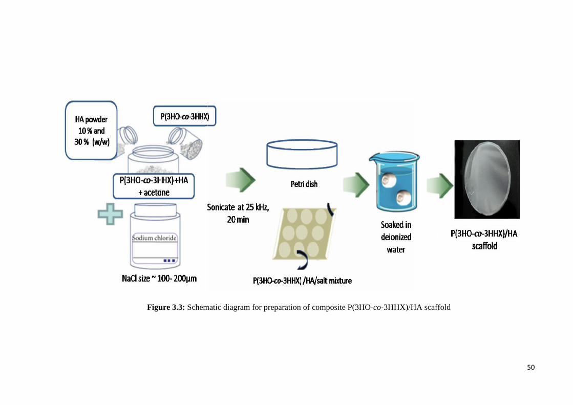
50
Figure 3.3: Schematic diagram for preparation of composite P(3HO-co-3HHX)/HA scaffold
50
Figure 3.3: Schematic diagram for preparation of composite P(3HO-co-3HHX)/HA scaffold
50
Figure 3.3: Schematic diagram for preparation of composite P(3HO-co-3HHX)/HA scaffold

51
3.3.3 Characterization of polymer composite
3.3.3.1 FTIR-ATR spectroscopy
A nondestructive attenuated total reflectance Fourier transform infrared spectra
of the control references and the composite polymer were recorded on Perkin-Elmer
Spectrum 400 FT-IR and FT-NIR Spectrometer (Perkin-Elmer Inc., Wellesley, MA,
USA) equipped with PIKE GladiATR hovering monolithic diamond ATR accessory
(Pike Technologies Inc., Fitchburg, USA). Control samples and their composites were
placed on the monolithic diamond ATR probe and clamped against the diamond crystal
plate using the force adapter. Thereafter, the samples were scanned over a range of
4000–400 cm−1 at 25 °C (Gumel et al., 2014).
3.3.3.2 X-ray diffraction (XRD) analysis
Crystallinity of mcl-PHA was investigated using a PANalytical EMPYREAN
(PANalytical, Almelo, Netherlands). Five milligrams of PHA was dissolved in 2 mL
dichloromethane. Then, it was casted on a glass slide, and allowed to dry overnight. A
20-minute scan was run on each sample with the X-ray settings held at 40 kV and 40
mA. The scan range was from 10° to 70° 2θ.
3.3.3.3 Differential scanning calorimetry (DSC)
The analysis was carried out using a Mettler-Toledo differential scanning
calorimeter (DSc 822e;Mettler-Toledo, Columbus, OH, USA) running on STARe DSc
ver 8.10 software, equipped with a HAAKE EK90/MT digital immersion cooler
(Thermo Fischer Scientific, USA). About 5 mg sample was encapsulated in an
aluminium pan. Analysis was performed at a programmed temperature range of −50 to
200 °C with a heating rate of 10 °C min−1 under a nitrogen flow rate 50 mL min−1 at a
head pressure of 1.5 bars. The melting temperature (Tm) was taken at the endothermic
52
peak of the DSC thermogram. The degree of crystallinity in polymer was calculated
based on the endothermic melting enthalpy (ΔHm) value obtained from DSC endotherm
with respect to ΔHm of PHB with 100 % crystallinity (140.1 J g−1) (Doi, 1990).
3.3.3.4 Surface analysis
The morphological characteristics of both control and composited polymers
were viewed in a high-resolution field emission scanning electron microscope (FESEM)
(Quanta FEG 450) (FEI, Oregon, USA). The microscope was operated at high vacuum
mode with an electron acceleration voltage of 5 kV and a working distance of about 10
mm. Thin films of neat polymer and polymer composites were mounted on brass stubs
using double-sided cellophane tape and introduced into the viewing chamber of the
instrument (Gumel et al., 2014). Energy dispersive X-ray spectrometry
(INCAEnergy200, Oxford Inst., UK) was performed in order to determine the presence
and distribution of HA particles in the composite scaffolds (Sultana & Wang, 2008).
3.3.3.5 Porosity of the scaffold
To evaluate the porosity of the composite scaffolds, they were weighed and
immersed in 95 % ethanol for one hour. Then, the samples were soaked in deionized
water overnight and their wet weights were recorded. The percentage of porosity of the
composite scaffolds was calculated using the following equation (Kuo & Leou, 2006;
Saadat et al., 2013):
Porosity (%) = − (Eq. 3.4)where, Ww is the wet weight of scaffold (g), Wd is the dry weight of scaffold (g) and Va
is the apparent scaffold volume (mL).

53
3.3.3.6 Biocompatibility study
3.3.3.6.1 In vitro cell culture
Prior to culture initiation, 5-mm diameter disks were cut from both pure P(3HO-
co-3HHX) and P(3HO-co-3HHX)/HA composite scaffolds, and sterilized through
immersion in 70 % ethanol for 15 minutes. Then, the scaffolds were rinsed with
sterilized phosphate-buffered saline and retained at room temperature for 2 hours under
aseptic condition prior to cell culture. Then, osteoblast cells were suspended in 500 µL
Dulbecco’s modified Eagle’s medium (DMEM) containing 10 % fetal bovine serum
(FBS), 2 mM L-glutamine, penicillin and streptomycin (all from Gibco-Invitrogen,
USA), and were placed on the surfaces of the disks located in the wells of 24-well
culture plates. Osteoblast cells were inoculated in a plating density of 1 x 10⁵ per
scaffold. Cell cultures were incubated at 37 °C, 5 % CO2 and saturated humidity to
allow cell adhesion to the surface of the materials and infiltration into the porous
structure. Three samples of each material were used in replicated experiments.
3.3.3.6.2 Alamar Blue assay
Alamar Blue (Biosource Int, Camarillo, USA), which detects the number of
viable cells, was used to measure the growth of cells on polymer scaffolds. At the
indicated time endpoints, the wells received 100 µL Alamar Blue per well. After
incubation at 37 °C for 3 h, the reduced resazurin dye was quantitated by FLUOstar®
Omega (BMG Optima, Ortenberg, Germany) fluorescence cell reader at 570 nm to 595
nm wavelengths. The percentage of resazurin dye reduction was calculated in order to
determine the growth of osteoblast cells.

54
3.3.3.6.3 Alkaline phosphatase (ALP) activity
The differentiation of osteoblast cells was evaluated by the expression of ALP
activity (Baek et al., 2012). For ALP activity measurement, total protein of cells on
scaffolds was extracted using 100 µl M-PER mammalian protein extraction reagent to
lyse the cells. The lysate was then centrifuged at 14,000 g at 4 °C for 15 minutes to
separate cell debris. Supernatant was collected and ALP activities were measured using
p-nitrophenyl phosphate (p-NPP) as a substrate. Following the ALP-catalyzed reaction,
p-nitrophenyl phosphate was converted to p-nitrophenol and the absorbance at 405 nm
was measured with a microplate reader (FLUOstar® Omega, BMG Optima, Ortenberg,
Germany).
3.4 Functionalization of mcl-PHA by graft copolymerization of P(3HO-co-
3HHX) with glycerol 1,3-diglycerolate acetate (GDD)
3.4.1 Material
Glycerol 1,3-diglycerol diacrylate (C15H24O9 MW 348.35 CAS 60453-84-1) was
purchased from Sigma Aldrich (Saint Louis, USA). Octanoic acid (CAS 124-07-2),
acetone (CAS 67-64-1) and benzoyl peroxide (with 25 % H₂O) for synthesis (C4H10O4
MW 242.23 CAS 94-36-0) were purchased from Merck Millipore (Darmstadt,
Germany). Analytical grade chemicals were used throughout the study.

55
3.4.2 Preparation of P(3HO-co-3HHX)-g-GDD copolymer
P(3HO-co-3HHX)-g-GDD copolymer was prepared by thermal treatment of
homogeneous acetone solution of P(3HO-co-3HHX), GDD monomer and benzoyl
peroxide (BPO) initiator (Figure 3.4). 0.2 g P(3HO-co-3HHX) was used throughout the
experiment. The reaction was carried out under nitrogen-saturated atmosphere. To
remove dissolved oxygen from the solution, nitrogen gas was bubbled into the solution
at 25 ± 1 °C for 10 minutes, and then the tube was sealed air-tight and incubated at 80
C for 4 hours. Then, the solution was poured into vigorously stirred methanol to
precipitate the copolymer. The resulting suspension was filtered and the solid product
was dried to a constant weight in a vacuum oven at 25 ± 1 °C. The product was purified
by dissolving in acetone and precipitating into methanol, followed by filtration and
vacuum drying. The mass increase of P(3HO-co-3HHX)-g-GDD due to successful
grafting was calculated according to the following equation:Mass increase due to succesful grafting (%) = × 100 (Eq. 3.5)where Mf is the mass after grafting and Mi is the initial mass of P(3HO-co-3HHX).
(Graft yield)
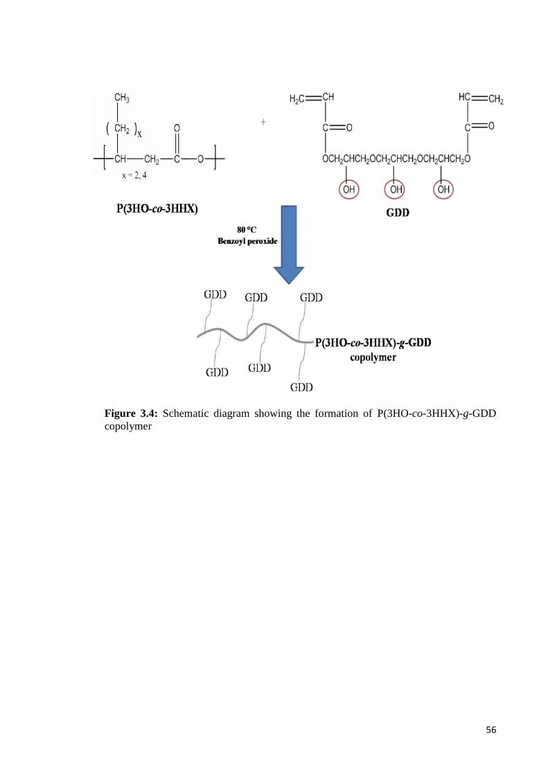
56
Figure 3.4: Schematic diagram showing the formation of P(3HO-co-3HHX)-g-GDDcopolymer
56
Figure 3.4: Schematic diagram showing the formation of P(3HO-co-3HHX)-g-GDDcopolymer
56
Figure 3.4: Schematic diagram showing the formation of P(3HO-co-3HHX)-g-GDDcopolymer
57
3.4.3 Effects of the initial monomer concentration
The procedure was carried out as described in section 3.4.2. The reaction
mixture was incubated at 80 °C with 0.04 mM BPO for 2 hours. The mass of P(3HO-
co-3HHX) was fixed at 0.2 g for the experiment. The concentrations investigated for
GDD monomer were 0.1, 0.3, 0.4, 0.6 and 0.9 mM.
3.4.4 Effects of reaction time
The procedure was carried out as described in section 3.4.2. Different
concentrations of GDD monomer (0.1, 0.3, 0.4, 0.6 and 0.9 mM) were studied at
different reaction times (0.5, 1.0, 2.0, 3.0 and 4.0 hours). The reaction mixture consisted
of 0.2 g P(3HO-co-3HHX), 0.04 mM BPO and pre-determined GDD monomer
concentration.
3.4.4.1 Determination of activation energy
The activation energy (Ea, J mol−1) for the graft copolymerization reaction was
calculated using the linearized Arrhenius equation:ln = ln − (Eq. 3.6)
where A is the frequency factor, R is the gas constant (8.3145 J mol−1 K−1), and T is the
absolute temperature (K).
3.4.5 Effects of reaction temperature
The procedure was carried out as described in section 3.4.2. Different reaction
temperatures viz. 70 °C, 80 °C and 90 °C were studied. The reaction mixture consisted
of 0.2 g P(3HO-co-3HHX), 0.4 mM GDD and 0.04 mM BPO incubated at pre-
determined temperature for 2 hours.
58
3.4.6 Effects of benzoyl peroxide
The procedure was carried out as described in section 3.4.2. Different
concentrations of radical (BPO) 0.01, 0.02, 0.04, 0.10, 0.15 and 0.20 mM were studied.
The reaction mixture consisted of 0.2 g P(3HO-co-3HHX), 0.4 mM GDD and BPO
incubated at 80 °C for 2 hours.
3.4.7 Characterization of P(3HO-co-3HHX)-g-GDD copolymers
3.4.7.1 FTIR-ATR Spectroscopy
The analysis was carried out as described in section 3.3.3.1.
3.4.7.2 Proton (1H) Nuclear Magnetic Resonance (NMR)
Five milligrams of mcl-PHA sample was dissolved in 2 mL deuterated
chloroform (CDCl3) and filtered into Nuclear Magnetic Resonance (NMR) tube using a
borosilicate glass syringe equipped with 0.22 μm polytetrafluoroethylene (PTFE)
disposable filter (11807–25; Sartorius Stedim, Goettingen, Germany). The spectrum
was acquired using a JEOL JNM-GSX 270 FT-NMR spectrometer (JOEL, Tokyo,
Japan) at 400 MHz against tetramethylsilane (TMS) as internal reference.
3.4.7.3 Simultaneous Thermal Analysis (STA)
The thermal stability of the neat polymer and its composite was evaluated by
simultaneous thermal analysis (STA) using Perkin Elmer STA 6000 machine (Perkin-
Elmer Inc., Wellesley, MA, USA) operated as tandem differential scanning calorimetry
(DSC) and thermogravimetric analysis (TGA) at nitrogen gas flow rate of 20 mL min−1.
Samples (8 mg) were compressed in an aluminium boat and heated from 30 to 550 °C at
a heating rate of 10 °C min−1.
59
3.4.7.4 Differential Scanning Calorimetry (DSC)
The analysis was carried out as described in section 3.3.3.3.
3.4.7.5 Gel Permeation Chromatography (GPC)
The number average molecular weight (Mn) and the polydispersity index
(Mw/Mn) of the grafted copolymer were investigated by gel permeation chromatography
(GPC) using Waters 600 (Waters Corporation, Milford, MA, USA) instrument equipped
with a Waters refractive index detector (model 2414) employing the following gel
columns (7.8 mm internal diameter, 300 mm length) in series: HR1, HR2, HR5E, and
HR5E Waters Styragel HR-THF. The sample was dissolved in tetrahydrofuran (THF) at
a concentration of 2.0 mg mL−1 and was filtered through a 0.22 μm PTFE filter. Then,
100 μL of the sample was injected at 40 °C. THF was used as the eluent at a flow rate of
1.0 mL min−1. The instrument was calibrated using monodisperse polystyrene standards
with different molecular weights (162, 380, 1020, 1320, 2930, 6770, 13030, 29150,
51150, 113300, 215000, and 483400 g mol-1).

60
3.4.7.6 Water Uptake Ability
The swelling behavior of grafted copolymers was studied using gravimetric
method (Kim et al., 2008). Dry films were immersed in phosphate-buffered saline (pH
7.4) at 37 °C. The swollen films were recovered, blotted quickly with absorbent paper to
eliminate surface water and weighed. The measurement was repeated until constant
weight of swollen sample was observed. The water uptake ability was calculated using
the following equation:Water uptake (%) = × 100 (Eq. 3.7)
where Wt is the weight of swollen film (g) at time t (min) and Wo is the initial weight of
the film (g).
3.5 Preparation of P(3HO-co-3HHX)-g-GDD/HA
P(3HO-co-3HHX)-g-GDD/HA was prepared by thermal treatment of
homogeneous solution of P(3HO-co-3HHX), 0.1 mM GDD monomer and 0.04 mM
benzoyl peroxide (BPO) initiator (as described in section 3.4.2). After the grafted
copolymer P(3HO-co-3HHX)-g-GDD was obtained, 10 weight % of HA powder was
added into the solution and sonicated at 25 kHz, 340 W for 20 minutes. Subsequently,
the mixture was poured into a glass Petri dish (6-cm diameter) as a casting mold. After
48 h of drying at room temperature (25 ± 1 °C), the samples were dried in an oven (40
°C) under vacuum for 24 h. After the drying process, dried and solidified samples were
removed from the Petri dishes.

61
3.5.1 Characterization of the P(3HO-co-3HHX)-g-GDD/HA
3.5.1.1 FTIR-ATR Spectroscopy
The analysis was carried out as described in section 3.3.3.1.
3.5.1.2 Energy Dispersive X-ray Analysis (EDX)
Energy dispersive X-ray spectrometry (INCAEnergy200, Oxford Inst., UK) was
performed in order to determine the presence and distribution of HA particles in the
composite scaffolds. The instrument was operated at high vacuum mode with an
electron acceleration voltage of 10 keV and a working distance of about 10 mm. Thin
films of P(3HO-co-3HHX)-g-GDD/HA were mounted on brass stubs using double-
sided cellophane tape and introduced into the viewing chamber of the instrument.
3.5.4 Toxicity test by brine shrimp lethality assay (BSLA)
In order to ascertain the potential toxicity of the P(3HO-co-3HHX)-g-GDD/HA
composite, cytotoxicity assay was carried out involving brine shrimp lethality assay
(BSLA) with Artemia franciscana as the test organism (Nunes et al., 2006; Rajabi et al.,
2015). The ARTOXKIT M toxicity kit employing A. franciscana cysts were purchased
from MicroBioTests Inc., Belgium. The toxicity assays were performed according to the
manufacturer’s instruction. The 24-hour assay was performed using instars II-III larvae
(nauplii) of the A. franciscana hatched from its cysts. Mortality of the test organism
upon exposure to the P(3HO-co-3HHX)-g-GDD/HA composite is the toxicity endpoint
employed in this assay.
Artificial seawater was prepared by dissolving salt and concentrated salt solution
(NaCl, KCl, CaCl2, MgCl2, MgSO4, NaHCO3 and H3BO3) provided with the kit in 1-L
of distilled water. A. franciscana cysts were hatched in the artificial seawater under
continuous illumination by light-emitting diode (LED) illuminator system at 6,000 lux
62
(MicroBioTests Inc.,) for 24 hours at room temperature (25 ± 1 °C). The nauplii (larva)
were then transferred into 24-well plates with 10 nauplii per well. Wells without
aqueous elution of test material served as a negative control.
Aqueous stock solutions of P(3HO-co-3HHX)-g-GDD/HA were prepared by
dissolving 10 mg material in 10 mL of distilled water and incubated at 37 °C under
gentle shaking for 48 hours (Pelka et al., 2000). Toxicity assays were performed on
aqueous dilutions of P(3HO-co-3HHX)-g-GDD/HA stock ranging from 12.5- to 100 %
and incubated for 24-hours in darkness at room temperature (25 ± 1 °C). The number of
surviving nauplii was counted and recorded after 24-hour exposure period. A nauplii is
considered to be dead if it does not exhibit any movement after 10 seconds observation.
The experiment was performed in triplicates and mortality of nauplii was recorded.

63
CHAPTER 4
RESULTS AND DISCUSSION
4.1 Biosynthesis of medium-chain-length poly(3-hydroxyalkanoates)
4.1.1 Determination of optimum carbon-to-nitrogen (C/N) mol ratio to be used as
supplementation solution in the fed-batch fermentation
The optimal C/N mol ratio for supplementation during mcl-PHA accumulation
by P. putida BET001 in fed-batch cultivation was determined using shake-flasks
fermentation as described in section 3.2.4. Different C/N mol ratios of carbon (octanoic
acid, C8H16O2) and nitrogen source (sodium ammonium hydrogen phosphate
tetrahydrate, NaNH4HPO4.H2O) were studied. Figure 4.1 shows the effects of different
C/N mol ratios (5, 10, 15 and 20) on dry cell weight and mcl-PHA content. The
optimum C/N mol ratio was observed at C/N 10 (3 g L-1 octanoic acid) with maximum
cell dry weight and mcl-PHA content of 0.83 g L-1 and 10.2 %, respectively.
Furthermore, higher C/N mol ratios of 15 and 20 led to lower cell dry weight and mcl-
PHA content, thus indicated possible inhibitory effects on both cell and mcl-PHA
accumulation at a relatively higher concentration of octanoic acid (Du and Yu, 2002;
Finkeova et al., 2013). The C/N ratios used in this study represented the octanoic acid
concentrations ranging from 1.5 to 6.0 g L-1. Fatty acids are toxic to cells of P. putida
even at low concentration, and for octanoic acid the inhibition was observed when its
concentration exceeds 4 g L-1 (Chardron et al., 2010). The optimum C/N mol ratio
obtained would be used in the subsequent batch and fed-batch experiments. The C/N
ratio experiment serves as a basis for the design of an ammonium-limited medium with
excess carbon source to be fed during the fed-batch cultivation (Annuar et al., 2007).
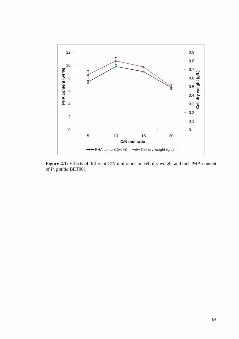
64
Figure 4.1: Effects of different C/N mol ratios on cell dry weight and mcl-PHA contentof P. putida BET001
0
0.1
0.2
0.3
0.4
0.5
0.6
0.7
0.8
0.9
0
2
4
6
8
10
12
5 10 15 20
Cel
l dry
wei
ght (
g/L)
PHA
cont
ent (
wt %
)
C/N mol ratio
PHA content (wt %) Cell dry weight (g/L)
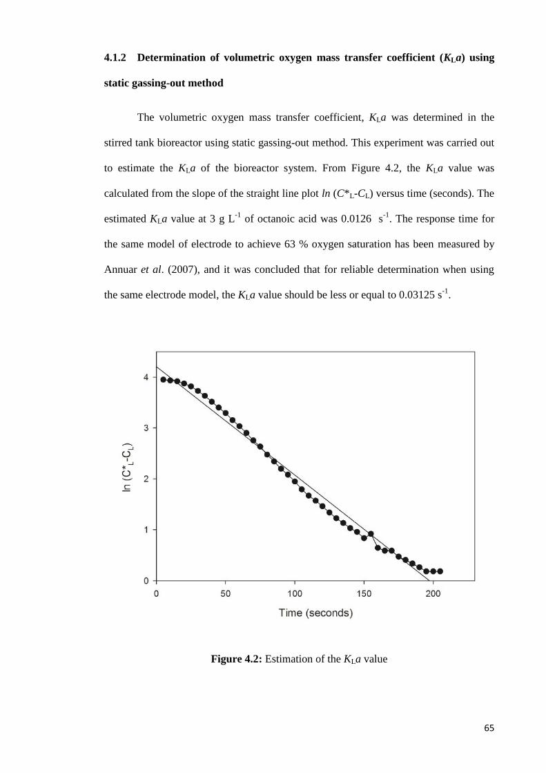
65
4.1.2 Determination of volumetric oxygen mass transfer coefficient (KLa) using
static gassing-out method
The volumetric oxygen mass transfer coefficient, KLa was determined in the
stirred tank bioreactor using static gassing-out method. This experiment was carried out
to estimate the KLa of the bioreactor system. From Figure 4.2, the KLa value was
calculated from the slope of the straight line plot ln (C*L-CL) versus time (seconds). The
estimated KLa value at 3 g L-1 of octanoic acid was 0.0126 s-1. The response time for
the same model of electrode to achieve 63 % oxygen saturation has been measured by
Annuar et al. (2007), and it was concluded that for reliable determination when using
the same electrode model, the KLa value should be less or equal to 0.03125 s-1.
Figure 4.2: Estimation of the KLa value
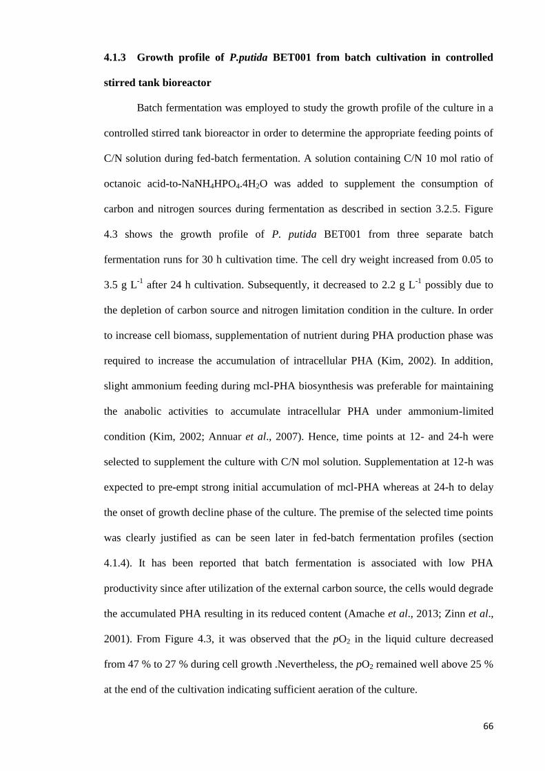
66
4.1.3 Growth profile of P.putida BET001 from batch cultivation in controlled
stirred tank bioreactor
Batch fermentation was employed to study the growth profile of the culture in a
controlled stirred tank bioreactor in order to determine the appropriate feeding points of
C/N solution during fed-batch fermentation. A solution containing C/N 10 mol ratio of
octanoic acid-to-NaNH4HPO4.4H2O was added to supplement the consumption of
carbon and nitrogen sources during fermentation as described in section 3.2.5. Figure
4.3 shows the growth profile of P. putida BET001 from three separate batch
fermentation runs for 30 h cultivation time. The cell dry weight increased from 0.05 to
3.5 g L-1 after 24 h cultivation. Subsequently, it decreased to 2.2 g L-1 possibly due to
the depletion of carbon source and nitrogen limitation condition in the culture. In order
to increase cell biomass, supplementation of nutrient during PHA production phase was
required to increase the accumulation of intracellular PHA (Kim, 2002). In addition,
slight ammonium feeding during mcl-PHA biosynthesis was preferable for maintaining
the anabolic activities to accumulate intracellular PHA under ammonium-limited
condition (Kim, 2002; Annuar et al., 2007). Hence, time points at 12- and 24-h were
selected to supplement the culture with C/N mol solution. Supplementation at 12-h was
expected to pre-empt strong initial accumulation of mcl-PHA whereas at 24-h to delay
the onset of growth decline phase of the culture. The premise of the selected time points
was clearly justified as can be seen later in fed-batch fermentation profiles (section
4.1.4). It has been reported that batch fermentation is associated with low PHA
productivity since after utilization of the external carbon source, the cells would degrade
the accumulated PHA resulting in its reduced content (Amache et al., 2013; Zinn et al.,
2001). From Figure 4.3, it was observed that the pO2 in the liquid culture decreased
from 47 % to 27 % during cell growth .Nevertheless, the pO2 remained well above 25 %
at the end of the cultivation indicating sufficient aeration of the culture.

67
Figure 4.3: Growth profile of P. putida BET001 in batch cultivations (Conditions: 600rpm; aeration 0.5 vvm; pH 7.0; 30 °C; arrows indicate potential feeding points of C/Nmol solution; open and closed symbols indicate cell dry weight data and % pO2 data,respectively)

68
4.1.4 Fed-batch fermentation of P. putida BET001
Culture condition is one of the main factors that affects the production of mcl-
PHA. Various types of mcl-PHA monomeric composition can be synthesized by
providing different carbon sources in the cultivation medium (Razaif‐Mazinah et al.,
2015). In this study, mcl-PHA was produced through fed-batch fermentation process by
P. putida BET001.It has been reported that P. putida BET001 is a growth-associated
mcl-PHA producer whereby the mcl-PHA fraction from the total biomass increases with
its specific growth rate (Gumel et al., 2012). Fed-batch fermentation was employed in
the investigation to extend the cell growth phase and concomitant mcl-PHA
accumulation, with the octanoic acid and nitrogen source supplementation. From Figure
4.4, a solution at C/N 10 mol ratio was added to supplement the consumption of carbon
and nitrogen sources by the cell culture that was growing and accumulationg mcl-PHA
at the same time. Combined solution of octanoic acid and NaNH4HPO4.4H2O (10.3 mL)
in the indicated mol ratio was added as a whole to the culture at 12- and 24 h. In
addition, 3 g L-1 of octanoic acid (3.3 mL) was supplied to the culture every 4 hours in
order to prevent depletion of the carbon substrate during fermentation. For efficient
growth and production of mcl-PHA in the producer organism used in the study, the
octanoic acid supplementation was kept below the inhibitory level of 4 g L-1 (Chardron
et al., 2010). Consequently, 8.6 g L-1 of cell dry weight with 63 % mcl-PHA content
was obtained after 48 hours fermentation. The cell mass and mcl-PHA content increased
with the fermentation time (Figure 4.4). The pO2 values decreased from 47 % to 23%
during cell growth (Figure 4.2). It was also concluded that sufficient aeration was
provided to the culture as no observable oxygen depletion was recorded throughout the
fermentation. Analyses showed that the mcl-PHA produced was composed of three
different monomers viz. 3-hydroxyoctanoate, 3-hydroxyhexanoate and 3-
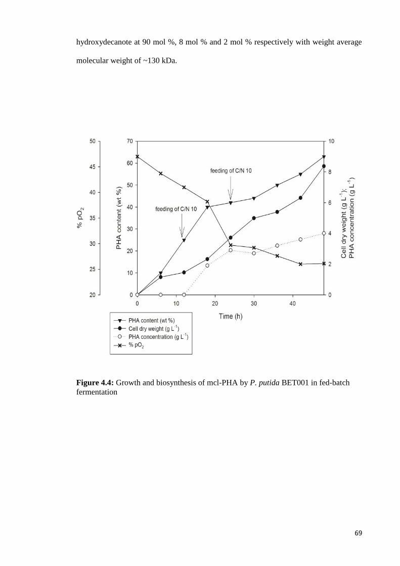
69
hydroxydecanote at 90 mol %, 8 mol % and 2 mol % respectively with weight average
molecular weight of ~130 kDa.
Figure 4.4: Growth and biosynthesis of mcl-PHA by P. putida BET001 in fed-batchfermentation
70
4.2 Blending of P(3HO-co-3HHX) with hydroxyapatite (HA)
4.2.1 Characterization of polymer composite
4.2.1.1 Fourier transform infrared spectroscopy (FTIR)
The chemical functional groups of P(3HO-co-3HHX), P(3HO-co-3HHX)/10 %
HA and P(3HO-co-3HHX)/30 % HA and HA were examined by FTIR spectroscopy, as
shown in Figure 4.5. Absorptions at 2926 and 2861 cm−1 were attributed to both
asymmetric CH3- and symmetric CH2- vibrations in the samples, respectively. The
presence of carbonyl ester bond in pure mcl-PHA sample was assigned to the absorption
at 1725 cm−1 (Figure 4.5(A)). In the composite of P(3HO-co-3HHX)/10 % HA and
P(3HO-co-3HHX)/30 % HA, carbonyl band absorptions were shifted to 1726 cm−1 and
1727 cm-1, respectively. It was reported that spectral changes (intensities and position)
of carbonyl band at 1740–1720 cm−1 was observed during PHA crystallization (Kansiz
et al., 2007; Xu et al., 2002). The bands at 563, 601, 604, 1030, 1032 and 1092 cm-1
corresponded to the phosphate group of HA (Moradi et al., 2013; Pramanik et al., 2009;
Wang et al., 2005). The spectra of P(3HO-co-3HHX)/HA composite (Figure 4.5 (B and
C)) showed the vibrational bands at 1727 cm-1 based on C=O of P(3HO-co-3HHX) and
1043 cm-1 based on PO of the hydroxyapatite, indicating the presence of P(3HO-co-
3HHX) and HA. Figure 4.5(D) showed the bands at 563, 600 and 1032 cm-1
corresponding to the phosphate group of HA. At 1042 cm-1, different intensities of
phosphate group for HA of P(3HO-co-3HHX)/10 % HA and P(3HO-co-3HHX)/30 %
HA were observed. Peak shifts from 1043 cm-1 (C–O) to 1021cm-1 (P–O) were
observed for both P(3HO-co-3HHX)/10 % HA and P(3HO-co-3HHX)/30 % HA
samples after blending, which indicated that HA had been well blended into P(3HO-co-
3HHX).

71
Figure 4.5: FTIR spectra of (A) P(3HO-co-3HHX); (B) P(3HO-co-3HHX)/10 % HA;(C) P(3HO-co-3HHX)/30 % HA; and (D) HA powder
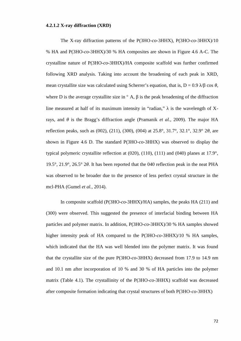
72
4.2.1.2 X-ray diffraction (XRD)
The X-ray diffraction patterns of the P(3HO-co-3HHX), P(3HO-co-3HHX)/10
% HA and P(3HO-co-3HHX)/30 % HA composites are shown in Figure 4.6 A-C. The
crystalline nature of P(3HO-co-3HHX)/HA composite scaffold was further confirmed
following XRD analysis. Taking into account the broadening of each peak in XRD,
mean crystallite size was calculated using Scherrer’s equation, that is, D = 0.9 λ/β cos θ,
where D is the average crystallite size in A, β is the peak broadening of the diffraction
line measured at half of its maximum intensity in “radian,” λ is the wavelength of X-
rays, and θ is the Bragg’s diffraction angle (Pramanik et al., 2009). The major HA
reflection peaks, such as (002), (211), (300), (004) at 25.8°, 31.7°, 32.1°, 32.9° 2θ, are
shown in Figure 4.6 D. The standard P(3HO-co-3HHX) was observed to display the
typical polymeric crystallite reflection at (020), (110), (111) and (040) planes at 17.9°,
19.5°, 21.9°, 26.5° 2θ. It has been reported that the 040 reflection peak in the neat PHA
was observed to be broader due to the presence of less perfect crystal structure in the
mcl-PHA (Gumel et al., 2014).
In composite scaffold (P(3HO-co-3HHX)/HA) samples, the peaks HA (211) and
(300) were observed. This suggested the presence of interfacial binding between HA
particles and polymer matrix. In addition, P(3HO-co-3HHX)/30 % HA samples showed
higher intensity peak of HA compared to the P(3HO-co-3HHX)/10 % HA samples,
which indicated that the HA was well blended into the polymer matrix. It was found
that the crystallite size of the pure P(3HO-co-3HHX) decreased from 17.9 to 14.9 nm
and 10.1 nm after incorporation of 10 % and 30 % of HA particles into the polymer
matrix (Table 4.1). The crystallinity of the P(3HO-co-3HHX) scaffold was decreased
after composite formation indicating that crystal structures of both P(3HO-co-3HHX)
73
Figure 4.6: XRD spectra of (A) P(3HO-co-3HHX); (B) P(3HO-co-3HHX)/10 % HA;(C) P(3HO-co-3HHX)/30 % HA and (D) HA

74
and HA have been altered after the composite formation. Similarly, it was reported that
the addition of HA particles has led to lower degree of crystallinity of the PHBV matrix
in a composite scaffold (Sultana & Khan, 2012). The XRD peaks, (211) and (300) were
shifted to higher 2θ values in the case of P(3HO-co-3HHX)/HA composite as compared
to pure HA (i.e. from 2θ = 31.9 to 32.1 and 32.9 to 33.3, respectively). This was
possibly due to compression from the contracting polymeric matrix through interfacial
bonding (Pramanik et al., 2009). In addition, the crystalline peak of neat mcl-PHA at 2θ
= 26.7 has shifted to 25.8. The shift and decrease in crystallinity of each peak of the
polymer as well as HA after composite formation clearly indicated the presence of
interfacial binding between HA particles and porous polymer matrix.
4.2.1.3 Differential scanning calorimetry (DSC)
The thermal properties, crystallinity and porosity data of the scaffolds are shown
in Table 4.1. The Tm values of the polymers were increased from 54.9 °C to 55.2 °C and
55.7 °C after blending of P(3HO-co-3HHX) with 10 % HA and 30 % HA, respectively.
The crystallinities of the polymers was decreased from 7.9 % to 6.9 % after blending
with HA. The observed decrease in polymer crystallinity as a result of HA composition
was corroborated by the data obtained from XRD analysis. It has been reported that
polymer with lower degree of crystallinity is degraded faster (El-Hadi et al., 2002).
Thus, it can be expected that P(3HO-co-3HHX)/HA composite scaffold would exhibit
higher rate of degradation in vitro and in vivo than neat P(3HO-co-3HHX) scaffolds. In
contrast, the porosity of the polymer did not show significant difference before and after
incorporation of HA particles.
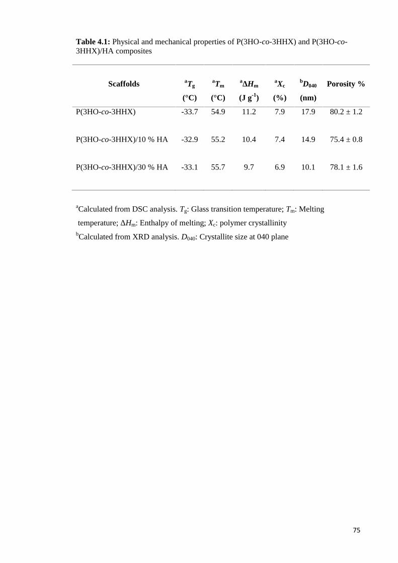
75
Table 4.1: Physical and mechanical properties of P(3HO-co-3HHX) and P(3HO-co-3HHX)/HA composites
Scaffolds aTg
(°C)
aTm
(°C)
aΔHm
(J g-1)
aXc
(%)
bD040
(nm)
Porosity %
P(3HO-co-3HHX) -33.7 54.9 11.2 7.9 17.9 80.2 ± 1.2
P(3HO-co-3HHX)/10 % HA -32.9 55.2 10.4 7.4 14.9 75.4 ± 0.8
P(3HO-co-3HHX)/30 % HA -33.1 55.7 9.7 6.9 10.1 78.1 ± 1.6
aCalculated from DSC analysis. Tg: Glass transition temperature; Tm: Melting
temperature; ΔHm: Enthalpy of melting; Xc: polymer crystallinitybCalculated from XRD analysis. D040: Crystallite size at 040 plane

76
4.2.1.4 Energy Dispersive X-ray Analysis (EDX)
Energy Dispersive X-ray Analysis (EDX, map of Ca) was performed in order to
investigate the distribution of hydroxyapatite particles in the P(3HO-co-3HHX)/HA
composite scaffold. Figure 4.7A showed the EDX spectra of neat P(3HO-co-3HHX)
with the presence of carbon and oxygen elements. In contrast, P(3HO-co-3HHX)
composite with HA showed the presence of carbon, oxygen, calcium and phosphorus
elements (Figure 4.7B and 4.7C), which clearly showed the presence of HA in the
composite matrix. EDX analysis showed a relatively higher level of calcium and
phosphorus on the surfaces of P(3HO-co-3HHX)/30 % HA composite scaffolds
compared to P(3HO-co-3HHX)/10 % HA samples. Ca/P molar ratios on the surfaces of
P(3HO-co-3HHX)/10 % HA and P(3HO-co-3HHX)/30 % HA were determined at 2.48
and 1.85, respectively. In the EDX spectra, the carbon and oxygen peaks were derived
from the polymer (Table 4.2). The presence of HA was revealed by the Ca and P peaks
(Rizzi et al., 2001).
In addition, EDX was applied to the composites to monitor HA exposure at the
composite surface of porous PHO/HA scaffolds. The weight and atomic percentage of
Ca and P element in P(3HO-co-3HHX)/30 % HA samples were higher (2.9, 7.0, 1.3,
2.4) than the P(3HO-co-3HHX)/10 % HA samples (0.4, 1.5, 0.2, 0.5), respectively.
These results showed that the HA particles were successfully incorporated into the
P(3HO-co-3HHX) matrix. Moreover, it is suggested that high energy ultrasound
irradiation (25 kHz) helped to enhance the dispersion of HA particle in the polymer
matrix. According to a previous study, ultrasonication has been shown to be an effective
means to overcome the agglomeration of particles in the biopolymer (Baei & Rezvani,
2011). Furthermore, good dispersion of inorganic filler in the composite assists to
improve the mechanical properties of the composite material (Chen et al., 2005).

77
Figure 4.7: EDX spectrum obtained at 10 keV on the (A) P(3HO-co-3HHX); (B)P(3HO-co-3HHX)/10 % HA and (C) P(3HO-co-3HHX)/30 % HA

78
Table 4.2: Elemental analysis of HA using EDX analysis of P(3HO-co-3HHX),P(3HO-co-3HHX)/10 % HA and P(3HO-co-3HHX)/30 % HA scaffolds.
Element Percentage of elements
Weight
%
Atomic
%
P(3HO-co-3HHX)
C 74.4 ± 0.5 79.4 ± 0.4
O 25.6 ± 0.5 20.5 ± 0.4
P(3HO-co-3HHX)/10 % HA
C 71.9 ± 0.8 78.0 ± 0.7
O 26.2 ± 0.7 21.3 ± 0.6
P 0.4 ± 0.0 0.2 ± 0.0
Ca 1.5 ± 0.1 0.5 ± 0.0
P(3HO-co-3HHX)/30 % HA
C 64.4 ± 1.7 74.1 ± 1.1
O 25.7 ± 0.3 22.2 ± 0.5
P 2.9 ± 0.5 1.3 ± 0.3
Ca 7.0 ± 0.9 2.4 ± 0.3

79
4.2.1.5 Field Emission Scanning Electron Microscope (FESEM)
The morphologies of the scaffolds are shown in Figure 4.8. Both the neat
P(3HO-co-3HHX) and composite scaffolds showed spongy appearance, high porosity
and extensive inter-pores connectivities (Figures 4.8 A, C and E). The porosity of the
scaffolds was observed to be in the range of 75 % to 80 % (Table 4.1). The P(3HO-co-
3HHX)/HA scaffolds (Figures 4.8 C and E) retained porous morphology as that of neat
P(3HO-co-3HHX) scaffolds (Figure 4.8 A). Pores are necessary for bone tissue
formation because they allow migration and proliferation of osteoblasts and
mesenchymal cells as well as vascularization. In addition, a porous surface improves
mechanical interlocking between the implant biomaterial and the surrounding natural
bone, thus providing greater mechanical stability at this critical interface (Karageorgiou
& Kaplan, 2005; Pramanik et al., 2009). From SEM images, the mean pore diameter
was determined approximately around 100-180 µm. Previous researches suggested that
human osteoblasts cells penetrate faster within the scaffolds containing large pores (>
100 µm), meanwhile the extent of mineralization was not affected by the pore size
(Akay et al., 2004; Nguyen et al., 2012).
It was found the osteoblast cells were favourably attached to the P(3HO-co-
3HHX)/30 % HA scaffolds (Figure 4.8 F) as compared to P(3HO-co-3HHX)/10 % HA
scaffolds (Figure 4.8 D), and significantly less osteoblast cells were found to be able to
attach themselves to neat P(3HO-co-3HHX) scaffolds (Figure 4.8 B). This was
attributed to the increased concentration of dispersed HA that helped to promote the
proliferation of osteoblast cells throughout the polymer matrixes. As far as the
composite scaffolds prepared in this study were concerned, their porosities were
sufficient for good interconnection and transportation of nutrition as evidenced by good
cell growth. Hence, the pore size of the scaffolds were favorable for good cell growth.

80
Figure 4.8: FESEM image of the scaffolds (A) P(3HO-co-3HHX) (B) cells on scaffoldsurface P(3HO-co-3HHX) (C) composite P(3HO-co-3HHX)/10 % HA (D) cells onscaffold surface P(3HO-co-3HHX)/10 % HA (E) composite P(3HO-co-3HHX)/30 %HA (F) cells on scaffold surface P(3HO-co-3HHX)/30 % HA (magnification 5000 ).
81
Jack et al. (2009) fabricated the PHBV/HA composite scaffolds with high porosity and
controlled pore architecture. Their results showed that the incorporation of HA
microparticles increased the stiffness and strength and improved the in vitro
bioactivities of the scaffolds. For bone tissue engineering, biodegradable composite
scaffolds containing HA hold great promises. The current investigation demonstrated
that HA particles could be homogeneously incorporated into the mcl-PHA to make
osteo-conductive porous composite scaffolds.
4.2.2 Biological response of osteoblast cells to P(3HO-co-3HHX)/HA composite
scaffolds
Further investigation was carried out with osteoblasts cells seeded on pure
P(3HO-co-3HHX) and P(3HO-co-3HHX) matrices containing 10 % and 30 % HA. The
attachment and growth of osteoblast cells were assessed using the Alamar Blue Assay
(Figure 4.9A). The metabolic activities of the osteoblast cells were analyzed at day 1, 7
and 14. It was found that the percentage of resazurin reduction increased with culture
time. From calibration data, it was found that the percentage of resazurin reduction was
positively correlated with the cell density. All matrices were able to support the growth
of osteoblast cells during 14 days of culture. The percentage of resazurin reduction in
neat scaffolds was lower compared to P(3HO-co-3HHX)/10 % HA and P(3HO-co-
3HHX)/30 % HA scaffolds, which indicated that the HA composite scaffolds provided a
more favourable physical environment for cell attachment and growth. It has been
reported that when the HA content were increased, more particles will be exposed on
the surface of the porous scaffold, hence favored the increase in the proliferation of the
cells (Huang et al., 2007; Wang et al., 2005). No significant differences were found
between P(3HO-co-3HHX)/10 % HA and P(3HO-co-3HHX)/30 % HA scaffold after 7
and 14 days of culture.
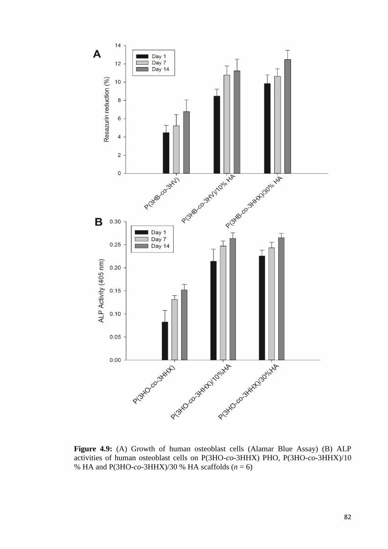
82
Figure 4.9: (A) Growth of human osteoblast cells (Alamar Blue Assay) (B) ALPactivities of human osteoblast cells on P(3HO-co-3HHX) PHO, P(3HO-co-3HHX)/10% HA and P(3HO-co-3HHX)/30 % HA scaffolds (n = 6)

83
Both P(3HO-co-3HHX)/10 % HA and P(3HO-co-3HHX)/30 % HA scaffolds
showed highest percentage of resazurin reduction on day 14 relative to neat P(3HO-co-
3HHX) scaffolds indicating that the recorded cells activities was primarily attributed to
their response from HA exposure. In addition, incorporation of hydroxyapatite particles
into the composite scaffold has been reported to significantly increase protein
adsorption (Wei & Ma, 2004). The microporous structure of composite scaffold
provided for greater surface-to-volume ratio, which could have contributed to further
increase the proliferation of the osteoblast cells.
The differentiation process of osteoblast cells on neat P(3HO-co-3HHX) and
P(3HO-co-3HHX)/HA composites have been studied from the alkaline phosphatase
(ALP) assay (Figure 4.9B). Generally, the ALP activities of cells grown on all scaffolds
increased continuously until day 14. In contrast, there was no significant difference on
osteoblast cells proliferation between P(3HO-co-3HHX)/10 % HA and P(3HO-co-
3HHX)/30 % HA scaffold after 7 and 14 days of culture. Both P(3HO-co-3HHX)/30 %
HA and P(3HO-co-3HHX)/10 % HA scaffolds showed the highest ALP activities on
day 14. The ALP activities of osteoblast cells grown on the neat P(3HO-co-3HHX)
scaffolds for various culture times were significantly lower than that of the composite
HA scaffolds. The results suggested that the presence of dispersed HA within the
polymer matrix helped to improve the differentiation of osteoblast cells. The
observation was consistent with the previous reports that the polymer–HA scaffolds
were superior to the pure polymer scaffolds for tissue engineering because the presence
of HA hydroxyl groups promote calcium and phosphate precipitations hence improved
interactions with osteoblast cells (Xi et al., 2008).
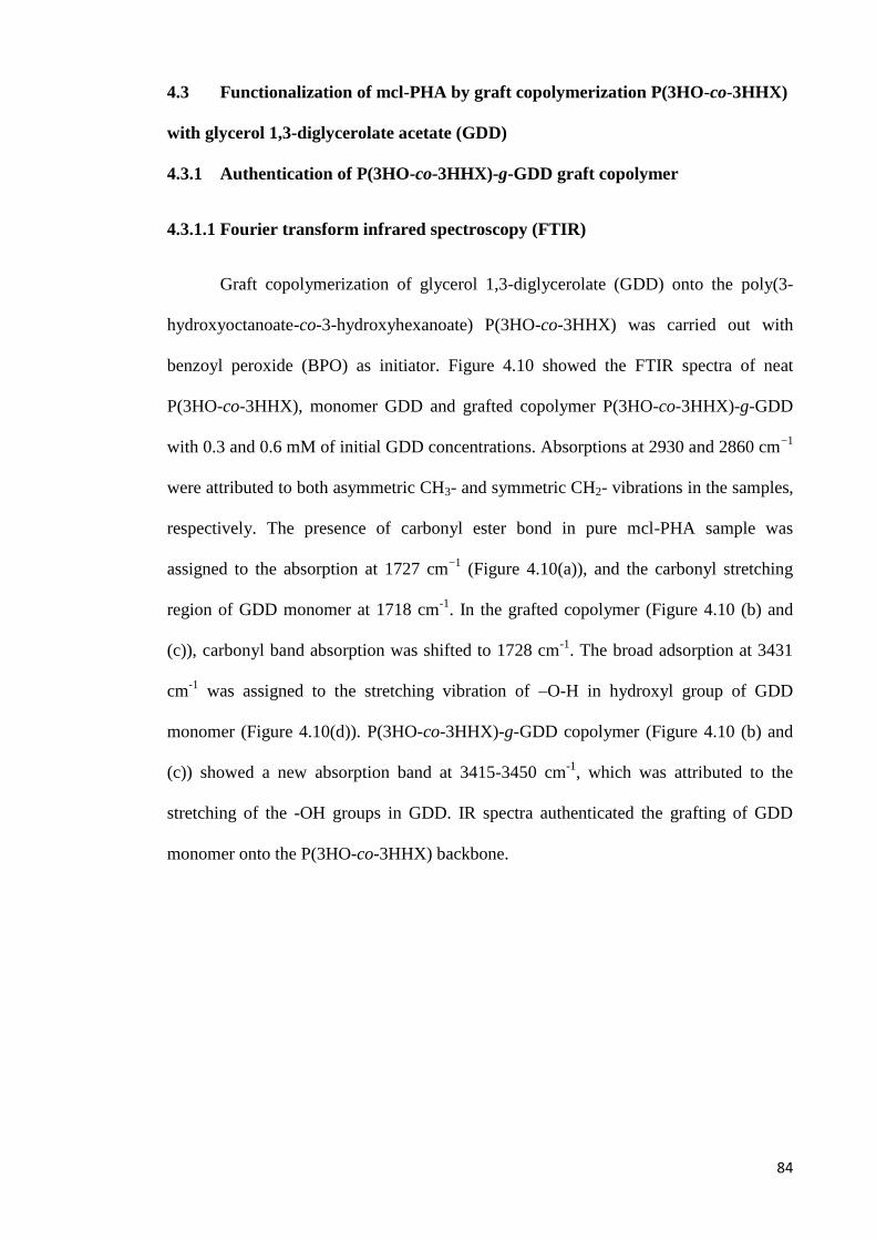
84
4.3 Functionalization of mcl-PHA by graft copolymerization P(3HO-co-3HHX)
with glycerol 1,3-diglycerolate acetate (GDD)
4.3.1 Authentication of P(3HO-co-3HHX)-g-GDD graft copolymer
4.3.1.1 Fourier transform infrared spectroscopy (FTIR)
Graft copolymerization of glycerol 1,3-diglycerolate (GDD) onto the poly(3-
hydroxyoctanoate-co-3-hydroxyhexanoate) P(3HO-co-3HHX) was carried out with
benzoyl peroxide (BPO) as initiator. Figure 4.10 showed the FTIR spectra of neat
P(3HO-co-3HHX), monomer GDD and grafted copolymer P(3HO-co-3HHX)-g-GDD
with 0.3 and 0.6 mM of initial GDD concentrations. Absorptions at 2930 and 2860 cm−1
were attributed to both asymmetric CH3- and symmetric CH2- vibrations in the samples,
respectively. The presence of carbonyl ester bond in pure mcl-PHA sample was
assigned to the absorption at 1727 cm−1 (Figure 4.10(a)), and the carbonyl stretching
region of GDD monomer at 1718 cm-1. In the grafted copolymer (Figure 4.10 (b) and
(c)), carbonyl band absorption was shifted to 1728 cm-1. The broad adsorption at 3431
cm-1 was assigned to the stretching vibration of –O-H in hydroxyl group of GDD
monomer (Figure 4.10(d)). P(3HO-co-3HHX)-g-GDD copolymer (Figure 4.10 (b) and
(c)) showed a new absorption band at 3415-3450 cm-1, which was attributed to the
stretching of the -OH groups in GDD. IR spectra authenticated the grafting of GDD
monomer onto the P(3HO-co-3HHX) backbone.
85
Figure 4.10: FTIR spectra of (a) P(3HO-co-3HHX); (b) P(3HO-co-3HHX)-g-GDD (0.6mM); (c) P(3HO-co-3HHX)-g-GDD (0.3 mM); and (d) GDD monomer
86
4.3.1.2 Proton (1H) nuclear magnetic resonance (NMR)
The chemical structure of the grafted polymer was further authenticated by
NMR spectroscopy. Figure 4.11 showed the 1H-NMR spectrum of the grafted
copolymer P(3HO-co-3HHX)-g-GDD. The grafted copolymer was dissolved in
deuterated chloroform (CDCl3). In the NMR spectrum of P(3HO-co-3HHX)-g-GDD,
typical signals of mcl-PHA and those of GDD segments were observed. Using internal
standard tetramethylsilane (TMS) as the reference, the observed multiplet peaks at 2.6
ppm (1) and at triplets peaks at 5.2 ppm (2) were assigned to methylene (-CH2) and
methine (-CH) protons of α- and β- carbon of mcl-PHA backbone, respectively. The
signals 1.3 ppm (4) and 0.9 ppm (5) were assigned to methylene (-CH2) proton and
terminal methyl (-CH3) proton of the P(3HO-co-3HHX) side chain, respectively. The
appearance of a new signal for (CH2-C) at 1.8 ppm (a) constituted evidence that GDD
was successfully grafted into the backbone of P(3HO-co-3HHX). Chemical shifts c, d,
and e were assigned to α, β-protons of methylene group of GDD monomer -COO-CH2,
CH2-CH(OH) and CH2-COO-, respectively. Hence, based on the signal analysis, we
proposed that the GDD monomers were grafted onto the C-3 carbon of mcl-PHA
backbone. The assignment was found to be in accordance with previously reported
literatures (Ilter et al., 2001; Lao et al., 2007; Nakas et al., 2015; Renard et al., 2003).
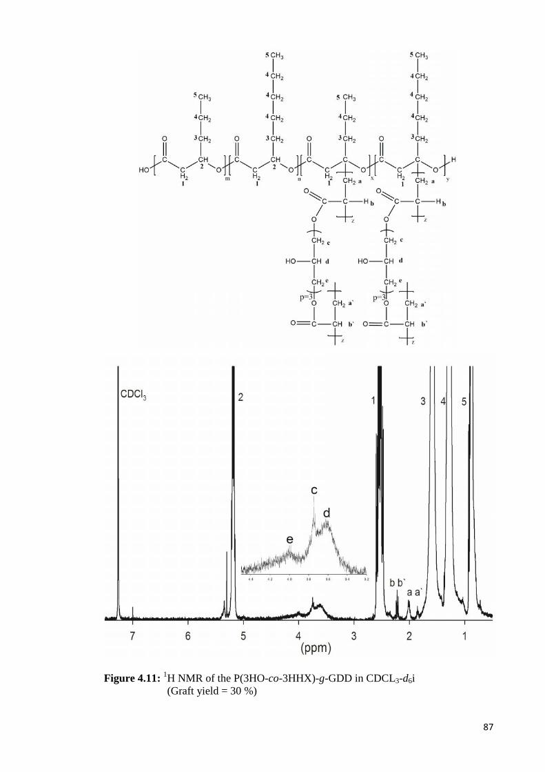
87
Figure 4.11: 1H NMR of the P(3HO-co-3HHX)-g-GDD in CDCL3-d6i(Graft yield = 30 %)
88
4.3.2 Mechanism of P(3HO-co-3HHX) grafting with GDD
Based on the experimental results and previous researches on graft
copolymerization, the mechanism for graft copolymerization of P(3HO-co-3HHX) by
GDD is proposed (Figure 4.12). It is hypothesized that the grafting of GDD monomer
onto P(3HO-co-3HHX) proceeds via three steps which include initiation, propagation
and termination reactions. Grafting of GDD onto P(3HO-co-3HHX) backbone is
proposed to occur via three ways: (1) random hydrogen abstraction from P(3HO-co-
3HHX) backbone by direct attack of initiator radicals (i.e. benzoyl peroxide); (2) chain
transfer of GDD growing radicals into P(3HO-co-3HHX) backbone; (3) the
recombination of GDD and P(3HO-co-3HHX) growing radicals. However, during free
radical polymerization reaction, both homopolymerization and graft copolymerization
could also take place. Chain transfer reaction of GDD proceeds via the creation of
secondary macroradical of GDD. Consequently, macroradicals could perform reaction
with each other, thus generating inter-grafted species of homopolymeric composition
(Figure 4.12).
During grafting reactions, polymer radicals produced from active polymers can
cause cleavage as well as proton abstraction from the polyester chain (Hazer, 1996; Lao
et al., 2007). In free radical polymerization reaction, initiation is the first reaction of a
chain carrier with a polymer or monomer molecule to generate a new covalent bond and
regenerate reactive centre at a new location. The thermodynamic driving force of these
reactions is the replacement of high energy π bond by σ bond (addition) or the
replacement of weaker R−H bond by a stronger R′−H (abstraction) (Bhattacharya et al.,
2009). The initiation could occur via addition, where the radical adds across a double
bond of GDD monomer, or by abstraction, where the radical removes a labile H- atom
from C-3 carbon of mcl-PHA macromolecule. Grafting is achieved when active

89
Figure 4.12: Proposed mechanism for the reaction of GDD monomer grafting ontoP(3HO-co-3HHX) (I: initiator benzoyl peroxide; m = 1, 2, 3, 4,…..)
90
polymer decomposes into macroradical, which involves abstracting the proton at the C-
3 carbon, and in turn creating radical(s) along the P(3HO-co-3HHX) main chain.
Coupling of the GDD macroradicals onto the free-radical sites of P(3HO-co-3HHX)
produced graft copolymer. The presence of diacrylate (-C=C-) in the structure of GDD
monomers promotes the formation of radical at both ends. Thus, rapid growth of active
chain is enhanced by the formation of graft copolymer. BPO is usually employed as a
source of radicals for the initiation of vinyl polymerization and cross-linking of both
saturated and unsaturated polymers (Lao et al., 20007). Therefore, the presence of
diacrylate in the structure of GDD monomers could promote crosslinking between the
chains of PHA.
Ilter et al. (2001) reported the presence of double bond opens up possibilities of
functionalization in biopolyesters to improve their mechanical and viscoelastic
properties. Lao et al. (2007) reported the methine protons of PHBHV, which are the
most acidic protons, may be abstracted from the backbone to generate macroradicals on
the chains of PHBV, thus initiating graft polymerization. Several studies of graft
polymerization of PHA also reported that free radical grafting can be initiated at C-3
carbon (-CH(R)-) of the PHA, where the protons are abstracted by free radicals such
benzoyl peroxide to form PHA macroradicals. These protons are the most acidic ones
along the PHA chains. Alternatively, free radical grafting could proceed via polymer
chain radical transfer to the PHA backbone (Lao et al., 2007; Lee & Lee, 1997; Nguyen,
2008; Wang et al., 2007). Active radical sites along the PHA backbone chain generated
from abstracting the protons at the C-3 carbon positions have been exploited to graft
vinyl chains via monomer polymerization (Li et al., 2016).

91
4.3.3 Thermal properties of P(3HO-co-3HHX)-g-GDD graft copolymer
Thermal analysis of graft copolymer was performed using TGA and DSC. The
thermal properties of neat P(3HO-co-3HHX) and P(3HO-co-3HHX)-g-GDD
copolymers are listed in Table 4.3. P(3HO-co-3HHX)-g-GDD copolymers showed
higher glass transition temperature (Tg) and lower melting temperature (Tm) compared to
neat P(3HO-co-3HHX). The Tg and Tm of graft copolymers increased slightly from -
33.7 °C to -32.9 °C and decreased signficantly from 54.9 °C to 51.1 °C, respectively, as
the concentration of GDD in the copolymers increased. The observation is equally
applicable with increasing graft yield from 23 to 127 % (Table 4.3). The presence of
GDD groups in the copolymer caused significant structural changes in the P(3HO-co-
3HHX) chains. It was reported that grafted GDD groups on the poly-3-
hydroxyoctanoate (PHO) backbone result in the formation of inter-molecular and intra-
molecular hydrogen bonding between GDD grafted chains (Kim et al., 2008). The
results suggested that GDD grafting led to the decrease of crystallization ability of
P(3HO-co-3HHX). Hence, introduction of GDD monomer hindered the crystallization
of P(3HO-co-3HHX) by introducing chain structural irregularities.
Figure 4.13 showed the TGA curves of neat P(3HO-co-3HHX), GDD monomer
and P(3HO-co-3HHX)-g-GDD copolymer samples with different initial concentrations
of GDD for grafting reaction. TGA analyses of P(3HO-co-3HHX) and graft copolymers
were performed to investigate the effects of different initial concentrations of GDD on
the thermal degradability of the composites (Figure 4.13(A) and 4.13(B)). Initial
degradation temperatures from TGA and maximum degradation temperatures from
derivative TGA were reported in Table 4.3. It can be observed that Td and Tmax values of
neat mcl-PHA were decreased from 277 to 269 °C and 295 to 287 °C, respectively, after
being grafted with different concentrations of GDD monomer (Table 4.3).

92
Table 4.3: Molecular weight, thermal, water uptake and graft yield data of neat P(3HO-co-3HHX) and copolymer P(3HO-co-3HHX)-g-GDD with different concentrations of GDD monomer
SamplesMolecular weights Thermal analysis Water
uptake(%)
Graftyield(%)aMn (x103)
Da
aMw/MnbTd
(°C)
bTmax
(°C)
cTm
(°C)
cTg
(°C)P(3HO-co-3HHX) 43 ± 0.8 1.89 277 295 54.9 -33.7 2.3 ± 0.3 -
P(3HO-co-3HHX)-g-GDD
0.1 mM 47 ± 1.3 1.78 270 285 54.4 -32.6 12.1 ± 0.7 23
0.3 mM 49 ± 2.5 1.67 271 286 50.4 -33.0 22.8 ± 1.9 39
0.4 mM 53 ± 1.9 1.64 273 287 51.4 -33.2 28.2 ± 0.8 59
0.6 mM 61 ± 2.1 1.47 269 287 51.1 -32.9 32.9 ± 1.8 127
aCalculated from GPC analysis; bCalculated from STA analysis; cCalculated from DSC analysis.
Mn number average molecular weight, Mw/Mn polydispersity index, Td: degradation temperature; Tm: Melting temperature,
Tg: glass transition temperature. Reaction condition: P(3HO-co-3HHX) 0.2 g; BPO 0.04 mM; 80 °C; 2 h
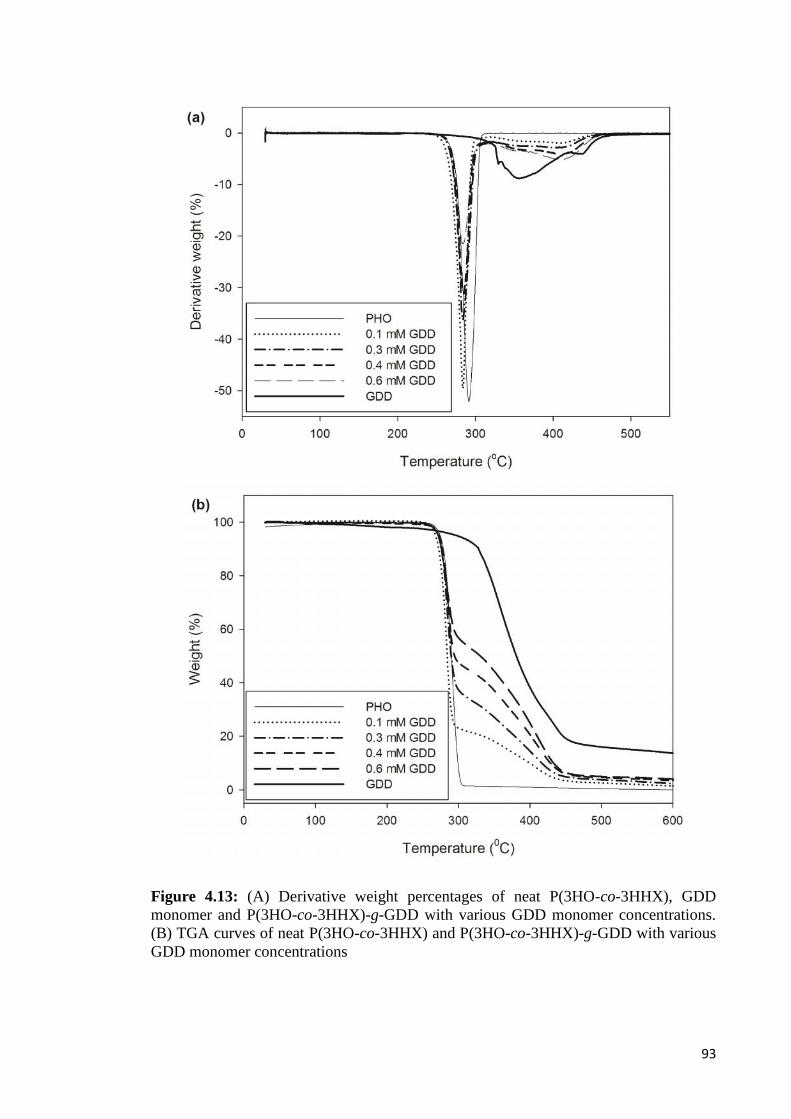
93
Figure 4.13: (A) Derivative weight percentages of neat P(3HO-co-3HHX), GDDmonomer and P(3HO-co-3HHX)-g-GDD with various GDD monomer concentrations.(B) TGA curves of neat P(3HO-co-3HHX) and P(3HO-co-3HHX)-g-GDD with variousGDD monomer concentrations
94
Degradation of GDD monomer and P(3HO-co-3HHX)-g-GDD began around
100 °C, which was attributed to loss of water (Lao et al., 2007). Although GDD was the
first to decompose (Td 316 °C), it showed the highest thermal stability (Tmax 327 °C).
The shape of all TGA curves was generally similar indicating the thermal degradation
of copolymer P(3HO-co-3HHX)-g-GDD with different graft yields followed similar
mechanism. Wang et al. (2007) reported that the pattern of TGA curves for PHBV-g-
PVP was also similar at different graft yields owing to similar mechanism of their
thermal degradation.
4.3.4 Molecular weight analysis of P(3HO-co-3HHX)-g-GDD graft copolymer
GPC was used to obtain the molecular weight and polydispersity data of the
graft copolymers (Table 4.3). The measurements showed a single peak corresponding to
the molecular weight of the polymer for each sample tested. The number average
molecular weights (Mn) of the P(3HO-co-3HHX)-g-GDD copolymers increased from
4.3 x 10⁴ to 6.1 x 10⁴ Da with increased initial concentration of GDD. In addition, the
polydispersity (Mw/Mn) of the grafted copolymer decreased from 1.89 to 1.47, as the
graft yield increased from 23 to 127 % (Table 4.3). It was reported that the radicals
produced by hydrogen abstraction following the initiation reaction were involved in
secondary reactions which could lead to a degradation of the polymer backbone or
crosslinking (Fei et al., 2004; Lao et al., 2007). Thus, the observed results could be
ascribed to the hydrogen bonding and possible crosslinking that took place during
modification. In other study, grafting of GDD monomer onto the PHO backbone also
resulted in a similar increase in molecular weight, hence increased degree of polymer
grafting (Kim et al., 2008). Moreover, the increase in molecular weights of P(3HO-co-
3HHX)-g-GDD copolymers and graft yield showed that GDD had been successfully
grafted onto P(3HO-co-3HHX) chains. Grafting of GDD onto P(3HO-co-3HHX)
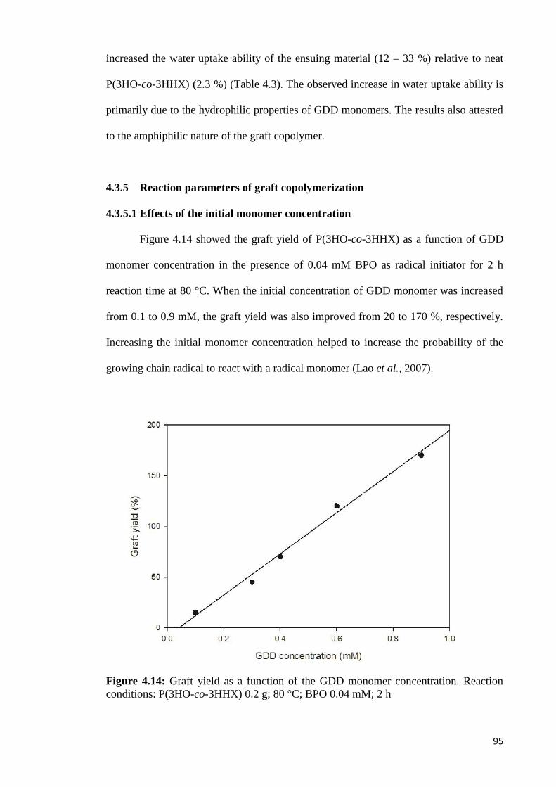
95
increased the water uptake ability of the ensuing material (12 – 33 %) relative to neat
P(3HO-co-3HHX) (2.3 %) (Table 4.3). The observed increase in water uptake ability is
primarily due to the hydrophilic properties of GDD monomers. The results also attested
to the amphiphilic nature of the graft copolymer.
4.3.5 Reaction parameters of graft copolymerization
4.3.5.1 Effects of the initial monomer concentration
Figure 4.14 showed the graft yield of P(3HO-co-3HHX) as a function of GDD
monomer concentration in the presence of 0.04 mM BPO as radical initiator for 2 h
reaction time at 80 °C. When the initial concentration of GDD monomer was increased
from 0.1 to 0.9 mM, the graft yield was also improved from 20 to 170 %, respectively.
Increasing the initial monomer concentration helped to increase the probability of the
growing chain radical to react with a radical monomer (Lao et al., 2007).
Figure 4.14: Graft yield as a function of the GDD monomer concentration. Reactionconditions: P(3HO-co-3HHX) 0.2 g; 80 °C; BPO 0.04 mM; 2 h

96
4.3.5.2 Effects of reaction time
Free radical reaction is known to be a fast reaction. Figure 4.15 (A) and (B)
showed the effects of reaction time and GDD monomer concentration on graft yield in
the presence of 0.04 mM BPO at 80 °C and 95 °C, respectively. The graft yield
increased as a function of monomer concentration and reaction time. The increase in
monomer concentration between 0.1 to 0.9 mM resulted in higher percentage of graft
yield. The yield increased rapidly during early phase of reaction period and levelled off
after 2 h reaction time. It was attributed to the termination of the polymerization
reaction thus formation of dead polymer. Wang et al. (2007) reported that after 60 min
of reaction time, the yield of PHBV grafted with vinyl group levelled off due to the
termination of polymerization reaction. The kinetic chain of radical polymerization
could also be ended by the pairing of two radicals to form non radical species
(Bhattacharya et al., 2009). The initial rate of grafting (Figure 4.16) was calculated from
the initial slope of the curves for each concentration of GDD shown in Figure 4.15 (A)
and (B). Higher initial rates of grafting were observed at all initial concentrations of
GDD as the reaction temperature was increased from 80 C to 95 C. The activation
energy (Ea) was calculated based on the Arrhenius equation and the apparent Ea
calculated for the graft copolymerization was ~ 51 kJ mol-1.
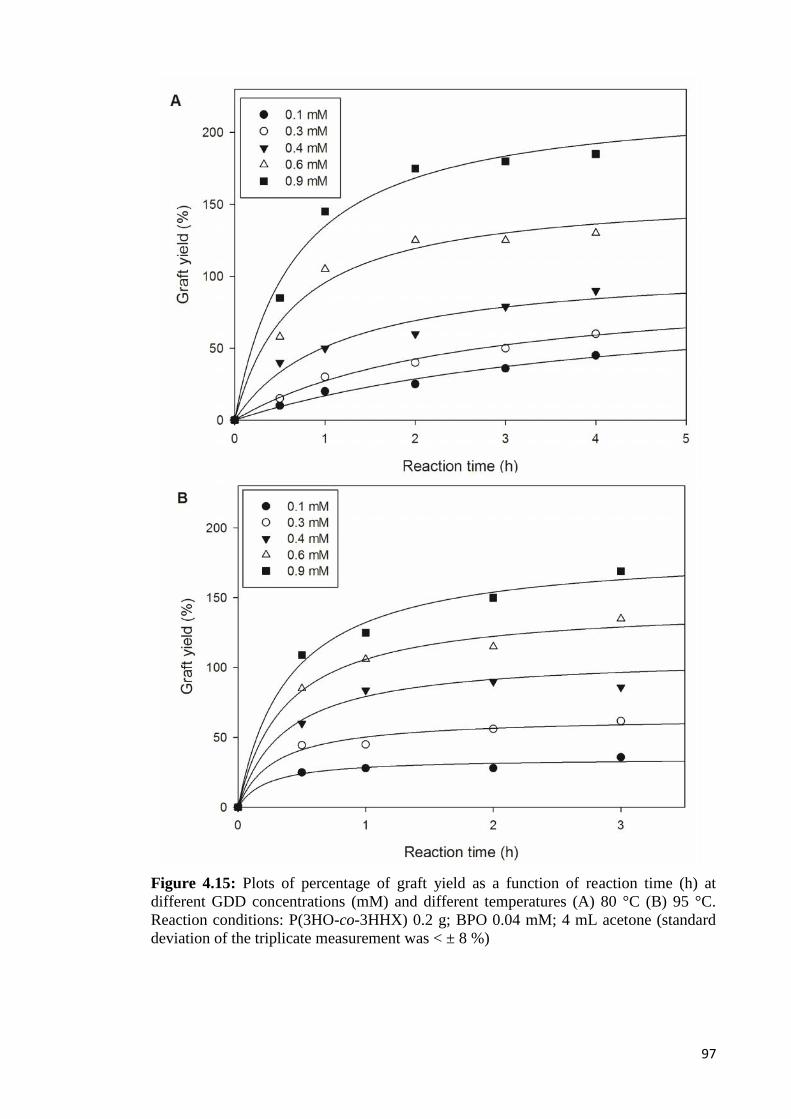
97
Figure 4.15: Plots of percentage of graft yield as a function of reaction time (h) atdifferent GDD concentrations (mM) and different temperatures (A) 80 °C (B) 95 °C.Reaction conditions: P(3HO-co-3HHX) 0.2 g; BPO 0.04 mM; 4 mL acetone (standarddeviation of the triplicate measurement was < ± 8 %)
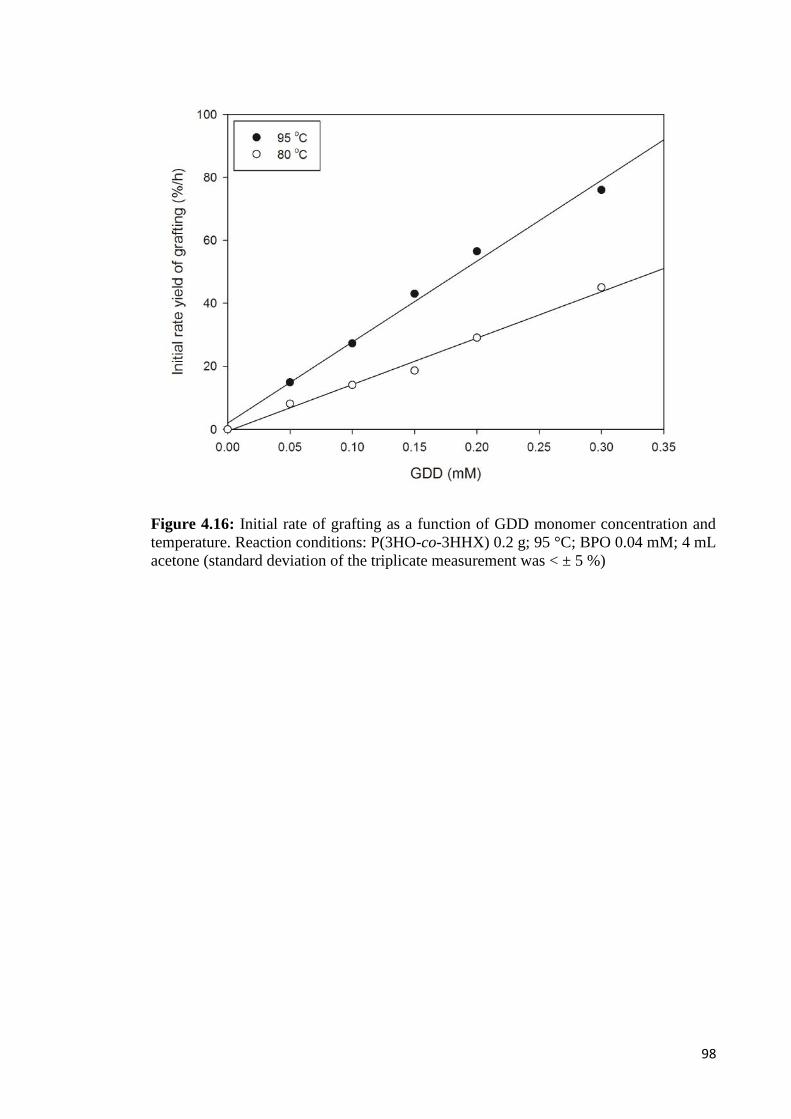
98
Figure 4.16: Initial rate of grafting as a function of GDD monomer concentration andtemperature. Reaction conditions: P(3HO-co-3HHX) 0.2 g; 95 °C; BPO 0.04 mM; 4 mLacetone (standard deviation of the triplicate measurement was < ± 5 %)

99
.3.5.3 Effects of reaction temperature
The effects of reaction temperature on the graft copolymerization are shown in
Figure 4.17. The concentration of GDD monomer and BPO were fixed at 0.4 mM and
0.04 mM, respectively, alongside with reaction time of 3 h. The percentage of graft
yield was increased as the reaction temperature increased from 70 to 90 °C. The
observed effect was associated with the increased in dissociation rate of initiator
benzoyl peroxide with temperature (Celik, 2004).
Furthermore, diffusion of GDD monomer into the reaction mixture was likely to
be enhanced by the thermal effects. In addition, swellability and mobility of P(3HO-co-
3HHX) and GDD monomer were also expected to increase as the temperature became
higher, and it improved the chances of successful collisions between GDD monomer
molecules and P(3HO-co-3HHX) macroradicals. Langer and Wilkie (1998) reported
that with higher temperature, the swelling of the polymer increased and more radical
initiators migrated into the polymer matrix. However, no grafting was observed at
temperatures below 65 °C, even after 3 h of reaction. It was attributed to the low rate of
initiator benzoyl peroxide dissociation and curtailment of GDD monomer diffusion to
penetrate the P(3HO-co-3HHX) polymer coil in solution.

100
Figure 4.17: Effects of reaction temperature on the graft yield copolymerization ofP(3HO-co-3HHX)-g-GDD. Reaction condition: 0.2 g P(3HO-co-3HHX), 0.4 mM GDD,0.04 mM BPO, 2 h (standard deviation of the triplicate measurement was < ± 5 %)
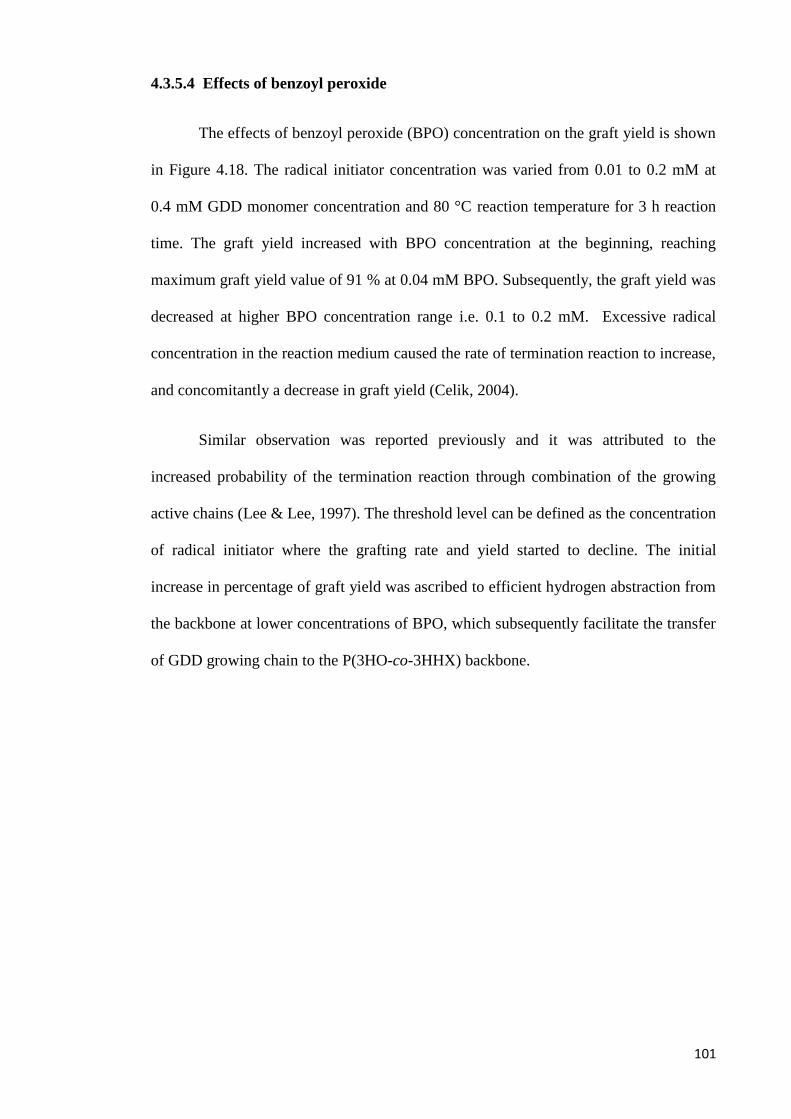
101
4.3.5.4 Effects of benzoyl peroxide
The effects of benzoyl peroxide (BPO) concentration on the graft yield is shown
in Figure 4.18. The radical initiator concentration was varied from 0.01 to 0.2 mM at
0.4 mM GDD monomer concentration and 80 °C reaction temperature for 3 h reaction
time. The graft yield increased with BPO concentration at the beginning, reaching
maximum graft yield value of 91 % at 0.04 mM BPO. Subsequently, the graft yield was
decreased at higher BPO concentration range i.e. 0.1 to 0.2 mM. Excessive radical
concentration in the reaction medium caused the rate of termination reaction to increase,
and concomitantly a decrease in graft yield (Celik, 2004).
Similar observation was reported previously and it was attributed to the
increased probability of the termination reaction through combination of the growing
active chains (Lee & Lee, 1997). The threshold level can be defined as the concentration
of radical initiator where the grafting rate and yield started to decline. The initial
increase in percentage of graft yield was ascribed to efficient hydrogen abstraction from
the backbone at lower concentrations of BPO, which subsequently facilitate the transfer
of GDD growing chain to the P(3HO-co-3HHX) backbone.
102
Figure 4.18: Effects of radical initiator (BPO) concentration on the graft yield

103
4.4 Authentication of P(3HO-co-3HHX)-g-GDD/HA
Modification of P(3HO-co-3HHX) via graft copolymerization with GDD (0.1
mM) and blending with 10 weight % of HA led to a novel composite P(3HO-co-
3HHX)-g-GDD/HA. The chemical functional groups of P(3HO-co-3HHX)-g-GDD/HA
were examined by FTIR spectroscopy, as shown in Figure 4.19. The spectra exhibited
vibrational bands at 1722 cm−1 based on C=O bond of P(3HO-co-3HHX) and 732 cm−1
based on PO of the hydroxyapatite, indicating the presence of HA in the polymer
matrix. Absorption at 3402 cm-1 corresponds to the presence of -OH group of GDD.
The results suggested that GDD and HA was successfully grafted and blend into the
P(3HO-co-3HHX) matrix, respectively.
Figure 4.19: FTIR spectra of P(3HO-co-3HHX)-g-GDD/HA
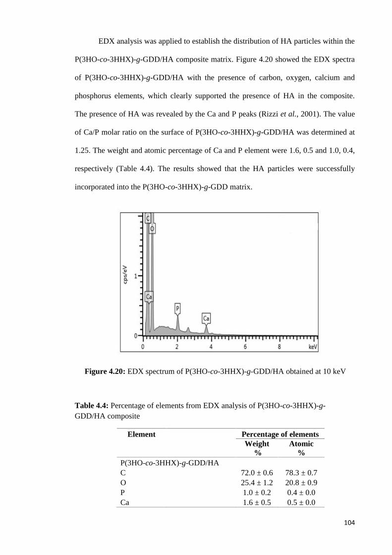
104
EDX analysis was applied to establish the distribution of HA particles within the
P(3HO-co-3HHX)-g-GDD/HA composite matrix. Figure 4.20 showed the EDX spectra
of P(3HO-co-3HHX)-g-GDD/HA with the presence of carbon, oxygen, calcium and
phosphorus elements, which clearly supported the presence of HA in the composite.
The presence of HA was revealed by the Ca and P peaks (Rizzi et al., 2001). The value
of Ca/P molar ratio on the surface of P(3HO-co-3HHX)-g-GDD/HA was determined at
1.25. The weight and atomic percentage of Ca and P element were 1.6, 0.5 and 1.0, 0.4,
respectively (Table 4.4). The results showed that the HA particles were successfully
incorporated into the P(3HO-co-3HHX)-g-GDD matrix.
Figure 4.20: EDX spectrum of P(3HO-co-3HHX)-g-GDD/HA obtained at 10 keV
Table 4.4: Percentage of elements from EDX analysis of P(3HO-co-3HHX)-g-GDD/HA composite
Element Percentage of elementsWeight
%Atomic
%P(3HO-co-3HHX)-g-GDD/HAC 72.0 ± 0.6 78.3 ± 0.7O 25.4 ± 1.2 20.8 ± 0.9P 1.0 ± 0.2 0.4 ± 0.0Ca 1.6 ± 0.5 0.5 ± 0.0
105
4.4.1 Toxicity test of P(3HO-co-3HHX)-g-GDD/HA using brine shrimp lethality
assay (BSLA)
Brine shrimp lethality assay (BSLA) was used to determine the toxicity of
polymeric composite of P(3HO-co-3HHX)-g-GDD/HA. The bioassay is a practical
implement for preliminary estimation of biological activities of a wide variety of
synthetic and natural products. BSLA is known as the most practical test for toxicology
and cytotoxicity of compounds due to its rapidity, convenience and cost-effectiveness
(Pelka et al., 2000; Rajabi et al., 2015).
Table 4.5 showed the mean percentage of mortality of A. franciscana nauplii
after 24 h exposure to aqueous solutions with different concentrations of P(3HO-co-
3HHX)-g-GDD/HA composite. The mean percentage of mortality in negative control
was 1.7 %. Meanwhile, the mean percentage of mortality at composite concentrations of
6.25, 12.5, 25, 50 and 100 % was 2.5, 3.3, 2.1, 2.5 and 1.2 %, respectively. No
significant difference was found between negative control and aqueous solutions of test
material at different concentrations after 24 hours exposure. Thus, it was concluded that
toxicity effect of P(3HO-co-3HHX)-g-GDD/HA composite towards A. franciscana
nauplii was not observed at all concentrations tested.

106
Table 4.5: Mean percentage of mortality of A. franciscana nauplii after 24 h exposureto aqueous solutions with different concentrations of P(3HO-co-3HHX)-g-GDD/HAcomposite
Concentration of sample (%) Mean mortality of A. franciscana (%)
Negative control 1.7 ± 0.2
6.25 2.5 ± 0.1
12.5 3.3 ± 0.2
25 2.1 ± 0.4
50 2.5 ± 0.1
100 1.2 ± 0.1*Values are mean of three independent experiments with three replicates per sampleconcentration

107
CHAPTER 5
CONCLUSIONS
5.1 Conclusions
Medium-chain-length poly(3-hydroxyalkanoates) (mcl-PHA) have been
produced by fed-batch fermentation of P. putida BET001. 8.6 g L-1 of cell dry weight
with 63 % mcl-PHA content was obtained after 48 hours fermentation. The mcl-PHA
was composed of three different monomers viz. 3-hydroxyoctanoate, 3-
hydroxyhexanoate and 3-hydroxydecanote at 90 mol %, 8 mol % and 2 mol %
respectively.
Modification of P(3HO-co-3HHX) via physical blending with hydroxyapatite
(HA) and chemical grafting with glycerol 1,3-diglecerol diacrylate (GDD) was carried
out. A porous composite scaffold made of mcl-PHA and hydroxyapatite (HA) was
successfully fabricated using facile particulate leaching technique. Based on FESEM
and EDX analyses, the scaffolds were found to be highly porous with interconnecting
pore structures and the HA particles were homogeneously dispersed in the polymer
matrix. The scaffolds showed excellent biocompatibility and osteoconductivity
characteristics based on strong proliferation and differentiation of osteoblast cells on
them. Thus, scaffolds made from P(3HO-co-3HHX)/HA composites are viable
candidate materials for bone tissue engineering application.
Moreover, GDD monomer was successfully grafted onto P(3HO-co-3HHX)
using benzoyl peroxide as radical initiator. P(3HO-co-3HHX)-g-GDD graft copolymer
were formed following chain transfer and chain termination by recombinant reactions.
Introduction of hydroxyl group onto P(3HO-co-3HHX) backbone improved the
wettability and impart amphilicity to the grafted polymer, thus potentially improving
their abilities for cellular interaction. The graft yield could be modulated by varying the

108
monomer concentration, initiator concentration, temperature and reaction time. The
optimum concentration of radical initiator was determined at 0.04 mM and graft yield
was observed to increase with initial monomer concentration. Amphiphilic copolymer
such as P(3HO-co-3HHX)-g-GDD obtained from the grafting of natural biopolyester
adds to the available repertoire of functional materials.
P(3HO-co-3HHX)-g-GDD/HA composite was also successfully fabricated via
graft copolymerization reaction and physical blending of HA to further improve the
properties of the neat mcl-PHA. Toxicity of the composite was studied against Artemia
franciscana in brine shrimp lethality assay. No significant mortality of the test organism
was recorded, thus implied that the novel scaffold posed negligible toxicity risk to the
cell. It is concluded that P(3HO-co-3HHX)-g-GDD/HA composite is potentially useful
for biomedical applications. It is also envisaged that future exploration with regards to
other composite materials and compositing methods for fabrication of biologically
active materials using medium-chain-length PHA as base component would
undoubtedly discover more potentialities, hence future applications.

109
5.2 Future research plan
The findings of this study have generated interesting directions for pursuing
future works. There are still much to be investigated regarding the modification of mcl-
PHA. Suggestions for future research following the current study are as follows:
1. The composite copolymer P(3HO-co-3HHX)/ HA can be further tailor-designed
using different techniques such as elctrospinning, freeze-drying method, melt-
compressed etc. in order to meet specific requirements of intended applications;
2. In vivo study of P(3HO-co-3HHX)/HA scaffold to be carried out by material
implantation in laboratory animals with femoral defect to determine associated
biological responses ;
3. The grafted copolymer P(3HO-co-3HHX)-g-GDD showed a good potential as
scaffold material, hence it is necessary to determine its biocompatibility using in vitro
and in vivo studies;
4. Further studies on the biodegradability behavior of the modified copolymer are
suggested in view of potential theranostic applications.
110
REFERENCES
Akay, G., Birch, M. & Bokhari, M. (2004). Microcellular polyhipe polymer supportsosteoblast growth and bone formation in vitro. Biomaterials, 25(18), 3991-4000.
Aldor, I. S. & Keasling, J. D. (2003). Process design for microbial plastic factories:metabolic engineering of polyhydroxyalkanoates. Current Opinion inBiotechnology, 14(5), 475-483.
Ali, I. & Jamil, N. (2016). Polyhydroxyalkanoates: current applications in the medicalfield. Frontiers in Biology, 11(1), 19-27.
Amache, R., Sukan, A., Safari, M., Roy, I. & Keshavarz, T. (2013). Advances in PHAsproduction. Chemical Engineering, 32, 931-936.
Annuar, M., Tan, I. K. & Ramachandran, K. (2008). Evaluation of nitrogen sources forgrowth and production of medium-chain-length poly-(3-hydroxyalkanoates)from palm kernel oil by Pseudomonas Putida PGA1. Asia Pacific Journal ofMolecular Biology and Biotechnology, 16(1), 11-15.
Annuar, M. S. M., Tan, I., Ibrahim, S. & Ramachandran, K. (2006). Production ofmedium-chain-length poly (3-hydroxyalkanoates) from saponified palm kerneloil by Pseudomonas Putida: kinetics of batch and fed-batch fermentations.Malaysian Journal of Microbiology, 2(2), 1-9.
Annuar, M. S. M. (2004). Production of medium-chain-length poly (3-hydroxyalkanoates) from saponified palm kernel oil by Pseudomonas putida.(Doctoral dissertation) University of Malaya.
Arslan, H., Hazer, B. & Yoon, S. C. (2007a). Grafting of poly (3‐hydroxyalkanoate) andlinoleic acid onto chitosan. Journal of Applied Polymer Science, 103(1), 81-89.
Arslan, H., Yeşilyurt, N. & Hazer, B. (2007b). The synthesis of poly(3‐hydroxybutyrate)‐g‐poly(methylmethacrylate) brush type graft copolymers byatom transfer radical polymerization Method. Journal of Applied PolymerScience, 106(3), 1742-1750.
Avci, D. & Mathias, L. J. (2004). Synthesis and photopolymerizations of new hydroxyl-containing dimethacrylate crosslinkers. Polymer, 45(6), 1763-1769.
Baei, M. S. & Rezvani, A. (2011). Nanocomposite (PHBHV/HA) fabrication frombiodegradable polymer. Middle-East Journal of Scientific Research, 7(1), 46-50.

111
Baek, J.-Y., Xing, Z.-C., Kwak, G., Yoon, K.-B., Park, S.-Y., Park, L. S. & Kang, I.-K.(2012). Fabrication and characterization of collagen-immobilized porousPHBV/HA nanocomposite scaffolds for bone tissue engineering. Journal ofNanomaterials, 2012, 1-11.
Bassas-Galià, M., Gonzalez, A., Micaux, F., Gaillard, V., Piantini, U., Schintke, S.,Zinn, M., & Mathieu, M. (2015). Chemical modification ofpolyhydroxyalkanoates (PHAs) for the preparation of hybrid biomaterials.CHIMIA International Journal for Chemistry, 69(10), 627-630.
Bayram, C., Denkbaş, E. B., Kiliçay, E., Hazer, B., Çakmak, H. B. & Noda, I. (2008).Preparation and characterization of triamcinolone acetonide-loaded poly (3-hydroxybutyrate-co-3-hydroxyhexanoate)(PHBHX) microspheres. Journal ofBioactive and Compatible Polymers, 23(4), 334-347.
Bhatt, R., Shah, D., Patel, K. & Trivedi, U. (2008). Pha–rubber blends: synthesis,characterization and biodegradation. Bioresource Technology, 99(11), 4615-4620.
Bhattacharya, A. & Misra, B. (2004). Grafting: a versatile means to modify polymers:techniques, factors and applications. Progress in Polymer Science, 29(8), 767-814.
Bhattacharya, A., Rawlins, J. W. & Ray, P. (2009). Polymer grafting and crosslinking:Wiley Online Library.
Bian, Y.Z., Wang, Y., Aibaidoula, G., Chen, G.-Q. & Wu, Q. (2009). Evaluation ofpoly (3-hydroxybutyrate-co-3-hydroxyhexanoate) conduits for peripheral nerveregeneration. Biomaterials, 30(2), 217-225
Boeree, N., Dove, J., Cooper, J., Knowles, J. & Hastings, G. (1993). Development of adegradable composite for orthopaedic use: mechanical evaluation of anhydroxyapatite-polyhydroxybutyrate composite material. Biomaterials, 14(10),793-796
Boccaccini, A. R. & Blaker, J. J. (2005). Bioactive composite materials for tissueengineering scaffolds. Expert Review of Medical Devices, 2(3), 303-317.
Brigham, C. J. & Sinskey, A. J. (2012). Applications of polyhydroxyalkanoates in themedical industry. International Journal of Biotechnology for WellnessIndustries, 1(1), 52.

112
Cakmakli, B., Hazer, B. & Borcakli, M. (2001). Poly(styrene peroxide) and poly(methylmethacrylate peroxide) for grafting on unsaturated bacterial polyesters.Macromolecular Bioscience, 1(8), 348-354.
Celik, M. (2004). Graft copolymerization of methacrylamide onto acrylic fibers initiatedby benzoyl peroxide. Journal of Applied Polymer Science, 94(4), 1519-1525.
Chan, S. M. (2012). Thermodegradation of medium-chain-length poly(3-hydroxyalkanoates), and assessment of the biopolyesters and oligoesters asplasticizer for poly(vinyl chloride). (Doctoral dissertation) University of Malaya.
Chanprateep, S., Katakura, Y., Visetkoop, S., Shimizu, H., Kulpreecha, S. & Shioya, S.(2008). characterization of new isolated Ralstonia Eutropha strain A-04 andkinetic study of biodegradable copolyester poly(3-hydroxybutyrate-co-4-hydroxybutyrate) production. Journal of Industrial Microbiology &Biotechnology, 35(11), 1205-1215.
Chardron, S., Bruzaud, S., Lignot, B., Elain, A. & Sire, O. (2010). Characterization ofbionanocomposites based on medium chain length polyhydroxyalkanoatessynthesized by Pseudomonas Oleovorans. Polymer Testing, 29(8), 966-971.
Chen, G. Q. & Wu, Q. (2005). The application of polyhydroxyalkanoates as tissueengineering materials. Biomaterials, 26(33), 6565-6578.
Chen, G., Ushida, T. & Tateishi, T. (2002). Scaffold design for tissue engineering.Macromolecular Bioscience, 2(2), 67-77.
Chen, Q., Liang, S. & Thouas, G. A. (2013). Elastomeric biomaterials for tissueengineering. Progress in Polymer Science, 38(3), 584-671.
Chen, W., Tao, X., Xue, P. & Cheng, X. (2005). Enhanced mechanical properties andmorphological characterizations of poly(vinyl alcohol)–carbon nanotubecomposite films. Applied Surface Science, 252(5), 1404-1409
Chen, X., Yang, X., Pan, J., Wang, L. & Xu, K. (2010). Degradation behaviors ofbioabsorbable P3/4HB monofilament suture in vitro and in vivo. Journal ofBiomedical Materials Research Part B: Applied Biomaterials, 92(2), 447-455
Chung, C. W., Kim, H. W., Kim, Y. B. & Rhee, Y. H. (2003). Poly(ethylene glycol)-grafted poly(3-hydroxyundecenoate) networks for enhanced blood compatibility.International Journal of Biological Macromolecules, 32(1), 17-22.
113
Chung, M. G., Kim, H. W., Kim, B. R., Kim, Y. B. & Rhee, Y. H. (2012).Biocompatibility and antimicrobial activity of poly(3-hydroxyoctanoate) graftedwith vinylimidazole. International Journal of Biological Macromolecules,50(2), 310-316.
Du, G. & Yu, J. (2002). Metabolic analysis on fatty acid utilization by Pseudomonasoleovorans: mcl-poly (3-hydroxyalkanoates) synthesis versus β-oxidation.Process Biochemistry, 38(3), 325-332.
Doi, Y. (1990). Microbial polyesters. New York: VCH Publishers, Inc., Yokohama,Japan.
Doyle, C., Tanner, E. & Bonfield, W. (1991). In vitro and in vivo evaluation ofpolyhydroxybutyrate and of polyhydroxybutyrate reinforced withhydroxyapatite. Biomaterials, 12(9), 841-847
El-Hadi, A., Schnabel, R., Straube, E., Müller, G. & Henning, S. (2002). Correlationbetween degree of crystallinity, morphology, glass temperature, mechanicalproperties and biodegradation of poly(3-hydroxyalkanoate) PHAs and theirblends. Polymer Testing, 21(6), 665-674.
Fei, B., Chen, C., Chen, S., Peng, S., Zhuang, Y., An, Y. & Dong, L. (2004).Crosslinking of poly[(3‐hydroxybutyrate)‐co‐(3‐hydroxyvalerate)] usingdicumyl peroxide as initiator. Polymer International, 53(7), 937-943.
Freier, T., Kunze, C., Nischan, C., Kramer, S., Sternberg, K., Saß, M., . . . Schmitz, K.-P. (2002). In vitro and in vivo degradation studies for development of abiodegradable patch based on poly (3-hydroxybutyrate). Biomaterials, 23(13),2649-2657.
Furrer, P., Panke, S. & Zinn, M. (2007). Efficient recovery of low endotoxin medium-chain-length poly([R]-3-hydroxyalkanoate) from bacterial biomass. Journal ofMicrobiological Methods, 69(1), 206-213.
Gumel, A. M., Annuar, M. S. & Heidelberg, T. (2012). Biosynthesis andcharacterization of polyhydroxyalkanoates copolymers produced byPseudomonas Putida Bet001 isolated from palm oil mill effluent. PLOS ONE,7(9), e45214.
Gumel, A., Annuar, M., Ishak, K. & Ahmad, N. (2014). Carbon nanofibers-poly-3-hydroxyalkanoates nanocomposite: ultrasound-assisted dispersion andthermostructural properties. Journal of Nanomaterials, 2014,123.

114
Gumel, A., Aris, M. & Annuar, M. (2014). Modification of polyhydroxyalkanoates(PHAs). In I. Roy & P. M. Visakh (Eds.), Polyhydroxyalkanoate (PHA) basedblends, composites and nanocomposites (pp. 141-182). UK: The Royal Societyof Chemistry.
Gursel, I., Yagmurlu, F., Korkusuz, F. & Hasirci, V. (2002). In vitro antibiotic releasefrom poly (3-hydroxybutyrate-co-3-hydroxyvalerate) rods. Journal ofMicroencapsulation, 19(2), 153-164
Guzmán, D., Kirsebom, H., Solano, C., Quillaguamán, J. & Hatti-Kaul, R. (2011).Preparation of hydrophilic poly(3-hydroxybutyrate) macroporous scaffoldsthrough enzyme-mediated modifications. Journal of Bioactive and CompatiblePolymers, 26(5), 452-463.
Hazer, B. (1996). Poly (β‐hydroxynonanoate) and polystyrene or poly(methylmethacrylate) graft copolymers: microstructure characteristics and mechanicaland thermal behavior. Macromolecular Chemistry and Physics, 197(2), 431-441.
Hazer, B. (2010). Amphiphilic poly (3-hydroxyalkanoate)s: potential candidates formedical applications. International Journal of Polymer Science, 2010, 1-8.
Hazer, B. & Steinbüchel, A. (2007). Increased diversification of polyhydroxyalkanoatesby modification reactions for industrial and medical applications. AppliedMicrobiology and Biotechnology, 74(1), 1-12.
Hazer, D. B., Kılıçay, E. & Hazer, B. (2012). Poly(3-hydroxyalkanoate)s:Diversification and biomedical applications: A state of the art review. MaterialsScience and Engineering: C, 32(4), 637-647.
Hu, S. G., Jou, C. H. & Yang, M. C. (2003). Antibacterial and biodegradable propertiesof polyhydroxyalkanoates grafted with chitosan and chitooligosaccharides viaozone treatment. Journal of Applied Polymer Science, 88(12), 2797-2803.
Huang, J., Lin, Y. W., Fu, X. W., Best, S. M., Brooks, R. A., Rushton, N. & Bonfield,W. (2007). Development of nano-sized hydroxyapatite reinforced compositesfor tissue engineering scaffolds. Journal of Materials Science: Materials inMedicine, 18(11), 2151-2157.
Ilter, S., Hazer, B., Borcakli, M. & Atici, O. (2001). Graft copolymerisation of methylmethacrylate onto a bacterial polyester containing unsaturated side chains.Macromolecular Chemistry and Physics, 202(11), 2281-2286.

115
Jack, K. S., Velayudhan, S., Luckman, P., Trau, M., Grøndahl, L. & Cooper-White, J.(2009). The fabrication and characterization of biodegradable ha/phbvnanoparticle–polymer composite scaffolds. Acta Biomaterialia, 5(7), 2657-2667.
Jenkins, D. W. & Hudson, S. M. (2001). Review of vinyl graft copolymerizationfeaturing recent advances toward controlled radical-based reactions andillustrated with chitin/chitosan trunk polymers. Chemical Reviews, 101(11),3245-3274.
Jiang, X. J., Sun, Z., Ramsay, J. A. & Ramsay, B. A. (2013). Fed-batch production ofmcl-pha with elevated 3-hydroxynonanoate content. AMB Express, 3(1), 1-8.
Jung, K., Hazenberg, W., Prieto, M. & Witholt, B. (2001). Two‐stage continuous
process development for the production of medium‐chain‐length
poly(3‐hydroxyalkanoates). Biotechnology and Bioengineering, 72(1), 19-24.
Kabilan, S., Ayyasamy, M., Jayavel, S. & Paramasamy, G. (2012). Pseudomonas sp. asa source of medium chain length polyhydroxyalkanoates for controlled drugdelivery: perspective. International Journal of Microbiology, 2012, 1-10.
Kai, D. & Loh, X. J. (2013). Polyhydroxyalkanoates: Chemical modifications towardbiomedical applications. ACS Sustainable Chemistry & Engineering, 2(2), 106-119.
Kai, Z., Ying, D. & Guo-Qiang, C. (2003). Effects of surface morphology on thebiocompatibility of polyhydroxyalkanoates. Biochemical Engineering Journal,16(2), 115-123
Kansiz, M., Domínguez-Vidal, A., Mcnaughton, D. & Lendl, B. (2007). Fourier-transform infrared (FTIR) spectroscopy for monitoring and determining thedegree of crystallisation of polyhydroxyalkanoates (PHAs). Analytical andBioanalytical Chemistry, 388(5-6), 1207-1213.
Karageorgiou, V. & Kaplan, D. (2005). Porosity of 3D biomaterial scaffolds andosteogenesis. Biomaterials, 26(27), 5474-5491.
Katti, D. S., Vasita, R. & Shanmugam, K. (2008). Improved biomaterials for tissueengineering applications: surface modification of polymers. Current Topics inMedicinal Chemistry, 8(4), 341-353.
Kim, D. Y., Kim, H. W., Chung, M. G. & Rhee, Y. H. (2007). Biosynthesis,modification, and biodegradation of bacterial medium-chain-lengthpolyhydroxyalkanoates. The Journal of Microbiology, 87-97.
116
Kim, H. W., Chung, C. W. & Rhee, Y. H. (2005a). UV-induced graft copolymerizationof monoacrylate-poly(ethylene glycol) onto poly(3-hydroxyoctanoate) to reduceprotein adsorption and platelet adhesion. International Journal of BiologicalMacromolecules, 35(1), 47-53.
Kim, H. W., Chung, C. W., Hwang, S. J. & Rhee, Y. H. (2005b). Drug release from andhydrolytic degradation of a poly (ethylene glycol) grafted poly (3-hydroxyoctanoate). International Journal of Biological Macromolecules, 36(1),84-89.
Kim, H. W., Chung, M. G., Kim, Y. B. & Rhee, Y. H. (2008). Graft copolymerizationof glycerol 1, 3-diglycerolate diacrylate onto poly(3-hydroxyoctanoate) toimprove physical properties and biocompatibility. International Journal ofBiological Macromolecules, 43(3), 307-313.
Knowles, J., Hastings, G., Ohta, H., Niwa, S. & Boeree, N. (1992). Development of adegradable composite for orthopaedic use: in vivo biomechanical andhistological evaluation of two bioactive degradable composites based on thepolyhydroxybutyrate polymer. Biomaterials, 13(8), 491-496
Köse, G. T., Korkusuz, F., Korkusuz, P., Purali, N., Özkul, A. & Hasırcı, V. (2003).Bone generation on PHBV matrices: an in vitro study. Biomaterials, 24(27),4999-5007
Kokubo, T., Kim, H.-M. & Kawashita, M. (2003). Novel bioactive materials withdifferent mechanical properties. Biomaterials, 24(13), 2161-2175.
Kunasundari, B. & Sudesh, K. (2011). Isolation and recovery of microbialpolyhydroxyalkanoates. Express Polymer Letters, 5(7), 620-634.
Langer, N. M. & Wilkie, C. A. (1998). Surface modification of polyamide‐6: graft
copolymerization of vinyl monomers onto polyamide‐6 and thermal analysis ofthe graft copolymers. Polymers for Advanced Technologies, 9(5), 290-296.
Lao, H. K., Renard, E., Langlois, V., Vallée‐Rehel, K. & Linossier, I. (2010). Surfacefunctionalization of PHBV by HEMA grafting via UV treatment: Comparisonwith thermal free radical polymerization. Journal of Applied Polymer Science,116(1), 288-297.
Lao, H.-K., Renard, E., Linossier, I., Langlois, V. & Vallée-Rehel, K. (2007).Modification of poly(3-hydroxybutyrate-co-3-hydroxyvalerate) film by chemicalgraft copolymerization. Biomacromolecules, 8(2), 416-423.

117
Le Meur, S., Zinn, M., Egli, T., Thöny-Meyer, L. & Ren, Q. (2012). Production ofmedium-chain-length polyhydroxyalkanoates by sequential feeding of xyloseand octanoic acid in engineered Pseudomonas Putida KT2440. BMCBiotechnology, 12(1), 53.
Lee, H. S. & Lee, T. Y. (1997). Graft polymerization of acrylamide ontopoly(hydroxybutyrate-co-hydroxyvalerate) films. Polymer, 38(17), 4505-4511.
Leenstra, T., Kuijpers-Jagtman, A. & Maltha, J. (1998). The healing process of palataltissues after palatal surgery with and without implantation of membranes: anexperimental study in dogs. Journal of Materials Science: Materials inMedicine, 9(5), 249-255
Li, H. & Chang, J. (2005). Preparation, characterization and in vitro release ofgentamicin from PHBV/wollastonite composite microspheres. Journal ofControlled Release, 107(3), 463-473
Li, Z. & Loh, X. J. (2015). Water soluble polyhydroxyalkanoates: Future materials fortherapeutic applications. Chemical Society Reviews, 44(10), 2865-2879.
Li, Z., Yang, J. & Loh, X. J. (2016). Polyhydroxyalkanoates: Opening doors for asustainable future. NPG Asia Materials, 8, e265.
Löbler, M., Sternberg, K., Stachs, O., Allemann, R., Grabow, N., Roock, A., . . . Hanh,B. D. (2011). Polymers and drugs suitable for the development of a drugdelivery drainage system in glaucoma surgery. Journal of Biomedical MaterialsResearch Part B: Applied Biomaterials, 97(2), 388-395
Madison, L. L. & Huisman, G. W. (1999). Metabolic engineering ofpoly(3hydroxyalkanoates): From DNA to plastic. Microbiology and MolecularBiology Reviews, 63(1), 21-53.
Malm, T., Bowald, S., Bylock, A., Busch, C. & Saldeen, T. (1994). Enlargement of theright ventricular outflow tract and the pulmonary artery with a newbiodegradable patch in transannular position. European Surgical Research,26(5), 298-308
Martin, D. P. & Williams, S. F. (2003). Medical applications of poly-4-hydroxybutyrate: a strong flexible absorbable biomaterial. BiochemicalEngineering Journal, 16(2), 97-105
Meng, D.-C., Shen, R., Yao, H., Chen, J.-C., Wu, Q. & Chen, G.-Q. (2014).Engineering the diversity of polyesters. Current Opinion in Biotechnology, 29,24-33.

118
Misra, S. K., Valappil, S. P., Roy, I. & Boccaccini, A. R. (2006). Polyhydroxyalkanoate(PHA)/inorganic phase composites for tissue engineering applications.Biomacromolecules, 7(8), 2249-2258.
Misra, S. K., Nazhat, S. N., Valappil, S. P., Moshrefi-Torbati, M., Wood, R. J., Roy, I.& Boccaccini, A. R. (2007). Fabrication and characterization of biodegradablepoly (3-hydroxybutyrate) composite containing bioglass. Biomacromolecules,8(7), 2112-2119
Mosahebi, A., Fuller, P., Wiberg, M. & Terenghi, G. (2002). Effect of allogeneicschwann cell transplantation on peripheral nerve regeneration. Experimentalneurology, 173(2), 213-223
Moradi, A., Dalilottojari, A., Pingguan-Murphy, B. & Djordjevic, I. (2013). Fabricationand characterization of elastomeric scaffolds comprised of a citric acid-basedpolyester/hydroxyapatite microcomposite. Materials & Design, 50, 446-450.
Muhr, A., Rechberger, E. M., Salerno, A., Reiterer, A., Schiller, M., Kwiecień, ...Koller, M. (2013). Biodegradable latexes from animal-derived waste:biosynthesis and characterization of mcl-pha accumulated by Ps. Citronellolis.Reactive and Functional Polymers, 73(10), 1391-1398.
Nair, L. S. & Laurencin, C. T. (2006). Polymers as biomaterials for tissue engineeringand controlled drug delivery. In K. Lee & D. Kaplan (Eds.), Tissue engineering I(pp. 47-90). Berlin, Heidelberg: Springer Berlin Heidelberg.
Nakas, J. P., Zhu, C., Perrotta, J. A. & Nomura, C. T. (2015). Methods for producingpolyhydroxyalkanoates from biodiesel-glycerol: U.S Patents 8,956,835,B2.
Nguyen, L. H., Annabi, N., Nikkhah, M., Bae, H., Binan, L., Park, S et al. (2012).Vascularized bone tissue engineering: Approaches for potential improvement.Tissue Engineering Part B: Reviews, 18(5), 363-382.
Nguyen, S. (2008). Graft copolymers containing poly(3-hydroxyalkanoates)- A reviewon their synthesis, properties, and applications. Canadian Journal of Chemistry,86(6), 570-578.
Nguyen, S. & Marchessault, R. H. (2004). Synthesis and properties of graft copolymersbased on poly(3‐hydroxybutyrate) macromonomers. MacromolecularBioscience, 4(3), 262-268.
Ni, J. & Wang, M. (2002). In vitro evaluation of hydroxyapatite reinforcedpolyhydroxybutyrate composite. Materials Science and Engineering: C, 20(1),101-109.

119
Novikov, L. N., Novikova, L. N., Mosahebi, A., Wiberg, M., Terenghi, G. & Kellerth,J.-O. (2002). A novel biodegradable implant for neuronal rescue andregeneration after spinal cord injury. Biomaterials, 23(16), 3369-3376
Panith, N., Assavanig, A., Lertsiri, S., Bergkvist, M., Surarit, R. & Niamsiri, N. (2016).Development of tunable biodegradable polyhydroxyalkanoates microspheres forcontrolled delivery of tetracycline for treating periodontal disease. Journal ofApplied Polymer Science, 133(42)
Peschel, G., Dahse, H. M., Konrad, A., Wieland, G. D., Mueller, P. J., Martin, D. P. &Roth, M. (2008). Growth of keratinocytes on porous films of poly(3‐hydroxybutyrate) and poly (4‐hydroxybutyrate) blended with hyaluronic acidand chitosan. Journal of Biomedical Materials Research Part A, 85(4), 1072-1081
Pelka, M., Danzl, C., Distler, W. & Petschelt, A. (2000). A new screening test fortoxicity testing of dental materials. Journal of Dentistry, 28(5), 341-345.
Pham, B. T., Tonge, M. P., Monteiro, M. J. & Gilbert, R. G. (2000). Grafting kinetics ofvinyl neodecanoate onto polybutadiene. Macromolecules, 33(7), 2383-2390.
Philip, S., Keshavarz, T. & Roy, I. (2007). Polyhydroxyalkanoates: Biodegradablepolymers with a range of applications. Journal of Chemical Technology &Biotechnology, 82(3), 233-247.
Poblete-Castro, I., Rodriguez, A. L., Lam, C. M. & Kessler, W. (2014). Improvedproduction of medium-chain-length polyhydroxyalkanoates in glucose-basedfed-batch cultivations of metabolically engineered Pseudomonas putida strains.Journal of Microbiology and Biotechnology, 24(1), 59-69.
Porter, J. R., Ruckh, T. T. & Popat, K. C. (2009). Bone tissue engineering: A review inbone biomimetics and drug delivery strategies. Biotechnology Progress, 25(6),1539-1560.
Pouton, C. W. & Akhtar, S. (1996). Biosynthetic polyhydroxyalkanoates and theirpotential in drug delivery. Advanced Drug Delivery Reviews, 18(2), 133-162.
Pramanik, N., Mishra, D., Banerjee, I., Maiti, T. K., Bhargava, P. & Pramanik, P.(2009). Chemical synthesis, characterization, and biocompatibility study ofhydroxyapatite/chitosan phosphate nanocomposite for bone tissue engineeringapplications. International Journal of Biomaterials, 2009, 512417.

120
Qu, X.-H., Wu, Q. & Chen, G.-Q. (2006). In vitro study on hemocompatibility andcytocompatibility of poly(3-hydroxybutyrate-co-3-hydroxyhexanoate). Journalof Biomaterials Science, Polymer Edition, 17(10), 1107-1121.
Rai, R., Keshavarz, T., Roether, J., Boccaccini, A. R. & Roy, I. (2011). Medium chainlength polyhydroxyalkanoates, promising new biomedical materials for thefuture. Materials Science and Engineering: R: Reports, 72(3), 29-47.
Rajabi, S., Ramazani, A., Hamidi, M. & Naji, T. (2015). Artemia Salina as a modelorganism in toxicity assessment of nanoparticles. DARU Journal ofPharmaceutical Sciences, 23(1), 1.
Razaif‐Mazinah, M., Rafais, M., Annuar, M., Suffian, M. & Sharifuddin, Y. (2015).Effects of even and odd number fatty acids cofeeding on pha production andcomposition in Pseudomonas Putida Bet001 isolated from palm oil mill effluent.Biotechnology and Applied Biochemistry, 63, 92-100.
Reddy, C., Ghai, R. & Kalia, V. C. (2003). Polyhydroxyalkanoates: An overview.Bioresource Technology, 87(2), 137-146.
Renard, E., Tanguy, P. Y., Samain, E. & Guerin, P. (2003). Synthesis of novel graftpolyhydroxyalkanoates. Paper presented at the Macromolecular Symposia.
Rezwan, K., Chen, Q. Z., Blaker, J. J. & Boccaccini, A. R. (2006). Biodegradable andbioactive porous polymer/inorganic composite scaffolds for bone tissueengineering. Biomaterials, 27(18), 3413-3431.
Rizzi, S. C., Heath, D., Coombes, A., Bock, N., Textor, M. & Downes, S. (2001).biodegradable polymer/hydroxyapatite composites: Surface analysis and initialattachment of human osteoblasts. Journal of Biomedical Materials Research,55(4), 475-486.
Saadat, A., Behnamghader, A., Karbasi, S., Abedi, D., Soleimani, M. & Shafiee, A.(2013). Comparison of acellular and cellular bioactivity of poly(3-hydroxybutyrate/hydroxyapatite nanocomposite and poly(3-hydroxybutyrate)scaffolds. Biotechnology and Bioprocess Engineering, 18(3), 587-593.
Sabir, M. I., Xu, X. & Li, L. (2009). A review on biodegradable polymeric materials forbone tissue engineering applications. Journal of Materials Science, 44(21),5713-5724.

121
Shabna, A., Saranya, V., Malathi, J., Shenbagarathai, R. & Madhavan, H. (2013).Indigenously produced polyhydroxyalkanoate based co‐polymer as cellularsupportive biomaterial. Journal of Biomedical Materials Research Part A102(10), 3470-3476.
Shishatskaya, E., Khlusov, I. & Volova, T. (2006). A hybrid PHB–hydroxyapatitecomposite for biomedical application: production, in vitro and in vivoinvestigation. Journal of Biomaterials Science, Polymer Edition, 17(5), 481-498
Shishatskaya, E., Volova, T., Puzyr, A., Mogilnaya, O. & Efremov, S. (2004). Tissueresponse to the implantation of biodegradable polyhydroxyalkanoate sutures.Journal of Materials Science: Materials in Medicine, 15(6), 719-728
Shrivastav, A., Kim, H.-Y. & Kim, Y.-R. (2013). Advances in the applications ofpolyhydroxyalkanoate nanoparticles for novel drug delivery system. BioMedResearch International, 2013,1-12.
Sodian, R., Sperling, J. S., Martin, D. P., Egozy, A., Stock, U., Mayer Jr, J. E. &Vacanti, J. P. (2000). Technical report: Fabrication of a trileaflet heart valvescaffold from a polyhydroxyalkanoate biopolyester for use in tissue engineering.Tissue Engineering, 6(2), 183-188.
Steinbüchel, A. & Lütke-Eversloh, T. (2003). Metabolic engineering and pathwayconstruction for biotechnological production of relevant polyhydroxyalkanoatesin microorganisms. Biochemical Engineering Journal, 16(2), 81-96.
Sudesh, K. & Iwata, T. (2008). Sustainability of biobased and biodegradable plastics.CLEAN–Soil, Air, Water, 36(5‐6), 433-442.
Sudesh, K., Abe, H. & Doi, Y. (2000). Synthesis, structure and properties ofpolyhydroxyalkanoates: biological polyesters. Progress in Polymer Science,25(10), 1503-1555.
Sultana, N. & Khan, T. H. (2012). In vitro degradation of PHBV scaffolds andnHA/PHBV composite scaffolds containing hydroxyapatite nanoparticles forbone tissue engineering. Journal of Nanomaterials, 2012, 1-12.
Sultana, N. & Wang, M. (2008). PHBV/PLLA-based composite scaffolds containingnano-sized hydroxyapatite particles for bone tissue engineering. Journal ofExperimental Nanoscience, 3(2), 121-132.
Tan, Q., Li, S., Ren, J. & Chen, C. (2011). Fabrication of porous scaffolds with acontrollable microstructure and mechanical properties by porogen fusiontechnique. International Journal of Molecular Sciences, 12(2), 890-904.
122
Tesema, Y., Raghavan, D. & Stubbs, J. (2004). Bone cell viability on collagenimmobilized poly (3‐hydroxybutrate‐co‐3‐hydroxyvalerate) membrane: Effect ofsurface chemistry. Journal of Applied Polymer Science, 93(5), 2445-2453
Tsuge, T. (2002). Metabolic improvements and use of inexpensive carbon sources inmicrobial production of polyhydroxyalkanoates. Journal of Bioscience andBioengineering, 94(6), 579-584
Verlinden, R. A., Hill, D. J., Kenward, M., Williams, C. D. & Radecka, I. (2007).Bacterial synthesis of biodegradable polyhydroxyalkanoates. Journal of AppliedMicrobiology, 102(6), 1437-1449.
Wang, W., Zhang, Y. & Chen, Y. (2007). Graft copolymerization of N-vinylpyrrolidoneonto poly(3-hydroxybutyrate-co-3-hydroxyvalerate) in homogeneous solution.Iranian Polymer Journal, 16(3), 195.
Wang, Y. W., Wu, Q., Chen, J. & Chen, G. Q. (2005). Evaluation of three-dimensionalscaffolds made of blends of hydroxyapatite and poly(3-hydroxybutyrate-co-3-hydroxyhexanoate) for bone reconstruction. Biomaterials, 26(8), 899-904.
Wang, Y.-W., Wu, Q. & Chen, G.-Q. (2004). Attachment, proliferation anddifferentiation of osteoblasts on random biopolyester poly(3-hydroxybutyrate-co-3-hydroxyhexanoate) scaffolds. Biomaterials, 25(4), 669-675.
Wei, G. & Ma, P. X. (2004). Structure and properties of nano-hydroxyapatite/polymercomposite scaffolds for bone tissue engineering. Biomaterials, 25(19), 4749-4757.
Williams, S. F. & Martin, D. P. (2005). Applications of polyhydroxyalkanoates (PHA)in medicine and pharmacy. Biopolymers 3,91-128.
Xi, J., Zhang, L., Zheng, Z. A., Chen, G., Gong, Y., Zhao, N. & Zhang, X. (2008).Preparation and evaluation of porous poly(3-hydroxybutyrate-co-3-hydroxyhexanoate)—hydroxyapatite composite scaffolds. Journal ofBiomaterials Applications, 22(4), 293-307.
Xu, J., Guo, B.-H., Yang, R., Wu, Q., Chen, G.-Q. & Zhang, Z.-M. (2002). In situ FTIRstudy on melting and crystallization of polyhydroxyalkanoates. Polymer, 43(25),6893-6899.
Yu, L., Dean, K. & Li, L. (2006). Polymer blends and composites from renewableresources. Progress in Polymer Science, 31(6), 576-602.

123
Zhao, K., Deng, Y., Chen, J. C. & Chen, G.-Q. (2003). Polyhydroxyalkanoate (PHA)scaffolds with good mechanical properties and biocompatibility. Biomaterials,24(6), 1041-1045
Zinn, M., Witholt, B. & Egli, T. (2001). Occurrence, synthesis and medical applicationof bacterial polyhydroxyalkanoate. Advanced Drug Delivery Reviews, 53(1), 5-21.
124
PUBLICATIONS AND PAPERS PRESENTED
Publications:
1. Ansari, N. F., Annuar, M. S. M., & Murphy, B. P. (2016). A porousmedium‐chain‐length poly (3‐hydroxyalkanoates)/hydroxyapatite composite as scaffoldfor bone tissue engineering. Engineering in Life Sciences 17: 420-429.
2. Ansari, N. F., Annuar, M. S. M. (2017). Functionalization of medium-chain-lengthpoly(3-hydroxyalkanoates) as amphiphilic material by graft copolymerization withglycerol 1,3-diglycerol diacrylate and its mechanism. Journal of MacromolecularScience, Part A (accepted – DOI: 10.1080/10601325.2017.1387490)
Conferences:
1. Nor Faezah Ansari, M. Suffian M. Annuar, Belinda Pingguan Murphy. (2015). Aporous composite PHA/hydroxyapatite scaffold for bone tissue engineering and itshydrophilicity enhancement by graft copolymerization with glycerol 1,3-diglyceroldiacrylate. Presented in 20th Biological Science Graduate Congress (BSGC),Chulalongkorn University, Thailand (Oral presentation).
2. Nor Faezah Ansari and M. Suffian M. Annuar. (2016). Fabrication andcharacterization of porous P(3HO-co-3HHX)/hydroxyapatite composite scaffold forbone tissue engineering and its hydrophilicity enhancement by graft copolymerizationwith glycerol 1,3-diglycerol diacrylate. Presented in 3rd International Conference ofChemical Engineering and Industrial Biotechnology (ICCEIB), Bayou Lagoon ParkResort, Melaka (Oral presentation).
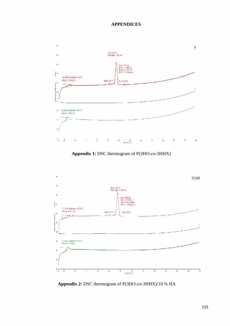
125
APPENDICES
Appendix 1: DSC thermogram of P(3HO-co-3HHX)
Appendix 2: DSC thermogram of P(3HO-co-3HHX)/10 % HA
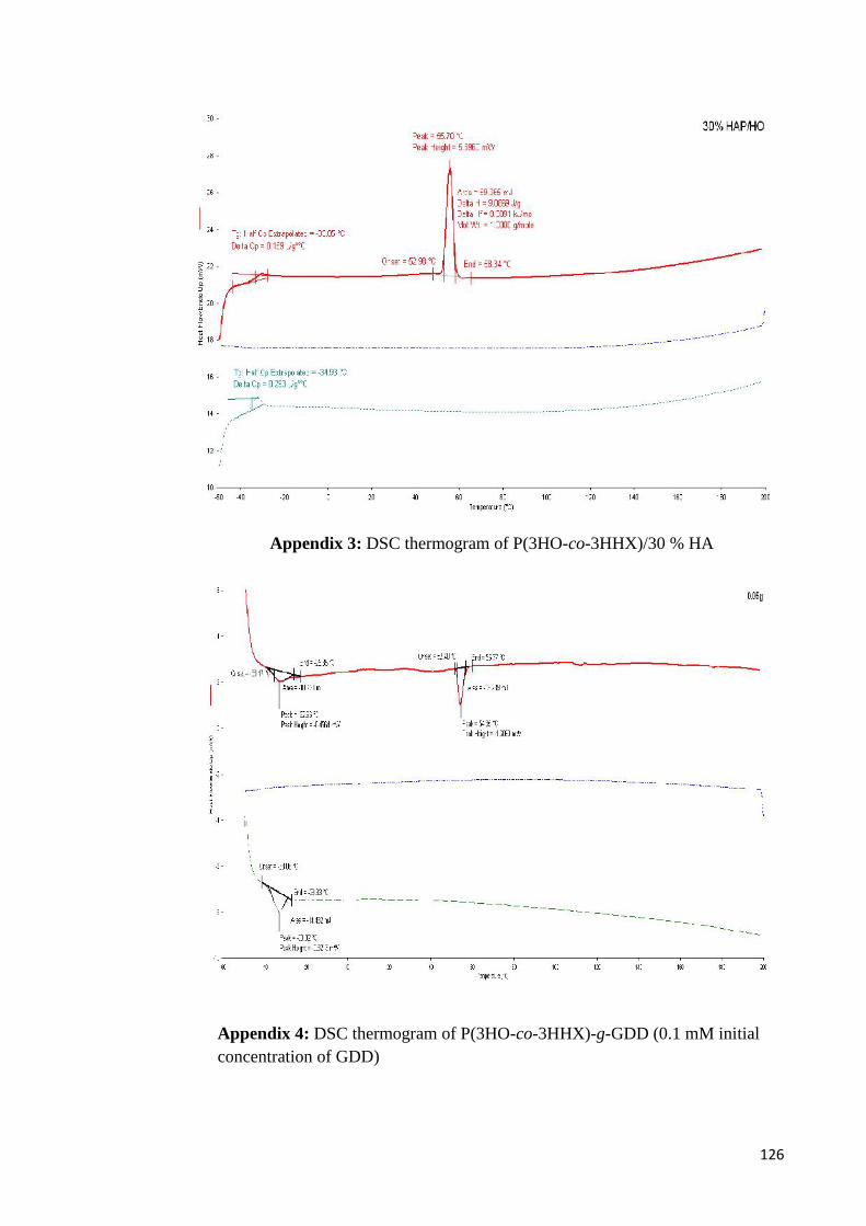
126
Appendix 3: DSC thermogram of P(3HO-co-3HHX)/30 % HA
Appendix 4: DSC thermogram of P(3HO-co-3HHX)-g-GDD (0.1 mM initialconcentration of GDD)
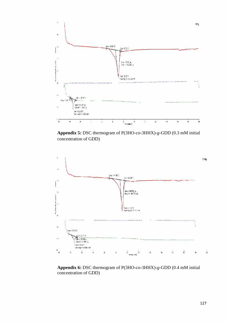
127
Appendix 5: DSC thermogram of P(3HO-co-3HHX)-g-GDD (0.3 mM initialconcentration of GDD)
Appendix 6: DSC thermogram of P(3HO-co-3HHX)-g-GDD (0.4 mM initialconcentration of GDD)

128
Appendix 7: DSC thermogram of P(3HO-co-3HHX)-g-GDD (0.6 mM initialconcentration of GDD)