Androgen metabolism via 17 -hydroxysteroid dehydrogenase ...
Upload
sheila-sharpCategory
view
213download
0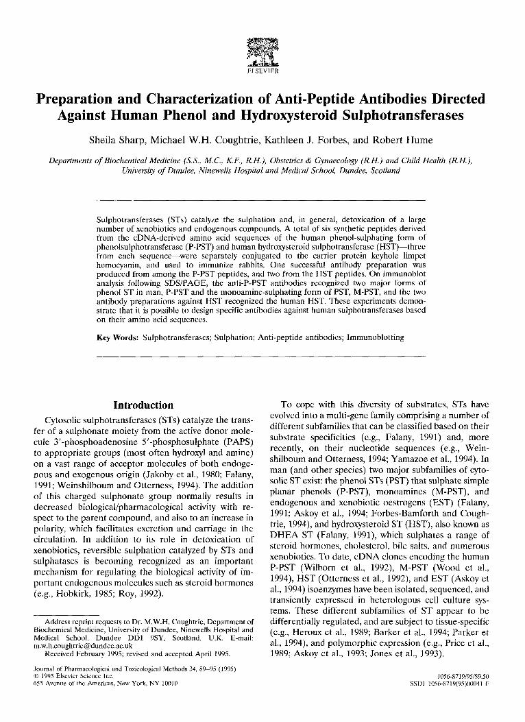
ELSEVIER
Preparation and Characterization of Anti-Peptide Antibodies Directed Against Human Phenol and Hydroxysteroid Sulphotransferases
Sheila Sharp, Michael W.H. Coughtrie, Kathleen J. Forbes, and Robert Hume
Departments of Biochemical Medicine (S.S., M.C., K.F., R.H.), Obstetrics & Gynaecology (R.H.) and Child Health (R.H.), University of Dundee, Ninewells Hospital and Medical School, Dundee, Scotland
Sulphotransferases (STs) catalyze the sulphation and, in general, detoxication of a large number of xenobiotics and endogenous compounds. A total of six synthetic peptides derived from the cDNA-derived amino acid sequences of the human phenol-sulphating form of phenolsulphotransferase (P-PST) and human hydroxysteroid sulphotransferase (HST)-three from each sequence-were separately conjugated to the carrier protein keyhole limpet hemocyanin, and used to immunize rabbits. One successful antibody preparation was produced from among the P-PST peptides, and two from the HST peptides. On immunoblot analysis following SDS/PAGE, the anti-P-PST antibodies recognized two major forms of phenol ST in man, P-PST and the monoamine-sulphating form of PST, M-PST, and the two antibody preparations against HST recognized the human HST. These experiments demon- strate that it is possible to design specific antibodies against human sulphotransferases based on their amino acid sequences.
Key Words: Sulphotransferases; Sulphation; Anti-peptide antibodies; Immunoblotting
Introduction Cytosolic sulphotransferases (STs) catalyze the trans-
fer of a sulphonate moiety from the active donor mole- cule 3’-phosphoadenosine 5’-phosphosulphate (PAPS) to appropriate groups (most often hydroxyl and amine) on a vast range of acceptor molecules of both endoge- nous and exogenous origin (Jakoby et al., 1980; Falany, 1991; Weinshilboum and Otterness, 1994). The addition of this charged sulphonate group normally results in decreased biological/pharmacological activity with re- spect to the parent compound, and also to an increase in polarity, which facilitates excretion and carriage in the circulation. In addition to its role in detoxication of xenobiotics, reversible sulphation catalyzed by STs and sulphatases is becoming recognized as an important mechanism for regulating the biological activity of im- portant endogenous molecules such as steroid hormones (e.g., Hobkirk, 1985; Roy, 1992).
Address reprint requests to Dr. M.W.H. Coughtrie, Department of Biochemical Medicine, University of Dundee, Ninewells Hospital and Medical School, Dundee DDl 9SY, Scotland, U.K. E-mail: [email protected]
Received February 1995; revised and accepted April 1995.
Journal of Pharmacological and Toxicological Methods 34, 89-95 (1995) 0 1995 Elsevier Science Inc. 655 Avenue of the Americas, New York, NY 10010
To cope with this diversity of substrates, STs have evolved into a multi-gene family comprising a number of different subfamilies that can be classified based on their substrate specificities (e.g., Falany, 1991) and, more recently, on their nucleotide sequences (e.g., Wein- shilboum and Otterness, 1994; Yamazoe et al., 1994). In man (and other species) two major subfamilies of cyto- solic ST exist: the phenol STs (PST) that sulphate simple planar phenols (P-PST), monoamines (M-PST), and endogenous and xenobiotic oestrogens (EST) (Falany, 1991; Askoy et al., 1994; Forbes-Bamforth and Cough- trie, 1994) and hydroxysteroid ST (HST), also known as DHEA ST (Falany, 1991) which sulphates a range of steroid hormones, cholesterol, bile salts, and numerous xenobiotics. To date, cDNA clones encoding the human P-PST (Wilborn et al., 1992) M-PST (Wood et al., 1994) HST (Otterness et al., 1992) and EST (Askoy et al., 1994) isoenzymes have been isolated, sequenced, and transiently expressed in heterologous cell culture sys- tems. These different subfamilies of ST appear to be differentially regulated, and are subject to tissue-specific (e.g., Heroux et al., 1989; Barker et al., 1994; Parker et al., 1994) and polymorphic expression (e.g., Price et al., 1989; Askoy et al., 1993; Jones et al., 1993).
1058719195/$9.50 SSDI 1056.871Y(95)00041-F
90 JPM Vol. 34, No. 2 October 1995:89-95
Our appreciation of the function of sulphation and of the STs will be enhanced through studying their local- ization and by identifying the factors governing their expression. Central to such studies are the availability of the appropriate molecular tools such as DNA probes and isoenzyme-specific antibodies, and we have previ- ously prepared antibodies against three purified rat liver ST isoenzymes, which have some cross-reactivity with human STs of the corresponding families (Coughtrie and Sharp, 1990; Borthwick et al., 1993; Sharp et al., 1993).
The aim of this work was to use the derived amino acid sequences of members of the human PST and HST families to design peptides that could be used to produce antibodies specific for these human proteins. We report the production of anti-peptide antibodies that specifi- cally recognize human HST and that recognize the very closely related members of the PST subfamily, P-PST and M-PST, as determined by their reaction with the partially purified human proteins, and with human liver and platelet cytosols.
Materials and Methods Materials
Peptides were synthesized by Severn Biotech Ltd., Kidderminster, U.K. Maleimide-activated keyhole lim- pet hemocyanin carrier protein, Immunopure antibody/ antigen immobilization kit, desalting columns, and IgG elution buffer were supplied by Pierce & Warriner, Chester, U.K. Freund’s adjuvant (complete and incom- plete) and peroxidase-conjugated goat anti-(rabbit IgG) (IgG fraction, absorbed with human serum proteins) were purchased from Sigma Chemical Company, Ltd., Poole, U.K. 1-[l-‘4C]Naphthol, Enhanced Chemilumi- nescence (ECL) western blot reagents, and X-ray film were obtained from Amersham International plc, Little Chalfont, U.K. [1,2,3,6,7-3H]DHEA (90 Ci/mmol), PAP35S (2.5 Ci/mmol), [2,4,6,7-3H]oestrone (93 Ci/ mmol) and [6,7-3H]17a-ethinyloestradiol (42.5 Ci/ mmol) were purchased from Du Pant/New England Nuclear, Stevenage, U.K. PAPS and all protein purifi- cation media were from Pharmacia Ltd., Milton Keynes, U.K. Scintillation fluid (Emulsifier Safe) was purchased from Canberra Packard, Pangbourne, U.K., and electro- phoresis chemicals were from MerckBDH, Glasgow, U.K. All other reagents were of analytical grade and purchased from commonly used local suppliers. Human platelet cytosols were prepared as previously described (Jones et al., 1993).
Antibody Production Using amino acid sequence data derived from the
published nucleotide sequences of the human liver P-
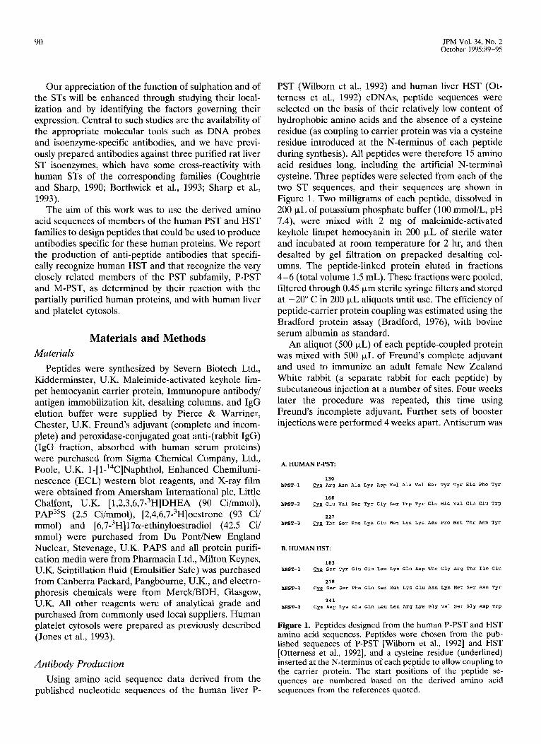
PST (Wilborn et al., 1992) and human liver HST (Ot- terness et al., 1992) cDNAs, peptide sequences were selected on the basis of their relatively low content of hydrophobic amino acids and the absence of a cysteine residue (as coupling to carrier protein was via a cysteine residue introduced at the N-terminus of each peptide during synthesis). All peptides were therefore 15 amino acid residues long, including the artificial N-terminal cysteine. Three peptides were selected from each of the two ST sequences, and their sequences are shown in Figure 1. Two milligrams of each peptide, dissolved in 200 ~J,L of potassium phosphate buffer (100 mmol/L, pH 7.4), were mixed with 2 mg of maleimide-activated keyhole limpet hemocyanin in 200 ~.LL of sterile water and incubated at room temperature for 2 hr, and then desalted by gel filtration on prepacked desalting col- umns. The peptide-linked protein eluted in fractions 4-6 (total volume 1.5 mL). These fractions were pooled, filtered through 0.45 pm sterile syringe filters and stored at -20” C in 200 PL aliquots until use. The efficiency of peptide-carrier protein coupling was estimated using the Bradford protein assay (Bradford, 1976), with bovine serum albumin as standard.
An aliquot (500 ~.LL) of each peptide-coupled protein was mixed with 500 l.r,L of Freund’s complete adjuvant and used to immunize an adult female New Zealand White rabbit (a separate rabbit for each peptide) by subcutaneous injection at a number of sites. Four weeks later the procedure was repeated, this time using Freund’s incomplete adjuvant. Further sets of booster injections were performed 4 weeks apart. Antiserum was
A. HUMAN P-PST:
Figure 1. Peptides designed from the human P-PST and HST amino acid sequences. Peptides were chosen from the pub- lished sequences of P-PST [Wilborn et al., 19921 and HST [Otterness et al., 19921, and a cysteine residue (underlined) inserted at the N-terminus of each peptide to allow coupling to the carrier protein. The start positions of the peptide se- quences are numbered based on the derived amino acid sequences from the references quoted.
S. SHARP ET AL, ANTI-PEPTIDE ANTIBODIES
91
collected 19 days after the final immunization by cardiac puncture under terminal anesthesia. The progress of antibody production was monitored at 2-week intervals using immunoblot analysis with human liver and platelet cytosols, and with partially purified human liver sulpho- transferase isoenzymes, P-PST and HST.
Afinity Purification of Anti-sulphotransferase Peptide Antibodies
Anti-peptide antibodies were isolated from rabbit serum by affinity purification. Peptide affinity columns were prepared using sulphhydryl-activated beaded aga- rose (Sulpholink Coupling Gel, Immunopure Ag/Ab Immobilization kit 2#) columns and 3 mg of the appro- priate peptide, as described by the manufacturer (Pierce & Warriner). Then 1 mL of appropriate rabbit serum was applied to the column and left for 2 hr at room temperature. Unbound serum protein was eluted with 16 mL of lOO-mM potassium phosphate buffer (pH 7.4) and bound IgG eluted with 1-mL aliquots of the IgG elution buffer supplied with the kit. Protein recovery was estimated using the method of Bradford (1976) and varied between 40 kg/mL and 300 kg/rnL for the differ- ent peptides, according to the efficiency of peptide coupling to the affinity gel.
Partial Putification of Human Liver Sulphotransferases
A 25-g sample of normal adult human liver that had been obtained at surgery and that was previously frozen at -70” C was homogenised in 4 volumes lo-mmol triethanolamine per liter/Cl-, pH 7.4, containing 3-mmol2-mercaptoethanol per liter and 10% (v/v) glyc- erol (buffer A). The homogenate was centrifuged at 12,OOOg for 15 min at 4” C, and the supernatant centri- fuged at 100,OOOg for 1 hr. The resulting supernatant (cytosol) was applied to a column of DEAE-Sepharose Fast Flow (35 X 2.6 cm) at a flow rate of 180 mL/hr, and following elution of unbound material with approxi- mately 4 column volumes of buffer A, the column was washed with buffer A containing lOO-mmol NaCl per liter. ST activity was eluted using a linear gradient of lOO-225-mmol NaCl per liter in buffer A at a flow rate of 60 mL/hr. DHEA, l-naphthol and oestrone ST activ- ities were determined (using radiochemical assays-see below) in the fractions, and those fractions containing high levels of ST activity towards DHEA (i.e., HST) and 1-naphthol (i.e., PST) were pooled and concentrated to
approximately 20 mL by ultrafiltration. Aliquots (1 mL) of this pooled material were applied to prepacked columns of Sephadex G-25 (PD-10, Pharmacia) and the buffer exchanged with buffer B (20-mmol triethano- lamine per liter/Cl-, pH 7.4 containing 3-mmol 2-mer- captoethanol per liter). Samples (1 mL) of the resulting protein fraction were applied to a MonoQ anion ex- change column attached to an FPLC system (Pharma- cia) at a flow rate of 60 mL/hr. Gradient elution of ST activity was performed using the following stepped gra- dient, all performed at a flow rate of 60 mL/hr: buffer B for 10 min, O-100 mmol NaCl per liter in buffer B over a period of 10 min, lOO-mmol NaCl per liter in buffer B for a further 10 min, and finally loo-225 mmol NaCl per liter in buffer B over 60 min. Fractions of 1 mL were collected and assayed for 4-nitrophenol, DHEA, dopa- mine and 17ol-ethinyloestradiol ST activities. Fractions containing the P-PST activity (determined with 4-nitro- phenol as substrate) and those containing HST activity (determined with DHEA as substrate) were separated by this method, and were pooled and frozen at -70” C prior to use in immunoblot analysis.
Sulphotransferase Enzyme Assays HST activity (DHEA as substrate) was measured
radiometrically as follows: reactions (final volume 215 ~.LL) contained liver cytosol or purification fraction (50 tr,L), DHEA (0.1 &i, 7.5-prnol final concentration per liter), PAPS (5-kmol, final concentration per liter) and buffer (lOO-mmol Tris per liter/Cl-, pH 7.4, 20-mmol MgCI, per liter). After 20 min at 37” C, incubations were stopped by the addition of 250~PL Tris/Cl--, pH 8.7 and 3 mL of chloroform. Following extraction by vigorous shaking, the phases were separated by centrifugation at 3OOOg for 3 min. 200 ~.LL of aqueous phase were mixed with 3 mL of scintillation fluid and the radioactivity quantitated by liquid scintillation spectrometry. Oes- trone, l-naphthol, 17B-oestradiol, and 17a-ethinylo- estradiol ST activities in 50-FL aliquots of human liver cytosol or the purification fractions were assayed as described previously (Barnforth et al., 1992, 1993). Do- pamine and 4-nitrophenol ST activities were assayed by the barium precipitation method using PAP3’S as de- scribed (Foldes and Meek, 1973; Anderson and Wein- shilboum, 1980).
SDSIPAGE and Immunoblotting Proteins were resolved on 11% acrylamide monomer
gels in the presence of 0.1% SDS by the method originally described by Laemmli (1970). For immunoblot analysis, proteins were electrophoretically transferred to nitrocellulose (Schleicher and Schuell) as described by
92 JPM Vol. 34, No. 2 October 1995:89-95
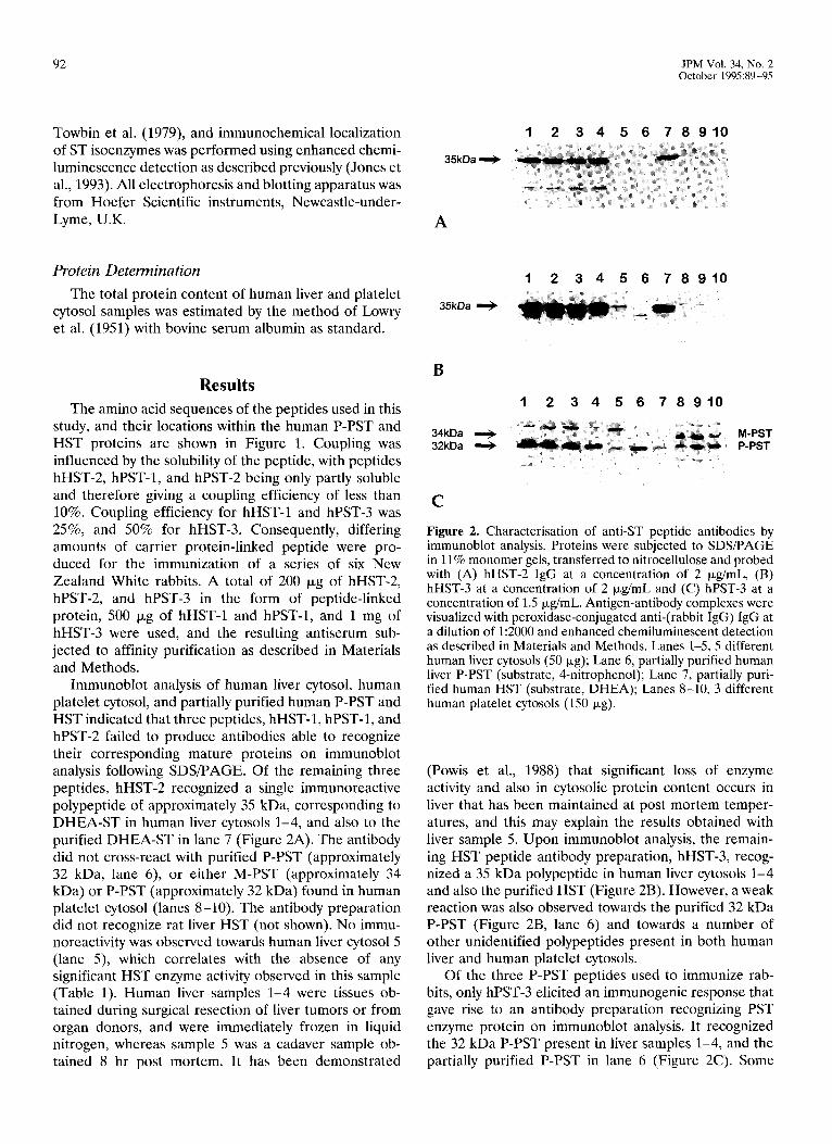
Towbin et al. (1979), and immunochemical localization 12345676910 of ST isoenzymes was performed using enhanced chemi- luminescence detection as described previously (Jones et
35kDa +
al., 1993). All electrophoresis and blotting apparatus was from Hoefer Scientific instruments, Newcastle-under- Lyme, U.K. A
Protein Determination The total protein content of human liver and platelet
cytosol samples was estimated by the method of Lowry et al. (1951) with bovine serum albumin as standard.
Results The amino acid sequences of the peptides used in this
study, and their locations within the human P-PST and HST proteins are shown in Figure 1. Coupling was influenced by the solubility of the peptide, with peptides hHST-2, hPST-1, and hPST-2 being only partly soluble and therefore giving a coupling efficiency of less than 10%. Coupling efficiency for hHST-1 and hPST-3 was 25%, and 50% for hHST-3. Consequently, differing amounts of carrier protein-linked peptide were pro- duced for the immunization of a series of six New Zealand White rabbits. A total of 200 Fg of hHST-2, hPST-2, and hPST-3 in the form of peptide-linked protein, 500 kg of hHST-1 and hPST-1, and 1 mg of hHST-3 were used, and the resulting antiserum sub- jected to affinity purification as described in Materials and Methods.
Immunoblot analysis of human liver cytosol, human platelet cytosol, and partially purified human P-PST and HST indicated that three peptides, hHST-1, hPST-1, and hPST-2 failed to produce antibodies able to recognize their corresponding mature proteins on immunoblot analysis following SDS/PAGE. Of the remaining three peptides, hHST-2 recognized a single immunoreactive polypeptide of approximately 35 kDa, corresponding to DHEA-ST in human liver cytosols 1-4, and also to the purified DHEA-ST in lane 7 (Figure 2A). The antibody did not cross-react with purified P-PST (approximately 32 kDa, lane 6), or either M-PST (approximately 34 kDa) or P-PST (approximately 32 kDa) found in human platelet cytosol (lanes S-10). The antibody preparation did not recognize rat liver HST (not shown). No immu- noreactivity was observed towards human liver cytosol5 (lane 5), which correlates with the absence of any significant HST enzyme activity observed in this sample (Table 1). Human liver samples l-4 were tissues ob- tained during surgical resection of liver tumors or from organ donors, and were immediately frozen in liquid nitrogen, whereas sample 5 was a cadaver sample ob- tained 8 hr post mortem. It has been demonstrated
12345676910
35kDa I,
B
12 345676910 .
34kDa + M-PST 32kDa + mm- ;=s P-PST
i 1_ i.
C
Figure 2. Characterisation of anti-ST peptide antibodies by immunoblot analysis. Proteins were subjected to SDS/PAGE in 11% monomer gels, transferred to nitrocellulose and probed with (A) hHST-2 IgG at a concentration of 2 &mL, (B) hHST-3 at a concentration of 2 &mL and (C) hPST-3 at a concentration of 1.5 &mL. Antigen-antibody complexes were visualized with peroxidase-conjugated anti-(rabbit IgG) IgG at a dilution of 1:2000 and enhanced chemiluminescent detection as described in Materials and Methods. Lanes 1-5, 5 different human liver cytosols (50 kg); Lane 6, partially purified human liver P-PST (substrate, 4-nitrophenol); Lane 7, partially puri- fied human HST (substrate, DHEA); Lanes S-10, 3 different human platelet cytosols (1.50 Fg).
(Powis et al., 1988) that significant loss of enzyme activity and also in cytosolic protein content occurs in liver that has been maintained at post mortem temper- atures, and this may explain the results obtained with liver sample 5. Upon immunoblot analysis, the remain- ing HST peptide antibody preparation, hHST-3, recog- nized a 35 kDa polypeptide in human liver cytosols l-4 and also the purified HST (Figure 2B). However, a weak reaction was also observed towards the purified 32 kDa P-PST (Figure 2B, lane 6) and towards a number of other unidentified polypeptides present in both human liver and human platelet cytosols.
Of the three P-PST peptides used to immunize rab- bits, only hPST-3 elicited an immunogenic response that gave rise to an antibody preparation recognizing PST enzyme protein on immunoblot analysis. It recognized the 32 kDa P-PST present in liver samples 1-4, and the partially purified P-PST in lane 6 (Figure 2C). Some
S. SHARP ET AL. ANTI-PEPTIDE ANTIBODIES
93
Table 1. Sulphotransferase Enzyme Activities in Cytosolic Fractions of Five Human Liver Samples
Liver 1 Liver 2 Liver 3 Liver 4 Liver 5
Substrate
DHEA 17B-Oestradiol Oestrone 17cu-Ethinyloestradiol 4-Nitrophenol 1-Naphthol
173 16
3.4 27 35
110
Specific Activity (pmoliminlmg protein)
146 125 200 12 13 9.7
2.7 2.3 n.d. 21 23 16 53 54 37 99 105 63
10 1.6
n.d. n.d. n.d. n.d.
Sulphotransferase enzyme activities were determined as described in Materials and Methods, using human liver cytosol samples which were subjected to immunoblot analysis in Figures 3A-C.
n.d. = No enzyme activity detectable, i.e. below the limit of sensitivity of the assay, which under the conditions employed was approximately 1 pmol/min/mg protein.
residual P-PST enzyme protein in the purified HST fraction (Figure 2C, lane 7) was also recognized by this antibody on immunoblot analysis. It did not recognize the HST isoform, but did cross-react with the 34 kDa M-PST found in human platelet cytosol (Figure 2C, lanes 8-10). This was confirmed by comparison of the level of protein expression demonstrated in the immu- noblot analysis with the enzyme activities observed in the individual platelet cytosols (Table 2). Platelet sample 1 expressed the low P-PST phenotype (Jones et al., 1993) as determined with 4-nitrophenol as substrate, and a correspondingly lower expression of P-PST can be seen on the immunoblot (Figure 2C). In contrast, platelet sample 3 exhibited a low level of expression of M-PST with lower M-PST activity with dopamine as substrate (Table 2). Little variation was observed in expression of liver P-PST enzyme protein in samples 1-4, where enzyme activities were found to be very similar but, as was the case for HST, little activity or enzyme protein was present in the post mortem liver sample, number 5 (Table 1 and Figure 2C).
Discussion We have used the amino acid sequences of two ST
isoenzymes, P-PST and HST, to design peptides that
Table 2. Sulphotransferase Enzyme Activities in Cytosolic Fractions of Three Human Platelet Samples
Platelet cytosol 1 Platelet cytosol 2 Platelet cytosol 3
Substrate Specific Activity (pmol/min/mg)
Dopamine 72 93 41 Vanillin 56 36 18 4-Nitrophenol 4.1 15 20
Sulphotransferase enzyme activities were determined as described in Materials and Methods, using the platelet cytosol samples which were subjected to immunoblot analysis in Figures 3A-C. Dopamine and vanillin are substrates for M-PST, and 4-nitrophenol is a substrate for P-PST.
were used to immunize rabbits. Of the six peptides used, a total of three (one from P-PST and two from HST) gave rise to antibody preparations that recognized the appropriate ST proteins on immunoblot analysis of human liver and platelet cytosols, and also of the corresponding partially purified human liver P-PST and HST isoenzymes. None of the antibodies recognized the corresponding proteins in rat liver (not shown). It is not clear why the peptides hHST-1 and hPST-1 and 2 failed to result in the successful production of anti-ST antibod- ies. One possible explanation for failure to produce antibodies is the solubility of the peptides, but of the three peptides that were the least soluble and that resulted in the lowest coupling efficiency (10%) to the carrier protein (keyhole limpet haemocyanin), two (hHST-2 and hPST-3) resulted in successful antibody production. Another possibility is that those peptides failing to produce antibody response in some way folded in a manner that, although producing an immunological response, resulted in antibodies that did not recognize the corresponding ST isoenzyme in the detection system used (immunoblot analysis of denatured proteins follow- ing SDS/PAGE). We tested the three successful antibod- ies by immunohistochemistry (on paraffin-embedded sections) of human fetal adrenal (a rich source of HST [Barker et al., 19941) and adult human liver, but there was no clear difference between the sections stained with the anti-peptide antibodies and the pre-immune IgG at the same concentration (not shown). This sug- gests that either the antibodies do not recognize their respective proteins in a “native” environment, or that the antibody titres were too low for use in this technique.
Of the two antibody preparations raised against HST, that which resulted from peptide hHST-2 appeared to have slightly higher specificity, as judged by immunoblot analysis (Figure 2A). It recognized only the 35 kDa HST protein in the partially purified HST preparation, noth- ing in the partially purified P-PST preparation, and the 35 kDa HST isoenzyme in human liver samples l-4.
94 JPM Vol. 34, No. 2 October 1995:89-95
Another, faster-migrating polypeptide was detected in liver samples 1-4, but we have reason to believe that this is a degradation fragment of the HST, because it also appears after prolonged storage of the purified enzyme (not shown). The antibody preparation that came from peptide hHST-3 had slightly stronger staining than hHST-2 at the same concentration, and recognized the small amount of protein in human liver sample 5 (ca- daver-derived), as well as exhibiting a slight cross- reaction with the P-PST isoenzyme in the partially purified preparation and also in the human liver samples (Figure 2B). The antibody raised against peptide hPST-3 recognized both P-PST, as determined by partially puri- fied human liver P-PST, human liver and human platelet cytosol, and also M-PST in human platelet cytosol. At the time when these peptides were designed and the antibodies raised, the cDNA corresponding to a M-PST isoform had not been cloned, and therefore the amino acid sequence was not known. The nucleotide sequence of an M-PST cDNA and the derived amino acid se- quence has recently been published [8], and found to be in excess of 92% identical at the amino acid level. Amino acid sequence alignment of peptide hPST-3 with the M-PST derived amino acid sequence (Wood et al., 1994) showed that there is 100% sequence identity over those 14 amino acids, and that the threonine residue that is in position 227 in the P-PST sequence (Wilborn et al., 1992) is also in position 227 in the M-PST sequence (Wood et al., 1994) indicating the molecular basis for our observation that the antibody produced from this peptide recognised both P-PST and M-PST on immuno- blot analysis. The availability of the M-PST amino acid sequence should now make it theoretically possible to produce antibodies against M-PST and P-PST that are mutually distinct, as the antibodies that have to date been produced that react with P-PST also react with M-PST and vice versa (e.g., Heroux et al., 1989; Jones et al., 1993). The sequences are, however, highly homolo- gous, and it remains to be seen whether or not this is possible in practice; these experiments are currently underway in our laboratory. The 50% success rate in raising satisfactory antibodies obtained here also indi- cates the potential difficulties in creating effective “de- signer” antibodies against such highly homologous pro- teins.
The antibodies prepared here should prove to be of value in investigating, by immunoblot analysis, the ex- pression of members of the PST and HST enzyme families in humans, and having demonstrated that the technique of raising anti-peptide antibodies can be ap- plied to the sulphotransferases, it should be possible to design further isoenzyme-specific antibodies as and when the amino acid sequences of novel isoforms be- come available.
This work was supported by the Sir Jules Thorn Charitable Trust (to MWHC), Action Research (to MWHC and RH) and by the Commis- sion of the European Communities (to MWHC, Contract BMHl- CT92-0097, Biomedicine and Health Programme). MWHC was a Caledonian Research Foundation/Royal Society of Edinburgh Re- search Fellow. We are grateful to Andy Cassidy for performing protein sequence database searches in relation to this work, and to the late Professor G.B. Ode11 for his help in obtaining human liver samples.
References Anderson RJ, Weinshilboum RM (1980) Phenolsulphotransferase in
human tissue: Radiochemical enzymatic assay and biochemical properties. Clin Chim Acta 103:79-80.
Askoy IA, Sochorova V, Weinshilboum RM (1993) Human liver dehydroepiandrosterone sulfotransferase: Nature and extent of individual variation. Clin Pharmacol Ther 54:498-506.
Askoy IA, Wood TC, Weinshilboum RM (1994) Human liver estrogen sulfotransferase: Identification by cDNA cloning and expression. Biochem Biophys Res Commun 200:1621-1629.
Bamforth KJ, Dalghesh K, Coughtrie MWH (1992) Inhibition of human liver steroid sulphotransferase activities by drugs: A novel mechanism of drug toxicity? Eur J Pharmacol228:15-21.
Bamforth KJ, Jones AL, Roberts RC, Coughtrie MWH (1993) Com- mon food additives are potent inhibitors of human liver 1701- ethinyloestradiol and dopamine sulphotransferases. Biochem Phar- macol46:1713-1720.
Barker EV, Hume R, Hallas A, Coughtrie MWH (1994) Dehydroepi- androsterone sulfotransferase in the developing human fetus: Quantitative biochemical and immunological characterization of the hepatic, renal and adrenal enzymes. Endoctinology 134:982- 989.
Borthwick EB, Burchell A, Coughtrie MWH (1993) Purification and immunochemical characterisation of a male-specific rat liver estro- gen sulfotransferase. Biochem J 289:719-725.
Bradford MM (1976) A rapid and sensitive method for the quantita- tion of microgram quantities of protein utilizing the principle of protein-dye binding. Anal Biochem 721248-254.
Coughtrie MWH, Sharp S (1990) Purification and immunochemical characterisation of a rat liver sulphotransferase conjugating para- cetamol. Biochem Pharmacol40:2305-2313.
Falany CN (1991) Molecular enzymology of human liver cytosolic sulfotransferases. Trends Pharmacol Sci 12:255-259.
Foldes A, Meek JL (1973) Rat brain phenolsulfotransferase: Partial purification and some properties. Biochim Biophys Acta 327:365- 374.
Forbes-Bamforth KJ, Coughtrie MWH (1994) Identification of a new adult human liver sulfotransferase with specificity for endogenous and xenobiotic estrogens. Biochem Biophys Res Commun 198:707- 711.
Heroux JA, Falany CN, Roth JA (1989) Immunological characteriza- tion of human phenol sulfotransferase. Mol Pharmacol 36129-33.
Hobkirk R (1985) Steroid sulfotransferases and steroid sulfate sulfa- tases: Characteristics and biological roles. Can J Biochem Cell Biol 63:1127-1144.
Jakoby WB, Sekura RD, Lyon ES, Marcus CJ, Wang J-L (1980) Sulfotransferases. In Enzymatic Basis of Detoxication, Vol 2. Ed., WB Jakoby. New York: Academic Press, pp. 199-228.
Jones AL, Roberts RC, Coughtrie MWH (1993) The human phenol sulphotransferase polymorphism is determined by the level of expression of the enzyme protein. Biochem J 296:287-290.
Laemmli UK (1970) Cleavage of structural proteins during the assem- bly of the head of bacteriophage T4. Nature 227:680-685.

S. SHARP ET AL. 95 ANTI-PEPTIDE ANTIBODIES
Lowry OH, Rosebrough NJ, Farr AL, Randall RJ (1951) Protein mea- surement with the Folin phenol reagent. J Biol Chem 193:265-275.
Otterness DM, Wieben ED, Wood TC, Watson WG, Madden BJ, McCormick DJ, Weinshilboum RM (1992) Human liver dehydro- epiandrosterone sulfotransferase: Molecular cloning and expres- sion of cDNA. Mol Pharmacol41:865-872.
Parker CR Jr, Falany CN, Stockard CR, Stankovic AK, Grizzle WE (1994) Immunohistochemical localization of dehydroepiandros- terone sulfotransferase in human fetal tissues. J Clin Endocrinol Metab 78~234-236.
Powis G, Jardine I, Van Dyke R, Weinshilboum R, Moore D, Wilke T, Rhodes W. Nelson R. Benson L, Szlumanski C (1988) Foreign I compound metabolism studies with human liver obtained as surgi- cal waste: Relation to donor characteristics and effects of tissue storage. Drug Metab Dispos 16:582-589.
Price RA, Spielman RS, Lucena AL, Van Loon JA, Maidak BL, Weinshilboum RM (1989) Genetic polymorphism for human plate- let thermostable phenol sulfotransferase (TS PST) activity. Genet- ics 122:905-914.
Roy AK (1992) Regulation of steroid hormone action in target cells by specific hormone-inactivating enzymes. Proc Sot Exp Biol Med 199:265-272.
Sharp S, Barker EV, Coughtrie MWH, Lowenstein PR, Hume R (1993) Immunochemical characterisation of a dehydroepiandros- terone sulfotransferase in rats and humans. Eur / Biochem 211: 539-548.
Towbin H, Stehelin T, Gordon J (1979) Electrophoretic transfer of proteins from polyacrylamide gels to nitrocellulose sheets: Proce- dures and some applications. Proc Nat1 Acad Sci USA 76:4045- 4049.
Weinshilboum R, Otterness D (1994) Sulfotransferase enzymes. In Conjugation-Deconjugation Reactions in Drug Metabolism and Tox- icity. Ed., FC Kauffman. Berlin: Springer-Verlag, pp. 45-78.
Wilborn TW, Comer KA, Dooley TP, Reardon IM, Heinrikson RL, Falany CN (1992) Sequence analysis and expression of the cDNA for the phenol-sulphating form of human liver phenol sulphotrans- ferase. Mol Pharmacol 43170-77.
Wood TC, Askoy IA. Askoy S, Weinshilboum RM (1994) Human thermolabile phenol sulfotransferase: cDNA Cloning, expression and characterization. Biochem Biophys Res Commun 198:1119- 1127.
Yamazoe Y, Nagata K, Ozawa S, Kato R (1994) Structural similarity and diversity of sulfotransferases. Chem Biol Interact 92:107-117.