Prenatal detection of a 1p36 deletion in a fetus with multiple malformations and a review of the...
5
Prenat. Diagn. 19: 49–53 (1999) SHORT COMMUNICATION Prenatal Detection of a 1p36 Deletion in a Fetus with Multiple Malformations and a Review of the Literature L. Faivre 1 *, N. Morichon-Delvallez 1 , G. Viot 1 , J. Martinovic 2 , M. P. Pinson 1 , J. P. Aubry 3 , V. Raclin 1 , P. Edery 1 , Y. Dumez 3 , A. Munnich 1 and M. Vekemans 1 1 De ´partement de Ge ´ne ´tique, Ho ˆpital Necker Enfants Malades, Paris, France 2 Unite ´ de Foetopathologie, Ho ˆpital Saint-Antoine, Paris, France 3 Department de Me ´decine Foetale, Ho ˆpital Necker Enfants Malades, Paris, France The prenatal diagnosis of a 1p36 deletion is reported. The pregnancy was ascertained at 24 weeks of gestation because of the discovery of multiple malformations at ultrasound including hypotelorism, moderate cerebral ventricular dilatation and Ebstein anomaly with secondary cardiac failure. Following cytogenetic studies and counselling, the pregnancy was terminated and a fetal autopsy performed. The phenotype of this antenatally- diagnosed case is compared with the clinical features of 44 previously reported cases with an identical deletion of the short arm of chromosome 1p36. Copyright ? 1999 John Wiley & Sons, Ltd. : deletion of the short arm of chromosome 1; Ebstein anomaly; cerebral ventricular dilatation; prenatal diagnosis INTRODUCTION With the advent of high-resolution chromosome analysis and molecular cytogenetic techniques, some small deletions such as deletions of the short arm of chromosome 1 are now being diagnosed more fre- quently. Since the initial observation (Hain et al., 1980), 42 additional cases with del (1)(p36) have been reported (Yunis et al., 1981; Deslanges et al., 1983; Steele et al., 1984; Magenis et al., 1987; Abbas et al., 1990; Wexler et al., 1991; Wargowski et al., 1991; Barbi et al., 1992; Biegel et al., 1993; Howard-Peebles and Black, 1994; Keppler-Noreuil et al., 1995; Sandlin et al., 1995; Reish et al., 1995; Blennow et al., 1996; Bos et al., 1996; Giraudeau et al., 1997; Eugster et al., 1997; Shapira et al., 1997; Robbins-Furman et al., 1997). The phenotype of this deletion is now well delineated (Shapira et al., 1997). The clinical features include mod- erate to severe mental retardation, hypotony, growth delay, eye/vision problems, hearing deficit, seizures, cardiac malformations and dysmorphic facies (pointed chin, flat nasal bridge, low-set ears, ear asymmetry, thickened ear helices, deep-set eyes, large anterior fontanel, clinodactyly and/or short fifth finger). Here, we report the first case of a 1p36 deletion ascertained prenatally because of abnormal ultrasound findings. CASE REPORT A 34-year-old Polish mother was referred to our centre at 24 weeks of gestation for her first pregnancy. Her past medical history was unremarkable. She had three brothers. Her 38-year-old husband is also Polish and the couple are not consanguineous. A second-trimester ultrasound examination showed a moderate cerebral ventricular dilatation, a hypotelorism and Ebstein anomaly. All parameters of fetal biometry were between the 50th and 97th percentile. A cytogenetic study performed on amniotic fluid cells showed a terminal deletion of the short arm of chromosome 1 (del(1)(p36)). Parental chromosomes were normal. After genetic counselling and because of the discovery of an important hydrops fetalis due to cardiac failure, the pregnancy was terminated at 28 weeks. External examination revealed a male fetus with a generalized hydrops and facial dysmorphism, including brachycephaly, a flat profile, low-set ears and a deep palate (Fig. 1). Unique transverse palmar creases are seen on both hands. Measurements (weight 1900 g; vertex–talus length 41·5 cm; vertex–coccyx length 26 cm; OFC 29·5 cm; foot 5·5 cm) were compatible with term. X-ray studies of the fetus showed 11 pairs of ribs with normal ossification. At necropsy, thymic hypoplasia, pleural effusion, abnormal pulmonary lobulation and Ebstein anomaly were found. Neuro- pathological examination revealed a moderate ven- tricular dilatation of the left ventricle and possible polymicrogyria of the left lobe. Histological examination showed pancreatic necrosis and an endomyocardic fibrosis. CYTOGENETIC AND MOLECULAR ANALYSIS Chromosome analysis of the fetus was performed on amniotic fluid cells using solid Giemsa, R and *Correspondence to: L. Faivre, Service de Ge ´ne ´tique Me ´dicale, Ho ˆ pital Necker Enfants Malades, 149 rue de Se `vres, 75015 Paris, France. CCC 0197–3851/99/010049–05$17.50 Copyright ? 1999 John Wiley & Sons, Ltd. Received 9 February 1998 Revised 26 May 1998 Accepted 19 June 1998
Transcript of Prenatal detection of a 1p36 deletion in a fetus with multiple malformations and a review of the...
Prenat. Diagn. 19: 49–53 (1999)
SHORT COMMUNICATION
Prenatal Detection of a 1p36 Deletion in a Fetus with MultipleMalformations and a Review of the Literature
L. Faivre1*, N. Morichon-Delvallez1, G. Viot1, J. Martinovic2, M. P. Pinson1, J. P. Aubry3,V. Raclin1, P. Edery1, Y. Dumez3, A. Munnich1 and M. Vekemans1
1Departement de Genetique, Hopital Necker Enfants Malades, Paris, France2Unite de Foetopathologie, Hopital Saint-Antoine, Paris, France3Department de Medecine Foetale, Hopital Necker Enfants Malades, Paris, France
The prenatal diagnosis of a 1p36 deletion is reported. The pregnancy was ascertained at 24 weeks of gestationbecause of the discovery of multiple malformations at ultrasound including hypotelorism, moderate cerebralventricular dilatation and Ebstein anomaly with secondary cardiac failure. Following cytogenetic studies andcounselling, the pregnancy was terminated and a fetal autopsy performed. The phenotype of this antenatally-diagnosed case is compared with the clinical features of 44 previously reported cases with an identicaldeletion of the short arm of chromosome 1p36. Copyright ? 1999 John Wiley & Sons, Ltd.
: deletion of the short arm of chromosome 1; Ebstein anomaly; cerebral ventricular dilatation;prenatal diagnosis
INTRODUCTION
With the advent of high-resolution chromosomeanalysis and molecular cytogenetic techniques, somesmall deletions such as deletions of the short arm ofchromosome 1 are now being diagnosed more fre-quently. Since the initial observation (Hain et al.,1980), 42 additional cases with del (1)(p36) have beenreported (Yunis et al., 1981; Deslanges et al., 1983;Steele et al., 1984; Magenis et al., 1987; Abbas et al.,1990; Wexler et al., 1991; Wargowski et al., 1991; Barbiet al., 1992; Biegel et al., 1993; Howard-Peebles andBlack, 1994; Keppler-Noreuil et al., 1995; Sandlinet al., 1995; Reish et al., 1995; Blennow et al., 1996; Boset al., 1996; Giraudeau et al., 1997; Eugster et al., 1997;Shapira et al., 1997; Robbins-Furman et al., 1997).The phenotype of this deletion is now well delineated(Shapira et al., 1997). The clinical features include mod-erate to severe mental retardation, hypotony, growthdelay, eye/vision problems, hearing deficit, seizures,cardiac malformations and dysmorphic facies (pointedchin, flat nasal bridge, low-set ears, ear asymmetry,thickened ear helices, deep-set eyes, large anteriorfontanel, clinodactyly and/or short fifth finger). Here,we report the first case of a 1p36 deletion ascertainedprenatally because of abnormal ultrasound findings.
CASE REPORT
A 34-year-old Polish mother was referred to our centreat 24 weeks of gestation for her first pregnancy. Her
CCC 0197–3851/99/010049–05$17.50Copyright ? 1999 John Wiley & Sons, Ltd.
past medical history was unremarkable. She had threebrothers. Her 38-year-old husband is also Polish andthe couple are not consanguineous. A second-trimesterultrasound examination showed a moderate cerebralventricular dilatation, a hypotelorism and Ebsteinanomaly. All parameters of fetal biometry werebetween the 50th and 97th percentile. A cytogeneticstudy performed on amniotic fluid cells showed aterminal deletion of the short arm of chromosome 1(del(1)(p36)). Parental chromosomes were normal.After genetic counselling and because of the discoveryof an important hydrops fetalis due to cardiac failure,the pregnancy was terminated at 28 weeks.
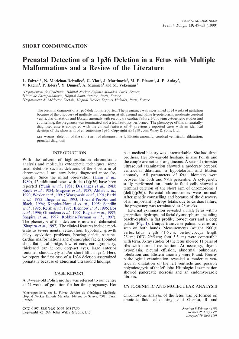
External examination revealed a male fetus with ageneralized hydrops and facial dysmorphism, includingbrachycephaly, a flat profile, low-set ears and a deeppalate (Fig. 1). Unique transverse palmar creases areseen on both hands. Measurements (weight 1900 g;vertex–talus length 41·5 cm; vertex–coccyx length26 cm; OFC 29·5 cm; foot 5·5 cm) were compatiblewith term. X-ray studies of the fetus showed 11 pairs ofribs with normal ossification. At necropsy, thymichypoplasia, pleural effusion, abnormal pulmonarylobulation and Ebstein anomaly were found. Neuro-pathological examination revealed a moderate ven-tricular dilatation of the left ventricle and possiblepolymicrogyria of the left lobe. Histological examinationshowed pancreatic necrosis and an endomyocardicfibrosis.
*Correspondence to: L. Faivre, Service de Genetique Medicale,Hopital Necker Enfants Malades, 149 rue de Sevres, 75015 Paris,France.
CYTOGENETIC AND MOLECULAR ANALYSIS
Chromosome analysis of the fetus was performed onamniotic fluid cells using solid Giemsa, R and
Received 9 February 1998Revised 26 May 1998
Accepted 19 June 1998
50 . .
Fig. 1(a, b)—Pictures of the fetus after termination at 24 weeks of gestation. Note hydrops fetalis and facialdysmorphism including brachycephaly, flat profile and low-set ears
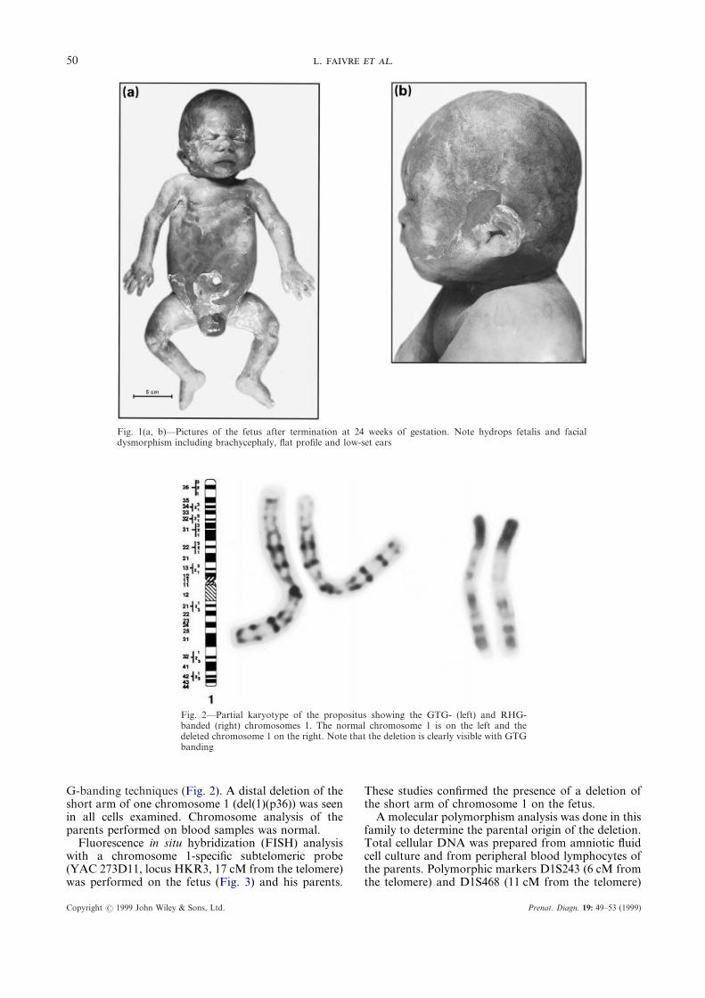
Fig. 2—Partial karyotype of the propositus showing the GTG- (left) and RHG-banded (right) chromosomes 1. The normal chromosome 1 is on the left and thedeleted chromosome 1 on the right. Note that the deletion is clearly visible with GTGbanding
G-banding techniques (Fig. 2). A distal deletion of theshort arm of one chromosome 1 (del(1)(p36)) was seenin all cells examined. Chromosome analysis of theparents performed on blood samples was normal.
Fluorescence in situ hybridization (FISH) analysiswith a chromosome 1-specific subtelomeric probe(YAC 273D11, locus HKR3, 17 cM from the telomere)was performed on the fetus (Fig. 3) and his parents.
Copyright ? 1999 John Wiley & Sons, Ltd.
These studies confirmed the presence of a deletion ofthe short arm of chromosome 1 on the fetus.
A molecular polymorphism analysis was done in thisfamily to determine the parental origin of the deletion.Total cellular DNA was prepared from amniotic fluidcell culture and from peripheral blood lymphocytes ofthe parents. Polymorphic markers D1S243 (6 cM fromthe telomere) and D1S468 (11 cM from the telomere)
Prenat. Diagn. 19: 49–53 (1999)

51 1p36
located between band 1p36.3 and the telomere wereused to establish which polymorphic loci were deletedin the fetus. The results showed that the deletion was ofpaternal origin (Fig. 4) and that the deletion was atleast 17 cM in size.
Fig. 3—Fluorescence in situ hybridization of the propositus with aspecific subtelomeric probe demonstrating the absence of signal onone chromosome 1
Fig. 4—Polymorphic marker analysis of the 1p36 deletion. Resultsfrom a fully informative 1p36 marker D1S243 are shown for thefetus and his parents. The fetus demonstrates inheritance of thematernal allele only
DISCUSSION
Here, we describe the prenatal diagnosis of a fetus withdeletion of the short arm of chromosome 1p36.Molecular cytogenetic analysis of the fetus confirmedthe terminal deletion and molecular studies showedthat the deletion was paternal in origin. To our knowl-edge, a total of 44 chromosome 1p36 deletions havebeen reported, representing today a newly delineateddeletion syndrome (Shapira et al., 1997) (Table 1). Thedetection of this deletion is difficult, however. For
Copyright ? 1999 John Wiley & Sons, Ltd.
example, in 12 out of 41 informative observations, thedeletion was not detected at the first cytogenetic analy-sis (Abbas et al., 1990; Wexler et al., 1991; Biegel et al.,1993; Howard-Peebles and Black, 1994; Giraudeau etal., 1997; Shapira et al., 1997). In addition, in one case(Giraudeau et al., 1997), the deletion was identified byscreening a sample of children with idiopathic mentalretardation to determine the frequency of subtelomericrearrangements using hypervariable polymorphicprobes. Commonly, diagnosis is made during earlychildhood in a five-year-old child with moderate tosevere mental retardation, hypotony, growth delay,eye/vision problems, hearing deficit, seizures, cardiacmalformation and a dysmorphic facies. In two cases,the diagnosis was made before birth. The first case wasdetected because of the presence of a maternal bal-anced translocation between the short arms of chromo-some 1 and 20, ascertained when the first child ofthe couple had inherited an unbalanced product ofthe translocation (Howard-Peebles et al., 1994). Thesecond case was ascertained because of the presence ofelevated maternal serum alpha-fetoprotein (Robbins-Furman et al., 1997). In both cases however, noabnormal sign was noted on ultrasound. Frequently,the deletion occurs de novo (31/39) although the segre-gation of a parental rearrangement is also observed(Hain et al., 1980; Deslanges et al., 1983; Barbi et al.,1992; Howard-Peebles and Black, 1994; Reish et al.,1995; Blennow et al., 1996). In 4/40 informative cases,the deletion resulted from a de novo unbalanced re-arrangement involving another chromosome. Studiesof the parental origin of the deletion have beenperformed in 15 de novo cases (Biegel et al., 1993;Giraudeau et al., 1997; Shapira et al., 1997). The originof the deletion was maternal in 12 cases and paternal in3 cases. Interestingly, in the present observation, thedeletion is paternal in origin.
Congenital heart malformation is a classical findingin 1p36 deletion. It is found in 20/33 cases (Table I),but no malformation is pathognomonic of the deletion.For example, cardiomyopathy is reported in fivecases (Reish et al., 1995; Keppler-Noreuil et al., 1995;Shapira et al., 1997), ductus arteriosus in four cases(Howard-Peebles and Black, 1994; Reish et al., 1995;Keppler-Noreuil et al., 1995; Shapira et al., 1997),tetralogy of Fallot in two cases (Howard-Peebles andBlack, 1994; Magenis et al., 1987), ventricular septaldefect in two cases (Biegel et al., 1993; Howard-Peeblesand Black, 1994), and left pulmonary artery branchstenosis in one case (Shapira et al., 1997). In thepresent case, Ebstein anomaly, leading to hydropsfetalis, was detected in utero. To our knowledge, nodeletion 1p36 has been associated with this malfor-mation. Ebstein anomaly is a rare defect, however,and the number of 1p36 deletions is still too small todecide whether this anomaly belongs to the phenotypicspectrum of the 1p36 deletion.
In conclusion, here we describe the first case of adeletion of the short arm of chromosome 1 ascertainedprenatally through the discovery of abnormal findingson ultrasound. A review of 44 previously reportedcases shows that the 1p36 deletion is well delineated
Prenat. Diagn. 19: 49–53 (1999)

lion Seizures Cardiopathy Hydrocephalus Other signs
1/2 Cardiomegaly 1/2 —" Murmur " Hydramnios, cleft lip,
finger hypoplasia? Cardiomegaly " Cleft lip+ " " Hallux valgus? + " Imperforate anus,
rectovaginal fistula+ ? Ventricular
dilatationScoliosis
+ ? ? Obesity+ ? ? Sturge–Weber, cleft of
the soft palate,nystagmus
" ? + Cleft palate, retinaldysfunction, deafness
" + " Neuroblastoma? + + ACC, eye abnormalities
1/1 2/3 " Hexadactyly, hearingloss, optic atrophy,cerebral atrophy,craniostenosis
2/2 2/3 Coloboma,polydactyly
? 2/3 2/2 Cleft palate, cleftalveolar ridge, hearingloss
e + ? " Scoliosis, deafness? + ? Alagille syndrome,
hearing losse + " " Deafness, scoliosis
+ ? " Obesity8/12 2/12 CHD
2/12 CM2/10 Cleft lip/palate 2/12,
hearing loss 6/8, eyeabnormalities 6/8,obesity 1/8
+ ? ? Abnormal serummaternal áFP
? + + Thymic hypoplasia18/26 20/33 12/35 Cleft lip/palate 8/41,
hearing loss 11/33, eyeabnormalities 11/33
52.
.
Cop
Table 1—Features of patients with del(1)(p36)
Reference(number of cases) Karyotype Inheritance
Prenataldiagnosis
Growthretardation Dysmorphy Hypotony
Mentaretardat
Hain et al. (1980) (2) der(1)t(1;5)(p36;q11) Parental " " + " ?Yunis et al. (1981) 45,XX,t(1.21)(p36;q11) De novo " + + + +
Deslanges et al. (1983) der(1)t(1;9)(p36;p12) Paternal " + + ? ?Steele et al. (1984) t(1;13)(p36.2;p11) De novo " " + + +Magenis et al. (1987) del(1)(p36) De novo " + + " ?
Abbas et al. (1990) 45,X,t(1;Y)(p36;p13) De novo " + + + +
Wargowski et al. (1991) (4) del(1)(p36) ? " + + + +Wexler et al. (1991) (2) del(1)(p36.3) De novo " ? 1/2 1/1 +
Barbi et al. (1992) der(1)t(1;15)(p36;p11.2) Maternal " + + + +
Biegel et al. (1993) del(1)(p36.1]p36.2) De novo " + + + +Howard-Peebles andBlack (1994)
der(1)t(1;20)(p36;pter) Maternal 1/2 ? + ? ?
Reish et al. (1995) (5) Translocation (4)Deletion (1)
Paternal (2)De novo (2)Unknown (1)
" 1/3 + 1/1 +
Keppler-Noreuilet al. (1995) (3)
del(1)(p36.22) De novo 2/3 + 2/2 + 1/1
Sandlin et al. (1995) (3) del(1)(p36) De novo " 2/3 + + +
Blennow et al. (1996) der(1)t(1;15)(p36;p11) Paternal " + + + SeverBos et al. (1996) del(1)(p36.31) ? " + + + +
Giraudeau et al. (1997) del(1)(p36.3) De novo " + + + SeverEugster et al. (1997) mos46,XX,del(1)(p36.33) De novo " + + + severeShapira et al. (1997) (12) del(1)(p36) De novo ? 8/8 12/12 8/8 7/8
Robbins-Furmanet al. (1997)
del(1)(p36.3) De novo + ? + ? ?
Present case 46,XY,del(1)(p36.3) De novo + ? + ? ?Total Deletion 28/45
Translocation 12/45Unknown 5/45
31/39 de novo 3/24 27/34 44/45 26/30 23/34
ACC: agenesis of corpus callosum; CHD: congenital heart disease; CM: cardiomyopathy.
yright?
1999John
Wiley
&Sons,
Ltd.
Prenat.
Diagn.
19:49–53
(1999)
53 1p36
postnatally but that its prenatal detection relies on thequality of cytogenetic techniques including FISH.
We would like to thank Dr T. Haaf for providing uswith the YAC 273D11 probe (http:/www.mpimg-berlin-dahlem-mpg.dc/).
REFERENCES
Abbas, N., Novelli, G., Carlo Stella, N., Triolo, O., Corrado,F., Fellous, M., Chery, M., Gilgenkrantz, S., Dallapiccola,B. (1990). A 45,X male with molecular evidence of atranslocation of Y euchromatin onto chromosome 1, Hum.Genet., 86, 94–98.
Barbi, G., Kennerknecht, I., Klett, C. (1992). Reciprocaltranslocation t(1;15)(p36.2;p11.2): confirmation of a sug-gestive cytogenetic diagnosis by in situ hybridization andclinical case report on resulting mososomy (1p), Am. J.Med. Genet., 43, 722–725.
Biegel, J.A., White, P.S., Marshall, H.N., Fujimori, M.,Zackai, E.H., Scher, C.D., Brodeur, G.M., Emmanuel,B.S. (1993). Constitutional 1p36 deletion in a child withneuroblastoma, Am. J. Hum. Genet., 52, 176–182.
Blennow, E., Bui, T., Wallin, A., Kogner, P. (1996). Mono-somy 1p36.31–33]pter due to a paternal reciprocal trans-location: prognostic significance of FISH analysis, Am. J.Med. Genet., 65, 60–67.
Bos, C., Opheim, K.E., Hudgins, L. (1996). Deletion(1)(p36.31) in a patient with features of Alagille syndrome(arteriohepatic dysplasia), Am. J. Hum. Genet., 59 (Suppl.),A112.
Deslanges, F., Mourrieras, P., Papouin-Rauzy, M., Saliou, P.(1983). Monosomy 1 pter due to familial t(1;9), Ann.Genet., 26, 53–55.
Eugster, E.A., Berry, S.A., Hirsch, B. (1997). Mosaicism fordeletion 1p36.33 in a patient with obesity and hyperphagia,Am. J. Med. Genet., 70, 409–412.
Giraudeau, F., Aubert, D., Young, I., Horsley, S., Knight,S., Kearney, L., Vergnaud, G., Flint, J. (1997). Molecular-cytogenetic detection of a deletion of 1p36.3, J. Med.Genet., 34, 314–317.
Hain, D., Leversha, M., Campbell, N., Daniel, A., Barr,P.A., Rogers, J.G. (1980). The ascertainment and impli-
Copyright ? 1999 John Wiley & Sons, Ltd.
cations of an unbalanced translocation in the neonate.Familial 1;15 translocation, Aust. Paediat. J., 16, 196–200.
Howard-Peebles, P.N., Black, S.H. (1994). FISH identifi-cation of a deletion 1p36 from a half cryptic maternaltranslocation, Am. J. Med. Genet., 52, 381.
Keppler-Noreuil, K.M., Carroll, A.J., Finley, W.H., LaneRutlrdge, S. (1995). Chromosome 1p terminal deletion:report of new findings and confirmation of two character-istic phenotypes, J. Med. Genet., 32, 619–622.
Magenis, R.E., Sheehy, R., Lacey, D., Brown, M.G., Litt, M.(1987). Small terminal deletion of chromosome 1 short armin an infant with multiple anomalies: confirmation by insitu hybridization of probe p1-79, Am. J. Hum. Genet.,41(Suppl.), A130.
Reish, O., Berry, S.A., Hirsh, B. (1995). Partial monosomy ofchromosome 1p36.3, Am. J. Med. Genet., 59, 467–475.
Robbins-Furman, P., Elder, F.F.B., Mastrobattista, J.M.,Northrup, H., Shapira, S.K. (1997). Prenatal diagnosis ofdeletion 1p36 syndrome, Am. J. Hum. Genet., 61(Suppl.),A139.
Sandlin, C.J., Dodd, B.S., Dumars, K.W., Bartley, J.A.,Bernstein, R., Lamb, A. (1995). Phenotypes associatedwith terminal deletion of the short arm of chromosome 1,Am. J. Hum. Genet., 57(Suppl.), A125.
Shapira, S.K., McCaskill, C., Northrup, H., Spikes, A.S.,Elder, F.F.B., Reid Sutton, V., Korenberg, J.R.,Greenberg, F., Shaffer, L.G. (1997). Chromosome 1p36deletions: the clinical phenotype and molecular character-ization of a common newly delineated syndrome, Am. J.Hum. Genet., 61, 642–650.
Steele, M.W., Wenger, S.L., Geweke, L.O., Golden, W.L.(1984). The level of 6-phosphogluconate deshydrogenase(6-PGD) activity in a patient with a 1p terminal deletionsuggests that the gene locus is not distal to sub-band p36.3on chromosome 1, Clin. Genet., 25, 59–62.
Wargowski, D., Sekkon, G., Laxova, R., Thompson, K.,Kent, C. (1991). Terminal deletions of band 1p36: emer-gence of two overlapping phenotypes, Am. J. Hum. Genet.,49(Suppl.), 278.
Wexler, P., Gilfillan, T., McGavran, L., Sujansky, E. (1991).Deletions (1)(p36.3) and the potential role of high resol-ution chromosome analysis, Am. J. Hum. Genet.,49(Suppl.), 278.
Yunis, E., Quintero, L., Leibovici, M. (1981). Monosomy1pter, Hum. Genet., 56, 279–282.
Prenat. Diagn. 19: 49–53 (1999)