Preclinical evaluation of TriMix and antigen mRNA-based...
35
1 Preclinical evaluation of TriMix and antigen mRNA-based anti-tumor therapy mRNA-based anti-tumor vaccination Sandra Van Lint 1 , Cleo Goyvaerts 1 , Sarah Maenhout 1 , Lode Goethals 2 , Aurélie Disy 1 , Daphné Benteyn 1 , Joeri Pen 1 , Aude Bonehill 1 , Carlo Heirman 1 , Karine Breckpot 1 and Kris Thielemans 1 1 Laboratory of Molecular and Cellular Therapy, Department of Immunology-Physiology, 2 In Vivo Cellular and Molecular Imaging Laboratory, Vrije Universiteit Brussel, Laarbeeklaan 103, 1090 Jette, Belgium Correspondence should be addressed to KB: Laboratory of Molecular and Cellular Therapy, Department of Immunology-Physiology, Vrije Universiteit Brussel, Laarbeeklaan 103/E, 1090 Jette, Belgium, tel: 0032-2-4774565, fax: 0032-2-4774568, [email protected]. This work was supported by grants from the Interuniversity Attraction Poles Program-Belgian State-Belgian Science Policy, the National Cancer Plan of the Federal Ministry of Health, the Stichting tegen Kanker, the Vlaamse Kanker Liga, an Integrated Project and an EU FP6- funded Network of Excellence, an IWT-TBM program, the FWO-Vlaanderen and the Scientific Fund Willy Gepts of the University hospital Brussels. CG, LG are doctoral fellows and AB, KB are postdoctoral fellows of the FWO-Vlaanderen. JP is a doctoral fellow of the IWT. SM is funded by an Emanuel Van Der Schueren grant from the Vlaamse Kanker Liga. The use of dendritic cells electroporated with TAA mRNA and TriMix is the topic of a patent application [WO2009/034172] on which AB and KT are filed as inventors. None of the authors receive any support or remuneration related to this platform. on May 6, 2019. © 2012 American Association for Cancer Research. cancerres.aacrjournals.org Downloaded from Author manuscripts have been peer reviewed and accepted for publication but have not yet been edited. Author Manuscript Published OnlineFirst on February 15, 2012; DOI: 10.1158/0008-5472.CAN-11-2957
Transcript of Preclinical evaluation of TriMix and antigen mRNA-based...
1
Preclinical evaluation of TriMix and antigen mRNA-based anti-tumor therapy
mRNA-based anti-tumor vaccination
Sandra Van Lint1, Cleo Goyvaerts1, Sarah Maenhout1, Lode Goethals2, Aurélie Disy1, Daphné
Benteyn1, Joeri Pen1, Aude Bonehill1, Carlo Heirman1, Karine Breckpot1 and Kris
Thielemans1
1Laboratory of Molecular and Cellular Therapy, Department of Immunology-Physiology, 2In
Vivo Cellular and Molecular Imaging Laboratory, Vrije Universiteit Brussel, Laarbeeklaan
103, 1090 Jette, Belgium
Correspondence should be addressed to KB: Laboratory of Molecular and Cellular Therapy,
Department of Immunology-Physiology, Vrije Universiteit Brussel, Laarbeeklaan 103/E, 1090
Jette, Belgium, tel: 0032-2-4774565, fax: 0032-2-4774568, [email protected].
This work was supported by grants from the Interuniversity Attraction Poles Program-Belgian
State-Belgian Science Policy, the National Cancer Plan of the Federal Ministry of Health, the
Stichting tegen Kanker, the Vlaamse Kanker Liga, an Integrated Project and an EU FP6-
funded Network of Excellence, an IWT-TBM program, the FWO-Vlaanderen and the
Scientific Fund Willy Gepts of the University hospital Brussels. CG, LG are doctoral fellows
and AB, KB are postdoctoral fellows of the FWO-Vlaanderen. JP is a doctoral fellow of the
IWT. SM is funded by an Emanuel Van Der Schueren grant from the Vlaamse Kanker Liga.
The use of dendritic cells electroporated with TAA mRNA and TriMix is the topic of a patent
application [WO2009/034172] on which AB and KT are filed as inventors. None of the
authors receive any support or remuneration related to this platform.
on May 6, 2019. © 2012 American Association for Cancer Research. cancerres.aacrjournals.org Downloaded from
Author manuscripts have been peer reviewed and accepted for publication but have not yet been edited. Author Manuscript Published OnlineFirst on February 15, 2012; DOI: 10.1158/0008-5472.CAN-11-2957
2
Abstract
The use of tumor-associated antigen [TAA] mRNA for therapeutic purposes is under active
investigation. To be effective, mRNA-vaccines need to deliver activation stimuli in addition to
TAAs to dendritic cells [DCs]. In this study, we evaluated whether intranodal delivery of TAA
mRNA together with TriMix, a mix of mRNA encoding CD40 ligand, constitutive active toll-
like receptor 4 and CD70, results in the in situ modification and maturation of DCs hence
priming of TAA-specific T-cells. We demonstrated selective uptake and translation of mRNA
in vivo by lymph node resident CD11c+ cells. This process was hampered by co-delivery of
classical maturation stimuli but not by TriMix mRNA. Importantly, TriMix mRNA induced a
T-cell attracting and stimulatory environment, including recruitment of antigen-specific CD4+
and CD8+ T-cells and cytotoxic T lymphocytes [CTLs] against various TAAs. In several
mouse tumor models, mRNA vaccination was as efficient in CTL induction and therapy
response as vaccination with mRNA electroporated DCs. Together, our findings suggest that
intranodal administration of TAA mRNA together with mRNA encoding immune modulating
molecules is a promising vaccination strategy.
on May 6, 2019. © 2012 American Association for Cancer Research. cancerres.aacrjournals.org Downloaded from
Author manuscripts have been peer reviewed and accepted for publication but have not yet been edited. Author Manuscript Published OnlineFirst on February 15, 2012; DOI: 10.1158/0008-5472.CAN-11-2957
3
Introduction
The immune system can mount immune responses against tumor-associated antigens [TAAs].
Such immune responses, mediated by CD4+ T helper 1 [TH1] cells and CD8+ cytotoxic T
lymphocytes [CTLs], can be enhanced or induced de novo by immunotherapeutic strategies
using antigen-loaded dendritic cells [DCs] (1-3). Several strategies have been developed to
deliver TAAs to DCs, including the use of mRNA (4-6). Autologous DCs loaded ex vivo with
TAA mRNA have been extensively tested in preclinical studies, demonstrating their ability to
induce functional TH1 cells and CTLs (7-10). Moreover, clinical testing demonstrated the
induction of antigen-specific immune responses by DC-vaccines (11). However, the logistics
of developing a specific vaccine for each patient may be prohibitive. Therefore, direct
administration of TAA mRNA has gained substantial interest (12-14). This method offers a
number of advantages, mRNA is not patient-specific, available at all times, safe and easy to
produce at low cost (12-14).
The success of mRNA-vaccination depends on the engulfment of mRNA by DCs and its
potential to mature DCs. Consequently, the route of mRNA delivery and the modus of DC
maturation are parameters that will critically impact on the efficiency of the mRNA-vaccine. It
was recently demonstrated that intranodal delivery of mRNA results in the engulfment of
mRNA by DCs, as well as the activation of toll-like receptors [TLRs] (15-17). Nevertheless, it
is suggested that naked mRNA is insufficient to fully harness the stimulatory potential of DCs
(9, 18). Therefore, co-delivery of additional stimuli, such as lipopolysaccharide [LPS], CD40
ligand [CD40L], polyinosinic:polycytidylic acid [polyI:C] and protamine-complexed mRNA,
has been evaluated (18, 19). However, defining the optimal protocol for in vivo DC
maturation, without abrogating the uptake/translation of mRNA has proven to be challenging.
The use of mRNA encoding immune modulating proteins might be an attractive alternative to
potentiate DCs in situ.
on May 6, 2019. © 2012 American Association for Cancer Research. cancerres.aacrjournals.org Downloaded from
Author manuscripts have been peer reviewed and accepted for publication but have not yet been edited. Author Manuscript Published OnlineFirst on February 15, 2012; DOI: 10.1158/0008-5472.CAN-11-2957
4
We previously demonstrated that electroporation of human DCs with CD40L mRNA and
mRNA encoding a constitutive active form of TLR4 [caTLR4] induces DC maturation. We
moreover introduced CD70 mRNA into these DCs to provide a co-stimulatory signal to
CD27+ T-cells. We showed that DCs modified with this so-called TriMix induce tumor-
specific T-cell responses in vitro as well as in vaccinated melanoma patients (20-23).
Here we report on the delivery of TAA and TriMix mRNA in situ to generate T-cell attracting
and stimulating DCs, a strategy that was shown to be as efficient as vaccination with in vitro
electroporated DCs in terms of CTL induction and anti-tumor therapy.
on May 6, 2019. © 2012 American Association for Cancer Research. cancerres.aacrjournals.org Downloaded from
Author manuscripts have been peer reviewed and accepted for publication but have not yet been edited. Author Manuscript Published OnlineFirst on February 15, 2012; DOI: 10.1158/0008-5472.CAN-11-2957
5
Materials and Methods
Mice
Female, 6 to 12 weeks old C57BL/6, DBA/2 and BALB/c mice were purchased from Harlan.
Transgenic mice were provided by B. Lambrecht [University of Ghent] and include OT-I mice
that carry a transgenic CD8 T-cell receptor [TCR] specific for the MHC I-restricted ovalbumin
[OVA] peptide SIINFEKL, OT-II mice that carry a transgenic CD4 TCR specific for the MHC
II-restricted OVA peptide ISQAVHAAHAEINEAGR and CD11c-diphtheria toxin receptor
[DTR] mice in which CD11c+ cells are depleted upon treatment with 4 ng diphtheria toxin
[DT]/g mouse [Sigma]. Where indicated mice received an intravenous hydrodynamic injection
with 10 μg of a plasmid encoding Flt3-ligand [a gift from O. Leo, Université Libre de
Bruxelles] in 0.9 NaCl in a final volume equal to 10% of the mouse body weight. Animals
were treated according to the European guidelines for animal experimentation. Experiments
were reviewed by the Ethical committee for use of laboratory animals of the Vrije Universiteit
Brussel.
Mouse cell lines and dendritic cells
The melanoma MO4, the T-cell lymphoma EG7-OVA, the mastocytoma P815 and the
myeloid leukemia C1498-WT1 were obtained from the American Type Culture Collection
[ATCC], C. Uytttenhove [Université Catholique de Louvain] and H.E. Kohrt [Stanford
University Medical Centre], respectively. No full authentication was carried out. Cell lines
were evaluated for the expression of MHC molecules and antigens [OVA: MO4 and EG7-
OVA, P1A: P815 and WT1: C1498-WT1] by RT-PCR or flow cytometry. Bone marrow-
derived DCs were generated as described (9).
on May 6, 2019. © 2012 American Association for Cancer Research. cancerres.aacrjournals.org Downloaded from
Author manuscripts have been peer reviewed and accepted for publication but have not yet been edited. Author Manuscript Published OnlineFirst on February 15, 2012; DOI: 10.1158/0008-5472.CAN-11-2957
6
Messenger RNA
The vector, pST1 was provided by U. Sahin [Johannes-Gutenberg University]. The vectors
pGEM-Ii80tOVA, pST1-tyrosinase-DC-LAMP, pST1-sig-WT1-ΔNLS-DC-LAMP, pST1-
caTLR4 and pGEM-tNGFR have been described (9, 21, 24, 25). The sequence encoding
Firefly Luciferase [FLuc] was cloned into pST1 with minor modifications. The vector pGEM-
Ii80P1A was cloned analogous to the cloning of pGEM-Ii80tOVA. The codon optimized
cDNA encoding mouse CD40L or CD70 were obtained from Geneart and cloned as a SpeI-
XhoI fragment in the pST1-vector. A fragment of the mouse Trp2 gene that encodes
SVYDFFVWL was amplified with the following primers: 5’-GGGGATCCGGCCATCCTAA
GACGG-3’ and 3’-GGGGGATCCGTGCACACGTCACAC TCGTTC-5’ and cloned as a
BamHI fragment in the BamHI linearized and shrimp alkaline phosphatase treated pST1-sig-
DC-LAMP. The sequence encoding enhanced green fluorescent protein [eGFP] was isolated
from p-eGFP-N1 as a HinDIII-NotI fragment and cloned into the HinDIII-NotI digested pST1-
vector. All enzymes were purchased from Fermentas.
Prior to in vitro transcription, pGEM- and pST1-vectors were linearized with SpeI and SapI,
respectively. In vitro transcription was performed as described (9). The mRNA was dissolved
in phosphate buffered saline [PBS], Ca2+-containing Hank’s balanced salt solution [HBSS,
Lonza] or 0.8 Ringer lactate [0.8 RL, Baxter].
Passive pulsing and electroporation of mRNA
To pulse DCs with mRNA, 5 x 10⁶ DCs were pelleted and incubated for 15 minutes with 10
μg tNGFR or FLuc mRNA in 15 μl. Where indicated pulsing was performed in the presence
of 1 ng/ml LPS from E. coli serotype 055:B5 [Sigma-Aldrich], 10 μg/ml polyI:C [Sigma] or
100 ng/ml monophosphoryl lipid A [MPL, GlaxoSmithKline]. DCs were cultured in RPMI
1640 medium supplemented with 5% FCI [Harlan], 50 µM β-mercaptoethanol and 20 ng/ml
on May 6, 2019. © 2012 American Association for Cancer Research. cancerres.aacrjournals.org Downloaded from
Author manuscripts have been peer reviewed and accepted for publication but have not yet been edited. Author Manuscript Published OnlineFirst on February 15, 2012; DOI: 10.1158/0008-5472.CAN-11-2957
7
mouse GM-CSF [prepared in-house] at a cell density of 106 DCs/ml. Four hours later, DCs
were lyzed using the Reporter lysis buffer from Promega. D-Luciferin [Xenogen] was added,
luminescence measured using the GlomaxTM 96-luminometer and data analyzed with
GlomaxTM software [Promega]. Electroporation of DCs with mRNA was performed as
described (9). Where indicated DCs were activated for 4 hours with 100 ng/ml LPS.
In situ delivery of mRNA
For intranodal delivery of mRNA, C57BL/6 mice were anesthetized with ketamine [70 mg/kg,
Ceva] and xylazine [10 mg/kg, Bayer]. The inguinal lymph node was surgically exposed and
injected with the indicated amount of mRNA [and where indicated 1 ng LPS]. Subsequently,
the wound was closed. On 3 consecutive days prior to intradermal delivery of mRNA, mice
were injected intradermally with PBS or 20 ng of mouse GM-CSF, after which the mRNA was
administered.
RNA isolation, cDNA synthesis and RT-PCR
RNA was extracted using the SV Total RNA Isolation System [Promega] and converted to
cDNA using the RevertAidTM H-Minus First strand cDNA synthesis kit [Fermentas]. The
sequence encoding FLuc was amplified with 5’-AAGGTGTGGCCCTTCC-3’ and 5’-CCAAG
AATGAAAATAGGGTTG-3’, whereas the sequence encoding β-actin was amplified with 5’-
TGCTATCCAGGCTGTGCTAT-3’ and 5’-GATGGAGTTGAAGGTAGTTT-3’ using the
following PCR program: 94°C 5’, 45x [94°C 30”, 52°C 30”, 72°C 30”], 72°C 10’, hold 4°C.
Immune array
RNA of lymph nodes injected with 0.8 RL, 10 µg antigen mRNA supplemented with 30 µg
tNGFR mRNA or TriMix [10 µg/component] was extracted and converted to cDNA.
on May 6, 2019. © 2012 American Association for Cancer Research. cancerres.aacrjournals.org Downloaded from
Author manuscripts have been peer reviewed and accepted for publication but have not yet been edited. Author Manuscript Published OnlineFirst on February 15, 2012; DOI: 10.1158/0008-5472.CAN-11-2957
8
Quantitative RT-PCR using the TaqMan® mouse immune response array [Applied
Biosystems] and analysis was performed according to the manufacturer’s instructions.
Flow cytometry
Allophycocyanin-conjugated anti-CD11c [HL3], -CCR7 [2H4] and phycoerythrin-conjugated
anti-CD40L [MR1] and -CD70 [FR70] antibodies were purchased from Pharmingen. The
antibodies against CD40 [FGK45], CD80 [16-10A1] and CD86 [GL-1] were prepared in-
house. Non-reactive isotype matched antibodies served as controls [Pharmingen]. Labeling of
DCs was performed as described (9). Data were collected using the FACSCanto flow
cytometer [Becton Dickinson] and analyzed using FACSDivaTM or FlowJoTM software.
Allogeneic mixed lymphocyte reaction
The ability of electroporated DCs to stimulate allogeneic CD90 purified [Miltenyi Biotec] T-
cells was assessed in a mixed lymphocyte reaction (26).
Enzyme-linked immunosorbent assay
Supernatants were screened in a sandwich enzyme-linked immunosorbent assay for the
presence of IL-6, IL-12p70, TNF-α or IFN-γ [eBioscience].
In vivo bioluminescence imaging
In vivo bioluminescence imaging was performed as described (27).
Fluorescence microscopy
Lymph nodes were injected with 10 μg eGFP mRNA, one day prior to isolation. Single cell
suspensions were prepared and stained with a phycoerythrin-conjugated anti-CD11c antibody.
Expression of CD11c and eGFP was evaluated using the Evosfl fluorescence microscope.
on May 6, 2019. © 2012 American Association for Cancer Research. cancerres.aacrjournals.org Downloaded from
Author manuscripts have been peer reviewed and accepted for publication but have not yet been edited. Author Manuscript Published OnlineFirst on February 15, 2012; DOI: 10.1158/0008-5472.CAN-11-2957
9
Immunization of mice
Mice were immunized intravenously with 5 x 10⁵ antigen-presenting DCs activated with
TriMix or LPS, or intranodally or intradermally with 10 µg antigen mRNA supplemented with
30 µg tNGFR mRNA or TriMix [10 µg/component]. Immunization with DCs electroporated
with tNGFR mRNA or with tNGFR mRNA as such served as a control. For assessment of
therapeutic efficacy, 5 x 10⁵ tumor cells were administered subcutaneously in the lower back,
7 days prior to immunization.
Intracytoplasmatic staining of IFN-γ
Spleen cells of immunized mice were stimulated for 24 hours with DCs pulsed for 2 hours
with 5 μM SIINFEKL peptide and matured with LPS. GolgiPlug was added 24 hours prior to
intracytoplasmatic staining of IFN-γ.
Pentamer staining
The staining of CD8+ T-cells with H2-Kb/SIINFEKL pentamers [Immunosource] was
performed as described (26).
In vivo cytotoxicity assay
Spleen cells from syngeneic mice were labeled with 10 μM carboxyfluorescein diacetate
succinimidine ester [CFSE] as described (9). These were pulsed with the peptide SIINFEKL
[OVA] or SVYDFFVWL [Trp2] [Thermo Electron Cooperation] or a set of overlapping
peptides covering WT1 [kind gift from V. Van Tendeloo, University of Antwerp] or
tyrosinase [EMC microcultures] at 5 µM for 2 hours. Peptide-pulsed cells were mixed at a 1:1
on May 6, 2019. © 2012 American Association for Cancer Research. cancerres.aacrjournals.org Downloaded from
Author manuscripts have been peer reviewed and accepted for publication but have not yet been edited. Author Manuscript Published OnlineFirst on February 15, 2012; DOI: 10.1158/0008-5472.CAN-11-2957
10
ratio with non-pulsed cells, labeled with 0.5 μM CFSE. Specific lysis of target cells was
analyzed 18 hours later by flow cytometry. The % of killing was calculated as described (28).
In vivo proliferation assay
One day prior to immunization, 106 purified and CFSE-labeled CD8+ OT-I or CD4+ OT-II
spleen cells were transferred to mice by intravenous injection. Five days post-immunization,
proliferation of T-cells was analyzed in peripheral blood, spleen and lymph nodes (28).
Statistical analyses
A one-way ANOVA followed by a Bonferroni’s multiple comparison test was performed.
Sample sizes and number of times experiments were repeated are indicated in the figure
legends. Number of asterisks in the figures indicates the level of statistical significance as
follows: *, p < 0.05; **, p < 0.01; ***, p < 0.001. The results are shown in a scatter plot in
which each mouse is depicted as a dot and the mean as a horizontal line, or in a column graph
or table as the mean ± SEM. Survival was visualized in a Kaplan-Meier plot. Differences in
survival were analyzed by the log-rank test.
on May 6, 2019. © 2012 American Association for Cancer Research. cancerres.aacrjournals.org Downloaded from
Author manuscripts have been peer reviewed and accepted for publication but have not yet been edited. Author Manuscript Published OnlineFirst on February 15, 2012; DOI: 10.1158/0008-5472.CAN-11-2957
11
Results
Dendritic cells matured through electroporation with TriMix mRNA efficiently stimulate
antigen-specific T-cells
We recently demonstrated that the T-cell stimulatory capacity of human DCs electroporated
with TAA mRNA is considerably increased by simultaneous co-electroporation with TriMix
(20). As we wanted to investigate the use of TriMix for the in situ modification of mouse DCs,
we evaluated whether electroporation of mouse DCs with TriMix results in immunogenic
DCs. We demonstrated that TriMix electroporated DCs displayed a phenotype [Fig. 1a],
cytokine secretion profile [Fig. 1b] and allogeneic T-cell stimulatory capacity [Fig. 1c]
comparable to that of LPS activated DCs. Importantly, we demonstrated that TriMix matured
DCs were superior to LPS matured DCs in stimulation of functional antigen-specific CD8+ T-
cells in vivo. This was demonstrated for OVA [Figs. 1d-f] and the TAA Trp2 [Fig. 1g].
Formulation and pharmacokinetics of mRNA for vaccination purposes
It was previously demonstrated that cellular uptake of mRNA can be influenced by the
composition of the injection solution (29). Therefore, we evaluated which buffer is best suited
to deliver mRNA to DCs. FLuc mRNA was dissolved in PBS, Ca2+-containing HBSS or 0.8
RL. Luminescence analysis of passively pulsed DCs demonstrated high FLuc expression when
the mRNA was dissolved in 0.8 RL or HBSS [Fig. 2a]. Next, we administered FLuc mRNA
intranodally. In vivo bioluminescence imaging demonstrated short-term FLuc expression when
mRNA was formulated in PBS when compared to high and long FLuc expression when
mRNA was formulated in HBSS or 0.8 RL [Fig. 2b]. The latter was unexpected as naked
mRNA is believed to have a short extracellular half life (30). To analyze the stability of
mRNA in vivo upon delivery in 0.8 RL, we resected lymph nodes injected with FLuc mRNA
6, 12 and 24 hours after injection. RT-PCR demonstrated the presence of FLuc mRNA up to
12 hours after injection. No FLuc mRNA was detectable at later time points [Fig. 2c].
on May 6, 2019. © 2012 American Association for Cancer Research. cancerres.aacrjournals.org Downloaded from
Author manuscripts have been peer reviewed and accepted for publication but have not yet been edited. Author Manuscript Published OnlineFirst on February 15, 2012; DOI: 10.1158/0008-5472.CAN-11-2957
12
Next, we evaluated the role of DCs in the uptake of mRNA in vivo. Lymph nodes were
injected with eGFP mRNA 24 hours prior to their isolation. Single cell suspensions were
prepared and stained for CD11c. Fluorescence microscopy showed a small number of eGFP+
cells. Importantly, all eGFP+ cells were CD11c+, demonstrating uptake and translation of
mRNA by DCs [Fig. 2d]. To further evidence a role for DCs, we used CD11c-DTR transgenic
mice in which administration of DT results in the depletion of CD11c+ cells. In vivo
bioluminescence imaging demonstrated the absence of FLuc expression in mice that were
treated with DT prior to intranodal administration of FLuc mRNA. Mice treated with PBS
served as a control [Fig. 2e]. Flow cytometric analysis of the lymph nodes of these mice
confirmed that the absence of luminescence was correlated with the depletion of DCs [Fig.
2e]. As delivery of mRNA into the inguinal lymph node is technically challenging, we finally
examined the feasibility of delivering mRNA intradermally. Since we demonstrated in the
former experiment that CD11c+ cells are responsible for the DC uptake, we pre-treated the
mice with an intradermal injection of PBS or GM-CSF on 3 consecutive days prior to the
intradermal injection of Fluc mRNA. In vivo bioluminescence imaging, performed 6 hours
later, demonstrated Fluc expression only in mice pre-treated with GM-CSF [Fig. 2f].
Intranodal delivery of TriMix generates an immune stimulatory environment
Induction of anti-tumor immune responses requires antigen-presentation by mature DCs (1-3).
To evaluate the effect of TriMix and classical maturation stimuli on the engulfment of mRNA
and the induction of an immune stimulatory environment, we first passively pulsed DCs in
vitro with FLuc mRNA and these maturation stimuli, demonstrating a reduction in FLuc
expression after pulsing of DCs with FLuc mRNA in the presence of LPS, MPL or polyI:C.
This reduction in protein expression was less pronounced when TriMix was co-delivered [Fig.
3a]. In addition, DCs pulsed with TriMix mRNA demonstrated a higher expression of CD40,
on May 6, 2019. © 2012 American Association for Cancer Research. cancerres.aacrjournals.org Downloaded from
Author manuscripts have been peer reviewed and accepted for publication but have not yet been edited. Author Manuscript Published OnlineFirst on February 15, 2012; DOI: 10.1158/0008-5472.CAN-11-2957
13
CD70, CD80 and CD86 compared to DCs pulsed with MPL [data not shown], LPS or polyI:C
[Fig. 3b].
Next we evaluated the uptake of FLuc mRNA when delivered as such or together with LPS or
TriMix in vivo. We demonstrated that co-delivery of TriMix had a lesser impact on the uptake
of mRNA than its co-delivery with LPS [Fig. 3c]. To increase the number of DCs that can be
recovered from the injected lymph node for analysis, we pre-treated the mice with a
hydrodynamic injection of a plasmid encoding Flt3-ligand. In analogy with the data described
by Kreiter et al (31), FLuc mRNA injected into these mice resulted in increased luminescence
reflecting the specific uptake by the DCs [data not shown]. Flow cytometry demonstrated that
DCs [CD11c+] from lymph nodes co-injected with TriMix displayed the highest expression of
CD40, CD80 and CD86 when compared to DCs isolated from lymph nodes injected with
FLuc mRNA alone or combined with LPS [Fig. 3d].
These findings prompted us to analyze, whether co-delivery of TriMix promotes a T-cell
attracting and activating environment, by profiling the expression levels of maturation-
associated markers by quantitative RT-PCR. We observed up-regulation of several markers in
lymph nodes injected with FLuc and tNGFR mRNA when compared to lymph nodes injected
with 0.8 RL. Importantly, the up-regulation of the following markers MHC II, IL-6, IL-15,
IFN-γ, MCP-1, IP-10, granzyme B, SOCS1 and STAT1 was at least two-fold higher when
TriMix was co-delivered [Table 1].
Intranodal delivery of TriMix but not LPS together with OVA mRNA results in
expansion of OVA-specific CD4+ and CD8+ T-cells with potent effector function
Activation of CD4+ T-cells is critical for the induction of long-lasting anti-tumor immunity
(32). Therefore, we evaluated the expansion of OVA-specific CD4+ T-cells upon intranodal
delivery of tNGFR mRNA, OVA mRNA or combined with TriMix or LPS. Proliferation of
CFSE labeled CD4+ OT-II cells was evaluated by flow cytometry, demonstrating enhanced
on May 6, 2019. © 2012 American Association for Cancer Research. cancerres.aacrjournals.org Downloaded from
Author manuscripts have been peer reviewed and accepted for publication but have not yet been edited. Author Manuscript Published OnlineFirst on February 15, 2012; DOI: 10.1158/0008-5472.CAN-11-2957
14
proliferation of OT-II cells in mice receiving OVA and TriMix mRNA. Of note, transferred T-
cells hardly proliferated when LPS was co-injected with OVA mRNA [Fig. 4a]. Similar
results were obtained with CD8+ OT-I cells [data not shown]. To further evaluate the
expansion and function of OVA-specific CD8+ T-cells, mice were immunized one day after
adoptive transfer of CD8+ OT-I cells. Five days post-immunization, we performed an H2-
kb/SIINFEKL pentamer staining or an in vivo cytotoxicity assay. Both assays demonstrated the
enhanced stimulation of OVA-specific CD8+ T-cells when mice were immunized with OVA
mRNA and TriMix when compared to mice immunized with OVA mRNA alone or combined
with LPS [Fig. 4b-c].
Using the model antigen OVA, we finally compared intradermal delivery of OVA and TriMix
mRNA in mice pre-treated with GM-CSF to its intranodal delivery. Using the in vivo
cytotoxicity assay we demonstrated that the lysis of target cells was the highest when the
mRNA was delivered intranodally [Fig. 4d].
Inclusion of TriMix in the mRNA-based anti-tumor vaccine enhances the induction of
TAA-specific cytotoxic T-cells
Next we assessed whether the results obtained with the antigen OVA are representative for
other TAAs. Mice were immunized with Trp2, WT1 or tyrosinase mRNA alone or combined
with TriMix. The in vivo cytotoxicity assay demonstrated enhanced lysis of target cells when
TriMix was included in the immunization regimen [Fig. 5a-c].
Immunization with antigen mRNA and TriMix is as efficient in stimulating cytotoxic T-
cells and in therapy as immunization with ex vivo modified dendritic cells
Therapeutic immunization with human DCs electroporated with TAA and TriMix mRNA has
shown promise in clinical evaluation (23). Therefore, we compared the efficacy of DC- to
mRNA-based immunization, evaluating the induction of antigen-specific CTLs in vivo. We
on May 6, 2019. © 2012 American Association for Cancer Research. cancerres.aacrjournals.org Downloaded from
Author manuscripts have been peer reviewed and accepted for publication but have not yet been edited. Author Manuscript Published OnlineFirst on February 15, 2012; DOI: 10.1158/0008-5472.CAN-11-2957
15
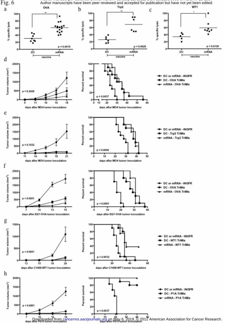
demonstrated that immunization with antigen and TriMix mRNA was as efficient as
immunization with antigen and TriMix mRNA electroporated DCs for the antigen OVA and
the TAAs Trp2 and WT1 [Fig. 6a-c]. We next evaluated the therapeutic efficacy of such
vaccines. Firstly, mice bearing MO4 tumors were treated with antigen and TriMix mRNA-
modified DCs or antigen and TriMix mRNA as such. Similar results were obtained upon
immunization with OVA [Fig. 6d] or Trp2 [Fig. 6e] as an antigen. Mice treated with tNGFR
electroporated DCs or tNGFR mRNA as such served as controls. Mice from control groups
showed rapid tumor growth, whereas mice immunized with a single intravenous injection of
DCs electroporated with antigen and TriMix mRNA or an intranodal injection of antigen and
TriMix mRNA showed a reduced tumor growth hence prolonged survival. These data were
extended to the mouse T-cell lymphoma EG7-OVA, the myeloid leukaemia C1498-WT1 in
C57BL/6 mice and the mastocytoma P815 in DBA-2 mice using OVA, WT1 and P1A as the
antigen applied for immunization, respectively [Fig. 6f-h].
on May 6, 2019. © 2012 American Association for Cancer Research. cancerres.aacrjournals.org Downloaded from
Author manuscripts have been peer reviewed and accepted for publication but have not yet been edited. Author Manuscript Published OnlineFirst on February 15, 2012; DOI: 10.1158/0008-5472.CAN-11-2957
16
Discussion
Delivery of TAA mRNA to DCs for cancer therapy offers many advantages, which can be
fully exploited when the mRNA is administered intranodally (15). It is proposed that mRNA
functions as a template for translation as well as a ligand for TLRs (33). It is not clear,
however, whether the intrinsic adjuvant effect of mRNA is sufficient to fully exploit the
immunostimulatory capacity of DCs (34).
Therefore, we evaluated the local delivery of mRNA encoding CD40L, CD70 and caTLR4
[referred to as TriMix] as an adjuvant in conjunction with intranodal TAA RNA vaccination.
We show that TriMix but not classical maturation stimuli potentiates the immunogenicity of
intranodal mRNA vaccination. We moreover demonstrated that the strength of TriMix is dual:
low impact on antigen mRNA immunobioavailability and simultaneous delivery of stimuli
that act synergistic in terms of activation of T-cell responses.
It has been suggested that the immunobioavailability of antigen mRNA is a critical success-
limiting factor in view of cancer therapy (35). First, we demonstrated high antigen expression
when mRNA was delivered in Ca2+-containing HBSS or the clinically applied 0.8 RL,
confirming the Ca2+-dependency for efficient uptake of mRNA (29). It was previously
demonstrated that several adjuvants hamper mRNA uptake, as it is critically dependent on
macropinocytosis, a process that is rapidly down-regulated upon DC activation (36).
Therefore, we next evaluated the engulfment of antigen mRNA when co-delivered with
TriMix or LPS. We confirmed the severe reduction in antigen expression when LPS was co-
administered. However, this phenomenon was less pronounced when TriMix was co-delivered
and might be explained by the timing of DC activation, which most likely is initiated after the
uptake and translation of the TriMix mRNA.
Recently, Diken et al (36) hypothesized that simultaneous delivery of classical activation
stimuli might result in imperilment of the induction of an immune response. We now
demonstrate that the co-delivery of LPS but not TriMix indeed completely abrogates the
on May 6, 2019. © 2012 American Association for Cancer Research. cancerres.aacrjournals.org Downloaded from
Author manuscripts have been peer reviewed and accepted for publication but have not yet been edited. Author Manuscript Published OnlineFirst on February 15, 2012; DOI: 10.1158/0008-5472.CAN-11-2957
17
stimulation of antigen-specific T-cells. In contrast, we demonstrated that the co-delivery of
TriMix mRNA significantly enhanced the induction of antigen-specific T-cells. The latter can
be explained in part by the fact that the intranodal delivery of TriMix mRNA resulted in
phenotypically more mature DCs and created an environment that is even better suited to
recruit and activate T-cells when compared to the use of antigen mRNA alone. However, this
cannot be the only explanation as co-delivery of LPS also resulted in highly mature DCs. The
explanation for the differences in T-cell stimulation in mice immunized with antigen mRNA
or the latter combined with TriMix or LPS might be found in the levels of MHC/peptide
complexes on DCs, which are determined by the availability of the antigen. It was
demonstrated that a certain threshold antigen dose is required for T-cells to decide to
participate in immune responses (37). We hypothesize that this threshold is not met when
classical adjuvants are co-delivered with antigen mRNA, as these almost completely abrogate
the engulfment of mRNA. We demonstrated that co-delivery of TriMix mRNA with antigen
mRNA resulted in a two-fold higher antigen expression when compared to the delivery in the
presence of LPS. This amount might surpass the required threshold for T-cell recognition and
engagement. Although the delivery of mRNA alone resulted in the highest availability of the
antigen mRNA and activated the DCs to a certain extent, we observed that the co-delivery of
TriMix resulted in enhanced T-cell responses. The latter might be partially explained by the
observation that lower antigen doses that exceed the above-mentioned threshold are correlated
with enhanced T-cell activation and functionality (37, 38).
Since we demonstrated that co-delivery of TriMix allows the uptake of antigen mRNA and has
an added benefit in terms of activation of adaptive T-cell responses, we next evaluated its
therapeutic efficacy. Since we described the induction of antigen-specific T-cells both in vitro
(20, 21) and in vaccinated melanoma patients (22) by TAA and TriMix mRNA electroporated
human DCs, we decided to compare DC- to mRNA-immunization. We demonstrated that the
therapeutic efficacy of antigen and TriMix mRNA is comparable to that of DCs electroporated
on May 6, 2019. © 2012 American Association for Cancer Research. cancerres.aacrjournals.org Downloaded from
Author manuscripts have been peer reviewed and accepted for publication but have not yet been edited. Author Manuscript Published OnlineFirst on February 15, 2012; DOI: 10.1158/0008-5472.CAN-11-2957
18
with this mRNA. As such we here highlight the feasibility and potency of the TriMix and
antigen mRNA-based immunization strategy.
It was recently implied that an adjuvant should be chosen on the basis of complementarity of
its mode of action with that of the vaccine format it will be combined with (31). In that regard,
the efficacy of mRNA administered into lymph nodes depends on its uptake and its ability to
create a CTL inducing milieu. We conclude that these prerequisites are met through the co-
delivery of TriMix mRNA, as it allows antigen mRNA uptake, confers a high T-cell
stimulatory capacity to DCs and as such enhances their ability to stimulate antigen-specific
immunity.
on May 6, 2019. © 2012 American Association for Cancer Research. cancerres.aacrjournals.org Downloaded from
Author manuscripts have been peer reviewed and accepted for publication but have not yet been edited. Author Manuscript Published OnlineFirst on February 15, 2012; DOI: 10.1158/0008-5472.CAN-11-2957

19
Acknowledgements
The authors wish to thank Petra Roman, Elsy Vaeremans and Xavier Debaere for their
technical assistance and Prof. M. Moser and Prof. O. Leo for critical reading of the
manuscript.
on May 6, 2019. © 2012 American Association for Cancer Research. cancerres.aacrjournals.org Downloaded from
Author manuscripts have been peer reviewed and accepted for publication but have not yet been edited. Author Manuscript Published OnlineFirst on February 15, 2012; DOI: 10.1158/0008-5472.CAN-11-2957
20
References
1. Breckpot K, Escors D. Dendritic cells for active anti-cancer immunotherapy: targeting
activation pathways through genetic modification. Endocr Metab Immune Disord Drug
Targets 2009; 9: 328-43.
2. Palucka K, Banchereau J, Mellman I. Designing vaccines based on biology of human
dendritic cell subsets. Immunity 2010; 33: 464-78.
3. Arce F, Kochan G, Breckpot K, Stephenson H, Escors D. Selective Activation of
Intracellular Signalling Pathways In Dendritic Cells For Cancer Immunotherapy. Anticancer
Agents Med Chem 2011 Jun 27. [Epub ahead of print].
4. Breckpot K, Heirman C, Neyns B, Thielemans K. Exploiting dendritic cells for cancer
immunotherapy: genetic modification of dendritic cells. J Gene Med 2004; 6: 1175-88.
5. Tuyaerts S, Michiels A, Corthals J, et al. Induction of Influenza Matrix Protein 1 and
MelanA-specific T lymphocytes in vitro using mRNA-electroporated dendritic cells. Cancer
Gene Ther 2003; 10: 696-706.
6. Gilboa E, Vieweg J. Cancer immunotherapy with mRNA-transfected dendritic cells.
Immunol Rev 2004; 199: 251-63.
7. Bonehill A, Heirman C, Tuyaerts S, et al. Messenger RNA-electroporated dendritic
cells presenting MAGE-A3 simultaneously in HLA class I and class II molecules. J Immunol
2004; 172: 6649-57.
8. Dullaers M, Breckpot K, Van Meirvenne S, et al. Side-by-side comparison of
lentivirally transduced and mRNA-electroporated dendritic cells: implications for cancer
immunotherapy protocols. Mol Ther 2004; 10: 768-79.
9. Van Meirvenne S, Straetman L, Heirman C, et al. Efficient genetic modification of
murine dendritic cells by electroporation with mRNA. Cancer Gene Ther 2002; 9: 787-97.
10. Michiels A, Tuyaerts S, Bonehill A, et al. Electroporation of immature and mature
dendritic cells: implications for dendritic cell-based vaccines. Gene Ther 2005; 12: 772-82.
on May 6, 2019. © 2012 American Association for Cancer Research. cancerres.aacrjournals.org Downloaded from
Author manuscripts have been peer reviewed and accepted for publication but have not yet been edited. Author Manuscript Published OnlineFirst on February 15, 2012; DOI: 10.1158/0008-5472.CAN-11-2957
21
11. Tuyaerts S, Aerts JL, Corthals J, et al. Current approaches in dendritic cell generation
and future implications for cancer immunotherapy. Cancer Immunol Immunother 2007; 56:
1513-37.
12. Van Lint S, Thielemans K, Breckpot K. mRNA: delivering an antitumor message?
Immunotherapy 2011; 3: 605-7.
13. Kreiter S, Diken M, Selmi A, Tureci O, Sahin U. Tumor vaccination using messenger
RNA: prospects of a future therapy. Curr Opin Immunol 2011; 23: 399-406.
14. Pascolo S. Vaccination with messenger RNA (mRNA). Handb Exp Pharmacol 2008:
221-35.
15. Kreiter S, Selmi A, Diken M, et al. Intranodal vaccination with naked antigen-
encoding RNA elicits potent prophylactic and therapeutic antitumoral immunity. Cancer Res
2010; 70: 9031-40.
16. Kariko K, Ni H, Capodici J, Lamphier M, Weissman D. mRNA is an endogenous
ligand for Toll-like receptor 3. J Biol Chem 2004; 279: 12542-50.
17. Kariko K, Buckstein M, Ni H, Weissman D. Suppression of RNA recognition by Toll-
like receptors: the impact of nucleoside modification and the evolutionary origin of RNA.
Immunity 2005; 23: 165-75.
18. Fotin-Mleczek M, Duchardt KM, Lorenz C, et al. Messenger RNA-based vaccines
with dual activity induce balanced TLR-7 dependent adaptive immune responses and provide
antitumor activity. J Immunother 2010; 34: 1-15.
19. Diken M, Kreiter S, Selmi A, et al. Selective uptake of naked vaccine RNA by
dendritic cells is driven by macropinocytosis and abrogated upon DC maturation. Gene Ther
2011;18:702-8.
20. Bonehill A, Van Nuffel AM, Corthals J, et al. Single-step antigen loading and
activation of dendritic cells by mRNA electroporation for the purpose of therapeutic
vaccination in melanoma patients. Clin Cancer Res 2009; 15: 3366-75.
on May 6, 2019. © 2012 American Association for Cancer Research. cancerres.aacrjournals.org Downloaded from
Author manuscripts have been peer reviewed and accepted for publication but have not yet been edited. Author Manuscript Published OnlineFirst on February 15, 2012; DOI: 10.1158/0008-5472.CAN-11-2957
22
21. Bonehill A, Tuyaerts S, Van Nuffel AM, et al. Enhancing the T-cell stimulatory
capacity of human dendritic cells by co-electroporation with CD40L, CD70 and constitutively
active TLR4 encoding mRNA. Mol Ther 2008; 16: 1170-80.
22. Van Nuffel AM, Corthals J, Neyns B, Heirman C, Thielemans K, Bonehill A.
Immunotherapy of cancer with dendritic cells loaded with tumor antigens and activated
through mRNA electroporation. Methods Mol Biol 2010; 629: 405-52.
23. Wilgenhof S, Van Nuffel AM, Corthals J, et al. Therapeutic vaccination with an
autologous mRNA electroporated dendritic cell vaccine in patients with advanced melanoma.
J Immunother 2011; 34: 448-56.
24. Benteyn D, Anguille S, Van Lint S, et al. Enhancing the Wilms’ Tumor 1 (WT1)
antigen expression and presentation by mRNA electroporated dendritic cells. Clin Canc Res.
Under revision.
25. Van Nuffel AM, Corthals J, Neyns B, Heirman C, Thielemans K, Bonehill A et al.
Dendritic cells loaded with mRNA encoding full-length tumor antigens prime CD4+ and
CD8+ T cells in melanoma patients. Molecular Therapy under revision.
26. Breckpot K, Escors D, Arce F, et al. HIV-1 lentiviral vector immunogenicity is
mediated by Toll-like receptor 3 (TLR3) and TLR7. J Virol 2010; 84: 5627-36.
27. Keyaerts M, Verschueren J, Bos TJ, et al. Dynamic bioluminescence imaging for
quantitative tumour burden assessment using IV or IP administration of D: -luciferin: effect on
intensity, time kinetics and repeatability of photon emission. Eur J Nucl Med Mol Imaging
2008; 35: 999-1007.
28. Dullaers M, Van Meirvenne S, Heirman C, et al. Induction of effective therapeutic
antitumor immunity by direct in vivo administration of lentiviral vectors. Gene Ther 2006; 13:
630-40.
on May 6, 2019. © 2012 American Association for Cancer Research. cancerres.aacrjournals.org Downloaded from
Author manuscripts have been peer reviewed and accepted for publication but have not yet been edited. Author Manuscript Published OnlineFirst on February 15, 2012; DOI: 10.1158/0008-5472.CAN-11-2957
23
29. Probst J, Weide B, Scheel B, et al. Spontaneous cellular uptake of exogenous
messenger RNA in vivo is nucleic acid-specific, saturable and ion dependent. Gene Ther
2007; 14: 1175-80.
30. Probst J, Brechtel S, Scheel B, et al. Characterization of the ribonuclease activity on
the skin surface. Genet Vaccines Ther 2006; 4: 4.
31. Kreiter S, Diken M, Selmi A, et al. FLT3 ligand enhances the cancer therapeutic
potency of naked RNA vaccines. Cancer Res 2011; 71: 6132-42.
32. Bevan MJ. Helping the CD8(+) T-cell response. Nat Rev Immunol 2004; 4: 595-602.
33. Reis e Sousa C. Toll-like receptors and dendritic cells: for whom the bug tolls. Semin
Immunol 2004; 16: 27-34.
34. Palucka K, Ueno H, Roberts L, Fay J, Banchereau J. Dendritic cells: are they clinically
relevant? Cancer J 2010; 16: 318-24.
35. Kuhn AN, Diken M, Kreiter S, Vallazza B, Tureci O, Sahin U. Determinants of
intracellular RNA pharmacokinetics: Implications for RNA-based immunotherapeutics. RNA
Biol 2011; 8: 35-43.
36. Diken M, Kreiter S, Selmi A, et al. Selective uptake of naked vaccine RNA by
dendritic cells is driven by macropinocytosis and abrogated upon DC maturation. Gene Ther
2011; 18: 702-8.
37. Henrickson SE, Mempel TR, Mazo IB, et al. T cell sensing of antigen dose governs
interactive behavior with dendritic cells and sets a threshold for T cell activation. Nat
Immunol 2008; 9: 282-91.
38. Rees W, Bender J, Teague TK, et al. An inverse relationship between T cell receptor
affinity and antigen dose during CD4(+) T cell responses in vivo and in vitro. Proc Natl Acad
Sci U S A 1999; 96: 9781-6.
on May 6, 2019. © 2012 American Association for Cancer Research. cancerres.aacrjournals.org Downloaded from
Author manuscripts have been peer reviewed and accepted for publication but have not yet been edited. Author Manuscript Published OnlineFirst on February 15, 2012; DOI: 10.1158/0008-5472.CAN-11-2957

24
Legends
Figure 1: Dendritic cells matured through electroporation of TriMix efficiently stimulate
antigen-specific T-cells. The histogram overlays in [a] show the phenotype of DCs
electroporated with tNGFR mRNA and left immature or matured by co-electroporation of
TriMix or addition of LPS [n = 10]. The graphs in [b] show the cytokines secreted by these
DCs [n = 6]. The graph in [c] depicts the incorporation of 3H thymidine by allogeneic spleen
cells cultured with these DCs [n = 3]. [d-f] Mice were immunized intravenously with 5 x 10⁵
DCs electroporated with OVA mRNA and matured by co-electroporation of TriMix mRNA or
addition of LPS. Five days later the expansion of functional OVA-specific CD8+ T-cells was
assessed. The results of [d] the pentamer staining, [e] the in vivo cytotoxicity assay and [f] the
intracytoplasmatic staining of IFN-γ on spleen cells restimulated with SIINFEKL-presenting
DCs are shown [n = 2]. [g] Mice, immunized with Trp2-presenting DCs, were subjected to an
in vivo cytotoxicity assay to evaluate the stimulation of Trp2-specific CD8+ T-cells [n = 2].
Figure 2: Formulation and pharmacokinetics of mRNA. [a] Mouse DCs were pulsed with
FLuc mRNA in the indicated buffer. Luminescence was measured 4 hours later. The graph
depicts the photon emission [n = 4]. [b-c] Mice were injected intranodally with FLuc mRNA.
[b] In vivo bioluminescence imaging was performed at the indicated time points [n = 4]. [c] To
evaluate the stability of FLuc mRNA in vivo, lymph nodes were isolated 6, 12 and 24 hours
after injection and PCR performed on cDNA synthesized from extracted mRNA [n = 4]. [d]
Mice received an intranodal injection of eGFP mRNA formulated in 0.8 RL. Four hours later,
the lymph node was resected, a single cell suspension prepared and stained for CD11c. The
photo obtained by fluorescence microscopy shows eGFP [green] expression by CD11c+ cells
[red, n = 4]. [e] Transgenic CD11c-DTR mice, which were pre-treated with PBS or DT,
received an intranodal injection with FLuc mRNA. In vivo bioluminescence imaging was
performed 4 hours later. Single cell suspensions were prepared from the lymph nodes and
on May 6, 2019. © 2012 American Association for Cancer Research. cancerres.aacrjournals.org Downloaded from
Author manuscripts have been peer reviewed and accepted for publication but have not yet been edited. Author Manuscript Published OnlineFirst on February 15, 2012; DOI: 10.1158/0008-5472.CAN-11-2957
25
analyzed by flow cytometry for the presence of CD11c+ cells [n = 3]. [f] Mice of which the
skin was pre-treated with PBS or GM-CSF, were injected intradermally with FLuc mRNA. In
vivo bioluminescence imaging was performed 6 hours later [n = 3].
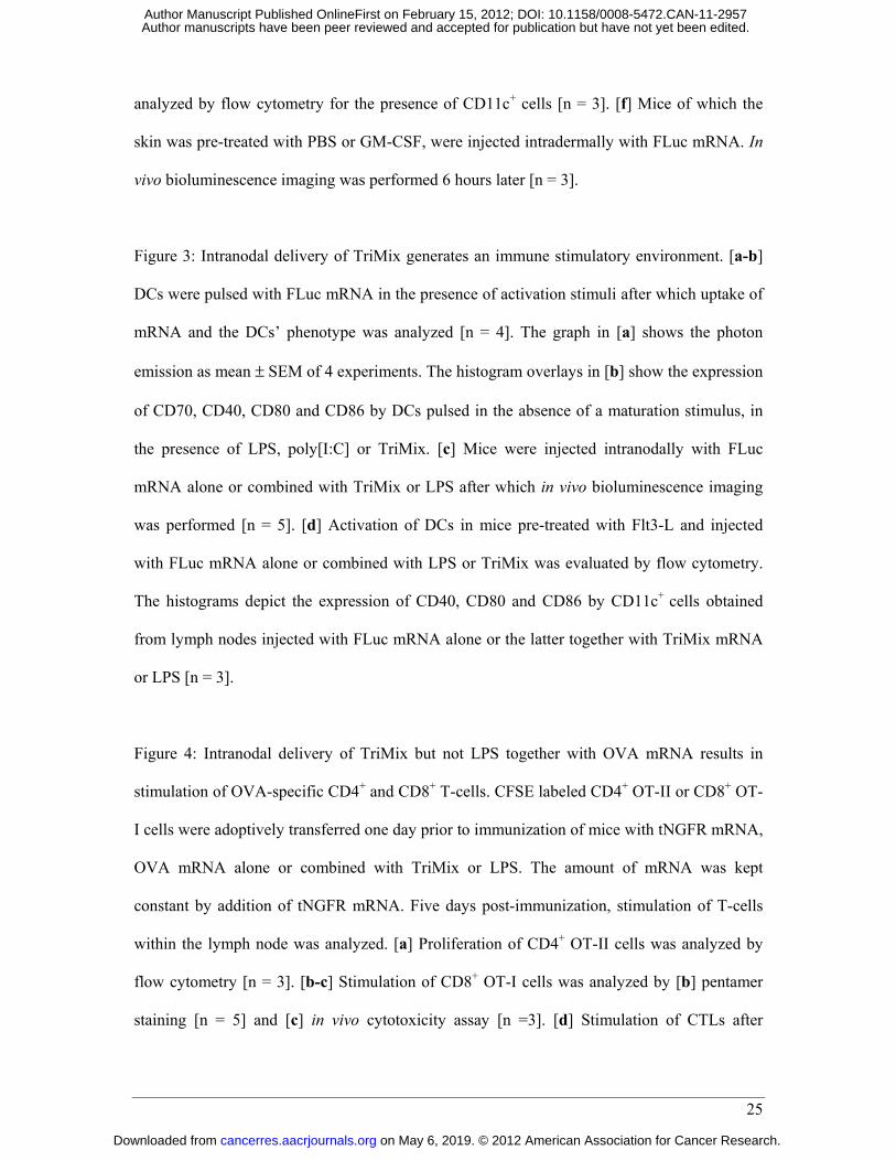
Figure 3: Intranodal delivery of TriMix generates an immune stimulatory environment. [a-b]
DCs were pulsed with FLuc mRNA in the presence of activation stimuli after which uptake of
mRNA and the DCs’ phenotype was analyzed [n = 4]. The graph in [a] shows the photon
emission as mean ± SEM of 4 experiments. The histogram overlays in [b] show the expression
of CD70, CD40, CD80 and CD86 by DCs pulsed in the absence of a maturation stimulus, in
the presence of LPS, poly[I:C] or TriMix. [c] Mice were injected intranodally with FLuc
mRNA alone or combined with TriMix or LPS after which in vivo bioluminescence imaging
was performed [n = 5]. [d] Activation of DCs in mice pre-treated with Flt3-L and injected
with FLuc mRNA alone or combined with LPS or TriMix was evaluated by flow cytometry.
The histograms depict the expression of CD40, CD80 and CD86 by CD11c+ cells obtained
from lymph nodes injected with FLuc mRNA alone or the latter together with TriMix mRNA
or LPS [n = 3].
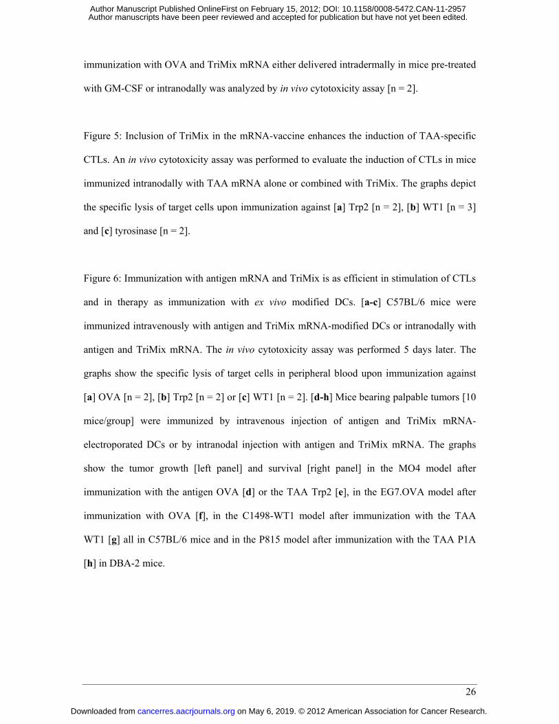
Figure 4: Intranodal delivery of TriMix but not LPS together with OVA mRNA results in
stimulation of OVA-specific CD4+ and CD8+ T-cells. CFSE labeled CD4+ OT-II or CD8+ OT-
I cells were adoptively transferred one day prior to immunization of mice with tNGFR mRNA,
OVA mRNA alone or combined with TriMix or LPS. The amount of mRNA was kept
constant by addition of tNGFR mRNA. Five days post-immunization, stimulation of T-cells
within the lymph node was analyzed. [a] Proliferation of CD4+ OT-II cells was analyzed by
flow cytometry [n = 3]. [b-c] Stimulation of CD8+ OT-I cells was analyzed by [b] pentamer
staining [n = 5] and [c] in vivo cytotoxicity assay [n =3]. [d] Stimulation of CTLs after
on May 6, 2019. © 2012 American Association for Cancer Research. cancerres.aacrjournals.org Downloaded from
Author manuscripts have been peer reviewed and accepted for publication but have not yet been edited. Author Manuscript Published OnlineFirst on February 15, 2012; DOI: 10.1158/0008-5472.CAN-11-2957
26
immunization with OVA and TriMix mRNA either delivered intradermally in mice pre-treated
with GM-CSF or intranodally was analyzed by in vivo cytotoxicity assay [n = 2].
Figure 5: Inclusion of TriMix in the mRNA-vaccine enhances the induction of TAA-specific
CTLs. An in vivo cytotoxicity assay was performed to evaluate the induction of CTLs in mice
immunized intranodally with TAA mRNA alone or combined with TriMix. The graphs depict
the specific lysis of target cells upon immunization against [a] Trp2 [n = 2], [b] WT1 [n = 3]
and [c] tyrosinase [n = 2].
Figure 6: Immunization with antigen mRNA and TriMix is as efficient in stimulation of CTLs
and in therapy as immunization with ex vivo modified DCs. [a-c] C57BL/6 mice were
immunized intravenously with antigen and TriMix mRNA-modified DCs or intranodally with
antigen and TriMix mRNA. The in vivo cytotoxicity assay was performed 5 days later. The
graphs show the specific lysis of target cells in peripheral blood upon immunization against
[a] OVA [n = 2], [b] Trp2 [n = 2] or [c] WT1 [n = 2]. [d-h] Mice bearing palpable tumors [10
mice/group] were immunized by intravenous injection of antigen and TriMix mRNA-
electroporated DCs or by intranodal injection with antigen and TriMix mRNA. The graphs
show the tumor growth [left panel] and survival [right panel] in the MO4 model after
immunization with the antigen OVA [d] or the TAA Trp2 [e], in the EG7.OVA model after
immunization with OVA [f], in the C1498-WT1 model after immunization with the TAA
WT1 [g] all in C57BL/6 mice and in the P815 model after immunization with the TAA P1A
[h] in DBA-2 mice.
on May 6, 2019. © 2012 American Association for Cancer Research. cancerres.aacrjournals.org Downloaded from
Author manuscripts have been peer reviewed and accepted for publication but have not yet been edited. Author Manuscript Published OnlineFirst on February 15, 2012; DOI: 10.1158/0008-5472.CAN-11-2957
Table
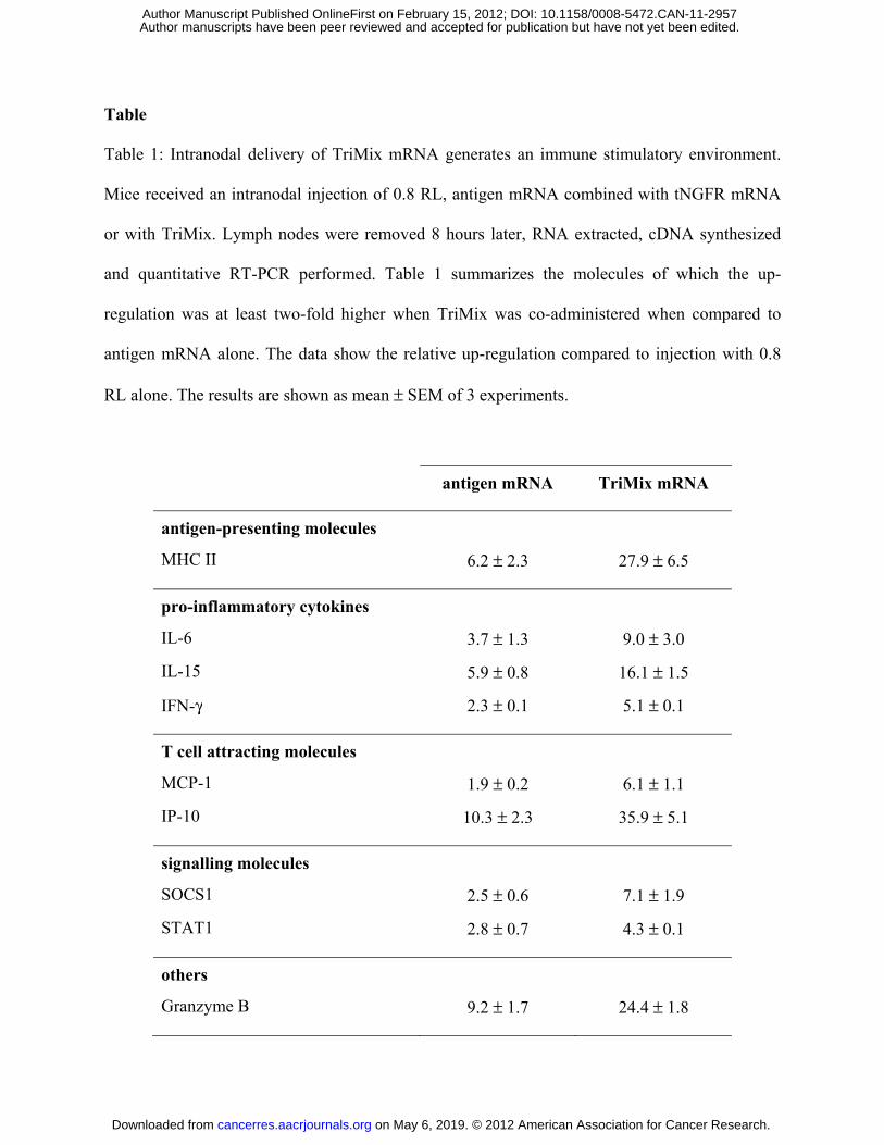
Table 1: Intranodal delivery of TriMix mRNA generates an immune stimulatory environment.
Mice received an intranodal injection of 0.8 RL, antigen mRNA combined with tNGFR mRNA
or with TriMix. Lymph nodes were removed 8 hours later, RNA extracted, cDNA synthesized
and quantitative RT-PCR performed. Table 1 summarizes the molecules of which the up-
regulation was at least two-fold higher when TriMix was co-administered when compared to
antigen mRNA alone. The data show the relative up-regulation compared to injection with 0.8
RL alone. The results are shown as mean ± SEM of 3 experiments.
antigen mRNA TriMix mRNA
antigen-presenting molecules
MHC II 6.2 ± 2.3 27.9 ± 6.5
pro-inflammatory cytokines
IL-6 3.7 ± 1.3 9.0 ± 3.0
IL-15 5.9 ± 0.8 16.1 ± 1.5
IFN-γ 2.3 ± 0.1 5.1 ± 0.1
T cell attracting molecules
MCP-1 1.9 ± 0.2 6.1 ± 1.1
IP-10 10.3 ± 2.3 35.9 ± 5.1
signalling molecules
SOCS1 2.5 ± 0.6 7.1 ± 1.9
STAT1 2.8 ± 0.7 4.3 ± 0.1
others
Granzyme B 9.2 ± 1.7 24.4 ± 1.8
on May 6, 2019. © 2012 American Association for Cancer Research. cancerres.aacrjournals.org Downloaded from
Author manuscripts have been peer reviewed and accepted for publication but have not yet been edited. Author Manuscript Published OnlineFirst on February 15, 2012; DOI: 10.1158/0008-5472.CAN-11-2957
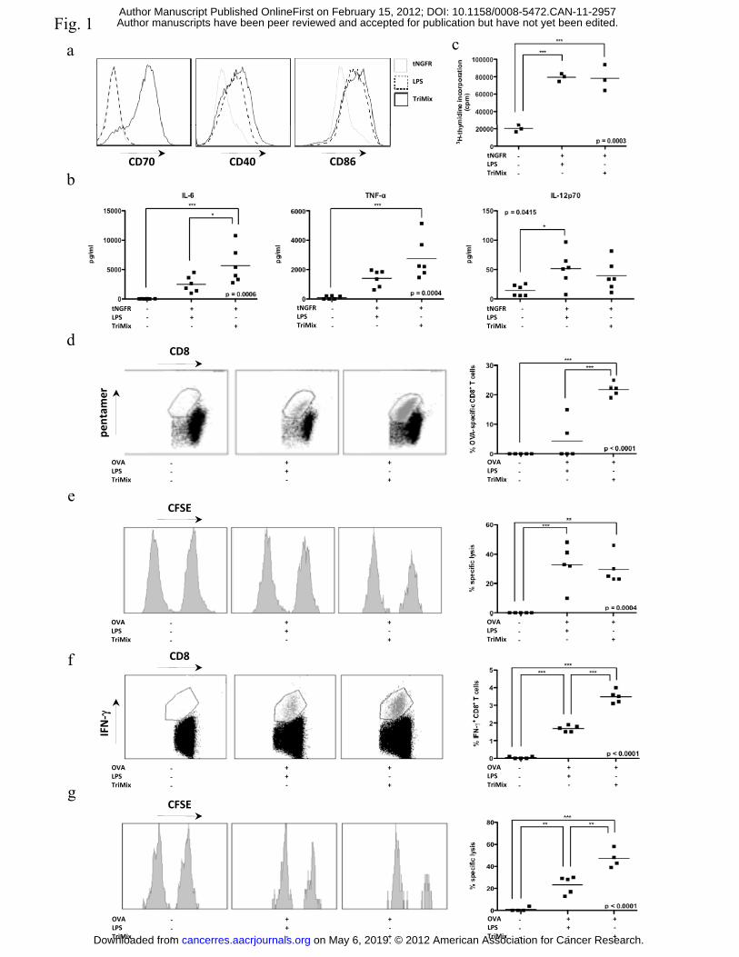
Fig. 1a c
tNGFR
LPS
TriMix
btNGFRLPSTriMix
---
++-
+-+
CD70 CD40 CD86
d
tNGFRLPSTriMix
---
++-
+-+
tNGFRLPSTriMix
---
++-
+-+
tNGFRLPSTriMix
---
++-
+-+
CD8
OVALPSTriMix
--
++
+-+
OVALPST iMi
--
++
+-+
pentam
er
eTriMix - - +TriMix - - +
CFSE
f
OVALPSTriMix
---
++-
+-+
OVALPSTriMix
---
++-
+-+
CD8
γ
g
OVALPSTriMix
---
++-
+-+
OVALPSTriMix
---
++-
+-+
CFSE
IFN-γ
OVALPSTriMix
---
++-
+-+
OVALPSTriMix
---
++-
+-+on May 6, 2019. © 2012 American Association for Cancer Research. cancerres.aacrjournals.org Downloaded from
Author manuscripts have been peer reviewed and accepted for publication but have not yet been edited. Author Manuscript Published OnlineFirst on February 15, 2012; DOI: 10.1158/0008-5472.CAN-11-2957
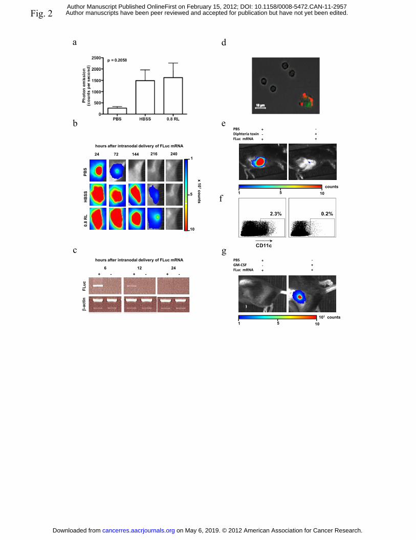
Fig. 2
a d
b ePBSDiphteria toxin
+-
-+
f
PBS
HB
SS
24 72 144 216 2401
5
x10
1counts
hours after intranodal delivery of FLuc mRNA
FLuc mRNA + +
1 5 10counts
c
0.8
RL
10
6 12 24
hours after intranodal delivery of FLuc mRNA PBSGM-CSFFLuc mRNA
+-+
-++
g
+ - + -+ -
FLuc
β-ac
tin
FLuc mRNA + +
1 5 10101 counts
on May 6, 2019. © 2012 American Association for Cancer Research. cancerres.aacrjournals.org Downloaded from
Author manuscripts have been peer reviewed and accepted for publication but have not yet been edited. Author Manuscript Published OnlineFirst on February 15, 2012; DOI: 10.1158/0008-5472.CAN-11-2957
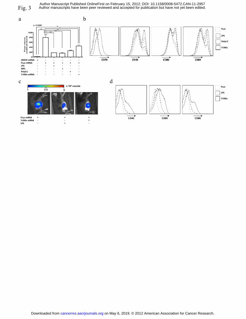
Fig. 3
a bFLuc
LPS
PolyI:C
tNGFR mRNAFLuc mRNALPSMPLPolyI:CTriMix mRNA
+-----
-+----
-++---
-+-+--
-+--+-
-+---+
dc
PolyI:C
TriMix
dc1 2.5 5
x 102 counts
FLuc mRNATriMix mRNA
+ + ++
FLuc
LPS
TriMix
TriMix mRNALPS
--
-+
+-
on May 6, 2019. © 2012 American Association for Cancer Research. cancerres.aacrjournals.org Downloaded from
Author manuscripts have been peer reviewed and accepted for publication but have not yet been edited. Author Manuscript Published OnlineFirst on February 15, 2012; DOI: 10.1158/0008-5472.CAN-11-2957
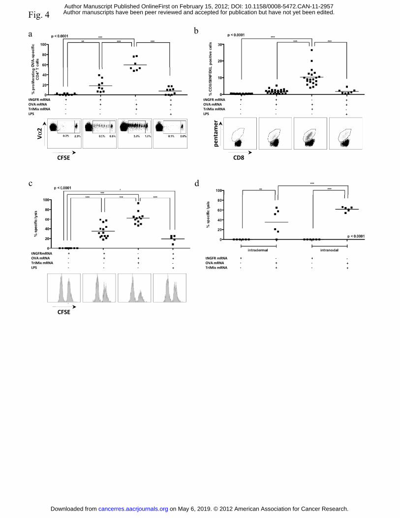
Fig. 4
ba
tNGFR mRNAOVA mRNATriMix mRNALPS
+---
++--
-++-
++-+
tNGFR mRNAOVA mRNATriMix mRNALPS
+---
++--
-++-
++-+
α2 mer
c d
CFSE
Vα
CD8
pent
a
tNGFRmRNAOVA mRNA
+-
++
-+
++ tNGFR mRNA + - + -OVA mRNA
TriMix mRNALPS
--
+--
++-
+-+
tNGFR mRNAOVA mRNATriMix mRNA
+--
++
+--
-++
CFSE
on May 6, 2019. © 2012 American Association for Cancer Research. cancerres.aacrjournals.org Downloaded from
Author manuscripts have been peer reviewed and accepted for publication but have not yet been edited. Author Manuscript Published OnlineFirst on February 15, 2012; DOI: 10.1158/0008-5472.CAN-11-2957
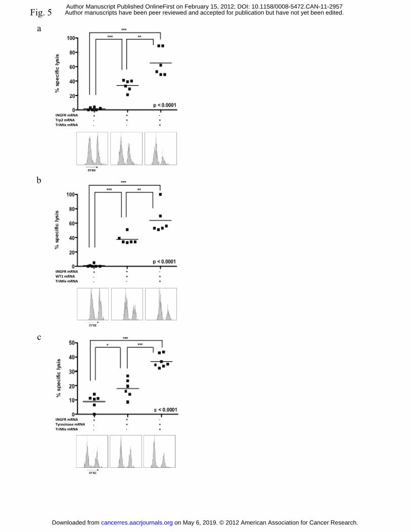
Fig. 5
a
tNGFR mRNATrp2 mRNATriMix mRNA
+--
++-
-++
b
tNGFR mRNAWT1 mRNATriMix mRNA
+--
++-
-++
c
tNGFR mRNATyrosinase mRNATriMix mRNA
+--
++-
-++
on May 6, 2019. © 2012 American Association for Cancer Research. cancerres.aacrjournals.org Downloaded from
Author manuscripts have been peer reviewed and accepted for publication but have not yet been edited. Author Manuscript Published OnlineFirst on February 15, 2012; DOI: 10.1158/0008-5472.CAN-11-2957
Published OnlineFirst February 15, 2012.Cancer Res Sandra Van Lint, Cleo Goyvaerts, Sarah Maenhout, et al. anti-tumour therapyPreclinical evaluation of TriMix and antigen mRNA-based
Updated version
10.1158/0008-5472.CAN-11-2957doi:
Access the most recent version of this article at:
Manuscript
Authoredited. Author manuscripts have been peer reviewed and accepted for publication but have not yet been
E-mail alerts related to this article or journal.Sign up to receive free email-alerts
Subscriptions
Reprints and
To order reprints of this article or to subscribe to the journal, contact the AACR Publications
Permissions
Rightslink site. Click on "Request Permissions" which will take you to the Copyright Clearance Center's (CCC)
.http://cancerres.aacrjournals.org/content/early/2012/02/15/0008-5472.CAN-11-2957To request permission to re-use all or part of this article, use this link
on May 6, 2019. © 2012 American Association for Cancer Research. cancerres.aacrjournals.org Downloaded from
Author manuscripts have been peer reviewed and accepted for publication but have not yet been edited. Author Manuscript Published OnlineFirst on February 15, 2012; DOI: 10.1158/0008-5472.CAN-11-2957