Pre-MDS Conditions: CHIP, ICUS, IDUS Pathobiology … · HU Dasatinib (100 mg/d) HU post SCT/ no HU...
43
Pre-MDS Conditions: CHIP, ICUS, IDUS Pathobiology and Diagnosis Peter Valent Department of Internal Medicine I and Ludwig Boltzmann Cluster Oncology Medical University of Vienna
Transcript of Pre-MDS Conditions: CHIP, ICUS, IDUS Pathobiology … · HU Dasatinib (100 mg/d) HU post SCT/ no HU...

Pre-MDS Conditions: CHIP, ICUS, IDUS Pathobiology and Diagnosis
Peter ValentDepartment of Internal Medicine I
and Ludwig Boltzmann Cluster OncologyMedical University of Vienna
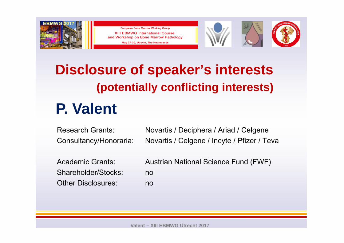
Disclosure of speaker’s interests(potentially conflicting interests)
P. ValentResearch Grants: Novartis / Deciphera / Ariad / CelgeneConsultancy/Honoraria: Novartis / Celgene / Incyte / Pfizer / Teva
Academic Grants: Austrian National Science Fund (FWF)Shareholder/Stocks: noOther Disclosures: no
Valent – XIII EBMWG Ütrecht 2017
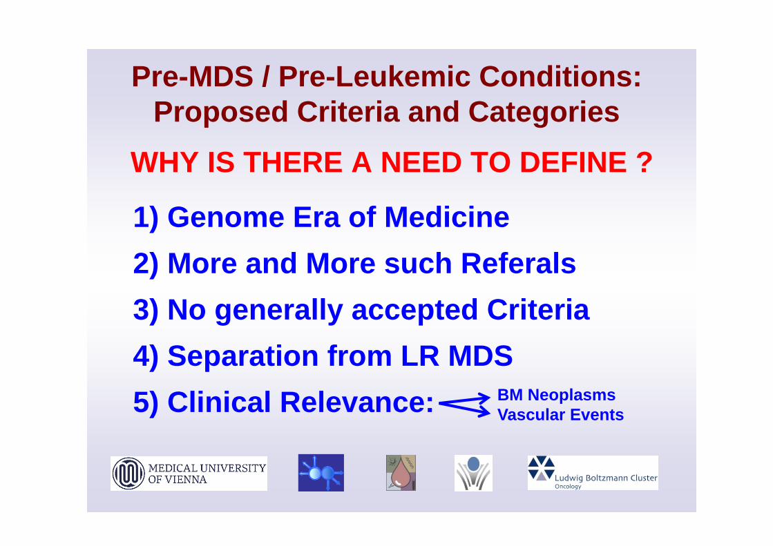
WHY IS THERE A NEED TO DEFINE ?
1) Genome Era of Medicine 2) More and More such Referals3) No generally accepted Criteria4) Separation from LR MDS5) Clinical Relevance:
Pre-MDS / Pre-Leukemic Conditions: Proposed Criteria and Categories
BM NeoplasmsVascular Events

Self renewal
Self renewal
Decisive Hits
Decisive Hits
Decisive Hits
Normal Mature Cells
Leukemic Cell Bulk
Leukemic Stem Cell
Normal Hematopoietic Stem Cell
NSC‐Niche Interactions
So far, only little is known about LSC-NicheInteractions and related Mechanisms that maycontribute to LSC Evolution and Resistance
ORIGIN AND BIOLOGY OF NEOPLASTIC STEM CELLS IN MYELOID NEOPLASMS
Nair et al., Biochem Pharmacol 2010;80:602
Valent – XIII EBMWG Ütrecht 2017

From Cytoreduction toDisease Eradication byElimination of LSC
‐ DEFINITION OF LSC: LONG‐TERM in vivo L‐PROPAGATING CELLS
‐ IDENTIFICATION AND PURIFICATION OF LSC‐ IDENTIFICATION AND VALIDATION OF TARGETS‐ EFFECTS OF TARGETED DRUGS ‐ PROVIDE A SUITABLE BASIS FOR THE DEVELOPMENT
OF LSC‐ERADICATING APPROACHES
How to translate the LSC Concept into Clinical Application ?
How to develop LSC-Eradicating (=Curative) Treatment Concepts ?
Valent – XIII EBMWG Ütrecht 2017

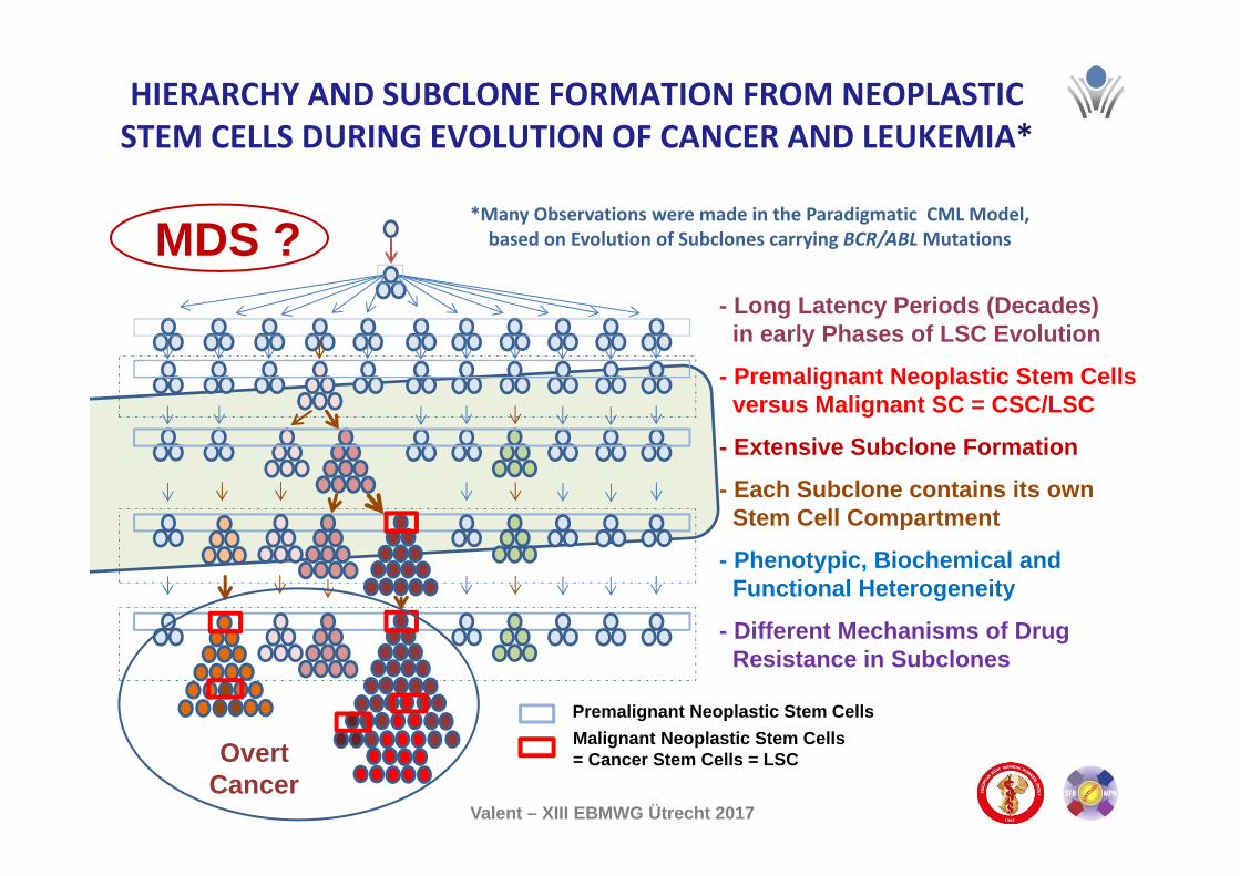
Premalignant Neoplastic Stem CellsMalignant Neoplastic Stem Cells= Cancer Stem Cells = LSC
- Long Latency Periods (Decades)in early Phases of LSC Evolution
- Premalignant Neoplastic Stem Cellsversus Malignant SC = CSC/LSC
- Extensive Subclone Formation
- Each Subclone contains its own Stem Cell Compartment
- Phenotypic, Biochemical andFunctional Heterogeneity
- Different Mechanisms of Drug Resistance in Subclones
HIERARCHY AND SUBCLONE FORMATION FROM NEOPLASTICSTEM CELLS DURING EVOLUTION OF CANCER AND LEUKEMIA*
*Many Observations were made in the Paradigmatic CML Model, based on Evolution of Subclones carrying BCR/ABLMutations
Overt Malignancy
Valent, Lancet Oncol 2010Valent, CCDT 2011Valent et al, Nat Rev Cancer 2012Valent et at, Cancer Res 2013
Valent – XIII EBMWG Ütrecht 2017

LSC-Marker:BCR-ABL1 mutants
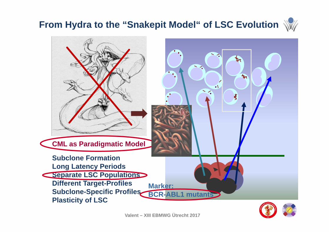
The Hydra Model of LSC Evolution: CML
CML as Paradigmatic Model
Subclone FormationLong Latency PeriodsSeparate LSC PopulationsDifferent Target-Profiles Subclone-Specific ProfilesPlasticity of LSC
Valent – XIII EBMWG Ütrecht 2017
Marker:BCR-ABL1 mutants
From Hydra to the “Snakepit Model“ of LSC Evolution
CML as Paradigmatic Model
Subclone FormationLong Latency PeriodsSeparate LSC PopulationsDifferent Target-Profiles Subclone-Specific ProfilesPlasticity of LSC
Valent – XIII EBMWG Ütrecht 2017

HUDasatinib (100 mg/d) HU
post SCT/ no
HU
A Patient with Imatinib‐Resistant CML: Subclone Formation
0102030405060708090
100
14.6.2007
4.10.2007
25.10.2007
30.1.2008
19.02.2008
08.4.2008
03.6.2008
24.10.2008
03.11.2008
21.11.2008
09.1.2009
18.3.2009
24.4.2009
08.5.2009
29.5.2009
17.6.2009
24.6.2009
Mut
ant c
lone
(% o
f tot
al B
CR
/AB
L)
0102030405060708090100
BC
R/A
BL of A
BL (%
)G250E BCR-ABL1/ABL Mutation‐Specific qPCR
SCT
G250E
Preuner et al, Leukemia 2008;22:1956Ligation‐Dependent PCR Technique
?
SCT, Hematopoietic Stem Cell TransplantationHU = Hydroxyurea
Preuner et al, Eur J Cancer, in press, 2011
?

HUDasatinib (100 mg/d) HU
post SCT/ no
HU
0102030405060708090
100
14.6.2007
4.10.2007
25.10.2007
30.1.2008
19.02.2008
08.4.2008
03.6.2008
24.10.2008
03.11.2008
21.11.2008
09.1.2009
18.3.2009
24.4.2009
08.5.2009
29.5.2009
17.6.2009
24.6.2009
0102030405060708090100
T315I BCR-ABL1/ABL1
Mut
ant c
lone
(% o
f tot
al B
CR
/AB
L)
BC
R/A
BL of A
BL (%
)
SCT
T315I?
A Patient with Imatinib‐Resistant CML: Subclone Formation
Preuner et al, Eur J Cancer 2012;48:233‐236

HUDasatinib (100 mg/d) HU
post SCT/ no
HU
0102030405060708090
100
14.6.2007
4.10.2007
25.10.2007
30.1.2008
19.02.2008
08.4.2008
03.6.2008
24.10.2008
03.11.2008
21.11.2008
09.1.2009
18.3.2009
24.4.2009
08.5.2009
29.5.2009
17.6.2009
24.6.2009
0102030405060708090100
E255K BCR-ABL1/ABL1
Mut
ant c
lone
(% o
f tot
al B
CR
/AB
L)
BC
R/A
BL of A
BL (%
)
SCT
E255K
E255K
A Patient with Imatinib‐Resistant CML: Subclone Formation
Preuner et al, Eur J Cancer 2012;48:233‐236

HUDasatinib (100 mg/d) HU
post SCT/ no
HU
0102030405060708090
100
14.6.2007
4.10.2007
25.10.2007
30.1.2008
19.02.2008
08.4.2008
03.6.2008
24.10.2008
03.11.2008
21.11.2008
09.1.2009
18.3.2009
24.4.2009
08.5.2009
29.5.2009
17.6.2009
24.6.2009
0102030405060708090100
T315I E255K G250E BCR-ABL1/ABL1M
utan
t clo
ne (%
of t
otal
BC
R/A
BL
)B
CR
/AB
L of AB
L (%)
SCT
A Patient with Imatinib‐Resistant CML: Subclone Formation
Preuner et al, Eur J Cancer 2012;48:233‐236
Preuner et al, Leukemia 2008;22:1956Ligation‐Dependent PCR Technique
Mutation‐Specific qPCR

Premalignant Neoplastic Stem CellsMalignant Neoplastic Stem Cells= Cancer Stem Cells = LSC
- Long Latency Periods (Decades)in early Phases of LSC Evolution
- Premalignant Neoplastic Stem Cellsversus Malignant SC = CSC/LSC
- Extensive Subclone Formation
- Each Subclone contains its own Stem Cell Compartment
- Phenotypic, Biochemical andFunctional Heterogeneity
- Different Mechanisms of Drug Resistance in Subclones
HIERARCHY AND SUBCLONE FORMATION FROM NEOPLASTICSTEM CELLS DURING EVOLUTION OF CANCER AND LEUKEMIA*
*Many Observations were made in the Paradigmatic CML Model, based on Evolution of Subclones carrying BCR/ABLMutations
Overt Leukemia
Valent, Lancet Oncol 2010Valent, CCDT 2011Valent et al, Nat Rev Cancer 2012Valent et at, Cancer Res 2013
Valent et al, Nat Rev Cancer 2012
Valent – XIII EBMWG Ütrecht 2017

Definition of Cure: Basic Considerations
cytoreductive therapy sparing cancer stem cells
early relapse
specific eradication of CSCs (sparing a few resistant NSCs)
late relapse
resistant subclones

>14 Years
>22 Years
Alternative Explanations:- New Clone / New Disease ? no- BCR-ABL1 in non-SC fraction ? no- Clonal Stability ? no- Immunosurveillance ? no- Competition in the SC-Niche ? no
most likely Explanation:
?How long
?How long
NORMAL STABLE BLOOD COUNTS !
Böhm et al, Leuk Lymphoma 2011;52:842-848
Valent, Lancet Oncol 2010;11:1010Valent – XIII EBMWG Ütrecht 2017
Ph+ ALL after Autologous HSCT: Untreated !Stable BCR/ABLp190+ MRD over a Decade

CML AS PARADIGMATIC DISEASE MODEL
Operational Cure with or without adetectable MRD
STIM Study MR-free Survival in CMLFrançois-Xavier Mahon et al,
Lancet Oncology 2010;11:1029-35
Effects of Treatment with Imatinib
often stable MRD
Valent, Lancet Oncol 2010Valent, CCDT 2011Valent et al, Nat Rev Cancer 2012Valent et at, Cancer Res 2013
Valent – XIII EBMWG Ütrecht 2017

Conditions Apparently Caused by Premalignant Neoplastic Stem Cells
• Clonal Hematopoiesis of IntederminatePotential = CHIP (Pre‐MDS)*
• Other Similar Conditions: MGUS, MCAS, …• ICUS, IDUS, CCUS, CHEP, ….• Low Risk MDS• Indolent Systemic Mastocytosis• Some Forms of Indolent NHLs• Early Phase MPN and CML, etc. etc.
*CHIP = Age‐RelatedClonal Hematopoiesis
= ARCH
Valent – XIII EBMWG Ütrecht 2017
Premalignant Neoplastic Stem CellsMalignant Neoplastic Stem Cells= Cancer Stem Cells = LSC
- Long Latency Periods (Decades)in early Phases of LSC Evolution
- Premalignant Neoplastic Stem Cellsversus Malignant SC = CSC/LSC
- Extensive Subclone Formation
- Each Subclone contains its own Stem Cell Compartment
- Phenotypic, Biochemical andFunctional Heterogeneity
- Different Mechanisms of Drug Resistance in Subclones
HIERARCHY AND SUBCLONE FORMATION FROM NEOPLASTICSTEM CELLS DURING EVOLUTION OF CANCER AND LEUKEMIA*
*Many Observations were made in the Paradigmatic CML Model, based on Evolution of Subclones carrying BCR/ABLMutations
Overt Cancer
MDS ?
Valent – XIII EBMWG Ütrecht 2017

Six Phases of Cancer Evolution0 = Genetic BackgroundI = Stable Somatic Process without
any LSC expansion (usually no Driver) II = Somatic Process with Driver Lesion
without relevant LSC expansionIII = Somatic Process with Driver Lesion
replacing and mimicking the normal organ (differentiated tissue cells)
IV = Premalignant Overt NeoplasmV = Overt Malignancy = CancerVI = Resistant Advanced Malignancy
Valent – XIII EBMWG Ütrecht 2017

Six Phases of sAML Evolution0 = Genetic backgroundI = Age-related somatic mutations (ARCH=
CHIP) in small-sized clones (e.g. DNMT3A, TET2)
II = Multiple somatic mutations (drivers) with (IDUS) or without (CHIP) dysplasia
III = Mutant clones replaces the normal BM: may cause cytopenia: CCUS or early LR MDS
IV = LR MDSV = HR MDSVI = sAML
MDS
Valent – XIII EBMWG Ütrecht 2017
Valent – MDS Conference – Vienna 2016
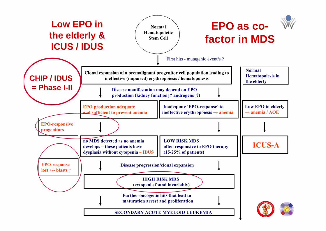
Normal Hematopoietic
Stem Cell
Clonal expansion of a premalignant progenitor cell population leading to ineffective (impaired) erythropoiesis / hematopoiesis
First hits - mutagenic event/s ?
EPO production adequate and sufficient to prevent anemia
Disease manifestation may depend on EPO production (kidney function↓? androgens↓?)
no MDS detected as no anemiadevelops – these patients have dysplasia without cytopenia = IDUS
HIGH RISK MDS (cytopenia found invariably)
Disease progression/clonal expansion
SECONDARY ACUTE MYELOID LEUKEMIA
Further oncogenic hits that lead to maturation arrest and proliferation
Inadequate ´EPO-response´ to ineffective erythropoiesis → anemia
LOW RISK MDS often responsive to EPO therapy(15-25% of patients)
Low EPO inthe elderly &ICUS / IDUS
EPO as co-factor in MDS
Low EPO in elderly→ anemia / AOE
ICUS-A
EPO-responsive progenitors
Normal Hematopoiesis in the elderly
EPO-response lost +/- blasts ↑
CHIP / IDUS= Phase I-II

Equation: Role of Decrease in EPO Production in the Etiology and
Manifestation of MDS
IDUS + ICUS = MDS

Six Phases in Ph+ CML0 = Genetic BackgroundI = Stable ARCH/CHIP mutations (may rarely
lead to a Ph-negative relapse/progress) II = BCR-ABL1 in healthy individuals (CHEP)
Stable Ph+ MRD during TKI therapy and:small subclones bearing BCR-ABL1 mutations
III = Very early chronic phase CMLIV = Chronic phase CML V = Accelerated phase CMLVI = Blast phase / blast crisis
Valent – XIII EBMWG Ütrecht 2017

IMPORTANT NOTES:• When an overt malignancy has developed, all the premalignant stages and ARCH+ subclonesare also still around (MRD) and:
1) may produce new malignantsub‐clones over time
2) may also increase the riskfor other diseases
3) may create the disease‐relatedmicroenvironment (niche)
4) may be highly resistant (MRDafter “successful“ therapy)
Valent – XIII EBMWG Ütrecht 2017

The two Clinically Important Endpoints of CHIP/ARCH
1. Hematopoietic/Myeloid Neoplasm (e.g. MDS or CML)
2. Severe Cardiovascular Disease (e.g. AOD)
Age‐related clonal hematopoiesis associated with adverse outcomesJaiswal et al, N Engl J Med. 2014;371:2488‐2498
CONCLUSIONSAge‐related clonal hematopoiesis is a common condition that is associated with increases in the risk of hematologic cancer and in all‐cause mortality, with the latter possibly due to an increased risk of cardiovascular disease
Valent – XIII EBMWG Ütrecht 2017

The two Clinically Important Endpoints of CHIP/ARCH
1. Hematopoietic/Myeloid Neoplasm (e.g. MDS)
2. Severe Cardiovascular Disease (e.g. AOD)
What is the clinical implication:We need to think in a more multi‐disciplinary way
Valent – XIII EBMWG Ütrecht 2017

Associations between Cardiovascular Problems and Myeloid / Myeloproliferative Disorders
1) Increased incidence of Thromboembolic Eventsin JAK V617F‐mutated MPN (often long beforean overt MPN is detected = clonal prephase?)
2) Increased risk of Occurrence of Thromboembolic Events in Patients with F/P+ MPN‐eo/CEL
3) Occurrence of Vascular Occlusive Diseases in CML Patients receiving Ponatinib or Nilotinib
4) Another Example may be: PNH Many more relationships may be deciphered in the near future !
Valent – XIII EBMWG Ütrecht 2017

EU-US multicenter cooperative initiative to
standardize parameters of disease and diagnostics for practice and clinical trials in
patients with MDS
10 YEAR ANNIVERSARYAND UPDATE 2016
July 7-9th 2006
EU-US initiative to standardize parameters of disease and diagnostics for practice and clinical trials
in patients with MDS- Morphology - Histopathology- Flow Cytometry- Cytogenetics and Molecular Markers- Prognostic Factors and Scoring System- Therapy and Clinical Outcome
Topics addressed in 2006 and 2016
Aims of the MDS WC in 2006:
- Minimal Diagnostic Criteria- Pre-MDS Conditions
- New Diagnostic Approaches- Diagnostic Standards- Diagnostic Algorithms- Prognostication Standards
- Position Paper- EU-US Collaborations- Topic-related Groups- Consecutive Meetings
Valent – XIII EBMWG Ütrecht 2017
FIRST ATTEMPT TO ESTABLISH CRITERIA, A NOMENCLATURE AND A CLASSIFICATION

MDS: Minimal Diagnostic Criteria 2007A. Prerequisite Criteria (BOTH MUST be fulfilled)
‐ Constant Cytopenia* (one or more lines, 6 mo unless abnormal karyotype present)‐ Exclusion of all other hematopoietic and non‐hematopoietic diseases as primaryreason for cytopenia/dysplasia . *Hb <11; ANC <1,500; PLT <100,000.
B. MDS‐related (decisive) Criteria (at least ONE)‐ Dysplasia in at least 10% of: erythrocytes or/and megakaryocyte or/and neutrophilsor/and >15% ring sideroblasts (iron stain) ‐ 5‐19% blast cells in bm smears‐ Typical karyotype abnormality (conventional cytogenetics or FISH)
C. Co‐Criteria* (pts fulfilling A but not B & typical clinical features)‐ Abnormal phenotype of bm cells by flow cytometry (or IHC)‐Molecular features indicative of a monoclonal disease process‐ Constantly reduced bm function (e.g. low CFU levels)
*In the absence of B, Co‐Criteria may lead to theprefinal diagnosis: highly suspective of MDS
Valent et al., Leuk Res 2007;31:727

FIRST ATTEMPTS TO ESTABLISH CRITERIA, A NOMENCLATURE AND A CLASSIFICATION
Position Papers:Meetings after 2006 in Vienna:
Hematopathology in MDS 2010Working Conference
June 25-26, 2010
Definitions and standards in the diagnosis and treatment of the myelodysplastic syndromes:
consensus statements and report from a working conference
Leuk Res 2007;31:727–736
Standards and impact of hematopathologyin myelodysplastic syndromes (MDS)
Oncotarget. 2010;1:483-496.
Major EU-US / Global Collaborations- Flow Group Advances- Cytogenetic and Molecular Studies- MDS Foundation Efforts- WHO Assists- IPSS-R Assists
Valent – XIII EBMWG Ütrecht 2017
MDS vs Pre-MDS Conditions

FIRST ATTEMPTS TO ESTABLISH CRITERIA, A NOMENCLATURE AND A CLASSIFICATION
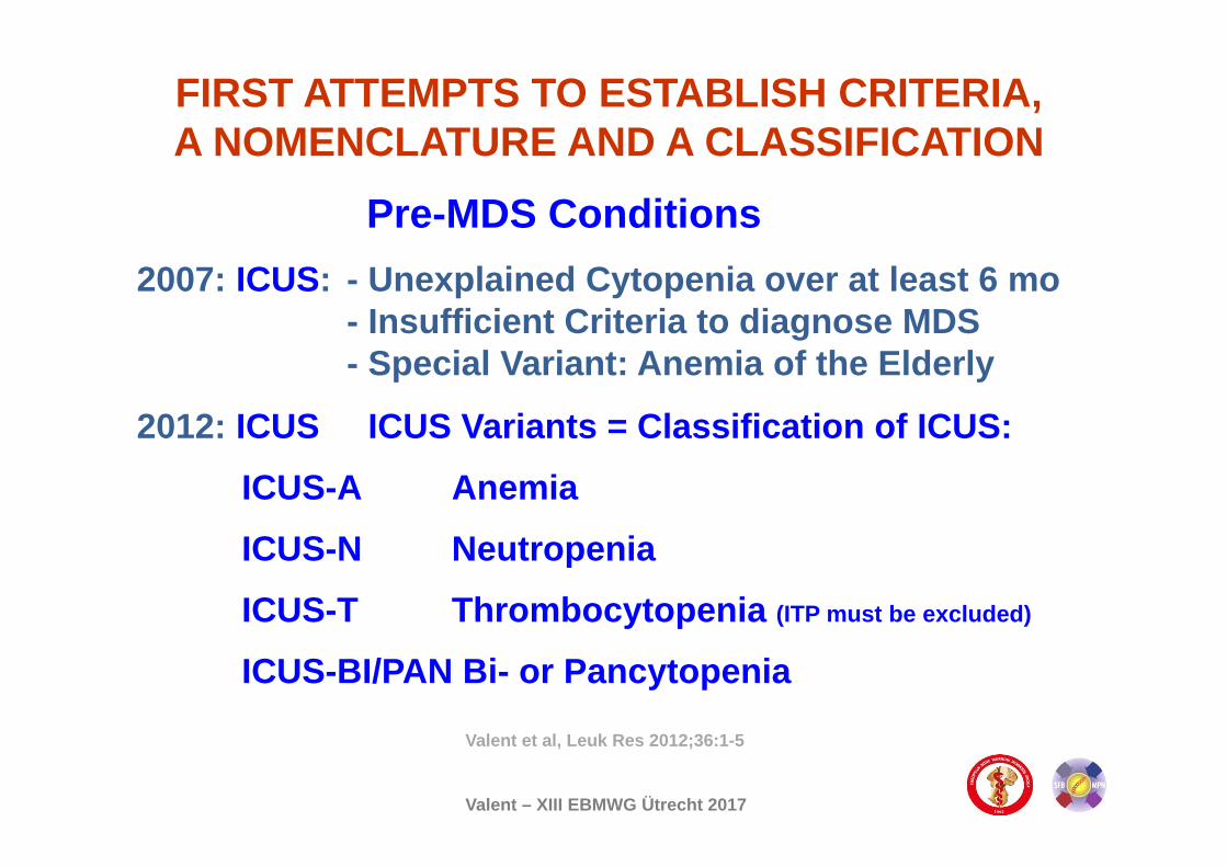
Pre-MDS Conditions 2007: ICUS: - Unexplained Cytopenia over at least 6 mo
- Insufficient Criteria to diagnose MDS- Special Variant: Anemia of the Elderly
2008: IDUS: - Unexplained Dysplasia over at least 6 mo- Insufficient Criteria to diagnose MDS
2012: CHIP: - Somatic Mutations in Myeloid Cells - No Cytopenia
2012: CCUS:- Somatic Mutations in Myeloid Cells- Cytopenia also present (often Anemia)- Insufficient Criteria to diagnose MDS
Valent – XIII EBMWG Ütrecht 2017
Valent et al, Leuk Res 2007 & 2012; Valent et al, Am J Cancer Res 2011, Steensma et al, Blood 2015
FIRST ATTEMPTS TO ESTABLISH CRITERIA, A NOMENCLATURE AND A CLASSIFICATION
Pre-MDS Conditions 2007: ICUS: - Unexplained Cytopenia over at least 6 mo
- Insufficient Criteria to diagnose MDS- Special Variant: Anemia of the Elderly
2012: ICUS ICUS Variants = Classification of ICUS:
ICUS-A Anemia
ICUS-N Neutropenia
ICUS-T Thrombocytopenia (ITP must be excluded)
ICUS-BI/PAN Bi- or Pancytopenia
Valent – XIII EBMWG Ütrecht 2017
Valent et al, Leuk Res 2012;36:1-5

Premalignant Neoplastic Stem CellsMalignant Neoplastic Stem Cells= Cancer Stem Cells = LSC
- Long Latency Periods (Decades)in early Phases of LSC Evolution
- Premalignant Neoplastic Stem Cellsversus Malignant SC = CSC/LSC
- Extensive Subclone Formation
- Each Subclone contains its own Stem Cell Compartment
- Phenotypic, Biochemical andFunctional Heterogeneity
- Different Mechanisms of Drug Resistance in Subclones
HIERARCHY AND SUBCLONE FORMATION FROM NEOPLASTICSTEM CELLS DURING EVOLUTION OF CANCER AND LEUKEMIA*
*Many Observations were made in the Paradigmatic CML Model, based on Evolution of Subclones carrying BCR/ABLMutations
Overt Cancer
MDS ?
Valent – XIII EBMWG Ütrecht 2017
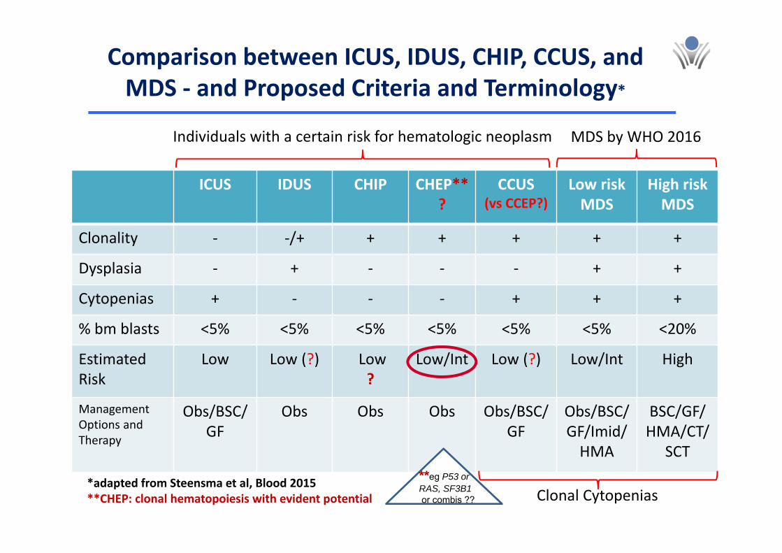
Comparison between ICUS, IDUS, CHIP, CCUS, and MDS ‐ and Proposed Criteria and Terminology*
ICUS IDUS CHIP CHEP**?
CCUS(vs CCEP?)
Low riskMDS
High riskMDS
Clonality ‐ ‐/+ + + + + +
Dysplasia ‐ + ‐ ‐ ‐ + +
Cytopenias + ‐ ‐ ‐ + + +
% bm blasts <5% <5% <5% <5% <5% <5% <20%
EstimatedRisk
Low Low (?) Low?
Low/Int Low (?) Low/Int High
ManagementOptions andTherapy
Obs/BSC/GF
Obs Obs Obs Obs/BSC/GF
Obs/BSC/GF/Imid/HMA
BSC/GF/HMA/CT/
SCT
MDS by WHO 2016Individuals with a certain risk for hematologic neoplasm
*adapted from Steensma et al, Blood 2015**CHEP: clonal hematopoiesis with evident potential Clonal Cytopenias
**eg P53 orRAS, SF3B1 or combis ??

Mutations detected in cases with CCUS & MDS
Kwok et al, Blood 2015
All pts MDS
DNMT3A 1.89% 11.4%TET2 0.35% 26.6%ASXL1 0.32% 17.6%JAK2 0.20% 3.6%GNB1 0.13% ‐TP53 0.13% 6.0%PPM1D 0.11% ‐SF3B1 0.10% 29.0%BCORL1 0.07% ‐SRSF2 0.06% 15.9%GNAS 0.06% ‐CBL 0.05% ‐MYD88 0.01% ‐U2AF1 0.01% 7.0%IDH2 0.01% 2.2%ATM 0.01% ‐
Sperr, MDS 2016

The real Question
Can we diagnose MDS in the absence of cytopenia ?
Can we diagnose MDS in individuals who have completely
normal blood counts ?
Valent – XIII EBMWG Ütrecht 2017

Definition of Diagnostic Cytopenia* regarding MDS and ICUS: proposal:
1) Simple Solution: WHO Cytopenia Definition
2) IWG-MDS (as WHO and: PLT = 100,000)
3) EU-US (2007) (Hb 11, ANC 1,500, PLT 100,000)
4) Other proposal ?*Must be persistent over at least 4 months
Valent – XIII EBMWG Ütrecht 2017
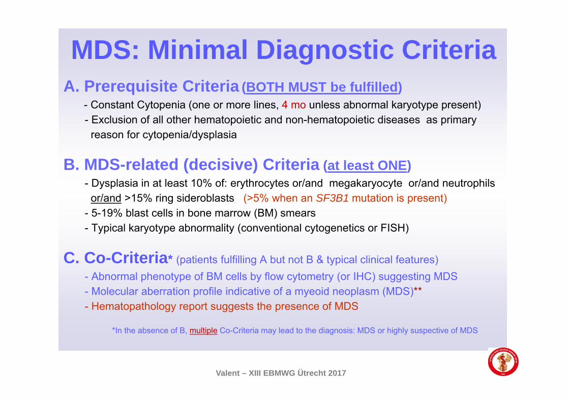
MDS: Minimal Diagnostic CriteriaA. Prerequisite Criteria (BOTH MUST be fulfilled)
- Constant Cytopenia (one or more lines, 4 mo unless abnormal karyotype present)- Exclusion of all other hematopoietic and non-hematopoietic diseases as primary
reason for cytopenia/dysplasia
B. MDS-related (decisive) Criteria (at least ONE)- Dysplasia in at least 10% of: erythrocytes or/and megakaryocyte or/and neutrophils
or/and >15% ring sideroblasts (>5% when an SF3B1 mutation is present) - 5-19% blast cells in bone marrow (BM) smears- Typical karyotype abnormality (conventional cytogenetics or FISH)
C. Co-Criteria* (patients fulfilling A but not B & typical clinical features)- Abnormal phenotype of BM cells by flow cytometry (or IHC) suggesting MDS- Molecular aberration profile indicative of a myeoid neoplasm (MDS)**- Hematopathology report suggests the presence of MDS
*In the absence of B, multiple Co-Criteria may lead to the diagnosis: MDS or highly suspective of MDS
Valent – XIII EBMWG Ütrecht 2017

MDS: Minimal Diagnostic CriteriaC. Co-Criteria* (patients fulfilling A but not B & typical clinical features)
- Abnormal phenotype of BM cells by flow cytometry (or IHC) suggesting MDS- Molecular aberration profile indicative of a myeoid neoplasm (MDS) - Hematopathology report suggests the presence of MDS
*In the absence of B, multiple Co-Criteria may lead to the diagnosis: MDS or highly suspective of MDS
OPEN QUESTIONS:- What combinations of co-criteria are most indicative of MDS?- What flow abnormalities qualify as MDS-related/specific?- What molecular markers (e.g. somatic mutations) qualify?- What allele burden is sufficient to count as a Co-Criterion of MDS?- What allele burden counts as Criterion of CHIP and CCUS (2%) ?- What assays (flow and molecular Seq) can be regarded standard? - Is there a standard algorithm for applying ´C´ co-criteria
Valent – XIII EBMWG Ütrecht 2017

ICUS: Minimal Diagnostic CriteriaA. Prerequisite Criteria (BOTH MUST be fulfilled)
- Constant Cytopenia (one or more lines, persisting for ≥4 mo)- Exclusion of hematopoietic and non-hematopoietic diseases including MDS
B. No MDS-related Criteria- No dysplasia in ≥10% of: erythrocytes or/and megakaryocyte or/and neutrophils
and <5% (<15%) ring sideroblasts (iron stain) and: - <5% blast cells in bone marrow (BM) smears and:- No karyotype abnormality by conventional cytogenetics and FISH
C. No Co-Criteria*- No abnormal (MDS-related) phenotype of BM cells by flow cytometry and IHC- No molecular features indicative of a myeloid neoplasm (MDS)[- No constantly reduced bm function (e.g. low CFU levels)]- Hematopathology report also excludes MDS
Valent – XIII EBMWG Ütrecht 2017
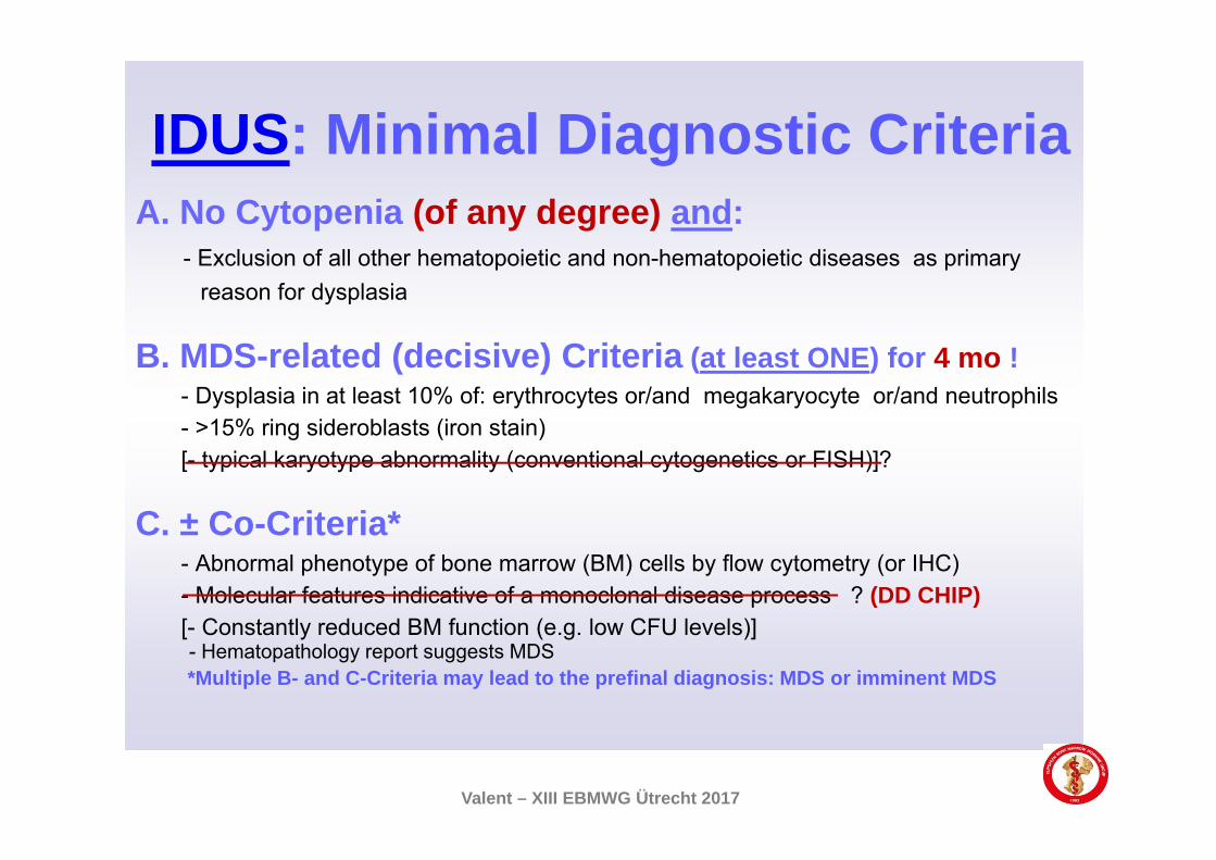
IDUS: Minimal Diagnostic CriteriaA. No Cytopenia (of any degree) and:
- Exclusion of all other hematopoietic and non-hematopoietic diseases as primaryreason for dysplasia
B. MDS-related (decisive) Criteria (at least ONE) for 4 mo !- Dysplasia in at least 10% of: erythrocytes or/and megakaryocyte or/and neutrophils- >15% ring sideroblasts (iron stain) [- typical karyotype abnormality (conventional cytogenetics or FISH)]?
C. ± Co-Criteria*- Abnormal phenotype of bone marrow (BM) cells by flow cytometry (or IHC)- Molecular features indicative of a monoclonal disease process ? (DD CHIP)[- Constantly reduced BM function (e.g. low CFU levels)]
*Multiple B- and C-Criteria may lead to the prefinal diagnosis: MDS or imminent MDS
Valent – XIII EBMWG Ütrecht 2017
- Hematopathology report suggests MDS

Thank You for Your AttentionPeter Valent and the M-Team in Vienna
Arock M.Germing U.Müllauer L.Bennett J.M.Bettelheim P.Lion T.Reiter A.Marian B.Holyoake T.Spittler A.
Akin C.Metcalfe D.D.Superti-Furga G.Grebien F.Hantschel O.Gotlib J.Fonatsch C.Schulenburg A.Triggiani M. Ogata K.
Horny H.P.Grunt T.Sotlar K.Mayerhofer M.Rülicke T.Willmann M.Ustun C.Rabitsch W.Orazi A.Zöchbauer S.
Zielinski C.C.Hartmann K.Pehamberger H.Moriggl R.Selzer E.Haase D.Valenta R.Pfeilstöcker M.Sexl V.Karlic H.
Geissler K.Ashman L.K.Van de Loosdrecht A.Haferlach T.Escribano L.Orfao A.Hoermann G.Mannhalter C.Eaves C.George T.
Cooperation Partners
E C N MEuropean Competence Network
on Mastocytosis
E C N MEuropean Competence Network
on Mastocytosis
Hadzijusufovic E. Neusiedler-N. J.Sonnleitner S. Herndlhofer S.Stefanzl G. Berger D.Blatt K. Keller A.Sadovnik I. Peter B.Bauer K. Eisenwort G.Herrmann H. Bauer K.Smiljković D. Müller N.Schneeweiss M. Gamperl S.Gleixner K.V. Füreder W.Wimazal F. Sperr W.R.
Valent – XIII EBMWG Ütrecht 2017