Pre-expanded Anterolateral Thigh Perforator Flap for Phalloplasty › uploads › general ›...
13
Pre-expanded Anterolateral Thigh Perforator Flap for Phalloplasty Salvatore D’Arpa, MD, PhD*, Britt Colebunders, MD, Filip Stillaert, MD, Stan Monstrey, MD, PhD INTRODUCTION Since the first description in 2005, 1 phalloplasty with a free or pedicled anterolateral thigh (ALT) flap has gradually gained popularity for penile reconstruction 2–14 as an alternative to the stan- dard radial forearm flap (RFF). 15 The main advantage of the ALT flap in this indi- cation is avoidance of the large forearm scar, which has become a recognizable sign of this operation because of the increasing attention received from the media. Very large flaps are needed for a phalloplasty and the donor site sub- sequently needs skin grafting. As a result, the donor site is quite noticeable because a hairless skin graft with a depression is left at the donor site. If the donor site is located in the forearm, it is not only quite visible and difficult to conceal un- less long sleeves are worn, but also a recognizable sign of the operation performed (Fig. 1). If an ALT flap is used for phalloplasty, the RFF donor site scars are avoided. However, a donor site scar will be present in the thigh, combined with the scars needed for skin graft harvest (Fig. 2). With the RFF and ALT, there is not only the flap donor site scar, but also the split thickness skin graft donor site, which is often more painful than the flap donor site itself. There is a particular subset of patients who want to avoid both scars because, although the thigh scars can be easily concealed whit a pair of shorts while dressed, they cannot be concealed when naked and are very close to the genital area. These patients would rather avoid disfigurement of the area that is the center of their masculinity and inti- macy. Pre-expansion of the ALT allows donor site scarring to be minimized in these patients (Fig. 3). TREATMENT GOALS AND PLANNED OUTCOMES Pre-expansion of a conventional ALT flap has 3 main goals: 1. Allowing primary donor site closure; 2. Improving the perforator’s vascular territory; 3. Thinning of the flap. Disclosure Statement: The authors have nothing to disclose. Department of Plastic and Reconstructive Surgery, Ghent University Hospital, De Pintelaan, 185, K12C, Gent 9000, Belgium * Corresponding author. E-mail address: [email protected] KEYWORDS Phalloplasty Skin expansion ALT flap LCFA flap Perforator flap Pre-expanded flap KEY POINTS The anterolateral thigh (ALT) perforator flap is a valuable alternative to the radial forearm flap for pa- tients who do not wish to have the forearm scar. ALT flap phalloplasty leaves visible scarring in the thighs owing to skin grafting of its donor site. Pre-expansion of an ALT flap allows primary donor site closure. Preoperative perforator location with computed tomography angiography is crucial to the success of the procedure. Clin Plastic Surg 44 (2017) 129–141 http://dx.doi.org/10.1016/j.cps.2016.08.004 0094-1298/17/Ó 2016 Elsevier Inc. All rights reserved. plasticsurgery.theclinics.com
Transcript of Pre-expanded Anterolateral Thigh Perforator Flap for Phalloplasty › uploads › general ›...

Pre-expandedAnterolateral ThighPerforator Flap for PhalloplastySalvatore D’Arpa, MD, PhD*, Britt Colebunders, MD,Filip Stillaert, MD, Stan Monstrey, MD, PhD
INTRODUCTION
Since the first description in 2005,1 phalloplastywith a free or pedicled anterolateral thigh (ALT)flap has gradually gained popularity for penilereconstruction2–14 as an alternative to the stan-dard radial forearm flap (RFF).15
The main advantage of the ALT flap in this indi-cation is avoidance of the large forearm scar,which has become a recognizable sign of thisoperation because of the increasing attentionreceived from the media. Very large flaps areneeded for a phalloplasty and the donor site sub-sequently needs skin grafting. As a result, thedonor site is quite noticeable because a hairlessskin graft with a depression is left at the donorsite. If the donor site is located in the forearm, itis not only quite visible and difficult to conceal un-less long sleeves are worn, but also a recognizablesign of the operation performed (Fig. 1).
If an ALT flap is used for phalloplasty, the RFFdonor site scars are avoided. However, a donorsite scar will be present in the thigh, combinedwith the scars needed for skin graft harvest
(Fig. 2). With the RFF and ALT, there is not onlythe flap donor site scar, but also the split thicknessskin graft donor site, which is often more painfulthan the flap donor site itself.
There is a particular subset of patients who wantto avoid both scars because, although the thighscars can be easily concealed whit a pair of shortswhile dressed, they cannot be concealed whennaked and are very close to the genital area. Thesepatients would rather avoid disfigurement of thearea that is the center of their masculinity and inti-macy. Pre-expansion of the ALT allows donor sitescarring to be minimized in these patients (Fig. 3).
TREATMENT GOALS AND PLANNEDOUTCOMES
Pre-expansion of a conventional ALT flap has 3main goals:
1. Allowing primary donor site closure;2. Improving the perforator’s vascular territory;3. Thinning of the flap.
Disclosure Statement: The authors have nothing to disclose.Department of Plastic and Reconstructive Surgery, Ghent University Hospital, De Pintelaan, 185, K12C, Gent9000, Belgium* Corresponding author.E-mail address: [email protected]
KEYWORDS
! Phalloplasty ! Skin expansion ! ALT flap ! LCFA flap ! Perforator flap ! Pre-expanded flap
KEY POINTS
! The anterolateral thigh (ALT) perforator flap is a valuable alternative to the radial forearm flap for pa-tients who do not wish to have the forearm scar.
! ALT flap phalloplasty leaves visible scarring in the thighs owing to skin grafting of its donor site.
! Pre-expansion of an ALT flap allows primary donor site closure.
! Preoperative perforator location with computed tomography angiography is crucial to the successof the procedure.
Clin Plastic Surg 44 (2017) 129–141http://dx.doi.org/10.1016/j.cps.2016.08.0040094-1298/17/! 2016 Elsevier Inc. All rights reserved. pl
astic
surgery.theclin
ics.com

In this particular application, the goal of preop-erative expansion of the ALT flap is achievingprimary donor site closure. Improving the perfora-tor’s vascular territory is not needed in this case.The flap measures 14 " 18 cm on average andsurvival is not an issue. Partial flap necrosis is avery uncommon occurrence, even without priorexpansion. Selection of the largest perforatorwith the aid of a preoperative computed tomogra-phy (CT) angiography warrants complete flapsurvival.Flap thinning would be extremely desirable and
was one of the goals we planned of pursuing whenwe first started expanding the ALT. Unfortunately,for this particular flap 2 expanders need to beplaced medially and laterally to the perforatorand expansion only results in a peripheral thinningof the flap with the flap’s fat being squeezed to-ward the perforator in the middle of the flap. Thiskind of deformation is of little use in a phalloplastybecause a lot of bulk is created in the middle of theflap, where it cannot be thinned out.The planned outcome of preoperative
ALT expansion in phalloplasty is to allow primarydonor site closure, avoid the disfiguring scar andthe painful skin graft donor site in the thigh.
PREOPERATIVE PLANNING ANDPREPARATION
Preoperative location of the perforator is crucial toflap planning. A CT angiography is used for thispurpose.10 The CT angiography allows the mostdistal perforator with the largest caliber, thelongest (to comfortably reach the pubis), with thebest subcutaneous branching and the mostconvenient intramuscular or septal course,providing a preoperative navigation that cannotbe obtained by simple Doppler location.The radiologist provides distances from the
anterior superior iliac spine based on an x–y axis(Fig. 4) drawn on the thigh and the position ofthe perforator is marked on the patient’s skin.The flap is drawn accordingly with the perforatorlying along its midline and close to its proximalmargin. Then the expander’s base (20 " 7 cm) isdrawn outside of the flap’s borders (Fig. 5)because, as described, placing the expanders inthe flap will squeeze the fat toward the midline,which is not desirable in this case.
PATIENT POSITIONING
The patient is placed in the supine position. Theipsilateral arm can be abducted or adducted
Fig. 1. Postoperative result of a radial forearm flap(RFF) phalloplasty showing the typical scar at thedonor site. Because this is the only application foruse of such a large RFF, this scar has become a recog-nizable sign of the operation, which not all patientslike to have. Scars in the thigh are also present dueto harvest of STSGs for coverage of the RFF donor site.
Fig. 2. Postoperative result of an anterolateral thigh(ALT)andsuperficial circumflex iliacperforator flapsphal-loplasty. Although concealable with regular clothing,when naked the scars in the thigh, owing to both ALTand split thickness skin graft harvest, are apparent.
D’Arpa et al130

based on the surgeon’s preference. Abduction willprovide greater room for the placement of thelateral expander because the hand, with the armadducted, comes in close proximity to the lateralincision and pocket.
PROCEDURAL APPROACHExpander Placement
Two remote “W” incisions16 are performed somecentimeters caudal to the inguinal ligament (seeFig. 5) and deepened to the deep fascia. Thenthe 2 pockets are dissected, bluntly or with thecautery, with the aid of a lighted retractor to obtainhemostasis. Care must be taken not to deepen theplane too much because the sensory nerves lie ontop of the fascia and they must not be damaged.Once the pocket is complete, a superficial(3–5 mm of fat left on the skin flap) pocket isdissected cranially to the incision to allow forremote port placement in a position that shall be
as easily accessible as possible (Fig. 6). Beforeexpander placement, two 12-F suction drains areplaced in the pocket. The air is emptied from theexpanders and they are partially filled with methy-lene blue–tinted saline, which allows easy visuali-zation of the fluid coming out of the expandersduring ambulatory postoperative expansion. Par-tial inflation keeps the expander distended and al-lows easy placement without folding (Fig. 7). Oncethe expander and ports are in position, easyaccessibility of the ports is double checked beforeclosure (Figs. 8 and 9).
Donor Site Closure
At the time of flap transfer (Fig. 10), the flap is har-vested first, with the expander left in place to main-tain skin stretch and inflated with extra 100 to150 mL to obtain some intraoperative expansion.
Donor site closure begins with expander andvalve removal through the easy access of thedefect left by the flap. Dissection is suprafascialand then on the plane of the deep capsule of theexpander, which will result in division of thecapsule into a superficial and a deep part. Whilethe deep part is not touched, the superficial isscored extensively to maximize the advancementof the skin flaps, extending the capsular incisionto the superficial fascia in a way very similar togaleal scoring in the scalp (Fig. 11). Then the flapsare brought together and temporarily held togetherwith skin staples. Two big dog ears will formdistally that are eventually resected, resulting ininverted “Y” or “T” scars (Figs. 12–16). Suctiondrains are placed underneath the flaps.
POTENTIAL COMPLICATIONS AND THEIRMANAGEMENT
There are no specific complications of ALT flappre-expansion; potential complications are thosecommonly related to tissue expansion. Like anyspecific body region, the anatomy accounts forsome peculiarities. In the thigh, the subcutaneousfat is quite dense, fibrous, and thick, and skinperforation and exposure is very unlikely.
We have observed 2 leaks from the inflationports that needed replacement likely owing topuncture with an exceedingly large needle. Infec-tion can be a complication and can be preventedwith appropriate technique. We have had an infec-tion when we associated liposuction to expanderplacement. Infection is treated with expanderremoval, culture-guided antibiotic therapy andexpander re-placement once the infection iscured. Placement without the aid of an CT angiog-raphy carries the risk of discontinuation of the pro-cedure because the right perforator is missed.
Fig. 3. Expanded anterolateral thigh and superficialcircumflex iliac perforator flaps phalloplasty donorsite, 5 months postoperatively. For comparison, hereis an early postoperative image of a phalloplasty afterexpansion. The donor site has been closed with an in-verted “Y” scar and no skin graft donor site is presentin the thigh. The scars are still red but already muchless disfiguring than those in Fig. 2.
Pre-expanded Anterolateral Thigh Perforator Flap 131

POSTPROCEDURAL CARE
Patients are immediately mobilized and dis-charged after drain removal. Expansions arebegun after 2 weeks and are usually performed
Fig. 4. An example of a preoperative computed tomography angiography. The sagittal (upper left), coronal (upperright), and axial (lower right) views of the perforator course together with a 3-dimensional reconstruction of theskin with the projection on the skin of the point of emergency from the fascia of the perforator (lower left), areprovided. In the upper right coronal view, the distances measured from the anterior superior iliac spine areprovided. Thus in a single image information about the course and position of the perforator are provided.
Fig. 5. Same patient as Fig. 4. A line is drawn connect-ing the anterior superior iliac spine (ASIS) to the upperlateral border of the patella. Using the angiographiccomputed tomographymeasurement in Fig. 4, the pro-jection on the skin of the perforator is marked with ablack, circledX, exactly 222mmbelowand32mm later-ally from the ASIS. Afterward the flap is drawn (blackrectangle). The skin projection of the 2 expanderpockets is drawn just lateral and medial to the flap tohave little overlapping with the flap once the ex-panders are inflated. The expander base and remoteports are drawn in green. The ports are placed in aneasily reachable position when the patient is lying su-pine. The “W” incisions (black) are placed in between.
Fig. 6. Medial view of a right thigh (the knee is on theleft hand side of the picture) at the time of expanderplacement. The pocket has already been dissectedthrough the “W” incision, which allows wider expo-sure with the same length compared with a linearincision. The drain is in place. Saline (150 mL) coloredwith methylene blue is injected in the expander afterall air has been removed. The expander is placed onthe skin in the same position that it will eventuallyhave inside the pocket.
D’Arpa et al132

weekly. The whole process usually takes 4 to6 months. It is thus initiated approximately6 months before phalloplasty.
Overexpansion is usually performed and isstopped until a circumference gain of at least14 cm has been obtained. During this period,especially when the expanders are fully inflated,physical activity is limited because the volume ofthe expanders restricts movement. Sports and ac-tivities that involve lower limb movements and thatare at risk for trauma to the thighs are restricted.The patients comply well and wear larger trousersto accommodate the inflated expanders.
REHABILITATION AND RECOVERY
Once the expanders are removed and the flaptransferred, recovery is relatively fast and no spe-cific rehabilitation is needed. Because of the phal-loplasty, the patients stay in bed for 10 days. When
allowed to walk, no specific problems have beenobserved.
OUTCOMES
The charts of 91 pedicled ALT flap phalloplastiesperformed between 2004 and2016were retrospec-tively reviewed. Nine patients (10%) underwent pre-expansion of the ALT flap in preparation for apedicled ALT flap phalloplasties. Seven patients
Fig. 7. Same view as in Fig. 6. The partial inflation ofthe expander facilitates insertion by keeping it dis-tended and avoiding folding.
Fig. 8. Same view as in Figs. 6 and 7. The remote portis inserted last. The pocket for the port is dissected in adifferent – more superficial – plane and with a bottle-neck to prevent the port from slipping back towardthe incision once inserted.
Fig. 9. Bird’s eye view, knee on the left hand side. Thefigure shows the 2 expanders with the procedurecompleted for the lateral one and to be completedfor the medial one, to show the 2 moments of place-ment of the needle in the port. The syringe on theright is connected to the medial port before closure.At this point, the port is probed to verify easy accessbefore closure, for eventual replacement. The syringeon the right has been used for a final inflation of theexpander after closure, to ensure obliteration of deadspace within the pocket to avoid fluid collection.
Fig. 10. Bird’s eye view, knees on the right hand side.Preoperative markings of a right pre-expanded ante-rolateral thigh flap. The CT scan showed 2 septal per-forators coming close to each other in this case. Thispicture shows how the expanders, although placedlaterally to the flap, eventually – with inflation – doexpand underneath the flap as well, causing some pe-ripheral thinning.
Pre-expanded Anterolateral Thigh Perforator Flap 133
wereoperated for female-to-malesex reassignmentsurgery, 1 for reconstruction after penile amputa-tion, and 1 for reconstruction after bladder extrophy(Table 1). Six patients had a preoperative CT scan.
Seven of 9 patients underwent eventual phallo-plasty with an ALT flap (see Table 1). In 1 patient(case 2 in Table 1) who had no preoperative CTangiography, no suitable perforator was identifiedand the procedure was converted in a free RFFphalloplasty. Another patient (case 8 in Table 1)had an infection that eventually forced removal ofthe implants. Because of this delay and of a signif-icant weight gain that caused fat thickening in the
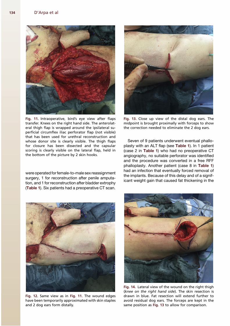
Fig. 11. Intraoperative, bird’s eye view after flapstransfer. Knees on the right hand side. The anterolat-eral thigh flap is wrapped around the ipsilateral su-perficial circumflex iliac perforator flap (not visible)that has been used for urethral reconstruction andwhose donor site is clearly visible. The thigh flapsfor closure has been dissected and the capsularscoring is clearly visible on the lateral flap, held inthe bottom of the picture by 2 skin hooks.
Fig. 12. Same view as in Fig. 11. The wound edgeshave been temporarily approximated with skin staplesand 2 dog ears form distally.
Fig. 13. Close up view of the distal dog ears. Themidpoint is brought proximally with forceps to showthe correction needed to eliminate the 2 dog ears.
Fig. 14. Lateral view of the wound on the right thigh(knee on the right hand side). The skin resection isdrawn in blue. Fat resection will extend further toavoid residual dog ears. The forceps are kept in thesame position as Fig. 13 to allow for comparison.
D’Arpa et al134

thigh, we abandoned the ALT flap phalloplasty andperformed an RFF phalloplasty instead.
In cases 2 and 3, there was a leak from theexpander inflation ports that required theirreplacement. The expanders did not deflate. Theleak was probably due to the use of a large needlefor inflation.
The only infection was observed in the patient inwhom we performed flap liposuction for thinningpurposes in the same operation as expanderplacement (case 8 in Table 1). Because it is only1 case, we cannot conclude that liposuction mightbe related to infection. More data are needed,although it seems that performing the 2 proced-ures simultaneously is better avoided if possible.
Donor site closure was primary with a doubledog ear resection distally that resulted in aninverted “T” or “Y” appearance in 6 cases andwith 2 opposed advancement flaps in 1 case. Afterflap harvest, all donor sites but one healed un-eventfully. This case (case 6 in Table 1) had 2advancement flaps for donor site closure, themedial of which had a partial necrosis with awound dehiscence that eventually required skingrafting. We would discourage use of these 2 flapsfor donor site closure and we would use 2 big rota-tion flaps—if flaps are needed because primaryclosure is not possible—instead.
There were no flap-related complications likepartial or total necroses.
In 2 cases (cases 1 and 2 in Table 1) lipofillingwas performed twice for correction of a contourdeformity in the thigh.
CASE DEMONSTRATIONSCase 1
A 36-year-old female to male transgender patientwas admitted for phalloplasty with a pre-expanded ALT flap combined with a free RFF forurethral reconstruction (Figs. 17–19; see Table 1,Case 5). Two rectangular expanders of 750 mLeach were implanted on the left thigh andexpanded with 1000 mL until a 14-cm circumfer-ence gain was achieved, which took 11 weeks.Seven months after expander placement, phallo-plasty was performed with the pre-expandedALT flap combined with a free RFF. The RFF wasanastomosed end to side to the femoral arteryand end-to-end to the greater saphenous vein.The pedicled ALT flap was transferred as a pedi-cled flap with 2 sensory nerves that were anasto-mosed to 1 ilio-inguinal nerve and to one of thedorsal clitoral nerves. The ALT flap donor sitewas closed primarily. Coronaplasty was per-formed 6 weeks after the initial operation. Erectileand testicular implants were placed 3 years afterthe operation. One year later, a minor correctionwas performed to reduce the penile size. At 5 yearsfollow-up the patient is doing fine.
Case 2
A 29-year-old female-to-male transgender patient(see Table 1, Case 9) was admitted for a
Fig. 15. Donor site after closure. The resulting scar isan inverted “Y”.
Fig. 16. Four years postoperative result. Despite somescar widening in the middle, the scar is little visible.No skin graft harvest scars are present. The inverted“T” can be seen distally above the knee.
Pre-expanded Anterolateral Thigh Perforator Flap 135
Table
1Patients
data
Patient
Diagnosis
FirstProcedure
Phalloplasty
DonorSiteClosu
reComplications
Date
Operated
(ALT
Flap)
Seco
ndary
Procedures
1FT
MExp
anderplacement
Previousphallo1
PedicledALT
Primary
None
10/2004
Lipofilling
2FT
MExp
anderplacement
PedicledALT
Primary
Exp
anderleakage
08/2009
Lipofilling
3BEX
Exp
anderplacement
RFF
(nogoodALT
perforators
NA
Exp
anderleakage
NA
None
4SC
CExp
anderplacement
PedicledALT
1freeRFF
Primary
None
06/2010
None
5FT
MExp
anderplacement
PedicledALT
1freeRFF
Primary
None
03/2011
None
6FT
MExp
anderplacement
PedicledALT
1pedicledSC
IAP
Adva
ncementflaps
Donorwounddehiscence
11/2015
None
7FT
MExp
anderplacement
PedicledALT
1pedicledSC
IAP
Primary
None
12/2015
None
8FT
MExp
anderplacement1
thinningbyliposuction
FreeRFF
(refusedALT
)NA
Infection
NA
None
9FT
MExp
anderplacement
PedicledALT
1pedicledSC
IAP
Primary
None
04/2016
None
Abbreviations:ALT,anterolateralthigh;BEX,bladderextrophy;
FTM,female
tomale;NA,notapplicable;RFF,radialforearm
flap;SC
C,squamouscellcarcinoma;SC
IAP,superficial
circumflexiliacperforatorflap.
D’Arpa et al136
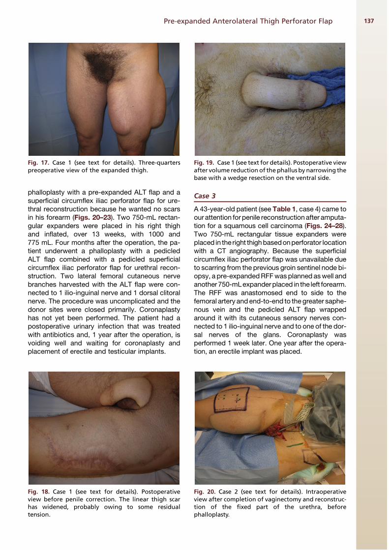
phalloplasty with a pre-expanded ALT flap and asuperficial circumflex iliac perforator flap for ure-thral reconstruction because he wanted no scarsin his forearm (Figs. 20–23). Two 750-mL rectan-gular expanders were placed in his right thighand inflated, over 13 weeks, with 1000 and775 mL. Four months after the operation, the pa-tient underwent a phalloplasty with a pedicledALT flap combined with a pedicled superficialcircumflex iliac perforator flap for urethral recon-struction. Two lateral femoral cutaneous nervebranches harvested with the ALT flap were con-nected to 1 ilio-inguinal nerve and 1 dorsal clitoralnerve. The procedure was uncomplicated and thedonor sites were closed primarily. Coronaplastyhas not yet been performed. The patient had apostoperative urinary infection that was treatedwith antibiotics and, 1 year after the operation, isvoiding well and waiting for coronaplasty andplacement of erectile and testicular implants.
Case 3
A 43-year-old patient (see Table 1, case 4) came toour attention for penile reconstruction after amputa-tion for a squamous cell carcinoma (Figs. 24–28).Two 750-mL rectangular tissue expanders wereplaced in the right thighbasedonperforator locationwith a CT angiography. Because the superficialcircumflex iliac perforator flap was unavailable dueto scarring from the previous groin sentinel node bi-opsy, a pre-expandedRFFwasplanned aswell andanother 750-mLexpanderplaced in the left forearm.The RFF was anastomosed end to side to thefemoral artery andend-to-end to the greater saphe-nous vein and the pedicled ALT flap wrappedaround it with its cutaneous sensory nerves con-nected to 1 ilio-inguinal nerve and to one of the dor-sal nerves of the glans. Coronaplasty wasperformed 1 week later. One year after the opera-tion, an erectile implant was placed.
Fig. 17. Case 1 (see text for details). Three-quarterspreoperative view of the expanded thigh.
Fig. 18. Case 1 (see text for details). Postoperativeview before penile correction. The linear thigh scarhas widened, probably owing to some residualtension.
Fig. 19. Case 1 (see text for details). Postoperative viewafter volume reductionof thephallus by narrowing thebase with a wedge resection on the ventral side.
Fig. 20. Case 2 (see text for details). Intraoperativeview after completion of vaginectomy and reconstruc-tion of the fixed part of the urethra, beforephalloplasty.
Pre-expanded Anterolateral Thigh Perforator Flap 137

DISCUSSION
The first case report of a pre-expanded free ALTcame from Tsai,17 who visualized the perforatorsthrough a large incision and placed a subfascialexpander. Other reports followed18–21 of its useas a free flap in burn wounds with the dual advan-tages of thinning the flap and closing the donor siteprimarily or reducing the skin graft and potentiallyincreasing vascularity. As described above, the in-crease in vascularity is not needed and the useful-ness of thinning the flap cannot be applied tophalloplasty. When the flap is thinned by expan-sion, the flap above the expander thins out butthe part where the pedicle enters the flap staysthicker.There are also some reports of pre-expanded
flaps in phalloplasty surgery with the RFF,22 thesuprapubic flap,23 or scapular flap.24 In thesecases, just a reduction of the area to be graftedand an insensate phallus are obtained, whereasa pre-expanded ALT flap always allows preserva-tion of flap’s innervation.Over the years, we have refined our technique to
optimize outcomes. A CT angiography is usedroutinely to accurately locate perforators and
expanders are placed accordingly. This way theunfortunate occurrence observed in patient 3 ofTable 1 is avoided (Fig. 29). Expansion is routinelycarried out until a circumference gain correspond-ing to the flap’s width is achieved. Closure is per-formed directly and a dog ear usually formsdistally, which is excised resulting in an inverted“T” or “Y” design. Patient are instructed on wearinglarge trousers to accommodate the bulk of the ex-panders. The flap can be farther thinned to the levelof the suprascarpal fat provided that the regionswere the nerves and the perforator lie are avoided.This thinning might sometimes result in a tempo-rary venous congestion (fast capillary refill) thatnormally subsides within 30 minutes. The ALTflap is harvested without any fascia and is besttunneled underneath the rectus femoris and sarto-rius muscles, and then through a wide subcutane-ous tunnel, to reach the pubic area. If anappropriate perforator is chosen based on CT angi-ography studies, the perforator is long enough totransfer a pedicled flap and avoid microsurgicalanastomoses. The patient is kept with his thighslightly bent to avoid any traction on the pedicle
Fig. 21. Case 2 (see text for details). Postoperativefrontal view showing 2 dog ears that will fade overtime and the contour deformity of the right thigh.
Fig. 22. Case 2 (see text for details). The left lateralview shows the linear scar of the superficial circumflexiliac perforator flap that extends far laterally toobtain an adequate pedicle length to reach the pubis.
D’Arpa et al138

for the first 3 days. Once the flap is tubed, any ten-sion must be avoided. If there is any tension thatmight cause flap compression with postoperativeedema, a skin graft is best placed ventrally to
relieve this tension and avoid vascularcompromise.
The presence of the expanders is cause ofdiscomfort, especially toward expander comple-tion, because the expanders hold a considerablevolume. Patients try to partially conceal the ex-panders by wearing very large trousers. It isindeed a procedure for a small group of patients(10%) in our series, who accept the presence ofthe expanders and the additional operation andambulatory inflations needed to reduce thedonor site scarring. These patients must bevery well-informed and have sufficient motivationbecause they have to put extra effort to go
Fig. 23. Case 2 (see text for details). Close up view ofthe external urinary meatus, made of the suture ofthe superficial circumflex iliac perforator and the an-terolateral thigh flaps.
Fig. 24. Case 3 (see text for details). Preoperative viewafter completion of tissue expansion in the forearmand thigh.
Fig. 25. Case 3 (see text for details). Left lateral intra-operative view. The belly is on the right hand side.The free radial forearm flap is wrapped around thedrain placed into the bladder through the urethraand the anterolateral thigh (ALT) flap is ready to bewrapped around it. The nerves ready for coaptationcan be seen coming from the ALT flap.
Fig. 26. Case 3 (see text for details). Intraoperativeview before expander removal. The fascia has beenclosed.
Pre-expanded Anterolateral Thigh Perforator Flap 139

through a longer process, additional operations,and extra costs.Also, it has to be pointed out that in this cases
reduction of scars is achieved at the expenses ofcontour. It has to be discussed with the patientthat indeed the patch like scars due to skingrafting will be avoided. But avoidance of scarscomes at the expenses of contour because thethinned expanded skin will cause a depressionand a contour deformity in the thigh thatwill need future lipofilling sessions to becorrected.
SUMMARY
Preoperative expansion is a valuable tool for mini-mizing donor site scarring in ALT flap phalloplastybecause it allows not only prevention of the un-sightly donor site graft, but also scarring and painrelated to a split thickness skin graft harvest. Preop-erative perforator locationwith a CT angiography al-lows minimally invasive expander placement.
REFERENCES
1. Ceulemans P. The Pedicled Anterolateral
Thigh Flap (ALT) Perforator Flap: A New Tech-
nique for Phallic Reconstruction. Paper pre-
sented at the XIV Biennial Symposium of the
Harry Benjamin International Gender Dysphoria
Association (HBIGDA). Bologna, Italy, April 6–9,
2005.
2. Felici N, Felici A. A new phalloplasty technique: the
free anterolateral thigh flap phalloplasty. J Plast Re-
constr Aesthet Surg 2006;59:153–7.
Fig. 27. Case 3 (see text for details). Three-quartersright postoperative view shows some widening anddiscoloration of the scar, not uncommon in peoplewith dark skin.
ValvesIncisionsExpander
Perforator
Flap
Fig. 29. This schematic drawingshows how incisions, expander,and valves shall be placed once theperforator is located by means of aCT scan. For more information,see Figs. 4 and 5 and the relativelegends, and the “preoperativeplanning and preparation” and“procedural approach: expanderplacement” sections.
Fig. 28. Case 3 (see text for details). Widening anddiscoloration are observed also in the forearm skin.
D’Arpa et al140

3. Mutaf M, Isik D, Bulut O, et al. A true nonmicrosurgi-
cal technique for total phallic reconstruction. Ann
Plast Surg 2006;57:100–6.
4. Lumen N, Monstrey S, Selvaggi G, et al. Phallo-
plasty: a valuable treatment for males with penile
insufficiency. Urology 2008;71(2):272–6.
5. Lumen N, Monstrey S, Ceulemans P, et al. Recon-
structive surgery for severe penile inadequacy:
phalloplasty with a free radial forearm flap or a pedi-
cled anterolateral thigh flap. Adv Urol 2008;704343.
6. Descamps MJ, Hayes PM, Hudson DA. Phalloplasty
in complete aphallia: pedicled anterolateral thigh
flap. J Plast Reconstr Aesthet Surg 2009;62(3):e51–4.
7. Lee GK, Lim AF, Bird ET. A novel single-flap tech-
nique for total penile reconstruction: the pedicled
anterolateral thigh flap. Plast Reconstr Surg 2009;
124(1):163–6.
8. Rubino C, Figus A, Dessy LA, et al. Innervated island
pedicled anterolateral thigh flap for neo-phallic
reconstruction in female-to-male transsexuals.
J Plast Reconstr Aesthet Surg 2009;62(3):e45–9.
9. Rashid M, Aslam A, Malik S, et al. Clinical applica-
tions of the pedicled anterolateral thigh flap in penile
reconstruction. J Plast Reconstr Aesthet Surg 2011;
64(8):1075–81.
10. Sinove Y, Kyriopoulos E, Ceulemans P, et al. Preop-
erative planning of a pedicled anterolateral thigh
(ALT) flap for penile reconstruction with the multide-
tector CT scan. Handchir Mikrochir Plast Chir 2013;
45(4):217–22.
11. Liu CY, Wei ZR, Jiang H, et al. Preconstruction of the
pars pendulans urethrae for phalloplasty with diges-
tive mucosa using a prefabricated anterolateral
thigh flap in a one-arm patient. Plast Reconstr
Surg Glob Open 2013;1(7):e53.
12. Holzbach T, Giunta RE, Machens HG, et al. Phallo-
plasty with pedicled anterolateral thigh flap (“ALT-
Flap”). Handchir Mikrochir Plast Chir 2011;43(4):
227–31 [in German].
13. Hasegawa K, Namba Y, Kimata Y. Phalloplasty with
an innervated island pedicled anterolateral thigh
flap in a female-to-male transsexual. Acta Med
Okayama 2013;67(5):325–31 [Erratum appears in
Acta Med Okayama 2014;68(3):183].
14. Morrison SD, Son J, Song J, et al. Modification of the
tube-in-tube pedicled anterolateral thigh flap for to-
tal phalloplasty: the mushroom flap. Ann Plast Surg
2014;72(Suppl 1):S22–6.
15. Monstrey S, Hoebeke P, Selvaggi G, et al. Penile
reconstruction: is the radial forearm flap really the
standard technique? Plast Reconstr Surg 2009;
124:510–8.
16. Matton GE, Tonnard PL, Monstrey SJ, et al.
A universal incision for tissue expander insertion.
Br J Plast Surg 1995;48(3):172–6.
17. Tsai FC. A new method: perforator-based tissue
expansion for a preexpanded free cutaneous perfo-
rator flap. Burns 2003;29:845–8.
18. Hallock GG. The preexpanded anterolateral thigh
free flap. Ann Plast Surg 2004;53:170–3.
19. HallockGG. Tissue expansion techniques tominimize
morbidity of the anterolateral thigh perforator flap
donor site. J Reconstr Microsurg 2013;29(9):565–70.
20. Acarturk TO. Aesthetic reconstruction of the postburn
neck contracture with a preexpanded anterolateral
thigh free flap. J Craniofac Surg 2014;25(1):e23–6.
21. Hocao!glu E, Arıncı A, Berkoz O, et al. Free
pre-expanded lateral circumflex femoral artery
perforator flap for extensive resurfacing and recon-
struction of the hand. J Plast Reconstr Aesthet
Surg 2013;66(12):1788–91.
22. Solinc M, Kosutic D, Stritar A, et al. Preexpanded
radial forearm free flap for one-stage total penile
reconstruction in female-to-male transsexuals.
J Reconstr Microsurg 2009;25(6):395–8.
23. Terrier JE, Courtois F, Ruffion A, et al. Surgical out-
comes and patients’ satisfaction with suprapubic
phalloplasty. J Sex Med 2014;11(1):288–98.
24. Dong L, Dong Y, He L, et al. Penile reconstruction by
preexpanded free scapular flap in severely burned
patient. Ann Plast Surg 2014;73(Suppl 1):S27–30.
Pre-expanded Anterolateral Thigh Perforator Flap 141