Practice Guidelines in Oncology - WordPress.com...Added a bullet "Optional FDG PET/CT (for T3,N1,...
147
Version 2.2011, 01/05/11 © National Comprehensive Cancer Network, Inc. 2011,All rights reserved. The NCCN Guidelines™ and this illustration may not be reproduced in any form without the express written permission of NCCN®. Continue NCCN Clinical Practice Guidelines in Oncology™ Breast Cancer NCCN.org Continue Version 2.2011 NCCN Guidelines for Patients™ available at www.nccn.com
Transcript of Practice Guidelines in Oncology - WordPress.com...Added a bullet "Optional FDG PET/CT (for T3,N1,...

NCCN®
NCCN Guidelines™ Version 2.2011
Version 2.2011, 01/05/11 © National Comprehensive Cancer Network, Inc. 2011, All rights reserved. The NCCN Guidelines™ and this illustration may not be reproduced in any form without the express written permission of NCCN®.
NCCN Guidelines IndexBreast Cancer Table of Contents
Staging, Discussion
Continue
NCCN Clinical Practice Guidelines in Oncology™
Breast Cancer
NCCN.org
Continue
Version 2.2011
NCCN Guidelines for Patients™ available at www.nccn.com
NCCN®
NCCN Guidelines™ Version 2.2011
Version 2.2011, 01/05/11 © National Comprehensive Cancer Network, Inc. 2011, All rights reserved. The NCCN Guidelines™ and this illustration may not be reproduced in any form without the express written permission of NCCN®.
NCCN Guidelines IndexBreast Cancer Table of Contents
Staging, Discussion
Robert W. Carlson, MD/Chair †Stanford Comprehensive Cancer Center
D. Craig Allred, MD
†
�
Siteman Cancer Center at Barnes-JewishHospital and Washington University School ofMedicine
Benjamin O. Anderson, MD ¶Fred Hutchinson Cancer ResearchCenter/Seattle Cancer Care Alliance
Harold J. Burstein, MD, PhD †Dana-Farber/Brigham and Women's CancerCenter
W. Bradford Carter, MD ¶H. Lee Moffitt Cancer Center & ResearchInstitute
Stephen B. Edge, MD ¶Roswell Park Cancer Institute
John K. Erban, MDMassachusetts General Hospital Cancer Center
William B. Farrar, MD ¶Arthur G. James Cancer Hospital & Richard J.Solove Research Institute at The Ohio StateUniversity
Andres Forero, MDUniversity of Alabama at BirminghamComprehensive Cancer Center
‡
Lori J. Pierce, MD §University of Michigan ComprehensiveCancer Center
Elizabeth C. Reed, MD †UNMC Eppley Cancer Center at TheNebraska Medical Center
Jasgit Sachdev, MD †St. Jude Children's ResearchHospital/University of Tennessee CancerInstitute
Mary Lou Smith, JD, MBA ¥Consultant
George Somlo, MD ‡City of Hope
John H. Ward, MD ‡Huntsman Cancer Institute at the Universityof Utah
Antonio C. Wolff, MD †The Sidney Kimmel Comprehensive CancerCenter at Johns Hopkins University
Richard Zellars, MD §The Sidney Kimmel Comprehensive CancerCenter at Johns Hopkins University
�
�
Sharon Hermes Giordano, MD MPH †The University of Texas M.D. AndersonCancer Center
Lori J. Goldstein, MD †Fox Chase Cancer Center
William J. Gradishar, MD ‡Robert H. Lurie Comprehensive CancerCenter of Northwestern University
Daniel F. Hayes, MD †University of Michigan ComprehensiveCancer Center
Clifford A. Hudis, MD †Memorial Sloan-Kettering Cancer Center
Britt-Marie Ljung, MDUCSF Helen Diller FamilyComprehensive Cancer Center
David A. Mankoff, MD, PhDFred Hutchinson Cancer ResearchCenter/Seattle Cancer Care Alliance
P. Kelly Marcom, MD †Duke Comprehensive Cancer Center
Ingrid A. Mayer, MDVanderbilt-Ingram Cancer Center
Beryl McCormick, MD §Memorial Sloan-Kettering Cancer Center
�
�
†
† Medical Oncology‡ Hematology/Oncology¶ Surgical Oncology
Pathology�
Ÿ Reconstructive Surgery
§ Radiation Oncology
Bone Marrow Transplantation¥ Patient Advocacy* Writing Committee Member
� Nuclear medicine
�
Breast CancerPanel Members
ContinueContinueContinue
Printed by paolo nitti on 2/3/2011 12:45:49 PM. For personal use only. Not approved for distribution. Copyright © 2011 National Comprehensive Cancer Network, Inc., All Rights Reserved.

NCCN®
NCCN Guidelines™ Version 2.2011
Version 2.2011, 01/05/11 © National Comprehensive Cancer Network, Inc. 2011, All rights reserved. The NCCN Guidelines™ and this illustration may not be reproduced in any form without the express written permission of NCCN®.
NCCN Guidelines IndexBreast Cancer Table of Contents
Staging, Discussion
NCCN Breast Cancer Panel Members
Summary of Guidelines Updates
Lobular Carcinoma In Situ (LCIS-1)
Ductal Carcinoma In Situ (DCIS-1)
Clinical Stage, Workup (BINV-1)
Locoregional Treatment of Clinical Stage l, llA,or llB Disease or T3,N1,M0 (BINV-2)
Systemic Adjuvant Treatment (BINV-4)
Preoperative Chemotherapy Guideline
Clinical Stage llA, llB, Workup (BINV-10)
Primary Treatment, Adjuvant Treatment(BINV-11)
Clinical Stage lllA, lllB, lllC, and Stage IV,Workup (BINV-13)
Preoperative Chemotherapy, LocoregionalTreatment, Adjuvant Treatment (BINV-14)
Surveillance/Follow-Up, Recurrence Workup or
Initial Workup for Stage lV Disease (BINV-15)
Treatment of Recurrence/Stage IV Disease
(BINV-16)
Principles of HER2 Testing (BINV-A)
Principles of Dedicated Breast MRI Testing
(BINV-B)
�
�
�
�
�
�
Noninvasive Breast Cancer
Invasive Breast Cancer
�
�
�
�
�
�
�
�
These guidelines are a statement of evidence and consensus of the authors regarding their views of currently accepted approaches to treatment. Anyclinician seeking to apply or consult these guidelines is expected to use independent medical judgment in the context of individual clinical circumstancesto determine any patient's care or treatment. The National Comprehensive Cancer Network makes no representations nor warranties of any kindwhatsoever regarding their content, use, or application and disclaims any responsibility for their application or use in any way. These guidelines arecopyrighted by National Comprehensive Cancer Network. All rights reserved. These guidelines and the illustrations herein may not be reproduced in anyform without the express written permission of NCCN. ©2011.
Clinical Trials:
Categories of Evidence andConsensus:NCCN
Thebelieves that the best managementfor any cancer patient is in a clinicaltrial. Participation in clinical trials isespecially encouraged.
To find clinical trials online at NCCNmember institutions,
All recommendationsare Category 2A unless otherwisespecified.
See
NCCN
click here:nccn.org/clinical_trials/physician.html
NCCN Categories of Evidenceand Consensus
Invasive Breast Cancer (continued)
Special Considerations
�
�
�
�
�
�
�
Fertility and Birth Control After Adjuvant
Breast Cancer (BINV-C)
Surgical Axillary Staging - Stage l, llA ,
and llB (BINV-D)
Axillary Lymph Node Staging (BINV-E)
Margin Status in Infiltrating Carcinoma(BINV-F)
Special Considerations to Breast-Conserving Therapy Requiring RadiationTherapy (BINV-G)
Principles of Breast ReconstructionFollowing Mastectomy (BINV-H)
Principles of Radiation Therapy (BINV-I)
Adjuvant Endocrine Therapy (BINV-J)
Adjuvant Chemotherapy (BINV-K)
Definition of Menopause (BINV-L)
Subsequent Endocrine Therapy (BINV-M)
Preferred Chemotherapy Regimens forRecurrent or Metastatic Breast Cancer(BINV-N)
�
�
�
�
�
�
�
�
�
Phyllodes Tumor (PHYLL-1)
Paget’s Disease (PAGET-1)
Breast Cancer During Pregnancy (PREG-1)
Inflammatory Breast Cancer (IBC-1)
Breast Cancer
NCCN Guidelines for Patients™available at www.nccn.com
Printed by paolo nitti on 2/3/2011 12:45:49 PM. For personal use only. Not approved for distribution. Copyright © 2011 National Comprehensive Cancer Network, Inc., All Rights Reserved.

NCCN®
NCCN Guidelines™ Version 2.2011
Version 2.2011, 01/05/11 © National Comprehensive Cancer Network, Inc. 2011, All rights reserved. The NCCN Guidelines™ and this illustration may not be reproduced in any form without the express written permission of NCCN®.
NCCN Guidelines IndexBreast Cancer Table of Contents
Staging, Discussion
UPDATES
Breast CancerUpdates
Note: All recommendations are category 2A unless otherwise indicated.Clinical Trials: NCCN believes that the best management of any cancer patient is in a clinical trial. Participation in clinical trials is especially encouraged.
Summary of the changes in the 2.2011 version of the Breast Cancer guidelines from the 1.2011 version include:BINV-17
BINV-N
DISCUSSION
�
�
�
�
�
If bone disease present, deleted "Add bisphosphonate" and replaced with "Add denosumab, zoledronic acid, or pamidronate."Modified footnote ee: "Denosumab, zoledronic acid, or pamidronate (all with calcium and vitamin D supplementation) should be given(category 1) in addition to chemotherapy or endocrine therapy if bone metastasis present, expected survival 3 months, and renalfunction is adequate. Patients should undergo a dental examination with preventive dentistry prior to initiation of this therapy. Theoptimal schedule and duration of denosumab, zoledronic acid, or pamidronate are unknown."
Added eribulin to list of preferred single agents, other microtubule inhibitors. Eribulin 1.4 mg/m IV days 1, 8 Cycled every 21 days.
The discussion section has been updated to reflect the changes in the algorithm.
2
LCIS-1
DCIS-1
BINV-1
BINV-2See Fertility and Birth Control After Adjuvant Breast Cancer Treatment (BINV-C)
�
�
�
�
�
�
�
�
�
�
�
�
Deleted "observation" from primary treatment, also removed "risk reduction with tamoxifen for premenopausal women, or with tamoxifen or raloxifene forpostmenopausal women."Deleted "In special circumstances, bilateral mastectomy ± reconstruction may be considered for risk reduction."Added pathway based on type of biopsy, core or surgical excision. If the initial biopsy was core needle biopsy the recommendation is for surgical excision.Added pathways for DCIS and invasive cancer based on surgical biopsy results. Recommend following the appropriate guideline.Added pathway for LCIS based on surgical biopsy results. Recommend counseling regarding risk reduction and surveillance per NCCN Breast Cancer RiskReduction Guidelines and NCCN Breast Cancer Screening and Diagnosis Guidelines.
Modified footnote k: "
Added a bullet "Optional FDG PET/CT (for T3,N1, M0) (category 2B)."Added a new footnote "The use of PET or PET/CT scanning is not indicated in the staging of clinical stage I, II, or operable III breast cancer. FDG PET/CT ismost helpful in situations where standard staging studies are equivocal or suspicious, especially in the setting of locally advanced or metastatic disease.FDG PET/CT may also be helpful in identifying unsuspected regional nodal disease and/or distant metastases in LABC when used in addition to standardstaging studies.Added "Consider fertility counseling if indicated."Added a new footnote " .
Added "infraclavicular region" to supraclavicula area for radiation recommendation. (also applies to BINV-3)Following the pathway for negative axillary nodes, added "or consideration of partial breast irradiation (PBI) in selected patients "
Whole breast radiation therapy following lumpectomy reduces recurrence rates in DCIS by about 50%. Approximately half of therecurrences are invasive and half DCIS. A number of factors determine that local recurrence risk; palpable mass, larger size, higher grade, close orinvolved margins, and age under 50 years. If the patient and physician view the individual risk as “low”, some patients may be treated by excision alone. Alldata evaluating the three local treatments show no differences in patient survival."
.Added a footnote: "PBI may be administered prior to chemotherapy."
�
Summary of the changes in the 1.2011 version of the Breast Cancer guidelines from the 3.2010 version include:
Continued on the next page
Printed by paolo nitti on 2/3/2011 12:45:49 PM. For personal use only. Not approved for distribution. Copyright © 2011 National Comprehensive Cancer Network, Inc., All Rights Reserved.
NCCN®
NCCN Guidelines™ Version 2.2011
Version 2.2011, 01/05/11 © National Comprehensive Cancer Network, Inc. 2011, All rights reserved. The NCCN Guidelines™ and this illustration may not be reproduced in any form without the express written permission of NCCN®.
NCCN Guidelines IndexBreast Cancer Table of Contents
Staging, Discussion
BINV-5
BINV-6
See Fertility and Birth Control After Adjuvant Breast Cancer Treatment (BINV-C)BINV-13
BINV-7
BINV-10 BINV-14
BINV-15
BINV-16
BINV-20
�
�
�
�
�
�
�
�
�
�
�
�
�
�
Removed "grade 1."Removed "grade 2 or 3, unfavorable features."Removed "± trastuzumab (category 3)."Deleted footnote r: "If ER-positive consider endocrine therapy for risk reduction and to diminish the small risk of disease recurrence.Following the pathway for pN0, added "consider adjuvant endocrine therapy."Following the pathway for pN1mi, added "adjuvant endocrine therapy ± adjuvant chemotherapy + trastuzumab."
Removed "grade 1, no unfavorable features."
Added a bullet "FDG PET/CT (category 2B)."Added a new footnote "The use of PET or PET/CT scanning is not indicated in the staging of clinical stage I, II, or operable III breast cancer. FDG PET/CT ismost helpful in situations where standard staging studies are equivocal or suspicious, especially in the setting of locally advanced or metastatic disease.FDG PET/CT may also be helpful in identifying unsuspected regional nodal disease and/or distant metastases in LABC when used in addition to standardstaging studies.Added "Consider fertility counseling if indicated."Added a new footnote " .
Changed "Mammogram every 12 mo (and 6-12 mo post-radiation therapy if breast conserved [category 2B])" to "annual mammography."Added "Evidence suggests that active lifestyle, achieving and maintaining an ideal body weight (20-25 BMI) may lead to optimal breast cancer outcomes."Added "First recurrence of disease should be biopsied."Modified footnote bb: "The use of estrogen, progesterone, or selective estrogen receptor modulators to treat osteoporosis or osteopenia in women withbreast cancer is discouraged. The use of a bisphosphonate is generally the preferred intervention to improve bone mineral density. Optimal duration ofbisphosphonate therapy has not been established. Factors to consider for duration of anti-osteoporosis therapy include bone mineral density, response totherapy, and risk factors for continued bone loss or fracture. Women treated with a bisphosphonate should undergo a dental examination with preventivedentistry prior to the initiation of therapy, and should take supplemental calcium and vitamin D."
When prior therapy with anthracycline, taxane, and trastuzumab: capecitabine + lapatinib (preferred). "Replaced with: "Continue HER2 targetedtherapy, typically in combination with other chemotherapy or trastuzumab + lapatinib."
�
�
�
�
Changed recommendation for pN1mi and tumors 0.6-1.0 cm to "Consider adjuvant chemotherapy + trastuzumab." Removed the category 3 designation, it isnow a category 2A recommendation.
Removed "If capecitabine administered as a radiation sensitizer, trastuzumab may be given concurrent with the capecitabine."
Preoperative chemotherapy ,deleted "anthracycline ± taxane preferred."
Removed "
(also applies to )�
Breast CancerUpdates
UPDATES
Note: All recommendations are category 2A unless otherwise indicated.Clinical Trials: NCCN believes that the best management of any cancer patient is in a clinical trial. Participation in clinical trials is especially encouraged.
Continued on the next page
Printed by paolo nitti on 2/3/2011 12:45:49 PM. For personal use only. Not approved for distribution. Copyright © 2011 National Comprehensive Cancer Network, Inc., All Rights Reserved.
NCCN®
NCCN Guidelines™ Version 2.2011
Version 2.2011, 01/05/11 © National Comprehensive Cancer Network, Inc. 2011, All rights reserved. The NCCN Guidelines™ and this illustration may not be reproduced in any form without the express written permission of NCCN®.
NCCN Guidelines IndexBreast Cancer Table of Contents
Staging, Discussion
BINV-B
BINV-D
BINV-I
BINV-C
�
�
�
�
�
Added the following bullet: "The utility of MRI in follow-up screening of women with prior breast cancer is undefined. It should generally be consideredonly in those whose lifetime risk of a second primary breast cancer is greater than 20% based on models largely dependent on family history, such asin those with the risk associated with inherited susceptibility of breast cancer."
Is a new page: Fertility And Birth Control After Adjuvant Breast Cancer Treatment.
Added a new footnote"Data from a single, randomized trial suggests that complete axillary lymph node dissection in women with clinically nodenegative T1-T2 tumors, fewer than 3 involved sentinel lymph nodes, and undergoing breast-conserving surgery and whole breast radiation results inmore morbidity, no improvement in locoregional recurrence rates, and no difference in overall survival compared with sentinel lymph node procedurealone."
Modified paragraph discussing whole breast radiation.Added paragraph for Accelerate Partial Breast Irradiation (APBI). Also added a paragraph for Optimizing Delivery and Individualized Therapy.
Breast CancerUpdates
UPDATES
Note: All recommendations are category 2A unless otherwise indicated.Clinical Trials: NCCN believes that the best management of any cancer patient is in a clinical trial. Participation in clinical trials is especially encouraged.
Printed by paolo nitti on 2/3/2011 12:45:49 PM. For personal use only. Not approved for distribution. Copyright © 2011 National Comprehensive Cancer Network, Inc., All Rights Reserved.
NCCN®
NCCN Guidelines™ Version 2.2011
Version 2.2011, 01/05/11 © National Comprehensive Cancer Network, Inc. 2011, All rights reserved. The NCCN Guidelines™ and this illustration may not be reproduced in any form without the express written permission of NCCN®.
NCCN Guidelines IndexBreast Cancer Table of Contents
Staging, Discussion
Note: All recommendations are category 2A unless otherwise indicated.Clinical Trials: NCCN believes that the best management of any cancer patient is in a clinical trial. Participation in clinical trials is especially encouraged.
LCIS-1
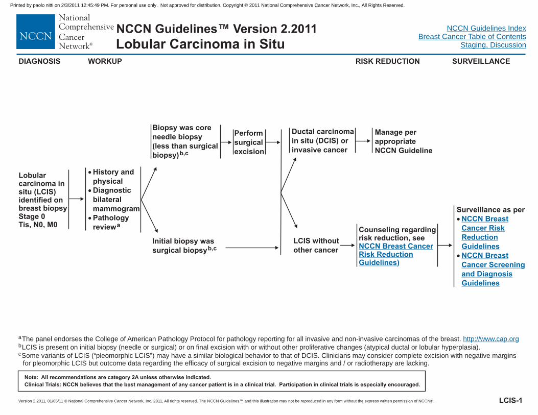
Lobular Carcinoma in SituWORKUPDIAGNOSIS RISK REDUCTION SURVEILLANCE
Lobularcarcinoma insitu (LCIS)identified onbreast biopsyStage 0Tis, N0, M0
�
�
�
History andphysicalDiagnosticbilateralmammogramPathologyreviewa
Counseling regardingrisk reduction, seeNCCN Breast CancerRisk ReductionGuidelines)
aThe panel endorses the College of American Pathology Protocol for pathology reporting for all invasive and non-invasive carcinomas of the breast.b
cLCIS is present on initial biopsy (needle or surgical) or on final excision with or without other proliferative changes (atypical ductal or lobular hyperplasia).
Some variants of LCIS (“pleomorphic LCIS”) may have a similar biological behavior to that of DCIS. Clinicians may consider complete excision with negative marginsfor pleomorphic LCIS but outcome data regarding the efficacy of surgical excision to negative margins and / or radiotherapy are lacking.
http://www.cap.org
Biopsy was coreneedle biopsy(less than surgicalbiopsy)b,c
Initial biopsy wassurgical biopsyb,c
Performsurgicalexcision
Ductal carcinomain situ (DCIS) orinvasive cancer
LCIS withoutother cancer
Surveillance as per�
�
NCCN BreastCancer RiskReductionGuidelinesNCCN BreastCancer Screeningand DiagnosisGuidelines
Manage perappropriateNCCN Guideline
Printed by paolo nitti on 2/3/2011 12:45:49 PM. For personal use only. Not approved for distribution. Copyright © 2011 National Comprehensive Cancer Network, Inc., All Rights Reserved.
NCCN®
NCCN Guidelines™ Version 2.2011
Version 2.2011, 01/05/11 © National Comprehensive Cancer Network, Inc. 2011, All rights reserved. The NCCN Guidelines™ and this illustration may not be reproduced in any form without the express written permission of NCCN®.
NCCN Guidelines IndexBreast Cancer Table of Contents
Staging, Discussion
Note: All recommendations are category 2A unless otherwise indicated.Clinical Trials: NCCN believes that the best management of any cancer patient is in a clinical trial. Participation in clinical trials is especially encouraged.
DCIS-1
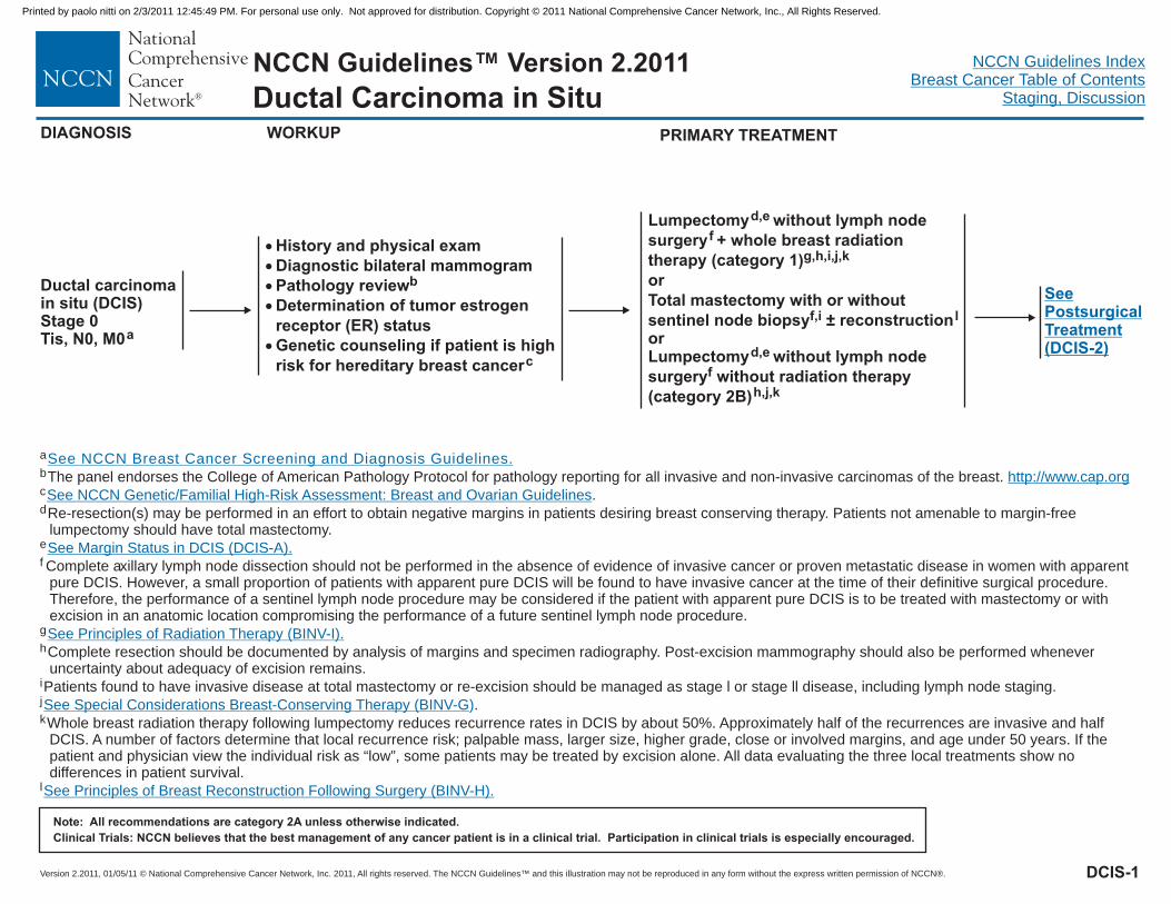
Ductal Carcinoma in SituWORKUPDIAGNOSIS
Ductal carcinomain situ (DCIS)Stage 0Tis, N0, M0a
�
�
�
�
�
History and physical examDiagnostic bilateral mammogramPathology reviewDetermination of tumor estrogenreceptor (ER) statusGenetic counseling if patient is highrisk for hereditary breast cancer
b
c
a
c
e
b
d
g
k
The panel endorses the College of American Pathology Protocol for pathology reporting for all invasive and non-invasive carcinomas of the breast.
.
Re-resection(s) may be performed in an effort to obtain negative margins in patients desiring breast conserving therapy. Patients not amenable to margin-freelumpectomy should have total mastectomy.
Whole breast radiation therapy following lumpectomy reduces recurrence rates in DCIS by about 50%. Approximately half of the recurrences are invasive and halfDCIS. A number of factors determine that local recurrence risk; palpable mass, larger size, higher grade, close or involved margins, and age under 50 years. If thepatient and physician view the individual risk as “low”, some patients may be treated by excision alone. All data evaluating the three local treatments show nodifferences in patient survival.
f
h
i
j
Complete axillary lymph node dissection should not be performed in the absence of evidence of invasive cancer or proven metastatic disease in women with apparentpure DCIS. However, a small proportion of patients with apparent pure DCIS will be found to have invasive cancer at the time of their definitive surgical procedure.Therefore, the performance of a sentinel lymph node procedure may be considered if the patient with apparent pure DCIS is to be treated with mastectomy or withexcision in an anatomic location compromising the performance of a future sentinel lymph node procedure.
Complete resection should be documented by analysis of margins and specimen radiography. Post-excision mammography should also be performed wheneveruncertainty about adequacy of excision remains.
Patients found to have invasive disease at total mastectomy or re-excision should be managed as stage l or stage ll disease, including lymph node staging.
.
l
See NCCN Breast Cancer Screening and Diagnosis Guidelines.
http://www.cap.org
See NCCN Genetic/Familial High-Risk Assessment: Breast and Ovarian Guidelines
See Margin Status in DCIS (DCIS-A).
See Principles of Radiation Therapy (BINV-I).
See Special Considerations Breast-Conserving Therapy (BINV-G
See Principles of Breast Reconstruction Following Surgery (BINV-H).
)
PRIMARY TREATMENT
Lumpectomy without lymph nodesurgery + whole breast radiationtherapy (category 1)orTotal mastectomy with or withoutsentinel node biopsy ± reconstruction
Lumpectomy without lymph nodesurgery without radiation therapy(category 2B)
d,e
f,i
d,e
f
f
g,h,i,j,k
l
h,j,k
or
SeePostsurgicalTreatment(DCIS-2)
Printed by paolo nitti on 2/3/2011 12:45:49 PM. For personal use only. Not approved for distribution. Copyright © 2011 National Comprehensive Cancer Network, Inc., All Rights Reserved.
NCCN®
NCCN Guidelines™ Version 2.2011
Version 2.2011, 01/05/11 © National Comprehensive Cancer Network, Inc. 2011, All rights reserved. The NCCN Guidelines™ and this illustration may not be reproduced in any form without the express written permission of NCCN®.
NCCN Guidelines IndexBreast Cancer Table of Contents
Staging, Discussion
Note: All recommendations are category 2A unless otherwise indicated.Clinical Trials: NCCN believes that the best management of any cancer patient is in a clinical trial. Participation in clinical trials is especially encouraged.
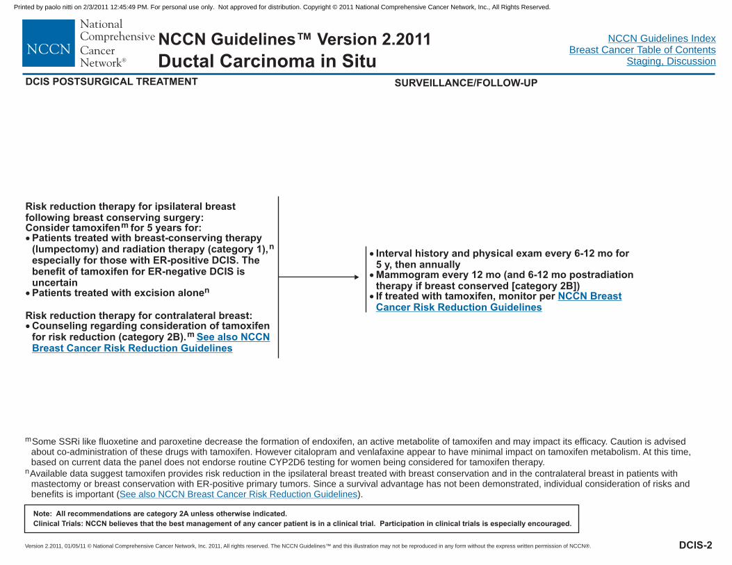
SURVEILLANCE/FOLLOW-UP
�
�
�
Interval history and physical exam every 6-12 mo for5 y, then annuallyMammogram every 12 mo (and 6-12 mo postradiationtherapy if breast conserved [category 2B])If treated with tamoxifen, monitor per NCCN BreastCancer Risk Reduction Guidelines
Risk reduction therapy for ipsilateral breastfollowing breast conserving surgery:Consider tamoxifen for 5 years for:
Patients treated with breast-conserving therapy(lumpectomy) and radiation therapy (category 1)
Patients treated with excision alone
Risk reduction therapy for contralateral breast:Counseling regarding consideration of tamoxifenfor risk reduction (category 2B).
m
n
n
m
�
�
�
,especially for those with ER-positive DCIS. Thebenefit of tamoxifen for ER-negative DCIS isuncertain
See also NCCNBreast Cancer Risk Reduction Guidelines
DCIS POSTSURGICAL TREATMENT
mSome SSRi like fluoxetine and paroxetine decrease the formation of endoxifen, an active metabolite of tamoxifen and may impact its efficacy. Caution is advisedabout co-administration of these drugs with tamoxifen. However citalopram and venlafaxine appear to have minimal impact on tamoxifen metabolism. At this time,based on current data the panel does not endorse routine CYP2D6 testing for women being considered for tamoxifen therapy.
nAvailable data suggest tamoxifen provides risk reduction in the ipsilateral breast treated with breast conservation and in the contralateral breast in patients withmastectomy or breast conservation with ER-positive primary tumors. Since a survival advantage has not been demonstrated, individual consideration of risks andbenefits is important ( ).See also NCCN Breast Cancer Risk Reduction Guidelines
DCIS-2
Ductal Carcinoma in Situ
Printed by paolo nitti on 2/3/2011 12:45:49 PM. For personal use only. Not approved for distribution. Copyright © 2011 National Comprehensive Cancer Network, Inc., All Rights Reserved.

NCCN®
NCCN Guidelines™ Version 2.2011
Version 2.2011, 01/05/11 © National Comprehensive Cancer Network, Inc. 2011, All rights reserved. The NCCN Guidelines™ and this illustration may not be reproduced in any form without the express written permission of NCCN®.
NCCN Guidelines IndexBreast Cancer Table of Contents
Staging, Discussion
Note: All recommendations are category 2A unless otherwise indicated.Clinical Trials: NCCN believes that the best management of any cancer patient is in a clinical trial. Participation in clinical trials is especially encouraged.
MARGIN STATUS IN DCIS
Substantial controversy exists regarding the definition of a negative pathologic margin in DCIS. Controversy arises out of the heterogeneityof the disease, difficulties in distinguishing the spectrum of hyperplastic conditions, anatomic considerations of the location of the margin,and inadequate prospective data on prognostic factors in DCIS.
Margins greater than 10 mm are widely accepted as negative (but may be excessive and may lead to a less optimal cosmetic outcome).
Margins less than 1 mm are considered inadequate.
With pathologic margins between 1-10 mm, wider margins are generally associated with lower local recurrence rates. However, close surgicalmargins (< 1 mm) at the fibroglandular boundary of the breast (chest wall or skin) do not mandate surgical re-excision but can be an indicationfor higher boost dose radiation to the involved lumpectomy site. (category 2B)
DCIS-A
Ductal Carcinoma in Situ
Printed by paolo nitti on 2/3/2011 12:45:49 PM. For personal use only. Not approved for distribution. Copyright © 2011 National Comprehensive Cancer Network, Inc., All Rights Reserved.

NCCN®
NCCN Guidelines™ Version 2.2011
Version 2.2011, 01/05/11 © National Comprehensive Cancer Network, Inc. 2011, All rights reserved. The NCCN Guidelines™ and this illustration may not be reproduced in any form without the express written permission of NCCN®.
NCCN Guidelines IndexBreast Cancer Table of Contents
Staging, Discussion
Note: All recommendations are category 2A unless otherwise indicated.Clinical Trials: NCCN believes that the best management of any cancer patient is in a clinical trial. Participation in clinical trials is especially encouraged.
CLINICAL STAGE WORKUP
Stage IT1, N0, M0
orStage IIAT0, N1, M0T1, N1, M0T2, N0, M0
orStage IIBT2, N1, M0T3, N0, M0
orStage IIIAT3, N1, M0
General workup including:
Optional studies for breast imaging:
If clinical stage lllA (T3, N1, M0) consider:Bone scan (category 2B)Abdominal ± pelvis CT or US or MRIChest imaging
Additional studies as directed by signs or symptoms:Bone scan indicated if localized bone pain or elevated alkaline phosphataseAbdominal ± pelvis CT or US or MRI if elevated alkaline phosphatase, abnormalliver function tests, abdominal symptoms, abnormal physical examination ofthe abdomen or pelvisChest imaging (if pulmonary symptoms are present)Optional FDG PET/CT (for T3,N1,M0) (category 2B)Consider fertility counseling if indicated
���
��
�
History and physical examCBC, plateletsLiver function tests and alkaline phosphatase
Pathology reviewDetermination of tumor estrogen/progesterone receptor (ER/PR) status andHER2 status
Breast MRI
�
�
�
�
�
�
�
�
�
Diagnostic bilateral mammogram, ultrasound as necessary
Genetic counseling if patient is high risk for hereditary breast cancer
a
b
c
d
�
e
f
See LocoregionalTreatment(BINV-2)
BINV-1
a
f
The panel endorses the College of American Pathology Protocol for pathology reporting for all invasive and non-invasive carcinomas of the breast.
The use of PET or PET/CT scanning is not indicated in the staging of clinical stage I, II, or operable III breast cancer. FDG PET/CT is most helpful in situations wherestandard staging studies are equivocal or suspicious, especially in the setting of locally advanced or metastatic disease. FDG PET/CT may also be helpful inidentifying unsuspected regional nodal disease and/or distant metastases in LABC when used in addition to standard staging studies.
b
c
e
d
http://www.cap.org.
See NCCN Genetics/Familial High-Risk Assessment: Breast and Ovarian Guidelines
See Fertility and Birth Control After Adjuvant Breast Cancer Treatment (BINV-C).
See Principles of HER2 Testing (BINV-A
See Principles of Dedicated Breast MRI Testing (BINV-B
).
).
.
Invasive Breast Cancer
Printed by paolo nitti on 2/3/2011 12:45:49 PM. For personal use only. Not approved for distribution. Copyright © 2011 National Comprehensive Cancer Network, Inc., All Rights Reserved.
NCCN®
NCCN Guidelines™ Version 2.2011
Version 2.2011, 01/05/11 © National Comprehensive Cancer Network, Inc. 2011, All rights reserved. The NCCN Guidelines™ and this illustration may not be reproduced in any form without the express written permission of NCCN®.
NCCN Guidelines IndexBreast Cancer Table of Contents
Staging, Discussion
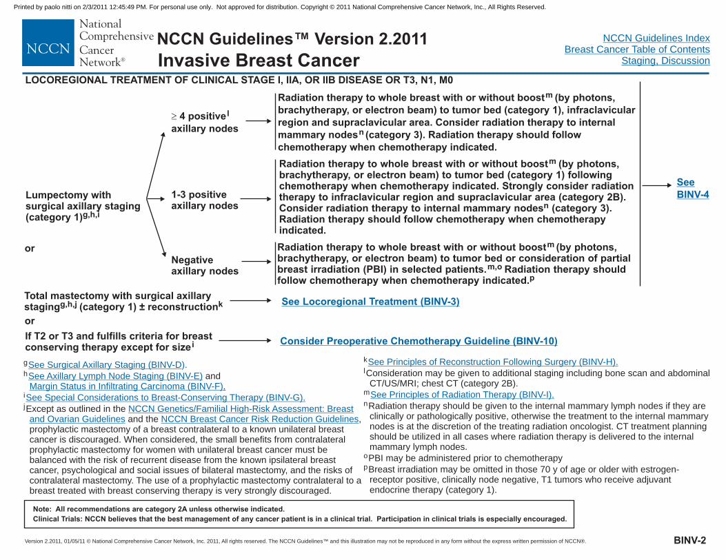
LOCOREGIONAL TREATMENT OF CLINICAL STAGE I, IIA, OR IIB DISEASE OR T3, N1, M0
g
h
i
and.
jExcept as outlined in theand the ,
prophylactic mastectomy of a breast contralateral to a known unilateral breastcancer is discouraged. When considered, the small benefits from contralateralprophylactic mastectomy for women with unilateral breast cancer must bebalanced with the risk of recurrent disease from the known ipsilateral breastcancer, psychological and social issues of bilateral mastectomy, and the risks ofcontralateral mastectomy. The use of a prophylactic mastectomy contralateral to abreast treated with breast conserving therapy is very strongly discouraged.
See Surgical Axillary Staging (BINV-D
See Axillary Lymph Node Staging (BINV-E)Margin Status in Infiltrating Carcinoma (BINV-F)
See Special Considerations to Breast-Conserving Therapy (BINV-G).
).
NCCN Genetics/Familial High-Risk Assessment: Breastand Ovarian Guidelines NCCN Breast Cancer Risk Reduction Guidelines
Lumpectomy withsurgical axillary staging(category 1)g,h,i
1-3 positivenodesaxillary
Negativenodesaxillary
Radiation therapy to whole breast with or without boost (by photons,brachytherapy, or electron beam) to tumor bed (category 1), infraclavicularregion and supraclavicular area. Consider radiation therapy to internalmammary nodes (category 3). Radiation therapy should followchemotherapy when chemotherapy indicated.
m
n
Radiation therapy to whole breast with or without boost (by photons,brachytherapy, or electron beam) to tumor bed (category 1)
Strongly consider radiationtherapy to supraclavicular area (category 2B).Consider radiation therapy to internal mammary nodes (category 3).Radiation therapy should follow chemotherapy when chemotherapyindicated.
m
n
followingchemotherapy when chemotherapy indicated.
infraclavicular region and
Radiation therapy to whole breast with or without boost (by photons,brachytherapy, or electron beam) to tumor bed or consideration of partialbreast irradiation (PBI) in selected patients. Radiation therapy shouldfollow chemotherapy when chemotherapy indicated.
m
m,op
Total mastectomy with surgical axillarystaging (category 1) ± reconstructiong,h,j k
Consider Preoperative Chemotherapy Guideline (BINV-10)If T2 or T3 and fulfills criteria for breastconserving therapy except for sizei
See Locoregional Treatment (BINV-3)
or
or
BINV-2
� 4 positiveaxillary nodes
l
SeeBINV-4
Note: All recommendations are category 2A unless otherwise indicated.Clinical Trials: NCCN believes that the best management of any cancer patient is in a clinical trial. Participation in clinical trials is especially encouraged.
k
o
p
l
m
n
Consideration may be given to additional staging including bone scan and abdominalCT/US/MRI; chest CT (category 2B).
Radiation therapy should be given to the internal mammary lymph nodes if they areclinically or pathologically positive, otherwise the treatment to the internal mammarynodes is at the discretion of the treating radiation oncologist. CT treatment planningshould be utilized in all cases where radiation therapy is delivered to the internalmammary lymph nodes.
PBI may be administered prior to chemotherapy
Breast irradiation may be omitted in those 70 y of age or older with estrogen-receptor positive, clinically node negative, T1 tumors who receive adjuvantendocrine therapy (category 1).
See Principles of Reconstruction Following Surgery (BINV-H).
See Principles of Radiation Therapy (BINV-I).
Invasive Breast Cancer
Printed by paolo nitti on 2/3/2011 12:45:49 PM. For personal use only. Not approved for distribution. Copyright © 2011 National Comprehensive Cancer Network, Inc., All Rights Reserved.

NCCN®
NCCN Guidelines™ Version 2.2011
Version 2.2011, 01/05/11 © National Comprehensive Cancer Network, Inc. 2011, All rights reserved. The NCCN Guidelines™ and this illustration may not be reproduced in any form without the express written permission of NCCN®.
NCCN Guidelines IndexBreast Cancer Table of Contents
Staging, Discussion
Note: All recommendations are category 2A unless otherwise indicated.Clinical Trials: NCCN believes that the best management of any cancer patient is in a clinical trial. Participation in clinical trials is especially encouraged.
Total mastectomy withsurgical axillarystaging (category 1)± reconstruction
g,hk
LOCOREGIONAL TREATMENT OF CLINICAL STAGE I, IIA, OR IIB DISEASE OR T3, N1, M0
� 4 positiveaxillary nodes l
Postchemotherapy radiation therapy to chest wall(category 1) + infraclavicular and supraclavicularareas. Consider radiation therapy to internal mammarynodes (category 3)
mm,n
1-3 positivenodesaxillary
Strongly consider postchemotherapy radiation therapyto chest wall + infraclavicular and supraclavicularareas; if radiation therapy is given, consider internalmammary node radiation therapy (category 3).
mm,n
Negative axillary nodesand tumor > 5 cmormargins positive
Postchemotherapy radiation therapy to chest wallm
Negative nodesand tumor 5 cm andmargins 1 mm
axillary�
�No radiation therapy
g
k
h
m
n
.
and
Radiation therapy should be given to the internal mammary lymph nodes that are clinically or pathologically positive, otherwise the treatment to the internal mammarynodes is at the discretion of the treating radiation oncologist. CT treatment planning should be utilized in all cases where radiation therapy is delivered to the internalmammary lymph nodes
lConsideration may be given to additional staging including bone scan; abdominal CT/US/MRI; chest CT (category 2B).
See Surgical Axillary Staging (BINV-D
See Axillary Lymph Node Staging (BINV-E Margin Status in Infiltrating Carcinoma (BINV-F
See Principles of Reconstruction Following Surgery (BINV-H)
See Principles of Radiation Therapy (BINV-I).
).
) ).
.
Negative nodesand tumor 5 cm and
axillary
close margins (< 1 mm)�
Consider radiation therapy to chest wall ± infraclavicularand supraclavicular nodes. Consider radiation therapy tointernal mammary nodes (category 3)m
BINV-3
SeeBINV-4
Invasive Breast Cancer
Printed by paolo nitti on 2/3/2011 12:45:49 PM. For personal use only. Not approved for distribution. Copyright © 2011 National Comprehensive Cancer Network, Inc., All Rights Reserved.

NCCN®
NCCN Guidelines™ Version 2.2011
Version 2.2011, 01/05/11 © National Comprehensive Cancer Network, Inc. 2011, All rights reserved. The NCCN Guidelines™ and this illustration may not be reproduced in any form without the express written permission of NCCN®.
NCCN Guidelines IndexBreast Cancer Table of Contents
Staging, Discussion
�
�
�
�
DuctalLobularMixedMetaplastic
q
�
�
TubularColloid
ER-positiveand/orPR positive
ER-negativeandPR-negative
ER-positiveand/orPR positive
ER-negativeandPR-negative
HER2 positiveb
HER2 negativeb
HER2 positiveb
HER2 negativeb
BINV-4
See Systemic Adjuvant Treatment - Hormone ReceptorPositive - HER2 Positive Disease (BINV-5)
Note: All recommendations are category 2A unless otherwise indicated.Clinical Trials: NCCN believes that the best management of any cancer patient is in a clinical trial. Participation in clinical trials is especially encouraged.
b .
This includes medullary and micropapillary subtypes.qSee Principles of HER2 Testing (BINV-A)
See Systemic Adjuvant Treatment - HormoneReceptor Positive - HER2 Negative Disease (BINV-6)
See Systemic Adjuvant Treatment - HormoneReceptor Negative - HER2 Positive Disease (BINV-7)
See Systemic Adjuvant Treatment - HormoneReceptor Negative - HER2 Negative Disease (BINV-8)
See Systemic Adjuvant Treatment -Favorable Histologies (BINV-9)
HISTOLOGY HER2 STATUS SYSTEMIC ADJUVANT TREATMENTHORMONERECEPTOR STATUS
Invasive Breast Cancer
Printed by paolo nitti on 2/3/2011 12:45:49 PM. For personal use only. Not approved for distribution. Copyright © 2011 National Comprehensive Cancer Network, Inc., All Rights Reserved.
NCCN®
NCCN Guidelines™ Version 2.2011
Version 2.2011, 01/05/11 © National Comprehensive Cancer Network, Inc. 2011, All rights reserved. The NCCN Guidelines™ and this illustration may not be reproduced in any form without the express written permission of NCCN®.
NCCN Guidelines IndexBreast Cancer Table of Contents
Staging, Discussion
Note: All recommendations are category 2A unless otherwise indicated.Clinical Trials: NCCN believes that the best management of any cancer patient is in a clinical trial. Participation in clinical trials is especially encouraged.
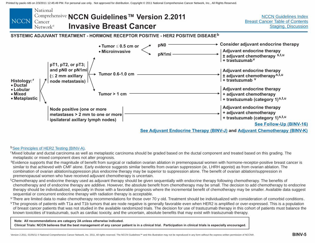
BINV-5
Tumor > 1 cm
SYSTEMIC ADJUVANT TREATMENT - HORMONE RECEPTOR POSITIVE - HER2 POSITIVE DISEASEb
Histology:DuctalLobularMixedMetaplastic
r
����
�
�
Tumor 0.5 cm orMicroinvasive
�
Tumor 0.6-1.0 cmAdjuvant endocrine therapy± adjuvant chemotherapys,t,u
v+ trastuzumab
pT1, pT2, or pT3;and pN0 or pN1mi( 2 mm axillarynode metastasis)�
Adjuvant endocrine therapy+ adjuvant chemotherapy+ trastuzumab (category 1)s,t,u
Adjuvant endocrine therapy+ adjuvant chemotherapy+ trastuzumab (category 1)s,t,u
Node positive (one or moremetastases > 2 mm to one or moreipsilateral axillary lymph nodes)
b .
Evidence supports that the magnitude of benefit from surgical or radiation ovarian ablation in premenopausal women with hormone-receptor-positive breast cancer issimilar to that achieved with CMF alone. Early evidence suggests similar benefits from ovarian suppression (ie, LHRH agonist) as from ovarian ablation. Thecombination of ovarian ablation/suppression plus endocrine therapy may be superior to suppression alone. The benefit of ovarian ablation/suppression inpremenopausal women who have received adjuvant chemotherapy is uncertain.
There are limited data to make chemotherapy recommendations for those over 70 y old. Treatment should be individualized with consideration of comorbid conditions.
The prognosis of patients with T1a and T1b tumors that are node negative is generally favorable even when HER2 is amplified or over-expressed. This is a populationof breast cancer patients that was not studied in the available randomized trials. The decision for use of trastuzumab therapy in this cohort of patients must balance theknown toxicities of trastuzumab, such as cardiac toxicity, and the uncertain, absolute benefits that may exist with trastuzumab therapy.
r
t
Mixed lobular and ductal carcinoma as well as metaplastic carcinoma should be graded based on the ductal component and treated based on this grading. Themetaplastic or mixed component does not alter prognosis.
s
u
v
Chemotherapy and endocrine therapy used as adjuvant therapy should be given sequentially with endocrine therapy following chemotherapy. The benefits ofchemotherapy and of endocrine therapy are additive. However, the absolute benefit from chemotherapy may be small. The decision to add chemotherapy to endocrinetherapy should be individualized, especially in those with a favorable prognosis where the incremental benefit of chemotherapy may be smaller. Available data suggestsequential or concurrent endocrine therapy with radiation therapy is acceptable.
See Principles of HER2 Testing (BINV-A)
See Follow-Up (BINV-16)
See Adjuvant Endocrine Therapy (BINV-J) Adjuvant Chemotherapy (BINV-K)and
Invasive Breast Cancer
pN0
pN1mi
Consider adjuvant endocrine therapy
Adjuvant endocrine therapy± adjuvant chemotherapy s,t,u
v+ trastuzumab
Printed by paolo nitti on 2/3/2011 12:45:49 PM. For personal use only. Not approved for distribution. Copyright © 2011 National Comprehensive Cancer Network, Inc., All Rights Reserved.
NCCN®
NCCN Guidelines™ Version 2.2011
Version 2.2011, 01/05/11 © National Comprehensive Cancer Network, Inc. 2011, All rights reserved. The NCCN Guidelines™ and this illustration may not be reproduced in any form without the express written permission of NCCN®.
NCCN Guidelines IndexBreast Cancer Table of Contents
Staging, Discussion
Note: All recommendations are category 2A unless otherwise indicated.Clinical Trials: NCCN believes that the best management of any cancer patient is in a clinical trial. Participation in clinical trials is especially encouraged.
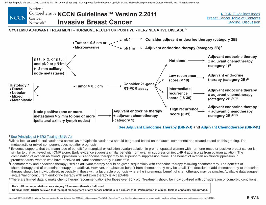
SYSTEMIC ADJUVANT TREATMENT - HORMONE RECEPTOR POSITIVE - HER2 NEGATIVE DISEASEb
Histology:DuctalLobularMixedMetaplastic
r
����
�
�
Tumor 0.5 cm orMicroinvasive
�pN0
pN1mi
BINV-6
pT1, pT2, or pT3;and pN0 or pN1mi( 2 mm axillarynode metastasis)�
Node positive (one or moremetastases > 2 mm to one or moreipsilateral axillary lymph nodes)
b .
Evidence supports that the magnitude of benefit from surgical or radiation ovarian ablation in premenopausal women with hormone-receptor-positive breast cancer issimilar to that achieved with CMF alone. Early evidence suggests similar benefits from ovarian suppression (ie, LHRH agonist) as from ovarian ablation. Thecombination of ovarian ablation/suppression plus endocrine therapy may be superior to suppression alone. The benefit of ovarian ablation/suppression inpremenopausal women who have received adjuvant chemotherapy is uncertain.
where the incremental benefit of chemotherapy may be smaller
There are limited data to make chemotherapy recommendations for those over 70 y old. Treatment should be individualized with consideration of comorbid conditions.
r
t
Mixed lobular and ductal carcinoma as well as metaplastic carcinoma should be graded based on the ductal component and treated based on this grading. Themetaplastic or mixed component does not alter prognosis.
s
u
Chemotherapy and endocrine therapy used as adjuvant therapy should be given sequentially with endocrine therapy following chemotherapy. The benefits ofchemotherapy and of endocrine therapy are additive. However, the absolute benefit from chemotherapy may be small. The decision to add chemotherapy to endocrinetherapy should be individualized, especially in those with a favorable prognosis . Available data suggestsequential or concurrent endocrine therapy with radiation therapy is acceptable.
See Principles of HER2 Testing (BINV-A)
� Tumor > 0.5 cmAdjuvant endocrine therapy± adjuvant chemotherapy(category 2B)s,t,u
Low recurrencescore (< 18)
Intermediaterecurrencescore (18-30)
High recurrencescore ( 31)�
Adjuvant endocrinetherapy (category 2B)s
Adjuvant endocrine therapy+ adjuvant chemotherapy(category 2B)s,t,u
Consider 21-geneRT-PCR assay
Not doneAdjuvant endocrine therapy± adjuvant chemotherapy(category 1)s
Adjuvant endocrine therapy+ adjuvant chemotherapy(category 1)
See Adjuvant Endocrine Therapy (BINV-J) Adjuvant Chemotherapy (BINV-K)and
Consider adjuvant endocrine therapy (category 2B)
Adjuvant endocrine therapy (category 2B)s
Invasive Breast Cancer
Printed by paolo nitti on 2/3/2011 12:45:49 PM. For personal use only. Not approved for distribution. Copyright © 2011 National Comprehensive Cancer Network, Inc., All Rights Reserved.
NCCN®
NCCN Guidelines™ Version 2.2011
Version 2.2011, 01/05/11 © National Comprehensive Cancer Network, Inc. 2011, All rights reserved. The NCCN Guidelines™ and this illustration may not be reproduced in any form without the express written permission of NCCN®.
NCCN Guidelines IndexBreast Cancer Table of Contents
Staging, Discussion
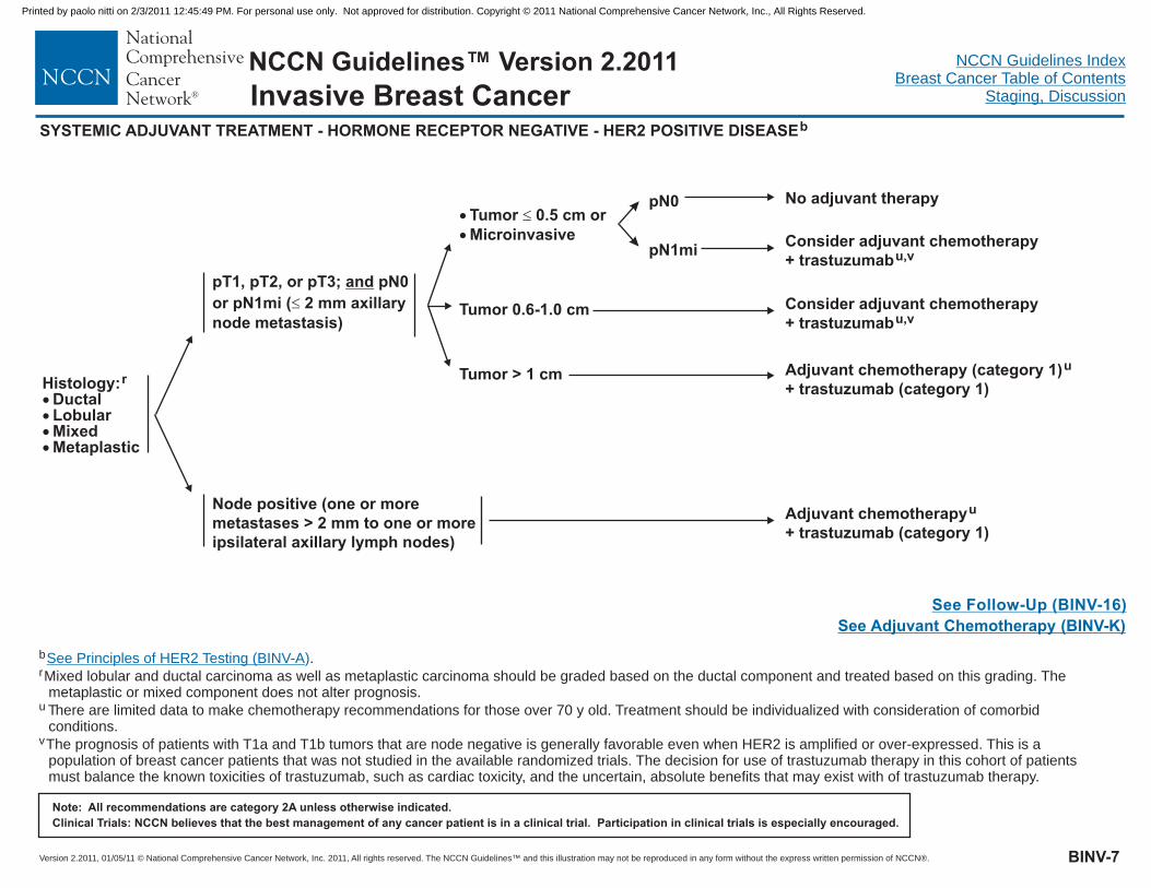
Consider adjuvant chemotherapy+ trastuzumabu,v
Note: All recommendations are category 2A unless otherwise indicated.Clinical Trials: NCCN believes that the best management of any cancer patient is in a clinical trial. Participation in clinical trials is especially encouraged.
BINV-7
SYSTEMIC ADJUVANT TREATMENT - HORMONE RECEPTOR NEGATIVE - HER2 POSITIVE DISEASEb
Tumor > 1 cmHistology:
DuctalLobularMixedMetaplastic
r
����
�
�
Tumor 0.5 cm orMicroinvasive
�
Tumor 0.6-1.0 cm
pN0
pN1mi
pT1, pT2, or pT3; pN0or pN1mi ( 2 mm axillarynode metastasis)
and�
No adjuvant therapy
Consider adjuvant chemotherapy+ trastuzumabu,v
Adjuvant chemotherapy (category 1)+ trastuzumab (category 1)
u
Adjuvant chemotherapy+ trastuzumab (category 1)
u
b .
There are limited data to make chemotherapy recommendations for those over 70 y old. Treatment should be individualized with consideration of comorbidconditions.
The prognosis of patients with T1a and T1b tumors that are node negative is generally favorable even when HER2 is amplified or over-expressed. This is apopulation of breast cancer patients that was not studied in the available randomized trials. The decision for use of trastuzumab therapy in this cohort of patientsmust balance the known toxicities of trastuzumab, such as cardiac toxicity, and the uncertain, absolute benefits that may exist with of trastuzumab therapy.
r
u
v
Mixed lobular and ductal carcinoma as well as metaplastic carcinoma should be graded based on the ductal component and treated based on this grading. Themetaplastic or mixed component does not alter prognosis.
See Principles of HER2 Testing (BINV-A)
See Follow-Up (BINV-16)See Adjuvant Chemotherapy (BINV-K)
Node positive (one or moremetastases > 2 mm to one or moreipsilateral axillary lymph nodes)
Invasive Breast Cancer
Printed by paolo nitti on 2/3/2011 12:45:49 PM. For personal use only. Not approved for distribution. Copyright © 2011 National Comprehensive Cancer Network, Inc., All Rights Reserved.

NCCN®
NCCN Guidelines™ Version 2.2011
Version 2.2011, 01/05/11 © National Comprehensive Cancer Network, Inc. 2011, All rights reserved. The NCCN Guidelines™ and this illustration may not be reproduced in any form without the express written permission of NCCN®.
NCCN Guidelines IndexBreast Cancer Table of Contents
Staging, Discussion
SYSTEMIC ADJUVANT TREATMENT - HORMONE RECEPTOR NEGATIVE - HER2 NEGATIVE DISEASEb
Adjuvant chemotherapy (category 1)u
Adjuvant chemotherapy (category 1)u
Note: All recommendations are category 2A unless otherwise indicated.Clinical Trials: NCCN believes that the best management of any cancer patient is in a clinical trial. Participation in clinical trials is especially encouraged.
BINV-8
Histology:DuctalLobularMixedMetaplastic
r
����
pT1, pT2, or pT3; pN0or pN1mi ( 2 mm axillarynode metastasis)
and�
b .
There are limited data to make chemotherapy recommendations for those over 70 y old. Treatment should be individualized with consideration of comorbid conditions.
r
u
Mixed lobular and ductal carcinoma as well as metaplastic carcinoma should be graded based on the ductal component and treated based on this grading. Themetaplastic or mixed component does not alter prognosis.
See Principles of HER2 Testing (BINV-A)
See Follow-Up (BINV-16)
Node positive (one or moremetastases > 2 mm to one or moreipsilateral axillary lymph nodes)
Tumor > 1 cm
�
�
Tumor 0.5 cm orMicroinvasive
�
Tumor 0.6-1.0 cm
pN0
pN1mi Consider adjuvant chemotherapyu
No adjuvant therapy
Consider adjuvant chemotherapy u
See Adjuvant Chemotherapy (BINV-J)
Invasive Breast Cancer
Printed by paolo nitti on 2/3/2011 12:45:49 PM. For personal use only. Not approved for distribution. Copyright © 2011 National Comprehensive Cancer Network, Inc., All Rights Reserved.
NCCN®
NCCN Guidelines™ Version 2.2011
Version 2.2011, 01/05/11 © National Comprehensive Cancer Network, Inc. 2011, All rights reserved. The NCCN Guidelines™ and this illustration may not be reproduced in any form without the express written permission of NCCN®.
NCCN Guidelines IndexBreast Cancer Table of Contents
Staging, Discussion
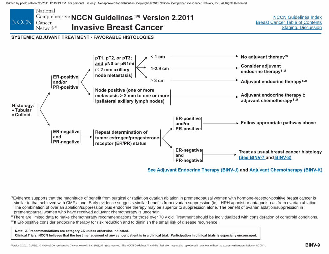
Histology:TubularColloid
��
SYSTEMIC ADJUVANT TREATMENT - FAVORABLE HISTOLOGIES
Note: All recommendations are category 2A unless otherwise indicated.Clinical Trials: NCCN believes that the best management of any cancer patient is in a clinical trial. Participation in clinical trials is especially encouraged.
BINV-9
See Adjuvant Endocrine Therapy (BINV-J) Adjuvant Chemotherapy (BINV-K)and
s
u
w
Evidence supports that the magnitude of benefit from surgical or radiation ovarian ablation in premenopausal women with hormone-receptor-positive breast cancer issimilar to that achieved with CMF alone. Early evidence suggests similar benefits from ovarian suppression (ie, LHRH agonist or antagonist) as from ovarian ablation.The combination of ovarian ablation/suppression plus endocrine therapy may be superior to suppression alone. The benefit of ovarian ablation/suppression inpremenopausal women who have received adjuvant chemotherapy is uncertain.
There are limited data to make chemotherapy recommendations for those over 70 y old. Treatment should be individualized with consideration of comorbid conditions.
If ER-positive consider endocrine therapy for risk reduction and to diminish the small risk of disease recurrence.
Node positive (one or moremetastasis > 2 mm to one or moreipsilateral axillary lymph nodes)
< 1 cm
1-2.9 cm
� 3 cm
No adjuvant therapyw
Consider adjuvantendocrine therapys,u
pT1, pT2, or pT3;pN0 or pN1mi
( 2 mm axillarynode metastasis)
and�
Adjuvant endocrine therapys,u
Adjuvant endocrine therapy ±adjuvant chemotherapys,u
ER-positiveand/orPR-positive
ER-negativeandPR-negative
Repeat determination oftumor estrogen/progesteronereceptor (ER/PR) status
ER-positiveand/orPR-positive
ER-negativeandPR-negative
Follow appropriate pathway above
Treat as usual breast cancer histologyand(See BINV-7 BINV-8)
Invasive Breast Cancer
Printed by paolo nitti on 2/3/2011 12:45:49 PM. For personal use only. Not approved for distribution. Copyright © 2011 National Comprehensive Cancer Network, Inc., All Rights Reserved.
NCCN®
NCCN Guidelines™ Version 2.2011
Version 2.2011, 01/05/11 © National Comprehensive Cancer Network, Inc. 2011, All rights reserved. The NCCN Guidelines™ and this illustration may not be reproduced in any form without the express written permission of NCCN®.
NCCN Guidelines IndexBreast Cancer Table of Contents
Staging, Discussion
Note: All recommendations are category 2A unless otherwise indicated.Clinical Trials: NCCN believes that the best management of any cancer patient is in a clinical trial. Participation in clinical trials is especially encouraged.
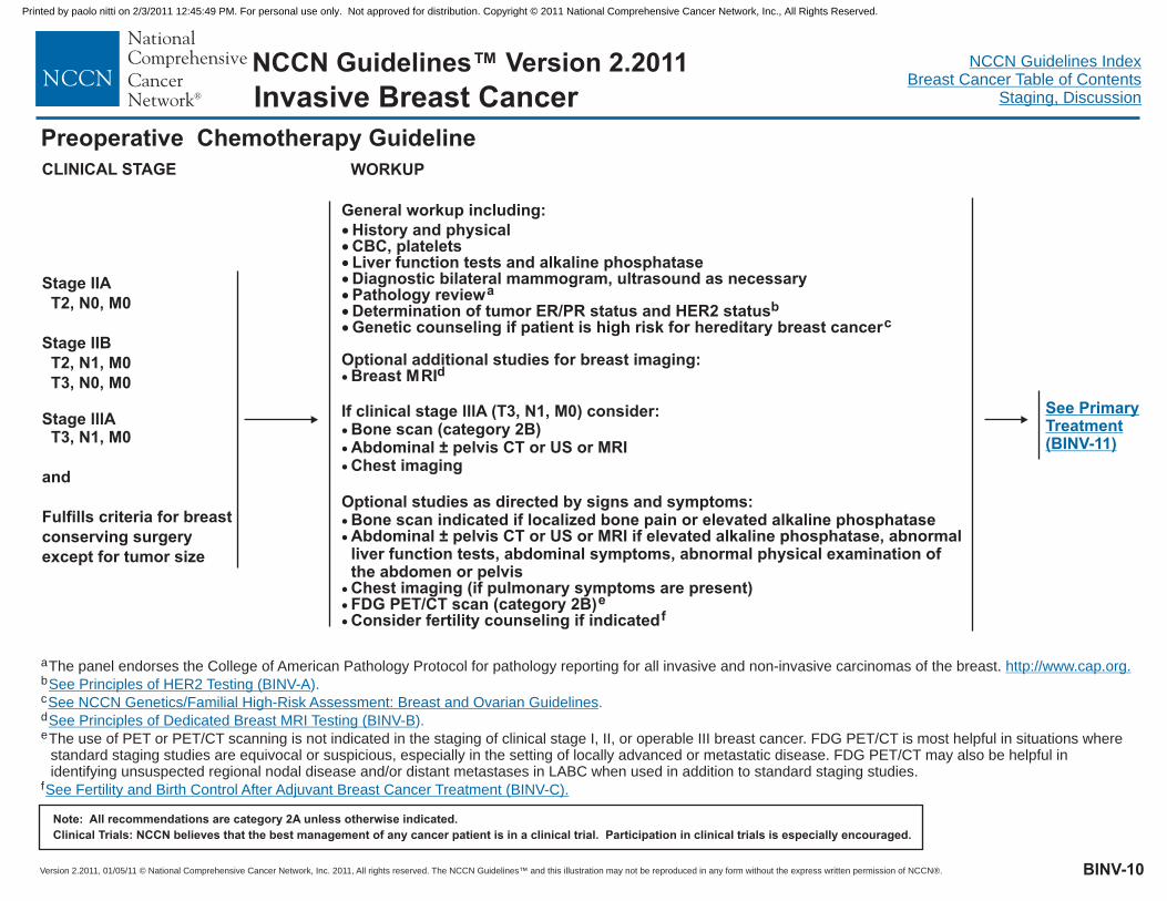
CLINICAL STAGE WORKUP
See PrimaryTreatment(BINV-11)
Preoperative Chemotherapy Guideline
Stage IIAT2, N0, M0
Stage IIBT2, N1, M0T3, N0, M0
Stage lllAT3, N1, M0
and
Fulfills criteria for breastconserving surgeryexcept for tumor size
General workup including:�������
History and physicalCBC, plateletsLiver function tests and alkaline phosphataseDiagnostic bilateral mammogram, ultrasound as necessaryPathology reviewDetermination of tumor ER/PR status and HER2 status
Breast MRI
Bone scan indicated if localized bone pain or elevated alkaline phosphataseAbdominal ± pelvis CT or US or MRI if elevated alkaline phosphatase, abnormalliver function tests, abdominal symptoms, abnormal physical examination ofthe abdomen or pelvisChest imaging (if pulmonary symptoms are present)FDG PET/CT scan (category 2B)
ab
c
d
e
Genetic counseling if patient is high risk for hereditary breast cancer
Optional additional studies for breast imaging:
If clinical stage lllA (T3, N1, M0) consider:Bone scan (category 2B)Abdominal ± pelvis CT or US or MRIChest imaging
Optional studies as directed by signs and symptoms:
Consider fertility counseling if indicated
�
�
�
�
�
�
�
�
� f
BINV-10
Invasive Breast Cancer
a
f
The panel endorses the College of American Pathology Protocol for pathology reporting for all invasive and non-invasive carcinomas of the breast.
The use of PET or PET/CT scanning is not indicated in the staging of clinical stage I, II, or operable III breast cancer. FDG PET/CT is most helpful in situations wherestandard staging studies are equivocal or suspicious, especially in the setting of locally advanced or metastatic disease. FDG PET/CT may also be helpful inidentifying unsuspected regional nodal disease and/or distant metastases in LABC when used in addition to standard staging studies.
b
c
e
d
http://www.cap.org.
See NCCN Genetics/Familial High-Risk Assessment: Breast and Ovarian Guidelines
See Fertility and Birth Control After Adjuvant Breast Cancer Treatment (BINV-C).
See Principles of HER2 Testing (BINV-A
See Principles of Dedicated Breast MRI Testing (BINV-B
).
).
.
Printed by paolo nitti on 2/3/2011 12:45:49 PM. For personal use only. Not approved for distribution. Copyright © 2011 National Comprehensive Cancer Network, Inc., All Rights Reserved.

NCCN®
NCCN Guidelines™ Version 2.2011
Version 2.2011, 01/05/11 © National Comprehensive Cancer Network, Inc. 2011, All rights reserved. The NCCN Guidelines™ and this illustration may not be reproduced in any form without the express written permission of NCCN®.
NCCN Guidelines IndexBreast Cancer Table of Contents
Staging, Discussion
Note: All recommendations are category 2A unless otherwise indicated.Clinical Trials: NCCN believes that the best management of any cancer patient is in a clinical trial. Participation in clinical trials is especially encouraged.
Desires breastpreservation
Does not desirebreast preservation
Core biopsy of breast tumor,localization of tumor bed forfuture surgical management
See Stage I and II Breast Cancer (BINV-3)
gSee Surgical Axillary Staging (BINV-D).
BINV-11
Preoperative Chemotherapy Guideline
Clinically negative axillary lymphnode(s), consider sentinel lymph nodeprocedureg
Consider axillary ultrasound; ifclinically positive see below
Clinically positive axillary lymphnode(s), consider core biopsy or FNA;or consider sentinel lymph nodeprocedure if FNA or core biopsynegative
Invasive Breast Cancer
See PrimaryTreatment (BINV-12)
Printed by paolo nitti on 2/3/2011 12:45:49 PM. For personal use only. Not approved for distribution. Copyright © 2011 National Comprehensive Cancer Network, Inc., All Rights Reserved.
NCCN®
NCCN Guidelines™ Version 2.2011
Version 2.2011, 01/05/11 © National Comprehensive Cancer Network, Inc. 2011, All rights reserved. The NCCN Guidelines™ and this illustration may not be reproduced in any form without the express written permission of NCCN®.
NCCN Guidelines IndexBreast Cancer Table of Contents
Staging, DiscussionInvasive Breast Cancer
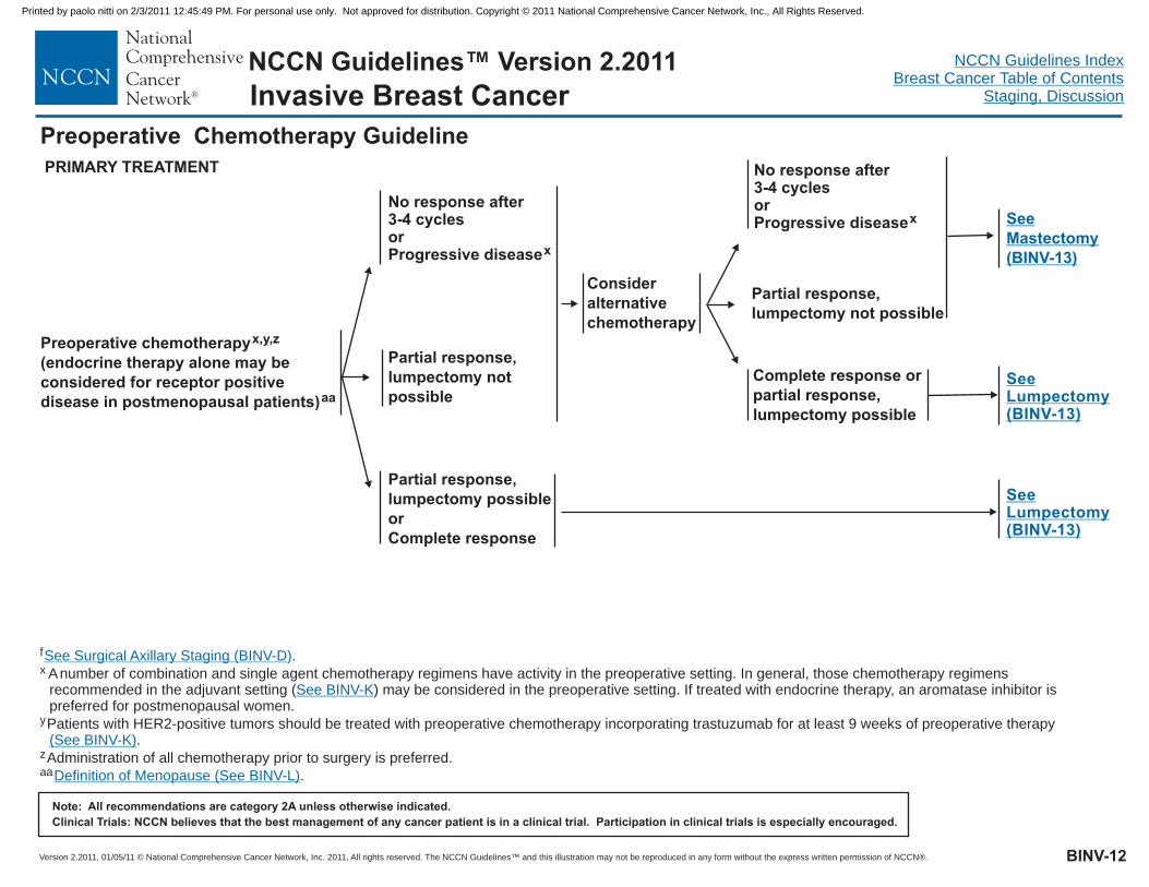
PRIMARY TREATMENT
Preoperative chemotherapy(endocrine therapy alone may beconsidered for receptor positivedisease in postmenopausal patients)
x,y,z
aa
Partial response,lumpectomypossible
not
Partial response,lumpectomy possibleorComplete response
No response after3-4 cyclesorProgressive diseasex
SeeLumpectomy(BINV-13)
Consideralternativechemotherapy
Partial response,lumpectomy possiblenot
No response after3-4 cyclesorProgressive diseasex See
Mastectomy(BINV-13)
Complete response orpartial response,lumpectomy possible
f
x Anumber of combination and single agent chemotherapy regimens have activity in the preoperative setting. In general, those chemotherapy regimensrecommended in the adjuvant setting ( ) may be considered in the preoperative setting. If treated with endocrine therapy, an aromatase inhibitor ispreferred for postmenopausal women.
Patients with HER2-positive tumors should be treated with preoperative chemotherapy incorporating trastuzumab for at least 9 weeks of preoperative therapy.
Administration of all chemotherapy prior to surgery is preferred.
.
y
z
aa
See Surgical Axillary Staging (BINV-D).
See BINV-K
(See BINV-K)
Definition of Menopause (See BINV-L)
Note: All recommendations are category 2A unless otherwise indicated.Clinical Trials: NCCN believes that the best management of any cancer patient is in a clinical trial. Participation in clinical trials is especially encouraged.
Preoperative Chemotherapy Guideline
SeeLumpectomy(BINV-13)
BINV-12
Printed by paolo nitti on 2/3/2011 12:45:49 PM. For personal use only. Not approved for distribution. Copyright © 2011 National Comprehensive Cancer Network, Inc., All Rights Reserved.

NCCN®
NCCN Guidelines™ Version 2.2011
Version 2.2011, 01/05/11 © National Comprehensive Cancer Network, Inc. 2011, All rights reserved. The NCCN Guidelines™ and this illustration may not be reproduced in any form without the express written permission of NCCN®.
NCCN Guidelines IndexBreast Cancer Table of Contents
Staging, Discussion
Mastectomy andsurgical axillarystaging ±reconstruction. Ifsentinel lymph nodebiopsy performedprechemotherapy andnegative findings, mayomit axillary lymphnode staging
bb
Lumpectomy withsurgical axillarystaging.bb If sentinellymph node biopsyperformedprechemotherapy andnegative findings, mayomit axillary lymphnode staging
�
�
�
Adjuvant radiation therapy post-mastectomy isbased on prechemotherapy tumor characteristicsas perandEndocrine therapy if ER-positive and/or PR-positive (category 1)Complete up to one year of trastuzumab therapy ifHER2-positive (category 1). May be administeredconcurrent with radiation therapy and withendocrine therapy if indicated.
m
t
m
BINV-3
See Adjuvant Endocrine Therapy (BINV-J)
�
�
�
Adjuvant radiation therapy post-lumpectomybased on prechemotherapy tumor characteristicsas perandEndocrine therapy if ER-positive and/or PR-positive (category 1)Complete up to one year of trastuzumab therapy ifHER2-positive (category 1). May be administeredconcurrent with radiation therapy and withendocrine therapy if indicated.
m
t
m
BINV-2
See Adjuvant Endocrine Therapy (BINV-J)
See Surveillance/Follow-up (BINV-16)
LOCAL TREATMENT ADJUVANT TREATMENT
Note: All recommendations are category 2A unless otherwise indicated.Clinical Trials: NCCN believes that the best management of any cancer patient is in a clinical trial. Participation in clinical trials is especially encouraged.
Consider additionalchemotherapy in thecontext of a clinical trial
m
tChemotherapy and endocrine therapy used as adjuvant therapy should be given sequentially with endocrine therapy following chemotherapy. The benefits ofchemotherapy and of endocrine therapy are additive. However, the absolute benefit from chemotherapy may be small. The decision to add chemotherapy to endocrinetherapy should be individualized, especially in those with a favorable prognosis where the incremental benefit of chemotherapy may be smaller. Available data suggestsequential or concurrent endocrine therapy with radiation therapy is acceptable.
Axillary staging may include sentinel node biopsy (category 3) or level l/ll dissection.bb
See Principles of Radiation Therapy (BINV-I).
BINV-13
Preoperative Chemotherapy Guideline
Consider additionalchemotherapy in thecontext of a clinical trial
Invasive Breast Cancer
Printed by paolo nitti on 2/3/2011 12:45:49 PM. For personal use only. Not approved for distribution. Copyright © 2011 National Comprehensive Cancer Network, Inc., All Rights Reserved.
NCCN®
NCCN Guidelines™ Version 2.2011
Version 2.2011, 01/05/11 © National Comprehensive Cancer Network, Inc. 2011, All rights reserved. The NCCN Guidelines™ and this illustration may not be reproduced in any form without the express written permission of NCCN®.
NCCN Guidelines IndexBreast Cancer Table of Contents
Staging, Discussion
Note: All recommendations are category 2A unless otherwise indicated.Clinical Trials: NCCN believes that the best management of any cancer patient is in a clinical trial. Participation in clinical trials is especially encouraged.
See Initial Workup for Stage IV Disease (BINV-16)Stage IVAny T, any N, M1
CLINICAL STAGE WORKUP
See PreoperativeChemotherapy andLocoregionalTreatment (BINV-15)Stage IIIB
T4, N0, M0T4, N1, M0T4, N2, M0
Stage lllCAny T, N3, M0
Stage IIIAT0, N2, M0T1, N2, M0T2, N2, M0T3, N2, M0
LOCALLY ADVANCED INVASIVE BREAST CANCER (NON-INFLAMMATORY)
(Stage IIIA patients with T3,N1, M0 disease, see BINV-1)
BINV-14
Invasive Breast Cancer
a
f
The panel endorses the College of American Pathology Protocol for pathology reporting for all invasive and non-invasive carcinomas of the breast.
The use of PET or PET/CT scanning is not indicated in the staging of clinical stage I, II, or operable III breast cancer. FDG PET/CT is most helpful in situations wherestandard staging studies are equivocal or suspicious, especially in the setting of locally advanced or metastatic disease. FDG PET/CT may also be helpful inidentifying unsuspected regional nodal disease and/or distant metastases in LABC when used in addition to standard staging studies.
b
c
e
d
http://www.cap.org.
See NCCN Genetics/Familial High-Risk Assessment: Breast and Ovarian Guidelines
See Fertility and Birth Control After Adjuvant Breast Cancer Treatment (BINV-C).
See Principles of HER2 Testing (BINV-A
See Principles of Dedicated Breast MRI Testing (BINV-B
).
).
.
General workup including:�������
History and physicalCBC, plateletsLiver function tests and alkaline phosphataseDiagnostic bilateral mammogram, ultrasound as necessaryPathology reviewDetermination of tumor ER/PR status and HER2 status
Breast MRI
Bone scan indicated if localized bone pain or elevated alkalinephosphataseAbdominal ± pelvis CT or US or MRI if elevated alkaline phosphatase,abnormal liver function tests, abdominal symptoms, abnormal physicalexamination of the abdomen or pelvisChest imaging (if pulmonary symptoms are present)FDG PET/CT scan (category 2B)
ab
c
d
e
Genetic counseling if patient is high risk for hereditary breast cancer
Optional additional studies for breast imaging:
If clinical stage lllA (T3, N1, M0) consider:Bone scan (category 2B)Abdominal ± pelvis CT or US or MRIChest imaging
Optional studies as directed by signs and symptoms:
Consider fertility counseling if indicated
�
�
�
�
�
�
�
�
� f
Printed by paolo nitti on 2/3/2011 12:45:49 PM. For personal use only. Not approved for distribution. Copyright © 2011 National Comprehensive Cancer Network, Inc., All Rights Reserved.
NCCN®
NCCN Guidelines™ Version 2.2011
Version 2.2011, 01/05/11 © National Comprehensive Cancer Network, Inc. 2011, All rights reserved. The NCCN Guidelines™ and this illustration may not be reproduced in any form without the express written permission of NCCN®.
NCCN Guidelines IndexBreast Cancer Table of Contents
Staging, Discussion
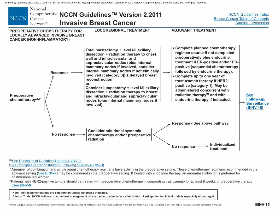
No response
Total mastectomy + level l/ll axillarydissection + radiation therapy to chestwall and infraclavicular andsupraclavicular nodes (plus internalmammary nodes if involved, considerinternal mammary nodes if not clinicallyinvolved [category 3]) ± delayed breastreconstructionorConsider lumpectomy + level l/ll axillarydissection + radiation therapy to breastand infraclavicular and supraclavicularnodes (plus internal mammary nodes ifinvolved)
j
LOCOREGIONAL TREATMENT
Response
ADJUVANT TREATMENT
Consider additional systemicchemotherapy and/or preoperativeradiation
SeeFollow-up/Surveillance(BINV-16)
Note: All recommendations are category 2A unless otherwise indicated.Clinical Trials: NCCN believes that the best management of any cancer patient is in a clinical trial. Participation in clinical trials is especially encouraged.
Response - See above pathway
No responseIndividualizedtreatment
Preoperativechemotherapyx,y
PREOPERATIVE CHEMOTHERAPY FORLOCALLY ADVANCED INVASIVE BREASTCANCER (NON-INFLAMMATORY)
BINV-15
m
x
j
y
Anumber of combination and single agent chemotherapy regimens have activity in the preoperative setting. Those chemotherapy regimens recommended in theadjuvant setting ( ) may be considered in the preoperative setting. If treated with endocrine therapy, an aromatase inhibitor is preferred forpostmenopausal women.
Patients with HER2-positive tumors should be treated with preoperative chemotherapy incorporating trastuzumab for at least 9 weeks of preoperative therapy
See Principles of Radiation Therapy (BINV-I).
See Principles of Reconstruction Following Surgery (BINV-H)
See BINV-K
See BINV-K)
.
( .
�
�
Complete planned chemotherapyregimen course if not completedpreoperatively plus endocrinetreatment if ER-positive and/or PR-positive (sequential chemotherapyfollowed by endocrine therapy).Complete up to one year oftrastuzumab therapy if HER2-positive (category 1). May beadministered concurrent withradiation therapy and withendocrine therapy if indicated.
m
Invasive Breast Cancer
Printed by paolo nitti on 2/3/2011 12:45:49 PM. For personal use only. Not approved for distribution. Copyright © 2011 National Comprehensive Cancer Network, Inc., All Rights Reserved.
NCCN®
NCCN Guidelines™ Version 2.2011
Version 2.2011, 01/05/11 © National Comprehensive Cancer Network, Inc. 2011, All rights reserved. The NCCN Guidelines™ and this illustration may not be reproduced in any form without the express written permission of NCCN®.
NCCN Guidelines IndexBreast Cancer Table of Contents
Staging, Discussion
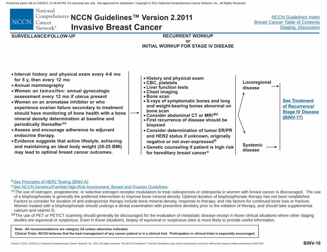
RECURRENT WORKUPor
INITIAL WORKUP FOR STAGE IV DISEASE
SURVEILLANCE/FOLLOW-UP
Locoregionaldisease
Systemicdisease
�
�
�
�
�
�
Interval history and physical exam every 4-6 mofor 5 y, then every 12 moAnnual mammographyWomen on tamoxifen: annual gynecologicassessment every 12 mo if uterus presentWomen on an aromatase inhibitor or whoexperience ovarian failure secondary to treatmentshould have monitoring of bone health with a bonemineral density determination at baseline andperiodically thereafterAssess and encourage adherence to adjuvantendocrine therapy.Evidence suggests that active lifestyle, achievingand maintaining an ideal body weight (20-25 BMI)may lead to optimal breast cancer outcomes.
cc
������
��
History and physical examCBC, plateletsLiver function testsChest imagingBone scanX-rays of symptomatic bones and longand weight-bearing bones abnormal onbone scanConsider abdominal CT or MRIdd
First recurrence of disease should bebiopsied
Genetic counseling if patient is high riskfor hereditary breast cancer
�
�
Consider determination of tumor ER/PRand HER2 status if unknown, originallynegative or not over-expressedb
c
Note: All recommendations are category 2A unless otherwise indicated.Clinical Trials: NCCN believes that the best management of any cancer patient is in a clinical trial. Participation in clinical trials is especially encouraged.
b
c
cc
dd
The use of estrogen, progesterone, or selective estrogen receptor modulators to treat osteoporosis or osteopenia in women with breast cancer is discouraged. The useof a bisphosphonate is generally the preferred intervention to improve bone mineral density. Optimal duration of bisphosphonate therapy has not been established.Factors to consider for duration of anti-osteoporosis therapy include bone mineral density, response to therapy, and risk factors for continued bone loss or fracture.Women treated with a bisphosphonate should undergo a dental examination with preventive dentistry prior to the initiation of therapy, and should take supplementalcalcium and vitamin D.
The use of PET or PET/CT scanning should generally be discouraged for the evaluation of metastatic disease except in those clinical situations where other stagingstudies are equivocal or suspicious. Even in these situations, biopsy of equivocal or suspicious sites is more likely to provide useful information.
See Principles of HER2 Testing (BINV-A
See NCCN Genetics/Familial High-Risk Assessment: Breast and Ovarian Guidelines
).
.
BINV-16
See Treatmentof Recurrence/Stage IV Disease(BINV-17)
Invasive Breast Cancer
Printed by paolo nitti on 2/3/2011 12:45:49 PM. For personal use only. Not approved for distribution. Copyright © 2011 National Comprehensive Cancer Network, Inc., All Rights Reserved.
NCCN®
NCCN Guidelines™ Version 2.2011
Version 2.2011, 01/05/11 © National Comprehensive Cancer Network, Inc. 2011, All rights reserved. The NCCN Guidelines™ and this illustration may not be reproduced in any form without the express written permission of NCCN®.
NCCN Guidelines IndexBreast Cancer Table of Contents
Staging, Discussion
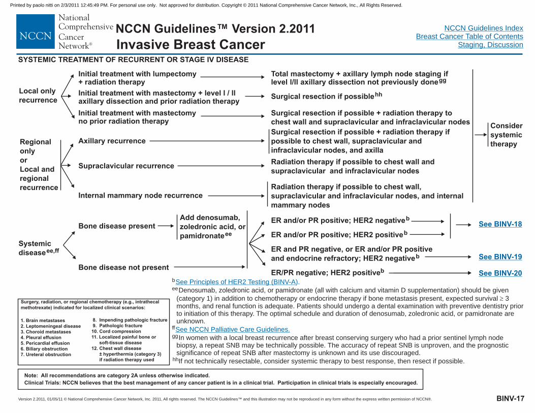
Total mastectomy + axillary lymph node staging iflevel l/ll axillary dissection not previously donegg
Considersystemictherapy
Note: All recommendations are category 2A unless otherwise indicated.Clinical Trials: NCCN believes that the best management of any cancer patient is in a clinical trial. Participation in clinical trials is especially encouraged.
b
ee
gg
Denosumab, zoledronic acid, or pamidronate (all with calcium and vitamin D supplementation) should be given
(category 1) in addition to chemotherapy or endocrine therapy if bone metastasis present, expected survival 3months, and renal function is adequate. Patients should undergo a dental examination with preventive dentistry priorto initiation of this therapy. The optimal schedule and duration of denosumab, zoledronic acid, or pamidronate areunknown.
In women with a local breast recurrence after breast conserving surgery who had a prior sentinel lymph nodebiopsy, a repeat SNB may be technically possible. The accuracy of repeat SNB is unproven, and the prognosticsignificance of repeat SNB after mastectomy is unknown and its use discouraged.
�
ff
hhIf not technically resectable, consider systemic therapy to best response, then resect if possible.
See Principles of HER2 Testing (BINV-A
See NCCN Palliative Care Guidelines.
).
Initial treatment with mastectomy + level l / llaxillary dissection and prior radiation therapy
Initial treatment with lumpectomy+ radiation therapy
Surgical resection if possible + radiation therapy tochest wall and supraclavicular and infraclavicular nodes
BINV-17
Surgery, radiation, or regional chemotherapy (e.g., intrathecalmethotrexate) indicated for localized clinical scenarios:
1. Brain metastases2. Leptomeningeal disease3. Choroid metastases4. Pleural effusion5. Pericardial effusion6. Biliary obstruction7. Ureteral obstruction
8. Impending pathologic fracture9. Pathologic fracture10. Cord compression11. Localized painful bone or
soft-tissue disease12. Chest wall disease
± hyperthermia (category 3)if radiation therapy used
SYSTEMIC TREATMENT OF RECURRENT OR STAGE IV DISEASE
Initial treatment with mastectomyno prior radiation therapy
Surgical resection if possiblehh
Systemicdiseaseee,ff
ER/PR negative; HER2 positiveb
ER and PR negative, or ER and/or PR positiveand endocrine refractory; HER2 negativeb
ER and/or PR positive; HER2 negativeb
ER and/or PR positive; HER2 positivebBone disease present
Bone disease not present
Add denosumab,zoledronic acid, orpamidronateee
See BINV-18
See BINV-19
See BINV-20
Invasive Breast Cancer
Local onlyrecurrence
RegionalonlyorLocal andregionalrecurrence
Axillary recurrence
Supraclavicular recurrence
Internal mammary node recurrence
Surgical resection if possible + radiation therapy ifpossible to chest wall, supraclavicular andinfraclavicular nodes, and axilla
Radiation therapy if possible to chest wall andsupraclavicular and infraclavicular nodes
Radiation therapy if possible to chest wall,supraclavicular and infraclavicular nodes, and internalmammary nodes
Printed by paolo nitti on 2/3/2011 12:45:49 PM. For personal use only. Not approved for distribution. Copyright © 2011 National Comprehensive Cancer Network, Inc., All Rights Reserved.

NCCN®
NCCN Guidelines™ Version 2.2011
Version 2.2011, 01/05/11 © National Comprehensive Cancer Network, Inc. 2011, All rights reserved. The NCCN Guidelines™ and this illustration may not be reproduced in any form without the express written permission of NCCN®.
NCCN Guidelines IndexBreast Cancer Table of Contents
Staging, Discussion
ER and/or PR POSITIVE; HER2 NEGATIVE OR POSITIVE
Note: All recommendations are category 2A unless otherwise indicated.Clinical Trials: NCCN believes that the best management of any cancer patient is in a clinical trial. Participation in clinical trials is especially encouraged.
BINV-18
ER and/or PR positive;HER2 negativeb
ER and/or PR positive;HER2 positiveb
Prior endocrinetherapy within 1y
No prior endocrinetherapy within 1y
Premenopausalaa
Postmenopausalaa,ii
Ovarian ablation orsuppression, plus endocrinetherapy as for postmenopausalwomenorAntiestrogen
jj,kk
kk
Aromatase inhibitororAntiestrogen
kk
kk
b
kk
aa .
Women presenting at time of initial diagnosis with metastatic disease may benefit from the performance of local breast surgery and/or radiation therapy. Generally thispalliative local therapy should be considered only after response to initial systemic therapy.
iiLimited studies document a progression free survival advantage of adding trastuzumab or lapatinib to aromatase inhibition in postmenopausal patients with ER-positive,HER2-positive disease. However, no overall survival advantage has been demonstrated.
ll
See Principles of HER2 Testing (BINV-A
Definition of Menopause (BINV-L)
).
jjSee Subsequent Endocrine Therapy (BINV-M).
See Preferred Chemotherapy Regimens for Recurrent or Metastatic Breast Cancer (BINV-N).
SYSTEMIC TREATMENT OF RECURRENT OR STAGE IV DISEASE
See
(BINV-20)
Follow-up TherapyFor Hormone Treatmentof Recurrence / Stage IVDisease
Visceral crisis
Premenopausalaa
Postmenopausalaa
Visceral crisis
Ovarian ablation orsuppression, plus endocrinetherapy as for postmenopausalwomenjj,kk
Consider initial chemotherapyand
ll
(See BINV-19 BINV-20)
Consider initial chemotherapyand
ll
(See BINV-19 BINV-21)
Invasive Breast Cancer
Printed by paolo nitti on 2/3/2011 12:45:49 PM. For personal use only. Not approved for distribution. Copyright © 2011 National Comprehensive Cancer Network, Inc., All Rights Reserved.
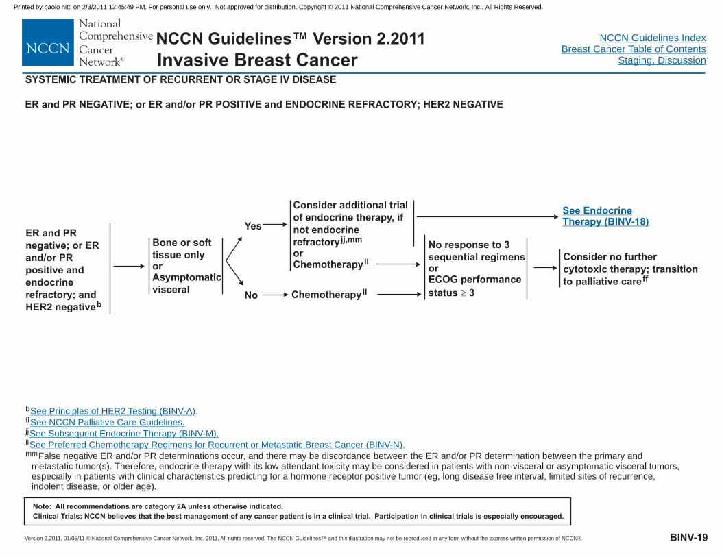
NCCN®
NCCN Guidelines™ Version 2.2011
Version 2.2011, 01/05/11 © National Comprehensive Cancer Network, Inc. 2011, All rights reserved. The NCCN Guidelines™ and this illustration may not be reproduced in any form without the express written permission of NCCN®.
NCCN Guidelines IndexBreast Cancer Table of Contents
Staging, Discussion
Note: All recommendations are category 2A unless otherwise indicated.Clinical Trials: NCCN believes that the best management of any cancer patient is in a clinical trial. Participation in clinical trials is especially encouraged.
b
jj
ff
mm
ll
False negative ER and/or PR determinations occur, and there may be discordance between the ER and/or PR determination between the primary andmetastatic tumor(s). Therefore, endocrine therapy with its low attendant toxicity may be considered in patients with non-visceral or asymptomatic visceral tumors,especially in patients with clinical characteristics predicting for a hormone receptor positive tumor (eg, long disease free interval, limited sites of recurrence,indolent disease, or older age).
See Principles of HER2 Testing (BINV-A
See Subsequent Endocrine Therapy (BINV-M).
).
See NCCN Palliative Care Guidelines.
See Preferred Chemotherapy Regimens for Recurrent or Metastatic Breast Cancer (BINV-N).
BINV-19
ER and PR NEGATIVE; or ER and/or PR POSITIVE and ENDOCRINE REFRACTORY; HER2 NEGATIVE
Bone or softtissue onlyorAsymptomaticvisceral
Yes
No
Consider additional trialof endocrine therapy, ifnot endocrinerefractoryorChemotherapy
jj,mm
ll
Chemotherapyll
No response to 3sequential regimensorECOG performancestatus 3�
Consider no furthercytotoxic therapy; transitionto palliative careff
See EndocrineTherapy (BINV-18)
SYSTEMIC TREATMENT OF RECURRENT OR STAGE IV DISEASE
ER and PRnegative; or ERand/or PRpositive andendocrinerefractory; andHER2 negativeb
Invasive Breast Cancer
Printed by paolo nitti on 2/3/2011 12:45:49 PM. For personal use only. Not approved for distribution. Copyright © 2011 National Comprehensive Cancer Network, Inc., All Rights Reserved.

NCCN®
NCCN Guidelines™ Version 2.2011
Version 2.2011, 01/05/11 © National Comprehensive Cancer Network, Inc. 2011, All rights reserved. The NCCN Guidelines™ and this illustration may not be reproduced in any form without the express written permission of NCCN®.
NCCN Guidelines IndexBreast Cancer Table of Contents
Staging, Discussion
SYSTEMIC TREATMENT OF RECURRENT OR STAGE IV DISEASE
Note: All recommendations are category 2A unless otherwise indicated.Clinical Trials: NCCN believes that the best management of any cancer patient is in a clinical trial. Participation in clinical trials is especially encouraged.
b
jj
ff
ll
nn
oo
mmFalse negative ER and/or PR determinations occur, and there may be discordance between the ER and/or PR determination between the primary and metastatictumor(s). Therefore, endocrine therapy with its low attendant toxicity may be considered in patients with non-visceral or asymptomatic visceral tumors, especially inpatients with clinical characteristics predicting for a hormone receptor positive tumor (eg, long disease free interval, limited sites of recurrence, indolent disease, or olderage).
Continued trastuzumab following progression on first line-trastuzumab containing chemotherapy for metastatic breast cancer. The optimal duration of trastuzumab inpatients with long-term control of disease is unknown.
Trastuzumab given in combination with an anthracycline is associated with significant cardiac toxicity.
See Principles of HER2 Testing (BINV-A
See Subsequent Endocrine Therapy (BINV-M).
).
See NCCN Palliative Care Guidelines.
See Preferred Chemotherapy Regimens for Recurrent or Metastatic Breast Cancer (BINV-N).
ER and PR NEGATIVE; or ER and/or PR POSITIVE and ENDOCRINE REFRACTORY; and HER2 POSITIVE
BINV-20
ER and PRnegative; or ERand/or PRpositive andendocrinerefractory; andHER2 positiveb
No responseto 3 sequentialregimensorECOGperformancestatus 3�
Consider nofurther cytotoxictherapy;transition topalliative careff
Bone or softtissue onlyorAsymptomaticvisceral
Yes
No
Consider trial ofendocrine therapy, if notendocrine refractoryjj,mm
Trastuzumab ±chemotherapyll,nn,oo
Continue HER2targeted therapy,typically incombination withother chemotherapyor trastuzumab +lapatinib
See EndocrineTherapy (BINV-18)
Invasive Breast Cancer
Printed by paolo nitti on 2/3/2011 12:45:49 PM. For personal use only. Not approved for distribution. Copyright © 2011 National Comprehensive Cancer Network, Inc., All Rights Reserved.
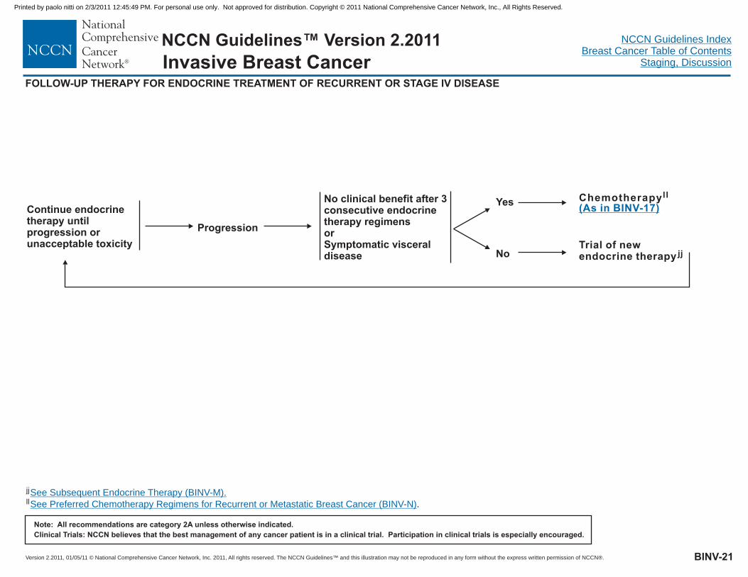
NCCN®
NCCN Guidelines™ Version 2.2011
Version 2.2011, 01/05/11 © National Comprehensive Cancer Network, Inc. 2011, All rights reserved. The NCCN Guidelines™ and this illustration may not be reproduced in any form without the express written permission of NCCN®.
NCCN Guidelines IndexBreast Cancer Table of Contents
Staging, Discussion
Note: All recommendations are category 2A unless otherwise indicated.Clinical Trials: NCCN believes that the best management of any cancer patient is in a clinical trial. Participation in clinical trials is especially encouraged.
FOLLOW-UP THERAPY FOR ENDOCRINE TREATMENT OF RECURRENT OR STAGE IV DISEASE
jj
ll .
See Subsequent Endocrine Therapy (BINV-M).
See Preferred Chemotherapy Regimens for Recurrent or Metastatic Breast Cancer (BINV-N)
BINV-21
Continue endocrinetherapy untilprogression orunacceptable toxicity
No clinical benefit after 3consecutive endocrinetherapy regimensorSymptomatic visceraldisease
Chemotherapyll
(As in BINV-17)Yes
NoTrial of newendocrine therapy jj
Progression
Invasive Breast Cancer
Printed by paolo nitti on 2/3/2011 12:45:49 PM. For personal use only. Not approved for distribution. Copyright © 2011 National Comprehensive Cancer Network, Inc., All Rights Reserved.

NCCN®
NCCN Guidelines™ Version 2.2011
Version 2.2011, 01/05/11 © National Comprehensive Cancer Network, Inc. 2011, All rights reserved. The NCCN Guidelines™ and this illustration may not be reproduced in any form without the express written permission of NCCN®.
NCCN Guidelines IndexBreast Cancer Table of Contents
Staging, Discussion
Note: All recommendations are category 2A unless otherwise indicated.Clinical Trials: NCCN believes that the best management of any cancer patient is in a clinical trial. Participation in clinical trials is especially encouraged.
BINV-A
PRINCIPLES OF HER2 TESTING1,2
Initial testingby IHC3
Laboratory meetsquality assurancestandards for IHCHER2 testingmethodology Yes
No
IHCtesting
IHC 0,1+
IHC 2+
IHC 3+
HER2 (-)
Borderlineresult4 FISH testing
HER2 (+)
Send sample to reference laboratory
Yes
No
FISH retest
FISH (-)
FISH+
HER2 (-)
Borderlineresult5
HER2 (+)
Send sample to reference laboratory
IHC testing
Countadditional cells
Borderline result
HER2 (-)FISHtesting
Initial testingby FISH3
Laboratory meetsquality assurancestandards for FISHHER2 testingmethodology
1
2
5
See also, Carlson RW, Moench SJ, Hammond, MEH, et al. HER2 testing in breast cancer: NCCN task force report and recommendations. JNCCN 4:S-1-S-24, 2006.
a
HER2 testing should be done only in laboratories accredited to perform such testing. Ongoing proficiency testing and full reporting of HER2 assay methods and resultsare required. A laboratory may perform only those tests which have been demonstrated to conform to these quality assurance standards. All other HER2 testing shouldbe sent to a qualified reference laboratory.
3Eithe an immunohistochemistry (IHC) assay or a fluorescence in situ hybridization (FISH) assay can be used to make an initial assessment of HER2 tumor status. AllHER2 assays, whether FDA-approved or not, mu be validated. Validation of a HER2 test is defined as at least 95% concordance when the testing method performedin a laboratory is compared with one of the following: a validated HER2 testing method performed in the same laboratory; a validated HER2 testing method performedin another laboratory; or validated reference lab results. Borderline samples should not be included in the validation study. These algorithms are based on theassumption that all validated HER2 tests have been shown to be at least 95% concordant with the complementary form of the HER2 test, either by direct testing orassociation with the levels of concordance between complementary testing achieved by the validating laboratory.
Borderline IHC samples (eg, IHC 2+) are subjected to reflex testing by a validated complementary (eg, FISH) method that has shown at least 95% concordancebetween IHC 0, 1+ results and FISH non-amplified results, and IHC 3+ results and FISH amplified results.
Borderline FISH samples (eg, an average HER2 gene/chromosome 17 ratio of 1.8-2.2 or an average HER2 gene copy number of > 4 - < 6) should undergo: counting ofadditional cells; retesting by FISH; or reflex testing by a valid ted IHC method which is at least 95% concordant with FISH as described above.
rst
4
Invasive Breast Cancer
Printed by paolo nitti on 2/3/2011 12:45:49 PM. For personal use only. Not approved for distribution. Copyright © 2011 National Comprehensive Cancer Network, Inc., All Rights Reserved.

NCCN®
NCCN Guidelines™ Version 2.2011
Version 2.2011, 01/05/11 © National Comprehensive Cancer Network, Inc. 2011, All rights reserved. The NCCN Guidelines™ and this illustration may not be reproduced in any form without the express written permission of NCCN®.
NCCN Guidelines IndexBreast Cancer Table of Contents
Staging, Discussion
Note: All recommendations are category 2A unless otherwise indicated.Clinical Trials: NCCN believes that the best management of any cancer patient is in a clinical trial. Participation in clinical trials is especially encouraged.
BINV-B
See NCCN Breast Screening and Diagnosis Guidelines for indications for screening MRI in women at increased breast cancer risk.
Personnel, facility and equipmentBreast MRI examinations should be performed and interpreted by an expert breast imaging team working in concert with themultidisciplinary treatment team.Breast MRI examinations require a dedicated breast coil and breast imaging radiologists familiar with the optimal timing sequences andother technical details for image interpretation. The imaging center should have the ability to perform MRI guided needle samplingand/or wire localization of MRI detected findings.
Clinical indications and applicationsMay be used for staging evaluation to define extent of cancer or presence of multifocal or multicentric cancer in the ipsilateral breast, oras screening of the contralateral breast cancer at time of initial diagnosis (category 2B). There are no data that demonstrate that use ofMRI to affect choice of local therapy improves outcome (local recurrence or survival).May be helpful for breast cancer evaluation before and after neoadjuvant therapy to define extent of disease, response to treatment, andpotential for breast conserving therapy.May be useful to detect additional disease in women with mammographically dense breast, but available data do not show differentialdetection rates by any subset by breast pattern (breast density) or disease type (eg. DCIS, invasive ductal cancer, invasive lobularcancer)May be useful for identifying primary cancer in women with axillary nodal adenocarcinoma or with Paget’s disease of the nipple withbreast primary not identified on mammography, ultrasound, or physical examinationFalsely positive findings on breast MRI are common. Surgical decisions should not be based solely on the MRI findings. Additionaltissue sampling of areas of concern identified by breast MRI is recommended.The utility of MRI in follow-up screening of women with prior breast cancer is undefined. It should generally be considered only in thosewhose lifetime risk of a second primary breast cancer is greater than 20% based on models largely dependent on family history, such asin those with the risk associated with inherited susceptibility to breast cancer.
�
�
�
�
�
�
�
�
PRINCIPLES OF DEDICATED BREAST MRI TESTING
Invasive Breast Cancer
Houssami N, Ciatto S, Macaskill P, Lord SJ, Warren RM, Dixon JM, Irwig L. Accuracy and surgical impact of magnetic resonance imaging in breast cancer staging:
systematic review and meta-analysis in detection of multifocal and multicentric cancer. J Clin Oncol 2008;26:3248-3258.
Printed by paolo nitti on 2/3/2011 12:45:49 PM. For personal use only. Not approved for distribution. Copyright © 2011 National Comprehensive Cancer Network, Inc., All Rights Reserved.

NCCN®
NCCN Guidelines™ Version 2.2011
Version 2.2011, 01/05/11 © National Comprehensive Cancer Network, Inc. 2011, All rights reserved. The NCCN Guidelines™ and this illustration may not be reproduced in any form without the express written permission of NCCN®.
NCCN Guidelines IndexBreast Cancer Table of Contents
Staging, Discussion
FERTILITY AND BIRTH CONTROL AFTER ADJUVANT BREAST CANCER TREATMENT
Although amenorrhea frequently occurs during or after chemotherapy it appears the majority of women younger than 35 y resume menseswithin 2 y of finishing adjuvant chemotherapyMenses and fertility are not necessarily linked. Absence of regular menses, particulary if the patient is taking tamoxifen does not necessarilyimply lack of fertility. Conversely, the presence of menses does not guarantee fertrility. There is limited data regarding continued fertility afterchemotherapyPatients should not become pregnant during treatment with radiation therapy, chemotherapy, or endocrine therapy.Although data are limited, hormone-based birth control is discouraged regardless of the hormone receptor status of the patient's cancerAlternative methods of birth control include intrauterine devices (IUD), barrier methods or for patients with no intent for future pregnancies,tubal ligation or vasectomy for the partner.No therapy has been shown to preserve fertility in patients receiving chemotherapyPatients who may desire future pregnancies should be referred to fertility specialists before chemotherapyBreast feeding following breast conserving cancer treatment is not contraindicated. However the quantity and quality of breast milkproduced by the breast conserved may not be sufficient or may be lacking some of the nutrients needed. Breast feeding during activetreatment with chemotherapy and endocrine therapy is not recommended.
�
�
�
�
�
�
�
�
Invasive Breast Cancer
Note: All recommendations are category 2A unless otherwise indicated.Clinical Trials: NCCN believes that the best management of any cancer patient is in a clinical trial. Participation in clinical trials is especially encouraged.
BINV-C
Printed by paolo nitti on 2/3/2011 12:45:49 PM. For personal use only. Not approved for distribution. Copyright © 2011 National Comprehensive Cancer Network, Inc., All Rights Reserved.

NCCN®
NCCN Guidelines™ Version 2.2011
Version 2.2011, 01/05/11 © National Comprehensive Cancer Network, Inc. 2011, All rights reserved. The NCCN Guidelines™ and this illustration may not be reproduced in any form without the express written permission of NCCN®.
NCCN Guidelines IndexBreast Cancer Table of Contents
Staging, Discussion
SURGICAL AXILLARY STAGING - STAGE I, IIA, AND IIB
ClinicalStage I/II
No
Yes
Refer to experienced sentinel node team1,5
Sentinel nodemapping andexcision 3,4,5
Clinically nodepositive at timeof diagnosis2
Clinically nodenegative at timeof diagnosis
Sentinel nodenegative6
No furthersurgery(category 1)
Sentinel nodepositive6
Sentinel nodenot identified
1
2
3
4
6
Sentinel node team must have documented experience with sentinel node biopsy in breast cancer. Team includes surgeon, radiologists, nuclear medicine physician,pathologist, and prior discussion with medical and radiation oncologists on use of sentinel node for treatment decisions.
Consider pathologic confirmation of malignancy in clinically positive nodes using ultrasound guided FNA or core biopsy in determining if patient needs axillary lymphnode dissection.
Axillary sentinel node biopsy in all cases; internal mammary sentinel node biopsy optional if drainage maps to internal mammary nodes (category 3).
Sentinel lymph node mapping injections may be peritumoral, subareolar or subdermal. However, only peritumoral injections map to the internal mammary lymph node(s).
Results of randomized clinical trials indicate that there is a lower risk of mobidity associated with sentinel node mapping and excision than with level l/ll axillarydissection.
Sentinel node involvement is defined by multilevel node sectioning with hematoxylin and eosin (H&E) staining. Cytokeratin Immunohistochemistry (IH ) may be used forequivocal cases on H&E. Routine cytokeratin IHC to define node involvement is not recommended in clinical decision making.
Data from a single, randomized trial suggests that complete axillary lymph node dissection in women with clinically node negative T1-T2 tumors, fewer than 3 involvedsentinel lymph nodes, and undergoing breast-conserving surgery and whole breast radiation results in more morbidity, no improvement in locoregional recurrence rates,and no difference in overall survival compared with sentinel lymph node procedure alone.
5
7
C
Sentinellymph nodecandidate
Note: All recommendations are category 2A unless otherwise indicated.Clinical Trials: NCCN believes that the best management of any cancer patient is in a clinical trial. Participation in clinical trials is especially encouraged.
Return to LocoregionalTreatment (BINV-2)
BINV-D
FNA or corebiopsy negative
Axillarydissectionlevel I/II7
Axillary dissection level I/IIExperiencedsentinel nodeteam1
Axillary dissection level I/II
No
Yes
Invasive Breast Cancer
Printed by paolo nitti on 2/3/2011 12:45:49 PM. For personal use only. Not approved for distribution. Copyright © 2011 National Comprehensive Cancer Network, Inc., All Rights Reserved.

NCCN®
NCCN Guidelines™ Version 2.2011
Version 2.2011, 01/05/11 © National Comprehensive Cancer Network, Inc. 2011, All rights reserved. The NCCN Guidelines™ and this illustration may not be reproduced in any form without the express written permission of NCCN®.
NCCN Guidelines IndexBreast Cancer Table of Contents
Staging, Discussion
In the absence of definitive data demonstrating superior survival from the performance of axillary lymph node dissection, patients who haveparticularly favorable tumors, patients for whom the selection of adjuvant systemic therapy is unlikely to be affected, for the elderly, or thosewith serious comorbid conditions, the performance of axillary lymph node dissection may be considered optional. The axillary dissectionshould be extended to include level lll nodes only if there is gross disease apparent in the level ll nodes.
Sentinel lymph node biopsy is the preferred method of axillary lymph node staging if there is an experienced sentinel node team and thepatient is an appropriate sentinel lymph node biopsy candidate (See BINV-D).
AXILLARY LYMPH NODE STAGING
Note: All recommendations are category 2A unless otherwise indicated.Clinical Trials: NCCN believes that the best management of any cancer patient is in a clinical trial. Participation in clinical trials is especially encouraged.
BINV-E
Invasive Breast Cancer
Printed by paolo nitti on 2/3/2011 12:45:49 PM. For personal use only. Not approved for distribution. Copyright © 2011 National Comprehensive Cancer Network, Inc., All Rights Reserved.

NCCN®
NCCN Guidelines™ Version 2.2011
Version 2.2011, 01/05/11 © National Comprehensive Cancer Network, Inc. 2011, All rights reserved. The NCCN Guidelines™ and this illustration may not be reproduced in any form without the express written permission of NCCN®.
NCCN Guidelines IndexBreast Cancer Table of Contents
Staging, Discussion
Note: All recommendations are category 2A unless otherwise indicated.Clinical Trials: NCCN believes that the best management of any cancer patient is in a clinical trial. Participation in clinical trials is especially encouraged.
BINV-F
MARGIN STATUS IN INFILTRATING CARCINOMA
The use of breast conserving therapy is predicated on achieving a pathologically negative margin of resection. Cases where there is a positivemargin should generally undergo further surgery, either a re-excision to achieve a negative margin or a mastectomy. If re-excision istechnically feasible to allow for breast conserving therapy, this can be done with resection of the involved margin guided by the orientation ofthe initial resection specimen or re-excision of the entire original excision cavity. If multiple margins remain positive, mastectomy may berequired for optimal local control.
It may be reasonable to treat selected cases with breast conserving therapy with a microscopically focally positive margin in the absence of anextensive intraductal component. For these patients, the use of a higher radiation boost dose to the tumor bed should be considered.
Margins should be evaluated on all surgical specimens from breast conserving surgery. Requirements for optimal margin evaluation include:Orientation of the surgical specimensDescription of the gross and microscopic margin statusReporting of the distance, orientation, and type of tumor (invasive or DCIS) in relation to the closest margin.
1
�
�
�
1An extensive intraductal component is defined as an infiltrating ductal cancer where greater than 25% of the tumor volume is DCIS andDCIS extends beyond the invasive cancer into surrounding normal breast parenchyma.
Invasive Breast Cancer
Printed by paolo nitti on 2/3/2011 12:45:49 PM. For personal use only. Not approved for distribution. Copyright © 2011 National Comprehensive Cancer Network, Inc., All Rights Reserved.
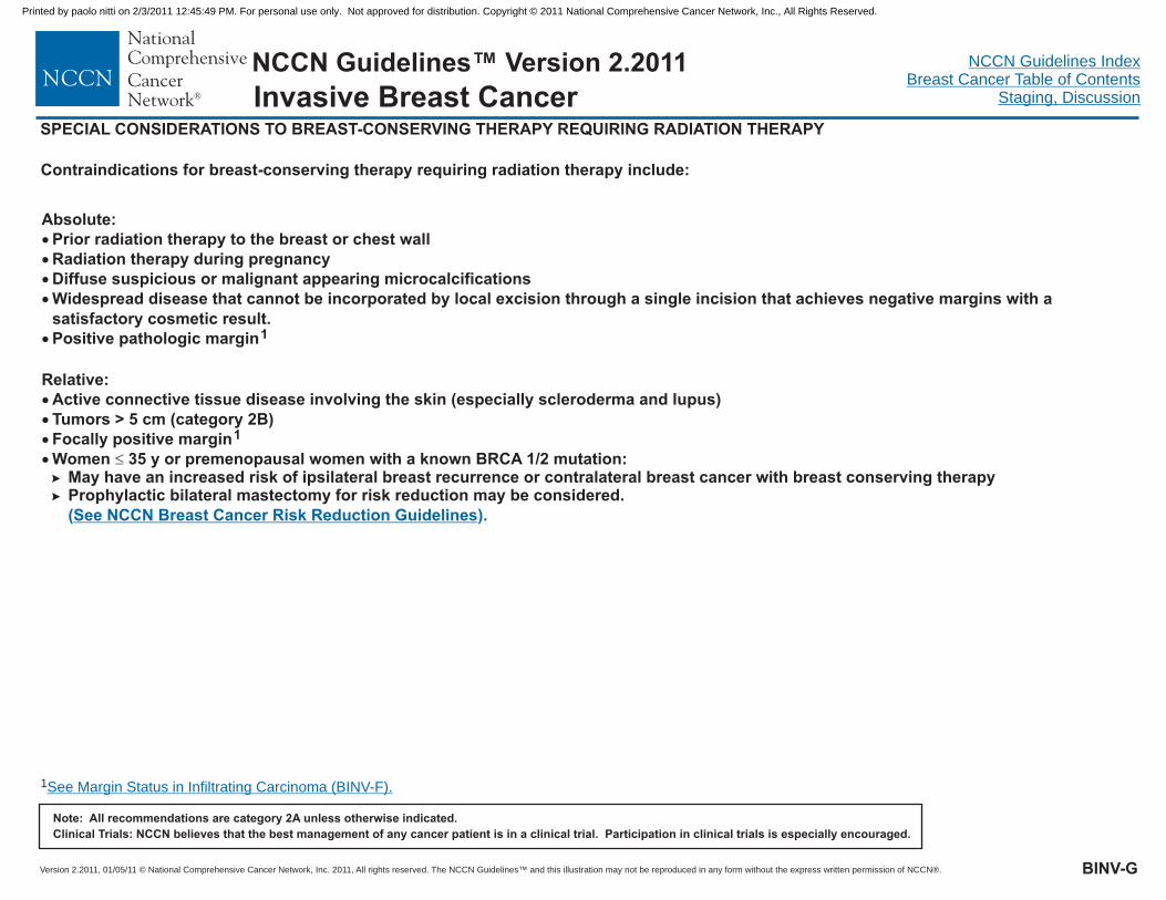
NCCN®
NCCN Guidelines™ Version 2.2011
Version 2.2011, 01/05/11 © National Comprehensive Cancer Network, Inc. 2011, All rights reserved. The NCCN Guidelines™ and this illustration may not be reproduced in any form without the express written permission of NCCN®.
NCCN Guidelines IndexBreast Cancer Table of Contents
Staging, Discussion
SPECIAL CONSIDERATIONS TO BREAST-CONSERVING THERAPY REQUIRING RADIATION THERAPY
Note: All recommendations are category 2A unless otherwise indicated.Clinical Trials: NCCN believes that the best management of any cancer patient is in a clinical trial. Participation in clinical trials is especially encouraged.
Contraindications for breast-conserving therapy requiring radiation therapy include:
Absolute:
Relative:
�
�
�
�
�
�
�
�
�
Prior radiation therapy to the breast or chest wallRadiation therapy during pregnancyDiffuse suspicious or malignant appearing microcalcificationsWidespread disease that cannot be incorporated by local excision through a single incision that achieves negative margins with asatisfactory cosmetic result.Positive pathologic margin
Active connective tissue disease involving the skin (especially scleroderma and lupus)Tumors > 5 cm (category 2B)Focally positive margin
May have an increased risk of ipsilateral breast recurrence or contralateral breast cancer with breast conserving therapyProphylactic bilateral mastectomy for risk reduction may be considered.
1
1
Women 35 y or premenopausal women with a known BRCA 1/2 mutation:��
�
( ).See NCCN Breast Cancer Risk Reduction Guidelines
BINV-G
1See Margin Status in Infiltrating Carcinoma (BINV-F).
Invasive Breast Cancer
Printed by paolo nitti on 2/3/2011 12:45:49 PM. For personal use only. Not approved for distribution. Copyright © 2011 National Comprehensive Cancer Network, Inc., All Rights Reserved.
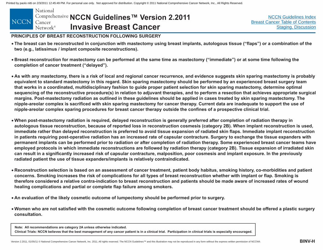
NCCN®
NCCN Guidelines™ Version 2.2011
Version 2.2011, 01/05/11 © National Comprehensive Cancer Network, Inc. 2011, All rights reserved. The NCCN Guidelines™ and this illustration may not be reproduced in any form without the express written permission of NCCN®.
NCCN Guidelines IndexBreast Cancer Table of Contents
Staging, Discussion
Note: All recommendations are category 2A unless otherwise indicated.Clinical Trials: NCCN believes that the best management of any cancer patient is in a clinical trial. Participation in clinical trials is especially encouraged.
BINV-H
�
�
�
�
�
�
�
The breast can be reconstructed in conjunction with mastectomy using breast implants, autologous tissue (“flaps”) or a combination of thetwo (e.g., latissimus / implant composite reconstructions).
Breast reconstruction for mastectomy can be performed at the same time as mastectomy (“immediate”) or at some time following thecompletion of cancer treatment (“delayed”).
As with any mastectomy, there is a risk of local and regional cancer recurrence, and evidence suggests skin sparing mastectomy is probablyequivalent to standard mastectomy in this regard. Skin sparing mastectomy should be performed by an experienced breast surgery teamthat works in a coordinated, multidisciplinary fashion to guide proper patient selection for skin sparing mastectomy, determine optimalsequencing of the reconstructive procedure(s) in relation to adjuvant therapies, and to perform a resection that achieves appropriate surgicalmargins. Post-mastectomy radiation as outlined in these guidelines should be applied in cases treated by skin sparing mastectomy. Thenipple-areolar complex is sacrificed with skin sparing mastectomy for cancer therapy. Current data are inadequate to support the use ofnipple-areolar complex sparing procedures for breast cancer therapy outside the confines of a prospective clinical trial.
When post-mastectomy radiation is required, delayed reconstruction is generally preferred after completion of radiation therapy inautologous tissue reconstruction, because of reported loss in reconstruction cosmesis (category 2B). When implant reconstruction is used,immediate rather than delayed reconstruction is preferred to avoid tissue expansion of radiated skin flaps. Immediate implant reconstructionin patients requiring post-operative radiation has an increased rate of capsular contracture. Surgery to exchange the tissue expanders withpermanent implants can be performed prior to radiation or after completion of radiation therapy. Some experienced breast cancer teams haveemployed protocols in which immediate reconstructions are followed by radiation therapy (category 2B). Tissue expansion of irradiated skincan result in a significantly increased risk of capsular contracture, malposition, poor cosmesis and implant exposure. In the previouslyradiated patient the use of tissue expanders/implants is relatively contraindicated.
Reconstruction selection is based on an assessment of cancer treatment, patient body habitus, smoking history, co-morbidities and patientconcerns. Smoking increases the risk of complications for all types of breast reconstruction whether with implant or flap. Smoking istherefore considered a relative contra-indication to breast reconstruction and patients should be made aware of increased rates of woundhealing complications and partial or complete flap failure among smokers.
An evaluation of the likely cosmetic outcome of lumpectomy should be performed prior to surgery.
Women who are not satisfied with the cosmetic outcome following completion of breast cancer treatment should be offered a plastic surgeryconsultation.
PRINCIPLES OF BREAST RECONSTRUCTION FOLLOWING SURGERY
Invasive Breast Cancer
Printed by paolo nitti on 2/3/2011 12:45:49 PM. For personal use only. Not approved for distribution. Copyright © 2011 National Comprehensive Cancer Network, Inc., All Rights Reserved.

NCCN®
NCCN Guidelines™ Version 2.2011
Version 2.2011, 01/05/11 © National Comprehensive Cancer Network, Inc. 2011, All rights reserved. The NCCN Guidelines™ and this illustration may not be reproduced in any form without the express written permission of NCCN®.
NCCN Guidelines IndexBreast Cancer Table of Contents
Staging, Discussion
PRINCIPLES OF RADIATION THERAPY
Note: All recommendations are category 2A unless otherwise indicated.Clinical Trials: NCCN believes that the best management of any cancer patient is in a clinical trial. Participation in clinical trials is especially encouraged.
BINV-I
Whole Breast Radiation:Target definition includes the majority of the breast tissue, and isbest done by both clinical assessment and CT-based treatmentplanning. A uniform dose distribution and minimal normal tissuetoxicity are the goals and can be accomplished using compensatorssuch as wedges, forward planning using segments, intensitymodulated radiation therapy (IMRT), respiratory gating, or pronepositioning. The breast should receive a dose of 45-50 Gy in 1.8 - 2Gy per fraction, or 42.5 Gy at 2.66 Gy per fraction. A boost to thetumor bed is recommended in patients at higher risk for local failure,(age < 50, positive axillary nodes, lymphovascular invasion, or closemargins). This can be achieved with brachytherapy or electron beamor photon fields. Typical doses are 10-16 Gy at 2 Gy/fx. All doseschedules are given 5 days per week.
Chest Wall Radiation (including breast reconstruction):The target includes the ipsilateral chest wall, mastectomy scar, anddrain sites where possible. Depending on whether the patient hasbeen reconstructed or not, several techniques using photons and/orelectrons are appropriate. CT-based treatment planning isencouraged, in order to identify lung and heart volumes, andminimize exposure of these organs. Special consideration should begiven to the use of bolus material when photon fields are used, toensure the skin dose is adequate.
Regional Nodal Radiation:Target delineation is best achieved by the use of CT-based treatmentplanning. For the paraclavicular and axillary nodes, prescriptiondepth varies based on the size of the patient. For internal mammarynode identification, the internal mammary artery and vein locationcan be used as a surrogate for the nodal locations, which usually arenot visible on imaging.
Dose is 50 - 50.4 Gy, given as 1.8 - 2.0 Gy fraction size (± scar boost at 2Gy per fraction to a total dose of approximately 60 Gy); all doseschedules given 5 days per week. If internal mammary lymph nodes areclinically or pathologically positive, radiation therapy should be given tothe internal mammary nodes, otherwise the treatment to the internalmammary nodes is at the discretion of the treating radiation oncologist.CT treatment planning should be utilized in all cases where radiationtherapy is delivered to the internal mammary lymph node field.
Accelerated Partial Breast Irradiation (APBI):Preliminary studies of APBI suggest rates of local control in selectedpatients with early stage breast cancer may be comparable to thosetreated with standard whole breast RT. Follow-up, however, is limitedand studies are on-going. Patients are encouraged to participate inclinical trials. If not trial eligible, per the consensus statement from theAmerican Society for Radiation Oncology (ASTRO), patients who may besuitable APBI are women 60 y and older who are not carriers of BRCA1/2 mutation treated with primary surgery for a unifocal T1N0 ER-positive cancer. Histology should be infiltrating ductal or a favorableductal subtype, not be associated with EIC or LCIS and margins shouldbe negative. 34 Gy in 10 fractions delivered twice per day withbrachytherapy or 38.5 Gy in 10 fractions delivered twice per day withexternal beam photon therapy is prescribed to the tumor bed. Otherfractionation schemes are currently under investigation.
Optimizing Delivery of Individual Therapy:It is important to individualize delivery of radiation therapy andconsiderations such as patient positioning (ie., prone vs supine) duringadministration of radiation therapy.
Neoadjuvant chemotherapy:Indications for radiation therapy and fields of treatment should be basedupon the pretreatment tumor characteristics in patients treated withneoadjuvant chemotherapy.
Invasive Breast Cancer
Printed by paolo nitti on 2/3/2011 12:45:49 PM. For personal use only. Not approved for distribution. Copyright © 2011 National Comprehensive Cancer Network, Inc., All Rights Reserved.
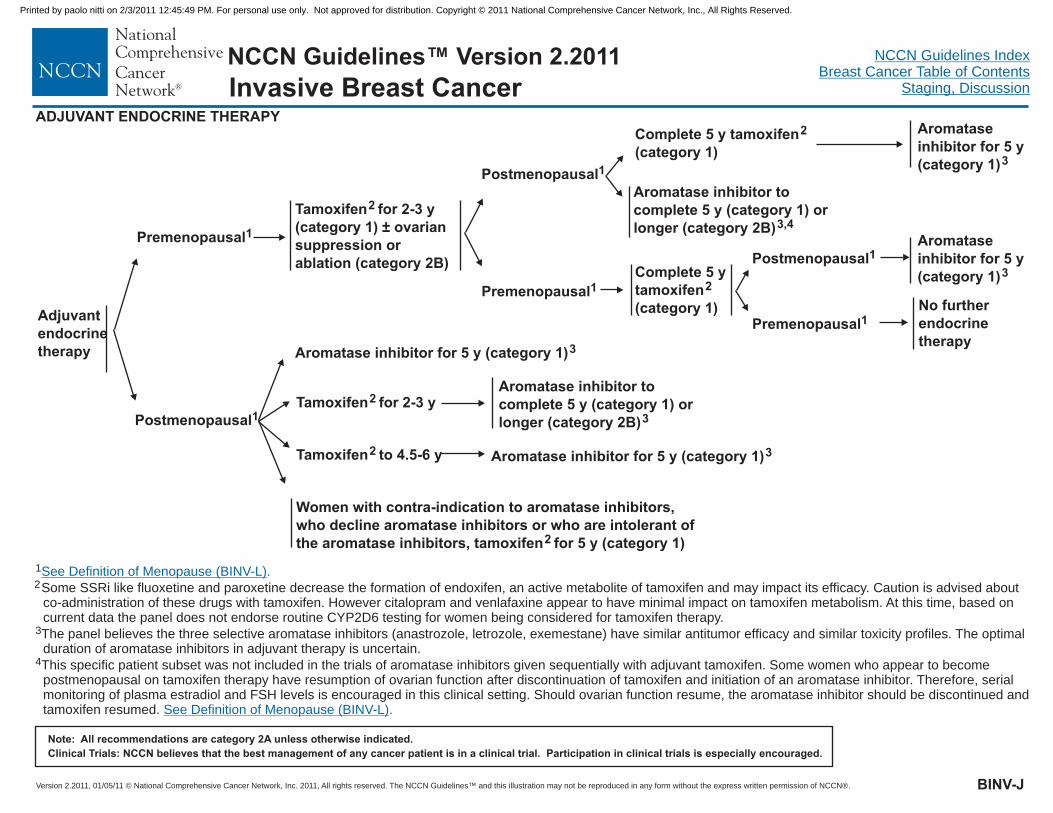
NCCN®
NCCN Guidelines™ Version 2.2011
Version 2.2011, 01/05/11 © National Comprehensive Cancer Network, Inc. 2011, All rights reserved. The NCCN Guidelines™ and this illustration may not be reproduced in any form without the express written permission of NCCN®.
NCCN Guidelines IndexBreast Cancer Table of Contents
Staging, Discussion
Note: All recommendations are category 2A unless otherwise indicated.Clinical Trials: NCCN believes that the best management of any cancer patient is in a clinical trial. Participation in clinical trials is especially encouraged.
1
2Some SSRi like fluoxetine and paroxetine decrease the formation of endoxifen, an active metabolite of tamoxifen and may impact its efficacy. Caution is advised aboutco-administration of these drugs with tamoxifen. However citalopram and venlafaxine appear to have minimal impact on tamoxifen metabolism. At this time, based oncurrent data the panel does not endorse routine CYP2D6 testing for women being considered for tamoxifen therapy.
3
4
The panel believes the three selective aromatase inhibitors (anastrozole, letrozole, exemestane) have similar antitumor efficacy and similar toxicity profiles. The optimalduration of aromatase inhibitors in adjuvant therapy is uncertain.
This specific patient subset was not included in the trials of aromatase inhibitors given sequentially with adjuvant tamoxifen. Some women who appear to becomepostmenopausal on tamoxifen therapy have resumption of ovarian function after discontinuation of tamoxifen and initiation of an aromatase inhibitor. Therefore, serialmonitoring of plasma estradiol and FSH levels is encouraged in this clinical setting. Should ovarian function resume, the aromatase inhibitor should be discontinued andtamoxifen resumed.
See Definition of Menopause (BINV-L)
See Definition of Menopause (BINV-L)
.
.
ADJUVANT ENDOCRINE THERAPY
Adjuvantendocrinetherapy
Premenopausal1
Postmenopausal1
Tamoxifen for 2-3 y(category 1) ± ovariansuppression orablation (category 2B)
2
Complete 5 ytamoxifen(category 1)
2
Aromataseinhibitor for 5 y(category 1)3
No furtherendocrinetherapy
Tamoxifen for 2-3 y2
Tamoxifen to 4.5-6 y2
Women with contra-indication to aromatase inhibitors,who decline aromatase inhibitors or who are intolerant ofthe aromatase inhibitors, tamoxifen for 5 y (category 1)2
Aromatase inhibitor tocomplete 5 y (category 1) orlonger (category 2B)3
Postmenopausal1
Premenopausal1
Postmenopausal1
Premenopausal1
Complete 5 y tamoxifen(category 1)
2 Aromataseinhibitor for 5 y(category 1)3
Aromatase inhibitor tocomplete 5 y (category 1) orlonger (category 2B)3,4
Aromatase inhibitor for 5 y (category 1)3
Aromatase inhibitor for 5 y (category 1)3
BINV-J
Invasive Breast Cancer
Printed by paolo nitti on 2/3/2011 12:45:49 PM. For personal use only. Not approved for distribution. Copyright © 2011 National Comprehensive Cancer Network, Inc., All Rights Reserved.

NCCN®
NCCN Guidelines™ Version 2.2011
Version 2.2011, 01/05/11 © National Comprehensive Cancer Network, Inc. 2011, All rights reserved. The NCCN Guidelines™ and this illustration may not be reproduced in any form without the express written permission of NCCN®.
NCCN Guidelines IndexBreast Cancer Table of Contents
Staging, Discussion
Preferred Adjuvant Regimens:TACDose-dense AC followed by paclitaxelevery 2 weeksAC followed by weekly paclitaxelTCAC
Other Adjuvant Regimens:FAC/CAFFEC/CEFCMF (AC followed by docetaxel every 3 weeksEC (A followed by T followed by C
FEC followed by T
�
�
�
�
�
�
�
�
�
�
�
�
�
(docetaxel/doxorubicin/cyclophosphamide)(doxorubicin/cyclophosphamide)
(doxorubicin/cyclophosphamide)(docetaxel and cyclophosphamide)(doxorubicin/cyclophosphamide)
(fluorouracil/doxorubicin/cyclophosphamide)(cyclophosphamide/epirubicin/fluorouracil)
cyclophosphamide/methotrexate/fluorouracil)
epirubicin/cyclophosphamide)(doxorubicin followed by paclitaxel followed
by cyclophosphamide) every 2 weekly regimen with filgrastim support
(fluorouracil/epirubicin/cyclophosphamide followed by docetaxel)FEC (fluorouracil/epirubicin/cyclophosphamide) followed by weekly paclitaxel
NON-TRASTUZUMAB CONTAINING REGIMENS (all category 1) TRASTUZUMAB CONTAINING REGIMENS (all category 1)
Preferred Adjuvant Regimen:
Other Adjuvant Regimens:Docetaxel + trastuzumab followed by FEC(Chemotherapy followed by trastuzumab sequentiallyAC followed by docetaxel + trastuzumab
Neoadjuvant:
�
�
�
�
�
�
AC followed by T + concurrent trastuzumab(doxorubicin/cyclophosphamide followed by paclitaxel plustrastuzumab, various schedules)TCH (docetaxel, carboplatin, trastuzumab)
T + trastuzumab followed by CEF + trastuzumab(paclitaxel plus trastuzumab followed bycyclophosphamide/epirubicin/fluorouracil plus trastuzumab)
fluorouracil/epirubicin/cyclophosphamide)
1
2
3
4
Retrospective evidence suggests that anthracycline-based chemotherapy regimens may be superior to non-anthracycline-based regimens in patients with HER2positive tumors.
CMF and radiation therapy may be given concurrently, or the CMF may be given first. All other chemotherapy regimens should be given prior to radiotherapy.
Chemotherapy and tamoxifen used as adjuvant therapy should be given sequentially with tamoxifen following chemotherapy.
Randomized clinical trials demonstrate that the addition of a taxane to anthracycline-based chemotherapy provides an improved outcome.
In patients with HER2 positive and axillary lymph node positive breast cancer, trastuzumab should be incorporated into the adjuvant therapy. (category 1)Trastuzumab should also be considered for patients with lymph node negative tumors greater than or equal to 1 cm. (category 1) Trastuzumab maybe given beginning either concurrent with paclitaxel as part of the AC followed by paclitaxel regimen, or alternatively after the completion of chemotherapy.Trastuzumab should not be given concurrent with an anthracycline because of cardiac toxicity, except as part of the neoadjuvant trastuzumab with paclitaxel followedby CEF regimen. Trastuzumab should be given for one year, (with the exception of the docetaxel + trastuzumab followed by FEC regimen in which trastuzumab isgiven for 9 weeks), with cardiac monitoring, and by either the weekly or every three weekly schedule.
HER2 positive
5
BINV-K1 of 5
Note: All recommendations are category 2A unless otherwise indicated.Clinical Trials: NCCN believes that the best management of any cancer patient is in a clinical trial. Participation in clinical trials is especially encouraged.
The selection, dosing, and administration of anti-cancer agents and the management
of associated toxicities are complex. Modifications of drug dose and schedule and
initiation of supportive care interventions are often necessary because of expected
toxicities and because of individual patient variability, prior treatment, and comorbidity.
The optimal delivery of anti-cancer agents therefore requires a health care delivery
team experienced in the use of anti-cancer agents and the management of
associated toxicities in patients with cancer.
NEOADJUVANT/ADJUVANT CHEMOTHERAPY1,2,3,4,5
Invasive Breast Cancer
Printed by paolo nitti on 2/3/2011 12:45:49 PM. For personal use only. Not approved for distribution. Copyright © 2011 National Comprehensive Cancer Network, Inc., All Rights Reserved.

NCCN®
NCCN Guidelines™ Version 2.2011
Version 2.2011, 01/05/11 © National Comprehensive Cancer Network, Inc. 2011, All rights reserved. The NCCN Guidelines™ and this illustration may not be reproduced in any form without the express written permission of NCCN®.
NCCN Guidelines IndexBreast Cancer Table of Contents
Staging, Discussion
OTHER ADJUVANT REGIMENS
TAC chemotherapyDocetaxel 75 mg/m IV day 1Doxorubicin 50 mg/m IV day 1Cyclophosphamide 500 mg/m IV day 1
Cycled every 21 days for 6 cycles.(All cycles are with filgrastim support).
Dose-dense AC followed by paclitaxel chemotherapyDoxorubicin 60 mg/m IV day 1Cyclophosphamide 600 mg/m IV day 1
Cycled every 14 days for 4 cycles.Followed by
Paclitaxel 175 mg/m by 3 h IV infusion day 1Cycled every 14 days for 4 cycles.(All cycles are with filgrastim support).
Doxorubicin 60 mg/m IV day 1Cyclophosphamide 600 mg/m IV day 1
Cycled every 21 days for 4 cycles.Followed byPaclitaxel 80 mg/m by 1 h IV infusion weeklyfor 12 wks.
TC chemotherapyDocetaxel 75 mg/m IV day 1Cyclophosphamide 600 mg/m IV day 1
Cycled every 21 days for 4 cycles
AC chemotherapyDoxorubicin 60 mg/m IV day 1Cyclophosphamide 600 mg/m IV day 1
Cycled every 21 days for 4 cycles.
12
22
152
2
2
22
2
62
2
72
2
�
�
�
�
�
�
�
�
�
�
�
�
�
�
AC followed by paclitaxel chemotherapy3,4,5
Note: All recommendations are category 2A unless otherwise indicated.Clinical Trials: NCCN believes that the best management of any cancer patient is in a clinical trial. Participation in clinical trials is especially encouraged.
BINV-K2 of 5
EC chemotherapyEpirubicin 100 mg/m IV day 1Cyclophosphamide 830 mg/m IV day 1
Cycled every 21 days for 8 cycles.
Dose-dense A-T-C chemotherapyDoxorubicin 60 mg/m IV day 1
Cycled every 14 days for 4 cycles.Followed by
Paclitaxel 175 mg/m by 3 h IV day 1Cycled every 14 days for 4 cycles.Followed by
Cyclophosphamide 600 mg/m IV day 1Cycled every 14 days for 4 cycles.(All cycles are with filgrastim support).
FEC followed by docetaxel chemotherapy5-Fluorouracil 500 mg/m IV day 1Epirubicin 100 mg/m IV day 1Cyclophosphamide 500 mg/m2 day 1
Cycled every 21 days for 3 cycles.Followed by
Docetaxel 100 mg/m day 1Cycled every 21 days for 3 cycles.
FEC followed by weekly paclitaxel5-fluorouracil 600 mg/m IV day 1Epirubicin 90 mg/m IV day 1Cyclophosphamide 600 mg/m IV day 1
3 weeks of no treatment
Paclitaxel 100 mg/m IV
132
2
142
2
2
142
2
2
162
22
2
�
�
�
�
�
�
�
�
�
�
�
�
�
�
Cycled every 21 days for 4 cyclesFollowed by:
Followed by:
Cycled every week for 8 cycles
FAC chemotherapy5-Fluorouracil 500 mg/m IV days 1 & 8 ordays 1 & 4Doxorubicin 50 mg/m IV day 1(or by 72 h continuous infusion)
Cyclophosphamide 100 mg/m IV day 1Doxorubicin 30 mg/m IV day 1 & 85-Fluorouracil 500 mg/m IV days 1 & 8
Cycled every 28 days for 6 cycles.
C FCyclophosphamide 75 mg/m POdays 1-14Epirubicin 60 mg/m IV days 1 & 85-Fluorouracil 500 mg/m IV days 1 & 8
With cotrimoxazole support.Cycled every 28 days for 6 cycles.
CMF chemotherapyCyclophosphamide 100 mg/m POdays 1-14Methotrexate 40 mg/m IV days 1 & 85-Fluorouracil 600 mg/m IV days 1 & 8
Cycled every 28 days for 6 cycles.
AC followed by docetaxel chemotherapyDoxorubicin 60 mg/m on day 1Cyclophosphamide 600 mg/m IV day 1
8,92
2
22
2
112
22
122
22
52
2
�
�
�
�
�
�
�
�
�
�
�
�
�
�
�
Cyclophosphamide 500 mg/m IV day 1Cycled every 21 days for 6 cycles.
CAF chemotherapy
E chemotherapy
Cycled every 21 days for 4 cycles.Followed by
Docetaxel 100 mg/m IV on day 1Cycled every 21 days for 4 cycles
2
10
2
NON-TRASTUZUMAB CONTAINING COMBINATIONSPREFERRED ADJUVANT REGIMENS
Invasive Breast Cancer
Printed by paolo nitti on 2/3/2011 12:45:49 PM. For personal use only. Not approved for distribution. Copyright © 2011 National Comprehensive Cancer Network, Inc., All Rights Reserved.

NCCN®
NCCN Guidelines™ Version 2.2011
Version 2.2011, 01/05/11 © National Comprehensive Cancer Network, Inc. 2011, All rights reserved. The NCCN Guidelines™ and this illustration may not be reproduced in any form without the express written permission of NCCN®.
NCCN Guidelines IndexBreast Cancer Table of Contents
Staging, Discussion
Note: All recommendations are category 2A unless otherwise indicated.Clinical Trials: NCCN believes that the best management of any cancer patient is in a clinical trial. Participation in clinical trials is especially encouraged.
BINV-K3 of 5
TRASTUZUMAB CONTAINING COMBINATIONS
AC followed by T chemotherapy with TrastuzumabDoxorubicin 60 mg/m IV day 1Cyclophosphamide 600 mg/m IV day 1
Cycled every 21 days for 4 cycles.Followed byPaclitaxel 80 mg/m by 1 h IV weekly for 12 wksWith
Trastuzumab 4 mg/kg IV with first dose of paclitaxelFollowed by
Trastuzumab 2 mg/kg IV weekly to complete 1 y of treatment. As analternative, trastuzumab 6 mg/kg IV every 3 wk may be used following thecompletion of paclitaxel, and given to complete 1 y of trastuzumab treatment.
Cardiac monitoring at baseline, 3, 6, and 9 mo.
Dose-dense AC followed by paclitaxel chemotherapyDoxorubicin 60 mg/m2 IV day 1Cyclophosphamide 600 mg/m2 IV day 1
Cycled every 14 days for 4 cycles.Followed by
Paclitaxel 175 mg/m2 by 3 h IV infusion day 1Cycled every 14 days for 4 cycles.(All cycles are with filgrastim support).With
Trastuzumab 4 mg/kg IV with first dose of paclitaxelFollowed by
Trastuzumab 2 mg/kg IV weekly to complete 1 y of treatment. As analternative, trastuzumab 6 mg/kg IV every 3 wk may be used following thecompletion of paclitaxel, and given to complete 1y of trastuzumab treatment.
Cardiac monitoring at baseline, 3, 6, and 9 mo.
172
2
2
2
�
�
�
�
�
�
�
�
�
The selection, dosing, and administration of anti-cancer agents and the management of associated toxicities are complex. Modifications of drug dose and schedule and initiation of
supportive care interventions are often necessary because of expected toxicities and because of individual patient variability, prior treatment, and comorbidity. The optimal delivery of anti-
cancer agents therefore requires a health care delivery team experienced in the use of anti-cancer agents and the management of associated toxicities in patients with cancer.
PREFERRED ADJUVANT REGIMENS AC followed by T chemotherapy with TrastuzumabDoxorubicin 60 mg/m IV day 1Cyclophosphamide 600 mg/m IV day 1
Cycled every 21 days for 4 cycles.Followed by
Paclitaxel 175 mg/m by 3 h IV day 1Cycled every 21 days for 4 cyclesWith
Trastuzumab 4 mg/kg IV with first dose of paclitaxelFollowed by
Trastuzumab 2 mg/kg IV weekly to complete 1 y of treatment. As analternative, trastuzumab 6 mg/kg IV every 3 wk may be used following thecompletion of paclitaxel, and given to complete 1y of trastuzumabtreatment.
Cardiac monitoring at baseline, 3, 6, and 9 mo.
TCH chemotherapyDocetaxel 75 mg/m IV day 1
Followed byCarboplatin AUC 6 IV day 1
Cycled every 21 days for 6 cyclesWith
Trastuzumab 4 mg/kg wk 1Followed by
Trastuzumab 2 mg/kg for 17 wksFollowed by
Trastuzumab 6 mg/kg IV every 3 wks to complete 1 year of trastuzumabtherapy
Cardiac monitoring at baseline, 3, 6, and 9 mo.
17
�
�
�
�
�
�
�
�
�
�
22
2
182
Invasive Breast Cancer
Printed by paolo nitti on 2/3/2011 12:45:49 PM. For personal use only. Not approved for distribution. Copyright © 2011 National Comprehensive Cancer Network, Inc., All Rights Reserved.
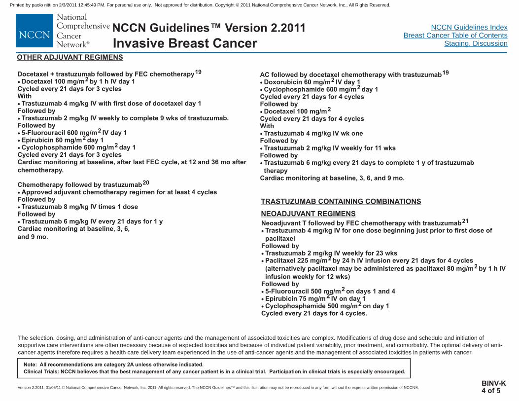
NCCN®
NCCN Guidelines™ Version 2.2011
Version 2.2011, 01/05/11 © National Comprehensive Cancer Network, Inc. 2011, All rights reserved. The NCCN Guidelines™ and this illustration may not be reproduced in any form without the express written permission of NCCN®.
NCCN Guidelines IndexBreast Cancer Table of Contents
Staging, Discussion
Note: All recommendations are category 2A unless otherwise indicated.Clinical Trials: NCCN believes that the best management of any cancer patient is in a clinical trial. Participation in clinical trials is especially encouraged.
BINV-K4 of 5
Neoadjuvant T followed by FEC chemotherapy with trastuzumabTrastuzumab 4 mg/kg IV for one dose beginning just prior to first dose ofpaclitaxel
Followed byTrastuzumab 2 mg/kg IV weekly for 23 wksPaclitaxel 225 mg/m by 24 h IV infusion every 21 days for 4 cycles(alternatively paclitaxel may be administered as paclitaxel 80 mg/m by 1 h IVinfusion weekly for 12 wks)
Followed by5-Fluorouracil 500 mg/m on days 1 and 4Epirubicin 75 mg/m2 IV on day 1Cyclophosphamide 500 mg/m on day 1
Cycled every 21 days for 4 cycles.
21
2
2
2
2
�
�
�
�
�
�
NEOADJUVANT REGIMENS
TRASTUZUMAB CONTAINING COMBINATIONS
Docetaxel + trastuzumab followed by FEC chemotherapyDocetaxel 100 mg/m by 1 h IV day 1
Cycled every 21 days for 3 cyclesWith
Trastuzumab 4 mg/kg IV with first dose of docetaxel day 1Followed by
Trastuzumab 2 mg/kg IV weekly to complete 9 wks of trastuzumab.Followed by
5-Fluorouracil 600 mg/m IV day 1Epirubicin 60 mg/m day 1Cyclophosphamide 600 mg/m day 1
Cycled every 21 days for 3 cyclesCardiac monitoring at baseline, after last FEC cycle, at 12 and 36 mo afterchemotherapy.
Chemotherapy followed by trastuzumabApproved adjuvant chemotherapy regimen for at least 4 cycles
Followed byTrastuzumab 8 mg/kg IV times 1 dose
Followed byTrastuzumab 6 mg/kg IV every 21 days for 1 y
Cardiac monitoring at baseline, 3, 6,and 9 mo.
192
22
2
20
�
�
�
�
�
�
�
�
�
AC followed by docetaxel chemotherapy with trastuzumabDoxorubicin 60 mg/m IV day 1Cyclophosphamide 600 mg/m day 1
Cycled every 21 days for 4 cyclesFollowed by
Docetaxel 100 mg/mCycled every 21 days for 4 cyclesWith
Trastuzumab 4 mg/kg IV wk oneFollowed by
Trastuzumab 2 mg/kg IV weekly for 11 wksFollowed by
Trastuzumab 6 mg/kg every 21 days to complete 1 y of trastuzumabtherapy
Cardiac monitoring at baseline, 3, 6, and 9 mo.
192
2
2
�
�
�
�
�
�
OTHER ADJUVANT REGIMENS
The selection, dosing, and administration of anti-cancer agents and the management of associated toxicities are complex. Modifications of drug dose and schedule and initiation of
supportive care interventions are often necessary because of expected toxicities and because of individual patient variability, prior treatment, and comorbidity. The optimal delivery of anti-
cancer agents therefore requires a health care delivery team experienced in the use of anti-cancer agents and the management of associated toxicities in patients with cancer.
Invasive Breast Cancer
Printed by paolo nitti on 2/3/2011 12:45:49 PM. For personal use only. Not approved for distribution. Copyright © 2011 National Comprehensive Cancer Network, Inc., All Rights Reserved.

NCCN®
NCCN Guidelines™ Version 2.2011
Version 2.2011, 01/05/11 © National Comprehensive Cancer Network, Inc. 2011, All rights reserved. The NCCN Guidelines™ and this illustration may not be reproduced in any form without the express written permission of NCCN®.
NCCN Guidelines IndexBreast Cancer Table of Contents
Staging, Discussion
Note: All recommendations are category 2A unless otherwise indicated.Clinical Trials: NCCN believes that the best management of any cancer patient is in a clinical trial. Participation in clinical trials is especially encouraged.
BINV-K5 of 5
1
5
7
9
10
11
13
14
15
16
17
18
19
20
Martin, Pienkowski T, Mackey J, et al: Adjuvant Docetaxel for Node-Positive Breast Cancer. N Engl J. Med 352: 22, 2005.
Dang C, Fomier M, Sugarman S, et al. The safety of dose-dense doxorubicin and cyclophosphamide followed by paclitaxel with trastuzumab in HER2/neuoverexpressed/amplified breast cancer. J Clin Oncol. 2008; 26:1216-1222.
Henderson IC, Berry DA, Demetri GD, et al: Improved outcomes from adding sequential paclitaxel but not from escalating doxorubicin dose in an adjuvantchemotherapy Regimen for Patients With Node-Positive Primary Breast Cancer. J Clin Oncol 21:976-983, 2003.
Mamounas EP, Bryant J, Lembersky BC, et al: Paclitaxel after doxorubicin plus cyclophosphamide as adjuvant chemotherapy for node-positive breast cancer: resultsfrom NSABP 13-28. J. Clin Oncol.:23:3686-96, 2005.Sparano JA, Wang M, Martino S, et al. Weekly paclitaxel in adjuvant treatment of breast cancer. N Engl J Med. 258:1663-1671, 2008.
Jones S, Holmes F, O’Shaughnessey J, et al. Extended follow-up and analysis by age of the US Oncology Adjuvant Trial 9735: DOcetaxwl/cyclophophamide isassociated with an overall survival benefit compared to doxorubicin/cyclophosphamide and is well tolerated in women 65 or older. San Antonio Breast CancerSymposium. Abstract 12, 2007.
Fisher B, Brown AM, Dimitrov NV, et al: Two months of doxorubicin-cyclophosphamide with and without interval reinduction therapy compared with six months ofcyclophosphamide, methotrexate, and fluorouracil in positive-node breast cancer patients with tamoxifen-nonresponsive tumors: Results from NSABP B-15. Journal ofClin Oncol 8:1483-1496, 1990.
Buzdar AU, Kau SW, Smith TL, Hortobagyi GN. Ten-year results of FAC adjuvant chemotherapy trial in breast cancer. Am J Clin Oncol 12; 123-128,1989
Assikis V, Buzdar A, Yang Y, et al: A phase III trial of sequential adjuvant chemotherapy for operable breast carcinoma: final analysis with 10-year follow-up. Cancer97:2716-23, 2003.
Bull JM, Tormey DC, Li SH, et al: A randomized comparative trial of adriamycin versus methotrexate in combination drug therapy. Cancer 41:1649-57, 1978
Levine MN, Bramwell VH, Pritchard KI, et al: Randomized trial of intensive cyclophosphamide, epirubicin, and fluorouracil chemotherapy compared withcyclophosphamide, methotrexate, and fluorouracil in premenopausal women with node-positive breast cancer. National Cancer Institute of Canada Clinical TrialsGroup. J Clin Oncol 16:2651-8, 1998.
Goldhirsch A, Colleoni M, Coates AS, et al: Adding adjuvant CMF chemotherapy to either radiotherapy or tamoxifen: are all CMFs alike? The International BreastCancer Study Group (IBCSG). Ann Oncol 9:489-93, 1998.
Piccart MJ, Di Leo A, Beauduin M, et al: Phase III Trial Comparing Two Dose Levels of Epirubicin Combined With Cyclophosphamide With Cyclophosphamide,Methotrexate, and Fluorouracil in Node-Positive Breast Cancer. J Clin Oncol 19:3103-3110, 2001.
Roche H, fumoleau P, Spielmann M, et al. Sequential adjuvant epirubicin-based and docetaxel chemotherapy for node-positive breast cancer patients: The FNCLCCPACS 001 trial. J Clin Oncol. 24:5664-5671, 2006.
Citron ML, Berry DA, Cirrincione C, et al: Randomized Trial of Dose-Dense Versus Conventionally Scheduled and Sequential Versus Concurrent CombinationChemotherapy as Postoperative Adjuvant Treatment of Node-Positive Primary Breast Cancer: First Report of Intergroup Trial C9741/Cancer and Leukemia Group BTrial 9741. J Clin Oncol 21:1431-1439, 2003.
Martin M, Rodriguez-Lescure A, Ruiz A, et al: Randomized phase 3 trial of fluorouracil, epirubicin, and cyclophosphamide alone or followed by paclitaxel for earlybreast cancer. J Natl Cancer Inst 2008; 100:805-814.
Romond EH, Perez EZ, Bryant J, et al. Trastuzumab plus adjuvant chemotherapy for operable HER2 positive breast cancer. N Engl J Med 353:1673-1684, 2005
Robert NJ, Eiermann W, Pienkowski T, et al. BCIRG 006: Docetaxel and trastuzumab-based regimens improve DFS and OS over AC followed by T in node positiveand high risk node negative HER2 positive early breast cancer patients: Quality of life at 36 mo. J Clin Oncol. 25:18S (June 20 suppl). Abstract 19647, 2007.
Joensuu H, Kellokumpu-Lehtinen P-L, Bono P, et al: Adjuvant docetaxel or vinorelbine with or without trastuzumab for breast cancer. N Engl J Med 354:809-20, 2006.
Piccart-Gebhart MJ, Procter M, Leyland-Jones B, et al. Trastuzumab after adjuvant chemotherapy in HER-2 positive breast cancer. N Engl J Med 353:1659-72, 2005.
2005.
2
3
4
6
8
12
21Buzdar A, Ibrahim N, Francis D, et al. Significantly higher pathologic complete remission rate after neoadjuvant therapy with trastuzumab, paclitaxel, and epirubicinchemotherapy: Results of a randomized trial in human epidermal growth factor receptor 2-positive operable breast cancer. J. Clin Oncol 23: 3676-3685,
Invasive Breast Cancer
Printed by paolo nitti on 2/3/2011 12:45:49 PM. For personal use only. Not approved for distribution. Copyright © 2011 National Comprehensive Cancer Network, Inc., All Rights Reserved.
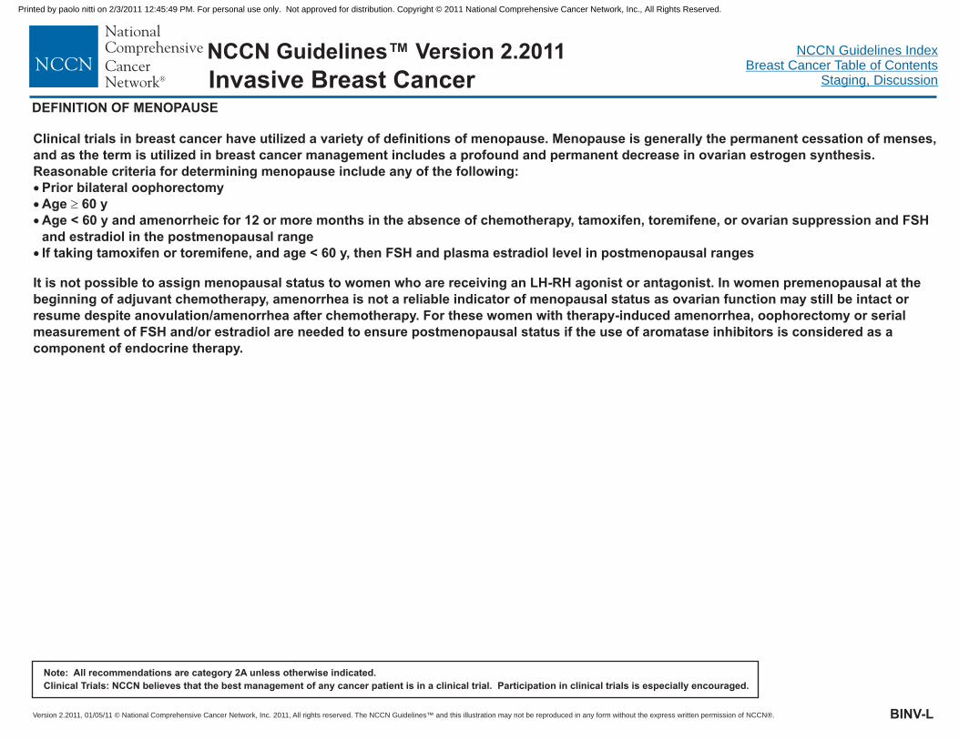
NCCN®
NCCN Guidelines™ Version 2.2011
Version 2.2011, 01/05/11 © National Comprehensive Cancer Network, Inc. 2011, All rights reserved. The NCCN Guidelines™ and this illustration may not be reproduced in any form without the express written permission of NCCN®.
NCCN Guidelines IndexBreast Cancer Table of Contents
Staging, Discussion
Note: All recommendations are category 2A unless otherwise indicated.Clinical Trials: NCCN believes that the best management of any cancer patient is in a clinical trial. Participation in clinical trials is especially encouraged.
BINV-L
DEFINITION OF MENOPAUSE
Clinical trials in breast cancer have utilized a variety of definitions of menopause. Menopause is generally the permanent cessation of menses,and as the term is utilized in breast cancer management includes a profound and permanent decrease in ovarian estrogen synthesis.Reasonable criteria for determining menopause include any of the following:
Prior bilateral oophorectomyAge 60 yAge < 60 y and amenorrheic for 12 or more months in the absence of chemotherapy, tamoxifen, toremifene, or ovarian suppression and FSHand estradiol in the postmenopausal rangeIf taking tamoxifen or toremifene, and age < 60 y, then FSH and plasma estradiol level in postmenopausal ranges
It is not possible to assign menopausal status to women who are receiving an LH-RH agonist or antagonist. In women premenopausal at thebeginning of adjuvant chemotherapy, amenorrhea is not a reliable indicator of menopausal status as ovarian function may still be intact orresume despite anovulation/amenorrhea after chemotherapy. For these women with therapy-induced amenorrhea, oophorectomy or serialmeasurement of FSH and/or estradiol are needed to ensure postmenopausal status if the use of aromatase inhibitors is considered as acomponent of endocrine therapy.
�
� �
�
�
Invasive Breast Cancer
Printed by paolo nitti on 2/3/2011 12:45:49 PM. For personal use only. Not approved for distribution. Copyright © 2011 National Comprehensive Cancer Network, Inc., All Rights Reserved.
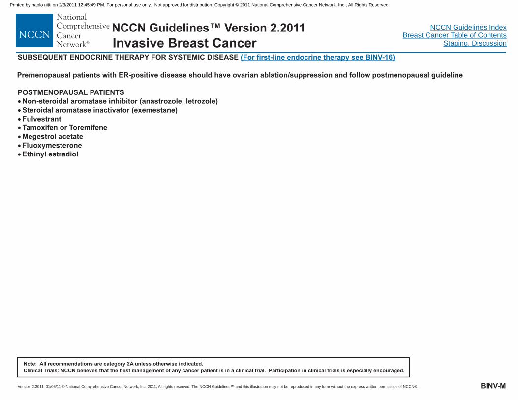
NCCN®
NCCN Guidelines™ Version 2.2011
Version 2.2011, 01/05/11 © National Comprehensive Cancer Network, Inc. 2011, All rights reserved. The NCCN Guidelines™ and this illustration may not be reproduced in any form without the express written permission of NCCN®.
NCCN Guidelines IndexBreast Cancer Table of Contents
Staging, Discussion
Note: All recommendations are category 2A unless otherwise indicated.Clinical Trials: NCCN believes that the best management of any cancer patient is in a clinical trial. Participation in clinical trials is especially encouraged.
SUBSEQUENT ENDOCRINE THERAPY FOR SYSTEMIC DISEASE (For first-line endocrine therapy see BINV-16)
POSTMENOPAUSAL PATIENTS�
�
�
�
�
�
�
Non-steroidal aromatase inhibitor (anastrozole, letrozole)Steroidal aromatase inactivator (exemestane)FulvestrantTamoxifen or ToremifeneMegestrol acetateFluoxymesteroneEthinyl estradiol
BINV-M
Premenopausal patients with ER-positive disease should have ovarian ablation/suppression and follow postmenopausal guideline
Invasive Breast Cancer
Printed by paolo nitti on 2/3/2011 12:45:49 PM. For personal use only. Not approved for distribution. Copyright © 2011 National Comprehensive Cancer Network, Inc., All Rights Reserved.

NCCN®
NCCN Guidelines™ Version 2.2011
Version 2.2011, 01/05/11 © National Comprehensive Cancer Network, Inc. 2011, All rights reserved. The NCCN Guidelines™ and this illustration may not be reproduced in any form without the express written permission of NCCN®.
NCCN Guidelines IndexBreast Cancer Table of Contents
Staging, Discussion
PREFERRED CHEMOTHERAPY REGIMENS FOR RECURRENT OR METASTATIC BREAST CANCER1
PREFERRED SINGLE AGENTS
OTHER SINGLE AGENTS
Anthracyclines
Taxanes
Anti-metabolites
Other microtubule inhibitors
�
�
�
�
�
�
�
�
�
�
�
�
�
�
�
�
�
DoxorubicinEpirubicinPegylated liposomal doxorubicin
PaclitaxelDocetaxelAlbumin-bound paclitaxel
CapecitabineGemcitabine
VinorelbineEribulin
CyclophosphamideMitoxantroneCisplatinEtoposide (po) (category 2B)VinblastineFluorouracil ClIxabepilone
PREFERRED AGENTS WITH BEVACIZUMAB2
� Paclitaxel
PREFERRED CHEMOTHERAPY COMBINATIONS�
�
�
�
�
�
�
�
CAF/FAC (FECACECATCMFDocetaxel/capecitabineGT (gemcitabine/paclitaxel)
cyclophosphamide/doxorubicin/fluorouracil)(fluorouracil/epirubicin/cyclophosphamide)
(doxorubicin/cyclophosphamide)(epirubicin/cyclophosphamide)(doxorubicin/docetaxel; doxorubicin/paclitaxel)
(cyclophosphamide/methotrexate/fluorouracil)
OTHER COMBINATIONS� Ixabepilone + capecitabine (category 2B)
PREFERRED FIRST-LINE AGENTS FOR HER2-POSITIVE DISEASETrastuzumab with:
�
�
�
�
Paclitaxel ± carboplatinDocetaxelVinorelbineCapecitabine
PREFERRED AGENTS FOR TRASTUZUMAB-EXPOSED HER2-POSITIVE DISEASE�
�
�
�
Lapatinib + capecitabineTrastuzumab + other first-line agentsTrastuzumab + capecitabineTrastuzumab + lapatinib (without cytotoxic therapy)
Note: All recommendations are category 2A unless otherwise indicated.Clinical Trials: NCCN believes that the best management of any cancer patient is in a clinical trial. Participation in clinical trials is especially encouraged.
1There is no compelling evidence that combination regimens are superior to sequential single agents.
Randomized clinical trials in metastatic breast cancer document that the addition of bevacizumab to some first or second line chemotherapy agents modestly improvestime to progression and response rates but does not improve overall survival. The time to progression impact may vary among cytotoxic agents and appears greatestwith bevacizumab in combination with weekly paclitaxel.
2
BINV-N1 of 6
Invasive Breast Cancer
Printed by paolo nitti on 2/3/2011 12:45:49 PM. For personal use only. Not approved for distribution. Copyright © 2011 National Comprehensive Cancer Network, Inc., All Rights Reserved.

NCCN®
NCCN Guidelines™ Version 2.2011
Version 2.2011, 01/05/11 © National Comprehensive Cancer Network, Inc. 2011, All rights reserved. The NCCN Guidelines™ and this illustration may not be reproduced in any form without the express written permission of NCCN®.
NCCN Guidelines IndexBreast Cancer Table of Contents
Staging, Discussion
Note: All recommendations are category 2A unless otherwise indicated.Clinical Trials: NCCN believes that the best management of any cancer patient is in a clinical trial. Participation in clinical trials is especially encouraged.
CAF chemotherapyCyclophosphamide 100 mg/m PO days 1-14Doxorubicin 30 mg/m IV days 1 & 85-Fluorouracil 500 mg/m IV days 1 & 8
Cycled every 28 days.
FAC chemotherapy5-Fluorouracil 500 mg/m IV days 1 & 8 or days 1 & 4Doxorubicin 50 mg/m IV day 1Cyclophosphamide 500 mg/m IV day 1
Cycled every 21 days.
AC chemotherapyDoxorubicin 60 mg/m IV day 1Cyclophosphamide 600 mg/m IV day 1
Cycled every 21 days.
EC chemotherapyEpirubicin 75 mg/m IV day 1Cyclophosphamide 600 mg/m IV day 1
Cycled every 21 days
1
2
4
5
2
2
�
�
�
�
�
�
�
�
�
�
2
2
2
2
2
2
3
2
2
2
2
2
FEC chemotherapyCyclophosphamide 400 mg/m IV days 1 & 8Epirubicin 50 mg/m IV days 1 & 85-Fluorouracil 500 mg/m IV days 1 & 8
Cycled every 28 days.
�
�
�
AT chemotherapyDoxorubicin 60 mg/m IV day 1
Paclitaxel 125-200 mg/m IV day 1Cycled every 21 days
AT chemotherapyDoxorubicin 50 mg/m IV day 1Docetaxel 75 mg/m IV day 1
CMF chemotherapyCyclophosphamide 100 mg/m PO days 1-14Methotrexate 40 mg/m IV days 1 & 85-Fluorouracil 600 mg/m IV days 1 & 8
Cycled every 28 days.
Docetaxel/capecitabine chemotherapyDocetaxel 75 mg/m IV day 1Capecitabine 950 mg/m PO twice daily days 1-14
Cycled every 21 days.
GT chemotherapyPaclitaxel 175 mg/m IV day 1Gemcitabine 1250 mg/m IV days 1 & 8 (following paclitaxel on day 1)
Cycled every 21 days.
6
2
2
7
2
2
8
2
2
2
9
2
2
10
2
2
�
�
�
�
�
�
�
�
�
�
�Cycled every 21 days
PREFERRED CHEMOTHERAPY COMBINATIONS
BINV-N2 of 6
OTHER COMBINATIONSIxabepilone/capecitabine (category 2B)
Ixabepilone 40 mg/m IV day 1Capecitabine 2000 mg/m PO days 1-14
�
�
2
2
Cycled every 21 days.The selection, dosing, and administration of anti-cancer agents and the management of associated toxicities are complex. Modifications of drug dose and schedule and initiation of
supportive care interventions are often necessary because of expected toxicities and because of individual patient variability, prior treatment, and comorbidity. The optimal delivery of anti-
cancer agents therefore requires a health care delivery team experienced in the use of anti-cancer agents and the management of associated toxicities in patients with cancer.
PREFERRED CHEMOTHERAPY REGIMENS FOR RECURRENT OR METASTATIC BREAST CANCER
Invasive Breast Cancer
Printed by paolo nitti on 2/3/2011 12:45:49 PM. For personal use only. Not approved for distribution. Copyright © 2011 National Comprehensive Cancer Network, Inc., All Rights Reserved.

NCCN®
NCCN Guidelines™ Version 2.2011
Version 2.2011, 01/05/11 © National Comprehensive Cancer Network, Inc. 2011, All rights reserved. The NCCN Guidelines™ and this illustration may not be reproduced in any form without the express written permission of NCCN®.
NCCN Guidelines IndexBreast Cancer Table of Contents
Staging, Discussion
PREFERRED SINGLE AGENTS
Note: All recommendations are category 2A unless otherwise indicated.Clinical Trials: NCCN believes that the best management of any cancer patient is in a clinical trial. Participation in clinical trials is especially encouraged.
BINV-N3 of 6
Anthracyclines:
Taxanes:
Doxorubicin 60-75 mg/m IV day 1Cycled every 21 daysOR
Doxorubicin 20 mg/m IV weekly
Epirubicin 60-90 mg/m IV day 1Cycled every 21 days.
Pegylated liposomal encapsulated doxorubicin 50 mg/m IV day 1Cycled every 28 days.
Paclitaxel 175 mg/m IV day 1Cycled every 21 days.OR
Paclitaxel 80 mg/m IV weekly
Cycled every 21 days.OR
Cycled every 21 days.
2 11
2 13
2 14
16
�
�
�
�
�
2 12
152
2
2 17,18
2 19
2 2 20,21
2 20
�
�
�
Docetaxel 60-100 mg/m IV day 1
Docetaxel 40 mg/m IV weekly for 6 wks followed by a 2 week rest, then repeat
Albumin-bound paclitaxel 100 mg/m or 150 mg/m days 1, 8, and 15 IVCycled every 28 days.
Albumin-bound paclitaxel 260 mg/m IV
Anti-metabolites:
Other microtubule inhibitors:
OTHER SINGLE AGENTS
�
�
�
�
Capecitabine 1000-1250 mg/m PO twice daily days 1-14Cycled every 21 days.
Gemcitabine 800-1200 mg/m IV days 1, 8 & 15Cycled every 28 days.
Vinorelbine 25 mg/m IV weeklyEribulin 1.4 mg/m IV days 1, 8Cycled every 21 days.
2 22
2 23
2 24
2
�
�
�
�
�
�
�
Cyclophosphamide
Mitoxantrone
Cisplatin
Etoposide (PO) (category 2B)
Vinblastine
Fluorouracil Cl
Ixabepilone
The selection, dosing, and administration of anti-cancer agents and the management of associated toxicities are complex. Modifications of drug dose and schedule and initiation of
supportive care interventions are often necessary because of expected toxicities and because of individual patient variability, prior treatment, and comorbidity. The optimal delivery of anti-
cancer agents therefore requires a health care delivery team experienced in the use of anti-cancer agents and the management of associated toxicities in patients with cancer.
PREFERRED AGENTS WITH BEVACIZUMABPaclitaxel plus bevacizumab
Paclitaxel 90 mg/m by 1 h IV days 1, 8 & 15Bevacizumab 10 mg/kg IV days 1 & 15
Cycled every 28 days.
25
2�
�
PREFERRED CHEMOTHERAPY REGIMENS FOR RECURRENT OR METASTATIC BREAST CANCER
Invasive Breast Cancer
Printed by paolo nitti on 2/3/2011 12:45:49 PM. For personal use only. Not approved for distribution. Copyright © 2011 National Comprehensive Cancer Network, Inc., All Rights Reserved.
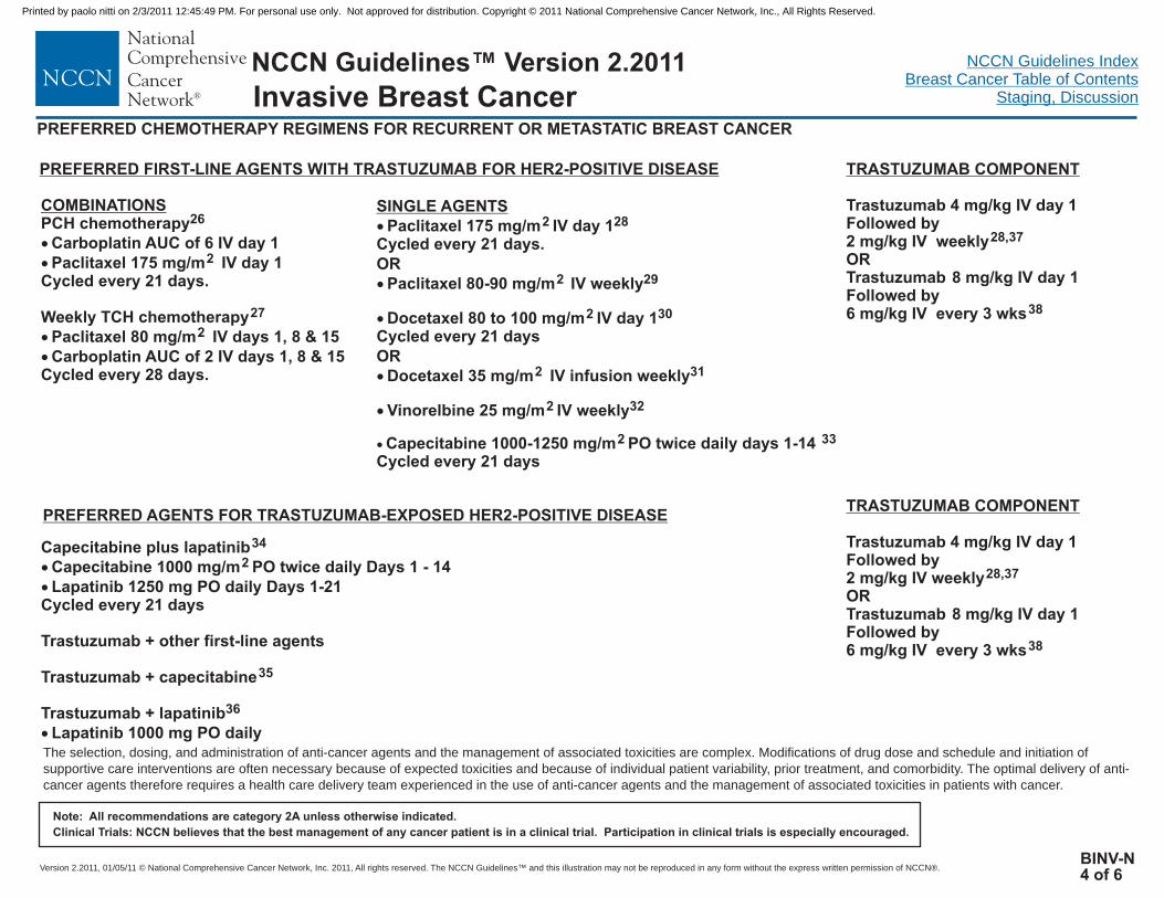
NCCN®
NCCN Guidelines™ Version 2.2011
Version 2.2011, 01/05/11 © National Comprehensive Cancer Network, Inc. 2011, All rights reserved. The NCCN Guidelines™ and this illustration may not be reproduced in any form without the express written permission of NCCN®.
NCCN Guidelines IndexBreast Cancer Table of Contents
Staging, Discussion
Capecitabine plus lapatinibCapecitabine 1000 mg/m PO twice daily Days 1 - 14Lapatinib 1250 mg PO daily Days 1-21
Cycled every 21 days
Trastuzumab + other first-line agents
Trastuzumab + capecitabine
Trastuzumab + lapatinibLapatinib 1000 mg PO daily
34
2
35
36
�
�
�
Note: All recommendations are category 2A unless otherwise indicated.Clinical Trials: NCCN believes that the best management of any cancer patient is in a clinical trial. Participation in clinical trials is especially encouraged.
PREFERRED FIRST-LINE AGENTS WITH TRASTUZUMAB FOR HER2-POSITIVE DISEASE
COMBINATIONSPCH chemotherapy
Carboplatin AUC of 6 IV day 1Paclitaxel 175 mg/m IV day 1
Cycled every 21 days.
Weekly TCH chemotherapyPaclitaxel 80 mg/m IV days 1, 8 & 15Carboplatin AUC of 2 IV days 1, 8 & 15
Cycled every 28 days.
26
27
2
�
�
�
�
2
TRASTUZUMAB COMPONENT
Trastuzumab 4 mg/kg IV day 1Followed by2 mg/kg IV weeklyORTrastuzumab 8 mg/kg IV day 1Followed by6 mg/kg IV every 3 wks
28,37
38
BINV-N4 of 6
PREFERRED AGENTS FOR TRASTUZUMAB-EXPOSED HER2-POSITIVE DISEASE
The selection, dosing, and administration of anti-cancer agents and the management of associated toxicities are complex. Modifications of drug dose and schedule and initiation of
supportive care interventions are often necessary because of expected toxicities and because of individual patient variability, prior treatment, and comorbidity. The optimal delivery of anti-
cancer agents therefore requires a health care delivery team experienced in the use of anti-cancer agents and the management of associated toxicities in patients with cancer.
SINGLE AGENTS�
�
�
�
�
Paclitaxel 175 mg/m IV day 1Cycled every 21 days.OR
Paclitaxel 80-90 mg/m IV weekly
Docetaxel 80 to 100 mg/m IV day 1Cycled every 21 daysOR
Docetaxel 35 mg/m IV infusion weekly
Vinorelbine 25 mg/m IV weekly
Capecitabine 1000-1250 mg/m PO twice daily days 1-14Cycled every 21 days
2
2 29
2 30
2 31
2 32
2 33
28
�
TRASTUZUMAB COMPONENT
Trastuzumab 4 mg/kg IV day 1Followed by2 mg/kg IV weeklyORTrastuzumab 8 mg/kg IV day 1Followed by6 mg/kg IV every 3 wks
28,37
38
PREFERRED CHEMOTHERAPY REGIMENS FOR RECURRENT OR METASTATIC BREAST CANCER
Invasive Breast Cancer
Printed by paolo nitti on 2/3/2011 12:45:49 PM. For personal use only. Not approved for distribution. Copyright © 2011 National Comprehensive Cancer Network, Inc., All Rights Reserved.

NCCN®
NCCN Guidelines™ Version 2.2011
Version 2.2011, 01/05/11 © National Comprehensive Cancer Network, Inc. 2011, All rights reserved. The NCCN Guidelines™ and this illustration may not be reproduced in any form without the express written permission of NCCN®.
NCCN Guidelines IndexBreast Cancer Table of Contents
Staging, Discussion
Note: All recommendations are category 2A unless otherwise indicated.Clinical Trials: NCCN believes that the best management of any cancer patient is in a clinical trial. Participation in clinical trials is especially encouraged.
1
2
3
4
5
6
7
8
10
11
1213
14
15
16
17
19
Bull JM, Tormey DC, Li SH, et al: A randomized comparative trial of adriamycin versus methotrexate in combination drug therapy. Cancer 41:1649-57, 1978.
Hortobagyi GN, Gutterman JU, Blumenschein GR, et al: Combination chemoimmunotherapy of metastatic breast cancer with 5-fluorouracil, adriamycin,cyclophosphamide, and BCG. Cancer 43:1225-33, 1979.
Ackland SP, Anton A, Breitbach GP, et al: Dose-Intensive Epirubicin-Based Chemotherapy Is Superior to an Intensive Intravenous Cyclophosphamide, Methotrexate,and Fluorouracil Regimen in Metastatic Breast Cancer: A Randomized Multinational Study. J Clin Oncol 19:943-953, 2001.
Fisher B, Brown AM, Dimitrov NV, et al: Two months of doxorubicin-cyclophosphamide with and without interval reinduction therapy compared with six months ofcyclophosphamide, methotrexate, and fluorouracil in positive-node breast cancer patients with tamoxifen-nonresponsive tumors: Results from NSABP B-15. Journal ofClinical Oncology 8:1483-1496, 1990.
Langley RE, Carmichel J, Jones AL, et al. Phase III trial of epirubicin plus paclitaxel compared with epirubicin plus cyclphosphamide as first-line chemotherapy formetastatic breast cancer: United Kingdom Cancer Research Institute. J Clin Oncol. 23:8322-8330,2005.
Bonadonna G, Brusamolino E, Valagussa P, et al: Combination chemotherapy as an adjuvant treatment in operable breast cancer. N Engl J Med 294:405-10, 1976.
O'Shaughnessy J, Miles D, Vukelja S, et al: Superior Survival With Capecitabine Plus Docetaxel Combination Therapy in Anthracycline-Pretreated Patients WithAdvanced Breast Cancer: Phase III Trial Results. J Clin Oncol 20:2812-2823, 2002.
Albain K.S., Nag S., et al: Global Phase lll Study of Gemcitabine Plus Paclitaxel (GT) vs. Paclitaxel (T) as Frontline Therapy for Metastatic Breast Cancer (MBC); FirstReport of Overall Survival. J Clin Oncol 22 No 14S: 510, 2004.
Chan S, Friedrichs K, Noel D, et al. Prospective randomized trial of docetaxel versus doxorubicin in patients with metastatic breast cancer. J Clin Oncol. 1999;17:2341-2454.
Gundersen S, Kvinnsland S, Klepp O, et al: Weekly adriamycin versus VAC in advanced breast cancer. A randomized trial. Eur J Cancer Clin Oncol. 22:1431-4, 1986.
Bastholt, L., Dalmark, M., Gjedde SB, et al: Dose-Response Relationship of Epirubicin in the Treatment of Postmenopausal Patients with Metastatic Breast Cancer: ARandomized Study of Epirubicin at Four Different Dose Levels Performed by the Danish Breast Cancer Coopertive Group.J Clin Oncol 14: 1146-1155, 1996.
O’Brien, M. E., Wigler, N., Inbar M, et al: Reduced Cardiotoxicity and Comparable Efficacy in a Phase lll Trial of Pegylated Liposomal Doxorubicin HC1(CAELYX/Doxil) vs. Conventional Doxorubicin for First-Line Treatment of Metastatic Breast Cancer. Ann Oncol 15(3): 440-9, 2004.
Seidman A, Tiersten A, Hudis C, et al: Phase II trial of paclitaxel by 3-hour infusion as initial and salvage chemotherapy for metastatic breast cancer. J Clin Oncol13:2575-2581, 1995.
Perez EA, Vogel CL, Irwin DH, et al: Multicenter Phase II Trial of Weekly Paclitaxel in Women With Metastatic Breast Cancer. J Clin Oncol 19:4216-4223, 2001.
Burris HAR: Single-agent docetaxel (Taxotere) in randomized phase III trials. Semin Oncol. 26:1-6, 1999.
Valero V: Docetaxel as single-agent therapy in metastatic breast cancer: clinical efficacy. Semin Oncol. 24(Suppl 13):S11-18, 1997.
Burstein HJ, Manola J, Younger J, et al: Docetaxel Administered on a Weekly Basis for Metastatic Breast Cancer. J Clin Oncol 18:1212-1219, 2000.
Gianni L, Munzone E, Capri G, et al. Paclitaxel by 3-hour infusion in combination with bolus doxorubicin in women with untreated metastatic breast cancer: highantitumor efficacy and cardiac effects in a dose-finding and sequence-finding study. J Clin Oncol. 13:2688-2699,1995.
Nabholtz J-M, Falkson C, Campos D, et al. Docetaxel and doxorubicin compared with doxorubicin and cyclophosphamide as first-line chemotherapy for metastaticbreast cancer :results of a randomized, multicenter, phase III trial. J Clin Oncol. 21:968-975,2003.
9
18
BINV-N5 of 6
The selection, dosing, and administration of anti-cancer agents and the management of associated toxicities are complex. Modifications of drug dose and schedule and initiation of
supportive care interventions are often necessary because of expected toxicities and because of individual patient variability, prior treatment, and comorbidity. The optimal delivery of anti-
cancer agents therefore requires a health care delivery team experienced in the use of anti-cancer agents and the management of associated toxicities in patients with cancer.
PREFERRED CHEMOTHERAPY REGIMENS FOR RECURRENT OR METASTATIC BREAST CANCER
Invasive Breast Cancer
Printed by paolo nitti on 2/3/2011 12:45:49 PM. For personal use only. Not approved for distribution. Copyright © 2011 National Comprehensive Cancer Network, Inc., All Rights Reserved.
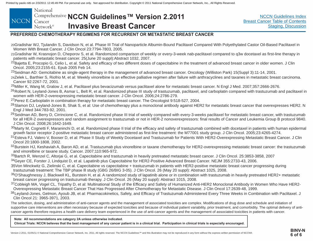
NCCN®
NCCN Guidelines™ Version 2.2011
Version 2.2011, 01/05/11 © National Comprehensive Cancer Network, Inc. 2011, All rights reserved. The NCCN Guidelines™ and this illustration may not be reproduced in any form without the express written permission of NCCN®.
NCCN Guidelines IndexBreast Cancer Table of Contents
Staging, Discussion
Note: All recommendations are category 2A unless otherwise indicated.Clinical Trials: NCCN believes that the best management of any cancer patient is in a clinical trial. Participation in clinical trials is especially encouraged.
BINV-N6 of 6
20
21
22
23
24
27
Gradishar WJ, Tjulandin S, Davidson N, et al. Phase III Trial of Nanoparticle Albumin-Bound Paclitaxel Compared With Polyethylated Castor Oil-Based Paclitaxel inWomen With Breast Cancer. J Clin Oncol 23:7794-7803, 2005.
Gradishar W, Krasnojon D, Cheporov S, et al. Randomized comparison of weekly or every-3-week nab-paclitaxel compared to q3w docetaxel as first-line therapy inpatients with metastatic breast cancer. 25(June 20 suppl):Abstract 1032, 2007.
Bajetta E, Procopio G, Celio L, et al. Safety and efficacy of two different doses of capecitabine in the treatment of advanced breast cancer in older women. J ClinOncol. 2005;23:2155-61. Epub 2005 Feb 14.
Seidman AD: Gemcitabine as single-agent therapy in the management of advanced breast cancer. Oncology (Williston Park) 15(Suppl 3):11-14, 2001.
Zelek L, Barthier S, Riofrio M, et al: Weekly vinorelbine is an effective palliative regimen after failure with anthracyclines and taxanes in metastatic breast carcinoma.Cancer 92:2267-72, 2001.
Miller K, Wang M, Gralow J, et al. Paclitaxel plus bevacizumab versus paclitaxel alone for metastatic breast cancer. N Engl J Med. 2007;357:2666-2676.
Robert N, Leyland-Jones B, Asmar L, Belt R, et al. Randomized phase III study of trastuzumab, paclitaxel, and carboplatin compared with trastuzumab and paclitaxel inwomen with HER-2-overexpressing metastatic breast cancer. J Clin Oncol. 2006;24:2786-279.
Perez E.Carboplatin in combination therapy for metastatic breast cancer. The Oncologist 9:518-527, 2004.
Slamon DJ, Leyland-Jones B, Shak S, et al: Use of chemotherapy plus a monoclonal antibody against HER2 for metastatic breast cancer that overexpresses HER2. NEngl J Med 344:783-92, 2001.
Seidman AD, Berry D, Cirrincione C, et al. Randomized phase III trial of weekly compared with every-3-weeks paclitaxel for metastatic breast cancer, with trastuzumabfor all HER-2 overexpressors and random assignment to trastuzumab or not in HER-2 nonoverexpressors: final results of Cancer and Leukemia Group B protocol 9840.J Clin Oncol. 2008;26:1642-1649.
Marty M, Cognetti F, Maraninchi D, et al. Randomized phase II trial of the efficacy and safety of trastuzumab combined with docetaxel in patients with human epidermalgrowth factor receptor 2-positive metastatic breast cancer administered as first-line treatment: the M77001 study group. J Clin Oncol. 2005;23:4265-4274.
Esteva FJ, Valero V, Booser D, et al: Phase II Study of Weekly Docetaxel and Trastuzumab for Patients With HER2-Overexpressing Metastatic Breast Cancer. J ClinOncol 20:1800-1808, 2002.
Burstein HJ, Keshaviah A, Baron AD, et al. Trastuzumab plus vinorelbine or taxane chemotherapy for HER2-overexpressing metastatic breast cancer: the trastuzumaband vinorelbine or taxane study. Cancer. 2007;110:965-972.
Bartch R, Wenzel C, Altorjai G, et al. Capecitabine and trastuzumab in heavily pretreated metastatic breast cancer. J Clin Oncol. 25:3853-3858, 2007
Geyer CE, Forster J, Lindquist D, et al. Lapatinib plus Capecitabine for HER2-Positive Advanced Breast Cancer. NEJM 355:2733-43, 2006.
35Von Minckwitz G, Zielinski C, et al. Capecitabine vs capecitabine + trastuzumab in patients with HER2-positive metastatic breast cancer progressing duringtrastuzumab treatment: The TBP phase lll study (GBG 26/BIG 3-05). J Clin Oncol. 26 (May 20 suppl): Abstract 1025, 2008.
O'Shaughnessy J, Blackwell KL, Burstein H, et al. A randomized study of lapatinib alone or in combination with trastuzumab in heavily pretreated HER2+ metastaticbreast cancer progressing on trastuzumab therapy. J Clin Oncol. 26 (May 20 suppl): Abstract 1015, 2008.
Cobleigh MA, Vogel CL, Tripathy D, et al: Multinational Study of the Efficacy and Safety of Humanized Anti-HER2 Monoclonal Antibody in Women Who Have HER2-Overexpressing Metastatic Breast Cancer That Has Progressed After Chemotherapy for Metastatic Disease. J Clin Oncol 17:2639-48, 1999.
Leyland-Jones, Gelmon, Ayoub JB, et al: Pharmacokinetics, Safety, and Efficacy of Trastuzumab Administered Every Three Weeks in Combination with Paclitaxel. JClin Oncol 21: 3965-3971, 2003.
25
26
28
29
30
31
32
33
34
36
37
38
The selection, dosing, and administration of anti-cancer agents and the management of associated toxicities are complex. Modifications of drug dose and schedule and initiation of
supportive care interventions are often necessary because of expected toxicities and because of individual patient variability, prior treatment, and comorbidity. The optimal delivery of anti-
cancer agents therefore requires a health care delivery team experienced in the use of anti-cancer agents and the management of associated toxicities in patients with cancer.
PREFERRED CHEMOTHERAPY REGIMENS FOR RECURRENT OR METASTATIC BREAST CANCER
Invasive Breast Cancer
Printed by paolo nitti on 2/3/2011 12:45:49 PM. For personal use only. Not approved for distribution. Copyright © 2011 National Comprehensive Cancer Network, Inc., All Rights Reserved.
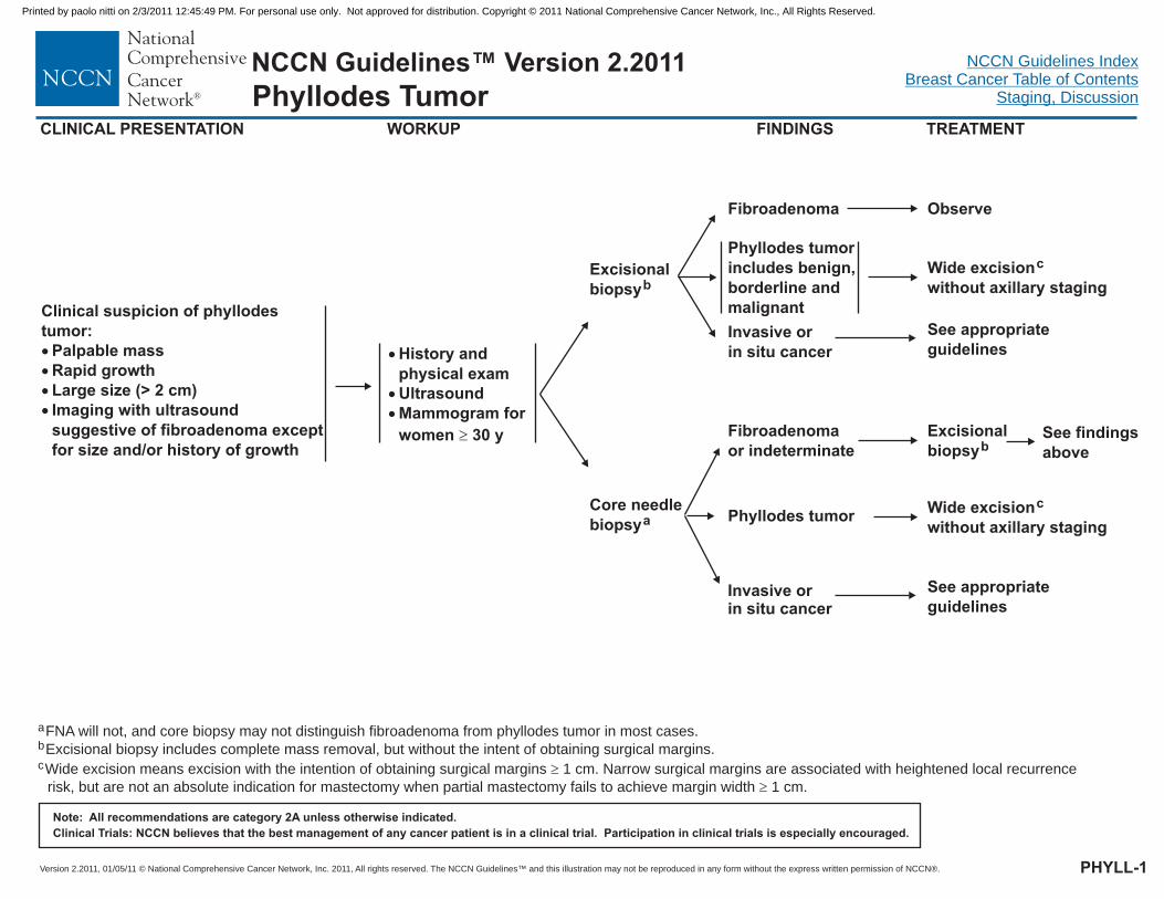
NCCN®
NCCN Guidelines™ Version 2.2011
Version 2.2011, 01/05/11 © National Comprehensive Cancer Network, Inc. 2011, All rights reserved. The NCCN Guidelines™ and this illustration may not be reproduced in any form without the express written permission of NCCN®.
NCCN Guidelines IndexBreast Cancer Table of Contents
Staging, Discussion
Note: All recommendations are category 2A unless otherwise indicated.Clinical Trials: NCCN believes that the best management of any cancer patient is in a clinical trial. Participation in clinical trials is especially encouraged.
PHYLL-1
Phyllodes TumorCLINICAL PRESENTATION
Clinical suspicion of phyllodestumor:
Palpable massRapid growthLarge size (> 2 cm)Imaging with ultrasoundsuggestive of fibroadenoma exceptfor size and/or history of growth
�
�
�
�
�
�
�
�
History andphysical examUltrasoundMammogram forwomen 30 y
Excisionalbiopsyb
Core needlebiopsya
Fibroadenomaor indeterminate
Fibroadenoma
Phyllodes tumorincludes benign,borderline andmalignant
Observe
Wide excisionwithout axillary staging
c
Excisionalbiopsyb
a
b
c
FNA will not, and core biopsy may not distinguish fibroadenoma from phyllodes tumor in most cases.
Excisional biopsy includes complete mass removal, but without the intent of obtaining surgical margins.
Wide excision means excision with the intention of obtaining surgical margins 1 cm. Narrow surgical margins are associated with heightened local recurrence
risk, but are not an absolute indication for mastectomy when partial mastectomy fails to achieve margin width 1 cm.
�
�
WORKUP FINDINGS TREATMENT
Invasive orin situ cancer
See appropriateguidelines
Phyllodes tumor Wide excisionwithout axillary staging
c
See findingsabove
Invasive orin situ cancer
See appropriateguidelines
Printed by paolo nitti on 2/3/2011 12:45:49 PM. For personal use only. Not approved for distribution. Copyright © 2011 National Comprehensive Cancer Network, Inc., All Rights Reserved.
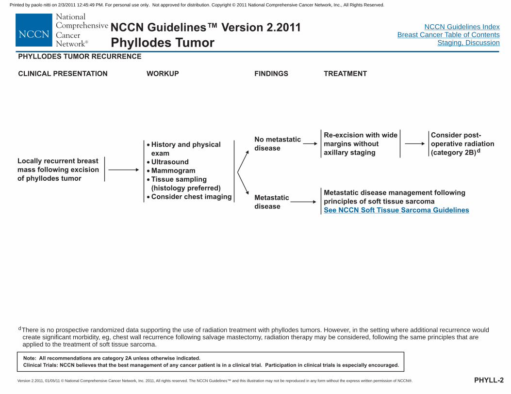
NCCN®
NCCN Guidelines™ Version 2.2011
Version 2.2011, 01/05/11 © National Comprehensive Cancer Network, Inc. 2011, All rights reserved. The NCCN Guidelines™ and this illustration may not be reproduced in any form without the express written permission of NCCN®.
NCCN Guidelines IndexBreast Cancer Table of Contents
Staging, Discussion
PHYLLODES TUMOR RECURRENCE
CLINICAL PRESENTATION WORKUP FINDINGS TREATMENT
Note: All recommendations are category 2A unless otherwise indicated.Clinical Trials: NCCN believes that the best management of any cancer patient is in a clinical trial. Participation in clinical trials is especially encouraged.
Locally recurrent breastmass following excisionof phyllodes tumor
�
�
�
�
�
History and physicalexamUltrasoundMammogramTissue sampling(histology preferred)Consider chest imaging
No metastaticdisease
Metastaticdisease
Re-excision with widemargins withoutaxillary staging
Metastatic disease management followingprinciples of soft tissue sarcomaSee NCCN Soft Tissue Sarcoma Guidelines
Consider post-operative radiation(category 2B)d
dThere is no prospective randomized data supporting the use of radiation treatment with phyllodes tumors. However, in the setting where additional recurrence wouldcreate significant morbidity, eg, chest wall recurrence following salvage mastectomy, radiation therapy may be considered, following the same principles that areapplied to the treatment of soft tissue sarcoma.
PHYLL-2
Phyllodes Tumor
Printed by paolo nitti on 2/3/2011 12:45:49 PM. For personal use only. Not approved for distribution. Copyright © 2011 National Comprehensive Cancer Network, Inc., All Rights Reserved.
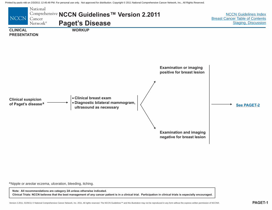
NCCN®
NCCN Guidelines™ Version 2.2011
Version 2.2011, 01/05/11 © National Comprehensive Cancer Network, Inc. 2011, All rights reserved. The NCCN Guidelines™ and this illustration may not be reproduced in any form without the express written permission of NCCN®.
NCCN Guidelines IndexBreast Cancer Table of Contents
Staging, Discussion
Clinical suspicionof Paget’s diseasea
�
�
Clinical breast examDiagnostic bilateral mammogram,ultrasound as necessary
Examination or imagingpositive for breast lesion
Examination and imagingnegative for breast lesion
CLINICALPRESENTATION
WORKUP
Note: All recommendations are category 2A unless otherwise indicated.Clinical Trials: NCCN believes that the best management of any cancer patient is in a clinical trial. Participation in clinical trials is especially encouraged.
aNipple or areolar eczema, ulceration, bleeding, itching.
See PAGET-2
PAGET-1
Paget’s Disease
Printed by paolo nitti on 2/3/2011 12:45:49 PM. For personal use only. Not approved for distribution. Copyright © 2011 National Comprehensive Cancer Network, Inc., All Rights Reserved.

NCCN®
NCCN Guidelines™ Version 2.2011
Version 2.2011, 01/05/11 © National Comprehensive Cancer Network, Inc. 2011, All rights reserved. The NCCN Guidelines™ and this illustration may not be reproduced in any form without the express written permission of NCCN®.
NCCN Guidelines IndexBreast Cancer Table of Contents
Staging, Discussion
Appropriatesystemicadjuvanttherapy
Full thickness skinbiopsy of involved NAC
Core biopsy ofbreast lesion andfull thickness skinbiopsy of involvednipple-a eolacomplex (NAC)
r
Breast and NACbiopsy negative
Breast DCISand NAC Paget’s
b
Breast invasive cancerand NAC Paget’sb,e
Breast negative forcancer and positiveNAC Paget’sb
NAC biopsy positivefor Paget’sb
NAC biopsynegative for Paget’s
Clinical follow-upRe-biopsy if not healing
Mastectomy ± axillary stagingorExcision of breast tumor and excision NAC withwhole breast radiation, consider boost to breastand NAC sites
c
d
Mastectomy + axillary staging ( )orExcision of breast tumor and excision NAC +axillary staging ( ) with whole breastradiation, consider boost to breast and NAC sites
See BINV-D
See BINV-D
Mastectomy + axillary staging( )orExcision of NAC with wholebreast radiation, consider boostto NAC sitesd
See BINV-D
Clinical follow-upRe-biopsy if not healing
TREATMENT
Examinationor imagingpositive forbreast lesion
Examinationand imagingnegative forbreast lesion
WORKUP
Note: All recommendations are category 2A unless otherwise indicated.Clinical Trials: NCCN believes that the best management of any cancer patient is in a clinical trial. Participation in clinical trials is especially encouraged.
PAGET-2
Paget’s Disease
bTo assess extent of disease or confirm additional disease consider MRI ( ).c
d
e
Mastectomy is always an option with any manifestation of Paget’s disease (see manuscript text).
With Paget’s disease and no associated peripheral cancer, or with associated DCIS, consider tamoxifen 20 mg per day for 5 years.
With associated invasive breast cancer, treat with appropriate systemic adjuvant therapy ( )
See BINV-B
See BINV-4
Consider MRIand tissuesampling
Printed by paolo nitti on 2/3/2011 12:45:49 PM. For personal use only. Not approved for distribution. Copyright © 2011 National Comprehensive Cancer Network, Inc., All Rights Reserved.
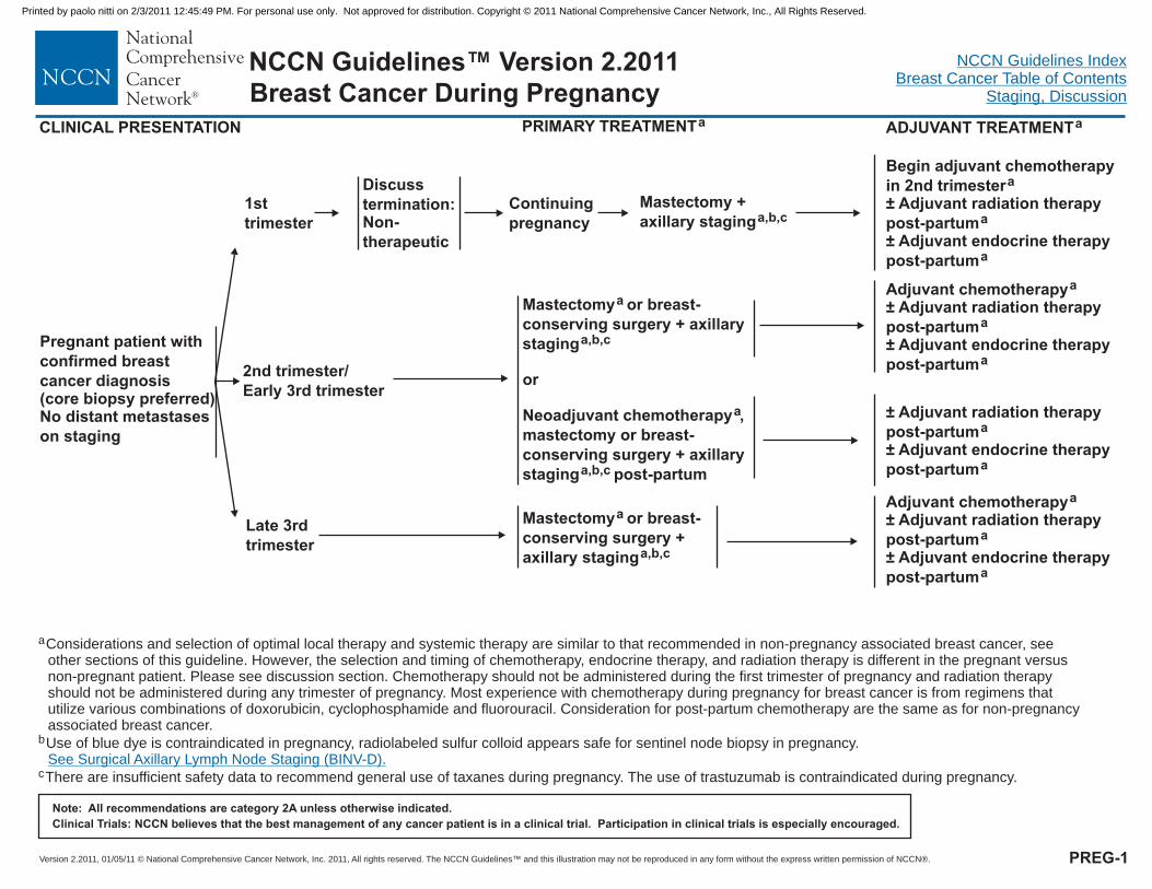
NCCN®
NCCN Guidelines™ Version 2.2011
Version 2.2011, 01/05/11 © National Comprehensive Cancer Network, Inc. 2011, All rights reserved. The NCCN Guidelines™ and this illustration may not be reproduced in any form without the express written permission of NCCN®.
NCCN Guidelines IndexBreast Cancer Table of Contents
Staging, Discussion
Note: All recommendations are category 2A unless otherwise indicated.Clinical Trials: NCCN believes that the best management of any cancer patient is in a clinical trial. Participation in clinical trials is especially encouraged.
Pregnant patient withconfirmed breastcancer diagnosis(core biopsy preferred)No distant metastaseson staging
1sttrimester
2nd trimester/Early 3rd trimester
Late 3rdtrimester
Discusstermination:Non-therapeutic
Continuingpregnancy
Mastectomy +axillary staginga,b,c
Begin adjuvant chemotherapyin 2nd trimester± Adjuvant radiation therapypost-partum± Adjuvant endocrine therapypost-partum
a
a
a
Mastectomy or breast-conserving surgery +
or
Neoadjuvant chemotherapy ,mastectomy or breast-conserving surgery +
post-partum
a
a,b,c
a
a,b,c
axillarystaging
axillarystaging
± Adjuvant radiation therapypost-partum± Adjuvant endocrine therapypost-partum
a
a
Adjuvant chemotherapy± Adjuvant radiation therapypost-partum± Adjuvant endocrine therapypost-partum
a
a
a
Mastectomy or breast-conserving surgery +
a
a,b,caxillary staging
Adjuvant chemotherapy± Adjuvant radiation therapypost-partum± Adjuvant endocrine therapypost-partum
a
a
a
aConsiderations and selection of optimal local therapy and systemic therapy are similar to that recommended in non-pregnancy associated breast cancer, seeother sections of this guideline. However, the selection and timing of chemotherapy, endocrine therapy, and radiation therapy is different in the pregnant versusnon-pregnant patient. Please see discussion section. Chemotherapy should not be administered during the first trimester of pregnancy and radiation therapyshould not be administered during any trimester of pregnancy. Most experience with chemotherapy during pregnancy for breast cancer is from regimens thatutilize various combinations of doxorubicin, cyclophosphamide and fluorouracil. Consideration for post-partum chemotherapy are the same as for non-pregnancyassociated breast cancer.
Use of blue dye is contraindicated in pregnancy, radiolabeled sulfur colloid appears safe for sentinel node biopsy in pregnancy.
There are insufficient safety data to recommend general use of
b
c taxanes during pregnancy. The use of trastuzumab is contraindicated during pregnancy.
See Surgical Axillary Lymph Node Staging (BINV-D).
CLINICAL PRESENTATION PRIMARY TREATMENTa ADJUVANT TREATMENTa
PREG-1
Breast Cancer During Pregnancy
Printed by paolo nitti on 2/3/2011 12:45:49 PM. For personal use only. Not approved for distribution. Copyright © 2011 National Comprehensive Cancer Network, Inc., All Rights Reserved.
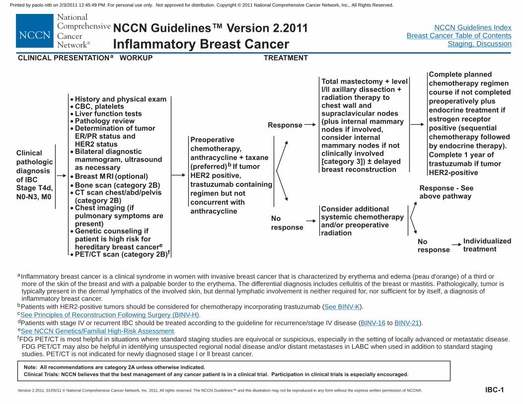
NCCN®
NCCN Guidelines™ Version 2.2011
Version 2.2011, 01/05/11 © National Comprehensive Cancer Network, Inc. 2011, All rights reserved. The NCCN Guidelines™ and this illustration may not be reproduced in any form without the express written permission of NCCN®.
NCCN Guidelines IndexBreast Cancer Table of Contents
Staging, Discussion
CLINICAL PRESENTATIONa
Inflammatory Breast Cancer
Clinicalpathologicdiagnosisof IBCStage T4d,N0-N3, M0
WORKUP
�����
�
��
�
�
�
History and physical examCBC, plateletsLiver function testsPathology reviewDetermination of tumorER/PR status andHER2
Bone scan (category 2B)CT scan chest/abd/pelvis(category 2B)Chest imaging (ifpulmonary symptoms arepresent)
PET/CT scan (category 2B)
statusBilateral diagnosticmammogram, ultrasoundas necessaryBreast MRI (optional)
Genetic counseling ifpatient is high risk forhereditary breast cancer
�
ef
Preoperativechemotherapy,anthracycline + taxane(preferred) If tumorHER2 positive,trastuzumab containingregimen but notconcurrent withanthracycline
b
Response
Noresponse
Total mastectomy + levell/ll axillary dissection +radiation therapy tochest wall andsupraclavicular nodes
) ± delayedbreast reconstruction
(plus internal mammarynodes if involved,consider internalmammary nodes if notclinically involved[category 3]
Consider additionalsystemic chemotherapyand/or preoperativeradiation
Response - Seeabove pathway
Noresponse
Individualizedtreatment
Complete plannedchemotherapy regimencourse if not completedpreoperatively plusendocrine treatment ifestrogen receptorpositive (sequentialchemotherapy followedby endocrine therapy).Complete 1 year oftrastuzumab if tumorHER2-positive
TREATMENT
Note: All recommendations are category 2A unless otherwise indicated.Clinical Trials: NCCN believes that the best management of any cancer patient is in a clinical trial. Participation in clinical trials is especially encouraged.
IBC-1
aInflammatory breast cancer is a clinical syndrome in women with invasive breast cancer that is characterized by erythema and edema (peau d'orange) of a third ormore of the skin of the breast and with a palpable border to the erythema. The differential diagnosis includes cellulitis of the breast or mastitis. Pathologically, tumor istypically present in the dermal lymphatics of the involved skin, but dermal lymphatic involvement is neither required for, nor sufficient for by itself, a diagnosis ofinflammatory breast cancer.
Patients with HER2-positive tumors should be considered for chemotherapy incorporating trastuzumab ( ).b
c
f
d
ePatients with stage IV or recurrent IBC should be treated according to the guideline for recurrence/stage IV disease ( to ).
FDG PET/CT is most helpful in situations where standard staging studies are equivocal or suspicious, especially in the setting of locally advanced or metastatic disease.FDG PET/CT may also be helpful in identifying unsuspected regional nodal disease and/or distant metastases in LABC when used in addition to standard stagingstudies. PET/CT is not indicated for newly diagnosed stage l or ll breast cancer.
See BINV-K
See Principles of Reconstruction Following Surgery (BINV-H)
BINV-16 BINV-21
See NCCN Genetics/Familial High-Risk Assessment
.
.
Printed by paolo nitti on 2/3/2011 12:45:49 PM. For personal use only. Not approved for distribution. Copyright © 2011 National Comprehensive Cancer Network, Inc., All Rights Reserved.
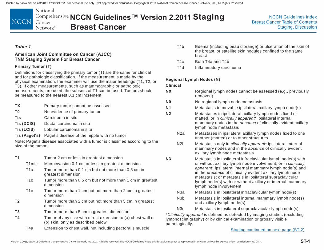
NCCN®
NCCN Guidelines™ Version 2.2011
Version 2.2011, 01/05/11 © National Comprehensive Cancer Network, Inc. 2011, All rights reserved. The NCCN Guidelines™ and this illustration may not be reproduced in any form without the express written permission of NCCN®.
NCCN Guidelines IndexBreast Cancer Table of Contents
Staging, Discussion
Table 1
American Joint Committee on Cancer (AJCC)TNM Staging System For Breast Cancer
Primary Tumor (T)
TX
T0
Tis
Tis (DCIS)
Tis (LCIS)
Tis (Paget's)
T1
T2
T3
T4
Regional Lymph Nodes (N)
Clinical
NX
N0
N1
N2
N3
Definitions for classifying the primary tumor (T) are the same for clinicaland for pathologic classification. If the measurement is made by thephysical examination, the examiner will use the major headings (T1, T2, orT3). If other measurements, such as mammographic or pathologicmeasurements, are used, the subsets of T1 can be used. Tumors shouldbe measured to the nearest 0.1 cm increment.
Primary tumor cannot be assessed
No evidence of primary tumor
Carcinoma in situ
Ductal carcinoma in situ
Lobular carcinoma in situ
Paget's disease of the nipple with no tumor
Note: Paget's disease associated with a tumor is classified according to thesize of the tumor.
Tumor 2 cm or less in greatest dimension
T1mic Microinvasion 0.1 cm or less in greatest dimension
T1a Tumor more than 0.1 cm but not more than 0.5 cm ingreatest dimension
T1b Tumor more than 0.5 cm but not more than 1 cm in greatestdimension
T1c Tumor more than 1 cm but not more than 2 cm in greatestdimension
Tumor more than 2 cm but not more than 5 cm in greatestdimension
Tumor more than 5 cm in greatest dimension
Tumor of any size with direct extension to (a) chest wall or(b) skin, only as described below
T4a Extension to chest wall, not including pectoralis muscle
T4b Edema (including peau d'orange) or ulceration of the skin ofthe breast, or satellite skin nodules confined to the samebreast
T4c Both T4a and T4b
T4d Inflammatory carcinoma
Regional lymph nodes cannot be assessed (e.g., previouslyremoved)
No regional lymph node metastasis
Metastasis to movable ipsilateral axillary lymph node(s)
Metastases in ipsilateral axillary lymph nodes fixed ormatted, or in * ipsilateral internalmammary nodes in the of clinically evident axillarylymph node metastasis
N2a Metastases in ipsilateral axillary lymph nodes fixed to oneanother (matted) or to other structures
N2b Metastasis only in * ipsilateral internalmammary nodes and in the of clinically evidentaxillary lymph node metastasis
Metastasis in ipsilateral infraclavicular lymph node(s) withor without axillary lymph node involvement, or in
* ipsilateral internal mammary lymph node(s) andin the of clinically evident axillary lymph nodemetastasis; or metastasis in ipsilateral supraclavicularlymph node(s) with or without axillary or internal mammarylymph node involvement
N3a Metastasis in ipsilateral infraclavicular lymph node(s)
N3b Metastasis in ipsilateral internal mammary lymph node(s)and axillary lymph node(s)
N3c Metastasis in ipsilateral supraclavicular lymph node(s)
* is defined as detected by imaging studies (excludinglymphoscintigraphy) or by clinical examination or grossly visiblepathologically.
clinically apparentabsence
clinically apparentabsence
clinicallyapparent
presence
Clinically apparent
Staging
ST-1
Staging continued on next page (ST-2)
Breast Cancer
Printed by paolo nitti on 2/3/2011 12:45:49 PM. For personal use only. Not approved for distribution. Copyright © 2011 National Comprehensive Cancer Network, Inc., All Rights Reserved.
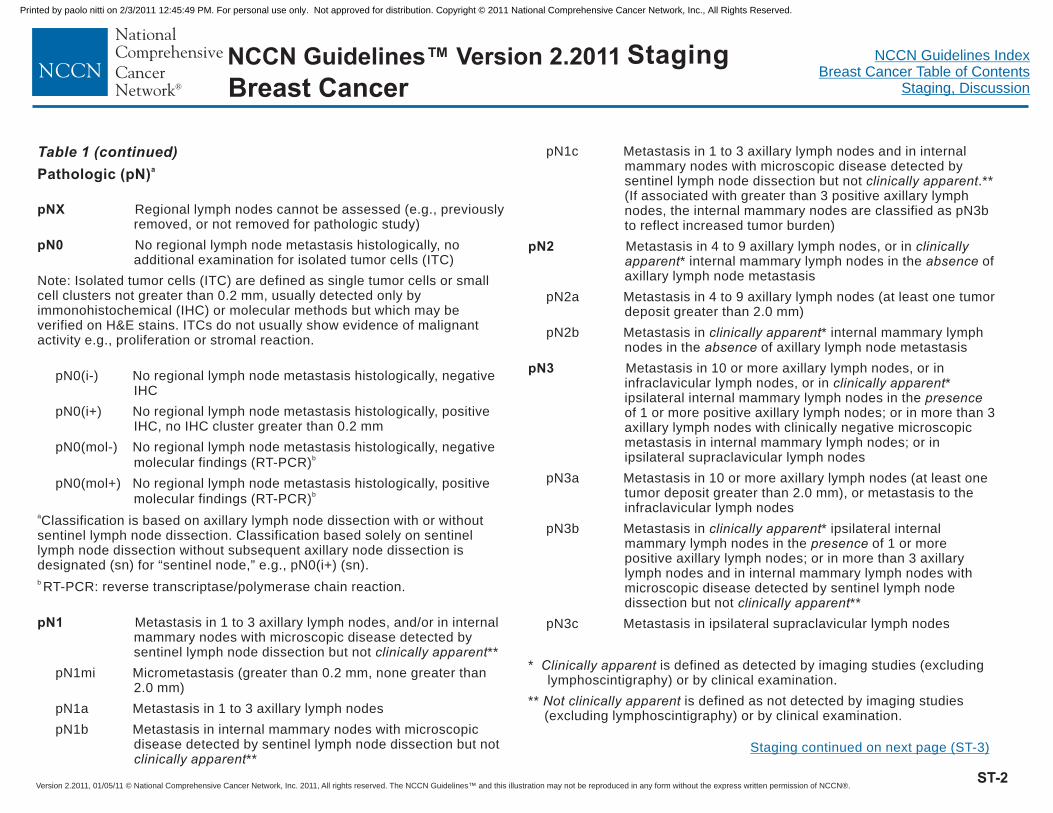
NCCN®
NCCN Guidelines™ Version 2.2011
Version 2.2011, 01/05/11 © National Comprehensive Cancer Network, Inc. 2011, All rights reserved. The NCCN Guidelines™ and this illustration may not be reproduced in any form without the express written permission of NCCN®.
NCCN Guidelines IndexBreast Cancer Table of Contents
Staging, Discussion
Table 1 (continued)
Pathologic (pN)a
pNX
pN0
pN1
pN2
pN3
Regional lymph nodes cannot be assessed (e.g., previouslyremoved, or not removed for pathologic study)
No regional lymph node metastasis histologically, noadditional examination for isolated tumor cells (ITC)
Note: Isolated tumor cells (ITC) are defined as single tumor cells or smallcell clusters not greater than 0.2 mm, usually detected only byimmonohistochemical (IHC) or molecular methods but which may beverified on H&E stains. ITCs do not usually show evidence of malignantactivity e.g., proliferation or stromal reaction.
pN0(i-) No regional lymph node metastasis histologically, negativeIHC
pN0(i+) No regional lymph node metastasis histologically, positiveIHC, no IHC cluster greater than 0.2 mm
pN0(mol-) No regional lymph node metastasis histologically, negativemolecular findings (RT-PCR)
pN0(mol+) No regional lymph node metastasis histologically, positivemolecular findings (RT-PCR)
Classification is based on axillary lymph node dissection with or withoutsentinel lymph node dissection. Classification based solely on sentinellymph node dissection without subsequent axillary node dissection isdesignated (sn) for “sentinel node,” e.g., pN0(i+) (sn).
RT-PCR: reverse transcriptase/polymerase chain reaction.
Metastasis in 1 to 3 axillary lymph nodes, and/or in internalmammary nodes with microscopic disease detected bysentinel lymph node dissection but not **
pN1mi Micrometastasis (greater than 0.2 mm, none greater than2.0 mm)
pN1a Metastasis in 1 to 3 axillary lymph nodes
pN1b Metastasis in internal mammary nodes with microscopicdisease detected by sentinel lymph node dissection but not
**
pN1c Metastasis in 1 to 3 axillary lymph nodes and in internalmammary nodes with microscopic disease detected bysentinel lymph node dissection but not .**(If associated with greater than 3 positive axillary lymphnodes, the internal mammary nodes are classified as pN3bto reflect increased tumor burden)
Metastasis in 4 to 9 axillary lymph nodes, or in* internal mammary lymph nodes in the of
axillary lymph node metastasis
pN2a Metastasis in 4 to 9 axillary lymph nodes (at least one tumordeposit greater than 2.0 mm)
pN2b Metastasis in * internal mammary lymphnodes in the of axillary lymph node metastasis
Metastasis in 10 or more axillary lymph nodes, or ininfraclavicular lymph nodes, or in *ipsilateral internal mammary lymph nodes in theof 1 or more positive axillary lymph nodes; or in more than 3axillary lymph nodes with clinically negative microscopicmetastasis in internal mammary lymph nodes; or inipsilateral supraclavicular lymph nodes
pN3a Metastasis in 10 or more axillary lymph nodes (at least onetumor deposit greater than 2.0 mm), or metastasis to theinfraclavicular lymph nodes
pN3b Metastasis in * ipsilateral internalmammary lymph nodes in the of 1 or morepositive axillary lymph nodes; or in more than 3 axillarylymph nodes and in internal mammary lymph nodes withmicroscopic disease detected by sentinel lymph nodedissection but not **
pN3c Metastasis in ipsilateral supraclavicular lymph nodes
* is defined as detected by imaging studies (excludinglymphoscintigraphy) or by clinical examination.
** is defined as not detected by imaging studies(excluding lymphoscintigraphy) or by clinical examination.
b
b
a
b
clinically apparent
clinically apparent
clinically apparent
clinicallyapparent absence
clinically apparentabsence
clinically apparentpresence
clinically apparentpresence
clinically apparent
Clinically apparent
Not clinically apparent
ST-2
Staging continued on next page (ST-3)
Breast CancerStaging
Printed by paolo nitti on 2/3/2011 12:45:49 PM. For personal use only. Not approved for distribution. Copyright © 2011 National Comprehensive Cancer Network, Inc., All Rights Reserved.

NCCN®
NCCN Guidelines™ Version 2.2011
Version 2.2011, 01/05/11 © National Comprehensive Cancer Network, Inc. 2011, All rights reserved. The NCCN Guidelines™ and this illustration may not be reproduced in any form without the express written permission of NCCN®.
NCCN Guidelines IndexBreast Cancer Table of Contents
Staging, Discussion
Table 1 (continued)
Distant Metastasis (M)
MX
M0
M1
STAGE GROUPING
Distant metastasis cannot be assessed
No distant metastasis
Distant metastasis
HISTOPATHOLOGIC TYPE
In situ Carcinomas
Invasive Carcinomas
The histopathologic types are thefollowing:
NOS (not otherwise specified)
Intraductal
Paget's disease and intraductal
NOS
Ductal
Inflammatory
Medullary, NOS
Medullary with lymphoid stroma
Mucinous
Papillary (predominantlymicropapillary pattern)
Tubular
Lobular
Paget's disease and infiltrating
Undifferentiated
Squamous cell
Adenoid cystic
Secretory
Cribriform
Stage 0
Stage I
Stage IIA
Stage IIB
Stage IIIA
Stage IIIB
Stage IIIC
Stage IV
Tis N0 M0
T1* N0 M0
T0 N1 M0
T1* N1 M0
T2 N0 M0
T2 N1 M0
T3 N0 M0
T0 N2 M0
T1* N2 M0
T2 N2 M0
T3 N1 M0
T3 N2 M0
* T1 includes T1mic
T4 N0 M0
T4 N1 M0
T4 N2 M0
Any T N3 M0
Any T Any N M1
Note: Stage designation may bechanged if post-surgical imagingstudies reveal the presence of distantmetastases, provided that the studiesare carried out within 4 months ofdiagnosis in the absence of diseaseprogression and provided that thepatient has not received neoadjuvanttherapy.
HISTOPATHOLOGIC GRADE (G)
HISTOLOGIC GRADE (NOTTINGHAM COMBINED HISTOLOGIC GRADE ISRECOMMENDED)
GX
G1
G2
G3
All invasive breast carcinomas with the exception of medullary carcinomashould be graded. The Nottingham combined histologic grade (Elston-Ellismodification of Scarff-Bloom-Richardson grading system) is recommended.The grade for a tumor is determined by assessing morphologic features (tubuleformation, nuclear pleomorphism, and mitotic count), assigning a value of 1(favorable) to 3 (unfavorable) for each feature, and adding together the scoresfor all three categories. A combined score of 3-5 points is grade 1; a combinedscore of 6-7 points is grade 2; a combined score of 8-9 points is grade 3.
Elston CW, Ellis IO. Pathological prognostic factors in breast cancer. I. Thevalue of histologic grade in breast cancer: experience from a large study withlong-term follow-up. Histopatholology 1991;19:403-410.
Fitzgibbons PL, Page DL, Weaver D et al. Prognostic factors in breast cancer.College of American Pathologists consensus statement 1999. Arch Pathol LabMed 2000;124:966-978.
Grade cannot be assessed
Low combined histologic grade (favorable)
Intermediate combined histologic grade (moderately favorable)
High combined histologic grade (unfavorable)
Used with the permission of the American Joint Committee on Cancer (AJCC),Chicago, Illinois. The original and primary source for this information is theAJCC Cancer Staging Manual, Sixth Edition (2002) published by Springer-Verlag New York. (For more information, visit .) Anycitation or quotation of this material must be credited to the AJCC as its primarysource. The inclusion of this information herein does not authorize any reuse orfurther distribution without the expressed, written permission of Springer-VerlagNew York, Inc., on behalf of the AJCC.
1,2
1
2.
www.cancerstaging.net
ST-3
StagingBreast Cancer
Printed by paolo nitti on 2/3/2011 12:45:49 PM. For personal use only. Not approved for distribution. Copyright © 2011 National Comprehensive Cancer Network, Inc., All Rights Reserved.
Version 2.2011, 01/05/11 © National Comprehensive Cancer Network, Inc. 2011, All rights reserved. The NCCN Guidelines™ and this illustration may not be reproduced in any form without the express written permission of NCCN®. MS-1
NCCN Guidelines IndexBreast Cancer TOC
Discussion
NCCN Guidelines™ Version 2.2011 Breast Cancer
Discussion
NCCN Categories of Evidence and Consensus
Category 1: The recommendation is based on high-level evidence (e.g., randomized controlled trials) and there is uniform NCCN consensus.
Category 2A: The recommendation is based on lower-level evidence and there is uniform NCCN consensus.
Category 2B: The recommendation is based on lower-level evidence and there is nonuniform NCCN consensus (but no major disagreement).
Category 3: The recommendation is based on any level of evidence but reflects major disagreement.
All recommendations are category 2A unless otherwise noted.
Overview The Breast Cancer Clinical Practice Guidelines presented here are the work of the members of the NCCN Breast Cancer Clinical Practice Guidelines Panel. Categories of evidence were assessed and are noted on the algorithms and in the text. Although not explicitly stated at every decision point of the Guidelines, patient participation in prospective clinical trials is the preferred option of treatment for all stages of breast cancer.
The American Cancer Society estimates that 209,060 new cases of invasive breast cancer will be diagnosed and 40,230 will die of breast cancer in the United States in 2010.1 In addition, about 54,010 women with be diagnosed with carcinoma in situ of the breast during the same
year. Breast cancer is the most common malignancy in women in the United States and is second only to lung cancer as a cause of cancer death.
The incidence of breast cancer has increased steadily in the United States over the past few decades, but breast cancer mortality appears to be declining,1, 2 suggesting a benefit from early detection and more effective treatment.
The etiology of the vast majority of breast cancer cases is unknown. However, numerous risk factors for the disease have been established. These risk factors include: female gender; increasing patient age; family history of breast cancer at a young age; early menarche; late menopause; older age at first live childbirth; prolonged hormone replacement therapy; previous exposure to therapeutic chest wall irradiation; benign proliferative breast disease; and genetic mutations such as of the BRCA1/2 genes. However, except for female gender and increasing patient age, these risk factors are associated with only a minority of breast cancers. Women with a strong family history of breast cancer should be evaluated according to the NCCN Genetic/Familial High-Risk Assessment Guidelines. Women at increased risk for breast cancer (generally those with ≥1.67% 5-year risk of breast cancer using the Gail model of risk assessment3) may consider risk reduction strategies (see NCCN Breast Cancer Risk Reduction Guidelines).
Proliferative abnormalities of the breast are limited to the lobular and ductal epithelium. In both the lobular and ductal epithelium, a spectrum of proliferative abnormalities may be seen, including hyperplasia, atypical hyperplasia, in situ carcinoma, and invasive carcinoma.4 Approximately 85% to 90% of invasive carcinomas are ductal in origin. The invasive ductal carcinomas include unusual variants of breast
Printed by paolo nitti on 2/3/2011 12:45:49 PM. For personal use only. Not approved for distribution. Copyright © 2011 National Comprehensive Cancer Network, Inc., All Rights Reserved.
Version 2.2011, 01/05/11 © National Comprehensive Cancer Network, Inc. 2011, All rights reserved. The NCCN Guidelines™ and this illustration may not be reproduced in any form without the express written permission of NCCN®. MS-2
NCCN Guidelines IndexBreast Cancer TOC
Discussion
NCCN Guidelines™ Version 2.2011 Breast Cancer
cancer, such as colloid or mucinous, adenoid cystic, and tubular carcinomas, which have especially favorable natural histories.
Staging All patients with breast cancer should be assigned a clinical stage of disease, and if appropriate evaluation is available, a pathologic stage of disease. The routine use of staging allows for efficient identification of local treatment options, assists in identifying systemic treatment options, allows the comparison of outcome results across institutions and clinical trials, and provides baseline prognostic information. Effective January 2010, the American Joint Committee on Cancer (AJCC) implemented a revision of the Cancer Staging Manual (seventh edition) containing important changes and additions in the TNM staging system for breast cancer (Table 1).5 This revision differs from the 2003 edition of the AJCC staging manual by providing more direction relating to the specific methods of clinical and pathological tumor measurement, recommending that all invasive cancers should be assigned a combined histologic tumor grade using the Elston-Ellis modification of the Scarff-Bloom-Richardson grading system, providing clarification of the classification of isolated tumor cells in axillary lymph node staging, subdividing stage I into stage IA and IB based upon the presence or absence of nodal micrometastases (N0 versus N0mi+), and defining a new category of M0(i+) disease referring to tumor cells detectable in bone marrow or circulating tumor cells or found incidentally in other tissues if not exceeding 0.2 mm. This version of the AJCC Cancer Staging Manual also recommends the collection of prognostic factors including tumor grade, estrogen receptor content, progesterone receptor content, and HER2 status, although these characteristics do not specifically influence assigned stage of disease.
Pathology Assessment A central component of the treatment of breast cancer is full knowledge of extent of disease and biologic features. These factors contribute to the determination of the stage of disease, assist in the estimation of the risk that the cancer will recur, and provide information that predicts response to therapy (e.g., hormone receptors and human epidermal growth factor receptor 2 [HER2]). These factors are determined by examination of excised tissue and provided in a written pathology report. Accurate pathology reporting requires communication between the clinician and the pathologist relating to relevant patient history, prior breast biopsies, prior irradiation to the chest, pregnancy status, characteristics of the abnormality biopsied (e.g., palpable, mammographically detected, microcalcifications), clinical state of lymph nodes, presence of inflammatory change or other skin abnormality, and any prior treatment administered (e.g., chemotherapy or radiation therapy). The specimens should be oriented for the pathologist, and specific requests for determination of biomarkers should be stated (e.g., estrogen receptor [ER], progesterone receptor [PR], and HER2 status). The use of consistent, unambiguous standards for reporting is strongly encouraged. Data from both national and local surveys show that as many as 50% of pathology reports for breast cancer are missing some elements critical to patient management.6, 7 Significant omissions include failure to orient and report surgical margins, and failure to report tumor grade consistently.
ER-tumor status should be determined for all samples of DCIS, and ER- and PR-tumor status should be determined for all samples of invasive breast cancer. ER- and PR-tumor status is normally determined by IHC testing. Although this method is considered reliable when performed by experienced pathology personnel, there have been a number of reports indicating that the reliability of ER and PR
Printed by paolo nitti on 2/3/2011 12:45:49 PM. For personal use only. Not approved for distribution. Copyright © 2011 National Comprehensive Cancer Network, Inc., All Rights Reserved.

Version 2.2011, 01/05/11 © National Comprehensive Cancer Network, Inc. 2011, All rights reserved. The NCCN Guidelines™ and this illustration may not be reproduced in any form without the express written permission of NCCN®. MS-3
NCCN Guidelines IndexBreast Cancer TOC
Discussion
NCCN Guidelines™ Version 2.2011 Breast Cancer
determinations can vary widely from one laboratory to another.8-10 These inter-laboratory differences may be attributable to the diverse methodologies and diverse interpretation schema used to evaluate tumor hormonal status. An NCCN Task Force and a panel of the American Society of Clinical Oncology and College of American Pathologists have reviewed this topic and issued recommendations on ER and PR testing in breast cancer.11, 12
Along with ER- and PR-, the determination of HER2-tumor status for all newly diagnosed invasive breast cancers is specified in the guidelines. HER2 status can be assessed by measuring the number of HER2 gene copies (fluorescence in situ hybridization [FISH]), or by a complementary method in which the quantity of HER2 cell surface receptors is assessed by IHC.13 Six methods currently have United States Food and Drug Administration approval for determining the HER2 status of breast cancer tumors. These methods include: 1) the IHC HercepTest® (DAKO, Glostrup, Denmark)14; 2) the IHC Pathway® HER2 test (Ventana Medical Systems, Tucson, AZ)15; 3) the INFORM® HER2 FISH test (Ventana Medical Systems)16; 4) the PathVysion® HER2 FISH test (Vysis, Downers Grove, IL)17; 5) the PharmaDX® HER2 FISH test (DAKO, Glostrup, Denmark)18 and 6) the SPOT-Light® HER2 CISH test (Invitrogen, Carmarillo, CA),19 although modifications of some of these methods are currently in use in many anatomic pathology laboratories. The accuracy of HER2 assays used in clinical practice is a major concern, and results from several studies have shown that false-positive20-24 as well as false-negative, 20, 25 HER2 test results are common. An NCCN Task Force has reviewed this topic and issued recommendations on HER2 testing in breast cancer26 which are summarized in the guideline (see BINV-A). The Panel considers either an IHC or FISH test as an acceptable method for making an initial determination of HER2 tumor status provided that the test method has
been validated and shown to be at least 95% concordant with another validated method. Evidence for 95% concordance between the HER2 assay used and a validated complementary HER2 testing method is also required. Breast cancer tumors are classified as HER2-positive if they demonstrate HER2 gene amplification by a FISH method or are scored as 3+ by an IHC method. Strategies for evaluating tumors with borderline or indeterminate HER2 status (e.g., FISH [Pathvysion®] scores of 1.8-2.2 HER2 genes/chromosome 17/cell, FISH [INFORM®] scores of greater than 4 to less than 6 HER2 genes/cell, or 2+ scores by IHC) are described in the guideline (see BINV-A). HER2 testing should be performed only in laboratories accredited to carry out such testing. Further, these laboratories should have standardized HER2 testing procedures in place, as well as programs to periodically evaluate the proficiency of personnel performing HER2 testing. Some of the information that HER2 test reports must provide include information on site of tumor; specimen type; histologic type; fixation method and time; block examined; and details on the HER2 testing method(s) used. Clinicians should be familiar with the significance of these criteria when making clinical recommendations for an individual patient.
A joint panel from ASCO and CAP has issued HER2 testing guidelines which are fully consistent with those recommended by NCCN, but which also provide detailed recommendations for a substantial ongoing quality assurance program for laboratory accreditation from CAP.27 The Panel endorses CAP accreditation for anatomic pathology laboratories performing HER2 testing.
The College of American Pathologists (CAP) has developed pathology reporting protocols to promote complete and standardized reporting of malignant specimens. CAP provides a protocol for each disease site that includes cancer case summaries (checklists) along with
Printed by paolo nitti on 2/3/2011 12:45:49 PM. For personal use only. Not approved for distribution. Copyright © 2011 National Comprehensive Cancer Network, Inc., All Rights Reserved.

Version 2.2011, 01/05/11 © National Comprehensive Cancer Network, Inc. 2011, All rights reserved. The NCCN Guidelines™ and this illustration may not be reproduced in any form without the express written permission of NCCN®. MS-4
NCCN Guidelines IndexBreast Cancer TOC
Discussion
NCCN Guidelines™ Version 2.2011 Breast Cancer
background documentation. These checklists form the basis for a synoptic, standardized reporting of pathologic findings. The checklists are available without charge through the CAP web site at www.cap.org.
Consistent, unambiguous, and complete pathology reporting is a cornerstone of quality breast cancer care, and the Panel endorses the use of the CAP protocols for reporting the pathological analysis of all breast specimens.
Treatment Approach Conceptually, the treatment of breast cancer includes the treatment of local disease with surgery, radiation therapy, or both, and the treatment of systemic disease with cytotoxic chemotherapy, endocrine therapy, biologic therapy or combinations of these. The need for and selection of various local or systemic therapies are based on a number of prognostic and predictive factors. These factors include tumor histology, clinical and pathologic characteristics of the primary tumor, axillary node status, tumor hormone receptor content, tumor HER2 status, presence or absence of detectable metastatic disease, patient comorbid conditions, patient age, and menopausal status. Breast cancer does occur in men, and men with breast cancer should be treated similarly to postmenopausal women, except that the use of aromatase inhibitors is ineffective without concomitant suppression of testicular steroidogenesis.28, 29 Patient preference is a major component of the decision-making process, especially in situations in which survival rates are equivalent among the available treatment options.
In terms of treatment, breast cancer may be divided into 1) the pure noninvasive carcinomas, which include lobular carcinoma in situ (LCIS) and ductal carcinoma in situ (DCIS) (stage 0); 2) operable, local-regional invasive carcinoma with or without associated
noninvasive carcinoma (clinical stage I, stage II, and some stage IIIA tumors); 3) inoperable local-regional invasive carcinoma with or without associated noninvasive carcinoma (clinical stage IIIB, stage IIIC, and some stage IIIA tumors); and 4) metastatic or recurrent carcinoma (stage IV).
Pure Noninvasive Carcinomas (Stage 0) Both LCIS and DCIS may be difficult to distinguish from atypical hyperplasia or from carcinomas with early invasion.30, 31 Therefore, pathology review of all cases is recommended. Bilateral diagnostic mammography should be performed to identify the presence of multiple primary tumors and to estimate the extent of the noninvasive lesion. Diagnostic evaluation of LCIS is described in the NCCN Breast Screening and Diagnosis Guidelines. Genetic counseling is recommended if the patient is considered to be at high risk of hereditary breast cancer as defined by the NCCN Genetic/Familial High-Risk Assessment: Breast and Ovarian Guidelines. Testing for genetic mutations without formal genetic counseling is discouraged.
The goal of treatment of pure in situ carcinoma is either preventing the occurrence of invasive disease or diagnosing the development of an invasive component when still localized to the breast. Patients found to have invasive disease, even if microinvasive, on pathology review or at the time of re-excision, mastectomy, or axillary lymph node staging should be treated according to the stage-appropriate guideline for invasive carcinoma.
Lobular carcinoma in situ Following a recommended work-up including history and physical examination, diagnostic bilateral mammography, and pathology review, observation alone is the preferred option for women diagnosed with pure LCIS because their risk of developing invasive carcinoma is low
Printed by paolo nitti on 2/3/2011 12:45:49 PM. For personal use only. Not approved for distribution. Copyright © 2011 National Comprehensive Cancer Network, Inc., All Rights Reserved.
Version 2.2011, 01/05/11 © National Comprehensive Cancer Network, Inc. 2011, All rights reserved. The NCCN Guidelines™ and this illustration may not be reproduced in any form without the express written permission of NCCN®. MS-5
NCCN Guidelines IndexBreast Cancer TOC
Discussion
NCCN Guidelines™ Version 2.2011 Breast Cancer
(approximately 21% over 15 years).32 The histologies of the invasive carcinomas tend to be favorable, and deaths from second invasive cancers are unusual in appropriately monitored women.33 Bilateral mastectomy, with or without reconstruction, should be considered in special circumstances such as in women with a BRCA1/2 mutation or a strong family history of breast cancer. The consensus of the Panel is that consideration of a risk-reduction mastectomy is an option for a woman with LCIS without additional risk factors; however, it is not a recommended approach for most of these women. Individualized decision-making relating to the choice of a risk-reduction mastectomy for a woman with LCIS should be made only following careful evaluation and multidisciplinary counseling (see NCCN Breast Cancer Risk Reduction Guidelines).
The risk of an invasive breast cancer after a diagnosis of LCIS is equal in both breasts.34 If mastectomy is considered as a risk reduction strategy, then a bilateral procedure is required to optimally minimize risk. Women treated with bilateral mastectomy are appropriate candidates for breast reconstruction (see BINV-H).
There is evidence to support the existence of histologically aggressive variants of LCIS (e.g., “pleomorphic” LCIS) which may have a greater potential than classic LCIS to develop into invasive lobular carcinoma.35 However, outcome data regarding treatment of patients with pleomorphic LCIS are lacking, due, in part, to a paucity of histologic categorization of variants of LCIS. Therefore, recommendations on the treatment of pleomorphic LCIS as a distinct entity of LCIS have not been made by the Panel.
Women with LCIS, whether they undergo observation only or are treated with bilateral mastectomy, have an excellent prognosis. Recent data from the National Surgical Adjuvant Breast and Bowel Project
(NSABP) Breast Cancer Prevention Trial show that tamoxifen given for 5 years is associated with an approximately 46% reduction (hazard ratio 0.54; 95% CI 0.27-1.02) in the risk of developing invasive breast cancer among women with LCIS.36, 37 Results from the NSABP Study of Tamoxifen and Raloxifene (STAR) trial have shown raloxifene to be as effective as tamoxifen in reducing the risk of invasive cancer in postmenopausal patients with LCIS.38 Therefore, the use of tamoxifen in premenopausal women or tamoxifen or raloxifene in postmenopausal women should be considered as a risk reduction strategy in women with LCIS who are followed with observation (category 1). (For recommendations on risk reduction, see also the NCCN Breast Cancer Risk Reduction Guidelines.)
Follow-up of patients with LCIS includes interval history and physical examinations every 6 to 12 months. Annual diagnostic mammography is recommended in patients being followed with clinical observation; see also the NCCN Breast Cancer Screening and Diagnosis Guidelines. Patients receiving tamoxifen or raloxifene therapy should be monitored as described in the NCCN Breast Cancer Risk Reduction Guidelines.
Ductal carcinoma in situ The recommended work-up and staging of DCIS includes: history and physical examination; bilateral diagnostic mammography; pathology review; and tumor ER determination (see DCIS-1). Genetic counseling is recommended if the patient is considered to be at high risk of hereditary breast cancer as defined by the NCCN Genetic/Familial High-Risk Assessment: Breast and Ovarian Guidelines.
Patients with DCIS and evidence of widespread disease (i.e., disease in two or more quadrants) on mammography or other imaging, physical examination, or biopsy require a total mastectomy without lymph node
Printed by paolo nitti on 2/3/2011 12:45:49 PM. For personal use only. Not approved for distribution. Copyright © 2011 National Comprehensive Cancer Network, Inc., All Rights Reserved.

Version 2.2011, 01/05/11 © National Comprehensive Cancer Network, Inc. 2011, All rights reserved. The NCCN Guidelines™ and this illustration may not be reproduced in any form without the express written permission of NCCN®. MS-6
NCCN Guidelines IndexBreast Cancer TOC
Discussion
NCCN Guidelines™ Version 2.2011 Breast Cancer
dissection. For the vast majority of patients with more limited disease where negative margins are achieved with the initial excision or with re-excision, breast-conserving therapy or total mastectomy are appropriate treatment options. Although mastectomy provides maximum local control, the long-term, cause-specific survival with mastectomy appears to be equivalent to that with excision and whole breast irradiation.39-41 Women treated with mastectomy are appropriate candidates for breast reconstruction (see BINV-H). Contraindications to breast-conserving therapy with radiation therapy are listed in the algorithm (see BINV-G).
Prospective randomized trials have shown that the addition of whole breast irradiation to a margin-free excision of pure DCIS decreases the rate of in-breast disease recurrence, but does not affect survival.39, 40,
42-44 or distant metastasis-free survival.45 Whole breast irradiation after breast-conserving surgery reduces the relative risk of a local failure by approximately one half. If whole breast radiation is used, the use of a radiation boost (by photons, brachytherapy, or electron beam) to the tumor bed is recommended to maximize local control, especially in patients 50 years of age or younger.
There is retrospective evidence suggesting that selected patients have a low risk of in-breast recurrence with excision alone without breast irradiation.46-49 For example, in a retrospective review, 10-year disease-free survival rates of 186 patients with DCIS treated with breast-conserving surgery alone were 94% for patients with low risk DCIS and 83% for patients with both intermediate and high-risk DCIS.46 In another retrospective study of 215 patients with DCIS treated with breast-conserving therapy without radiation therapy, endocrine therapy or chemotherapy, the recurrence rate over 8 years was 0%, 21.5%, and 32.1% in patients with low, intermediate or high risk DCIS, respectively.47 A multi-institutional nonrandomized
prospective study of selected patients with low-risk DCIS treated without radiation has also provided some support for the use of excision without radiation in the treatment of DCIS.50 At a median follow-up of 6.2 years, the 5-year risk of ipsilateral breast recurrence was 6.1% (95% CI, 4.1%-8.2%) in the subset of patients with low/intermediate grade DCIS and median tumor size of 6 mm. However, the 5-year rate of local ipsilateral recurrence observed in the group of patients with high-grade DCIS (median tumor size of 5 mm) was considerably higher (15.3%; 95% CI, 8.2%-22.5%) at a median follow-up of 6.7 years. Margin widths were ≥ 5 mm in 69.2% and 82.9% of patients in the low/intermediate risk and high risk arms, respectively, with margins widths of ≥10 mm or no tumor on re-excision observed in 48.5% and 53.3% of patients in the respective groups. Although an acceptably low ipsilateral recurrence rate was observed in the low/intermediate grade arm of the study at 5 years, the 7-year ipsilateral recurrence rate in this group of patients was considerably higher (10.5%; 95% CI, 7.5%-13.6%), suggesting that these events may be delayed but not prevented in this population. Ipsilateral breast recurrences were approximately equally divided between invasive breast cancer and DCIS in the low/intermediate risk group but only about one-third of patients with an in-breast recurrence in the high-risk group had invasive disease.
Many factors, including patient age, tumor size, tumor grade, and margin width, impact recurrence risk. The definition of a negative margin has not been firmly established in DCIS. There appears to be a consensus that margins greater than 10 mm are adequate and margins less than 1 mm are inadequate, but no uniform consensus exists for margin status between these values. Results from a retrospective study of 445 patients with pure DCIS treated by excision alone indicated that margin width was the most important independent predictor of local
Printed by paolo nitti on 2/3/2011 12:45:49 PM. For personal use only. Not approved for distribution. Copyright © 2011 National Comprehensive Cancer Network, Inc., All Rights Reserved.
Version 2.2011, 01/05/11 © National Comprehensive Cancer Network, Inc. 2011, All rights reserved. The NCCN Guidelines™ and this illustration may not be reproduced in any form without the express written permission of NCCN®. MS-7
NCCN Guidelines IndexBreast Cancer TOC
Discussion
NCCN Guidelines™ Version 2.2011 Breast Cancer
recurrence, although the trend for decreasing local recurrence risk with increasing margin width was most apparent with margins less than 1 mm and greater than or equal to 6 mm.51 In a recent meta-analysis of 4660 patients with DCIS treated with breast conserving surgery and radiation, a surgical margin of < 2 mm was associated with increased rates of ipsilateral breast tumor recurrence compared with margins of 2 mm, although no significant differences were observed when margins of >2mm - 5mm or >5 mm were compared with 2 mm margins.52 The results of this study suggest that wide margins (≥ 2 mm), which can compromise cosmetic outcome, do not provide additional benefit in the population of patients with DCIS receiving radiation therapy following breast conserving therapy. Further complicating the issue of margin width is the impact of the fibroglandular boundary – the pectoral fascia and the superficial skin where narrower tumor free margins may provide adequate local control. Finally, because the choice of local treatment does not impact disease-related survival, the individual patient’s acceptance of the potential for an increased risk of local recurrence must be considered.
Axillary dissection is not recommended for patients with pure DCIS, and axillary nodal involvement in patients with pure DCIS in the breast is rare.53 However, a small proportion of women with apparent pure DCIS on initial biopsy will be found to have invasive breast cancer at the time of the definitive surgical procedure and thus ultimately require axillary lymph node staging. In patients with apparent pure DCIS to be treated with mastectomy or with excision in an anatomic location (e.g., tail of the breast), which could compromise the performance of a future sentinel lymph node procedure, a sentinel lymph node procedure may be considered.54-56
The primary treatment options for women with DCIS along with their respective categories of consensus are:
Lumpectomy plus radiation (category 1); Total mastectomy, with or without reconstruction (category 2A); Lumpectomy alone followed by clinical observation (category 2B).
There is no evidence that survival differs between the three treatment options. Decreased rates of local recurrence following lumpectomy have been observed in randomized trials with the addition of whole breast radiation (category 1). Although randomized trials evaluating the effectiveness of total mastectomy in DCIS have not been performed, mastectomy is a highly effective strategy to decrease risk of local recurrence (category 2A). The option of lumpectomy alone should be considered only in cases where the patient and the physician view the individual risks as “low” (category 2B).
An analysis of specimen margins and specimen radiographs should be performed to ensure that all mammographically detectable DCIS has been excised. In addition, a post-excision mammogram should be considered where appropriate (e.g., the mass and/or microcalcifications are not clearly within the specimen).57 Clips are used by some NCCN institutions to demarcate the biopsy area because DCIS may be clinically occult and further surgery may be required, pending the margin status review by pathology.
DCIS falls between atypical ductal hyperplasia and invasive ductal carcinoma within the spectrum of breast proliferative abnormalities. The NSABP Breast Cancer Prevention Trial showed a 75% reduction in the occurrence of invasive breast cancer in patients with atypical ductal hyperplasia treated with tamoxifen.36, 37 These data also showed that tamoxifen led to a substantial reduction in the risk of developing benign breast disease.58 The Early Breast Cancer Trialists’ overview analysis showed that, with five years of tamoxifen therapy, women with ER-
Printed by paolo nitti on 2/3/2011 12:45:49 PM. For personal use only. Not approved for distribution. Copyright © 2011 National Comprehensive Cancer Network, Inc., All Rights Reserved.
Version 2.2011, 01/05/11 © National Comprehensive Cancer Network, Inc. 2011, All rights reserved. The NCCN Guidelines™ and this illustration may not be reproduced in any form without the express written permission of NCCN®. MS-8
NCCN Guidelines IndexBreast Cancer TOC
Discussion
NCCN Guidelines™ Version 2.2011 Breast Cancer
positive or receptor-unknown invasive tumors had a 39% reduction in the annual odds of recurrence of invasive breast cancer.2
Similarly, the NSABP B-24 trial found a benefit from tamoxifen for women with DCIS after treatment with breast conservation surgery and radiation therapy. In that study, women with DCIS who were treated with breast-conserving therapy were randomized to receive placebo or tamoxifen. The women treated with tamoxifen had a 5% absolute reduction in recurrence risk and a 37% reduction in relative risk of recurrence. The women receiving tamoxifen had an 8.2% total incidence of breast cancer (4.1% invasive and 4.2% noninvasive) compared with a 13.4% incidence of breast cancer (7.2% invasive and 6.2% noninvasive) in the placebo-treated women at a median follow-up of 74 months.59 The cumulative incidence of invasive breast cancer at five years in the ipsilateral breast was 4.2% and 2.1% in women receiving placebo and tamoxifen, respectively, and in the contralateral breast, 2.3% and 1.8% in the placebo and tamoxifen groups, respectively). A retrospective analysis of ER expression in NSABP B-24 suggests that increased levels of ER expression predict for tamoxifen benefit in terms of reduction of risk for the development of both ipsilateral and contralateral breast cancer following breast-conserving therapy.60
Tamoxifen treatment, therefore, may be considered as a strategy to reduce the risk of ipsilateral breast cancer recurrence in women with DCIS treated with breast-conserving therapy, especially in those with ER-positive DCIS (category 1 for those undergoing breast-conserving surgery plus radiation therapy; category 2A for those undergoing excision alone). Tamoxifen may also be considered as a risk reduction therapy to decrease risk of contralateral breast cancer in women with DCIS who have undergone a lumpectomy (with or without radiation) and in women with DCIS treated with mastectomy (category 2B).
Follow-up of women with DCIS includes interval history and physical examination every 6 to 12 months for five years and then annually, as well as yearly diagnostic mammography. In patients undergoing breast-conserving therapy, the first follow-up mammogram should be performed 6 - 12 months after the completion of breast-conserving radiation therapy (category 2B). Patients receiving tamoxifen should be monitored as described in the NCCN Breast Cancer Risk Reduction Guidelines.
The vast majority of recurrences of DCIS are in-breast recurrences following breast conserving therapy, and most recurrences occur close to the site of prior disease. In those women for whom the initial DCIS was treated with excision alone, the treatment decision making for a recurrence of DCIS is similar to that followed previously. In those women for whom the initial DCIS was treated with breast conserving surgery plus radiation therapy, mastectomy is usually necessary following a recurrence of DCIS. Local recurrences following mastectomy for DCIS should be treated with wide local excision with consideration for chest wall irradiation.
Overall, approximately half of the local recurrences following initial treatment for a pure DCIS are again DCIS, and the others are invasive cancer. Those with local recurrences that are invasive should receive systemic treatment as appropriate for a newly diagnosed invasive breast cancer.
Stage I, IIA, IIB, or T3N1M0 Invasive Breast Cancer The recommended work-up and staging of invasive breast cancer includes: history and physical exam; a complete blood cell count; platelet count; liver function tests; bilateral diagnostic mammography; breast ultrasonography, if necessary; tumor ER and PR determinations; HER2 tumor status determination; and pathology review (see BINV-1).
Printed by paolo nitti on 2/3/2011 12:45:49 PM. For personal use only. Not approved for distribution. Copyright © 2011 National Comprehensive Cancer Network, Inc., All Rights Reserved.
Version 2.2011, 01/05/11 © National Comprehensive Cancer Network, Inc. 2011, All rights reserved. The NCCN Guidelines™ and this illustration may not be reproduced in any form without the express written permission of NCCN®. MS-9
NCCN Guidelines IndexBreast Cancer TOC
Discussion
NCCN Guidelines™ Version 2.2011 Breast Cancer
Genetic counseling is recommended if the patient is considered to be at high risk of hereditary breast cancer as defined by the NCCN Genetic/Familial High-Risk Assessment: Breast and Ovarian Guidelines.
Use of MRI to evaluate women considering breast-conserving therapy is optional. If MRI imaging of the breast is performed, it should be done with a dedicated breast coil, with consultation with the multidisciplinary treatment team, and by a breast imaging team capable of performing MRI-guided biopsy (see BINV-B). The limitations of breast MRI include a high percentage of false-positive findings.61-63 MRI imaging of the breast, therefore, should generally be considered in the staging of breast cancer for patients whose breasts cannot be imaged adequately with mammography and ultrasound (e.g., women with very dense breast tissue, women with positive axillary nodal status and occult primary tumor presumed to originate in the breast, or to evaluate the chest wall).64 No randomized, prospective assessment of the utility of MRI in staging or treatment decision making in breast cancer treatment is available. One retrospective study suggested an outcome benefit65 whereas another did not.66 One systematic review63 documented breast MRI staging to alter surgical treatment in 7.8% to 33.3% of women.63 However, no differences in outcome, if any, can be demonstrated in that analysis. Patients should not be denied the option of breast conservation therapy based upon MRI findings alone in the absence of tissue sampling.
For patients with clinical stage T3N1M0 disease, additional staging studies including bone scan (category 2B), abdominal imaging using CT, ultrasound, or MRI, and chest imaging should be considered. These studies are not indicated in patients with stage I disease without signs/symptoms of metastatic disease, nor are they needed in many other patients with early-stage breast cancer.67 For patients with stage
I, stage II or T3N1M0 disease, radionuclide bone scanning, abdominal imaging with CT, ultrasound, or MRI, and chest imaging are typically indicated only for those patients with signs or symptoms related to the bone, abdomen or chest (e.g., pain, abnormal laboratory tests, pulmonary symptoms). These recommendations are supported by a study evaluating patients with newly diagnosed breast cancer by bone scan, liver ultrasonography, and chest radiography.68 Metastases were identified by bone scan in 5.1%, 5.6% and 14% of patients with stage I, II and III disease, respectively, and no evidence of metastasis was detected by liver ultrasonography or chest radiography in patients with stage I or II disease.
The panel recommends against the use of positron emission tomography (PET) or PET/CT scanning in the staging of these patients. The recommendation against the use of PET scanning is supported by the high false negative rate in the detection of lesions that are small (< 1 cm) and/or low grade, the low sensitivity for detection of axillary nodal metastases, the low prior probability of these patients having detectable metastatic disease, and the high rate of false-positive scans.69-74
Fertility Numerous epidemiological studies have demonstrated that child-bearing subsequent to treatment for invasive breast cancer does not increase rates of recurrence or death from breast cancer 75. The off-spring of pregnancies subsequent to treatment for breast cancer do not have an increased rate of birth defects or other serious childhood illness. However, treatment for breast cancer, especially with cytotoxic agents, may impair fertility. Therefore, it is reasonable and appropriate to consider fertility preservation prior to breast cancer treatment in young women who desire to bear children following breast cancer therapy.76-78 No high-level evidence demonstrates that ovarian
Printed by paolo nitti on 2/3/2011 12:45:49 PM. For personal use only. Not approved for distribution. Copyright © 2011 National Comprehensive Cancer Network, Inc., All Rights Reserved.
Version 2.2011, 01/05/11 © National Comprehensive Cancer Network, Inc. 2011, All rights reserved. The NCCN Guidelines™ and this illustration may not be reproduced in any form without the express written permission of NCCN®. MS-10
NCCN Guidelines IndexBreast Cancer TOC
Discussion
NCCN Guidelines™ Version 2.2011 Breast Cancer
suppression or other interventions decrease the toxicity of cytotoxic chemotherapy on the premenopausal ovary.79 However, many women, especially those under age 35, regain menstrual function within 2 years of completing chemotherapy. 80 Resumption of menses does not necessarily correlate with fertility, and fertility may be preserved in the absence of menses. Should a newly diagnosed premenopausal woman with non-metastatic breast cancer desire to bear children subsequent to breast cancer treatment, she should receive consultation with a physician with expertise in fertility. Multiple factors to consider in making a decision for fertility preservation include the age of the woman, risk of premature ovarian failure based upon anticipated chemotherapy, and length of optimal endocrine therapy. It is important for fetal safety that women not become pregnant during breast cancer treatment (see BINV-C).
Local-regional treatment A number of randomized trials document that mastectomy with axillary lymph node dissection is equivalent to breast-conserving therapy with lumpectomy, axillary dissection, and whole breast irradiation, as primary breast treatment for the majority of women with stage I and stage II breast cancers (category 1).81-84
The Panel recommends whole breast irradiation to include the majority of the breast tissue; breast irradiation should be performed following CT-based treatment planning so as to limit irradiation exposure of the heart and lungs, and to assure adequate coverage of the primary tumor and surgical site. Tissue wedging, forward planning with segments (step and shoot), or intensity-modulated radiation therapy (IMRT) is recommended.85 Dose/fraction schedules of either 50 Gy in 25 fractions over 35 days or 42.5 Gy in 16 fractions over 22 days have been prospectively evaluated and are comparable with respect to DFS and
overall survival in a study of women with node-negative early-stage breast cancer with a median follow-up of 69 months.86 Randomized trials have demonstrated a decrease in in-breast recurrences with an additional “boost” dose of radiation (by photons, brachytherapy, or electron beam) to the tumor bed.87, 88 The relative reduction in risk of local recurrence with the addition of a “boost” is similar across age groups (from ≤ 40 years to > 60 years) while the absolute gain in local control is highest in the younger patients. There is a demonstrated benefit favoring a boost in patients with positive axillary nodes, lymphovascular invasion, or close margins. (See BINV-I [Principles of Radiation Therapy]). For example, a subset analysis from an EORTC trial including only those patients (1724 patients out of 5318 total) for whom central pathology review of tumor margins was available demonstrated that the 10-year relapse rate was significantly lower when women with positive tumor margins received a “boost” (4% vs. 13%; P = 0.0001). However, a “boost” did not significantly lower the relapse rate in the group with negative margins.89 Hence, the Panel recommends consideration of a “boost” after post-lumpectomy whole breast irradiation (see BINV-2). Administration of whole-breast irradiation therapy with or without a “boost” to the tumor bed following lumpectomy is a category 1 recommendation for patients with node-positive disease (category 2A recommendation for patients with node-negative disease). The guideline includes a recommendation for regional lymph node irradiation in patients treated with breast-conserving surgery (see BINV-2) in situations analogous to those recommended for patients treated with post-mastectomy irradiation (see BINV-3; subsequent discussion; Principles of Radiation [BINV-I]). Radiation therapy to the infraclavicular region and supraclavicular area is recommended for patients with four or more positive lymph nodes (category 2A) and should be strongly considered in those with 1-3 positive lymph nodes (category 2B). Although data
Printed by paolo nitti on 2/3/2011 12:45:49 PM. For personal use only. Not approved for distribution. Copyright © 2011 National Comprehensive Cancer Network, Inc., All Rights Reserved.
Version 2.2011, 01/05/11 © National Comprehensive Cancer Network, Inc. 2011, All rights reserved. The NCCN Guidelines™ and this illustration may not be reproduced in any form without the express written permission of NCCN®. MS-11
NCCN Guidelines IndexBreast Cancer TOC
Discussion
NCCN Guidelines™ Version 2.2011 Breast Cancer
from ongoing randomized clinical trials evaluating regional lymph node irradiation in patients with node-positive disease treated with breast conserving surgery are not yet available, extrapolation of results of studies of patients undergoing mastectomy is supported by similarities in tumor biology. In addition, consideration should be given to irradiation of the internal mammary nodes (category 3) (see section on Radiation Therapy after Mastectomy) (see BINV-I).
The use of breast-conserving therapy is absolutely contraindicated for patients who have received previous moderate- or high-dose radiation to the breast or chest wall, are pregnant and would require radiation during pregnancy, have diffuse suspicious or malignant-appearing microcalcifications on mammography, have widespread disease that cannot be incorporated by local excision through a single incision with a satisfactory cosmetic result, or have positive pathologic margins (see BINV-F; BINV-G). Patients with a pathologically positive margin should generally undergo re-excision(s) to achieve a negative pathologic margin. If the margins remain positive after re-excision(s), then mastectomy may be required for optimal local disease control. In order to adequately assess margins following lumpectomy, the Panel recommends that the surgical specimens be oriented, that the pathologist provide descriptions of the gross and microscopic margin status, and the distance, orientation, and type of tumor (invasive or DCIS) in relation to the closest margin.
Relative contraindications to breast-conserving therapy include active connective tissue disease involving the skin (especially scleroderma and lupus), tumors greater than 5 cm (category 2B), and focally positive pathologic margins (see BINV-G). Those patients with focally positive pathologic margins who do not undergo re-excision should be considered for a higher radiation boost dose to the tumor bed.
Several studies of women with early-stage breast cancer treated with breast-conserving therapy have identified young age as a significant predictor of an increased likelihood of ipsilateral breast tumor recurrence after breast conserving surgery.90-93 Risk factors, such as a family history of breast cancer or a genetic predisposition for breast cancer (e.g., BRCA 1/2 or other mutation), are more likely to exist in the population of young women with breast cancer, thereby confounding the independent contributions of age and treatment to clinical outcome.94 Survival outcomes for young women with breast cancer receiving either breast-conserving therapy or mastectomy are similar.95
Several studies have been reported using accelerated partial breast irradiation (APBI) rather than whole breast irradiation following complete surgical excision of in breast disease. The Panel generally views the use of APBI as investigational, and encourages its use within the confines of a high-quality, prospective clinical trial.96 For patients who are not trial eligible, recommendations from the American Society for Radiation Oncology (ASTRO) indicate that APBI may be suitable in selected patients with early stage breast cancer and may be comparable to treatment with standard whole breast RT 97 Patients who may be suitable for APBI are women 60 years and older who are not carriers of a known BRCA1/2 mutation, have been treated with primary surgery for a unifocal Stage I, ER positive cancer. Tumors should be infiltrating ductal or a favorable histology, not be associated with an extensive intraductal component or LCIS, and margins should be negative. 34 Gy in 10 fractions delivered twice per day with brachytherapy or 38.5 Gy in 10 fractions delivered twice per day with external beam photon therapy to the tumor bed is recommended. Other fractionation schemes are under investigation.
Ongoing studies have suggested that the ASTRO stratification guidelines may not adequately predict ipsilateral breast tumor
Printed by paolo nitti on 2/3/2011 12:45:49 PM. For personal use only. Not approved for distribution. Copyright © 2011 National Comprehensive Cancer Network, Inc., All Rights Reserved.
Version 2.2011, 01/05/11 © National Comprehensive Cancer Network, Inc. 2011, All rights reserved. The NCCN Guidelines™ and this illustration may not be reproduced in any form without the express written permission of NCCN®. MS-12
NCCN Guidelines IndexBreast Cancer TOC
Discussion
NCCN Guidelines™ Version 2.2011 Breast Cancer
recurrence (IBTR) following APBI. 98, 99 Followup is limited, and studies are ongoing.
Only limited data are available on the survival impact of mastectomy contralateral to a unilateral breast cancer.100 A recent analysis of women included in the Surveillance, Epidemiology, and End Results (SEER) database treated with mastectomy for a unilateral breast cancer during 1998-2002 showed that contralateral mastectomy performed at the time of treatment of a unilateral cancer was associated with a reduction in breast cancer-specific mortality only in the population of young women (18-49 years of age) with stage I/II, ER-negative breast cancer (hazard ratio 0.64; 95% CI, 0.44-0.94; P = 0.025).101 The Panel recommends that women with breast cancer who are ≤35 years or premenopausal and carriers of a known BRCA 1/2 mutation consider additional risk reduction strategies following appropriate risk assessment and counseling (see BINV-G; NCCN Breast Risk Reduction Guidelines and NCCN Genetic/Familial High-Risk Assessment Guidelines). This process should involve multidisciplinary consultations prior to surgery, and include a discussion of the risks associated with development of a contralateral breast cancer as compared with the risks associated with recurrent disease from the primary cancer. Except as specifically outlined in these Guidelines, prophylactic mastectomy of a breast contralateral to a known unilateral breast cancer treated with mastectomy is discouraged by the Panel. The use of a prophylactic mastectomy contralateral to a breast treated with breast conserving surgery is very strongly discouraged in all patients.
Whole breast irradiation as a component of breast conserving therapy is not always necessary in selected women 70 years of age or older. In a study of women with clinical stage I, ER-positive breast cancer, who were 70 years of age or older at diagnosis, patients were randomized to
receive lumpectomy with whole breast radiation or lumpectomy alone, both with tamoxifen for five years. Local-regional recurrence rates were 1% in the lumpectomy, radiation and tamoxifen arm, and 4% in the lumpectomy plus tamoxifen arm. There were no differences in overall survival, disease-free survival or need for mastectomy.102 These results were confirmed in an updated analysis of this study with a median follow-up of 10.5 years.103
Similar results were obtained in another study of similar design.104 The Guidelines allow for the use of breast-conserving surgery (pathologically negative margin required) plus tamoxifen or an aromatase inhibitor without breast irradiation in women age 70 or older with clinically negative lymph nodes and ER-positive, T1 breast cancer (category 1 with tamoxifen; category 2A with an aromatase inhibitor).
If adjuvant chemotherapy is indicated following breast-conserving surgery, radiation should be given after chemotherapy is completed.105,
106 This recommendation is based in part on results of the “Upfront-Outback” trial in which patients who had undergone breast conserving surgery and axillary dissection were randomly assigned to receive chemotherapy following radiation therapy or radiation therapy following chemotherapy. An increased rate of distant recurrence was seen in the group with delayed radiotherapy at a median follow-up of 58 months, although differences in rates of distant or local recurrence were not significant when the two arms were compared at 135 months follow-up.105, 106
The NCCN Breast Cancer Treatment Guidelines include a guideline for surgical staging of the axilla for stages I, IIA, and IIB breast cancer (see BINV-D). A typical woman with clinical stage I or stage II breast cancer requires pathologic assessment of the axillary lymph node status.
Printed by paolo nitti on 2/3/2011 12:45:49 PM. For personal use only. Not approved for distribution. Copyright © 2011 National Comprehensive Cancer Network, Inc., All Rights Reserved.
Version 2.2011, 01/05/11 © National Comprehensive Cancer Network, Inc. 2011, All rights reserved. The NCCN Guidelines™ and this illustration may not be reproduced in any form without the express written permission of NCCN®. MS-13
NCCN Guidelines IndexBreast Cancer TOC
Discussion
NCCN Guidelines™ Version 2.2011 Breast Cancer
Performance of sentinel lymph node (SLN) mapping and resection in the surgical staging of the clinically negative axilla is recommended by the Panel for assessment of the pathologic status of the axillary lymph nodes (ALN) in patients with clinical stage I or stage II breast cancer 56,
107-115 (category 1, see BINV-D). This recommendation is supported by results of recent randomized clinical trials showing decreased arm and shoulder morbidity (e.g., pain, lymphedema, and sensory loss) in patients with breast cancer undergoing SLN biopsy compared with patients undergoing standard axillary node dissection.115, 116 No significant differences in the effectiveness of the SLN procedure or level I and II dissection in determining the presence or absence of metastases in axillary nodes were seen in these studies. However, not all women are candidates for SLN resection. An experienced SLN team is mandatory for the use of SLN mapping and excision.117, 118 Women who have clinical stage I or II disease and do not have immediate access to an experienced SLN team should be referred to an experienced sentinel lymph node team for the definitive surgical treatment of the breast and surgical axillary lymph node staging. In addition, potential candidates for sentinel lymph node mapping and excision should have clinically negative axillary lymph nodes, or a negative core or fine needle aspiration (FNA) biopsy of any clinically suspicious axillary lymph node(s). If the sentinel lymph node cannot be identified or is positive for metastasis, a formal axillary lymph node dissection should be performed (category 2A) or axillary irradiation administered (category 2B). The optimal technique for axillary radiation is not established in studies, but the axillary nodes can be included in the breast tangential fields. If lymph node mapping identifies sentinel lymph nodes in the internal mammary chain, internal mammary node excision is considered optional (category 3). In many institutions, sentinel lymph nodes are assessed for the presence of metastases by both hematoxylin and eosin (H&E) staining and cytokeratin
immunohistochemistry (IHC). The clinical significance of a lymph node that is negative by H&E staining but positive by cytokeratin IHC is not clear. Because the historical and clinical trial data on which treatment decisions are based have relied on H&E staining, the Panel does not recommend routine cytokeratin IHC to define node involvement and believes that current treatment decisions should be made based solely on H&E staining. This recommendation is further supported by a recently reported randomized clinical trial for patients with H&E negative nodes where further examination by cytokeratin IHC did not lead to significantly improved overall survival (OS) at five years.119 In the uncommon situation in which H&E staining is equivocal, reliance on the results of cytokeratin IHC is appropriate. Multiple attempts have been made to identify cohorts of women with involved sentinel lymph nodes who have a low enough risk for non-sentinel lymph node involvement that complete axillary dissection might be avoided if the sentinel lymph node is positive. Unfortunately, none of them can identify a low enough risk group of patients with positive SLNBs to eliminate the need for ALND.120-126 A randomized trial compared SLN resection alone with ALND in women aged 18 and above with T1/T2 tumors, fewer than 3 positive SLNs, and undergoing breast conserving surgery and whole breast irradiation. In this study, no difference in local recurrence, disease free survival (DFS) or OS was found between the two treatment groups. Only ER negative status, age less than 50 and lack of adjuvant systemic therapy were associated with decreased OS.127, 128
Level I or II axillary dissection is the recommended staging study in women with stage III breast cancer. In addition, axillary lymph node dissection remains indicated in women found to have axillary lymph node involvement on sentinel lymph node excision. Traditional level I and level II axillary dissection required that at least 10 lymph nodes
Printed by paolo nitti on 2/3/2011 12:45:49 PM. For personal use only. Not approved for distribution. Copyright © 2011 National Comprehensive Cancer Network, Inc., All Rights Reserved.
Version 2.2011, 01/05/11 © National Comprehensive Cancer Network, Inc. 2011, All rights reserved. The NCCN Guidelines™ and this illustration may not be reproduced in any form without the express written permission of NCCN®. MS-14
NCCN Guidelines IndexBreast Cancer TOC
Discussion
NCCN Guidelines™ Version 2.2011 Breast Cancer
should be provided for pathologic evaluation to accurately stage the axilla.129, 130 Axillary dissection should be extended to include level III nodes only if gross disease is apparent in the level I or II nodes.
Furthermore, in the absence of definitive data demonstrating superior survival with axillary lymph node dissection or sentinel lymph node resection, these procedures may be considered optional in patients who have particularly favorable tumors, patients for whom the selection of adjuvant systemic therapy is unlikely to be affected by the results of the procedure, elderly patients, and patients with serious co-morbid conditions (see BINV-E). Women who do not undergo axillary dissection or axillary lymph node irradiation are at increased risk for ipsilateral lymph node recurrence.131 Women who undergo mastectomy are appropriate candidates for breast reconstruction.
Preoperative chemotherapy for large clinical stage IIA and IIB tumors and T3N1M0 tumors Preoperative chemotherapy should be considered for women with large clinical stage IIA, stage IIB, and T3N1M0 tumors who meet the criteria for breast-conserving therapy except for tumor size and who wish to undergo breast-conserving therapy. In the available clinical trials of preoperative chemotherapy, pretreatment biopsies have been limited to core needle biopsy or FNA cytology. Therefore, in patients anticipated to receive preoperative chemotherapy, core biopsy of the breast tumor and localization of the tumor bed for future surgical management should be performed. For patients with clinically negative axillary nodes, sentinel lymph node biopsy can be considered. For those with clinically suspicious axillary lymph nodes, the Panel recommends consideration of either a core biopsy or FNA of these nodes, along with a sentinel node biopsy if FNA or core biopsy results are negative.132 Preoperative chemotherapy is not indicated unless invasive breast
cancer is confirmed. Recommended staging studies are outlined on BINV-10.
The current guideline lists pre-chemotherapy sentinel lymph node resection as the preferred option for surgical axillary staging in those women with clinically negative ipsilateral axillary examinations (see BINV-D). If the sentinel lymph node is histologically negative, omission of the axillary dissection may be considered at the time of local, surgical therapy. If the sentinel lymph node is histologically positive, then level I and II axillary dissection should be performed at the time of definitive surgical therapy. If a pre-chemotherapy sentinel lymph node excision is not performed, then a level I and II axillary dissection (category 2A) or sentinel lymph node excision (category 3) (with level I and II axillary dissection if sentinel lymph node is positive) should be performed at the time of definitive surgical therapy. The false negative rate of sentinel lymph node biopsy in either the pre- or post- chemotherapy settings is low.112, 133, 134 Nevertheless, the possibility remains that a pathologic complete response following chemotherapy may occur in lymph node metastases previously undetected by clinical exam. Therefore, the Panel generally recommends a pre-chemotherapy sentinel lymph node excision because it provides additional information to guide local and systemic treatment decisions. In the event that sentinel lymph node resection is performed after administration of preoperative chemotherapy, both the pre-chemotherapy clinical and the post-chemotherapy pathologic nodal stages must be used to determine the risk of local recurrence. Close communication between members of the multidisciplinary team, including the pathologist, is particularly important when any treatment strategy involving preoperative chemotherapy is planned.
In some patients, preoperative chemotherapy results in sufficient tumor response that breast-conserving therapy becomes possible. Because
Printed by paolo nitti on 2/3/2011 12:45:49 PM. For personal use only. Not approved for distribution. Copyright © 2011 National Comprehensive Cancer Network, Inc., All Rights Reserved.

Version 2.2011, 01/05/11 © National Comprehensive Cancer Network, Inc. 2011, All rights reserved. The NCCN Guidelines™ and this illustration may not be reproduced in any form without the express written permission of NCCN®. MS-15
NCCN Guidelines IndexBreast Cancer TOC
Discussion
NCCN Guidelines™ Version 2.2011 Breast Cancer
complete or near-complete clinical responses are common, the use of percutaneously placed clips into the breast under mammographic or ultrasound guidance or other method of localizing pre-chemotherapy tumor volume aids in the post-chemotherapy resection of the original area of tumor and is encouraged. The results of the NSABP B-18 trial show that breast conservation rates are higher after preoperative chemotherapy.135 However, preoperative chemotherapy has no demonstrated disease specific survival advantage over postoperative adjuvant chemotherapy in patients with stage II tumors. NSABP B-27 is a three-arm, randomized phase III trial of women with invasive breast cancer treated with preoperative doxorubicin and cyclophosphamide (AC) chemotherapy for four cycles followed by local therapy alone, preoperative AC followed by preoperative docetaxel for four cycles followed by local therapy, or AC followed by local therapy followed by four cycles of postoperative docetaxel. Results from this study which involved 2411 women documented a higher rate of complete pathologic response at the time of local therapy in patients treated preoperatively with four cycles of AC followed by four cycles of docetaxel versus four cycles of preoperative AC. Disease-free survival and overall survival have not been shown to be superior following docetaxel treatment in B-27.136 A disease-free survival advantage was observed (hazard ratio 0.71; 95% CI, 0.55 – 0.91; P = 0.007) favoring preoperative versus postoperative docetaxel in the subset of patients experiencing a clinical partial response to AC.
A number of chemotherapy regimens have been studied as preoperative chemotherapy in the neoadjuvant setting. The Panel believes that the regimens recommended in the adjuvant setting (see BINV-K) are appropriate to consider in the preoperative chemotherapy setting. The benefits of “tailoring” preoperative chemotherapy (i.e., switching following limited response) or using preoperative
chemotherapy to evaluate disease responsiveness have not been well studied.137 In women with HER2-positive tumors treated with neoadjuvant chemotherapy, the addition of neoadjuvant trastuzumab to paclitaxel followed by FEC chemotherapy was associated with an increase in the pathologic complete response rate from 26% to 65.2% (P = 0.016).138 Thus, the incorporation of trastuzumab into neoadjuvant chemotherapy regimens appears important in HER2-positive tumors.139
Several randomized trials have assessed the value of neoadjuvant endocrine therapy in postmenopausal women with ER-positive breast cancer. These studies have generally compared the rates of objective response and rates of breast-conserving surgery among treatment with tamoxifen, anastrozole, anastrozole plus tamoxifen, or letrozole. These studies consistently demonstrate that the use of either anastrozole or letrozole alone provides superior rates of breast-conserving surgery and usually objective response when compared with tamoxifen.140, 141 On the basis of these trials, if preoperative endocrine therapy is to be utilized, an aromatase inhibitor is preferred in the treatment of postmenopausal women with hormone receptor-positive disease.
If the tumor responds to preoperative chemotherapy, lumpectomy plus (if pre-chemotherapy sentinel lymph node staging was not done or was positive) axillary lymph node dissection (category 2A) or (if pre-chemotherapy axillary lymph node staging not performed) sentinel lymph node procedure (category 3) may be considered if the requirements for breast-conserving therapy are fulfilled (see BINV-11; BINV-12). If a pre-chemotherapy sentinel lymph node procedure was performed and the sentinel lymph node was pathologically negative, then further axillary lymph node staging is not necessary. If a pre-chemotherapy sentinel lymph node procedure was performed and the sentinel lymph node was positive, then a level I/II axillary lymph node dissection should be performed. Surgery should be followed by
Printed by paolo nitti on 2/3/2011 12:45:49 PM. For personal use only. Not approved for distribution. Copyright © 2011 National Comprehensive Cancer Network, Inc., All Rights Reserved.
Version 2.2011, 01/05/11 © National Comprehensive Cancer Network, Inc. 2011, All rights reserved. The NCCN Guidelines™ and this illustration may not be reproduced in any form without the express written permission of NCCN®. MS-16
NCCN Guidelines IndexBreast Cancer TOC
Discussion
NCCN Guidelines™ Version 2.2011 Breast Cancer
individualized chemotherapy such as taxanes (category 2B) if the full course of planned chemotherapy was not administered preoperatively, as well as breast and regional lymph node irradiation. The consensus of the Panel is that there is no role for postoperative chemotherapy if a full course of standard chemotherapy has been completed preoperatively. If after several cycles of preoperative chemotherapy, the tumor fails to respond, the response is minimal, or if the disease progresses at any point, an alternative chemotherapy should be considered, followed by local therapy, usually a mastectomy plus axillary dissection, with or without breast reconstruction.
Postoperative treatment for these patients consists of individualized chemotherapy, and endocrine therapy following chemotherapy in women with ER- and/or PR-positive tumors. Up to one year of trastuzumab therapy should be completed if tumor is HER2-positive (category 1). Radiation should be delivered to the chest wall and supraclavicular lymph nodes (see Principles of Radiation Therapy [BINV-I]). Inclusion of the internal mammary lymph nodes in the radiation therapy field can be considered, but this recommendation generated substantial controversy among Panel members (category 3). Postmastectomy radiation therapy in patients with T2N0M0 tumors may be considered optional. Endocrine therapy and trastuzumab can be administered concurrently with radiation therapy if indicated.
Radiation therapy after mastectomy Node positive disease Three randomized clinical trials have shown that a disease-free and overall survival advantage is conferred by the addition of chest wall and regional lymph node irradiation in women with positive axillary lymph nodes after mastectomy and axillary lymph node dissection.142-146 In these trials, the ipsilateral chest wall and the ipsilateral local-regional lymph nodes were irradiated. On the basis of these studies, the current
Guidelines call for postmastectomy irradiation in women with four or more positive axillary lymph nodes and strong consideration of postmastectomy irradiation in women with 1-3 positive axillary lymph nodes. Two retrospective analyses have provided evidence for benefit of radiation therapy for only selected patients receiving preoperative chemotherapy prior to mastectomy.147, 148 However, the Panel recommends that decisions related to administration of radiation therapy for patients receiving neoadjuvant chemotherapy should be made on the basis of pre-chemotherapy tumor characteristics, irrespective of tumor response to preoperative chemotherapy (i.e., radiation therapy is recommended in patients with clinical stage III disease and a pathologic complete response to neoadjuvant chemotherapy).
Women with four or more positive axillary lymph nodes are at substantially increased risk for locoregional recurrence of disease. The use of prophylactic chest wall irradiation in this setting substantially reduces the risk of local recurrence.82 The use of postmastectomy, post-chemotherapy chest wall and regional lymph node irradiation is recommended (category 1).
The recommendation for strong consideration of chest wall and supraclavicular irradiation in women with 1-3 involved axillary lymph nodes (see BINV-3) generated substantial controversy among Panel members. The use of regional nodal irradiation is supported by a subgroup analysis of studies from the Danish Breast Cancer Collaborative Group.149 In this analysis, a substantial survival benefit was associated with postmastectomy radiation therapy for women with 1-3 positive axillary lymph nodes. Some Panel members believe chest wall and supraclavicular irradiation should be used routinely after mastectomy and chemotherapy in this subgroup of patients. However, other Panel members believe radiation should be considered in this
Printed by paolo nitti on 2/3/2011 12:45:49 PM. For personal use only. Not approved for distribution. Copyright © 2011 National Comprehensive Cancer Network, Inc., All Rights Reserved.

Version 2.2011, 01/05/11 © National Comprehensive Cancer Network, Inc. 2011, All rights reserved. The NCCN Guidelines™ and this illustration may not be reproduced in any form without the express written permission of NCCN®. MS-17
NCCN Guidelines IndexBreast Cancer TOC
Discussion
NCCN Guidelines™ Version 2.2011 Breast Cancer
setting but should not be mandatory, given the studies that do not show an advantage. This is an unusual situation in which high-level evidence exists but is contradictory.82, 144-146, 149 Women with one to three involved axillary lymph nodes and tumors greater than 5 cm or tumors with pathologic margins postmastectomy should receive radiation therapy to the chest wall and supraclavicular area.
The Panel also recommends consideration of ipsilateral internal mammary field radiation therapy in women with positive axillary lymph nodes (category 3). However, there is considerable disagreement regarding the inclusion of the ipsilateral internal mammary field. Some Panel members believe that irradiation of the internal mammary nodes is unnecessary and produces possible morbidity. Internal mammary node radiation has not been isolated as an independent factor in decreasing recurrence. Others believe internal mammary nodes should be included in the radiation fields, as used in the studies that demonstrated an advantage for postmastectomy, post-chemotherapy radiation therapy. The consensus of the Panel is that radiation therapy should be given to clinically or pathologically positive ipsilateral internal mammary lymph nodes; otherwise, treatment of the internal mammary lymph nodes is at the discretion of the treating radiation oncologist.
Postmastectomy irradiation should be performed using CT-based treatment planning to assure reduced radiation dose to the heart and lungs. The recommended radiation is 50 Gy in fractions of 1.8-2.0 Gy to the ipsilateral chest wall, mastectomy scar, and drain sites. Additional “boost” dose of radiation to the mastectomy scar can be delivered (e.g., 2 Gy fractionated in 5 doses, typically with electrons). Radiation dose to regional lymph nodes is 50 Gy given using 1.8-2.0 Gy fraction size.
Node negative disease Features in node-negative tumors that predict a high rate of local recurrence include primary tumors greater than 5 cm and close (less than 1 mm) or positive pathologic margins. Chest wall irradiation is recommended for these patients.150 Consideration should be given to radiation to the ipsilateral supraclavicular area and to the ipsilateral internal mammary lymph nodes (category 3), especially in patients with inadequate axillary evaluation or extensive lymphovascular invasion. Postmastectomy radiation therapy is not recommended for patients with negative margins, tumors 5 cm or smaller, and no positive axillary lymph nodes.
The Panel recommends that decisions related to administration of radiation therapy for patients receiving preoperative chemotherapy should be made on the basis of pre-chemotherapy tumor characteristics irrespective of response to neoadjuvant chemotherapy.
Breast reconstruction Breast reconstruction following mastectomy Mastectomy results in loss of the breast for breast feeding, loss of sensation in the skin of the breast and nipple-areolar complex, and loss of the breast for cosmetic, body image, and psychosocial purposes. The loss of the breast for cosmetic, body image, and psychosocial issues may be partially overcome through the performance of breast reconstruction with or without reconstruction of the nipple areolar complex. Reconstruction can be performed either immediately following mastectomy and under the same anesthetic or in a delayed fashion following mastectomy.
A number of factors must be considered in the decision-making about breast reconstruction following mastectomy (see BINV-H). There are a number of different types of breast reconstruction that include the use
Printed by paolo nitti on 2/3/2011 12:45:49 PM. For personal use only. Not approved for distribution. Copyright © 2011 National Comprehensive Cancer Network, Inc., All Rights Reserved.

Version 2.2011, 01/05/11 © National Comprehensive Cancer Network, Inc. 2011, All rights reserved. The NCCN Guidelines™ and this illustration may not be reproduced in any form without the express written permission of NCCN®. MS-18
NCCN Guidelines IndexBreast Cancer TOC
Discussion
NCCN Guidelines™ Version 2.2011 Breast Cancer
of implants, autogenous tissues, or both.151 Reconstruction with implants can be performed either by immediate placement of a permanent subpectoral implant or initial placement of a subpectoral expander implant followed by gradual expansion of the implant envelope with stretching of the pectoralis major muscle and overlying skin followed by replacement of the expander with a permanent implant. A wide variety of implants are available that contain saline, silicone gel, or a combination of saline and silicone gel inside a solid silicone envelope. Autogenous tissue methods of reconstruction use various combinations of fat, muscle, skin and vasculature from donor sites (e.g., abdomen, buttock, or back) that may be brought to the chest wall with their original blood supply (pedicle flap) or as free flaps with microvascular anastamoses to blood supply from the chest wall/thorax. A number of procedures using autologous tissue are available including trans rectus abdominis myocutaneous (TRAM) flap, latissimus dorsi flap, and gluteus myocutaneous flap reconstruction. Composite reconstruction techniques use implants in combination with autogenous tissue reconstruction to provide volume and symmetry. Patients with underlying diabetes or who smoke tobacco have increased rates of complications following autogenous tissue breast cancer reconstruction, presumably because of underlying microvascular disease.
“Skin-sparing” mastectomy procedures are appropriate for some patients and involve removal of the breast parenchyma including the nipple areolar complex while preserving the majority of the original skin envelope and followed by immediate reconstruction with autogenous tissue, a prosthetic implant, or a composite of autogenous tissue and an implant. Advantages of skin-sparing procedure include an improved cosmetic outcome resulting in a reduction in the size of the mastectomy scar and a more natural breast shape, especially when autologous
tissue is used in reconstruction,152 and the ability to perform immediate reconstruction. Although no randomized studies have been performed, results of a number of mostly retrospective studies have indicated that the risk of local recurrence is not increased when patients receiving skin-sparing mastectomies are compared with those undergoing non-skin-sparing procedures although strong selection biases almost certainly exist in the identification of patients appropriate for skin sparing procedures.153-157 Reconstruction of the nipple areolar complex (NAC) may also be performed in a delayed fashion if desired by the patient. Reconstructed nipples are devoid of sensation.
Plans for post-mastectomy radiation therapy can impact decisions related to breast reconstruction since there is a significantly increased risk of implant capsular contracture following irradiation of an implant. Furthermore, postmastectomy irradiation may have a negative impact on breast cosmesis when autologous tissue is used in immediate breast reconstruction, and may interfere with the targeted delivery of radiation when immediate reconstruction is performed using either autologous tissue or breast implants.158, 159 Some studies, however, have not found a significant compromise in reconstruction cosmesis following irradiation.160 Although the Panel generally recommends delayed reconstruction for patients who will undergo postmastectomy radiation therapy, the preferred approach to breast reconstruction for these patients was a subject of controversy among the Panel, and a number of reconstructive approaches are summarized on BINV-H for these patients.
The decision regarding type of reconstruction includes patient preference, body habitus, smoking history, comorbidities, plans for irradiation, and expertise and experience of the reconstruction team. Reconstruction is an optional procedure that does not impact the probability of recurrence or death, but it is associated with an improved
Printed by paolo nitti on 2/3/2011 12:45:49 PM. For personal use only. Not approved for distribution. Copyright © 2011 National Comprehensive Cancer Network, Inc., All Rights Reserved.
Version 2.2011, 01/05/11 © National Comprehensive Cancer Network, Inc. 2011, All rights reserved. The NCCN Guidelines™ and this illustration may not be reproduced in any form without the express written permission of NCCN®. MS-19
NCCN Guidelines IndexBreast Cancer TOC
Discussion
NCCN Guidelines™ Version 2.2011 Breast Cancer
quality of life for many patients. It is sometimes necessary to perform surgery on the contralateral breast (e.g., breast reduction, implantation) to achieve optimal symmetry between the ipsilateral reconstructed breast and the contralateral breast.
Recently, skin-sparing mastectomy involving preservation of the skin of the nipple areola complex has become the subject of increased attention. Possible advantages of this procedure include improvements in breast cosmesis, body image, and nipple sensation following mastectomy, although the impact of this procedure on these quality of life issues has not been well studied.161-163 There are limited data from recent surgical series with relatively short follow-up which suggest that performance of NAC-sparing mastectomy in selected patients is associated with low rates of both occult involvement of the NAC with breast cancer and local recurrence of disease.162, 164, 165 Nevertheless, the Panel recommends that mastectomy in the setting of breast cancer involve removal of the NAC (see BINV-H) since long-term follow-up is not available and selection criteria for appropriate candidates have not been defined. Several prospective trials are underway to evaluate NAC-sparing mastectomy in the setting of cancer, and enrollment in such trials is encouraged.
As breast reconstruction does not impact disease recurrence or survival, the expectations and desires of the patient are paramount in the decision making process. When breast reconstruction following mastectomy is planned, close prospective evaluation and collaboration between members of the breast cancer treatment team is essential including both the oncologic and reconstructive surgeons, other members of the multidisciplinary breast cancer team, and the patient.
Breast reconstruction following breast conserving surgery Issues related to breast reconstruction also pertain to women who undergo or have undergone a lumpectomy, particularly in situations where the surgical defect is large and/or expected to be cosmetically unsatisfactory. The evolving field of oncoplastic surgery includes the use of “volume displacement” techniques performed in conjunction with a large partial mastectomy.166 Oncoplastic volume-displacement procedures combine the removal of generous regions of breast tissue (typically designed to conform to the segmentally distributed cancer in the breast) with “mastopexy” techniques in which remaining breast tissues are shifted together within the breast envelope to fill the resulting surgical defect and thereby avoid the creation of significant breast deformity. Volume displacement techniques are generally performed during the same operative setting as the breast conserving lumpectomy by the same surgeon who is performing the cancer resection.167, 168
Advantages of oncoplastic volume displacement techniques are that they permit the removal of larger regions of breast tissue, thereby achieving wider surgical margins around the cancer, at the same time that they better preserve the natural shape and appearance of the breast than do standard breast resections.169 Limitations of oncoplastic volume displacement techniques include lack of standardization among centers, performance at only a limited number of sites in the U.S, and the possible necessity for subsequent mastectomy if pathologic margins are positive when further breast conserving attempts are deemed impractical or unrealistic. Nevertheless, the consensus of the Panel is that these issues should be considered prior to surgery for women who are likely to have a surgical defect that is cosmetically unsatisfactory, and that women who undergo lumpectomy and are dissatisfied with the cosmetic outcome after treatment should be offered a consultation with a plastic surgeon to address the repair of
Printed by paolo nitti on 2/3/2011 12:45:49 PM. For personal use only. Not approved for distribution. Copyright © 2011 National Comprehensive Cancer Network, Inc., All Rights Reserved.
Version 2.2011, 01/05/11 © National Comprehensive Cancer Network, Inc. 2011, All rights reserved. The NCCN Guidelines™ and this illustration may not be reproduced in any form without the express written permission of NCCN®. MS-20
NCCN Guidelines IndexBreast Cancer TOC
Discussion
NCCN Guidelines™ Version 2.2011 Breast Cancer
resulting breast defects. Finally, it is important to note that the primary focus should be on treatment of the tumor, and such treatment should not be compromised when decisions regarding breast reconstruction are made.
Systemic adjuvant therapy After surgical treatment, adjuvant systemic therapy should be considered. The published results of the Early Breast Cancer Trialists’ Collaborative Group overview analyses of adjuvant polychemotherapy and tamoxifen show convincing reductions in the odds of recurrence and of death in all age groups under 70 years for polychemotherapy and in all age groups for tamoxifen.2 Thus, for those under age 70, the current Guidelines recommend adjuvant therapy without regard to patient age (category 1). The decision to use systemic adjuvant therapy requires considering and balancing risk for disease recurrence with local therapy alone, the magnitude of benefit from applying adjuvant therapy, toxicity of the therapy and comorbidity.170, 171 The decision-making process requires a collaboration involving the health care team and the patient. The consensus of the Panel is that there are insufficient data to make definitive chemotherapy recommendations for those over 70 years of age. Although AC/CMF was found to be superior to capecitabine in a randomized trial of women aged ≥65 years with early-stage breast cancer, enrollment in that study was discontinued early.172 There is also a possibility that AC/CMF is not superior to no chemotherapy in this cohort. Therefore, treatment should be individualized for women in this age group, with consideration given to comorbid conditions.
Estimating risk of relapse or death and benefits of systemic treatment A number of prognostic factors predict for future recurrence or death from breast cancer. The strongest prognostic factors are patient age, comorbidity, tumor size, tumor grade, number of involved axillary lymph
nodes, and possibly HER2 tumor status. Algorithms have been published estimating rates of recurrence,170 and a validated computer-based model (Adjuvant! Online; www.adjuvantonline.com) is available to estimate 10-year disease-free and overall survival that incorporates all of the above prognostic factors except for HER2 tumor status.171, 173 These tools aid the clinician in objectively estimating outcome with local treatment only, and also assist in estimating the absolute benefits expected from systemic adjuvant endocrine therapy and chemotherapy. These estimates may be utilized by the clinician and patient in their shared decision-making regarding the toxicities, costs, and benefits of systemic adjuvant therapy.174
A determination of the HER2 status of the tumor is recommended for prognostic purposes for patients with node-negative breast cancer. 175 More importantly, HER2 tumor status also provides predictive information used in selecting optimal adjuvant/neoadjuvant therapy and in the selection of therapy for recurrent or metastatic disease (category 1). For example, retrospective analyses have demonstrated that anthracycline-based adjuvant therapy is superior to non-anthracycline-based adjuvant chemotherapy in patients with HER2-positive tumors,176-180 and that the dose of doxorubicin may be important in the treatment of tumors that are HER2-positive.181 Prospective evidence of the predictive utility of HER2 status in early stage 182-185 and metastatic breast cancer186-188 is available for trastuzumab-containing therapies.
Use of DNA microarray technologies to characterize breast cancer has allowed for development of classification systems of breast cancer by gene expression profile.189 Five major subtypes of breast cancer have been identified by DNA microarray gene expression profiling: ER-positive/HER2-negative (Luminal A and Luminal B subtypes); ER-negative/HER2-negative (Basal subtype); HER2-positive; and
Printed by paolo nitti on 2/3/2011 12:45:49 PM. For personal use only. Not approved for distribution. Copyright © 2011 National Comprehensive Cancer Network, Inc., All Rights Reserved.
Version 2.2011, 01/05/11 © National Comprehensive Cancer Network, Inc. 2011, All rights reserved. The NCCN Guidelines™ and this illustration may not be reproduced in any form without the express written permission of NCCN®. MS-21
NCCN Guidelines IndexBreast Cancer TOC
Discussion
NCCN Guidelines™ Version 2.2011 Breast Cancer
tumors that have characteristics similar to normal breast tissue (Normal breast-like).190-192 In retrospective analyses, these gene expression subtypes are associated with differing relapse-free and overall survival. A similar approach has been used to define more limited sets of genes for prognostic and predictive purposes.193 For example, the Mammaprint assay uses microarray technology to analyze a 70-gene expression profile from frozen breast tumor tissue as a means of selecting patients with early-stage, node-negative breast cancer who are more likely to develop distant metastases.194-196
Another gene-based approach is the 21-gene assay using reverse transcription polymerase chain reaction (RT-PCR) on RNA isolated from paraffin-embedded breast cancer tissue (Oncotype Dx). On retrospective analysis of two trials (NSABP B-14 and B-20) performed in women with hormone receptor-positive, axillary lymph node-negative invasive breast cancer, this assay system was able to quantify risk of recurrence as a continuous variable (e.g., Oncotype Dx recurrence score) and to predict responsiveness to both tamoxifen and CMF or methotrexate/5-fluorouracil/leucovorin chemotherapy.197, 198 A comparison of simultaneous analyses of breast cancer tumors using five different gene-expression models indicated that four of these methods (including Mammaprint and Oncotype Dx) provided similar predictions of clinical outcome.199
While many of the DNA microarray technologies are able to stratify patients into prognostic and/or predictive subsets on retrospective analysis, the gene subsets differ from study to study, and prospective clinical trials testing the utility of these techniques have yet to be reported. Currently, two prospective randomized clinical trials (TAILORx and MINDACT) are addressing the use of Oncotype DX and MammaPrint, respectively, as predictive and/or prognostic tools in populations of women with early-stage lymph node-negative breast
cancer. Pending the results of the prospective trials, the Panel considers the 21-gene RT-PCR assay as an option when evaluating patients with primary tumors characterized as 0.6-1.0 cm with unfavorable features or > 1cm, and node-negative, hormone receptor-positive and HER2-negative (category 2A). In this circumstance, the recurrence score may be determined to assist in estimating likelihood of recurrence and benefit from chemotherapy (category 2B). The Panel emphasizes that the recurrence score should be used for decision- making only in the context of other elements of risk stratification for an individual patient. All recommendations involving use of the recurrence score in treatment decision-making are categorized as 2B (see BINV-6).
Retrospective subset analysis from a single randomized clinical trial in post menopausal axillary lymph node positive ER-positive breast cancer found that the 21 gene RT PCR assay may provide predictive information for chemotherapy benefit in addition to tamoxifen.200 Patient selection for assay use remains controversial (category 3).
Axillary lymph node negative tumors Small tumors (up to 0.5 cm in greatest diameter) that do not involve the lymph nodes are so favorable that adjuvant systemic therapy is of minimal incremental benefit and is not recommended as treatment of the invasive breast cancer. Endocrine therapy may be considered to reduce the risk of a second contralateral breast cancer, especially in those with ER-positive disease. The NSABP database demonstrated a correlation between the ER status of a new contralateral breast tumor and the original primary tumor, which reinforced the notion that endocrine therapy is unlikely to be an effective strategy to reduce the risk of contralateral breast cancer in patients diagnosed with ER-negative tumors.201 Patients with invasive ductal or lobular tumors 0.6 to 1 cm in diameter and no lymph node involvement may be divided
Printed by paolo nitti on 2/3/2011 12:45:49 PM. For personal use only. Not approved for distribution. Copyright © 2011 National Comprehensive Cancer Network, Inc., All Rights Reserved.

Version 2.2011, 01/05/11 © National Comprehensive Cancer Network, Inc. 2011, All rights reserved. The NCCN Guidelines™ and this illustration may not be reproduced in any form without the express written permission of NCCN®. MS-22
NCCN Guidelines IndexBreast Cancer TOC
Discussion
NCCN Guidelines™ Version 2.2011 Breast Cancer
into patients with a low risk of recurrence and those with unfavorable prognostic features that warrant consideration of adjuvant therapy. Unfavorable prognostic features include intramammary angiolymphatic invasion, high nuclear grade, high histological grade, HER2-positive status, or hormone receptor-negative status (category 2B). The use of endocrine therapy and chemotherapy in these relatively lower risk subsets of women must be based on balancing the expected absolute risk reduction and the individual patient’s willingness to experience toxicity to achieve that incremental risk reduction.
Patients with lymph node involvement or with tumors greater than 1 cm in diameter are appropriate candidates for adjuvant systemic therapy (category 1). For women with lymph node-negative, hormone receptor-negative tumors greater than 1 cm in diameter, chemotherapy is recommended (category 1). For those with lymph node-negative, hormone receptor-positive breast cancer tumors greater than 1 cm, endocrine therapy with chemotherapy is recommended (category 1). Incremental benefit of combination chemotherapy in patients with lymph node-negative, hormone receptor-positive breast cancer may be relatively small.202 Therefore, the Panel recommends that tumor hormone receptor status be included as one of the factors considered when making chemotherapy-related treatment decisions for patients with node-negative, hormone receptor-positive breast cancer. Patients for whom this evaluation may be especially important are those with tumors characterized as 0.6-1.0 cm and hormone receptor-positive that are grade 2 or 3 or have unfavorable features, or greater than 1 cm and hormone receptor-positive and HER2-negative (see BINV-5; BINV-6). However, chemotherapy should not be withheld from these patients solely on the basis of ER-positive tumor status.2, 202, 203
The use of genomic/gene expression array data which also incorporate additional prognostic/predictive biomarkers (e.g., Oncotype Dx
recurrence score) may provide additional prognostic and predictive information beyond anatomic staging and determination of ER/PR and HER2 status. Assessment of the role of the genomic/gene expression array technology is difficult because of the retrospective nature of the studies, the evolution of chemotherapy and hormone therapy regimens, and the overall more favorable prognosis of the patients with lymph node-negative disease compared with those enrolled in the historically-controlled clinical trials. Some NCCN institutions consider performing RT-PCR analysis (e.g., Oncotype DX assay) to further refine risk stratification for adjuvant chemotherapy for patients with node-negative, ER-positive, HER2-negative breast cancers greater than 0.5 cm, whereas others do not (category 2B).
Axillary lymph node positive tumors Patients with lymph node-positive disease are candidates for chemotherapy and, if the tumor is hormone receptor-positive, for the addition of endocrine therapy (category 1). In postmenopausal women with hormone receptor-positive disease, an aromatase inhibitor should be utilized either as initial adjuvant therapy, sequential with tamoxifen, or as extended therapy following tamoxifen, unless a contraindication exists or the woman declines such therapy. In premenopausal women, adjuvant tamoxifen is recommended. If both chemotherapy and tamoxifen are administered, data from the Intergroup trial 0100 suggest that delaying initiation of tamoxifen until after completion of chemotherapy improves disease-free survival compared with concomitant administration.203 Consequently, chemotherapy followed by endocrine therapy should be the preferred therapy sequence.
Guideline stratification for systemic adjuvant therapy The current version of the Guidelines first recognizes subsets of patients with early breast cancer of the usual histologies based upon responsiveness to endocrine therapy and trastuzumab (i.e., hormone
Printed by paolo nitti on 2/3/2011 12:45:49 PM. For personal use only. Not approved for distribution. Copyright © 2011 National Comprehensive Cancer Network, Inc., All Rights Reserved.
Version 2.2011, 01/05/11 © National Comprehensive Cancer Network, Inc. 2011, All rights reserved. The NCCN Guidelines™ and this illustration may not be reproduced in any form without the express written permission of NCCN®. MS-23
NCCN Guidelines IndexBreast Cancer TOC
Discussion
NCCN Guidelines™ Version 2.2011 Breast Cancer
receptor status, HER2 status) (see BINV-4). Patients are then further stratified based upon risk for recurrence of disease based upon anatomic and pathologic characteristics (i.e., tumor grade, tumor size, axillary lymph node status, angiolymphatic invasion) (see BINV-5; BINV-6; BINV-7; BINV-8).
Adjuvant endocrine therapy The NCCN Guidelines call for the determination of ER and PR content in all primary invasive breast cancers.11 Patients with invasive breast cancers that are ER- or PR-positive should be considered for adjuvant endocrine therapy regardless of patient age, lymph node status, or whether or not adjuvant chemotherapy is to be administered.204 Selected studies suggest that HER2-positive breast cancers may be less sensitive to some endocrine therapies, although other studies have failed to confirm this finding.178, 205-212 A retrospective analysis of tumor blocks collected in the Arimidex, Tamoxifen, Alone or in Combination (ATAC) Trial indicated that HER2 amplification is a marker of relative endocrine resistance independent of type of endocrine therapy.213 However, given the favorable toxicity profile of the available endocrine therapies, the Panel recommends the use of adjuvant endocrine therapy in the majority of women with hormone receptor-positive breast cancer regardless of menopausal status, age, or HER2 status of the tumor. Possible exceptions to the recommendation of adjuvant endocrine therapy for patients with hormone receptor-positive disease are those patients with lymph node-negative cancers less than or equal to 0.5 cm or 0.6 to 1.0 cm in diameter with favorable prognostic features where the prognosis is so favorable that the benefits of adjuvant endocrine therapy are very small.
The most firmly established adjuvant endocrine therapy is tamoxifen for both premenopausal and postmenopausal women.2 In women with ER-positive breast cancer, adjuvant tamoxifen decreases the annual
odds of recurrence by 39% and the annual odds of death by 31% irrespective of the use of chemotherapy, patient age, menopausal status, or axillary lymph node status.2 Prospective, randomized trials demonstrate that the optimal duration of tamoxifen appears to be five years. In patients receiving both tamoxifen and chemotherapy, chemotherapy should be given first, followed by sequential tamoxifen.203
A number of studies have evaluated aromatase inhibitors in the treatment of postmenopausal women with early-stage breast cancer. These studies have utilized the aromatase inhibitors as initial adjuvant therapy, as sequential therapy following 2-3 years of tamoxifen, or as extended therapy following 4.5 -6 years of tamoxifen. The aromatase inhibitors are not active in the treatment of women with functioning ovaries and should not be used in women whose ovarian function cannot be reliably assessed owing to treatment-induced amenorrhea (see Definition of Menopause, BINV-L). The results from two prospective, randomized clinical trials have provided evidence of an overall survival benefit for patients with early-stage breast cancer receiving initial endocrine therapy with tamoxifen followed sequentially by anastrozole (hazard ratio 0.53; 95% CI, 0.28-0.99; P = 0.045) or exemestane (hazard ratio 0.83; 95% CI, 0.69-1.00; P = 0.05 [excluding patients with ER-negative disease]) when compared with tamoxifen as the only endocrine therapy.214, 215 In addition, the National Cancer Institute Canada Clinical Trials Group (NCIC CTG) MA-17 trial demonstrated a survival advantage with extended therapy with letrozole compared with placebo in women with axillary lymph node-positive (but not lymph node-negative), ER-positive breast cancer.216 However, no survival differences have been reported for patients receiving initial adjuvant therapy with an aromatase inhibitor versus first-line tamoxifen.217, 218 Tamoxifen and aromatase inhibitors have different side
Printed by paolo nitti on 2/3/2011 12:45:49 PM. For personal use only. Not approved for distribution. Copyright © 2011 National Comprehensive Cancer Network, Inc., All Rights Reserved.
Version 2.2011, 01/05/11 © National Comprehensive Cancer Network, Inc. 2011, All rights reserved. The NCCN Guidelines™ and this illustration may not be reproduced in any form without the express written permission of NCCN®. MS-24
NCCN Guidelines IndexBreast Cancer TOC
Discussion
NCCN Guidelines™ Version 2.2011 Breast Cancer
effect profiles. Both contribute to hot flashes, night sweats and may cause vaginal dryness. Aromatase inhibitors are more commonly associated with musculoskeletal symptoms, osteoporosis, and increased rate of bone fracture, while tamoxifen is associated with an increased risk of uterine cancer and deep venous thrombosis.
Two studies have examined initial adjuvant endocrine treatment with either tamoxifen or an aromatase inhibitor. The ATAC Trial demonstrated that anastrozole is superior to tamoxifen or the combination of tamoxifen and anastrozole in the adjuvant endocrine therapy of postmenopausal women with hormone receptor-positive breast cancer.219, 220 With a median of 100 months follow-up, results in 5216 postmenopausal women with hormone receptor-positive, early breast cancer enrolled in the ATAC trial demonstrated fewer recurrences (hazard ratio for DFS=0.85; 95% CI, 0.76-0.94; P = 0.003) with anastrozole compared with tamoxifen.217 No difference in survival has been observed (hazard ratio 0.90; 95% CI, 0.75-1.07; P = 0.2). Patients in the combined tamoxifen and anastrozole group gained no benefit over those in the tamoxifen group, suggesting a possible deleterious effect from the weak estrogenic effect of tamoxifen in patients with near complete elimination of endogenous estrogen levels.220 ATAC trial sub-protocols show a lesser effect of anastrozole compared with tamoxifen on endometrial tissue,221 similar effects of anastrozole and tamoxifen on quality of life, with most patients reporting that their overall quality of life was not significantly impaired,222 a greater loss of bone mineral density with anastrozole,223 a small pharmacokinetic interference of anastrozole in the presence of tamoxifen of unclear significance,224 and no evidence for an interaction between prior chemotherapy and anastrozole.225
Breast International Group (BIG) 1-98 is a randomized trial testing the use of tamoxifen alone for five years, letrozole alone for five years, or
tamoxifen for two years followed sequentially by letrozole for three years, or letrozole for two years followed sequentially by tamoxifen for three years. An early analysis compared tamoxifen alone versus letrozole alone, including those patients in the sequential arms during their first two years of treatment only.218 With 8,010 women included in the analysis, disease-free survival was superior in the letrozole treated women (hazard rate 0.81; 95% CI 0.70 – 0.93; log rank P = 0.003). No interaction between progesterone receptor expression and benefit was observed. No difference in overall survival has been observed. A comparison of the cardiovascular side effects in the tamoxifen and letrozole arms for of the BIG 1-98 trial showed that the overall incidence of cardiac adverse events was similar (letrozole, 4.8%; tamoxifen, 4.7%). However, the incidence of grade 3 to 5 cardiac adverse events was significantly higher in the letrozole arm, and both the overall incidence and incidence of grade 3 to 5 thromboembolic events was significantly higher in the tamoxifen arm.226 In addition, a higher incidence of bone fracture was observed for women in the letrozole arm compared with those in the tamoxifen arm (9.5% versus 6.5%).227
Four trials have studied the use of tamoxifen for 2-3 years followed sequentially by a third generation aromatase inhibitor versus continued tamoxifen. The Italian Tamoxifen Anastrozole (ITA) trial randomized 426 postmenopausal women with breast cancer who had completed 2-3 years of tamoxifen to either continue tamoxifen or to switch to anastrozole to complete a total of five years of endocrine therapy.228 The hazard rate for relapse strongly favored sequential treatment with anastrozole (hazard ratio 0.35; 95% CI, 0.18 - 0.68; P = 0.001) with a trend towards fewer deaths (P = 0.10).228 Updated results from this study show the hazard ratio for relapse-free survival as 0.56 (95% CI, 0.35-0.89; P = 0.01); P value for overall survival analysis remained at 0.1.229 The Intergroup Exemestane Study (IES) trial randomized 4742
Printed by paolo nitti on 2/3/2011 12:45:49 PM. For personal use only. Not approved for distribution. Copyright © 2011 National Comprehensive Cancer Network, Inc., All Rights Reserved.
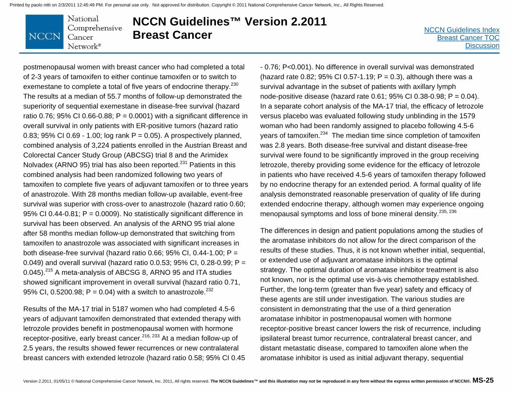
Version 2.2011, 01/05/11 © National Comprehensive Cancer Network, Inc. 2011, All rights reserved. The NCCN Guidelines™ and this illustration may not be reproduced in any form without the express written permission of NCCN®. MS-25
NCCN Guidelines IndexBreast Cancer TOC
Discussion
NCCN Guidelines™ Version 2.2011 Breast Cancer
postmenopausal women with breast cancer who had completed a total of 2-3 years of tamoxifen to either continue tamoxifen or to switch to exemestane to complete a total of five years of endocrine therapy.230 The results at a median of 55.7 months of follow-up demonstrated the superiority of sequential exemestane in disease-free survival (hazard ratio 0.76; 95% CI 0.66-0.88; P = 0.0001) with a significant difference in overall survival in only patients with ER-positive tumors (hazard ratio 0.83; 95% CI 0.69 - 1.00; log rank P = 0.05). A prospectively planned, combined analysis of 3,224 patients enrolled in the Austrian Breast and Colorectal Cancer Study Group (ABCSG) trial 8 and the Arimidex Nolvadex (ARNO 95) trial has also been reported.231 Patients in this combined analysis had been randomized following two years of tamoxifen to complete five years of adjuvant tamoxifen or to three years of anastrozole. With 28 months median follow-up available, event-free survival was superior with cross-over to anastrozole (hazard ratio 0.60; 95% CI 0.44-0.81; P = 0.0009). No statistically significant difference in survival has been observed. An analysis of the ARNO 95 trial alone after 58 months median follow-up demonstrated that switching from tamoxifen to anastrozole was associated with significant increases in both disease-free survival (hazard ratio 0.66; 95% CI, 0.44-1.00; P = 0.049) and overall survival (hazard ratio 0.0.53; 95% CI, 0.28-0.99; P = 0.045).215 A meta-analysis of ABCSG 8, ARNO 95 and ITA studies showed significant improvement in overall survival (hazard ratio 0.71, 95% CI, 0.5200.98; P = 0.04) with a switch to anastrozole.232
Results of the MA-17 trial in 5187 women who had completed 4.5-6 years of adjuvant tamoxifen demonstrated that extended therapy with letrozole provides benefit in postmenopausal women with hormone receptor-positive, early breast cancer.216, 233 At a median follow-up of 2.5 years, the results showed fewer recurrences or new contralateral breast cancers with extended letrozole (hazard ratio 0.58; 95% CI 0.45
- 0.76; P<0.001). No difference in overall survival was demonstrated (hazard rate 0.82; 95% CI 0.57-1.19; P = 0.3), although there was a survival advantage in the subset of patients with axillary lymph node-positive disease (hazard rate 0.61; 95% CI 0.38-0.98; P = 0.04). In a separate cohort analysis of the MA-17 trial, the efficacy of letrozole versus placebo was evaluated following study unblinding in the 1579 woman who had been randomly assigned to placebo following 4.5-6 years of tamoxifen.234 The median time since completion of tamoxifen was 2.8 years. Both disease-free survival and distant disease-free survival were found to be significantly improved in the group receiving letrozole, thereby providing some evidence for the efficacy of letrozole in patients who have received 4.5-6 years of tamoxifen therapy followed by no endocrine therapy for an extended period. A formal quality of life analysis demonstrated reasonable preservation of quality of life during extended endocrine therapy, although women may experience ongoing menopausal symptoms and loss of bone mineral density.235, 236
The differences in design and patient populations among the studies of the aromatase inhibitors do not allow for the direct comparison of the results of these studies. Thus, it is not known whether initial, sequential, or extended use of adjuvant aromatase inhibitors is the optimal strategy. The optimal duration of aromatase inhibitor treatment is also not known, nor is the optimal use vis-à-vis chemotherapy established. Further, the long-term (greater than five year) safety and efficacy of these agents are still under investigation. The various studies are consistent in demonstrating that the use of a third generation aromatase inhibitor in postmenopausal women with hormone receptor-positive breast cancer lowers the risk of recurrence, including ipsilateral breast tumor recurrence, contralateral breast cancer, and distant metastatic disease, compared to tamoxifen alone when the aromatase inhibitor is used as initial adjuvant therapy, sequential
Printed by paolo nitti on 2/3/2011 12:45:49 PM. For personal use only. Not approved for distribution. Copyright © 2011 National Comprehensive Cancer Network, Inc., All Rights Reserved.
Version 2.2011, 01/05/11 © National Comprehensive Cancer Network, Inc. 2011, All rights reserved. The NCCN Guidelines™ and this illustration may not be reproduced in any form without the express written permission of NCCN®. MS-26
NCCN Guidelines IndexBreast Cancer TOC
Discussion
NCCN Guidelines™ Version 2.2011 Breast Cancer
therapy, or extended therapy. Thus, the current version of the guideline recommends that postmenopausal women with early breast cancer receive an aromatase inhibitor as initial adjuvant therapy, sequential with tamoxifen, or as extended therapy in those situations where endocrine therapy is to be utilized. The Panel finds no compelling evidence that there are meaningful efficacy or toxicity differences between anastrozole, letrozole, and exemestane. In postmenopausal women, the use of tamoxifen alone for five years is limited to those who decline or who have a contraindication to aromatase inhibitors (see BINV-J).
It should be re-emphasized that the aromatase inhibitors are associated with the development of benign ovarian pathology and do not adequately suppress ovarian estrogen synthesis in women with functioning ovaries. Premenopausal women should not be given therapy with an aromatase inhibitor outside the confines of a clinical trial. Women who are premenopausal at the time of diagnosis and who become amenorrheic with chemotherapy may have continued estrogen production from the ovaries in the absence of menses. Serial assessment of circulating LH, FSH, and estradiol to assure a true postmenopausal status is mandatory if this subset of women is to be considered for therapy with an aromatase inhibitor.237, 238 (see BINV-L).
Adjuvant cytotoxic chemotherapy
A number of combination chemotherapy regimens are appropriate to consider when adjuvant cytotoxic chemotherapy is utilized (see BINV-K). All adjuvant chemotherapy regimens listed in the Guidelines have been evaluated in phase III clinical trials, and the current version of the adjuvant chemotherapy guideline does not distinguish between options for chemotherapy regimens by axillary lymph node status. Those regimens listed as preferred include: docetaxel, doxorubicin, and cyclophosphamide (TAC); doxorubicin, cyclophosphamide (AC);
dose-dense AC with sequential paclitaxel; AC followed by weekly paclitaxel; and docetaxel plus cyclophosphamide (TC). Other regimens included in the guidelines are: fluorouracil, doxorubicin, and cyclophosphamide (FAC/CAF) or cyclophosphamide, epirubicin, and fluorouracil (FEC/CEF); epirubicin and cyclophosphamide (EC); cyclophosphamide, methotrexate and fluorouracil (CMF); AC with sequential docetaxel administered every 3 weeks; doxorubicin, paclitaxel, cyclophosphamide each as a single agent for four cycles given every 2 weeks (dose-dense A – T– C); FEC followed by docetaxel; and FEC followed by weekly paclitaxel. The adjuvant chemotherapy guideline also includes specific representative doses and schedules for the recommended adjuvant chemotherapy regimens (see BINV-K). Recent studies document substantial improvement in outcome with the incorporation of trastuzumab in the adjuvant treatment of HER2-positive breast cancer (see Adjuvant trastuzumab therapy).
The purpose of distinguishing between ‘preferred’ and ‘other’ adjuvant chemotherapy regimens is to convey the sense of the Panel regarding the relative efficacy and toxicity of the regimens.239 Factors considered by the Panel include the efficacy, toxicity, and treatment schedules of the regimens. This initial attempt at categorizing preferred regimens will be followed in the future by a more comprehensive, systematic evaluation of comparative effectiveness which will also include cost considerations. Summarized below are clinical trial results focusing on treatment efficacy.
Studies of CMF chemotherapy versus no chemotherapy have shown disease-free and overall survival advantages with CMF chemotherapy. 2, 240 Studies using CAF/FAC (cyclophosphamide, doxorubicin, 5-fluorouracil) chemotherapy have shown that the use of full-dose chemotherapy regimens is important. 241 In the Early Breast Cancer
Printed by paolo nitti on 2/3/2011 12:45:49 PM. For personal use only. Not approved for distribution. Copyright © 2011 National Comprehensive Cancer Network, Inc., All Rights Reserved.
Version 2.2011, 01/05/11 © National Comprehensive Cancer Network, Inc. 2011, All rights reserved. The NCCN Guidelines™ and this illustration may not be reproduced in any form without the express written permission of NCCN®. MS-27
NCCN Guidelines IndexBreast Cancer TOC
Discussion
NCCN Guidelines™ Version 2.2011 Breast Cancer
Trialists’ overview of polychemotherapy, comparison of anthracycline-containing regimens with CMF showed a 12% further reduction in the annual odds of recurrence (P = 0.006) and an 11% further reduction in the annual odds of death (P = 0.02) with anthracycline-containing regimens.240 Based on these data, the Panel qualified the appropriate chemotherapy regimens by the statement that anthracycline-containing regimens are preferred for node-positive patients. The Early Breast Cancer Trialists’ analysis, however, did not consider the potential interaction between HER2 tumor status and efficacy of anthracycline-containing versus CMF chemotherapy regimens. Retrospective analysis has suggested that the superiority of anthracycline-containing chemotherapy may be limited to the treatment of those breast cancers that are HER2-positive.175, 177, 180, 210, 242-244 The retrospective finding across several clinical trials that anthracycline-based chemotherapy may be more efficacious in patients whose tumors are HER2-positive has led to a footnote stating that anthracycline-based chemotherapy may be superior to non-anthracycline-containing regimens in the adjuvant treatment of such patients (see BINV-K).
Doxorubicin and cyclophosphamide chemotherapy for four cycles has been studied in randomized trials, resulting in relapse-free and overall survival equivalent to CMF chemotherapy.245-247 No benefit from dose escalation of either doxorubicin or cyclophosphamide was shown.248, 249
The results of two randomized trials comparing AC chemotherapy with or without sequential paclitaxel chemotherapy in women with axillary node-positive breast cancer suggest improved disease-free rates, and results from one of the trials showed an improvement in overall survival, with the addition of paclitaxel.249, 250 On retrospective analysis, the apparent advantage of the paclitaxel-containing regimen appears greater in women with ER-negative breast cancers.
A randomized trial evaluated the use of concurrent versus sequential chemotherapy (doxorubicin followed by paclitaxel followed by cyclophosphamide versus doxorubicin plus cyclophosphamide followed by paclitaxel) given either every two weeks with filgrastim support versus every three weeks. The results show no significant difference between the two chemotherapy regimens, but demonstrate a 26% reduction in hazard of recurrence (P = 0.01) and a 31% reduction in the hazard of death (P = 0.013) for the dose-dense regimens.251
Two randomized prospective trials of CEF chemotherapy in axillary lymph node-positive breast cancer are available. In one trial, premenopausal women with node-positive breast cancer were randomized to receive classic CMF therapy versus CEF chemotherapy using high-dose epirubicin. Both ten-year relapse-free survival (52% vs. 45%; P = 0.007) and overall survival (62% vs. 58%; P = 0.085) favored the CEF arm of the trial.252 The second trial compared CEF given all intravenously every 3 weeks at 2 dose levels of epirubicin (50 mg/m2 vs. 100 mg/m2) in premenopausal and postmenopausal women with node-positive breast cancer. Five-year disease-free survival (55% vs. 66%; P = 0.03) and overall survival (65% vs. 76%; P = 0.007) both favored the epirubicin 100 mg/m2 arm.253 Another trial compared 2 dose levels of EC chemotherapy with CMF chemotherapy in women with node-positive breast cancer. 254 This study showed that higher dose EC chemotherapy was equivalent to CMF chemotherapy and superior to moderate dose EC in event-free survival and overall survival. Another randomized trial in women with axillary lymph node-positive breast cancer compared six cycles of FEC with three cycles of FEC followed by three cycles of docetaxel.255 Five-year disease-free survival (78.4% versus 73.2%, adjusted P = 0.012) and overall survival (90.7% versus 86.7%, P = 0.017) were superior with sequential FEC followed by docetaxel. However, no significant disease-free survival differences
Printed by paolo nitti on 2/3/2011 12:45:49 PM. For personal use only. Not approved for distribution. Copyright © 2011 National Comprehensive Cancer Network, Inc., All Rights Reserved.
Version 2.2011, 01/05/11 © National Comprehensive Cancer Network, Inc. 2011, All rights reserved. The NCCN Guidelines™ and this illustration may not be reproduced in any form without the express written permission of NCCN®. MS-28
NCCN Guidelines IndexBreast Cancer TOC
Discussion
NCCN Guidelines™ Version 2.2011 Breast Cancer
were seen in a recent large randomized study comparing adjuvant chemotherapy with four cycles of every three weekly FEC followed by four cycles of every three weekly docetaxel with standard anthracycline chemotherapy regimens (e.g., FEC or epirubicin followed by CMF) in women with node-positive or high-risk node-negative operable breast cancer.256
Final results from a randomized trial comparing docetaxel, doxorubicin, and cyclophosphamide (TAC) versus FAC chemotherapy in axillary lymph node-positive breast cancer demonstrated that TAC is superior to FAC.257 Estimated 5-year disease-free survival with TAC was 75% and FAC 68% (hazard ratio 0.72; 95% CI 0.59-0.88; P = 0.001) and survival 87% with TAC and 81% with FAC (hazard ratio 0.70; 95% CI 0.53-0.91; P = 0.008). Disease-free survival favored TAC in both ER-positive and ER-negative tumors. At a median follow-up of 73 months, results from the 3-arm randomized NSABP B-30 trial comparing TAC versus AT versus AC followed by docetaxel (ACT) demonstrated that ACT had significant advantage in disease-free survival (hazard ratio 0.83; P = 0.006) but not in overall survival (hazard ratio 0.86; P = 0.086) when compared with TAC. In addition, both disease-free (hazard ratio 0.080; P = 0.001) and overall survival (hazard ratio 0.83; P = 0.034) were significantly increased when ACT was compared with AT, with AT demonstrating non-inferiority compared with TAC.258
The Eastern Cooperative Oncology Group E1199 study was a four arm trial that randomized 4,950 women to receive AC chemotherapy followed by either paclitaxel or docetaxel given by either an every three weekly schedule or a weekly schedule.259, 260 At a median 63.8 months follow-up, no statistically significant differences in disease-free or overall survival were observed when comparing paclitaxel to docetaxel or weekly versus every three weekly administration. In a secondary
series of comparisons, weekly paclitaxel was superior to every three weekly paclitaxel in disease-free survival (hazard ratio 1.27, 95% CI 1.03 – 1.57; P = 0.006) and overall survival (hazard ratio 1.32, 95% CI 1.02-1.72; P = 0.01), and every three weekly docetaxel was superior to every three weekly paclitaxel in disease-free survival (hazard ratio 1.23, 95% CI 1.00-1.52; P-0.02) but not in overall survival.260 Based on these results as well as the findings from the CALGB trial 9741 which showed dose dense AC followed by paclitaxel every two weeks to have a survival benefit when compared with the regimen of AC followed by every three weekly paclitaxel,251 the every three weekly paclitaxel regimen has been removed from the Guidelines.
Combination docetaxel and cyclophosphamide (TC) was compared with AC chemotherapy in a trial that randomized 1016 women with stage I – III breast cancer.261 At a median follow-up of seven years, overall disease-free survival (81% versus 75%; P = 0.033; hazard ratio 0.74; 95% CI, 0.56-0.98) and overall survival (87% versus 82%; P = 0.032; hazard ratio 0.69; 95% CI, 0.50-0.97) were significantly improved with TC compared with AC.
The addition of weekly paclitaxel following FEC was shown to be superior to FEC alone in a randomized study of 1246 women with early-stage breast cancer.262 The former regimen was associated with a 23% reduction in the risk of relapse compared with FEC (hazard ratio 0.77; 95% CI, 0.62-0.95; P = 0.022) although no significant difference in overall survival was seen when the two arms were compared at a median follow-up of 66 months.
Several retrospective studies have evaluated the potential interaction of chemotherapy benefit and ER status.2, 202 These studies assessed the effect of chemotherapy on the risk of breast cancer recurrence in patients with ER-positive tumors receiving adjuvant endocrine therapy
Printed by paolo nitti on 2/3/2011 12:45:49 PM. For personal use only. Not approved for distribution. Copyright © 2011 National Comprehensive Cancer Network, Inc., All Rights Reserved.

Version 2.2011, 01/05/11 © National Comprehensive Cancer Network, Inc. 2011, All rights reserved. The NCCN Guidelines™ and this illustration may not be reproduced in any form without the express written permission of NCCN®. MS-29
NCCN Guidelines IndexBreast Cancer TOC
Discussion
NCCN Guidelines™ Version 2.2011 Breast Cancer
when compared with patients with ER-negative tumor status not undergoing adjuvant endocrine therapy. These analyses suggest that the benefits of chemotherapy are significantly greater in patients with ER-negative disease. For example, the results of Berry et al. demonstrated that 22.8% more patients with ER-negative tumors survived without disease for five years if they received chemotherapy; this benefit was only 7% for patients with ER-positive tumors receiving chemotherapy.202 The guideline therefore includes a recommendation for endocrine therapy and consideration of chemotherapy for patients with node-negative disease and tumors characterized as ER-positive which are greater than 1 cm and HER2-negative or tumors 0.6 to 1.0 cm that are grade 2 or 3 or with unfavorable features (see BINV-6).
Adjuvant trastuzumab therapy Trastuzumab is a humanized, monoclonal antibody with specificity for the extracellular domain of the human epidermal growth factor receptor 2 (HER2/neu; HER2).263 Results of five randomized trials testing trastuzumab as adjuvant therapy have been reported182-185 In NSABP B-31 patients with HER2-positive, node-positive breast cancer were randomly assigned to 4 cycles of AC every three weeks followed by paclitaxel 4 cycles every three weeks or the same regimen with 52 weeks of trastuzumab commencing with the paclitaxel. In the North Central Cancer Treatment Group (NCCTG) N9831 trial, patients with HER2-positive breast cancer that was node-positive, or, if node-negative, with primary tumors greater than 1 cm in size if ER- and PR- negative or greater than 2 cm in size if ER- or PR-positive, were similarly randomized except that paclitaxel was given by a low dose weekly schedule for 12 weeks and a third arm delayed trastuzumab until the completion of paclitaxel. The B-31 and NCCTG N9831 trials were jointly analyzed with the merged control arms for both trials compared with the merged arms using trastuzumab begun concurrently with the paclitaxel. There were 3968 patients included in the joint
analysis performed at four years median follow-up. A 52% reduction in the risk of recurrence (hazard ratio 0.48; 95% CI 0.41-0.57; P<0.0001) and a 35% reduction in the risk of death (hazard ratio 0.65; 95% CI 0.51-0.84; log-rank P = 0.0007) were documented.264 Similar significant effects on disease-free survival were observed when results of the NSABP B-31 and NCCTG N9831 trials were analyzed separately. Cardiac toxicity was increased in patients treated with trastuzumab.184,
265, 266 In the adjuvant trastuzumab trials, the rates of grade III/IV congestive heart failure (CHF) or cardiac-related death for patients receiving treatment regimens containing trastuzumab ranged from 0% (FinHer trial) to 4.1% (NSABP B-31 trial) overall 182-185, 265, 266 The frequency of cardiac dysfunction appears to be related to both age and baseline left ventricular ejection fraction. An analysis of data from N9831 showed the 3-year cumulative incidence of congestive heart failure or cardiac death to be 0.3%, 2.8% and 3.3% in the arms of the trial without trastuzumab, with trastuzumab following chemotherapy, and with trastuzumab initially combined with paclitaxel, respectively. 265 The acceptable rate of significant cardiac toxicity observed in the trastuzumab adjuvant trials in part reflects rigorous monitoring for cardiac dysfunction. Furthermore, concerns have been raised regarding the long-term cardiac risks associated with trastuzumab therapy based on results of follow-up evaluations of cardiac function in patients enrolled in some of these trials.267, 268
A third trial (HERA) (N=5081) tested trastuzumab for one or for two years compared to none following all local therapy and a variety of standard chemotherapy regimens in patients with node-positive disease or node-negative disease with tumor ≥ 1 cm.183 At a median follow-up of one year, comparing one year versus not of trastuzumab, trastuzumab resulted in a 46% reduction in the risk of recurrence compared to no trastuzumab (hazard ratio 0.54; 95% CI 0.43-0.67; P < 0.0001), no
Printed by paolo nitti on 2/3/2011 12:45:49 PM. For personal use only. Not approved for distribution. Copyright © 2011 National Comprehensive Cancer Network, Inc., All Rights Reserved.
Version 2.2011, 01/05/11 © National Comprehensive Cancer Network, Inc. 2011, All rights reserved. The NCCN Guidelines™ and this illustration may not be reproduced in any form without the express written permission of NCCN®. MS-30
NCCN Guidelines IndexBreast Cancer TOC
Discussion
NCCN Guidelines™ Version 2.2011 Breast Cancer
difference in overall survival, and acceptable cardiac toxicity. The two year data indicate that 1-year of trastuzumab therapy is associated with an overall survival benefit when compared with observation (hazard ratio for risk of death = 0.66; 95% CI, 0.47-0.91; P = 0.0115).269
The Breast Cancer International Research Group (BCIRG) 006 study randomized 3,222 women with HER2-positive, node-positive or high-risk node negative breast cancer to AC followed by docetaxel, AC followed by docetaxel plus trastuzumab for one year, or carboplatin, docetaxel plus trastuzumab for one year.185 At 36 months of follow-up, patients receiving AC followed by docetaxel with trastuzumab (ACTH) had a hazard ratio for disease-free recurrence of 0.61 (95% CI, 0.48-0.76; P<0.0001) when compared with the group of patients in the control arm receiving the same chemotherapy regimen without trastuzumab (ACT). The hazard ratio for disease-free survival was 0.67 (95% CI, 0.54-0.83; P = 0.0003) when patients in the carboplatin/docetaxel/ trastuzumab (TCH) containing arm were compared to patients in the control arm. No statistically significant difference in the hazard ratio for disease-free survival was observed between the two trastuzumab-containing arms. An overall survival advantage was reported for patients in both trastuzumab-containing arms relative to the control arm (hazard ratio for AC-TH vs AC-T = 0.59; 95% CI, 0.42-0.85; P = 0.004; hazard ratio for TCH vs AC-T = 0.66; 95% CI, 0.47-0.93; P = 0.017). Cardiac toxicity was significantly lower in the TCH arm (8.6% patients with >10% relative decline in left ventricular ejection fraction) compared with the AC-TH arm (18%; P<0.0001); differences in cardiac toxicity between the TCH arm and the AC-T control arm (10%) were not significant.
A fifth trial (FinHer) randomized 1010 women to either nine weeks of vinorelbine followed by three cycles of FEC chemotherapy versus docetaxel for three cycles followed by three cycles of FEC
chemotherapy.182 Patients (n = 232) with HER2-positive cancers that were either node-positive or node-negative and ≥ 2 cm and progesterone receptor- negative were further randomized to receive or not trastuzumab for 9 weeks during the vinorelbine or docetaxel portions of the chemotherapy only. With a median follow-up of three years, the addition of trastuzumab was associated with a reduction in risk of recurrence (hazard ratio 0.42; 95% CI 0.21-0.83; P = 0.01). No statistically significant differences in overall survival (hazard ratio 0.41; 95% CI 0.16 – 1.08; P = 0.07) or cardiac toxicity were observed with the addition of trastuzumab.182 At five years follow-up, a comparison of the two arms (i.e., chemotherapy with and without trastuzumab) demonstrated that the hazard ratios for distant disease-free survival (hazard ratio 0.65; 95% CI, 0.38-1.12; P = 0.12) and overall survival (hazard ratio 0.55; 95% CI, 0.27-1.11; P = 0.094) were higher relative to those reported at three years.270
All of the adjuvant trials of trastuzumab have demonstrated clinically significant improvements in disease-free survival, and the combined analysis from the NSABP B31 and NCCTG N9831 trials, and the HERA trial, showed significant improvement in overall survival with the use of trastuzumab in patients with high-risk, HER2-positive breast cancer. Therefore, regimens from each of these trials are included as trastuzumab-containing adjuvant regimen choices in the guideline (category 1) (see BINV-K). The benefits of trastuzumab are independent of ER status.184 On the basis of these studies, the Panel has designated use of trastuzumab with chemotherapy as a category 1 recommendation in patients with HER2-positive tumors >1cm. The Panel recommends AC followed by paclitaxel with trastuzumab for 1 year commencing with the first dose of paclitaxel as a preferred trastuzumab-containing adjuvant regimen since the efficacy of this regimen has been demonstrated in two randomized clinical trials, and it
Printed by paolo nitti on 2/3/2011 12:45:49 PM. For personal use only. Not approved for distribution. Copyright © 2011 National Comprehensive Cancer Network, Inc., All Rights Reserved.

Version 2.2011, 01/05/11 © National Comprehensive Cancer Network, Inc. 2011, All rights reserved. The NCCN Guidelines™ and this illustration may not be reproduced in any form without the express written permission of NCCN®. MS-31
NCCN Guidelines IndexBreast Cancer TOC
Discussion
NCCN Guidelines™ Version 2.2011 Breast Cancer
has been associated with significant improvements in overall survival. The TCH regimen is also classified as a preferred regimen, especially in those with risk factors for cardiac toxicity, given the results of BCIG 006 study that demonstrated superior disease-free survival in patients receiving either TCH or AC followed by docetaxel plus trastuzumab both compared with AC followed by docetaxel alone. Since patients with borderline FISH (Pathvysion®) scores of greater than 2.0 to 2.2 HER2 genes/chromosome 17/cell in early-stage breast cancer were eligible for the adjuvant trials, the Panel cannot recommend exclusion of these patients from adjuvant treatment with trastuzumab if HER2 tumor status remains equivocal following retesting by the same or a complementary method (see BINV-A). The Panel has also included a recommendation for consideration of adjuvant trastuzumab in women with node-negative tumors that are 0.6-1.0 cm (category 2A) (see BINV-5; BINV-7). Some support for this recommendation comes from results of a retrospective study of 1245 women with early-stage breast cancer tumors characterized as T1pN0.271 Ten-year breast cancer specific survival and 10-year recurrence-free survival were 85% and 75%, respectively, in women with tumors characterized as HER2-positive, ER-positive, and 70% and 61%, respectively, in women with HER2-positive, ER-negative tumors. Two more recent retrospective studies have also investigated recurrence-free survival in this patient population. In one large study, 5-year recurrence-free survival rates of 77.1% and 93.7% (P<0.001) were observed for patients with HER2-positive and HER2-negative T1a,b,N0,M0 breast tumors, respectively, with no recurrence-free survival differences seen in the HER2-positive group when hormonal receptor status was considered.272 In another retrospective study of women with small HER2-positive tumors, the risk of recurrence at 5 years was low, although disease-free survival was inferior in the group with HER2-positive, hormone receptor-positive disease.273 None of the
patients in these two retrospective studies had received trastuzumab. Subgroup analyses from several of the randomized trials have shown consistent benefit of trastuzumab irrespective of tumor size or nodal status.264, 274 The recommendation for consideration of trastuzumab in patients with HER2-positive tumors that are 0.6-1.0 cm is now designated as category 2A.
Dose dense ACT with trastuzumab is another trastuzumab-containing adjuvant chemotherapy regimen included in the Guidelines. Data from a single arm study of 70 patients have provided support for the safety and feasibility of this regimen.275
Finally, no statistically significant disease-free or overall survival benefit for the addition of trastuzumab was observed in the FNCLCC-PACS-04 trial in which 528 women with HER2-positive, node-positive breast cancer were randomly assigned to receive trastuzumab or observation following completion of adjuvant anthracycline-based chemotherapy with or without docetaxel.276 These results suggest that the sequential administration of trastuzumab following chemotherapy is not as efficacious as a schedule involving concomitant chemotherapy and trastuzumab.
Adjuvant therapy of favorable histology tumors The Guidelines provide systemic treatment recommendations for the favorable-histology invasive breast cancers, such as tubular and colloid cancers, based on tumor size and axillary lymph node status (see BINV-9). If used, the treatment options for endocrine therapy, chemotherapy, and sequencing of treatment with other modalities are similar to those of the usual histology breast cancers. The vast majority of tubular breast cancers are both ER-positive and HER2-negative. Thus, the pathology evaluation and accuracy of the ER and/or HER2 determination should be reviewed if a tubular breast cancer is found to
Printed by paolo nitti on 2/3/2011 12:45:49 PM. For personal use only. Not approved for distribution. Copyright © 2011 National Comprehensive Cancer Network, Inc., All Rights Reserved.
Version 2.2011, 01/05/11 © National Comprehensive Cancer Network, Inc. 2011, All rights reserved. The NCCN Guidelines™ and this illustration may not be reproduced in any form without the express written permission of NCCN®. MS-32
NCCN Guidelines IndexBreast Cancer TOC
Discussion
NCCN Guidelines™ Version 2.2011 Breast Cancer
be ER-negative and/or HER2-positive, or if a tumor with a ER- and PR-negative status is found to be grade 1.11 Should a breast cancer be histologically identified as a tubular or colloid (mucinous) breast cancer and be confirmed as ER-negative, then the tumor should be treated according to the guideline for the usual histology, ER-negative breast cancers. The Panel acknowledges that prospective data regarding systemic adjuvant therapy of favorable histology tumors are lacking.
Medullary carcinoma is an uncommon variant of infiltrating ductal carcinoma characterized by high nuclear grade, lymphocytic infiltration, a pushing tumor border, and the presence of a syncytial growth pattern. It was previously thought that medullary carcinoma has a lower potential for metastases and a better prognosis than typical infiltrating ductal carcinoma. However, the best available evidence suggests that the risk of metastases equals that of other high-grade carcinomas, even for cases that meet all the pathologic criteria for typical medullary carcinoma. Furthermore, typical medullary carcinoma is uncommon, and there is marked interobserver variation in diagnosing this entity. Many cases classified as medullary carcinoma do not have all the pathologic features on subsequent pathologic review. Given these facts, there is concern that patients may be harmed if a high-grade infiltrating ductal carcinoma is misclassified as typical medullary carcinoma and this classification used as the basis for withholding otherwise indicated adjuvant systemic therapy. Therefore, the NCCN Panel believes that including medullary carcinoma with other special histology cancers that carry a very favorable prognosis and often do not require systemic therapy is not appropriate. The Panel recommends that cases classified as medullary carcinoma be treated as other infiltrating ductal carcinomas based on tumor size, grade, and lymph node status.
Stage III Invasive Breast Cancer The staging evaluation for most patients with stage III invasive breast cancer is similar to the one for patients with T3N1M0 disease (see BINV-14; BINV-1). The workup includes history and physical exam, a complete blood cell count, platelet count, liver function and alkaline phosphatase tests, chest imaging, pathology review, pre-chemotherapy determination of tumor ER/PR receptor status and HER2 status, diagnostic bilateral mammogram and breast ultrasound as clinically warranted. Genetic counseling is recommended if the patient is considered to be at high risk of hereditary breast cancer as defined by the NCCN Genetic/Familial High-Risk Assessment: Breast and Ovarian Guidelines
The performance of other studies, such as a breast MRI, a bone scan (category 2B) and abdominal imaging with CT (with or without pelvic CT), ultrasound or MRI (all category 2A) are optional unless directed by symptoms or other abnormal study results. PET/CT scan is also included as an optional additional study (category 2B).
The consensus of the Panel is that PET/CT is most helpful in situations where standard imaging results are equivocal or suspicious. However, limited recent studies72, 73, 277-280 support a potential role for FDG PET/CT to detect regional node involvement as well as distant metastases in locally advanced breast cancer including T3, N1, M0 disease.
Equivocal or suspicious sites identified by PET/CT scanning should be biopsied for confirmation whenever possible and the site of disease would impact the course of treatment. In the past decade, the advent of PET/CT scanners has significantly changed the approach to PET imaging.281 However, the terminology has also created confusion regarding the nature of the scans obtained from a PET/CT device.
Printed by paolo nitti on 2/3/2011 12:45:49 PM. For personal use only. Not approved for distribution. Copyright © 2011 National Comprehensive Cancer Network, Inc., All Rights Reserved.
Version 2.2011, 01/05/11 © National Comprehensive Cancer Network, Inc. 2011, All rights reserved. The NCCN Guidelines™ and this illustration may not be reproduced in any form without the express written permission of NCCN®. MS-33
NCCN Guidelines IndexBreast Cancer TOC
Discussion
NCCN Guidelines™ Version 2.2011 Breast Cancer
PET/CT scanners have both a PET and CT scanner in the same gantry that allows precise co-registration of molecular (PET) and anatomic (CT) imaging. Almost all current clinical PET imaging is performed using combined PET/CT devices.
In PET/CT tomographs, the CT scanner has a second important role beyond diagnostic CT scanning.281 For PET applications, the CT scan is also used for photon attenuation correction and for anatomic localization of the PET imaging findings. For these tasks, the CT scan is usually taken without breathholding, to match PET image acquisition, and typically uses relatively low-dose (non-diagnostic) CT. Radiation exposure for these non-diagnostic CT scans is lower than for diagnostic CT. Intravenous contrast is not needed for this task.
PET/CT scanners typically include a high-quality CT device that can also be used for stand-alone, optimized and fully diagnostic CT. Diagnostic CT scans are acquired using breathholding for optimal chest imaging, and are often performed with intravenous contrast. For fully diagnostic CT, the CT beam current, and therefore patient radiation exposure, is considerably higher than for the low-dose CT needed for PET requirements. Radiation exposures for fully diagnostic CT are often greater than for the emission (PET) component of the study.
Currently, the approach to clinical PET/CT imaging varies widely across centers.282 Many centers perform low-dose CT as part of a PET/CT scan, and perform optimized, fully diagnostic CT only when diagnostic CT has also been requested in addition to PET/CT. Other centers combine diagnostic CT scans with PET on all of their PET/CT images. The CT scans described in sections BINV-1, BINV-10 and BINV-14 refer to fully optimized diagnostic CT scans, while the PET or PET/CT scans refer to scans primarily directed towards the PET component, not necessarily using diagnostic-quality CT. It is important for referring
physicians to understand the differences between PET/CT performed primarily for PET imaging and fully optimized CT performed as a stand-alone diagnostic CT examination.282
Operable locally advanced breast cancer (clinical stage T3N1M0) Locally advanced breast cancer describes a subset of invasive breast cancer where the initial clinical and radiographic evaluation documents advanced disease confined to the breast and regional lymph nodes. The AJCC clinical staging system used in these Guidelines and for the determination of operability is recommended and locally advanced disease is represented by the stage III category. Patients with stage III disease may be further divided into those where an initial surgical approach is unlikely to be successful in removal of all disease or to provide long-term local control and those with disease where a reasonable initial surgical approach is likely to achieve pathologically negative margins and provide long-term local control. Thus, stage IIIA patients are divided into those who have clinical T3N1M0 disease versus those who have clinical TanyN2M0 disease, based on evaluation by a multidisciplinary team. For patients with operable locally advanced disease, generally patients with clinical T3N1M0 disease, treatment is as outlined in BINV-1 through BINV-6.
Postsurgical systemic adjuvant therapy for patients with stage IIIA breast cancer who do not receive neoadjuvant chemotherapy is similar to that for patients with stage II disease.
Inoperable locally advanced breast cancer (clinical stage IIIA [except for T3N1M0], clinical stage IIIB, or clinical stage IIIC) The workup of locally advanced breast cancer is described on BINV-14. For patients with inoperable non-inflammatory locally advanced disease at presentation, the initial use of anthracycline-based preoperative chemotherapy with or without a taxane is standard therapy.283 Patients
Printed by paolo nitti on 2/3/2011 12:45:49 PM. For personal use only. Not approved for distribution. Copyright © 2011 National Comprehensive Cancer Network, Inc., All Rights Reserved.
Version 2.2011, 01/05/11 © National Comprehensive Cancer Network, Inc. 2011, All rights reserved. The NCCN Guidelines™ and this illustration may not be reproduced in any form without the express written permission of NCCN®. MS-34
NCCN Guidelines IndexBreast Cancer TOC
Discussion
NCCN Guidelines™ Version 2.2011 Breast Cancer
with locally advanced breast cancer that is HER2-positive should receive an initial chemotherapy program that incorporates preoperative trastuzumab (BINV-K). Local therapy following a clinical response to preoperative chemotherapy usually consists of (1) total mastectomy with level I/II axillary lymph node dissection, with or without delayed breast reconstruction, or (2) lumpectomy and level I/II axillary dissection. Both local treatment groups are considered to have sufficient risk of local recurrence to warrant the use of chest wall (or breast) and supraclavicular node irradiation. If internal mammary lymph nodes are involved, they should also be irradiated. In the absence of detected internal mammary node involvement, consideration may be given to including the internal mammary lymph nodes in the radiation field (category 3) (see BINV-15). Adjuvant therapy may involve completion of planned chemotherapy regimen course if not completed preoperatively, followed by endocrine therapy in patients with hormone receptor-positive disease (see BINV-15). Up to one year of total trastuzumab therapy should be completed, if the tumor is HER2-positive (category 1). Endocrine therapy and trastuzumab can be administered concurrent with radiation therapy if indicated.
Patients with an inoperable stage III tumor with disease progression during preoperative chemotherapy should be considered for palliative breast irradiation in an attempt to enhance local control. In all subsets of patients, further systemic adjuvant chemotherapy after local therapy is felt to be standard. Tamoxifen (or an aromatase inhibitor if postmenopausal) should be added for those with hormone receptor-positive tumors, and trastuzumab should be given to those with HER2-positive tumors. Post-treatment follow-up for women with stage III disease is the same as for women with earlier-stage, invasive breast cancer. Treatment recommendations for inflammatory locally advanced breast cancer are described on IBC-1.
Post-therapy Surveillance and Follow-up Post-therapy follow-up is optimally performed by members of the treatment team and includes the performance of regular physical examinations and mammography. In patients undergoing breast-conserving therapy, mammography should be performed annually (category 2A). The routine performance of alkaline phosphatase and liver function tests are not included in the Guidelines.284-286 In addition, the Panel notes no evidence to support the use of “tumor markers” for breast cancer, and routine bone scans, CT scans, MRI scans, PET scans, or ultrasound examinations in the asymptomatic patient provide no advantage in survival or ability to palliate recurrent disease and are, therefore, not recommended.72, 287
The use of dedicated breast MRI may be considered as an option for post-therapy surveillance and follow-up in women at high risk of bilateral disease, such as carriers of BRCA 1/2 mutations. Rates of contralateral breast cancer following either breast-conserving therapy or mastectomy have been reported to be increased in women with BRCA 1/2 mutations when compared with patients with sporadic breast cancer.288-290(see NCCN Genetic/Familial High-Risk Assessment: Breast and Ovarian Guidelines; NCCN Breast Cancer Screening and Diagnosis Guidelines).
The Panel recommends that women with intact uteri who are taking tamoxifen should have yearly gynecologic assessments and rapid evaluation of any vaginal spotting that might occur because of the risk of tamoxifen-associated endometrial carcinoma in postmenopausal women291(see BINV-16). The performance of routine endometrial biopsy or ultrasonography in the asymptomatic woman is not recommended. Neither test has demonstrated utility as a screening test
Printed by paolo nitti on 2/3/2011 12:45:49 PM. For personal use only. Not approved for distribution. Copyright © 2011 National Comprehensive Cancer Network, Inc., All Rights Reserved.

Version 2.2011, 01/05/11 © National Comprehensive Cancer Network, Inc. 2011, All rights reserved. The NCCN Guidelines™ and this illustration may not be reproduced in any form without the express written permission of NCCN®. MS-35
NCCN Guidelines IndexBreast Cancer TOC
Discussion
NCCN Guidelines™ Version 2.2011 Breast Cancer
in any population of women. The vast majority of women with tamoxifen-associated uterine carcinoma have early vaginal spotting.
Symptom management for women on adjuvant endocrine therapies often requires treatment of hot flashes and the treatment of concurrent depression. Venlafaxine has specifically been studied and is an effective intervention in decreasing hot flashes.292 Recent evidence has suggested that concomitant use of tamoxifen with certain selective serotonin reuptake inhibitors (SSRIs) (e.g., paroxetine and fluoxetine) may decrease plasma levels of endoxifen, an active metabolite of tamoxifen.293, 294 These SSRIs may interfere with the enzymatic conversion of tamoxifen to endoxifen by inhibiting a particular isoform of cytochrome P-450 enzyme (CYP2D6) involved in the metabolism of tamoxifen. However, the SSRIs citalopram, escitalopram, fluvoxamine, gabapentin, sertraline, and venlafaxine appear to have no or only minimal effect on tamoxifen metabolism.237, 295 If an aromatase inhibitor is considered in women with amenorrhea following treatment, baseline levels of estradiol and gonadotropin followed by serial monitoring of these hormones should be performed if endocrine therapy with an aromatase inhibitor is initiated237(see BINV-L). Bilateral oophorectomy assures postmenopausal status in young women with therapy-induced amenorrhea and may be considered prior to initiating therapy with an aromatase inhibitor in a young woman.
Follow-up also includes assessment of patient adherence to ongoing medication regimens such as endocrine therapies. Predictors of poor adherence to medication include the presence of side effects associated with the medication, and incomplete understanding by the patient of the benefits associated with regular administration of the medication.296 The Panel recommends the implementation of simple strategies to enhance patient adherence to endocrine therapy, such as direct questioning of the patient during office visits, as well as brief,
clear explanations on the value of taking the medication regularly and the therapeutic importance of longer durations of endocrine therapy (see BINV-16).
Evidence suggests that a healthy lifestyle may lead to better breast cancer outcomes. A nested case control study of 369 women with ER-positive tumors who developed a second primary breast cancer compared with 734 matched control patients who did not develop a second primary tumor, showed an association between obesity (BMI>=30), smoking and alcohol consumption and contralateral breast cancer.297 A prospective study of 1490 women diagnosed with stage I-III breast cancer showed an association between high fruit and vegetable consumption, physical activity and improved survivorship, regardless of obesity.298 Thus, the panel recommends an active lifestyle and ideal body weight (BMI 20-25) for optimal overall health and breast cancer outcomes.
Many young women treated for breast cancer remain or regain premenopausal status following treatment for breast cancer. For these women, the panel discourages the use of hormonal birth control methods, regardless of the hormone receptor status of the tumor.299 Alternative birth control methods are recommended; including intrauterine devices (IUDs), barrier methods, and for those with no intent of future pregnancy, tubal ligation or vasectomy for the partner. Breast feeding during endocrine or chemotherapy treatment is not recommended by the panel because of risks to the infant. Breast feeding after breast conserving treatment for breast cancer is not contraindicated. However, lactation from an irradiated breast may not be possible, or may occur only with a diminished capacity.299, 300 (see BINV-C).
Printed by paolo nitti on 2/3/2011 12:45:49 PM. For personal use only. Not approved for distribution. Copyright © 2011 National Comprehensive Cancer Network, Inc., All Rights Reserved.

Version 2.2011, 01/05/11 © National Comprehensive Cancer Network, Inc. 2011, All rights reserved. The NCCN Guidelines™ and this illustration may not be reproduced in any form without the express written permission of NCCN®. MS-36
NCCN Guidelines IndexBreast Cancer TOC
Discussion
NCCN Guidelines™ Version 2.2011 Breast Cancer
Stage IV Metastatic or Recurrent Breast Cancer The staging evaluation of women who present with metastatic or recurrent breast cancer includes history and physical exam, the performance of a CBC, platelet count, liver function tests, chest imaging, bone scan, radiographs of any long or weight-bearing bones that are painful or appear abnormal on bone scan, consideration of CT or MRI scan of the abdomen, biopsy documentation of first recurrence if possible, and determination of hormone receptor status (ER and PR) and HER2 status should be repeated, especially if unknown, originally negative or not over-expressed. Genetic counseling is recommended if patient is considered to be at high risk of hereditary breast cancer as defined by the NCCN Genetic/Familial High-Risk Assessment: Breast and Ovarian Guidelines.
The Panel generally discourages the use of PET or PET/CT scans for the evaluation of patients with recurrent disease, except in those situations where other staging studies are equivocal or suspicious. Although there is limited, mostly retrospective, evidence to support the use of PET/CT scanning to guide treatment planning through determination of the extent of disease in select patients with recurrent or metastatic disease,72, 73, 301, 302 the Panel considers biopsy of equivocal or suspicious sites to be more likely than PET/CT scanning to provide accurate staging information in this population of patients.
Local disease only Patients with local recurrence only are divided into 3 groups: those who had been treated initially by mastectomy alone, mastectomy with radiation therapy and those who had received breast-conserving therapy (see BINV-16).
In one retrospective study of local recurrence patterns in women with breast cancer who had undergone mastectomy and adjuvant
chemotherapy without radiation therapy, the most common sites of local recurrence were at the chest wall and the supraclavicular lymph nodes.303 The recommendations for treatment of the population of patients experiencing a local recurrence only are supported by analyses of a combined database of patients from the EORTC 10801 and Danish Breast Cancer Group 82TM trials. The analyses compared breast-conserving therapy with mastectomy in patients with stage I and stage II disease. The 133 (approximately 8%) patients experiencing a local recurrence as an initial event were approximately equally divided between those who had undergone mastectomy and those who had received breast-conserving therapy as initial treatment for breast cancer. Of those in the former group, 51 (76%) were able to undergo radiation therapy with or without surgery as treatment for local disease recurrence. No difference in survival emerged when patients receiving salvage treatment after initial treatment with mastectomy or breast-conserving therapy were compared; approximately 50% of both groups were alive at 10 years.304
Mastectomy-treated patients should undergo surgical resection of the local recurrence (if it can be accomplished without heroic surgery) and involved-field radiation therapy to chest wall and supraclavicular area (if the chest wall was not previously treated or if additional radiation therapy may be safely administered). The use of surgical resection in this setting implies the use of limited excision of disease with the goal of obtaining clear margins of resection. Unresectable chest wall recurrent disease should be treated with radiation therapy if no prior radiation has been given. Women with a local recurrence of disease after initial breast-conserving therapy should undergo a total mastectomy and axillary staging if a level I/II axillary dissection was not previously performed. Limited data suggest that a repeat sentinel lymph node biopsy following local recurrence of disease may be successfully
Printed by paolo nitti on 2/3/2011 12:45:49 PM. For personal use only. Not approved for distribution. Copyright © 2011 National Comprehensive Cancer Network, Inc., All Rights Reserved.

Version 2.2011, 01/05/11 © National Comprehensive Cancer Network, Inc. 2011, All rights reserved. The NCCN Guidelines™ and this illustration may not be reproduced in any form without the express written permission of NCCN®. MS-37
NCCN Guidelines IndexBreast Cancer TOC
Discussion
NCCN Guidelines™ Version 2.2011 Breast Cancer
performed in 80% of women who have previously undergone breast conserving therapy and sentinel node biopsy.305 The consensus of the Panel is that the preferred surgical approach for most women with a local recurrence following breast conserving therapy and sentinel node biopsy is mastectomy and a level I/II axillary dissection, although sentinel node biopsy in lieu of a level I/II axillary dissection can be considered if prior axillary staging was done by sentinel node biopsy only.
After local treatment, women with local recurrences only should be considered for limited duration systemic chemotherapy or endocrine therapy similar to that outlined in the adjuvant chemotherapy section. The ongoing BIG 1-01/IBCSG 27-02/NSABP B-37 study is evaluating the utility of chemotherapy in women who develop an isolated local and/or regional ipsilateral recurrence following primary treatment for early breast cancer.306 The Panel emphasized the importance of individualizing treatment strategies in patients with a recurrence of disease limited to a local site.
The Guidelines include consideration of the addition of hyperthermia to irradiation for localized recurrences/metastasis (category 3) (see BINV-17). There have been several prospective randomized trials comparing radiation to radiation plus hyperthermia in the treatment of locally advanced/recurrent cancers, primarily breast cancer chest wall recurrences.307, 308 While there is heterogeneity among the study results, a recent series with strict quality assurance demonstrated a statistically significant increase in local tumor response and greater duration of local control with the addition of hyperthermia to radiation compared to radiation alone.307 No differences in overall survival have been demonstrated. Delivery of local hyperthermia is technically demanding and requires specialized expertise and equipment (e.g., the monitoring of temperatures and management of possible tissue burns).
The Panel thus recommends that the use of hyperthermia be limited to treatment centers with appropriate training, expertise, and equipment. The addition of hyperthermia generated substantial discussion and controversy among the Panel and is a category 3 recommendation.
Systemic disease The systemic treatment of breast cancer recurrence or stage IV disease prolongs survival and enhances quality of life but is not curative. Therefore, treatments associated with minimal toxicity are preferred. Thus, the use of the minimally toxic endocrine therapies is preferred to the use of cytotoxic therapy whenever reasonable.309
Guideline stratification for therapy in systemic disease Patients with recurrence of breast cancer or metastatic breast cancer at diagnosis are initially stratified according to whether or not bone metastasis is present (see section on Supportive therapy for bone metastasis below). These two patient subsets are then stratified further by tumor hormone receptor and HER2 status (see BINV-17).
Supportive therapy for bone metastasis Treatment targeting osteoclast activity is of value in patients with metastatic breast cancer in bone to prevent bone fractures, bone pain requiring radiation therapy, spinal cord compression, and hypercalcemia (skeletal related events; SREs).310-312 The bisphosphonates zoledronic acid or pamidronate have been used for this purpose, and there is extensive clinical trial support for their efficacy in prevention of SREs (see below section on Bisphosphonates). Recently, a single randomized, active controlled trial in metastatic breast cancer met the primary endpoint of equivalency and achieved a secondary endpoint of superiority of time to the occurrence of SRE with denosumab, a fully human monoclonal antibody directed against RANK ligand, a mediator of osteoclast
Printed by paolo nitti on 2/3/2011 12:45:49 PM. For personal use only. Not approved for distribution. Copyright © 2011 National Comprehensive Cancer Network, Inc., All Rights Reserved.

Version 2.2011, 01/05/11 © National Comprehensive Cancer Network, Inc. 2011, All rights reserved. The NCCN Guidelines™ and this illustration may not be reproduced in any form without the express written permission of NCCN®. MS-38
NCCN Guidelines IndexBreast Cancer TOC
Discussion
NCCN Guidelines™ Version 2.2011 Breast Cancer
function313 as compared with zoledronic acid.312 Thus it appears that denosumab is at least as efficacious as zoledronic acid in preventing SREs. No study of bisphosphonate or denosumab has demonstrated an impact on overall survival in patients with metastatic disease.
The bisphosphonates and denosumab are associated with the occurance of osteonecrosis of the jaw. Poor baseline dental health or the requirement for dental procedures during treatment are known risk factors for osteonecrosis of the jaw. Thus, a dental examination with preventive dentistry intervention is recommended prior to treatment with intravenous bisphosphonate or denosumab, and dental procedures during treatment should be avoided if at all possible. Additional risk factors for the development of osteonecrosis of the jaw include administration of chemotherapy or corticosteroids and poor oral hygiene with periodontal disease and dental abscess.314
Confirmation of metastatic disease by imaging including x-ray, CT or MRI; and initial evaluation of serum calcium, creatinine, phosphorous and magnesium levels should be undertaken prior to the initiation of intravenous bisphosphonate treatment or subcutaneous denosumab treatment in patients with metastatic disease. Frequent measurement of calcium, phosphorous and magnesium may be prudent since hypophosphatemia and hypocalcemia have been reported.
Bisphosphonates Women with bone metastasis, especially if lytic, can be given a bisphosphonate (e.g., pamidronate or zoledronic acid) in combination with calcium citrate and vitamin D if expected survival is 3 months or longer and creatinine levels are below 3.0 mg/dL (category 1).311, 315-320 Bisphosphonates are given in addition to chemotherapy or endocrine therapy. Zoledronic acid may be superior to pamidronate in lytic breast metastasis.321, 322
There are extensive data from randomized trials in support of the use of bisphosphonates for patients with metastatic disease to bone. The randomized clinical trial data includes the use of zoledronic acid and pamidronate in the United States and ibandronate and clodronate in European countries.318, 320, 322-327 In metastatic bone disease, bisphosphonate treatment is associated with fewer skeletal-related events, pathologic fractures, and less need for radiation therapy and surgery to treat bone pain.
The use of bisphosphonates in metastatic disease is a palliative care measure. No impact on overall survival has been observed in patients treated with bisphosphonates. The data indicate that zoledronic acid and pamidronate may be given on a 3-5 weekly schedule in conjunction with antineoplastic therapy (i.e., endocrine therapy, chemotherapy or biologic therapy). The use of bisphosphonates should be accompanied by calcium and vitamin D supplementation with daily doses of calcium of 1200 to 1500mg and Vitamin D3 400 – 800 IU. Recommended agents for use in the United States are pamidronate 90 mg intravenously over two hours or zoledronic acid 4mg intravenously over 15 minutes. The original studies continued treatment for up to 24 months; however, there are limited long-term safety data indicating treatment can continue beyond that time.325, 327, 328 The risk of renal toxicity necessitates monitoring of serum creatinine prior to administration of each dose and dose reduction or discontinuation if renal function is reduced. Current clinical trial results support the use of bisphosphonates for up to two years. Longer durations of bisphosphonate therapy may provide additional benefit, but this has not yet been tested in clinical trials.
Osteonecrosis of the jaw, a recently reported complication of bisphosphonate treatment, has been described. In a review of more than 16,000 cancer patients, an increased risk of jaw or facial bone
Printed by paolo nitti on 2/3/2011 12:45:49 PM. For personal use only. Not approved for distribution. Copyright © 2011 National Comprehensive Cancer Network, Inc., All Rights Reserved.
Version 2.2011, 01/05/11 © National Comprehensive Cancer Network, Inc. 2011, All rights reserved. The NCCN Guidelines™ and this illustration may not be reproduced in any form without the express written permission of NCCN®. MS-39
NCCN Guidelines IndexBreast Cancer TOC
Discussion
NCCN Guidelines™ Version 2.2011 Breast Cancer
surgery along with an increased risk of being diagnosed with inflammatory conditions or osteomyelitis of the jaw with the use of intravenous bisphosphonates was documented. An absolute risk of 5.48 events per 100 patients treated was seen, with an increase in risk associated with an increase in cumulative dose of drug.329
Denosumab Women with metastatic breast cancer to bone who are candidates for bisphosphonate therapy may also be considered for treatment with denosumab, based upon the results of a single randomized trial comparing denosumab to zoledronic acid.312 All trial patients were recommended to supplement with vitamin D and calcium. Patients on the experimental arm were given 120 mg of denosumab injected subcutaneously every four weeks plus intravenous placebo, vs. the control arm where patients were given an intravenous infusion of 4 mg of zoledronic acid every four weeks, and a subcutaneous placebo. In this trial with non-inferiority as the primary endpoint, denosumab was shown to significantly delay time to first SRE by 18% as compared with zoledronic acid (HR, 0.82;95% CI, 0.71-0.95 p<0.001 for non-inferiority, P = 0.01 for superiority). No difference in time to progression or overall survival was observed. Adverse event profiles were similar for the two groups, including incidence of ONJ, with a reduced risk of renal related and acute phase adverse events in the denosumab treatment group. Long term risks of denosumab treatment are as yet unknown. The optimal duration of treatment with denosumab is not known.
Endocrine therapy Women with recurrent or metastatic disease characterized by tumors that are ER- and/or PR-positive are appropriate candidates for initial endocrine therapy (see BINV-18). In postmenopausal women who have received previous antiestrogen therapy and are within one year of antiestrogen exposure, evidence supports the use of a selective
aromatase inhibitor as the preferred first-line therapy for their recurrent disease.330, 331 For postmenopausal women who are antiestrogen naive or who are more than 1 year from previous antiestrogen therapy, the aromatase inhibitors appear to have superior outcome compared with tamoxifen, although the differences are modest.332-335 A recent Cochrane review has also suggested a survival benefit favoring the aromatase inhibitors over other endocrine therapies, although the advantage is small.336 A randomized phase III trial comparing tamoxifen and exemestane as first-line endocrine therapy for postmenopausal women with metastatic breast cancer showed no significant differences in progression-free survival or overall survival between the two arms.334 Therefore, either tamoxifen or an aromatase inhibitor is an appropriate option in this setting.
In premenopausal women with previous antiestrogen therapy who are within one year of antiestrogen exposure, the preferred second-line therapy is either surgical or radiotherapeutic oophorectomy or leuteinizing hormone-releasing hormone (LHRH) agonists with endocrine therapy as for postmenopausal women. In premenopausal women without previous exposure to an antiestrogen, initial treatment is with an antiestrogen alone, or ovarian suppression or ablation plus endocrine therapy as for postmenopausal women.337(see BINV-18).
Limited studies document a progression-free survival advantage of adding trastuzumab or lapatinib to aromatase inhibition in postmenopausal women with hormone receptor-positive metastatic breast cancer.338, 339
Many premenopausal and postmenopausal women with hormone-responsive breast cancer benefit from sequential use of endocrine therapies at the time of disease progression. Therefore, women with breast cancers who respond to an endocrine maneuver
Printed by paolo nitti on 2/3/2011 12:45:49 PM. For personal use only. Not approved for distribution. Copyright © 2011 National Comprehensive Cancer Network, Inc., All Rights Reserved.
Version 2.2011, 01/05/11 © National Comprehensive Cancer Network, Inc. 2011, All rights reserved. The NCCN Guidelines™ and this illustration may not be reproduced in any form without the express written permission of NCCN®. MS-40
NCCN Guidelines IndexBreast Cancer TOC
Discussion
NCCN Guidelines™ Version 2.2011 Breast Cancer
with either shrinkage of the tumor or long-term disease stabilization (clinical benefit) should receive additional endocrine therapy at the time of disease progression (see BINV-21). Additional endocrine therapies for second-line and subsequent therapy are listed in the endocrine algorithm (see BINV-M). The antiestrogen fulvestrant is an option for the treatment of postmenopausal women with hormone receptor-positive metastatic breast cancer previously treated with an antiestrogen or an aromatase inhibitor. Fulvestrant lacks the estrogen agonistic activity of tamoxifen and is well tolerated as a single monthly gluteal intramuscular injection. Fulvestrant appears to be at least as effective as anastrozole in patients whose disease progressed on previous tamoxifen,340, 341 and a reanalysis of these studies suggests a longer duration of response favoring fulvestrant.342A phase II study of fulvestrant in postmenopausal women with advanced breast cancer and disease progression following aromatase inhibitor therapy documented a partial response rate of 14.3% with an additional 20.8% of patients achieving stable disease for at least 6 months.343 Furthermore, the clinical benefit rates of exemestane and fulvestrant observed in a phase III trial of postmenopausal women with hormone receptor-positive advanced breast cancer who experienced disease progression on prior nonsteroidal aromatase inhibitor therapy were comparable (32.2% vs. 31.5%; P = 0.853).344 In that study, fulvestrant was administered as a 500 mg loading dose followed by doses of 250 mg on day 14, 28, and then monthly. A pharmacokinetic analysis demonstrated that steady-state levels of the drug were achieved earlier than with the FDA-approved standard dosing regimen of 250 mg monthly.345 The Panel considers both this loading-dose regimen and the FDA-approved regimen to be appropriate for administration of fulvestrant.
Endocrine therapies in postmenopausal women include selective, nonsteroidal aromatase inhibitors (anastrozole and letrozole); steroidal
aromatase inhibitors (exemestane); pure anti-estrogens (fulvestrant); progestin (megestrol acetate); androgens (fluoxymesterone); and high-dose estrogen (ethinyl estradiol). In premenopausal women, therapies include LHRH agonists (goserelin and luprolide); surgical or radiotherapeutic oophorectomy; progestin (megestrol acetate); androgens (fluoxymesterone); and high-dose estrogen (ethinyl estradiol). After second-line endocrine therapy, little high-level evidence exists to assist in selecting the optimal sequence of endocrine therapy.
Endocrine therapy may be active in patients with negative ER and PR determinations, especially on the primary tumor and in soft tissue disease and/or bone dominant disease.346-348 Endocrine therapy is also associated with relatively low toxicity. Further, false negative determinations of ER and PR tumor status are not unusual and the hormone receptor status of primary and metastatic sites of disease may differ. The Panel recommends consideration of a trial of endocrine therapy for patients with disease characterized as hormone receptor-negative or hormone receptor-positive and endocrine refractory, and localized to the bone or soft tissue only or asymptomatic visceral disease, irrespective of HER2 tumor status (see BINV-19; BINV-20).
Cytotoxic chemotherapy Women with hormone receptor-negative tumors not localized to the bone or soft tissue only or that are associated with symptomatic visceral metastasis, or that have hormone receptor-positive tumors that are refractory to endocrine therapy, should receive chemotherapy (see BINV-19; BINV-20). A variety of chemotherapy regimens are felt to be appropriate, as outlined in the treatment algorithm (see BINV-N). Combination chemotherapy generally provides higher rates of objective response and longer time to progression, in comparison to single agent chemotherapy. Combination chemotherapy is, however, associated
Printed by paolo nitti on 2/3/2011 12:45:49 PM. For personal use only. Not approved for distribution. Copyright © 2011 National Comprehensive Cancer Network, Inc., All Rights Reserved.
Version 2.2011, 01/05/11 © National Comprehensive Cancer Network, Inc. 2011, All rights reserved. The NCCN Guidelines™ and this illustration may not be reproduced in any form without the express written permission of NCCN®. MS-41
NCCN Guidelines IndexBreast Cancer TOC
Discussion
NCCN Guidelines™ Version 2.2011 Breast Cancer
with an increase in toxicity, and is of little survival benefit.349-352 Furthermore, administering single agents sequentially decreases the likelihood that dose reductions will be needed. Thus, the Panel finds little compelling evidence that combination chemotherapy is superior to sequential single agents. Standard clinical practice is to continue first-line chemotherapy until progression. Adverse effects may require dose reduction and cessation of chemotherapy prior to disease progression. Limited information suggests that progression-free survival can be prolonged with the use of continuous chemotherapy versus shorter course chemotherapy.353, 354 Due to the lack of overall survival differences, the use of prolonged versus shorter chemotherapy needs to be weighed against the detrimental effects of continuous chemotherapy on overall quality of life.
Listed on BINV-N are single cytotoxic agents and combination chemotherapy regimens recommended by the Panel for the treatment of patients with metastatic disease. Single agents are categorized as either preferred or other single agents on the basis of a balance of the efficacy, toxicity, and treatment schedules of the drugs. Likewise, combination regimens are categorized as either preferred or other combinations.
Preferred chemotherapies thus include sequential single agents or combination chemotherapy. Among preferred first-line single agents, the Panel includes: the anthracyclines- doxorubicin, epirubicin, and pegylated liposomal doxorubicin; the taxanes; paclitaxel, docetaxel, and albumin-bound paclitaxel; anti-metabolites- capecitabine and gemcitabine; and non-taxane microtubule inhibitors – eribulin and vinorelbine. Among preferred first-line combination regimens, the Panel includes cyclophosphamide, doxorubicin, and fluorouracil (FAC/CAF); fluorouracil, epirubicin, cyclophosphamide (FEC); doxorubicin, cyclophosphamide (AC); epirubicin, cyclophosphamide (EC);
doxorubicin in combination with either docetaxel or paclitaxel (AT); cyclophosphamide, methotrexate, fluorouracil (CMF); docetaxel, capecitabine; and gemcitabine, paclitaxel. Under the heading of other single agents are cyclophosphamide, cisplatin, etoposide orally (category 2B), vinblastine, mitoxantrone, ixabepilone, and fluorouracil by continuous infusion. As with endocrine therapy, sequential responses are often observed with chemotherapy, supporting the use of sequential single agents and combination chemotherapy regimens. The current guideline includes doses and schedules of these single agents and combination regimens for metastatic breast cancer (see BINV-N).
A series of recent trials have sought to define the role for bevacizumab, a humanized monoclonal antibody against the vascular endothelial growth factor (VEGF) in the treatment of metastatic breast cancer. The E2100 trial randomized 722 women with recurrent or metastatic breast cancer to first-line chemotherapy with paclitaxel with or without bevacizumab.355 This trial documented superior progression-free survival (11.8 months vs. 5.9 months; hazard ratio 0.60; P <0.001) favoring bevacizumab plus paclitaxel compared with paclitaxel alone. A similar trial (Avastin and Docetaxel (AVADO)) 356 enrolled 736 patients and randomized to treatment with docetaxel and bevacizumab or docetaxel and placebo. This trial also documented increased PFS in the arm containing bevacizumab (10.1 months vs. 8.2 months with docetaxel alone; hazard ratio 0.77, P = 0.006). An additional trial, RIBBON-1, combined bevacizumab with capecitabine, docetaxel, nab-paclitaxel, FEC/CAF/AC/EC or with the same chemotherapy alone. Results of this trial show a statistically significant increase in PFS with bevacizumab and capecitabine (8.6 months vs. 5.7 months, hazard ratio 0.688, P = 0.0002) and taxane or anthracycline (9.2 months vs 8.0 months, hazard ratio 0.644, P < 0.0001) containing arms.357
Printed by paolo nitti on 2/3/2011 12:45:49 PM. For personal use only. Not approved for distribution. Copyright © 2011 National Comprehensive Cancer Network, Inc., All Rights Reserved.
Version 2.2011, 01/05/11 © National Comprehensive Cancer Network, Inc. 2011, All rights reserved. The NCCN Guidelines™ and this illustration may not be reproduced in any form without the express written permission of NCCN®. MS-42
NCCN Guidelines IndexBreast Cancer TOC
Discussion
NCCN Guidelines™ Version 2.2011 Breast Cancer
None of these studies demonstrate an increase in overall survival or quality of life when analyzed alone or in a meta-analysis combining the trials.358 The increase in PFS with bevacizumab is modest, and appears the greatest in combination with paclitaxel, especially as reported in an unpublished analysis provided to the FDA.359
Eribulin is a non-taxane microtubule inhibitor approved by the FDA in November of 2010 for the treatment of patients with metastatic breast cancer who have previously received at least two chemotherapeutic regimens for the treatment of metastatic disease. Prior therapy should have included an anthracycline and a taxane in either the adjuvant or metastatic setting. A phase III study of eribulin vs. treatment of physicians choice in heavily pretreated metastatic breast cancer patients showed an improved overall survival of approximately 2.5 months for those on the eribulin arm (hazard ratio 0.81, 95% CI, 0.66 to 0.99; P = 0.041). No difference in time to progression was observed (Hazard ratio 0.87, 95% CI, 0.71 to 1.05; P = 0.14).360
Ixabepilone, an epothilone B analogue, is a newer agent for treatment of recurrent or metastatic breast cancer as a single agent (category 2A) or in combination with capecitabine (category 2B), both in the “other active options” grouping (see BINV-N). Use of ixabepilone as monotherapy has been evaluated in several phase II trials of women with metastatic breast cancer: in a first-line setting in patients previously treated with anthracycline chemotherapy361; in patients with taxane-resistant metastatic breast cancer362; and in patients with advanced breast cancer resistant to an anthracycline, a taxane, and capecitabine.363 In the phase II trials, objective response rate, median duration of response, and median overall survival duration was 41.5% (95% CI, 29.4% to 54.4%), 8.2 months (95% CI, 5.7 to 10.2 months), and 22.0 months (95% CI, 15.6 to 27.0 months) in the first line setting,361 12% (95% CI, 4.7% to 26.5%), 10.4 months, and 7.9 months
for the taxane-resistant patients,362 and 11.5% (95% CI, 6.3% to 18.9%), 5.7 months, and 8.6 months for the patients previously treated with an anthracycline, a taxane, and capecitabine.363 In the study of Perez et al., grade 3/4 treatment-related toxicities included peripheral sensory neuropathy (14%) and neutropenia (54%).363 In addition, a phase III study compared ixabepilone plus capecitabine to capecitabine alone in women with metastatic breast cancer which progressed after anthracycline and taxane treatment.364 The primary endpoint, progression-free survival (PFS), was 5.8 months vs. 4.2 months (hazard ratio 0.75, 95% CI, 0.64 to 0.88; P = 0.0003), and objective response rate was 35% vs. 14% (P<0.0001) in the 2 arms of the trial. No data on overall survival were reported, although the incidence of treatment-related death resulting from neutropenia was substantially higher in the combination arm.
Failure to achieve a tumor response to 3 sequential chemotherapy regimens or an Eastern Cooperative Oncology Group (ECOG) performance status of 3 or greater is an indication for supportive therapy only. In this context, failure to respond to a chemotherapy regimen means the absence of even a marginal response to the use of a given chemotherapy regimen. Response to a chemotherapy regimen followed by progression of disease is not considered a failure to experience response.
Patients with metastatic breast cancer frequently develop a number of anatomically localized problems that may benefit from local irradiation, surgery, or regional chemotherapy (e.g., intrathecal methotrexate for leptomeningeal carcinomatosis).
HER2-targeted therapy Patients with tumors that are HER2-positive may derive benefit from treatment with trastuzumab as a single agent or in combination with
Printed by paolo nitti on 2/3/2011 12:45:49 PM. For personal use only. Not approved for distribution. Copyright © 2011 National Comprehensive Cancer Network, Inc., All Rights Reserved.
Version 2.2011, 01/05/11 © National Comprehensive Cancer Network, Inc. 2011, All rights reserved. The NCCN Guidelines™ and this illustration may not be reproduced in any form without the express written permission of NCCN®. MS-43
NCCN Guidelines IndexBreast Cancer TOC
Discussion
NCCN Guidelines™ Version 2.2011 Breast Cancer
selected chemotherapeutic agents, or the combination of capecitabine plus lapatinib for those refractory to therapy with an anthracycline, a taxane, and trastuzumab (BINV-20). The Panel recommends selecting patients for HER2-targeted therapy if their tumors are either positive for HER2 by FISH or 3+ by IHC. HER2 testing recommendations are described in the guideline (see BINV-A). Patients with tumors IHC 0 or 1+ for HER2 or FISH not amplified have very low rates of HER2-targeted response, and therapy with trastuzumab or lapatinib is not warranted.365 Adequate standardization and validation of HER2 assays by FISH and IHC used in clinical practice is a concern, and data suggest that false-positive determinations are common.21, 23, 26, 27, 366 Recommendations regarding HER2 testing have been published.26, 27
In patients with metastatic breast cancer with HER2-positive tumors that are hormone receptor-negative, first-line trastuzumab in combination with selected chemotherapeutics 187 or as a single agent186, 188 is recommended (see BINV-N). Randomized trials demonstrate benefit from adding trastuzumab to other agents including paclitaxel with or without carboplatin, 187, 365, 367, 368 docetaxel,367 and vinorelbine, 367 or as a single agent188 for patients with HER2-positive disease. In addition, the combination of trastuzumab and capecitabine has also shown efficacy as a first-line trastuzumab-containing regimen in this population of patients.369, 370 For those patients with hormone receptor-positive, HER2-positive disease, the Panel recommends initial treatment with endocrine therapy, an approach consistent with most of these studies. The Panel believes the 27% frequency of significant cardiac dysfunction in patients treated with the combination of trastuzumab and doxorubicin/cyclophosphamide chemotherapy in the metastatic setting is too high for use of this combination outside the confines of a prospective clinical trial.187, 370, 371
The Panel recommends continuation of HER2 blockade for patients with HER2-positive metastatic breast cancer which progresses on first-line trastuzumab-containing regimens. This recommendation also applies to the relatively new class of patients who are diagnosed with HER2-positive metastatic disease following prior exposure to trastuzumab in the adjuvant setting. Several recent trials have demonstrated benefit of continuation of trastuzumab therapy following disease progression on a trastuzumab-containing regimen. 372-374 However, the optimal duration of trastuzumab in patients with long-term control of disease is unknown. The regimen of capecitabine plus lapatinib is also an option for patients with HER2-positive disease following progression on a trastuzumab-containing regimen. A phase III study compared lapatinib plus capecitabine with capecitabine alone in women with advanced or metastatic breast cancer refractory to trastuzumab in the metastatic setting and with prior treatment with an anthracycline and a taxane in either the metastatic or adjuvant setting.375 Time to progression was increased in the group receiving combination therapy when compared with the group receiving capecitabine monotherapy (8.4 months vs. 4.4 months; hazard ratio 0.49, 95% CI, 0.34-0.71; P<0.001). Another study of women with metastatic breast cancer showed that lapatinib in combination with letrozole increased progression free survival over letrozole alone in the subset of women with HER2 positive cancer (3.0 months for letrozole-placebo vs. 8.2 months for letrozole-lapatinib; HR=0.71, 95% CI, 0.53-0.96 P = 0.019).338 In addition, results from a phase III trial in which patients with heavily pretreated metastatic breast cancer and disease progression on trastuzumab therapy were randomly assigned to monotherapy with lapatinib or trastuzumab plus lapatinib showed that progression-free survival was increased from 8.1 weeks to 12 weeks (P = 0.008) with the combination.376, 377 The current guideline includes doses and schedules of representative chemotherapy single agents
Printed by paolo nitti on 2/3/2011 12:45:49 PM. For personal use only. Not approved for distribution. Copyright © 2011 National Comprehensive Cancer Network, Inc., All Rights Reserved.
Version 2.2011, 01/05/11 © National Comprehensive Cancer Network, Inc. 2011, All rights reserved. The NCCN Guidelines™ and this illustration may not be reproduced in any form without the express written permission of NCCN®. MS-44
NCCN Guidelines IndexBreast Cancer TOC
Discussion
NCCN Guidelines™ Version 2.2011 Breast Cancer
and regimens for use in combination with either trastuzumab or lapatinib for metastatic breast cancer, and for the combination of lapatinib and trastuzumab (see BINV-N). Based on the absence of data, the Panel does not recommend the addition of chemotherapy to the trastuzumab/lapatinib combination. The optimal duration of HER2-targeted therapy in patients with long-term disease control is unknown.
Surgery The primary treatment approach recommended by the NCCN Panel for women with metastatic breast cancer and an intact primary tumor is systemic therapy, with consideration of surgery after initial systemic treatment for those women requiring palliation of symptoms or with impending complications, such as skin ulceration, bleeding, fungation, and pain.378 Generally such surgery should be undertaken only if complete local clearance of tumor may be obtained and if other sites of disease are not immediately threatening to life. Alternatively, radiation therapy may be considered as an option to surgery. Often such surgery requires collaboration between the breast surgeon and the reconstructive surgeon to provide optimal cancer control and wound closure.
Recent retrospective studies suggest a potential survival benefit from complete excision of the in breast tumor in select patients with metastatic breast cancer.379-382 Substantial selection biases exist in all of these studies and are likely to confound the study results. 383, 384 Nevertheless, the Panel recognizes the need for randomized clinical trials that will address the risks and benefits of local therapy for patients with stage IV disease while eliminating selection biases. Patient enrollment in such trials is encouraged.
Special Situations Paget’s disease Paget’s disease of the breast is a rare manifestation of breast cancer characterized by neoplastic cells in the epidermis of the nipple areolar complex.385 It most commonly presents with eczema of the areola, bleeding, ulceration, and itching of the nipple. The diagnosis is often delayed because of the rare nature of the condition and confusion with other dermatologic conditions. There is an associated cancer elsewhere in the breast in up to about 80-90% of cases.386-388 The associated cancers are not necessarily located adjacent to the nipple areolar complex and may be either DCIS or invasive cancer.
Women with clinical signs that raise suspicion for Paget’s disease require a complete history and physical examination and diagnostic breast imaging (see PAGET-1). Any breast lesion identified by imaging or examination should be evaluated according to the NCCN Breast Screening and Diagnostic Guidelines. The skin of the nipple areolar complex should undergo surgical biopsy including the full thickness of the epidermis including at least a portion of any clinically involved nipple areola complex. When biopsy of the nipple areola complex is positive for Paget’s disease, breast MRI is recommended to define the extent of disease and identify additional disease (see PAGET-2; BINV-B).388, 389
There are no category 1 data that specifically address local management of Paget’s disease. Systemic therapy is based on the stage and biological characteristics of any underlying cancer, and is supported by the evidence cited in the relevant stage-specific breast cancer treatment guidelines.
Management of Paget’s disease has traditionally been total mastectomy with axillary dissection. Total mastectomy remains a
Printed by paolo nitti on 2/3/2011 12:45:49 PM. For personal use only. Not approved for distribution. Copyright © 2011 National Comprehensive Cancer Network, Inc., All Rights Reserved.
Version 2.2011, 01/05/11 © National Comprehensive Cancer Network, Inc. 2011, All rights reserved. The NCCN Guidelines™ and this illustration may not be reproduced in any form without the express written permission of NCCN®. MS-45
NCCN Guidelines IndexBreast Cancer TOC
Discussion
NCCN Guidelines™ Version 2.2011 Breast Cancer
reasonable option for patients regardless of the absence or presence of an associated breast cancer.387 Recent data demonstrate that satisfactory local control may be achieved with breast-conserving surgery including the excision with negative margins of any underlying breast cancer along with resection of the nipple areolar complex followed by whole breast radiation therapy.390-394 The risk of ipsilateral breast recurrence after breast-conserving nipple areola complex resection and radiation therapy with or without an associated cancer is similar to that with breast-conserving surgery and radiation therapy with the typical invasive or in situ cancer.
For Paget’s disease without an associated cancer (i.e., no palpable mass or imaging abnormality), it is recommended that breast-conserving surgery consist of removal of the entire nipple areola complex with a negative margin of underlying breast tissue. In cases with an associated cancer elsewhere in the breast, the surgery includes removal of the nipple areolar complex with a negative margin, and removal of the peripheral cancer using standard breast-conserving technique to achieve a negative margin. It is not necessary to remove the nipple areolar complex and the peripheral cancer in continuity in a single surgical specimen or through a single incision. Mastectomy also remains an appropriate treatment option (see PAGET-2).
Axillary lymph node staging is not necessary when breast-conserving therapy is used to treat Paget’s disease with underlying DCIS in the absence of evidence of invasive cancer following clinical examination, imaging evaluation, and full thickness skin biopsy of the involved nipple areola complex. In the presence of an underlying invasive breast cancer treated with breast-conserving surgery, axillary surgery should be performed according to the Surgical Axillary Staging guideline (see BINV-D). In cases treated by total mastectomy, axillary staging is recommended for patients with invasive disease and should also be
considered for patients with underlying DCIS without evidence of invasive disease because the final pathology may reveal an invasive cancer in the mastectomy specimen and the mastectomy precludes subsequent sentinel node biopsy. Two retrospective studies have provided evidence for a high degree of accuracy in the identification of the sentinel node(s) in patients with Paget’s disease.395, 396 Patients treated with breast conservation should receive whole breast radiation. Extended field radiation to regional lymph nodes should be used in cases of an associated invasive breast cancer with involved lymph nodes as for any breast cancer as described in BINV-2. A radiation boost should be considered to the site of the resected nipple areolar complex and any associated resected cancer site, if applicable.
Women with an associated invasive cancer have substantial risk of developing metastases. Adjuvant systemic therapy should be administered according to the stage of the cancer. Women with Paget’s disease treated with breast conservation and without an associated cancer or those with associated DCIS should consider tamoxifen for risk reduction. Those with an associated invasive cancer should receive adjuvant systemic therapy based on the stage and hormone receptor status as outlined in BINV-4 to BINV-9.
Phyllodes tumors of the breast (also known as phylloides tumors, cystosarcoma phyllodes)
Phyllodes tumors of the breast are rare tumors comprised of both stromal and epithelial elements.397 Phyllodes tumors exist in benign, borderline, and malignant subtypes, although there is not uniform agreement on the criteria for assigning subtype or for predicting biological behavior.398 Subtype of phyllodes tumor appears less important for risk of recurrence than does the margin of tumor-free resection achieved by surgical treatment. Diagnosis of phyllodes tumors prior to excisional biopsy/lumpectomy is uncommon. Phyllodes
Printed by paolo nitti on 2/3/2011 12:45:49 PM. For personal use only. Not approved for distribution. Copyright © 2011 National Comprehensive Cancer Network, Inc., All Rights Reserved.

Version 2.2011, 01/05/11 © National Comprehensive Cancer Network, Inc. 2011, All rights reserved. The NCCN Guidelines™ and this illustration may not be reproduced in any form without the express written permission of NCCN®. MS-46
NCCN Guidelines IndexBreast Cancer TOC
Discussion
NCCN Guidelines™ Version 2.2011 Breast Cancer
tumors occur in an older age distribution than fibroadenoma, a younger age distribution than the invasive ductal and lobular cancers, and with a mean age in the 40s.399 Phyllodes tumors often enlarge rapidly and are usually painless. Phyllodes tumors often appear on ultrasound and mammography as fibroadenomas, and fine needle aspiration cytology and even core needle biopsy are inadequate to reliably distinguish phyllodes tumors from fibroadenoma.399 Thus in the setting of a large or rapidly enlarging clinical fibroadenoma, excisional biopsy should be considered to pathologically exclude a phyllodes tumor. Patients with the Li-Fraumeni Syndrome (germline TP53 mutation, see NCCN Genetic/Familial High Risk Assessment Guidelines) have an increased risk of phyllodes tumors.400 Local recurrences of phyllodes tumors are the most common site of recurrence. Most distant recurrences occur in the lung, and may be solid nodules or thin-walled cavities.
Treatment of phyllodes tumors (which includes benign, borderline and malignant subtypes) is with local surgical excision with tumor free margins of 1 cm or greater. Lumpectomy or partial mastectomy is the preferred surgical therapy. Total mastectomy is necessary only if negative margins cannot be obtained by lumpectomy or partial mastectomy (see PHYLL-1).401 Since phyllodes tumors rarely metastasize to the axillary lymph nodes, surgical axillary staging or axillary lymph node dissection is not necessary unless the lymph nodes are pathologic on clinical examination.402 In those patients who experience a local recurrence, resection of the recurrence with wide tumor-free surgical margins should be performed (see PHYLL-2). Some members of the Panel recommend local radiation therapy of the remaining breast or chest wall following resection of a local recurrence, but this recommendation is controversial (category 2B).403
While the epithelial component of most phyllodes tumors contains estrogen receptor (58%) and/or progesterone receptor (75%),404
endocrine therapy has no proven role in the treatment of phyllodes tumors. Similarly, there is no evidence that adjuvant cytotoxic chemotherapy provides benefit in reduction of recurrences or death. In the rare patient who experiences a systemic recurrence (usually in the lung), treatment should be as recommended in the NCCN Soft Tissue Sarcoma Guidelines.
Breast cancer during pregnancy Breast cancer occurring concurrent with pregnancy is an infrequent clinical event. In a California registry study, there were 1.3 breast cancers diagnosed per 10,000 live births.405 Unfortunately, breast cancer during pregnancy is most often axillary lymph node-positive and with larger primary tumor size. Histologically the tumors are poorly differentiated, more frequently estrogen and progesterone receptor-negative and approximately 30% are HER2-positive.406, 407 The diagnosis is often delayed because neither the patient nor the physician suspects malignancy.
Evaluation of the pregnant patient with suspected breast cancer should include a physical examination with particular attention to the breast and regional lymph nodes. Mammogram of the breast with shielding can be done safely and the accuracy is reported to be greater than 80%.408 Ultrasound of the breast and regional lymph nodes can be used to assess the extent of disease and also to guide biopsy. Ultrasound has been reported to be abnormal in up to 100% of breast cancers occurring during pregnancy.408 Biopsies for cytologic evaluation of a suspicious breast mass may be done with a fine needle aspiration (FNA) of the breast and suspicious lymph nodes. However, the preferred technique is core needle biopsy. This provides tissue for histologic confirmation of invasive disease as well as providing adequate tissue for hormone receptor and HER2 analyses.
Printed by paolo nitti on 2/3/2011 12:45:49 PM. For personal use only. Not approved for distribution. Copyright © 2011 National Comprehensive Cancer Network, Inc., All Rights Reserved.
Version 2.2011, 01/05/11 © National Comprehensive Cancer Network, Inc. 2011, All rights reserved. The NCCN Guidelines™ and this illustration may not be reproduced in any form without the express written permission of NCCN®. MS-47
NCCN Guidelines IndexBreast Cancer TOC
Discussion
NCCN Guidelines™ Version 2.2011 Breast Cancer
Staging assessment of the pregnant patient with breast cancer may be guided by clinical disease stage. For clinically node-negative T1-T2 tumors, a chest x-ray (with shielding), liver function and renal function assessment and complete blood count with differential are appropriate. In patients who have clinically node-positive or T3 breast lesions, in addition to the aforementioned, an ultrasound of the liver and consideration of a screening MRI of the thoracic and lumbar spine without contrast may be employed. The documentation of the presence of metastases may alter the treatment plan and influence the patient’s decision regarding maintenance of the pregnancy. Assessment of the pregnancy should include a maternal fetal medicine consultation and review of antecedent maternal risks such as hypertension, diabetes and complications with prior pregnancies. Documentation of fetal growth and development and fetal age by means of ultrasonographic assessment is appropriate. Estimation of the date of the delivery will help with systemic chemotherapy planning. In addition, maternal fetal medicine consultation should include counseling regarding maintaining or terminating pregnancy. Counseling of the pregnant patient with breast cancer should include a review of the treatment options which include mastectomy or breast-conserving surgery as well as the use of systemic therapy. The most common surgical procedure has been modified radical mastectomy. However, Kuerer et al. have shown that breast-conserving surgery is possible if radiation therapy can be delayed to the postpartum period,409 and breast-conserving therapy during pregnancy does not appear to have a negative impact on survival.409, 410 When surgery is performed at 25 weeks gestation or later, obstetrical and prenatal specialists must be on site and immediately available in the event of precipitous delivery of a viable fetus.
Although there are a limited number of isolated case reports and small retrospective studies evaluating use of sentinel lymph node biopsy in the pregnant patients, 411, 412 the sensitivity and specificity of the procedure has not been established in this setting. Thus, there are insufficient data on which to base recommendations for its use in the pregnant woman. Decisions related to use of sentinel lymph node biopsy in pregnancy should be individualized. A recent review of the relative and absolute contraindications to sentinel node biopsy concluded that sentinel node biopsy should not be offered to pregnant women under 30 weeks gestation.413 There are limited data with only case reports and estimations of fetal radiation dose regarding use of radioactive tracer (e.g., technectium 99m sulfur colloid).414-416 Isosulfan blue or methylene blue dye for sentinel node biopsy procedures is discouraged during pregnancy.
The indications for systemic chemotherapy are the same in the pregnant patient as in the non-pregnant breast cancer patient, although chemotherapy should not be administered at any point during the first trimester of pregnancy. The greatest experience in pregnancy has been with anthracycline and alkylating agent chemotherapy.417, 418 Collected data of chemotherapy exposure in utero indicates that the first trimester has the greatest risk of fetal malformation.419, 420 Fetal malformation risks in the second and third trimester are approximately 1.3%, not different than that of fetuses not exposed to chemotherapy during pregnancy. If systemic therapy is initiated, fetal monitoring prior to each chemotherapy cycle is appropriate. Chemotherapy during pregnancy should not be given after week 35 of pregnancy or within 3 weeks of planned delivery in order to avoid the potential for hematologic complications at the time of delivery. Recent data from a single institution prospective study indicate that FAC chemotherapy (5-FU 500 mg/m2 IV day 1 and 4, doxorubicin 50 mg/m2 by IV infusion over 72
Printed by paolo nitti on 2/3/2011 12:45:49 PM. For personal use only. Not approved for distribution. Copyright © 2011 National Comprehensive Cancer Network, Inc., All Rights Reserved.
Version 2.2011, 01/05/11 © National Comprehensive Cancer Network, Inc. 2011, All rights reserved. The NCCN Guidelines™ and this illustration may not be reproduced in any form without the express written permission of NCCN®. MS-48
NCCN Guidelines IndexBreast Cancer TOC
Discussion
NCCN Guidelines™ Version 2.2011 Breast Cancer
hours and cyclophosphamide 500 mg/m2 IV day 1) may be given with relative safety during the second and third trimesters of pregnancy.418 Ondansetron, lorazepam and dexamethasone can be used as part of the pre-chemotherapy antiemetic regimen. As reported by Gwyn et al., the median gestational age at delivery was 38 weeks, more than 50% of the patients had vaginal delivery and there have been no fetal deaths.406 An update of this experience reported on 57 women treated with FAC in the adjuvant or neoadjuvant setting. There were 57 live births. A survey of parents/guardians reported on the health of 40 children. There was one child with Down’s syndrome and two with congenital abnormalities (club foot; congenital bilateral ureteral reflux). The children are reported to be healthy and progressing well in school.418, 421
Ondansetron, lorazepam and dexamethasone can be used as part of the pre-chemotherapy antiemetic regimen.
There are limited data on the use of taxanes during pregnancy and their use is not recommended during pregnancy.422-426 If a taxane is indicated clinically, it may be used in the post-delivery setting. Preferred chemotherapy choices are those doxorubicin-based regimens that have already been evaluated in pregnant patients.
There are only case reports of trastuzumab use during pregnancy.427-434 The majority of these case reports indicated oligo- or anhydramnios with administration of trastuzumab; fetal renal failure occurred in one case. If trastuzumab is otherwise indicated, it should be administered in the postpartum period; the Panel recommends against its use during pregnancy.
A single case report of first trimester exposure to lapatinib during treatment for breast cancer reported an uncomplicated delivery of a healthy female neonate.435
Endocrine therapy and radiation therapy are contraindicated during pregnancy. Endocrine therapy and radiation therapy, if indicated, should thus not be initiated until the post-partum period.
Communication between the oncologist and maternal fetal medicine specialist is essential at every visit and treatment decision point for the patient (see PREG-1)
Inflammatory breast cancer Inflammatory breast cancer (IBC) is a rare, aggressive form of breast cancer estimated to account for 1%-6% of breast cancer cases in the United States.436, 437 IBC is a clinical diagnosis that requires erythema and dermal edema (peau d’orange) of a third or more of the skin of the breast with a palpable border to the erythema. IBC is classified according to the 7th edition of the AJCC Cancer Staging Manual as stage IIIB, stage IIIC, or stage IV breast cancer, depending on the degree of nodal involvement and whether distant metastases are present. The primary tumor of IBC is classified as T4d by definition, even when no mass is specifically apparent in the breast. On radiographic imaging, findings of skin thickening and, in some cases, an underlying mass is observed. Despite use of the term “inflammatory”, the characteristic clinical features of IBC are due to blockage of dermal lymphatics by tumor emboli. Although a biopsy is required to evaluate for the presence of cancer in breast tissue and the dermal lymphatics, a diagnosis of IBC is based of clinical findings, and dermal lymphatic involvement is neither required for, nor sufficient by itself, to assign a diagnosis of IBC.5, 438 The differential diagnosis includes cellulitis of the breast and mastitis.
Printed by paolo nitti on 2/3/2011 12:45:49 PM. For personal use only. Not approved for distribution. Copyright © 2011 National Comprehensive Cancer Network, Inc., All Rights Reserved.

Version 2.2011, 01/05/11 © National Comprehensive Cancer Network, Inc. 2011, All rights reserved. The NCCN Guidelines™ and this illustration may not be reproduced in any form without the express written permission of NCCN®. MS-49
NCCN Guidelines IndexBreast Cancer TOC
Discussion
NCCN Guidelines™ Version 2.2011 Breast Cancer
In the past, IBC has often been placed under the general heading of locally advanced breast cancer. There is a growing body of evidence that IBC patients, when compared with those with noninflammatory forms of locally advanced breast cancer, are more likely to have disease that is HER2-positive and hormone receptor-negative, 439, 440to have a less favorable prognosis 441, 442(i.e., disease-free survival at 5 years were 35% and 50% for inflammatory vs. non-inflammatory status [P = 0.020]443 ), and to be younger at the time of disease presentation.444 The Panel acknowledges that studies focusing on genetic characterization of IBC are needed to more clearly define IBC as a disease entity and to optimize treatment.445, 446 Nevertheless, current evidence provides justification for a separate guideline for the work-up and treatment of patients diagnosed with IBC (see IBC-1).
Women with a clinical/pathologic diagnosis of IBC without distant metastasis (stage T4d, N0-N3, M0) should undergo a thorough staging evaluation. Recommendations include a complete history and physical examination involving a complete blood cell count and platelet count. Evaluations for the presence of distant metastasis include liver function testing, bone scan (category 2B), CT imaging of the chest, abdomen and pelvis (category 2B; category 2A for CT imaging of the chest when pulmonary symptoms are present). Evaluation of the extent of local disease is determined using diagnostic bilateral mammogram, with the addition of ultrasound as necessary. A breast MRI scan is optional. A pathology review and pre-chemotherapy determinations of tumor hormone-receptor and HER2-receptor status should be performed. Genetic counseling is recommended if the patient is considered to be at high risk of hereditary breast cancer as defined by the NCCN Genetic/Familial High-Risk Assessment: Breast and Ovarian Guidelines. PET/CT scan is also included as an optional additional study (category 2B). The consensus of the Panel is that PET/CT is
most helpful in situations where standard imaging results are equivocal or suspicious. However, there is limited evidence suggesting that PET/CT may be a useful adjunct to standard imaging in the setting of inflammatory breast cancer due to the increased risk of regional lymph node involvement and distant spread of disease in this group of patients.72, 73, 447, 448 Nevertheless, equivocal or suspicious sites identified by PET/CT scanning or other imaging methods should be biopsied for confirmation of stage IV disease whenever possible.
The treatment of patients with IBC should involve a combined modality approach.436 The benefit of preoperative chemotherapy followed by mastectomy over preoperative chemotherapy alone in patients with IBC was shown in a retrospective analysis in which lower local recurrence rates and longer disease-specific survival were reported for the combined modality approach. 449 Results from a retrospective study of patients with IBC performed over a 20-year period at M.D. Anderson demonstrated that initial treatment with doxorubicin-based chemotherapy followed by local therapy (i.e., radiation therapy or mastectomy, or both) and additional postoperative chemotherapy resulted in a 15-year disease-free survival rate of 28%.450 Additional support for the use of anthracycline-based preoperative chemotherapy comes from the only randomized trial of patients with IBC. In this study, 5-year survival rates of 44% were observed when epirubicin/cyclophosphamide-based regimens were administered as initial therapy.451 A recent retrospective study has demonstrated that addition of a taxane to an anthracycline-based regimen improved PFS and overall survival in patients with ER-negative IBC.452 A recent systematic review found evidence for an association between the intensity of preoperative therapy and the likelihood of a pathologic complete response.453
Printed by paolo nitti on 2/3/2011 12:45:49 PM. For personal use only. Not approved for distribution. Copyright © 2011 National Comprehensive Cancer Network, Inc., All Rights Reserved.

Version 2.2011, 01/05/11 © National Comprehensive Cancer Network, Inc. 2011, All rights reserved. The NCCN Guidelines™ and this illustration may not be reproduced in any form without the express written permission of NCCN®. MS-50
NCCN Guidelines IndexBreast Cancer TOC
Discussion
NCCN Guidelines™ Version 2.2011 Breast Cancer
It has been known for many years that primary surgical treatment of patients with IBC is associated with very poor outcomes.454 Use of breast-conserving surgery in patients with IBC has been associated with poor cosmesis, and limited data suggest that rates of local recurrence may be higher when compared with mastectomy.
The Panel recommends preoperative chemotherapy with an anthracycline-based regimen with or without taxanes for the initial treatment of patients with IBC (see IBC-1). Inclusion of trastuzumab in the chemotherapy regimen is recommended for patients with HER2-positive disease. Patients with a clinical/pathologic diagnosis of IBC should not be treated with pre-chemotherapy surgery. Patients responding to preoperative chemotherapy should undergo mastectomy with axillary lymph node dissection; breast-conserving therapy is not recommended for patients with IBC. Any remaining planned chemotherapy should be completed postmastectomy followed sequentially by endocrine therapy in patients with hormone receptor-positive disease. If the IBC is HER2 positive, completion of one year of trastuzumab is recommended. Finally, post-mastectomy chest wall and regional node irradiation is recommended following the completion of any planned chemotherapy (see IBC-1). Mastectomy is not recommended for patients with IBC who do not respond to preoperative chemotherapy. Additional systemic chemotherapy and/or preoperative radiation should be considered for these patients, and patients responding to this secondary therapy should undergo mastectomy and subsequent treatment as described above. Patients with stage IV or recurrent IBC should be treated according to the guideline for recurrence/stage IV disease (BINV-16 to BINV-21).
Axillary breast cancer Axillary metastasis from an occult breast cancer represents approximately 3-5% of breast cancers. Evidence to support
recommendations on the management of these patients comes from a limited number of retrospective studies involving small numbers of patients 455-457(see also references therein). Although treatment of women with axillary metastases from an unknown primary tumor has typically involved mastectomy and axillary nodal dissection, some of these patients have also been successfully treated with axillary nodal dissection followed by radiation therapy.456, 457
There is some evidence to indicate that MRI of the breast can facilitate the identification of occult breast cancer, and help select those patients most likely to benefit from mastectomy. For example, in a study of 40 patients with biopsy-proven breast cancer in the axilla, and a negative or indeterminate mammogram, MRI identified the primary breast lesion in 70% of the patients.456 In addition, of the 7 patients with a negative MRI who subsequently underwent axillary lymph node dissection and radiation therapy to the whole breast, no evidence of local recurrence was evident at a median follow-up of 19 months.
The NCCN Occult Primary Guidelines provide guidance on the diagnosis and initial work-up of patients with a suspicious axillary mass in the absence of any signs of a primary tumor. (It is also worth noting that a small subset of these patients may have a primary cancer in the axillary tail of the breast.) These guidelines also provide recommendations for additional work-up, including chest and abdominal CT to evaluate for evidence of distant metastases for patients diagnosed with adenocarcinoma (or carcinoma not otherwise specified) of the axillary nodes without evidence of a primary breast lesion; in particular, breast MRI and ultrasound are recommended. Axillary ultrasound should also be performed.
Patients with MRI-positive disease should undergo further evaluation with ultrasound or MRI-guided biopsy and receive treatment according
Printed by paolo nitti on 2/3/2011 12:45:49 PM. For personal use only. Not approved for distribution. Copyright © 2011 National Comprehensive Cancer Network, Inc., All Rights Reserved.
Version 2.2011, 01/05/11 © National Comprehensive Cancer Network, Inc. 2011, All rights reserved. The NCCN Guidelines™ and this illustration may not be reproduced in any form without the express written permission of NCCN®. MS-51
NCCN Guidelines IndexBreast Cancer TOC
Discussion
NCCN Guidelines™ Version 2.2011 Breast Cancer
to the clinical stage of the breast cancer. Treatment recommendations for those with MRI-negative disease are based on nodal status. For patients with T0,N1,M0 disease, options include either mastectomy plus axillary nodal dissection or axillary nodal dissection plus whole breast irradiation with or without nodal irradiation (see BINV-D). Systemic chemotherapy, endocrine therapy, or trastuzumab is given according to the recommendations for Stage II or III disease (BINV-4). Neoadjuvant chemotherapy, trastuzumab, and endocrine therapy should be considered for patients with T0, N2-N3,M0 disease followed by axillary nodal dissection and mastectomy as for patients with locally advanced disease (BINV-14).
Summary The therapeutic options for patients with noninvasive or invasive breast cancer are complex and varied. In many situations, the patient and physician have the responsibility to jointly explore and select the most appropriate option from among the available alternatives.
With few exceptions, the evaluation, treatment, and follow-up recommendations in these Guidelines are based on the results of past and present clinical trials. However, there is not a single clinical situation in which the treatment of breast cancer has been optimized with respect to either maximizing cure or minimizing toxicity and disfigurement. Therefore, patient/physician participation in prospective clinical trials allows patients to not only receive state-of-the-art cancer treatment but also to contribute to improving the treatment of future patients.
Printed by paolo nitti on 2/3/2011 12:45:49 PM. For personal use only. Not approved for distribution. Copyright © 2011 National Comprehensive Cancer Network, Inc., All Rights Reserved.
Version 2.2011, 01/05/11 © National Comprehensive Cancer Network, Inc. 2011, All rights reserved. The NCCN Guidelines™ and this illustration may not be reproduced in any form without the express written permission of NCCN®. REF-1
NCCN Guidelines IndexBreast Cancer TOC
Discussion
NCCN Guidelines™ Version 2.2011 Breast Cancer
References 1. Jemal A, Siegel R, Xu J, Ward E. Cancer statistics, 2010. CA Cancer J Clin 2010;60:277-300. Available at: http://www.ncbi.nlm.nih.gov/pubmed/20610543.
2. Effects of chemotherapy and hormonal therapy for early breast cancer on recurrence and 15-year survival: an overview of the randomised trials. Lancet 2005;365:1687-1717. Available at: http://www.ncbi.nlm.nih.gov/pubmed/15894097.
3. Gail MH, Costantino JP, Bryant J, et al. Weighing the risks and benefits of tamoxifen treatment for preventing breast cancer. J Natl Cancer Inst 1999;91:1829-1846. Available at: http://www.ncbi.nlm.nih.gov/pubmed/10547390.
4. Dupont WD, Page DL. Risk factors for breast cancer in women with proliferative breast disease. N Engl J Med 1985;312:146-151. Available at: http://www.ncbi.nlm.nih.gov/pubmed/3965932.
5. Edge SB, Byrd DR, Compton CC, et al., eds. AJCC Cancer Staging Manual, 7th Edition. New York: Springer; 2010.
6. White J, Morrow M, Moughan J, et al. Compliance with breast-conservation standards for patients with early-stage breast carcinoma. Cancer 2003;97:893-904. Available at: http://www.ncbi.nlm.nih.gov/pubmed/12569588.
7. Wilkinson NW, Shahryarinejad A, Winston JS, et al. Concordance with breast cancer pathology reporting practice guidelines. J Am Coll Surg 2003;196:38-43. Available at: http://www.ncbi.nlm.nih.gov/pubmed/12517547.
8. Allred DC, Harvey JM, Berardo M, Clark GM. Prognostic and predictive factors in breast cancer by immunohistochemical analysis. Mod Pathol 1998;11:155-168. Available at: http://www.ncbi.nlm.nih.gov/pubmed/9504686.
9. Rhodes A, Jasani B, Barnes DM, et al. Reliability of immunohistochemical demonstration of oestrogen receptors in routine practice: interlaboratory variance in the sensitivity of detection and evaluation of scoring systems. J Clin Pathol 2000;53:125-130. Available at: http://www.ncbi.nlm.nih.gov/pubmed/10767828.
10. Rudiger T, Hofler H, Kreipe HH, et al. Quality assurance in immunohistochemistry: results of an interlaboratory trial involving 172 pathologists. Am J Surg Pathol 2002;26:873-882. Available at: http://www.ncbi.nlm.nih.gov/pubmed/12131154.
11. Allred DC, Carlson RW, Berry DA, et al. NCCN Task Force Report: Estrogen Receptor and Progesterone Receptor Testing in Breast Cancer by Immunohistochemistry. J Natl Compr Canc Netw 2009;7 Suppl 6:1-1. Available at: http://www.ncbi.nlm.nih.gov/pubmed/19755043.
12. Hammond MEH, Hayes DF, Dowsett M, et al. American Society of Clinical Oncology/College of American Pathologists Guideline Recommendations for Immunohistochemical Testing of Estrogen and Progesterone Receptors in Breast Cancer. J Clin Oncol 2010;28:2784-2795. Available at: http://www.ncbi.nlm.nih.gov/pubmed/20404251.
13. Wang S, Saboorian MH, Frenkel E, et al. Laboratory assessment of the status of Her-2/neu protein and oncogene in breast cancer specimens: comparison of immunohistochemistry assay with fluorescence in situ hybridisation assays. J Clin Pathol 2000;53:374-381. Available at: http://www.ncbi.nlm.nih.gov/pubmed/10889820.
14. DAKO HercepTest ® [Package Insert] Carpinteria, Calif: DAKO Corp;2004.
15. Pathway ® HER2 [Package Insert]. Tucson, Ariz: Ventana Medical Systems, Inc.;2004.
Printed by paolo nitti on 2/3/2011 12:45:49 PM. For personal use only. Not approved for distribution. Copyright © 2011 National Comprehensive Cancer Network, Inc., All Rights Reserved.
Version 2.2011, 01/05/11 © National Comprehensive Cancer Network, Inc. 2011, All rights reserved. The NCCN Guidelines™ and this illustration may not be reproduced in any form without the express written permission of NCCN®. REF-2
NCCN Guidelines IndexBreast Cancer TOC
Discussion
NCCN Guidelines™ Version 2.2011 Breast Cancer
16. INFORM® HER2/neu gene detection system [Package Insert]. Tucson, Ariz: Ventana Medical Systems, Inc.;2001.
17. Pathvysion® HER2/neu method [Package Insert]. Downers Grove, Ill. Vysis, Inc.;2001.
18. HER2 FISH pharmDx™ Kit [Package Insert]. Dako Denmark A/S, Glostrup, Denmark;2005.
19. SPOT-Light® HER2 CISH kit [Package Insert], Camarillo, CA, Invitrogen Corp.;2008.
20. Dybdal N, Leiberman G, Anderson S, et al. Determination of HER2 gene amplification by fluorescence in situ hybridization and concordance with the clinical trials immunohistochemical assay in women with metastatic breast cancer evaluated for treatment with trastuzumab. Breast Cancer Res Treat 2005;93:3-11. Available at: http://www.ncbi.nlm.nih.gov/pubmed/16184453.
21. Paik S, Bryant J, Tan-Chiu E, et al. Real-world performance of HER2 testing--National Surgical Adjuvant Breast and Bowel Project experience. J Natl Cancer Inst 2002;94:852-854. Available at: http://www.ncbi.nlm.nih.gov/pubmed/12048273.
22. Paik S, Tan-Chiu E, Bryant J, al. e. Successful quality assurance program for HER2 testing in the NSABP trial for Herceptin [abstract]. Breast Cancer Res Treat 2002;76(suppl):Abstract S31. Available at:
23. Perez EA, Suman VJ, Davidson NE, et al. HER2 testing by local, central, and reference laboratories in specimens from the North Central Cancer Treatment Group N9831 intergroup adjuvant trial. J Clin Oncol 2006;24:3032-3038. Available at: http://www.ncbi.nlm.nih.gov/pubmed/16809727.
24. Tubbs RR, Pettay JD, Roche PC, et al. Discrepancies in clinical laboratory testing of eligibility for trastuzumab therapy: apparent immunohistochemical false-positives do not get the message. J Clin
Oncol 2001;19:2714-2721. Available at: http://www.ncbi.nlm.nih.gov/pubmed/11352964.
25. Press MF, Sauter G, Bernstein L, et al. Diagnostic evaluation of HER-2 as a molecular target: an assessment of accuracy and reproducibility of laboratory testing in large, prospective, randomized clinical trials. Clin Cancer Res 2005;11:6598-6607. Available at: http://www.ncbi.nlm.nih.gov/pubmed/16166438.
26. Carlson RW, Moench SJ, Hammond MEH, et al. HER2 testing in breast cancer: NCCN Task Force report and recommendations. J Natl Compr Canc Netw 2006;4 Suppl 3:1-22. Available at: http://www.ncbi.nlm.nih.gov/pubmed/16813731.
27. Wolff AC, Hammond MEH, Schwartz JN, et al. American Society of Clinical Oncology/College of American Pathologists guideline recommendations for human epidermal growth factor receptor 2 testing in breast cancer. J Clin Oncol 2007;25:118-145. Available at: http://www.ncbi.nlm.nih.gov/pubmed/17159189.
28. Giordano SH, Buzdar AU, Hortobagyi GN. Breast cancer in men. Ann Intern Med 2002;137:678-687. Available at: http://www.ncbi.nlm.nih.gov/pubmed/12379069.
29. Giordano SH, Valero V, Buzdar AU, Hortobagyi GN. Efficacy of anastrozole in male breast cancer. Am J Clin Oncol 2002;25:235-237. Available at: http://www.ncbi.nlm.nih.gov/pubmed/12040279.
30. Rosai J. Borderline epithelial lesions of the breast. Am J Surg Pathol 1991;15:209-221. Available at: http://www.ncbi.nlm.nih.gov/pubmed/1847606.
31. Schnitt SJ, Connolly JL, Tavassoli FA, et al. Interobserver reproducibility in the diagnosis of ductal proliferative breast lesions using standardized criteria. Am J Surg Pathol 1992;16:1133-1143. Available at: http://www.ncbi.nlm.nih.gov/pubmed/1463092.
Printed by paolo nitti on 2/3/2011 12:45:49 PM. For personal use only. Not approved for distribution. Copyright © 2011 National Comprehensive Cancer Network, Inc., All Rights Reserved.

Version 2.2011, 01/05/11 © National Comprehensive Cancer Network, Inc. 2011, All rights reserved. The NCCN Guidelines™ and this illustration may not be reproduced in any form without the express written permission of NCCN®. REF-3
NCCN Guidelines IndexBreast Cancer TOC
Discussion
NCCN Guidelines™ Version 2.2011 Breast Cancer
32. Haagensen CD, Bodian C, Haagensen DE, Jr. . Breast carcinoma. Risk and detection. Diseases of the Breast (ed 2nd). Philadelphia: WB Saunders; 1981.
33. Bradley SJ, Weaver DW, Bouwman DL. Alternatives in the surgical management of in situ breast cancer. A meta-analysis of outcome. Am Surg 1990;56:428-432. Available at: http://www.ncbi.nlm.nih.gov/pubmed/2164336.
34. Chuba PJ, Hamre MR, Yap J, et al. Bilateral risk for subsequent breast cancer after lobular carcinoma-in-situ: analysis of surveillance, epidemiology, and end results data. J Clin Oncol 2005;23:5534-5541. Available at: http://www.ncbi.nlm.nih.gov/pubmed/16110014.
35. Anderson BO, Calhoun KE, Rosen EL. Evolving concepts in the management of lobular neoplasia. J Natl Compr Canc Netw 2006;4:511-522. Available at: http://www.ncbi.nlm.nih.gov/pubmed/16687097.
36. Fisher B, Costantino JP, Wickerham DL, et al. Tamoxifen for the prevention of breast cancer: current status of the National Surgical Adjuvant Breast and Bowel Project P-1 study. J Natl Cancer Inst 2005;97:1652-1662. Available at: http://www.ncbi.nlm.nih.gov/pubmed/16288118.
37. Fisher B, Costantino JP, Wickerham DL, et al. Tamoxifen for prevention of breast cancer: report of the National Surgical Adjuvant Breast and Bowel Project P-1 Study. J Natl Cancer Inst 1998;90:1371-1388. Available at: http://www.ncbi.nlm.nih.gov/pubmed/9747868.
38. Vogel VG, Costantino JP, Wickerham DL, et al. Effects of Tamoxifen vs Raloxifene on the Risk of Developing Invasive Breast Cancer and Other Disease Outcomes: The NSABP Study of Tamoxifen and Raloxifene (STAR) P-2 Trial. JAMA 2006;295:2727-2741. Available at: http://www.ncbi.nlm.nih.gov/pubmed/16754727
39. Bijker N, Meijnen P, Peterse JL, et al. Breast-conserving treatment with or without radiotherapy in ductal carcinoma-in-situ: ten-year results of European Organisation for Research and Treatment of Cancer randomized phase III trial 10853--a study by the EORTC Breast Cancer Cooperative Group and EORTC Radiotherapy Group. J Clin Oncol 2006;24:3381-3387. Available at: http://www.ncbi.nlm.nih.gov/pubmed/16801628.
40. Fisher B, Dignam J, Wolmark N, et al. Lumpectomy and radiation therapy for the treatment of intraductal breast cancer: findings from National Surgical Adjuvant Breast and Bowel Project B-17. J Clin Oncol 1998;16:441-452. Available at: http://www.ncbi.nlm.nih.gov/pubmed/9469327.
41. Vargas C, Kestin L, Go N, et al. Factors associated with local recurrence and cause-specific survival in patients with ductal carcinoma in situ of the breast treated with breast-conserving therapy or mastectomy. Int J Radiat Oncol Biol Phys 2005;63:1514-1521. Available at: http://www.ncbi.nlm.nih.gov/pubmed/16005576.
42. Emdin SO, Granstrand B, Ringberg A, et al. SweDCIS: Radiotherapy after sector resection for ductal carcinoma in situ of the breast. Results of a randomised trial in a population offered mammography screening. Acta Oncol 2006;45:536-543. Available at: http://www.ncbi.nlm.nih.gov/pubmed/16864166.
43. Houghton J, George WD, Cuzick J, et al. Radiotherapy and tamoxifen in women with completely excised ductal carcinoma in situ of the breast in the UK, Australia, and New Zealand: randomised controlled trial. Lancet 2003;362:95-9102. Available at: http://www.ncbi.nlm.nih.gov/pubmed/12867108.
44. Julien JP, Bijker N, Fentiman IS, et al. Radiotherapy in breast-conserving treatment for ductal carcinoma in situ: first results of the EORTC randomised phase III trial 10853. EORTC Breast Cancer Cooperative Group and EORTC Radiotherapy Group. Lancet 2000;355:528-533. Available at: http://www.ncbi.nlm.nih.gov/pubmed/10683002.
Printed by paolo nitti on 2/3/2011 12:45:49 PM. For personal use only. Not approved for distribution. Copyright © 2011 National Comprehensive Cancer Network, Inc., All Rights Reserved.
Version 2.2011, 01/05/11 © National Comprehensive Cancer Network, Inc. 2011, All rights reserved. The NCCN Guidelines™ and this illustration may not be reproduced in any form without the express written permission of NCCN®. REF-4
NCCN Guidelines IndexBreast Cancer TOC
Discussion
NCCN Guidelines™ Version 2.2011 Breast Cancer
45. Holmberg L, Garmo H, Granstrand B, et al. Absolute risk reductions for local recurrence after postoperative radiotherapy after sector resection for ductal carcinoma in situ of the breast. J Clin Oncol 2008;26:1247-1252. Available at: http://www.ncbi.nlm.nih.gov/pubmed/18250350.
46. Di Saverio S, Catena F, Santini D, et al. 259 Patients with DCIS of the breast applying USC/Van Nuys prognostic index: a retrospective review with long term follow up. Breast Cancer Res Treat 2008;109:405-416. Available at: http://www.ncbi.nlm.nih.gov/pubmed/17687650.
47. Gilleard O, Goodman A, Cooper M, et al. The significance of the Van Nuys prognostic index in the management of ductal carcinoma in situ. World J Surg Oncol 2008;6:61-61. Available at: http://www.ncbi.nlm.nih.gov/pubmed/18564426.
48. Silverstein MJ, Lagios MD, Craig PH, et al. A prognostic index for ductal carcinoma in situ of the breast. Cancer 1996;77:2267-2274. Available at: http://www.ncbi.nlm.nih.gov/pubmed/8635094.
49. Silverstein MJ, Lagios MD, Groshen S, et al. The influence of margin width on local control of ductal carcinoma in situ of the breast. N Engl J Med 1999;340:1455-1461. Available at: http://www.ncbi.nlm.nih.gov/pubmed/10320383.
50. Hughes LL, Wang M, Page DL, et al. Local excision alone without irradiation for ductal carcinoma in situ of the breast: a trial of the Eastern Cooperative Oncology Group. J Clin Oncol 2009;27:5319-5324. Available at: http://www.ncbi.nlm.nih.gov/pubmed/19826126.
51. MacDonald HR, Silverstein MJ, Mabry H, et al. Local control in ductal carcinoma in situ treated by excision alone: incremental benefit of larger margins. Am J Surg 2005;190:521-525. Available at: http://www.ncbi.nlm.nih.gov/pubmed/16164913.
52. Dunne C, Burke JP, Morrow M, Kell MR. Effect of margin status on local recurrence after breast conservation and radiation therapy for ductal carcinoma in situ. J Clin Oncol 2009;27:1615-1620. Available at: http://www.ncbi.nlm.nih.gov/pubmed/19255332.
53. Julian TB, Land SR, Fourchotte V, et al. Is sentinel node biopsy necessary in conservatively treated DCIS? Ann Surg Oncol 2007;14:2202-2208. Available at: http://www.ncbi.nlm.nih.gov/pubmed/17534687.
54. Cody HS, Van Zee KJ. Point: sentinel lymph node biopsy is indicated for patients with DCIS. J Natl Compr Canc Netw 2003;1:199-206. Available at: http://www.ncbi.nlm.nih.gov/pubmed/19768878.
55. Edge SB, Sheldon DG. Counterpoint: sentinel lymph node biopsy is not indicated for ductal carcinoma in situ. J Natl Compr Canc Netw 2003;1:207-212. Available at: http://www.ncbi.nlm.nih.gov/pubmed/19768879.
56. Lyman GH, Giuliano AE, Somerfield MR, et al. American Society of Clinical Oncology guideline recommendations for sentinel lymph node biopsy in early-stage breast cancer. J Clin Oncol 2005;23:7703-7720. Available at: http://www.ncbi.nlm.nih.gov/pubmed/16157938.
57. Waddell BE, Stomper PC, DeFazio JL, et al. Postexcision mammography is indicated after resection of ductal carcinoma-in-situ of the breast. Ann Surg Oncol 2000;7:665-668. Available at: http://www.ncbi.nlm.nih.gov/pubmed/11034243.
58. Tan-Chiu E, Wang J, Costantino JP, et al. Effects of tamoxifen on benign breast disease in women at high risk for breast cancer. J Natl Cancer Inst 2003;95:302-307. Available at: http://www.ncbi.nlm.nih.gov/pubmed/12591986.
59. Fisher B, Dignam J, Wolmark N, et al. Tamoxifen in treatment of intraductal breast cancer: National Surgical Adjuvant Breast and Bowel
Printed by paolo nitti on 2/3/2011 12:45:49 PM. For personal use only. Not approved for distribution. Copyright © 2011 National Comprehensive Cancer Network, Inc., All Rights Reserved.

Version 2.2011, 01/05/11 © National Comprehensive Cancer Network, Inc. 2011, All rights reserved. The NCCN Guidelines™ and this illustration may not be reproduced in any form without the express written permission of NCCN®. REF-5
NCCN Guidelines IndexBreast Cancer TOC
Discussion
NCCN Guidelines™ Version 2.2011 Breast Cancer
Project B-24 randomised controlled trial. Lancet 1999;353:1993-2000. Available at: http://www.ncbi.nlm.nih.gov/pubmed/10376613.
60. Allred DC, Bryant J, Land S, et al. Estrogen receptor expression as a predictive marker of the effectiveness of tamoxifen in the treatment of DCIS: Findings from the NSABP Protocol B-24 [abstract]. Breast Cancer Res Treat 2002;76(Suppl 1):Abstract A30.
61. Esserman L. Integration of Imaging in the Management of Breast Cancer. J Clin Oncol 2005;23:1601-1602. Available at: http://www.ncbi.nlm.nih.gov/pubmed/15755961.
62. Gundry KR. The application of breast MRI in staging and screening for breast cancer. Oncology (Williston Park) 2005;19:159-169. Available at: http://www.ncbi.nlm.nih.gov/pubmed/15770888.
63. Houssami N, Ciatto S, Macaskill P, et al. Accuracy and surgical impact of magnetic resonance imaging in breast cancer staging: systematic review and meta-analysis in detection of multifocal and multicentric cancer. J Clin Oncol 2008;26:3248-3258. Available at: http://www.ncbi.nlm.nih.gov/pubmed/18474876.
64. Lehman CD, DeMartini W, Anderson BO, Edge SB. Indications for breast MRI in the patient with newly diagnosed breast cancer. J Natl Compr Canc Netw 2009;7:193-201. Available at: http://www.ncbi.nlm.nih.gov/pubmed/19200417.
65. Fischer U, Zachariae O, Baum F, et al. The influence of preoperative MRI of the breasts on recurrence rate in patients with breast cancer. Eur Radiol 2004;14:1725-1731. Available at: http://www.ncbi.nlm.nih.gov/pubmed/15248080.
66. Solin LJ, Orel SG, Hwang W-T, et al. Relationship of breast magnetic resonance imaging to outcome after breast-conservation treatment with radiation for women with early-stage invasive breast carcinoma or ductal carcinoma in situ. J Clin Oncol 2008;26:386-391. Available at: http://www.ncbi.nlm.nih.gov/pubmed/18202414.
67. Baseline Staging Tests in Primary Breast Cancer: Practice Guideline Report # 1-14: Members of the Breast Cancer Disease Site Group. 2003. Available at: http://www.cancercare.on.ca/common/pages/UserFile.aspx?serverId=6&path=/File%20Database/CCO%20Files/PEBC/pebc1-14f.pdf. Accessed December 06, 2010
68. Puglisi F, Follador A, Minisini AM, et al. Baseline staging tests after a new diagnosis of breast cancer: further evidence of their limited indications. Ann Oncol 2005;16:263-266. Available at: http://www.ncbi.nlm.nih.gov/pubmed/15668281.
69. Carr CE, Conant EF, Rosen MA, et al. The impact of FDG PET in the staging of breast cancer [abstract]. J Clin Oncol 2006;24(Suppl 18):Abstract 530. Available at: http://meeting.ascopubs.org/cgi/content/abstract/24/18_suppl/530.
70. Khan QJ, O'Dea AP, Dusing R, et al. Integrated FDG-PET/CT for initial staging of breast cancer [abstract]. J Clin Oncol 2007;25(Suppl 18):Abstract 558. Available at: http://meeting.ascopubs.org/cgi/content/abstract/25/18_suppl/558.
71. Kumar R, Chauhan A, Zhuang H, et al. Clinicopathologic factors associated with false negative FDG-PET in primary breast cancer. Breast Cancer Res Treat 2006;98:267-274. Available at: http://www.ncbi.nlm.nih.gov/pubmed/16555126.
72. Podoloff DA, Advani RH, Allred C, et al. NCCN task force report: positron emission tomography (PET)/computed tomography (CT) scanning in cancer. J Natl Compr Canc Netw 2007;5 Suppl 1:1-1. Available at: http://www.ncbi.nlm.nih.gov/pubmed/17509259.
73. Rosen EL, Eubank WB, Mankoff DA. FDG PET, PET/CT, and breast cancer imaging. Radiographics 2007;27 Suppl 1:S215-229. Available at: http://www.ncbi.nlm.nih.gov/pubmed/18180228.
74. Wahl RL, Siegel BA, Coleman RE, Gatsonis CG. Prospective multicenter study of axillary nodal staging by positron emission
Printed by paolo nitti on 2/3/2011 12:45:49 PM. For personal use only. Not approved for distribution. Copyright © 2011 National Comprehensive Cancer Network, Inc., All Rights Reserved.
Version 2.2011, 01/05/11 © National Comprehensive Cancer Network, Inc. 2011, All rights reserved. The NCCN Guidelines™ and this illustration may not be reproduced in any form without the express written permission of NCCN®. REF-6
NCCN Guidelines IndexBreast Cancer TOC
Discussion
NCCN Guidelines™ Version 2.2011 Breast Cancer
tomography in breast cancer: a report of the staging breast cancer with PET Study Group. J Clin Oncol 2004;22:277-285. Available at: http://www.ncbi.nlm.nih.gov/pubmed/14722036.
75. Kranick JA, Schaefer C, Rowell S, et al. Is pregnancy after breast cancer safe? Breast J 2010;16:404-411. Available at: http://www.ncbi.nlm.nih.gov/pubmed/20522097.
76. Cruz MR, Prestes JC, Gimenes DL, Fanelli MF. Fertility preservation in women with breast cancer undergoing adjuvant chemotherapy: a systematic review. Fertil Steril 2010;94:138-143. Available at: http://www.ncbi.nlm.nih.gov/pubmed/19339000.
77. Dunn L, Fox KR. Techniques for fertility preservation in patients with breast cancer. Curr Opin Obstet Gynecol 2009;21:68-73. Available at: http://www.ncbi.nlm.nih.gov/pubmed/19125006.
78. Oktem O, Oktay K. Fertility preservation for breast cancer patients. Semin Reprod Med 2009;27:486-492. Available at: http://www.ncbi.nlm.nih.gov/pubmed/19806518.
79. Lobo RA. Potential options for preservation of fertility in women. N Engl J Med 2005;353:64-73. Available at: http://www.ncbi.nlm.nih.gov/pubmed/16000356.
80. Sukumvanich P, Case LD, Van Zee K, et al. Incidence and time course of bleeding after long-term amenorrhea after breast cancer treatment: a prospective study. Cancer 2010;116:3102-3111. Available at: http://www.ncbi.nlm.nih.gov/pubmed/20564648.
81. Arriagada R, Le MG, Rochard F, Contesso G. Conservative treatment versus mastectomy in early breast cancer: patterns of failure with 15 years of follow-up data. Institut Gustave-Roussy Breast Cancer Group. J Clin Oncol 1996;14:1558-1564. Available at: http://www.ncbi.nlm.nih.gov/pubmed/8622072.
82. Clarke M, Collins R, Darby S, et al. Effects of radiotherapy and of differences in the extent of surgery for early breast cancer on local
recurrence and 15-year survival: an overview of the randomised trials. Lancet 2005;366:2087-2106. Available at: http://www.ncbi.nlm.nih.gov/pubmed/16360786.
83. Fisher B, Anderson S, Bryant J, et al. Twenty-year follow-up of a randomized trial comparing total mastectomy, lumpectomy, and lumpectomy plus irradiation for the treatment of invasive breast cancer. N Engl J Med 2002;347:1233-1241. Available at: http://www.ncbi.nlm.nih.gov/pubmed/12393820.
84. Veronesi U, Cascinelli N, Mariani L, et al. Twenty-year follow-up of a randomized study comparing breast-conserving surgery with radical mastectomy for early breast cancer. N Engl J Med 2002;347:1227-1232. Available at: http://www.ncbi.nlm.nih.gov/pubmed/12393819.
85. Pignol J, Olivotto I, Rakovitch E, et al. Plenary 1: Phase III Randomized Study of Intensity Modulated Radiation Therapy Versus Standard Wedging Technique for Adjuvant Breast Radiotherapy[Abstract]. Int J Radiat Oncol Biol Phys 2006;66(Suppl 1):S1. Available at: http://www.sciencedirect.com/science/article/B6T7X-4M3HN2Y-G/2/884fa5eebd14ea7b80e38fc3ea39664c.
86. Whelan T, MacKenzie R, Julian J, et al. Randomized trial of breast irradiation schedules after lumpectomy for women with lymph node-negative breast cancer. J Natl Cancer Inst 2002;94:1143-1150. Available at: http://www.ncbi.nlm.nih.gov/pubmed/12165639.
87. Antonini N, Jones H, Horiot JC, et al. Effect of age and radiation dose on local control after breast conserving treatment: EORTC trial 22881-10882. Radiother Oncol 2007;82:265-271. Available at: http://www.ncbi.nlm.nih.gov/pubmed/17126434.
88. Bartelink H, Horiot JC, Poortmans P, et al. Recurrence rates after treatment of breast cancer with standard radiotherapy with or without additional radiation. N Engl J Med 2001;345:1378-1387. Available at: http://www.ncbi.nlm.nih.gov/pubmed/11794170.
Printed by paolo nitti on 2/3/2011 12:45:49 PM. For personal use only. Not approved for distribution. Copyright © 2011 National Comprehensive Cancer Network, Inc., All Rights Reserved.
Version 2.2011, 01/05/11 © National Comprehensive Cancer Network, Inc. 2011, All rights reserved. The NCCN Guidelines™ and this illustration may not be reproduced in any form without the express written permission of NCCN®. REF-7
NCCN Guidelines IndexBreast Cancer TOC
Discussion
NCCN Guidelines™ Version 2.2011 Breast Cancer
89. Jones H, Antonini N, Colette L, et al. The Impact of Boost Dose and Margins on the Local Recurrence Rate in Breast Conserving Therapy: Results From the EORTC Boost-No Boost Trial [abstract]. Int J Radiat Oncol Biol Phys 2007;69(Suppl 1):Plenary 4, S2-S3. Available at: http://www.sciencedirect.com/science/article/B6T7X-4PS4X0B-J/2/7d5dbc43cc2c8a8e18450de19bc8ecc4.
90. Fourquet A, Campana F, Zafrani B, et al. Prognostic factors of breast recurrence in the conservative management of early breast cancer: a 25-year follow-up. Int J Radiat Oncol Biol Phys 1989;17:719-725. Available at: http://www.ncbi.nlm.nih.gov/pubmed/2777661.
91. Komoike Y, Akiyama F, Iino Y, et al. Ipsilateral breast tumor recurrence (IBTR) after breast-conserving treatment for early breast cancer: risk factors and impact on distant metastases. Cancer 2006;106:35-41. Available at: http://www.ncbi.nlm.nih.gov/pubmed/16333848.
92. Pierce LJ, Griffith KA, Buys S, et al. Outcomes following breast conservation versus mastectomy in BRCA1/2 carriers with early-stage breast cancer [abstract]. J Clin Oncol 2008;26(Suppl 15):Abstract 536. Available at: http://meeting.ascopubs.org/cgi/content/abstract/26/15_suppl/536.
93. Zhou P, Gautam S, Recht A. Factors affecting outcome for young women with early stage invasive breast cancer treated with breast-conserving therapy. Breast Cancer Res Treat 2007;101:51-57. Available at: http://www.ncbi.nlm.nih.gov/pubmed/16821084.
94. Golshan M, Miron A, Nixon AJ, et al. The prevalence of germline BRCA1 and BRCA2 mutations in young women with breast cancer undergoing breast-conservation therapy. Am J Surg 2006;192:58-62. Available at: http://www.ncbi.nlm.nih.gov/pubmed/16769276.
95. Kroman N, Holtveg H, Wohlfahrt J, et al. Effect of breast-conserving therapy versus radical mastectomy on prognosis for young women with
breast carcinoma. Cancer 2004;100:688-693. Available at: http://www.ncbi.nlm.nih.gov/pubmed/14770422.
96. McCormick B. Partial-breast radiation for early staged breast cancers: hypothesis, existing data, and a planned phase III trial. J Natl Compr Canc Netw 2005;3:301-307. Available at: http://www.ncbi.nlm.nih.gov/pubmed/16002002.
97. Smith BD, Arthur DW, Buchholz TA, et al. Accelerated partial breast irradiation consensus statement from the American Society for Radiation Oncology (ASTRO). Int J Radiat Oncol Biol Phys 2009;74:987-1001. Available at: http://www.ncbi.nlm.nih.gov/pubmed/19545784.
98. Shaitelman SF, Vicini FA, Beitsch P, et al. Five-year outcome of patients classified using the American Society for Radiation Oncology consensus statement guidelines for the application of accelerated partial breast irradiation: an analysis of patients treated on the American Society of Breast Surgeons MammoSite Registry Trial. Cancer 2010;116:4677-4685. Available at: http://www.ncbi.nlm.nih.gov/pubmed/20602483.
99. Vicini F, Arthur D, Wazer D, et al. Limitations of the American Society of Therapeutic Radiology and Oncology Consensus Panel Guidelines on the Use of Accelerated Partial Breast Irradiation. Int J Radiat Oncol Biol Phys 2010. Available at: http://www.ncbi.nlm.nih.gov/pubmed/20510540.
100. Recht A. Contralateral Prophylactic Mastectomy: Caveat Emptor. J Clin Oncol 2009;27:1347-1349. Available at: http://www.ncbi.nlm.nih.gov/pubmed/19224834.
101. Bedrosian I, Hu C, Chang GH. Contralateral prophylactic mastectomy and survival [abstract]. Presented at the ASCO Breast Cancer Symposium; Washington, D.C. Abstract 2.
102. Hughes KS, Schnaper LA, Berry D, et al. Lumpectomy plus tamoxifen with or without irradiation in women 70 years of age or older
Printed by paolo nitti on 2/3/2011 12:45:49 PM. For personal use only. Not approved for distribution. Copyright © 2011 National Comprehensive Cancer Network, Inc., All Rights Reserved.
Version 2.2011, 01/05/11 © National Comprehensive Cancer Network, Inc. 2011, All rights reserved. The NCCN Guidelines™ and this illustration may not be reproduced in any form without the express written permission of NCCN®. REF-8
NCCN Guidelines IndexBreast Cancer TOC
Discussion
NCCN Guidelines™ Version 2.2011 Breast Cancer
with early breast cancer. N Engl J Med 2004;351:971-977. Available at: http://www.ncbi.nlm.nih.gov/pubmed/15342805.
103. Hughes KS, Schnaper LA, Cirrincione C, et al. Lumpectomy plus tamoxifen with or without irradiation in women age 70 or older with early breast cancer [abstract]. J Clin Oncol 2010;28(Suppl 15):Abstract 507. Available at: http://meeting.ascopubs.org/cgi/content/abstract/28/15_suppl/507.
104. Fyles AW, McCready DR, Manchul LA, et al. Tamoxifen with or without breast irradiation in women 50 years of age or older with early breast cancer. N Engl J Med 2004;351:963-970. Available at: http://www.ncbi.nlm.nih.gov/pubmed/15342804.
105. Bellon JR, Come SE, Gelman RS, et al. Sequencing of Chemotherapy and Radiation Therapy in Early-Stage Breast Cancer: Updated Results of a Prospective Randomized Trial. J Clin Oncol 2005;23:1934-1940. Available at: http://www.ncbi.nlm.nih.gov/pubmed/15774786.
106. Recht A, Come SE, Henderson IC, et al. The sequencing of chemotherapy and radiation therapy after conservative surgery for early-stage breast cancer. N Engl J Med 1996;334:1356-1361. Available at: http://www.ncbi.nlm.nih.gov/pubmed/8614420.
107. Bass SS, Lyman GH, McCann CR, et al. Lymphatic Mapping and Sentinel Lymph Node Biopsy. Breast J 1999;5:288-295. Available at: http://www.ncbi.nlm.nih.gov/pubmed/11348304.
108. Cox CE. Lymphatic mapping in breast cancer: combination technique. Ann Surg Oncol 2001;8:67S-70S. Available at: http://www.ncbi.nlm.nih.gov/pubmed/11599905.
109. Cox CE, Nguyen K, Gray RJ, et al. Importance of lymphatic mapping in ductal carcinoma in situ (DCIS): why map DCIS? Am Surg 2001;67:513-519. Available at: http://www.ncbi.nlm.nih.gov/pubmed/11409797.
110. Krag D, Weaver D, Ashikaga T, et al. The sentinel node in breast cancer--a multicenter validation study. N Engl J Med 1998;339:941-946. Available at: http://www.ncbi.nlm.nih.gov/pubmed/9753708.
111. Krag DN, Anderson SJ, Julian TB, et al. Primary outcome results of NSABP B-32, a randomized phase III clinical trial to compare sentinel node resection (SNR) to conventional axillary dissection (AD) in clinically node-negative breast cancer patients [abstract]. J Clin Oncol 2010;28(Suppl 18):Abstract LBA505. Available at: http://meeting.ascopubs.org/cgi/content/abstract/28/18_suppl/LBA505.
112. Kuehn T, Vogl FD, Helms G, et al. Sentinel-node biopsy for axillary staging in breast cancer: results from a large prospective German multi-institutional trial. Eur J Surg Oncol 2004;30:252-259. Available at: http://www.ncbi.nlm.nih.gov/pubmed/15028305.
113. McMasters KM, Giuliano AE, Ross MI, et al. Sentinel-lymph-node biopsy for breast cancer--not yet the standard of care. N Engl J Med 1998;339:990-995. Available at: http://www.ncbi.nlm.nih.gov/pubmed/9753717.
114. O'Hea BJ, Hill AD, El-Shirbiny AM, et al. Sentinel lymph node biopsy in breast cancer: initial experience at Memorial Sloan-Kettering Cancer Center. J Am Coll Surg 1998;186:423-427. Available at: http://www.ncbi.nlm.nih.gov/pubmed/9544956.
115. Veronesi U, Paganelli G, Viale G, et al. A randomized comparison of sentinel-node biopsy with routine axillary dissection in breast cancer. N Engl J Med 2003;349:546-553. Available at: http://www.ncbi.nlm.nih.gov/pubmed/12904519.
116. Mansel RE, Fallowfield L, Kissin M, et al. Randomized multicenter trial of sentinel node biopsy versus standard axillary treatment in operable breast cancer: the ALMANAC Trial. J Natl Cancer Inst 2006;98:599-609. Available at: http://www.ncbi.nlm.nih.gov/pubmed/16670385.
Printed by paolo nitti on 2/3/2011 12:45:49 PM. For personal use only. Not approved for distribution. Copyright © 2011 National Comprehensive Cancer Network, Inc., All Rights Reserved.
Version 2.2011, 01/05/11 © National Comprehensive Cancer Network, Inc. 2011, All rights reserved. The NCCN Guidelines™ and this illustration may not be reproduced in any form without the express written permission of NCCN®. REF-9
NCCN Guidelines IndexBreast Cancer TOC
Discussion
NCCN Guidelines™ Version 2.2011 Breast Cancer
117. Cox CE, Salud CJ, Cantor A, et al. Learning curves for breast cancer sentinel lymph node mapping based on surgical volume analysis. J Am Coll Surg 2001;193:593-600. Available at: http://www.ncbi.nlm.nih.gov/pubmed/11768674.
118. Dupont E, Cox C, Shivers S, et al. Learning curves and breast cancer lymphatic mapping: institutional volume index. J Surg Res 2001;97:92-96. Available at: http://www.ncbi.nlm.nih.gov/pubmed/11319887.
119. Cote R, Giuliano AE, Hawes D, et al. ACOSOG Z0010: A multicenter prognostic study of sentinel node (SN) and bone marrow (BM) micrometastases in women with clinical T1/T2 N0 M0 breast cancer [abstract]. J Clin Oncol 2010;28(Suppl 18): Abstract CRA504. Available at: http://meeting.ascopubs.org/cgi/content/abstract/28/18_suppl/CRA504.
120. Degnim AC, Reynolds C, Pantvaidya G, et al. Nonsentinel node metastasis in breast cancer patients: assessment of an existing and a new predictive nomogram. Am J Surg 2005;190:543-550. Available at: http://www.ncbi.nlm.nih.gov/pubmed/16164917.
121. Houvenaeghel G, Nos C, Giard S, et al. A nomogram predictive of non-sentinel lymph node involvement in breast cancer patients with a sentinel lymph node micrometastasis. Eur J Surg Oncol 2009;35:690-695. Available at: http://www.ncbi.nlm.nih.gov/pubmed/19046847.
122. Katz A, Smith BL, Golshan M, et al. Nomogram for the prediction of having four or more involved nodes for sentinel lymph node-positive breast cancer. J Clin Oncol 2008;26:2093-2098. Available at: http://www.ncbi.nlm.nih.gov/pubmed/18445838.
123. Kohrt HE, Olshen RA, Bermas HR, et al. New models and online calculator for predicting non-sentinel lymph node status in sentinel lymph node positive breast cancer patients. BMC Cancer 2008;8:66-66. Available at: http://www.ncbi.nlm.nih.gov/pubmed/18315887.
124. Scow JS, Degnim AC, Hoskin TL, et al. Assessment of the performance of the Stanford Online Calculator for the prediction of nonsentinel lymph node metastasis in sentinel lymph node-positive breast cancer patients. Cancer 2009;115:4064-4070. Available at: http://www.ncbi.nlm.nih.gov/pubmed/19517477.
125. van la Parra RFD, Ernst MF, Bevilacqua JLB, et al. Validation of a nomogram to predict the risk of nonsentinel lymph node metastases in breast cancer patients with a positive sentinel node biopsy: validation of the MSKCC breast nomogram. Ann Surg Oncol 2009;16:1128-1135. Available at: http://www.ncbi.nlm.nih.gov/pubmed/19252954.
126. Werkoff G, Lambaudie E, Fondrinier E, et al. Prospective multicenter comparison of models to predict four or more involved axillary lymph nodes in patients with breast cancer with one to three metastatic sentinel lymph nodes. J Clin Oncol 2009;27:5707-5712. Available at: http://www.ncbi.nlm.nih.gov/pubmed/19826125.
127. Giuliano AE, McCall L, Beitsch P, et al. Locoregional recurrence after sentinel lymph node dissection with or without axillary dissection in patients with sentinel lymph node metastases: the American College of Surgeons Oncology Group Z0011 randomized trial. Ann Surg 2010;252:426-432; discussion 432-423. Available at: http://www.ncbi.nlm.nih.gov/pubmed/20739842.
128. Giuliano AE, McCall LM, Beitsch PD, et al. ACOSOG Z0011: A randomized trial of axillary node dissection in women with clinical T1-2 N0 M0 breast cancer who have a positive sentinel node [abstract]. J Clin Oncol 2010;28(Suppl 18):Abstract CRA506. Available at: http://meeting.ascopubs.org/cgi/content/abstract/28/18_suppl/CRA506.
129. Axelsson CK, Mouridsen HT, Zedeler K. Axillary dissection of level I and II lymph nodes is important in breast cancer classification. The Danish Breast Cancer Cooperative Group (DBCG). Eur J Cancer 1992;28A:1415-1418. Available at: http://www.ncbi.nlm.nih.gov/pubmed/1515262.
Printed by paolo nitti on 2/3/2011 12:45:49 PM. For personal use only. Not approved for distribution. Copyright © 2011 National Comprehensive Cancer Network, Inc., All Rights Reserved.

Version 2.2011, 01/05/11 © National Comprehensive Cancer Network, Inc. 2011, All rights reserved. The NCCN Guidelines™ and this illustration may not be reproduced in any form without the express written permission of NCCN®.REF-10
NCCN Guidelines IndexBreast Cancer TOC
Discussion
NCCN Guidelines™ Version 2.2011 Breast Cancer
130. Kiricuta CI, Tausch J. A mathematical model of axillary lymph node involvement based on 1446 complete axillary dissections in patients with breast carcinoma. Cancer 1992;69:2496-2501. Available at: http://www.ncbi.nlm.nih.gov/pubmed/1568171.
131. Fisher B, Redmond C, Fisher ER, et al. Ten-year results of a randomized clinical trial comparing radical mastectomy and total mastectomy with or without radiation. N Engl J Med 1985;312:674-681. Available at: http://www.ncbi.nlm.nih.gov/pubmed/3883168.
132. Alkuwari E, Auger M. Accuracy of fine-needle aspiration cytology of axillary lymph nodes in breast cancer patients: a study of 115 cases with cytologic-histologic correlation. Cancer 2008;114:89-93. Available at: http://www.ncbi.nlm.nih.gov/pubmed/18286535.
133. Classe J-M, Bordes V, Campion L, et al. Sentinel lymph node biopsy after neoadjuvant chemotherapy for advanced breast cancer: results of Ganglion Sentinelle et Chimiotherapie Neoadjuvante, a French prospective multicentric study. J Clin Oncol 2009;27:726-732. Available at: http://www.ncbi.nlm.nih.gov/pubmed/19114697.
134. Hunt KK, Yi M, Mittendorf EA, et al. Sentinel Lymph Node Surgery After Neoadjuvant Chemotherapy is Accurate and Reduces the Need for Axillary Dissection in Breast Cancer Patients. Ann Surg 2009. Available at: http://www.ncbi.nlm.nih.gov/pubmed/19730235.
135. Fisher B, Bryant J, Wolmark N, et al. Effect of preoperative chemotherapy on the outcome of women with operable breast cancer. J Clin Oncol 1998;16:2672-2685. Available at: http://www.ncbi.nlm.nih.gov/pubmed/9704717.
136. Bear HD, Anderson S, Smith RE, et al. Sequential preoperative or postoperative docetaxel added to preoperative doxorubicin plus cyclophosphamide for operable breast cancer:National Surgical Adjuvant Breast and Bowel Project Protocol B-27. J Clin Oncol 2006;24:2019-2027. Available at: http://www.ncbi.nlm.nih.gov/pubmed/16606972.
137. Hudis C, Modi S. Preoperative chemotherapy for breast cancer: miracle or mirage? JAMA 2007;298:2665-2667. Available at: http://www.ncbi.nlm.nih.gov/pubmed/18073362.
138. Buzdar AU, Ibrahim NK, Francis D, et al. Significantly higher pathologic complete remission rate after neoadjuvant therapy with trastuzumab, paclitaxel, and epirubicin chemotherapy: results of a randomized trial in human epidermal growth factor receptor 2-positive operable breast cancer. J Clin Oncol 2005;23:3676-3685. Available at: http://www.ncbi.nlm.nih.gov/pubmed/15738535.
139. Buzdar AU, Valero V, Ibrahim NK, et al. Neoadjuvant therapy with paclitaxel followed by 5-fluorouracil, epirubicin, and cyclophosphamide chemotherapy and concurrent trastuzumab in human epidermal growth factor receptor 2-positive operable breast cancer: an update of the initial randomized study population and data of additional patients treated with the same regimen. Clin Cancer Res 2007;13:228-233. Available at: http://www.ncbi.nlm.nih.gov/pubmed/17200359.
140. Ellis MJ, Coop A, Singh B, et al. Letrozole is more effective neoadjuvant endocrine therapy than tamoxifen for ErbB-1- and/or ErbB-2-positive, estrogen receptor-positive primary breast cancer: evidence from a phase III randomized trial. J Clin Oncol 2001;19:3808-3816. Available at: http://www.ncbi.nlm.nih.gov/pubmed/11559718.
141. Smith IE, Dowsett M, Ebbs SR, et al. Neoadjuvant Treatment of Postmenopausal Breast Cancer With Anastrozole, Tamoxifen, or Both in Combination: The Immediate Preoperative Anastrozole, Tamoxifen, or Combined With Tamoxifen (IMPACT) Multicenter Double-Blind Randomized Trial. J Clin Oncol 2005;23:5108-5116. Available at: http://www.ncbi.nlm.nih.gov/pubmed/15998903.
142. Hellman S. Stopping metastases at their source. N Engl J Med 1997;337:996-997. Available at: http://www.ncbi.nlm.nih.gov/pubmed/9309106.
Printed by paolo nitti on 2/3/2011 12:45:49 PM. For personal use only. Not approved for distribution. Copyright © 2011 National Comprehensive Cancer Network, Inc., All Rights Reserved.

Version 2.2011, 01/05/11 © National Comprehensive Cancer Network, Inc. 2011, All rights reserved. The NCCN Guidelines™ and this illustration may not be reproduced in any form without the express written permission of NCCN®.REF-11
NCCN Guidelines IndexBreast Cancer TOC
Discussion
NCCN Guidelines™ Version 2.2011 Breast Cancer
143. Overgaard M, Hansen PS, Overgaard J, et al. Postoperative radiotherapy in high-risk premenopausal women with breast cancer who receive adjuvant chemotherapy. Danish Breast Cancer Cooperative Group 82b Trial. N Engl J Med 1997;337:949-955. Available at: http://www.ncbi.nlm.nih.gov/pubmed/9395428.
144. Overgaard M, Jensen MB, Overgaard J, et al. Postoperative radiotherapy in high-risk postmenopausal breast-cancer patients given adjuvant tamoxifen: Danish Breast Cancer Cooperative Group DBCG 82c randomised trial. Lancet 1999;353:1641-1648. Available at: http://www.ncbi.nlm.nih.gov/pubmed/10335782.
145. Ragaz J, Olivotto IA, Spinelli JJ, et al. Locoregional radiation therapy in patients with high-risk breast cancer receiving adjuvant chemotherapy: 20-year results of the British Columbia randomized trial. J Natl Cancer Inst 2005;97:116-126. Available at: http://www.ncbi.nlm.nih.gov/pubmed/15657341.
146. Recht A, Edge SB, Solin LJ, et al. Postmastectomy radiotherapy: clinical practice guidelines of the American Society of Clinical Oncology. J Clin Oncol 2001;19:1539-1569. Available at: http://www.ncbi.nlm.nih.gov/pubmed/11230499.
147. Huang EH, Tucker SL, Strom EA, et al. Postmastectomy radiation improves local-regional control and survival for selected patients with locally advanced breast cancer treated with neoadjuvant chemotherapy and mastectomy. J Clin Oncol 2004;22:4691-4699. Available at: http://www.ncbi.nlm.nih.gov/pubmed/15570071.
148. McGuire SE, Gonzalez-Angulo AM, Huang EH, et al. Postmastectomy radiation improves the outcome of patients with locally advanced breast cancer who achieve a pathologic complete response to neoadjuvant chemotherapy. Int J Radiat Oncol Biol Phys 2007;68:1004-1009. Available at: http://www.ncbi.nlm.nih.gov/pubmed/17418973.
149. Overgaard M, Nielsen HM, Overgaard J. Is the benefit of postmastectomy irradiation limited to patients with four or more positive
nodes, as recommended in international consensus reports? A subgroup analysis of the DBCG 82 b&c randomized trials. Radiother Oncol 2007;82:247-253. Available at: http://www.ncbi.nlm.nih.gov/pubmed/17306393.
150. Nielsen HM, Overgaard M, Grau C, et al. Study of failure pattern among high-risk breast cancer patients with or without postmastectomy radiotherapy in addition to adjuvant systemic therapy: long-term results from the Danish Breast Cancer Cooperative Group DBCG 82 b and c randomized studies. J Clin Oncol 2006;24:2268-2275. Available at: http://www.ncbi.nlm.nih.gov/pubmed/16618947.
151. Ahmed S, Snelling A, Bains M, Whitworth IH. Breast reconstruction. BMJ 2005;330:943-948. Available at: http://www.ncbi.nlm.nih.gov/pubmed/15845976.
152. Toth BA, Forley BG, Calabria R. Retrospective study of the skin-sparing mastectomy in breast reconstruction. Plast Reconstr Surg 1999;104:77-84. Available at: http://www.ncbi.nlm.nih.gov/pubmed/10597677.
153. Carlson GW, Styblo TM, Lyles RH, et al. The use of skin sparing mastectomy in the treatment of breast cancer: The Emory experience. Surg Oncol 2003;12:265-269. Available at: http://www.ncbi.nlm.nih.gov/pubmed/14998566.
154. Downes KJ, Glatt BS, Kanchwala SK, et al. Skin-sparing mastectomy and immediate reconstruction is an acceptable treatment option for patients with high-risk breast carcinoma. Cancer 2005;103:906-913. Available at: http://www.ncbi.nlm.nih.gov/pubmed/15651068.
155. Foster RD, Esserman LJ, Anthony JP, et al. Skin-sparing mastectomy and immediate breast reconstruction: a prospective cohort study for the treatment of advanced stages of breast carcinoma. Ann Surg Oncol 2002;9:462-466. Available at: http://www.ncbi.nlm.nih.gov/pubmed/12052757.
Printed by paolo nitti on 2/3/2011 12:45:49 PM. For personal use only. Not approved for distribution. Copyright © 2011 National Comprehensive Cancer Network, Inc., All Rights Reserved.

Version 2.2011, 01/05/11 © National Comprehensive Cancer Network, Inc. 2011, All rights reserved. The NCCN Guidelines™ and this illustration may not be reproduced in any form without the express written permission of NCCN®.REF-12
NCCN Guidelines IndexBreast Cancer TOC
Discussion
NCCN Guidelines™ Version 2.2011 Breast Cancer
156. Medina-Franco H, Vasconez LO, Fix RJ, et al. Factors associated with local recurrence after skin-sparing mastectomy and immediate breast reconstruction for invasive breast cancer. Ann Surg 2002;235:814-819. Available at: http://www.ncbi.nlm.nih.gov/pubmed/12035037.
157. Newman LA, Kuerer HM, Hunt KK, et al. Presentation, treatment, and outcome of local recurrence afterskin-sparing mastectomy and immediate breast reconstruction. Ann Surg Oncol 1998;5:620-626. Available at: http://www.ncbi.nlm.nih.gov/pubmed/9831111.
158. Kronowitz SJ, Robb GL. Radiation therapy and breast reconstruction: a critical review of the literature. Plast Reconstr Surg 2009;124:395-408. Available at: http://www.ncbi.nlm.nih.gov/pubmed/19644254.
159. Tran NV, Chang DW, Gupta A, et al. Comparison of immediate and delayed free TRAM flap breast reconstruction in patients receiving postmastectomy radiation therapy. Plast Reconstr Surg 2001;108:78-82. Available at: http://www.ncbi.nlm.nih.gov/pubmed/11420508.
160. Mehta VK, Goffinet D. Postmastectomy radiation therapy after TRAM flap breast reconstruction. Breast J 2004;10:118-122. Available at: http://www.ncbi.nlm.nih.gov/pubmed/15009038.
161. Garcia-Etienne CA, Cody Iii HS, Disa JJ, et al. Nipple-Sparing Mastectomy: Initial Experience at the Memorial Sloan-Kettering Cancer Center and a Comprehensive Review of Literature. Breast J 2009;15:440-449. Available at: http://www.ncbi.nlm.nih.gov/pubmed/19496781.
162. Petit JY, Veronesi U, Orecchia R, et al. Nipple sparing mastectomy with nipple areola intraoperative radiotherapy: one thousand and one cases of a five years experience at the European institute of oncology of Milan (EIO). Breast Cancer Res Treat 2009;117:333-338. Available at: http://www.ncbi.nlm.nih.gov/pubmed/19152026.
163. Yueh JH, Houlihan MJ, Slavin SA, et al. Nipple-sparing mastectomy: evaluation of patient satisfaction, aesthetic results, and sensation. Ann Plast Surg 2009;62:586-590. Available at: http://www.ncbi.nlm.nih.gov/pubmed/19387167.
164. Chung AP, Sacchini V. Nipple-sparing mastectomy: Where are we now? Surg Oncol 2008;17:261-266. Available at: http://www.ncbi.nlm.nih.gov/pubmed/18456492.
165. Gerber B, Krause A, Dieterich M, et al. The oncological safety of skin sparing mastectomy with conservation of the nipple-areola complex and autologous reconstruction: an extended follow-up study. Ann Surg 2009;249:461-468. Available at: http://www.ncbi.nlm.nih.gov/pubmed/19247035.
166. Anderson BO, Masetti R, Silverstein MJ. Oncoplastic approaches to partial mastectomy: an overview of volume-displacement techniques. Lancet Oncol 2005;6:145-157. Available at: http://www.ncbi.nlm.nih.gov/pubmed/15737831.
167. Huemer GM, Schrenk P, Moser F, et al. Oncoplastic techniques allow breast-conserving treatment in centrally located breast cancers. Plast Reconstr Surg 2007;120:390-398. Available at: http://www.ncbi.nlm.nih.gov/pubmed/17632339.
168. Malycha PL, Gough IR, Margaritoni M, et al. Oncoplastic breast surgery: a global perspective on practice, availability, and training. World J Surg 2008;32:2570-2577. Available at: http://www.ncbi.nlm.nih.gov/pubmed/18581169.
169. Kaur N, Petit J-Y, Rietjens M, et al. Comparative study of surgical margins in oncoplastic surgery and quadrantectomy in breast cancer. Ann Surg Oncol 2005;12:539-545. Available at: http://www.ncbi.nlm.nih.gov/pubmed/15889210.
170. Loprinzi CL, Thome SD. Understanding the utility of adjuvant systemic therapy for primary breast cancer. J Clin Oncol
Printed by paolo nitti on 2/3/2011 12:45:49 PM. For personal use only. Not approved for distribution. Copyright © 2011 National Comprehensive Cancer Network, Inc., All Rights Reserved.
Version 2.2011, 01/05/11 © National Comprehensive Cancer Network, Inc. 2011, All rights reserved. The NCCN Guidelines™ and this illustration may not be reproduced in any form without the express written permission of NCCN®.REF-13
NCCN Guidelines IndexBreast Cancer TOC
Discussion
NCCN Guidelines™ Version 2.2011 Breast Cancer
2001;19:972-979. Available at: http://www.ncbi.nlm.nih.gov/pubmed/11181659.
171. Ravdin PM, Siminoff LA, Davis GJ, et al. Computer program to assist in making decisions about adjuvant therapy for women with early breast cancer. J Clin Oncol 2001;19:980-991. Available at: http://www.ncbi.nlm.nih.gov/pubmed/11181660.
172. Muss HB, Berry DA, Cirrincione CT, et al. Adjuvant chemotherapy in older women with early-stage breast cancer. N Engl J Med 2009;360:2055-2065. Available at: http://www.ncbi.nlm.nih.gov/pubmed/19439741.
173. Olivotto IA, Bajdik CD, Ravdin PM, et al. Population-Based Validation of the Prognostic Model ADJUVANT! for Early Breast Cancer. J Clin Oncol 2005;23:2716-2725. Available at: http://www.ncbi.nlm.nih.gov/pubmed/15837986.
174. Loprinzi CL, Ravdin PM. Decision-making for patients with resectable breast cancer: individualized decisions for and by patients and their physicians. J Natl Compr Canc Netw 2003;1:189-196. Available at: http://www.ncbi.nlm.nih.gov/pubmed/19768877.
175. Cooke T, Reeves J, Lanigan A, Stanton P. HER2 as a prognostic and predictive marker for breast cancer. Ann Oncol 2001;12 Suppl 1:23-28. Available at: http://www.ncbi.nlm.nih.gov/pubmed/11521717.
176. Paik S, Bryant J, Park C, et al. erbB-2 and response to doxorubicin in patients with axillary lymph node-positive, hormone receptor-negative breast cancer. J Natl Cancer Inst 1998;90:1361-1370. Available at: http://www.ncbi.nlm.nih.gov/pubmed/9747867.
177. Paik S, Bryant J, Tan-Chiu E, et al. HER2 and choice of adjuvant chemotherapy for invasive breast cancer: National Surgical Adjuvant Breast and Bowel Project Protocol B-15. J Natl Cancer Inst 2000;92:1991-1998. Available at: http://www.ncbi.nlm.nih.gov/pubmed/11121461.
178. Piccart MJ, Di Leo A, Hamilton A. HER2. a 'predictive factor' ready to use in the daily management of breast cancer patients? Eur J Cancer 2000;36:1755-1761. Available at: http://www.ncbi.nlm.nih.gov/pubmed/10974622.
179. Pritchard KI, Shepherd LE, O'Malley FP, et al. HER2 and responsiveness of breast cancer to adjuvant chemotherapy. N Engl J Med 2006;354:2103-2111. Available at: http://www.ncbi.nlm.nih.gov/pubmed/16707747.
180. Thor AD, Berry DA, Budman DR, et al. erbB-2, p53, and efficacy of adjuvant therapy in lymph node-positive breast cancer. J Natl Cancer Inst 1998;90:1346-1360. Available at: http://www.ncbi.nlm.nih.gov/pubmed/9747866.
181. Dressler LG, Berry DA, Broadwater G, et al. Comparison of HER2 status by fluorescence in situ hybridization and immunohistochemistry to predict benefit from dose escalation of adjuvant doxorubicin-based therapy in node-positive breast cancer patients. J Clin Oncol 2005;23:4287-4297. Available at: http://www.ncbi.nlm.nih.gov/pubmed/15994142.
182. Joensuu H, Kellokumpu-Lehtinen P-L, Bono P, et al. Adjuvant docetaxel or vinorelbine with or without trastuzumab for breast cancer. N Engl J Med 2006;354:809-820. Available at: http://www.ncbi.nlm.nih.gov/pubmed/16495393.
183. Piccart-Gebhart MJ, Procter M, Leyland-Jones B, et al. Trastuzumab after adjuvant chemotherapy in HER2-positive breast cancer. N Engl J Med 2005;353:1659-1672. Available at: http://www.ncbi.nlm.nih.gov/pubmed/16236737.
184. Romond EH, Perez EA, Bryant J, et al. Trastuzumab plus adjuvant chemotherapy for operable HER2-positive breast cancer. N Engl J Med 2005;353:1673-1684. Available at: http://www.ncbi.nlm.nih.gov/pubmed/16236738.
Printed by paolo nitti on 2/3/2011 12:45:49 PM. For personal use only. Not approved for distribution. Copyright © 2011 National Comprehensive Cancer Network, Inc., All Rights Reserved.
Version 2.2011, 01/05/11 © National Comprehensive Cancer Network, Inc. 2011, All rights reserved. The NCCN Guidelines™ and this illustration may not be reproduced in any form without the express written permission of NCCN®.REF-14
NCCN Guidelines IndexBreast Cancer TOC
Discussion
NCCN Guidelines™ Version 2.2011 Breast Cancer
185. Slamon D, Eiermann W, Robert N, et al. BCIRG 006: 2nd interim analysis phase III randomized trial comparing doxorubicin and cyclophosphamide followed by docetaxel (ACT) with doxorubicin and cyclophosphamide followed by docetaxel and trastuzumab (ACTH) with docetaxel, carboplatin and trastuzumab (TCH) in Her2neu positive early breast cancer patients [abstract]. SABCS 2006:Abstract 52.
186. Cobleigh MA, Vogel CL, Tripathy D, et al. Multinational study of the efficacy and safety of humanized anti-HER2 monoclonal antibody in women who have HER2-overexpressing metastatic breast cancer that has progressed after chemotherapy for metastatic disease. J Clin Oncol 1999;17:2639-2648. Available at: http://www.ncbi.nlm.nih.gov/pubmed/10561337.
187. Slamon DJ, Leyland-Jones B, Shak S, et al. Use of chemotherapy plus a monoclonal antibody against HER2 for metastatic breast cancer that overexpresses HER2. N Engl J Med 2001;344:783-792. Available at: http://www.ncbi.nlm.nih.gov/pubmed/11248153.
188. Vogel CL, Cobleigh MA, Tripathy D, et al. Efficacy and safety of trastuzumab as a single agent in first-line treatment of HER2-overexpressing metastatic breast cancer. J Clin Oncol 2002;20:719-726. Available at: http://www.ncbi.nlm.nih.gov/pubmed/11821453.
189. Jeffrey SS, Lonning PE, Hillner BE. Genomics-based prognosis and therapeutic prediction in breast cancer. J Natl Compr Canc Netw 2005;3:291-300. Available at: http://www.ncbi.nlm.nih.gov/pubmed/16002001.
190. Perou CM, Jeffrey SS, van de Rijn M, et al. Distinctive gene expression patterns in human mammary epithelial cells and breast cancers. Proc Natl Acad Sci U S A 1999;96:9212-9217. Available at: http://www.ncbi.nlm.nih.gov/pubmed/10430922.
191. Sorlie T, Perou CM, Tibshirani R, et al. Gene expression patterns of breast carcinomas distinguish tumor subclasses with clinical
implications. Proc Natl Acad Sci U S A 2001;98:10869-10874. Available at: http://www.ncbi.nlm.nih.gov/pubmed/11553815.
192. Sorlie T, Tibshirani R, Parker J, et al. Repeated observation of breast tumor subtypes in independent gene expression data sets. Proc Natl Acad Sci U S A 2003;100:8418-8423. Available at: http://www.ncbi.nlm.nih.gov/pubmed/12829800.
193. Wang Y, Klijn JGM, Zhang Y, et al. Gene-expression profiles to predict distant metastasis of lymph-node-negative primary breast cancer. Lancet 2005;365:671-679. Available at: http://www.ncbi.nlm.nih.gov/pubmed/15721472.
194. Glas AM, Floore A, Delahaye LJMJ, et al. Converting a breast cancer microarray signature into a high-throughput diagnostic test. BMC Genomics 2006;7:278-278. Available at: http://www.ncbi.nlm.nih.gov/pubmed/17074082.
195. van de Vijver MJ, He YD, van't Veer LJ, et al. A gene-expression signature as a predictor of survival in breast cancer. N Engl J Med 2002;347:1999-2009. Available at: http://www.ncbi.nlm.nih.gov/pubmed/12490681.
196. van 't Veer LJ, Dai H, van de Vijver MJ, et al. Gene expression profiling predicts clinical outcome of breast cancer. Nature 2002;415:530-536. Available at: http://www.ncbi.nlm.nih.gov/pubmed/11823860.
197. Paik S, Shak S, Tang G, et al. A multigene assay to predict recurrence of tamoxifen-treated, node-negative breast cancer. N Engl J Med 2004;351:2817-2826. Available at: http://www.ncbi.nlm.nih.gov/pubmed/15591335.
198. Paik S, Tang G, Shak S, et al. Gene expression and benefit of chemotherapy in women with node-negative, estrogen receptor-positive breast cancer. J Clin Oncol 2006;24:3726-3734. Available at: http://www.ncbi.nlm.nih.gov/pubmed/16720680.
Printed by paolo nitti on 2/3/2011 12:45:49 PM. For personal use only. Not approved for distribution. Copyright © 2011 National Comprehensive Cancer Network, Inc., All Rights Reserved.
Version 2.2011, 01/05/11 © National Comprehensive Cancer Network, Inc. 2011, All rights reserved. The NCCN Guidelines™ and this illustration may not be reproduced in any form without the express written permission of NCCN®.REF-15
NCCN Guidelines IndexBreast Cancer TOC
Discussion
NCCN Guidelines™ Version 2.2011 Breast Cancer
199. Fan C, Oh DS, Wessels L, et al. Concordance among gene-expression-based predictors for breast cancer. N Engl J Med 2006;355:560-569. Available at: http://www.ncbi.nlm.nih.gov/pubmed/16899776.
200. Albain KS, Barlow WE, Shak S, et al. Prognostic and predictive value of the 21-gene recurrence score assay in postmenopausal women with node-positive, oestrogen-receptor-positive breast cancer on chemotherapy: a retrospective analysis of a randomised trial. Lancet Oncol 2010;11:55-65. Available at: http://www.ncbi.nlm.nih.gov/pubmed/20005174.
201. Swain SM, Wilson JW, Mamounas EP, et al. Estrogen receptor status of primary breast cancer is predictive of estrogen receptor status of contralateral breast cancer. J Natl Cancer Inst 2004;96:516-523. Available at: http://www.ncbi.nlm.nih.gov/pubmed/15069113.
202. Berry DA, Cirrincione C, Henderson IC, et al. Estrogen-receptor status and outcomes of modern chemotherapy for patients with node-positive breast cancer. JAMA 2006;295:1658-1667. Available at: http://www.ncbi.nlm.nih.gov/pubmed/16609087.
203. Albain KS, Barlow WE, Ravdin PM, et al. Adjuvant chemotherapy and timing of tamoxifen in postmenopausal patients with endocrine-responsive, node-positive breast cancer: a phase 3, open-label, randomised controlled trial. Lancet 2009;374:2055-2063. Available at: http://www.ncbi.nlm.nih.gov/pubmed/20004966.
204. Tamoxifen for early breast cancer: an overview of the randomised trials. Early Breast Cancer Trialists' Collaborative Group. Lancet 1998;351:1451-1467. Available at: http://www.ncbi.nlm.nih.gov/pubmed/9605801.
205. Arpino G, Green SJ, Allred DC, et al. HER-2 amplification, HER-1 expression, and tamoxifen response in estrogen receptor-positive metastatic breast cancer: a southwest oncology group study. Clin Cancer Res 2004;10:5670-5676. Available at: http://www.ncbi.nlm.nih.gov/pubmed/15355892.
206. Berry DA, Muss HB, Thor AD, et al. HER-2/neu and p53 expression versus tamoxifen resistance in estrogen receptor-positive, node-positive breast cancer. J Clin Oncol 2000;18:3471-3479. Available at: http://www.ncbi.nlm.nih.gov/pubmed/11032587.
207. De Laurentiis M, Arpino G, Massarelli E, et al. A meta-analysis on the interaction between HER-2 expression and response to endocrine treatment in advanced breast cancer. Clin Cancer Res 2005;11:4741-4748. Available at: http://www.ncbi.nlm.nih.gov/pubmed/16000569.
208. Eppenberger-Castori S, Kueng W, Benz C, et al. Prognostic and predictive significance of ErbB-2 breast tumor levels measured by enzyme immunoassay. J Clin Oncol 2001;19:645-656. Available at: http://www.ncbi.nlm.nih.gov/pubmed/11157014.
209. Knoop AS, Bentzen SM, Nielsen MM, et al. Value of epidermal growth factor receptor, HER2, p53, and steroid receptors in predicting the efficacy of tamoxifen in high-risk postmenopausal breast cancer patients. J Clin Oncol 2001;19:3376-3384. Available at: http://www.ncbi.nlm.nih.gov/pubmed/11454885.
210. Mass R. The role of HER-2 expression in predicting response to therapy in breast cancer. Semin Oncol 2000;27:46-52. Available at: http://www.ncbi.nlm.nih.gov/pubmed/11236028.
211. Paik S, Shak S, Tang G, et al. Expression of the 21 genes in the Recurrence Score assay and tamoxifen clinical benefit in the NSABP study B-14 of node negative, estrogen receptor positive breast cancer [abstract]. J Clin Oncol 2005;23(Suppl 16):Abstract 510. Available at: http://meeting.ascopubs.org/cgi/content/abstract/23/16_suppl/510.
212. Pegram MD, Pauletti G, Slamon DJ. HER-2/neu as a predictive marker of response to breast cancer therapy. Breast Cancer Res Treat 1998;52:65-77. Available at: http://www.ncbi.nlm.nih.gov/pubmed/10066073.
Printed by paolo nitti on 2/3/2011 12:45:49 PM. For personal use only. Not approved for distribution. Copyright © 2011 National Comprehensive Cancer Network, Inc., All Rights Reserved.

Version 2.2011, 01/05/11 © National Comprehensive Cancer Network, Inc. 2011, All rights reserved. The NCCN Guidelines™ and this illustration may not be reproduced in any form without the express written permission of NCCN®.REF-16
NCCN Guidelines IndexBreast Cancer TOC
Discussion
NCCN Guidelines™ Version 2.2011 Breast Cancer
213. Dowsett M, Allred C, Knox J, et al. Relationship between quantitative estrogen and progesterone receptor expression and human epidermal growth factor receptor 2 (HER-2) status with recurrence in the Arimidex, Tamoxifen, Alone or in Combination trial. J Clin Oncol 2008;26:1059-1065. Available at: http://www.ncbi.nlm.nih.gov/pubmed/18227529.
214. Coombes RC, Kilburn LS, Snowdon CF, et al. Survival and safety of exemestane versus tamoxifen after 2-3 years' tamoxifen treatment (Intergroup Exemestane Study): a randomised controlled trial. Lancet 2007;369:559-570. Available at: http://www.ncbi.nlm.nih.gov/pubmed/17307102.
215. Kaufmann M, Jonat W, Hilfrich J, et al. Improved overall survival in postmenopausal women with early breast cancer after anastrozole initiated after treatment with tamoxifen compared with continued tamoxifen: the ARNO 95 Study. J Clin Oncol 2007;25:2664-2670. Available at: http://www.ncbi.nlm.nih.gov/pubmed/17563395.
216. Goss PE, Ingle JN, Martino S, et al. Randomized trial of letrozole following tamoxifen as extended adjuvant therapy in receptor-positive breast cancer: updated findings from NCIC CTG MA.17. J Natl Cancer Inst 2005;97:1262-1271. Available at: http://www.ncbi.nlm.nih.gov/pubmed/16145047.
217. Forbes JF, Cuzick J, Buzdar A, et al. Effect of anastrozole and tamoxifen as adjuvant treatment for early-stage breast cancer: 100-month analysis of the ATAC trial. Lancet Oncol 2008;9:45-53. Available at: http://www.ncbi.nlm.nih.gov/pubmed/18083636.
218. Thurlimann B, Keshaviah A, Coates AS, et al. A comparison of letrozole and tamoxifen in postmenopausal women with early breast cancer. N Engl J Med 2005;353:2747-2757. Available at: http://www.ncbi.nlm.nih.gov/pubmed/16382061.
219. Baum M, Budzar AU, Cuzick J, et al. Anastrozole alone or in combination with tamoxifen versus tamoxifen alone for adjuvant treatment of postmenopausal women with early breast cancer: first
results of the ATAC randomised trial. Lancet 2002;359:2131-2139. Available at: http://www.ncbi.nlm.nih.gov/pubmed/12090977.
220. Howell A, Cuzick J, Baum M, et al. Results of the ATAC (Arimidex, Tamoxifen, Alone or in Combination) trial after completion of 5 years' adjuvant treatment for breast cancer. Lancet 2005;365:60-62. Available at: http://www.ncbi.nlm.nih.gov/pubmed/15639680.
221. Duffy S, Jackson TL, Lansdown M, et al. The ATAC ('Arimidex', Tamoxifen, Alone or in Combination) adjuvant breast cancer trial: first results of the endometrial sub-protocol following 2 years of treatment. Hum Reprod 2006;21:545-553. Available at: http://www.ncbi.nlm.nih.gov/pubmed/16210385.
222. Fallowfield L, Cella D, Cuzick J, et al. Quality of life of postmenopausal women in the Arimidex, Tamoxifen, Alone or in Combination (ATAC) Adjuvant Breast Cancer Trial. J Clin Oncol 2004;22:4261-4271. Available at: http://www.ncbi.nlm.nih.gov/pubmed/15514369.
223. Eastell R, Adams JE, Coleman RE, et al. Effect of anastrozole on bone mineral density: 5-year results from the anastrozole, tamoxifen, alone or in combination trial 18233230. J Clin Oncol 2008;26:1051-1057. Available at: http://www.ncbi.nlm.nih.gov/pubmed/18309940.
224. Dowsett M, Cuzick J, Howell A, Jackson I. Pharmacokinetics of anastrozole and tamoxifen alone, and in combination, during adjuvant endocrine therapy for early breast cancer in postmenopausal women: a sub-protocol of the 'Arimidex and tamoxifen alone or in combination' (ATAC) trial. Br J Cancer 2001;85:317-324. Available at: http://www.ncbi.nlm.nih.gov/pubmed/11487258.
225. Buzdar AU, Guastalla J-P, Nabholtz J-M, et al. Impact of chemotherapy regimens prior to endocrine therapy: Results from the ATAC (Anastrozole and Tamoxifen, Alone or in Combination) trial. Cancer 2006;107:472-480. Available at: http://www.ncbi.nlm.nih.gov/pubmed/16804925.
Printed by paolo nitti on 2/3/2011 12:45:49 PM. For personal use only. Not approved for distribution. Copyright © 2011 National Comprehensive Cancer Network, Inc., All Rights Reserved.

Version 2.2011, 01/05/11 © National Comprehensive Cancer Network, Inc. 2011, All rights reserved. The NCCN Guidelines™ and this illustration may not be reproduced in any form without the express written permission of NCCN®.REF-17
NCCN Guidelines IndexBreast Cancer TOC
Discussion
NCCN Guidelines™ Version 2.2011 Breast Cancer
226. Mouridsen H, Keshaviah A, Coates AS, et al. Cardiovascular adverse events during adjuvant endocrine therapy for early breast cancer using letrozole or tamoxifen: safety analysis of BIG 1-98 trial. J Clin Oncol 2007;25:5715-5722. Available at: http://www.ncbi.nlm.nih.gov/pubmed/17998546.
227. Rabaglio M, Sun Z, Price KN, et al. Bone fractures among postmenopausal patients with endocrine-responsive early breast cancer treated with 5 years of letrozole or tamoxifen in the BIG 1-98 trial. Ann Oncol 2009;20:1489-1498. Available at: http://www.ncbi.nlm.nih.gov/pubmed/19474112.
228. Boccardo F, Rubagotti A, Puntoni M, et al. Switching to anastrozole versus continued tamoxifen treatment of early breast cancer: preliminary results of the Italian Tamoxifen Anastrozole Trial. J Clin Oncol 2005;23:5138-5147. Available at: http://www.ncbi.nlm.nih.gov/pubmed/16009955.
229. Boccardo F, Rubagotti A, Guglielmini P, et al. Switching to anastrozole versus continued tamoxifen treatment of early breast cancer. Updated results of the Italian tamoxifen anastrozole (ITA) trial. Ann Oncol 2006;17 Suppl 7:10-14. Available at: http://www.ncbi.nlm.nih.gov/pubmed/16760270.
230. Coombes RC, Hall E, Gibson LJ, et al. A randomized trial of exemestane after two to three years of tamoxifen therapy in postmenopausal women with primary breast cancer. N Engl J Med 2004;350:1081-1092. Available at: http://www.ncbi.nlm.nih.gov/pubmed/15014181.
231. Jakesz R, Jonat W, Gnant M, et al. Switching of postmenopausal women with endocrine-responsive early breast cancer to anastrozole after 2 years' adjuvant tamoxifen: combined results of ABCSG trial 8 and ARNO 95 trial. Lancet 2005;366:455-462. Available at: http://www.ncbi.nlm.nih.gov/pubmed/16084253.
232. Jonat W, Gnant M, Boccardo F, et al. Effectiveness of switching from adjuvant tamoxifen to anastrozole in postmenopausal women with
hormone-sensitive early-stage breast cancer: a meta-analysis. Lancet Oncol 2006;7:991-996. Available at: http://www.ncbi.nlm.nih.gov/pubmed/17138220.
233. Goss PE, Ingle JN, Martino S, et al. A randomized trial of letrozole in postmenopausal women after five years of tamoxifen therapy for early-stage breast cancer. N Engl J Med 2003;349:1793-1802. Available at: http://www.ncbi.nlm.nih.gov/pubmed/14551341.
234. Goss PE, Ingle JN, Pater JL, et al. Late extended adjuvant treatment with letrozole improves outcome in women with early-stage breast cancer who complete 5 years of tamoxifen. J Clin Oncol 2008;26:1948-1955. Available at: http://www.ncbi.nlm.nih.gov/pubmed/18332475.
235. Perez EA, Josse RG, Pritchard KI, et al. Effect of letrozole versus placebo on bone mineral density in women with primary breast cancer completing 5 or more years of adjuvant tamoxifen: a companion study to NCIC CTG MA.17. J Clin Oncol 2006;24:3629-3635. Available at: http://www.ncbi.nlm.nih.gov/pubmed/16822845.
236. Whelan TJ, Goss PE, Ingle JN, et al. Assessment of quality of life in MA.17: a randomized, placebo-controlled trial of letrozole after 5 years of tamoxifen in postmenopausal women. J Clin Oncol 2005;23:6931-6940. Available at: http://www.ncbi.nlm.nih.gov/pubmed/16157934.
237. Smith IE, Dowsett M, Yap Y-S, et al. Adjuvant aromatase inhibitors for early breast cancer after chemotherapy-induced amenorrhoea: caution and suggested guidelines. J Clin Oncol 2006;24:2444-2447. Available at: http://www.ncbi.nlm.nih.gov/pubmed/16735701.
238. Yu B, Douglas N, Ferin MJ, et al. Changes in markers of ovarian reserve and endocrine function in young women with breast cancer undergoing adjuvant chemotherapy. Cancer 2010;116:2099-2105. Available at: http://www.ncbi.nlm.nih.gov/pubmed/20187091.
Printed by paolo nitti on 2/3/2011 12:45:49 PM. For personal use only. Not approved for distribution. Copyright © 2011 National Comprehensive Cancer Network, Inc., All Rights Reserved.
Version 2.2011, 01/05/11 © National Comprehensive Cancer Network, Inc. 2011, All rights reserved. The NCCN Guidelines™ and this illustration may not be reproduced in any form without the express written permission of NCCN®.REF-18
NCCN Guidelines IndexBreast Cancer TOC
Discussion
NCCN Guidelines™ Version 2.2011 Breast Cancer
239. Erban JK, Lau J. On the toxicity of chemotherapy for breast cancer--the need for vigilance. J Natl Cancer Inst 2006;98:1096-1097. Available at: http://www.ncbi.nlm.nih.gov/pubmed/16912256.
240. Polychemotherapy for early breast cancer: an overview of the randomised trials. Early Breast Cancer Trialists' Collaborative Group. Lancet 1998;352:930-942. Available at: http://www.ncbi.nlm.nih.gov/pubmed/9752815.
241. Wood WC, Budman DR, Korzun AH, et al. Dose and dose intensity of adjuvant chemotherapy for stage II, node-positive breast carcinoma. N Engl J Med 1994;330:1253-1259. Available at: http://www.ncbi.nlm.nih.gov/pubmed/8080512.
242. Menard S, Valagussa P, Pilotti S, et al. Response to cyclophosphamide, methotrexate, and fluorouracil in lymph node-positive breast cancer according to HER2 overexpression and other tumor biologic variables. J Clin Oncol 2001;19:329-335. Available at: http://www.ncbi.nlm.nih.gov/pubmed/11208823.
243. Muss HB, Thor AD, Berry DA, et al. c-erbB-2 expression and response to adjuvant therapy in women with node-positive early breast cancer. N Engl J Med 1994;330:1260-1266. Available at: http://www.ncbi.nlm.nih.gov/pubmed/7908410.
244. Watanabe T, Kuranami M, Inoue K, et al. Phase III trial comparing 4-cycle doxorubicin plus cyclophosphamide followed by 4-cycle taxan with 8-cycle taxan as adjuvant therapy for node-positive breast cancer: Results of N-SAS-BC02 trial [abstract]. J Clin Oncol 2009;27(Suppl 15):Abstract 516. Available at: http://meeting.ascopubs.org/cgi/content/abstract/27/15S/516.
245. Bang SM, Heo DS, Lee KH, et al. Adjuvant doxorubicin and cyclophosphamide versus cyclophosphamide, methotrexate, and 5-fluorouracil chemotherapy in premenopausal women with axillary lymph node positive breast carcinoma. Cancer 2000;89:2521-2526. Available at: http://www.ncbi.nlm.nih.gov/pubmed/11135211.
246. Fisher B, Anderson S, Tan-Chiu E, et al. Tamoxifen and chemotherapy for axillary node-negative, estrogen receptor-negative breast cancer: findings from National Surgical Adjuvant Breast and Bowel Project B-23. J Clin Oncol 2001;19:931-942. Available at: http://www.ncbi.nlm.nih.gov/pubmed/11181655.
247. Fisher B, Brown AM, Dimitrov NV, et al. Two months of doxorubicin-cyclophosphamide with and without interval reinduction therapy compared with 6 months of cyclophosphamide, methotrexate, and fluorouracil in positive-node breast cancer patients with tamoxifen-nonresponsive tumors: results from the National Surgical Adjuvant Breast and Bowel Project B-15. J Clin Oncol 1990;8:1483-1496. Available at: http://www.ncbi.nlm.nih.gov/pubmed/2202791.
248. Fisher B, Anderson S, Wickerham DL, et al. Increased intensification and total dose of cyclophosphamide in a doxorubicin-cyclophosphamide regimen for the treatment of primary breast cancer: findings from National Surgical Adjuvant Breast and Bowel Project B-22. J Clin Oncol 1997;15:1858-1869. Available at: http://www.ncbi.nlm.nih.gov/pubmed/9164196.
249. Henderson IC, Berry DA, Demetri GD, et al. Improved outcomes from adding sequential Paclitaxel but not from escalating Doxorubicin dose in an adjuvant chemotherapy regimen for patients with node-positive primary breast cancer. J Clin Oncol 2003;21:976-983. Available at: http://www.ncbi.nlm.nih.gov/pubmed/12637460.
250. Mamounas EP, Bryant J, Lembersky B, et al. Paclitaxel after doxorubicin plus cyclophosphamide as adjuvant chemotherapy for node-positive breast cancer: results from NSABP B-28. J Clin Oncol 2005;23:3686-3696. Available at: http://www.ncbi.nlm.nih.gov/pubmed/15897552.
251. Citron ML, Berry DA, Cirrincione C, et al. Randomized trial of dose-dense versus conventionally scheduled and sequential versus concurrent combination chemotherapy as postoperative adjuvant treatment of node-positive primary breast cancer: first report of
Printed by paolo nitti on 2/3/2011 12:45:49 PM. For personal use only. Not approved for distribution. Copyright © 2011 National Comprehensive Cancer Network, Inc., All Rights Reserved.

Version 2.2011, 01/05/11 © National Comprehensive Cancer Network, Inc. 2011, All rights reserved. The NCCN Guidelines™ and this illustration may not be reproduced in any form without the express written permission of NCCN®.REF-19
NCCN Guidelines IndexBreast Cancer TOC
Discussion
NCCN Guidelines™ Version 2.2011 Breast Cancer
Intergroup Trial C9741/Cancer and Leukemia Group B Trial 9741. J Clin Oncol 2003;21:1431-1439. Available at: http://www.ncbi.nlm.nih.gov/pubmed/12668651.
252. Levine MN, Pritchard KI, Bramwell VHC, et al. Randomized trial comparing cyclophosphamide, epirubicin, and fluorouracil with cyclophosphamide, methotrexate, and fluorouracil in premenopausal women with node-positive breast cancer: update of National Cancer Institute of Canada Clinical Trials Group Trial MA5. J Clin Oncol 2005;23:5166-5170. Available at: http://www.ncbi.nlm.nih.gov/pubmed/16051958.
253. Benefit of a High-Dose Epirubicin Regimen in Adjuvant Chemotherapy for Node-Positive Breast Cancer Patients With Poor Prognostic Factors: 5-Year Follow-Up Results of French Adjuvant Study Group 05 Randomized Trial. J Clin Oncol 2001;19:602-611. Available at: http://www.ncbi.nlm.nih.gov/pubmed/11157009.
254. Piccart MJ, Di Leo A, Beauduin M, et al. Phase III trial comparing two dose levels of epirubicin combined with cyclophosphamide with cyclophosphamide, methotrexate, and fluorouracil in node-positive breast cancer. J Clin Oncol 2001;19:3103-3110. Available at: http://www.ncbi.nlm.nih.gov/pubmed/11408507.
255. Roche H, Fumoleau P, Spielmann M, et al. Sequential adjuvant epirubicin-based and docetaxel chemotherapy for node-positive breast cancer patients: the FNCLCC PACS 01 Trial. J Clin Oncol 2006;24:5664-5671. Available at: http://www.ncbi.nlm.nih.gov/pubmed/17116941.
256. Ellis P, Barrett-Lee P, Johnson L, et al. Sequential docetaxel as adjuvant chemotherapy for early breast cancer (TACT): an open-label, phase III, randomised controlled trial. Lancet 2009;373:1681-1692. Available at: http://www.ncbi.nlm.nih.gov/pubmed/19447249.
257. Martin M, Pienkowski T, Mackey J, et al. Adjuvant docetaxel for node-positive breast cancer. N Engl J Med 2005;352:2302-2313. Available at: http://www.ncbi.nlm.nih.gov/pubmed/15930421.
258. Swain SM, Jeong J-H, Geyer CE, et al. NSABP B-30: definitive analysis of patient outcome from a randomized trial evaluating different schedules and combinations of adjuvant therapy containing doxorubicin, docetaxel and cyclophosphamide in women with operable, node-positive breast cancer [abstract]. SABCS abstract 2008:Abstract 75.
259. Sparano JA, Wang M, Martino S, et al. Phase III study of doxorubicin-cyclophosphamide followed by paclitaxel or docetaxel given every 3 weeks or weekly in patients with axillary node positive or high risk node negative breast cancer [abstract]. SABCS abstract 2005:Abstract 48. Available at: http://meeting.ascopubs.org/cgi/content/abstract/25/18_suppl/516.
260. Sparano JA, Wang M, Martino S, et al. Weekly paclitaxel in the adjuvant treatment of breast cancer. N Engl J Med 2008;358:1663-1671. Available at: http://www.ncbi.nlm.nih.gov/pubmed/18420499.
261. Jones S, Holmes FA, O'Shaughnessy J, et al. Docetaxel With Cyclophosphamide Is Associated With an Overall Survival Benefit Compared With Doxorubicin and Cyclophosphamide: 7-Year Follow-Up of US Oncology Research Trial 9735. J Clin Oncol 2009;27:1177-1183. Available at: http://www.ncbi.nlm.nih.gov/pubmed/19204201.
262. Martin M, Rodriguez-Lescure A, Ruiz A, et al. Randomized phase 3 trial of fluorouracil, epirubicin, and cyclophosphamide alone or followed by Paclitaxel for early breast cancer. J Natl Cancer Inst 2008;100:805-814. Available at: http://www.ncbi.nlm.nih.gov/pubmed/18505968.
263. Burstein HJ. The distinctive nature of HER2-positive breast cancers. N Engl J Med 2005;353:1652-1654. Available at: http://www.ncbi.nlm.nih.gov/pubmed/16236735.
264. Perez EA, Romond EH, Suman VJ, et al. Updated results of the combined analysis of NCCTG N9831 and NSABP B-31 adjuvant chemotherapy with/without trastuzumab in patients with HER2-positive
Printed by paolo nitti on 2/3/2011 12:45:49 PM. For personal use only. Not approved for distribution. Copyright © 2011 National Comprehensive Cancer Network, Inc., All Rights Reserved.
Version 2.2011, 01/05/11 © National Comprehensive Cancer Network, Inc. 2011, All rights reserved. The NCCN Guidelines™ and this illustration may not be reproduced in any form without the express written permission of NCCN®.REF-20
NCCN Guidelines IndexBreast Cancer TOC
Discussion
NCCN Guidelines™ Version 2.2011 Breast Cancer
breast cancer [abstract]. J Clin Oncol 2007;25(Suppl 18):Abstract 512. Available at: http://meeting.ascopubs.org/cgi/content/abstract/25/18_suppl/512.
265. Perez EA, Suman VJ, Davidson NE, et al. Cardiac safety analysis of doxorubicin and cyclophosphamide followed by paclitaxel with or without trastuzumab in the North Central Cancer Treatment Group N9831 adjuvant breast cancer trial. J Clin Oncol 2008;26:1231-1238. Available at: http://www.ncbi.nlm.nih.gov/pubmed/18250349.
266. Tan-Chiu E, Yothers G, Romond E, et al. Assessment of cardiac dysfunction in a randomized trial comparing doxorubicin and cyclophosphamide followed by paclitaxel, with or without trastuzumab as adjuvant therapy in node-positive, human epidermal growth factor receptor 2-overexpressing breast cancer: NSABP B-31. J Clin Oncol 2005;23:7811-7819. Available at: http://www.ncbi.nlm.nih.gov/pubmed/16258083.
267. Geyer CE, Jr., Bryant JL, Romond EH, et al. Update of cardiac dysfunction on NSABP B-31, a randomized trial of sequential doxorubicin/cyclophosphamide (AC)->paclitaxel (T) vs. AC->T with trastuzumab (H) [abstract]. J Clin Oncol 2006;24(Suppl 18):Abstract 581. Available at: http://meeting.ascopubs.org/cgi/content/abstract/24/18_suppl/581.
268. Telli ML, Hunt SA, Carlson RW, Guardino AE. Trastuzumab-related cardiotoxicity: calling into question the concept of reversibility. J Clin Oncol 2007;25:3525-3533. Available at: http://www.ncbi.nlm.nih.gov/pubmed/17687157.
269. Smith I, Procter M, Gelber RD, et al. 2-year follow-up of trastuzumab after adjuvant chemotherapy in HER2-positive breast cancer: a randomised controlled trial. Lancet 2007;369:29-36. Available at: http://www.ncbi.nlm.nih.gov/pubmed/17208639.
270. Joensuu H, Bono P, Kataja V, et al. Fluorouracil, Epirubicin, and Cyclophosphamide With Either Docetaxel or Vinorelbine, With or Without Trastuzumab, As Adjuvant Treatments of Breast Cancer: Final
Results of the FinHer Trial. J Clin Oncol 2009;27:5685-5692. Available at: http://www.ncbi.nlm.nih.gov/pubmed/19884557.
271. Norris B, Chia S, Cheang M, et al. Poor 10 yr breast cancer specific survival and relapse free survival for HER2 positive T1N0 tumors [abstract]. SABCS abstract 2006:Abstract 2031. Available at:
272. Gonzalez-Angulo AM, Litton JK, Broglio KR, et al. High Risk of Recurrence for Patients With Breast Cancer Who Have Human Epidermal Growth Factor Receptor 2–Positive, Node-Negative Tumors 1 cm or Smaller. J Clin Oncol 2009;27:5700-5706. Available at: http://www.ncbi.nlm.nih.gov/pubmed/19884543.
273. Curigliano G, Viale G, Bagnardi V, et al. Clinical relevance of HER2 overexpression/amplification in patients with small tumor size and node-negative breast cancer. J Clin Oncol 2009;27:5693-5699. Available at: http://www.ncbi.nlm.nih.gov/pubmed/19884553.
274. Untch M, Gelber RD, Jackisch C, et al. Estimating the magnitude of trastuzumab effects within patient subgroups in the HERA trial. Ann Oncol 2008;19:1090-1096. Available at: http://www.ncbi.nlm.nih.gov/pubmed/18296421.
275. Dang C, Fornier M, Sugarman S, et al. The safety of dose-dense doxorubicin and cyclophosphamide followed by paclitaxel with trastuzumab in HER-2/neu overexpressed/amplified breast cancer. J Clin Oncol 2008;26:1216-1222. Available at: http://www.ncbi.nlm.nih.gov/pubmed/18323546.
276. Spielmann M, Roché H, Delozier T, et al. Trastuzumab for Patients With Axillary-Node–Positive Breast Cancer: Results of the FNCLCC-PACS 04 Trial. J Clin Oncol 2009;27:6129-6134. Available at: http://www.ncbi.nlm.nih.gov/pubmed/19917839.
277. Aukema TS, Straver ME, Peeters MJ, et al. Detection of extra-axillary lymph node involvement with FDG PET/CT in patients with stage II-III breast cancer. Eur J Cancer 2010. Available at: http://www.ncbi.nlm.nih.gov/pubmed/20719497.
Printed by paolo nitti on 2/3/2011 12:45:49 PM. For personal use only. Not approved for distribution. Copyright © 2011 National Comprehensive Cancer Network, Inc., All Rights Reserved.
Version 2.2011, 01/05/11 © National Comprehensive Cancer Network, Inc. 2011, All rights reserved. The NCCN Guidelines™ and this illustration may not be reproduced in any form without the express written permission of NCCN®.REF-21
NCCN Guidelines IndexBreast Cancer TOC
Discussion
NCCN Guidelines™ Version 2.2011 Breast Cancer
278. Fuster D, Duch J, Paredes P, et al. Preoperative staging of large primary breast cancer with [18F]fluorodeoxyglucose positron emission tomography/computed tomography compared with conventional imaging procedures. J Clin Oncol 2008;26:4746-4751. Available at: http://www.ncbi.nlm.nih.gov/pubmed/18695254.
279. Groheux D, Moretti JL, Baillet G, et al. Effect of (18)F-FDG PET/CT imaging in patients with clinical Stage II and III breast cancer. Int J Radiat Oncol Biol Phys 2008;71:695-704. Available at: http://www.ncbi.nlm.nih.gov/pubmed/18436392.
280. van der Hoeven JJM, Krak NC, Hoekstra OS, et al. 18F-2-fluoro-2-deoxy-d-glucose positron emission tomography in staging of locally advanced breast cancer. J Clin Oncol 2004;22:1253-1259. Available at: http://www.ncbi.nlm.nih.gov/pubmed/15051773.
281. Alessio AM, Kinahan PE, Cheng PM, et al. PET/CT scanner instrumentation, challenges, and solutions. Radiol Clin North Am 2004;42:1017-1032, vii. Available at: http://www.ncbi.nlm.nih.gov/pubmed/15488555.
282. Wong TZ, Paulson EK, Nelson RC, et al. Practical approach to diagnostic CT combined with PET. AJR Am J Roentgenol 2007;188:622-629. Available at: http://www.ncbi.nlm.nih.gov/pubmed/17312045.
283. Hortobagyi GN, Singletary SE, Strom EA. Locally advanced breast cancer. In: Harris JR, Lippman ME, Morrow M, Osborne CK, eds. Diseases of the Breast. Philadelphia: Lippincott Williams & Wilkins; 2004.
284. Impact of follow-up testing on survival and health-related quality of life in breast cancer patients. A multicenter randomized controlled trial. The GIVIO Investigators. JAMA 1994;271:1587-1592. Available at: http://www.ncbi.nlm.nih.gov/pubmed/8182811.
285. Rosselli Del Turco M, Palli D, Cariddi A, et al. Intensive diagnostic follow-up after treatment of primary breast cancer. A randomized trial. National Research Council Project on Breast Cancer follow-up. JAMA 1994;271:1593-1597. Available at: http://www.ncbi.nlm.nih.gov/pubmed/7848404.
286. Smith TJ, Davidson NE, Schapira DV, et al. American Society of Clinical Oncology 1998 update of recommended breast cancer surveillance guidelines. J Clin Oncol 1999;17:1080-1082. Available at: http://www.ncbi.nlm.nih.gov/pubmed/10071303.
287. Bast RC, Ravdin P, Hayes DF, et al. 2000 update of recommendations for the use of tumor markers in breast and colorectal cancer: clinical practice guidelines of the American Society of Clinical Oncology. J Clin Oncol 2001;19:1865-1878. Available at: http://www.ncbi.nlm.nih.gov/pubmed/11251019.
288. Kirova YM, Stoppa-Lyonnet D, Savignoni A, et al. Risk of breast cancer recurrence and contralateral breast cancer in relation to BRCA1 and BRCA2 mutation status following breast-conserving surgery and radiotherapy. Eur J Cancer 2005;41:2304-2311. Available at: http://www.ncbi.nlm.nih.gov/pubmed/16140006.
289. Metcalfe K, Lynch HT, Ghadirian P, et al. Contralateral breast cancer in BRCA1 and BRCA2 mutation carriers. J Clin Oncol 2004;22:2328-2335. Available at: http://www.ncbi.nlm.nih.gov/pubmed/15197194.
290. Pierce LJ, Levin AM, Rebbeck TR, et al. Ten-year multi-institutional results of breast-conserving surgery and radiotherapy in BRCA1/2-associated stage I/II breast cancer. J Clin Oncol 2006;24:2437-2443. Available at: http://www.ncbi.nlm.nih.gov/pubmed/16636335.
291. ACOG committee opinion. No. 336: Tamoxifen and uterine cancer. Obstet Gynecol 2006;107:1475-1478. Available at: http://www.ncbi.nlm.nih.gov/pubmed/16738185.
Printed by paolo nitti on 2/3/2011 12:45:49 PM. For personal use only. Not approved for distribution. Copyright © 2011 National Comprehensive Cancer Network, Inc., All Rights Reserved.

Version 2.2011, 01/05/11 © National Comprehensive Cancer Network, Inc. 2011, All rights reserved. The NCCN Guidelines™ and this illustration may not be reproduced in any form without the express written permission of NCCN®.REF-22
NCCN Guidelines IndexBreast Cancer TOC
Discussion
NCCN Guidelines™ Version 2.2011 Breast Cancer
292. Loprinzi CL, Kugler JW, Sloan JA, et al. Venlafaxine in management of hot flashes in survivors of breast cancer: a randomised controlled trial. Lancet 2000;356:2059-2063. Available at: http://www.ncbi.nlm.nih.gov/pubmed/11145492.
293. Garber K. Tamoxifen pharmacogenetics moves closer to reality. J Natl Cancer Inst 2005;97:412-413. Available at: http://www.ncbi.nlm.nih.gov/pubmed/15770000.
294. Jin Y, Desta Z, Stearns V, et al. CYP2D6 genotype, antidepressant use, and tamoxifen metabolism during adjuvant breast cancer treatment. J Natl Cancer Inst 2005;97:30-39. Available at: http://www.ncbi.nlm.nih.gov/pubmed/15632378.
295. Henry NL, Stearns V, Flockhart DA, et al. Drug Interactions and Pharmacogenomics in the Treatment of Breast Cancer and Depression. Am J Psychiatry 2008;165:1251-1255. Available at: http://www.ncbi.nlm.nih.gov/pubmed/18829880.
296. Osterberg L, Blaschke T. Adherence to medication. N Engl J Med 2005;353:487-497. Available at: http://www.ncbi.nlm.nih.gov/pubmed/16079372.
297. Li CI, Daling JR, Porter PL, et al. Relationship between potentially modifiable lifestyle factors and risk of second primary contralateral breast cancer among women diagnosed with estrogen receptor-positive invasive breast cancer. J Clin Oncol 2009;27:5312-5318. Available at: http://www.ncbi.nlm.nih.gov/pubmed/19738113.
298. Pierce JP, Stefanick ML, Flatt SW, et al. Greater survival after breast cancer in physically active women with high vegetable-fruit intake regardless of obesity. J Clin Oncol 2007;25:2345-2351. Available at: http://www.ncbi.nlm.nih.gov/pubmed/17557947.
299. Hickey M, Peate M, Saunders CM, Friedlander M. Breast cancer in young women and its impact on reproductive function. Human Reproduction Update 2009;15:323-339. Available at: http://www.ncbi.nlm.nih.gov/pubmed/19174449.
300. Moran MS, Colasanto JM, Haffty BG, et al. Effects of breast-conserving therapy on lactation after pregnancy. Cancer J 2005;11:399-403. Available at: http://www.ncbi.nlm.nih.gov/pubmed/16259870.
301. Eubank WB, Mankoff D, Bhattacharya M, et al. Impact of FDG PET on defining the extent of disease and on the treatment of patients with recurrent or metastatic breast cancer. AJR Am J Roentgenol 2004;183:479-486. Available at: http://www.ncbi.nlm.nih.gov/pubmed/15269044.
302. Moon DH, Maddahi J, Silverman DH, et al. Accuracy of whole-body fluorine-18-FDG PET for the detection of recurrent or metastatic breast carcinoma. J Nucl Med 1998;39:431-435. Available at: http://www.ncbi.nlm.nih.gov/pubmed/9529287.
303. Katz A, Strom EA, Buchholz TA, et al. Locoregional recurrence patterns after mastectomy and doxorubicin-based chemotherapy: implications for postoperative irradiation. J Clin Oncol 2000;18:2817-2827. Available at: http://www.ncbi.nlm.nih.gov/pubmed/10920129.
304. van Tienhoven G, Voogd AC, Peterse JL, et al. Prognosis after treatment for loco-regional recurrence after mastectomy or breast conserving therapy in two randomised trials (EORTC 10801 and DBCG-82TM). EORTC Breast Cancer Cooperative Group and the Danish Breast Cancer Cooperative Group. Eur J Cancer 1999;35:32-38. Available at: http://www.ncbi.nlm.nih.gov/pubmed/10211085.
305. Cox CE, Furman BT, Kiluk JV, et al. Use of reoperative sentinel lymph node biopsy in breast cancer patients. J Am Coll Surg 2008;207:57-61. Available at: http://www.ncbi.nlm.nih.gov/pubmed/18589362.
306. Wapnir IL, Aebi S, Gelber S, et al. Progress on BIG 1-02/IBCSG 27-02/NSABP B-37, a prospective randomized trial evaluating chemotherapy after local therapy for isolated locoregional recurrences
Printed by paolo nitti on 2/3/2011 12:45:49 PM. For personal use only. Not approved for distribution. Copyright © 2011 National Comprehensive Cancer Network, Inc., All Rights Reserved.

Version 2.2011, 01/05/11 © National Comprehensive Cancer Network, Inc. 2011, All rights reserved. The NCCN Guidelines™ and this illustration may not be reproduced in any form without the express written permission of NCCN®.REF-23
NCCN Guidelines IndexBreast Cancer TOC
Discussion
NCCN Guidelines™ Version 2.2011 Breast Cancer
of breast cancer. Ann Surg Oncol 2008;15:3227-3231. Available at: http://www.ncbi.nlm.nih.gov/pubmed/18784962.
307. Jones EL, Oleson JR, Prosnitz LR, et al. Randomized trial of hyperthermia and radiation for superficial tumors. J Clin Oncol 2005;23:3079-3085. Available at: http://www.ncbi.nlm.nih.gov/pubmed/15860867.
308. Vernon CC, Hand JW, Field SB, et al. Radiotherapy with or without hyperthermia in the treatment of superficial localized breast cancer: results from five randomized controlled trials. International Collaborative Hyperthermia Group. Int J Radiat Oncol Biol Phys 1996;35:731-744. Available at: http://www.ncbi.nlm.nih.gov/pubmed/8690639.
309. Higgins MJ, Wolff AC. Therapeutic options in the management of metastatic breast cancer. Oncology (Williston Park) 2008;22:614-623. Available at: http://www.ncbi.nlm.nih.gov/pubmed/18561551.
310. Gralow JR, Biermann JS, Farooki A, et al. NCCN Task Force Report: Bone Health in Cancer Care. J Natl Compr Canc Netw 2009;7 Suppl 3:1-1. Available at: http://www.ncbi.nlm.nih.gov/pubmed/19555589.
311. Hillner BE, Ingle JN, Chlebowski RT, et al. American Society of Clinical Oncology 2003 update on the role of bisphosphonates and bone health issues in women with breast cancer. J Clin Oncol 2003;21:4042-4057. Available at: http://www.ncbi.nlm.nih.gov/pubmed/12963702.
312. Stopeck AT, Lipton A, Body JJ, et al. Denosumab Compared With Zoledronic Acid for the Treatment of Bone Metastases in Patients With Advanced Breast Cancer: A Randomized, Double-Blind Study. J Clin Oncol 2010;28:epub ahead of print. Available at: http://www.ncbi.nlm.nih.gov/pubmed/21060033.
313. Lacey DL, Timms E, Tan HL, et al. Osteoprotegerin ligand is a cytokine that regulates osteoclast differentiation and activation. Cell
1998;93:165-176. Available at: http://www.ncbi.nlm.nih.gov/pubmed/9568710.
314. Woo S-B, Hellstein JW, Kalmar JR. Narrative [corrected] review: bisphosphonates and osteonecrosis of the jaws. Ann Intern Med 2006;144:753-761. Available at: http://www.ncbi.nlm.nih.gov/pubmed/16702591.
315. Ali SM, Esteva FJ, Hortobagyi G, et al. Safety and efficacy of bisphosphonates beyond 24 months in cancer patients. J Clin Oncol 2001;19:3434-3437. Available at: http://www.ncbi.nlm.nih.gov/pubmed/11454892.
316. Berenson JR, Rosen LS, Howell A, et al. Zoledronic acid reduces skeletal-related events in patients with osteolytic metastases. Cancer 2001;91:1191-1200. Available at: http://www.ncbi.nlm.nih.gov/pubmed/11283917.
317. Conte PF, Latreille J, Mauriac L, et al. Delay in progression of bone metastases in breast cancer patients treated with intravenous pamidronate: results from a multinational randomized controlled trial. The Aredia Multinational Cooperative Group. J Clin Oncol 1996;14:2552-2559. Available at: http://www.ncbi.nlm.nih.gov/pubmed/8823335.
318. Hortobagyi GN, Theriault RL, Lipton A, et al. Long-term prevention of skeletal complications of metastatic breast cancer with pamidronate. Protocol 19 Aredia Breast Cancer Study Group. J Clin Oncol 1998;16:2038-2044. Available at: http://www.ncbi.nlm.nih.gov/pubmed/9626201.
319. Theriault RL. The role of bisphosphonates in breast cancer. J Natl Compr Canc Netw 2003;1:232-241. Available at: http://www.ncbi.nlm.nih.gov/pubmed/19768882.
320. Theriault RL, Lipton A, Hortobagyi GN, et al. Pamidronate reduces skeletal morbidity in women with advanced breast cancer and lytic bone lesions: a randomized, placebo-controlled trial. Protocol 18 Aredia
Printed by paolo nitti on 2/3/2011 12:45:49 PM. For personal use only. Not approved for distribution. Copyright © 2011 National Comprehensive Cancer Network, Inc., All Rights Reserved.
Version 2.2011, 01/05/11 © National Comprehensive Cancer Network, Inc. 2011, All rights reserved. The NCCN Guidelines™ and this illustration may not be reproduced in any form without the express written permission of NCCN®.REF-24
NCCN Guidelines IndexBreast Cancer TOC
Discussion
NCCN Guidelines™ Version 2.2011 Breast Cancer
Breast Cancer Study Group. J Clin Oncol 1999;17:846-854. Available at: http://www.ncbi.nlm.nih.gov/pubmed/10071275.
321. Rosen LS, Gordon D, Kaminski M, et al. Zoledronic acid versus pamidronate in the treatment of skeletal metastases in patients with breast cancer or osteolytic lesions of multiple myeloma: a phase III, double-blind, comparative trial. Cancer J 2001;7:377-387. Available at: http://www.ncbi.nlm.nih.gov/pubmed/11693896.
322. Rosen LS, Gordon DH, Dugan W, et al. Zoledronic acid is superior to pamidronate for the treatment of bone metastases in breast carcinoma patients with at least one osteolytic lesion. Cancer 2004;100:36-43. Available at: http://www.ncbi.nlm.nih.gov/pubmed/14692022.
323. Diel IJ, Body JJ, Lichinitser MR, et al. Improved quality of life after long-term treatment with the bisphosphonate ibandronate in patients with metastatic bone disease due to breast cancer. Eur J Cancer 2004;40:1704-1712. Available at: http://www.ncbi.nlm.nih.gov/pubmed/15251160.
324. Hortobagyi GN, Theriault RL, Porter L, et al. Efficacy of pamidronate in reducing skeletal complications in patients with breast cancer and lytic bone metastases. Protocol 19 Aredia Breast Cancer Study Group. N Engl J Med 1996;335:1785-1791. Available at: http://www.ncbi.nlm.nih.gov/pubmed/8965890.
325. Lipton A, Theriault RL, Hortobagyi GN, et al. Pamidronate prevents skeletal complications and is effective palliative treatment in women with breast carcinoma and osteolytic bone metastases: long term follow-up of two randomized, placebo-controlled trials. Cancer 2000;88:1082-1090. Available at: http://www.ncbi.nlm.nih.gov/pubmed/10699899.
326. McLachlan SA, Cameron D, Murray R, et al. Safety of oral ibandronate in the treatment of bone metastases from breast cancer : long-term follow-up experience. Clin Drug Investig 2006;26:43-48. Available at: http://www.ncbi.nlm.nih.gov/pubmed/17163234.
327. Pecherstorfer M, Rivkin S, Body J-J, et al. Long-term safety of intravenous ibandronic acid for up to 4 years in metastatic breast cancer: an open-label trial. Clin Drug Investig 2006;26:315-322. Available at: http://www.ncbi.nlm.nih.gov/pubmed/17163265.
328. Rosen LS, Gordon D, Kaminski M, et al. Long-term efficacy and safety of zoledronic acid compared with pamidronate disodium in the treatment of skeletal complications in patients with advanced multiple myeloma or breast carcinoma: a randomized, double-blind, multicenter, comparative trial. Cancer 2003;98:1735-1744. Available at: http://www.ncbi.nlm.nih.gov/pubmed/14534891.
329. Wilkinson GS, Kuo Y-F, Freeman JL, Goodwin JS. Intravenous bisphosphonate therapy and inflammatory conditions or surgery of the jaw: a population-based analysis. J Natl Cancer Inst 2007;99:1016-1024. Available at: http://www.ncbi.nlm.nih.gov/pubmed/17596574.
330. Buzdar A, Douma J, Davidson N, et al. Phase III, multicenter, double-blind, randomized study of letrozole, an aromatase inhibitor, for advanced breast cancer versus megestrol acetate. J Clin Oncol 2001;19:3357-3366. Available at: http://www.ncbi.nlm.nih.gov/pubmed/11454883.
331. Buzdar AU, Jonat W, Howell A, et al. Anastrozole versus megestrol acetate in the treatment of postmenopausal women with advanced breast carcinoma: results of a survival update based on a combined analysis of data from two mature phase III trials. Arimidex Study Group. Cancer 1998;83:1142-1152. Available at: http://www.ncbi.nlm.nih.gov/pubmed/9740079.
332. Bonneterre J, Thurlimann B, Robertson JF, et al. Anastrozole versus tamoxifen as first-line therapy for advanced breast cancer in 668 postmenopausal women: results of the Tamoxifen or Arimidex Randomized Group Efficacy and Tolerability study. J Clin Oncol 2000;18:3748-3757. Available at: http://www.ncbi.nlm.nih.gov/pubmed/11078487.
Printed by paolo nitti on 2/3/2011 12:45:49 PM. For personal use only. Not approved for distribution. Copyright © 2011 National Comprehensive Cancer Network, Inc., All Rights Reserved.

Version 2.2011, 01/05/11 © National Comprehensive Cancer Network, Inc. 2011, All rights reserved. The NCCN Guidelines™ and this illustration may not be reproduced in any form without the express written permission of NCCN®.REF-25
NCCN Guidelines IndexBreast Cancer TOC
Discussion
NCCN Guidelines™ Version 2.2011 Breast Cancer
333. Nabholtz JM, Buzdar A, Pollak M, et al. Anastrozole is superior to tamoxifen as first-line therapy for advanced breast cancer in postmenopausal women: results of a North American multicenter randomized trial. Arimidex Study Group. J Clin Oncol 2000;18:3758-3767. Available at: http://www.ncbi.nlm.nih.gov/pubmed/11078488.
334. Paridaens RJ, Dirix LY, Beex LV, et al. Phase III study comparing exemestane with tamoxifen as first-line hormonal treatment of metastatic breast cancer in postmenopausal women: the European Organisation for Research and Treatment of Cancer Breast Cancer Cooperative Group. J Clin Oncol 2008;26:4883-4890. Available at: http://www.ncbi.nlm.nih.gov/pubmed/18794551.
335. Vergote I, Bonneterre J, Thurlimann B, et al. Randomised study of anastrozole versus tamoxifen as first-line therapy for advanced breast cancer in postmenopausal women. Eur J Cancer 2000;36 Suppl 4:S84-85. Available at: http://www.ncbi.nlm.nih.gov/pubmed/11056332.
336. Gibson L, Lawrence D, Dawson C, Bliss J. Aromatase inhibitors for treatment of advanced breast cancer in postmenopausal women. Cochrane Database Syst Rev 2009. Available at: http://www.ncbi.nlm.nih.gov/pubmed/19821307.
337. Klijn JG, Blamey RW, Boccardo F, et al. Combined tamoxifen and luteinizing hormone-releasing hormone (LHRH) agonist versus LHRH agonist alone in premenopausal advanced breast cancer: a meta-analysis of four randomized trials. J Clin Oncol 2001;19:343-353. Available at: http://www.ncbi.nlm.nih.gov/pubmed/11208825.
338. Johnston S, Pippen J, Pivot X, et al. Lapatinib combined with letrozole versus letrozole and placebo as first-line therapy for postmenopausal hormone receptor-positive metastatic breast cancer. J Clin Oncol 2009;27:5538-5546. Available at: http://www.ncbi.nlm.nih.gov/pubmed/19786658.
339. Kaufman B, Mackey JR, Clemens MR, et al. Trastuzumab plus anastrozole versus anastrozole alone for the treatment of
postmenopausal women with human epidermal growth factor receptor 2-positive, hormone receptor-positive metastatic breast cancer: results from the randomized phase III TAnDEM study. J Clin Oncol 2009;27:5529-5537. Available at: http://www.ncbi.nlm.nih.gov/pubmed/19786670.
340. Howell A, Robertson JFR, Quaresma Albano J, et al. Fulvestrant, formerly ICI 182,780, is as effective as anastrozole in postmenopausal women with advanced breast cancer progressing after prior endocrine treatment. J Clin Oncol 2002;20:3396-3403. Available at: http://www.ncbi.nlm.nih.gov/pubmed/12177099.
341. Osborne CK, Pippen J, Jones SE, et al. Double-Blind, Randomized Trial Comparing the Efficacy and Tolerability of Fulvestrant Versus Anastrozole in Postmenopausal Women With Advanced Breast Cancer Progressing on Prior Endocrine Therapy: Results of a North American Trial. J Clin Oncol 2002;20:3386-3395. Available at: http://www.ncbi.nlm.nih.gov/pubmed/12177098
342. Robertson JFR, Osborne CK, Howell A, et al. Fulvestrant versus anastrozole for the treatment of advanced breast carcinoma in postmenopausal women: a prospective combined analysis of two multicenter trials. Cancer 2003;98:229-238. Available at: http://www.ncbi.nlm.nih.gov/pubmed/12872340.
343. Ingle JN, Suman VJ, Rowland KM, et al. Fulvestrant in women with advanced breast cancer after progression on prior aromatase inhibitor therapy: North Central Cancer Treatment Group Trial N0032. J Clin Oncol 2006;24:1052-1056. Available at: http://www.ncbi.nlm.nih.gov/pubmed/16505423.
344. Chia S, Gradishar W, Mauriac L, et al. Double-blind, randomized placebo controlled trial of fulvestrant compared with exemestane after prior nonsteroidal aromatase inhibitor therapy in postmenopausal women with hormone receptor-positive, advanced breast cancer: results from EFECT. J Clin Oncol 2008;26:1664-1670. Available at: http://www.ncbi.nlm.nih.gov/pubmed/18316794.
Printed by paolo nitti on 2/3/2011 12:45:49 PM. For personal use only. Not approved for distribution. Copyright © 2011 National Comprehensive Cancer Network, Inc., All Rights Reserved.
Version 2.2011, 01/05/11 © National Comprehensive Cancer Network, Inc. 2011, All rights reserved. The NCCN Guidelines™ and this illustration may not be reproduced in any form without the express written permission of NCCN®.REF-26
NCCN Guidelines IndexBreast Cancer TOC
Discussion
NCCN Guidelines™ Version 2.2011 Breast Cancer
345. Robertson JFR, Erikstein B, Osborne KC, et al. Pharmacokinetic profile of intramuscular fulvestrant in advanced breast cancer. Clin Pharmacokinet 2004;43:529-538. Available at: http://www.ncbi.nlm.nih.gov/pubmed/15170367.
346. Buzdar A, Jonat W, Howell A, et al. Anastrozole, a potent and selective aromatase inhibitor, versus megestrol acetate in postmenopausal women with advanced breast cancer: results of overview analysis of two phase III trials. Arimidex Study Group. J Clin Oncol 1996;14:2000-2011. Available at: http://www.ncbi.nlm.nih.gov/pubmed/8683230.
347. Dombernowsky P, Smith I, Falkson G, et al. Letrozole, a new oral aromatase inhibitor for advanced breast cancer: double-blind randomized trial showing a dose effect and improved efficacy and tolerability compared with megestrol acetate. J Clin Oncol 1998;16:453-461. Available at: http://www.ncbi.nlm.nih.gov/pubmed/9469328.
348. Lonning PE, Bajetta E, Murray R, et al. Activity of exemestane in metastatic breast cancer after failure of nonsteroidal aromatase inhibitors: a phase II trial. J Clin Oncol 2000;18:2234-2244. Available at: http://www.ncbi.nlm.nih.gov/pubmed/10829043.
349. Albain KS, Nag S, Calderillo-Ruiz G, et al. Global phase III study of gemcitabine plus paclitaxel (GT) vs. paclitaxel (T) as frontline therapy for metastatic breast cancer (MBC): First report of overall survival [Abstract]. J Clin Oncol 2004;22(Suppl 14):Abstract 510 Available at: http://meeting.ascopubs.org/cgi/content/abstract/22/14_suppl/510.
350. Carrick S, Parker S, Wilcken N, et al. Single agent versus combination chemotherapy for metastatic breast cancer. Cochrane Database Syst Rev 2005. Available at: http://www.ncbi.nlm.nih.gov/pubmed/15846660.
351. O'Shaughnessy J, Miles D, Vukelja S, et al. Superior survival with capecitabine plus docetaxel combination therapy in anthracycline-pretreated patients with advanced breast cancer: phase
III trial results. J Clin Oncol 2002;20:2812-2823. Available at: http://www.ncbi.nlm.nih.gov/pubmed/12065558.
352. Sledge GW, Neuberg D, Bernardo P, et al. Phase III trial of doxorubicin, paclitaxel, and the combination of doxorubicin and paclitaxel as front-line chemotherapy for metastatic breast cancer: an intergroup trial (E1193). J Clin Oncol 2003;21:588-592. Available at: http://www.ncbi.nlm.nih.gov/pubmed/12586793.
353. Falkson G, Gelman RS, Pandya KJ, et al. Eastern Cooperative Oncology Group randomized trials of observation versus maintenance therapy for patients with metastatic breast cancer in complete remission following induction treatment. J Clin Oncol 1998;16:1669-1676. Available at: http://www.ncbi.nlm.nih.gov/pubmed/9586877.
354. Muss HB, Case LD, Richards F, et al. Interrupted versus continuous chemotherapy in patients with metastatic breast cancer. The Piedmont Oncology Association. N Engl J Med 1991;325:1342-1348. Available at: http://www.ncbi.nlm.nih.gov/pubmed/1922236.
355. Miller K, Wang M, Gralow J, et al. Paclitaxel plus bevacizumab versus paclitaxel alone for metastatic breast cancer. N Engl J Med 2007;357:2666-2676. Available at: http://www.ncbi.nlm.nih.gov/pubmed/18160686.
356. Miles DW, Chan A, Dirix LY, et al. Phase III Study of Bevacizumab Plus Docetaxel Compared With Placebo Plus Docetaxel for the First-Line Treatment of Human Epidermal Growth Factor Receptor 2–Negative Metastatic Breast Cancer. J Clin Oncol 2010;28:3239-3247. Available at: http://www.ncbi.nlm.nih.gov/pubmed/20498403.
357. Robert NJ, Dieras V, Glaspy J, et al. RIBBON-1: Randomized, double-blind, placebo-controlled, phase III trial of chemotherapy with or without bevacizumab (B) for first-line treatment of HER2-negative locally recurrent or metastatic breast cancer (MBC) [abstract]. J Clin Oncol 2009;27(Suppl 15):Abstract 1005. Available at: http://meeting.ascopubs.org/cgi/content/abstract/27/15S/1005.
Printed by paolo nitti on 2/3/2011 12:45:49 PM. For personal use only. Not approved for distribution. Copyright © 2011 National Comprehensive Cancer Network, Inc., All Rights Reserved.

Version 2.2011, 01/05/11 © National Comprehensive Cancer Network, Inc. 2011, All rights reserved. The NCCN Guidelines™ and this illustration may not be reproduced in any form without the express written permission of NCCN®.REF-27
NCCN Guidelines IndexBreast Cancer TOC
Discussion
NCCN Guidelines™ Version 2.2011 Breast Cancer
358. O'Shaughnessy J, Miles D, Gray RJ, et al. A meta-analysis of overall survival data from three randomized trials of bevacizumab (BV) and first-line chemotherapy as treatment for patients with metastatic breast cancer (MBC) [abstract]. J Clin Oncol 2010;28(Suppl 15):Abstract 1005. Available at: http://meeting.ascopubs.org/cgi/content/abstract/28/15_suppl/1005.
359. BRIEFING BOOK, ONCOLOGY DRUGS ADVISORY COMMITTEE MEETING, AVASTIN (Bevacizumab). Genentech, Inc., A Member of the Roche Group; 2010. Available at: http://www.fda.gov/downloads/AdvisoryCommittees/CommitteesMeetingMaterials/Drugs/OncologicDrugsAdvisoryCommittee/UCM219228.pdf. Accessed December 06, 2010.
360. Twelves C, Loesch D, Blum JL, et al. A phase III study (EMBRACE) of eribulin mesylate versus treatment of physician's choice in patients with locally recurrent or metastatic breast cancer previously treated with an anthracycline and a taxane [abstract]. J Clin Oncol 2010;28(Suppl 18):Abstract CRA1004. Available at: http://meeting.ascopubs.org/cgi/content/abstract/28/18_suppl/CRA1004.
361. Roche H, Yelle L, Cognetti F, et al. Phase II clinical trial of ixabepilone (BMS-247550), an epothilone B analog, as first-line therapy in patients with metastatic breast cancer previously treated with anthracycline chemotherapy. J Clin Oncol 2007;25:3415-3420. Available at: http://www.ncbi.nlm.nih.gov/pubmed/17606972.
362. Thomas E, Tabernero J, Fornier M, et al. Phase II clinical trial of ixabepilone (BMS-247550), an epothilone B analog, in patients with taxane-resistant metastatic breast cancer. J Clin Oncol 2007;25:3399-3406. Available at: http://www.ncbi.nlm.nih.gov/pubmed/17606975.
363. Perez EA, Lerzo G, Pivot X, et al. Efficacy and safety of ixabepilone (BMS-247550) in a phase II study of patients with advanced breast cancer resistant to an anthracycline, a taxane, and
capecitabine. J Clin Oncol 2007;25:3407-3414. Available at: http://www.ncbi.nlm.nih.gov/pubmed/17606974.
364. Thomas ES, Gomez HL, Li RK, et al. Ixabepilone plus capecitabine for metastatic breast cancer progressing after anthracycline and taxane treatment. J Clin Oncol 2007;25:5210-5217. Available at: http://www.ncbi.nlm.nih.gov/pubmed/17968020.
365. Seidman AD, Berry D, Cirrincione C, et al. Randomized phase III trial of weekly compared with every-3-weeks paclitaxel for metastatic breast cancer, with trastuzumab for all HER-2 overexpressors and random assignment to trastuzumab or not in HER-2 nonoverexpressors: final results of Cancer and Leukemia Group B protocol 9840. J Clin Oncol 2008;26:1642-1649. Available at: http://www.ncbi.nlm.nih.gov/pubmed/18375893.
366. Roche PC, Suman VJ, Jenkins RB, et al. Concordance between local and central laboratory HER2 testing in the breast intergroup trial N9831. J Natl Cancer Inst 2002;94:855-857. Available at: http://www.ncbi.nlm.nih.gov/pubmed/12048274.
367. Burstein HJ, Keshaviah A, Baron AD, et al. Trastuzumab plus vinorelbine or taxane chemotherapy for HER2-overexpressing metastatic breast cancer: the trastuzumab and vinorelbine or taxane study. Cancer 2007;110:965-972. Available at: http://www.ncbi.nlm.nih.gov/pubmed/17614302.
368. Robert N, Leyland-Jones B, Asmar L, et al. Randomized Phase III Study of Trastuzumab, Paclitaxel, and Carboplatin Compared With Trastuzumab and Paclitaxel in Women With HER-2–Overexpressing Metastatic Breast Cancer. J Clin Oncol 2006;24:2786-2792. Available at: http://www.ncbi.nlm.nih.gov/pubmed/16782917.
369. Schaller G, Bangemann N, Weber J, et al. Efficacy and safety of trastuzumab plus capecitabine in a German multicentre phase II study of pre-treated metastatic breast cancer [abstract]. J Clin Oncol 2005;23(Suppl 16):Abstract 717. Available at: http://meeting.ascopubs.org/cgi/content/abstract/23/16_suppl/717.
Printed by paolo nitti on 2/3/2011 12:45:49 PM. For personal use only. Not approved for distribution. Copyright © 2011 National Comprehensive Cancer Network, Inc., All Rights Reserved.
Version 2.2011, 01/05/11 © National Comprehensive Cancer Network, Inc. 2011, All rights reserved. The NCCN Guidelines™ and this illustration may not be reproduced in any form without the express written permission of NCCN®.REF-28
NCCN Guidelines IndexBreast Cancer TOC
Discussion
NCCN Guidelines™ Version 2.2011 Breast Cancer
370. Yamamoto D, Iwase S, Kitamura K, et al. A phase II study of trastuzumab and capecitabine for patients with HER2-overexpressing metastatic breast cancer: Japan Breast Cancer Research Network (JBCRN) 00 Trial. Cancer Chemother Pharmacol 2008;61:509-514. Available at: http://www.ncbi.nlm.nih.gov/pubmed/17516068.
371. Seidman A, Hudis C, Pierri MK, et al. Cardiac dysfunction in the trastuzumab clinical trials experience. J Clin Oncol 2002;20:1215-1221. Available at: http://www.ncbi.nlm.nih.gov/pubmed/11870163.
372. Bartsch R, Wenzel C, Altorjai G, et al. Capecitabine and trastuzumab in heavily pretreated metastatic breast cancer. J Clin Oncol 2007;25:3853-3858. Available at: http://www.ncbi.nlm.nih.gov/pubmed/17679724.
373. von Minckwitz G, du Bois A, Schmidt M, et al. Trastuzumab beyond progression in human epidermal growth factor receptor 2-positive advanced breast cancer: a german breast group 26/breast international group 03-05 study. J Clin Oncol 2009;27:1999-2006. Available at: http://www.ncbi.nlm.nih.gov/pubmed/19289619.
374. Von Minckwitz G, Zielinski C, Maarteense E, et al. Capecitabine vs. capecitabine + trastuzumab in patients with HER2-positive metastatic breast cancer progressing during trastuzumab treatment: The TBP phase III study (GBG 26/BIG 3-05) [abstract]. J Clin Oncol 2008;26(Suppl 15):Abstract 1025. Available at: http://meeting.ascopubs.org/cgi/content/abstract/26/15_suppl/1025.
375. Geyer CE, Forster J, Lindquist D, et al. Lapatinib plus capecitabine for HER2-positive advanced breast cancer. N Engl J Med 2006;355:2733-2743. Available at: http://www.ncbi.nlm.nih.gov/pubmed/17192538.
376. Blackwell KL, Burstein HJ, Storniolo AM, et al. Randomized study of Lapatinib alone or in combination with trastuzumab in women with ErbB2-positive, trastuzumab-refractory metastatic breast cancer. J Clin Oncol 2010;28:1124-1130. Available at: http://www.ncbi.nlm.nih.gov/pubmed/20124187.
377. O'Shaughnessy J, Blackwell KL, Burstein H, et al. A randomized study of lapatinib alone or in combination with trastuzumab in heavily pretreated HER2+ metastatic breast cancer progressing on trastuzumab therapy [abstract]. J Clin Oncol 2008;26(Suppl 15):Abstract 1015. Available at: http://meeting.ascopubs.org/cgi/content/abstract/26/15_suppl/1015.
378. Hortobagyi GN. Multidisciplinary management of advanced primary and metastatic breast cancer. Cancer 1994;74:416-423. Available at: http://www.ncbi.nlm.nih.gov/pubmed/8004615.
379. Babiera GV, Rao R, Feng L, et al. Effect of primary tumor extirpation in breast cancer patients who present with stage IV disease and an intact primary tumor. Ann Surg Oncol 2006;13:776-782. Available at: http://www.ncbi.nlm.nih.gov/pubmed/16614878.
380. Khan SA, Stewart AK, Morrow M. Does aggressive local therapy improve survival in metastatic breast cancer? Surgery 2002;132:620-626. Available at: http://www.ncbi.nlm.nih.gov/pubmed/12407345.
381. Rao R, Feng L, Kuerer HM, et al. Timing of surgical intervention for the intact primary in stage IV breast cancer patients. Ann Surg Oncol 2008;15:1696-1702. Available at: http://www.ncbi.nlm.nih.gov/pubmed/18357493.
382. Rapiti E, Verkooijen HM, Vlastos G, et al. Complete excision of primary breast tumor improves survival of patients with metastatic breast cancer at diagnosis. J Clin Oncol 2006;24:2743-2749. Available at: http://www.ncbi.nlm.nih.gov/pubmed/16702580.
383. Morrow M, Goldstein L. Surgery of the Primary Tumor in Metastatic Breast Cancer: Closing the Barn Door After the Horse Has Bolted? J Clin Oncol 2006;24:2694-2696. Available at: http://www.ncbi.nlm.nih.gov/pubmed/16702578.
384. Olson JA, Marcom PK. Benefit or bias? The role of surgery to remove the primary tumor in patients with metastatic breast cancer.
Printed by paolo nitti on 2/3/2011 12:45:49 PM. For personal use only. Not approved for distribution. Copyright © 2011 National Comprehensive Cancer Network, Inc., All Rights Reserved.

Version 2.2011, 01/05/11 © National Comprehensive Cancer Network, Inc. 2011, All rights reserved. The NCCN Guidelines™ and this illustration may not be reproduced in any form without the express written permission of NCCN®.REF-29
NCCN Guidelines IndexBreast Cancer TOC
Discussion
NCCN Guidelines™ Version 2.2011 Breast Cancer
Ann Surg 2008;247:739-740. Available at: http://www.ncbi.nlm.nih.gov/pubmed/18438109.
385. Sakorafas GH, Blanchard K, Sarr MG, Farley DR. Paget's disease of the breast. Cancer Treat Rev 2001;27:9-18. Available at: http://www.ncbi.nlm.nih.gov/pubmed/11237774.
386. Kollmorgen DR, Varanasi JS, Edge SB, Carson WE. Paget's disease of the breast: a 33-year experience. J Am Coll Surg 1998;187:171-177. Available at: http://www.ncbi.nlm.nih.gov/pubmed/9704964.
387. Marcus E. The management of Paget's disease of the breast. Curr Treat Options Oncol 2004;5:153-160. Available at: http://www.ncbi.nlm.nih.gov/pubmed/14990209.
388. Morrogh M, Morris EA, Liberman L, et al. MRI identifies otherwise occult disease in select patients with Paget disease of the nipple. J Am Coll Surg 2008;206:316-321. Available at: http://www.ncbi.nlm.nih.gov/pubmed/18222386.
389. Frei KA, Bonel HM, Pelte M-F, et al. Paget disease of the breast: findings at magnetic resonance imaging and histopathologic correlation. Invest Radiol 2005;40:363-367. Available at: http://www.ncbi.nlm.nih.gov/pubmed/15905723.
390. Bijker N, Rutgers EJ, Duchateau L, et al. Breast-conserving therapy for Paget disease of the nipple: a prospective European Organization for Research and Treatment of Cancer study of 61 patients. Cancer 2001;91:472-477. Available at: http://www.ncbi.nlm.nih.gov/pubmed/11169928.
391. Kawase K, Dimaio DJ, Tucker SL, et al. Paget's disease of the breast: there is a role for breast-conserving therapy. Ann Surg Oncol 2005;12:391-397. Available at: http://www.ncbi.nlm.nih.gov/pubmed/15915373.
392. Marshall JK, Griffith KA, Haffty BG, et al. Conservative management of Paget disease of the breast with radiotherapy: 10- and 15-year results. Cancer 2003;97:2142-2149. Available at: http://www.ncbi.nlm.nih.gov/pubmed/12712465.
393. Pierce LJ, Haffty BG, Solin LJ, et al. The conservative management of Paget's disease of the breast with radiotherapy. Cancer 1997;80:1065-1072. Available at: http://www.ncbi.nlm.nih.gov/pubmed/9305706.
394. Singh A, Sutton RJ, Baker CB, Sacks NP. Is mastectomy overtreatment for Paget's disease of the nipple? Breast 1999;8:191-194. Available at: http://www.ncbi.nlm.nih.gov/pubmed/14731439.
395. Laronga C, Hasson D, Hoover S, et al. Paget's disease in the era of sentinel lymph node biopsy. Am J Surg 2006;192:481-483. Available at: http://www.ncbi.nlm.nih.gov/pubmed/16978954.
396. Sukumvanich P, Bentrem DJ, Cody HS, et al. The role of sentinel lymph node biopsy in Paget's disease of the breast. Ann Surg Oncol 2007;14:1020-1023. Available at: http://www.ncbi.nlm.nih.gov/pubmed/17195914.
397. Telli ML, Horst KC, Guardino AE, et al. Phyllodes tumors of the breast: natural history, diagnosis, and treatment. J Natl Compr Canc Netw 2007;5:324-330. Available at: http://www.ncbi.nlm.nih.gov/pubmed/17439760.
398. Anderson BO, Lawton TJ, Lehman CD, Moe RE. Phyllodes Tumors. In: Harris JR, Lippman ME, Morrow M, Osborne CK, eds. Diseases of the Breast (ed 3rd). Philadelphia: Lippincott Williams & Wilkins; 2004.
399. Salvadori B, Cusumano F, Del Bo R, et al. Surgical treatment of phyllodes tumors of the breast. Cancer 1989;63:2532-2536. Available at: http://www.ncbi.nlm.nih.gov/pubmed/2541890.
Printed by paolo nitti on 2/3/2011 12:45:49 PM. For personal use only. Not approved for distribution. Copyright © 2011 National Comprehensive Cancer Network, Inc., All Rights Reserved.

Version 2.2011, 01/05/11 © National Comprehensive Cancer Network, Inc. 2011, All rights reserved. The NCCN Guidelines™ and this illustration may not be reproduced in any form without the express written permission of NCCN®.REF-30
NCCN Guidelines IndexBreast Cancer TOC
Discussion
NCCN Guidelines™ Version 2.2011 Breast Cancer
400. Birch JM, Alston RD, McNally RJ, et al. Relative frequency and morphology of cancers in carriers of germline TP53 mutations. Oncogene 2001;20:4621-4628. Available at: http://www.ncbi.nlm.nih.gov/pubmed/11498785.
401. Chaney AW, Pollack A, McNeese MD, et al. Primary treatment of cystosarcoma phyllodes of the breast. Cancer 2000;89:1502-1511. Available at: http://www.ncbi.nlm.nih.gov/pubmed/11013364.
402. Mangi AA, Smith BL, Gadd MA, et al. Surgical management of phyllodes tumors. Arch Surg 1999;134:487-492. Available at: http://www.ncbi.nlm.nih.gov/pubmed/10323420.
403. Pandey M, Mathew A, Kattoor J, et al. Malignant phyllodes tumor. Breast J 2001;7:411-416. Available at: http://www.ncbi.nlm.nih.gov/pubmed/11843853.
404. Tse GMK, Lee CS, Kung FYL, et al. Hormonal receptors expression in epithelial cells of mammary phyllodes tumors correlates with pathologic grade of the tumor: a multicenter study of 143 cases. Am J Clin Pathol 2002;118:522-526. Available at: http://www.ncbi.nlm.nih.gov/pubmed/12375638.
405. Smith LH, Dalrymple JL, Leiserowitz GS, et al. Obstetrical deliveries associated with maternal malignancy in California, 1992 through 1997. Am J Obstet Gynecol 2001;184:1504-1512. Available at: http://www.ncbi.nlm.nih.gov/pubmed/11408874.
406. Gwyn K, Theriault R. Breast cancer during pregnancy. Oncology (Williston Park) 2001;15:39-46. Available at: http://www.ncbi.nlm.nih.gov/pubmed/11271981.
407. Middleton LP, Amin M, Gwyn K, et al. Breast carcinoma in pregnant women: assessment of clinicopathologic and immunohistochemical features. Cancer 2003;98:1055-1060. Available at: http://www.ncbi.nlm.nih.gov/pubmed/12942575.
408. Yang WT, Dryden MJ, Gwyn K, et al. Imaging of breast cancer diagnosed and treated with chemotherapy during pregnancy. Radiology 2006;239:52-60. Available at: http://www.ncbi.nlm.nih.gov/pubmed/16484353.
409. Kuerer HM, Gwyn K, Ames FC, Theriault RL. Conservative surgery and chemotherapy for breast carcinoma during pregnancy. Surgery 2002;131:108-110. Available at: http://www.ncbi.nlm.nih.gov/pubmed/11812971.
410. Annane K, Bellocq J-P, Brettes J-P, Mathelin C. Infiltrative breast cancer during pregnancy and conservative surgery. Fetal Diagn Ther 2005;20:442-444. Available at: http://www.ncbi.nlm.nih.gov/pubmed/16113569.
411. Khera SY, Kiluk JV, Hasson DM, et al. Pregnancy-associated breast cancer patients can safely undergo lymphatic mapping. Breast J 2008;14:250-254. Available at: http://www.ncbi.nlm.nih.gov/pubmed/18476883.
412. Mondi MM, Cuenca RE, Ollila DW, et al. Sentinel lymph node biopsy during pregnancy: initial clinical experience. Ann Surg Oncol 2007;14:218-221. Available at: http://www.ncbi.nlm.nih.gov/pubmed/17066225.
413. Filippakis GM, Zografos G. Contraindications of sentinel lymph node biopsy: are there any really? World J Surg Oncol 2007;5:10. Available at: http://www.ncbi.nlm.nih.gov/pubmed/17261174.
414. Gentilini O, Cremonesi M, Trifiro G, et al. Safety of sentinel node biopsy in pregnant patients with breast cancer. Ann Oncol 2004;15:1348-1351. Available at: http://www.ncbi.nlm.nih.gov/pubmed/15319240.
415. Keleher A, Wendt R, Delpassand E, et al. The safety of lymphatic mapping in pregnant breast cancer patients using Tc-99m sulfur colloid. Breast J 2004;10:492-495. Available at: http://www.ncbi.nlm.nih.gov/pubmed/15569204.
Printed by paolo nitti on 2/3/2011 12:45:49 PM. For personal use only. Not approved for distribution. Copyright © 2011 National Comprehensive Cancer Network, Inc., All Rights Reserved.
Version 2.2011, 01/05/11 © National Comprehensive Cancer Network, Inc. 2011, All rights reserved. The NCCN Guidelines™ and this illustration may not be reproduced in any form without the express written permission of NCCN®.REF-31
NCCN Guidelines IndexBreast Cancer TOC
Discussion
NCCN Guidelines™ Version 2.2011 Breast Cancer
416. Pandit-Taskar N, Dauer LT, Montgomery L, et al. Organ and fetal absorbed dose estimates from 99mTc-sulfur colloid lymphoscintigraphy and sentinel node localization in breast cancer patients. J Nucl Med 2006;47:1202-1208. Available at: http://www.ncbi.nlm.nih.gov/pubmed/16818956.
417. Germann N, Goffinet F, Goldwasser F. Anthracyclines during pregnancy: embryo-fetal outcome in 160 patients. Ann Oncol 2004;15:146-150. Available at: http://www.ncbi.nlm.nih.gov/pubmed/14679135.
418. Johnson PH, Gwyn K, Gordon N, et al. The treatment of pregnant women with breast cancer and the outcomes of the children exposed to chemotherapy in utero [abstract]. J Clin Oncol 2005;23(SUppl 16):Abstract 540. Available at: http://meeting.ascopubs.org/cgi/content/abstract/23/16_suppl/540.
419. Doll DC, Ringenberg QS, Yarbro JW. Antineoplastic agents and pregnancy. Semin Oncol 1989;16:337-346. Available at: http://www.ncbi.nlm.nih.gov/pubmed/2678485.
420. Ebert U, Loffler H, Kirch W. Cytotoxic therapy and pregnancy. Pharmacol Ther 1997;74:207-220. Available at: http://www.ncbi.nlm.nih.gov/pubmed/9336023.
421. Hahn KME, Johnson PH, Gordon N, et al. Treatment of pregnant breast cancer patients and outcomes of children exposed to chemotherapy in utero. Cancer 2006;107:1219-1226. Available at: http://www.ncbi.nlm.nih.gov/pubmed/16894524.
422. Gainford MC, Clemons M. Breast cancer in pregnancy: are taxanes safe? Clinical Oncol (R Coll Radiol) 2006;18:159. Available at: http://www.ncbi.nlm.nih.gov/pubmed/16523825.
423. Garcia-Manero M, Royo MP, Espinos J, et al. Pregnancy associated breast cancer. Eur J Surg Oncol 2009;35:215-218. Available at: http://www.ncbi.nlm.nih.gov/pubmed/18550321.
424. Gonzalez-Angulo AM, Walters RS, Carpenter RJ, et al. Paclitaxel chemotherapy in a pregnant patient with bilateral breast cancer. Clin Breast Cancer 2004;5:317-319. Available at: http://www.ncbi.nlm.nih.gov/pubmed/15507181.
425. Mir O, Berveiller P, Ropert S, et al. Emerging therapeutic options for breast cancer chemotherapy during pregnancy. Ann Oncol 2008;19:607-613. Available at: http://www.ncbi.nlm.nih.gov/pubmed/17921242.
426. Potluri V, Lewis D, Burton GV. Chemotherapy with taxanes in breast cancer during pregnancy: case report and review of the literature. Clin Breast Cancer 2006;7:167-170. Available at: http://www.ncbi.nlm.nih.gov/pubmed/16800979.
427. Bader AA, Schlembach D, Tamussino KF, et al. Anhydramnios associated with administration of trastuzumab and paclitaxel for metastatic breast cancer during pregnancy. Lancet Oncol 2007;8:79-81. Available at: http://www.ncbi.nlm.nih.gov/pubmed/17196514.
428. Fanale MA, Uyei AR, Theriault RL, et al. Treatment of metastatic breast cancer with trastuzumab and vinorelbine during pregnancy. Clin Breast Cancer 2005;6:354-356. Available at: http://www.ncbi.nlm.nih.gov/pubmed/16277887.
429. Pant S, Landon MB, Blumenfeld M, et al. Treatment of breast cancer with trastuzumab during pregnancy. J Clin Oncol 2008;26:1567-1569. Available at: http://www.ncbi.nlm.nih.gov/pubmed/18349415.
430. Sekar R, Stone PR. Trastuzumab use for metastatic breast cancer in pregnancy. Obstet Gynecol 2007;110:507-510. Available at: http://www.ncbi.nlm.nih.gov/pubmed/17666645.
431. Shrim A, Garcia-Bournissen F, Maxwell C, et al. Favorable pregnancy outcome following Trastuzumab (Herceptin) use during pregnancy--Case report and updated literature review. Reprod Toxicol
Printed by paolo nitti on 2/3/2011 12:45:49 PM. For personal use only. Not approved for distribution. Copyright © 2011 National Comprehensive Cancer Network, Inc., All Rights Reserved.

Version 2.2011, 01/05/11 © National Comprehensive Cancer Network, Inc. 2011, All rights reserved. The NCCN Guidelines™ and this illustration may not be reproduced in any form without the express written permission of NCCN®.REF-32
NCCN Guidelines IndexBreast Cancer TOC
Discussion
NCCN Guidelines™ Version 2.2011 Breast Cancer
2007;23:611-613. Available at: http://www.ncbi.nlm.nih.gov/pubmed/17399946.
432. Waterston AM, Graham J. Effect of Adjuvant Trastuzumab on Pregnancy. J Clin Oncol 2006;24:321-322. Available at: http://www.ncbi.nlm.nih.gov/pubmed/16401684.
433. Watson WJ. Herceptin (trastuzumab) therapy during pregnancy: association with reversible anhydramnios. Obstet Gynecol 2005;105:642-643. Available at: http://www.ncbi.nlm.nih.gov/pubmed/15738038.
434. Witzel ID, Müller V, Harps E, et al. Trastuzumab in pregnancy associated with poor fetal outcome. Ann Oncol 2008;19:191-192. Available at: http://www.ncbi.nlm.nih.gov/pubmed/18084047.
435. Kelly H, Graham M, Humes E, et al. Delivery of a healthy baby after first-trimester maternal exposure to lapatinib. Clin Breast Cancer 2006;7:339-341. Available at: http://www.ncbi.nlm.nih.gov/pubmed/17092403.
436. Dawood, S, Cristofanilli M. What progress have we made in managing inflammatory breast cancer? Oncology 2007;21:673-679. Available at: http://www.ncbi.nlm.nih.gov/pubmed/17564325
437. Jaiyesimi IA, Buzdar AU, Hortobagyi G. Inflammatory breast cancer: a review. J Clin Oncol 1992;10:1014-1024. Available at: http://www.ncbi.nlm.nih.gov/pubmed/1588366.
438. Haagensen CD. Inflammatory Carcinoma. Diseases of the Breast. Philadelphia: WB Saunders; 1956:488-498.
439. Paradiso A, Tommasi S, Brandi M, et al. Cell kinetics and hormonal receptor status in inflammatory breast carcinoma. Comparison with locally advanced disease. Cancer 1989;64:1922-1927. Available at: http://www.ncbi.nlm.nih.gov/pubmed/2790702.
440. Turpin E, Bieche I, Bertheau P, et al. Increased incidence of ERBB2 overexpression and TP53 mutation in inflammatory breast cancer. Oncogene 2002;21:7593-7597. Available at: http://www.ncbi.nlm.nih.gov/pubmed/12386822.
441. Cristofanilli M, Valero V, Buzdar AU, et al. Inflammatory breast cancer (IBC) and patterns of recurrence: understanding the biology of a unique disease. Cancer 2007;110:1436-1444. Available at: http://www.ncbi.nlm.nih.gov/pubmed/17694554.
442. Panades M, Olivotto IA, Speers CH, et al. Evolving treatment strategies for inflammatory breast cancer: a population-based survival analysis. J Clin Oncol 2005;23:1941-1950. Available at: http://www.ncbi.nlm.nih.gov/pubmed/15774787.
443. Merajver SD, Weber BL, Cody R, et al. Breast conservation and prolonged chemotherapy for locally advanced breast cancer: the University of Michigan experience. J Clin Oncol 1997;15:2873-2881. Available at: http://www.ncbi.nlm.nih.gov/pubmed/9256131.
444. Hance KW, Anderson WF, Devesa SS, et al. Trends in inflammatory breast carcinoma incidence and survival: the surveillance, epidemiology, and end results program at the National Cancer Institute. J Natl Cancer Inst 2005;97:966-975. Available at: http://www.ncbi.nlm.nih.gov/pubmed/15998949.
445. Bleicher RJ, Morrow M. Inflammatory Breast Cancer: Still Poorly Characterized. The Dawood/Cristofanilli article reviewed. Oncology 2007;21:679-680. Available at: http://www.cancernetwork.com/display/article/10165/61508?pageNumber=1.
446. Nguyen DM, Sam K, Tsimelzon A, et al. Molecular heterogeneity of inflammatory breast cancer: a hyperproliferative phenotype. Clin Cancer Res 2006;12:5047-5054. Available at: http://www.ncbi.nlm.nih.gov/pubmed/16951220.
Printed by paolo nitti on 2/3/2011 12:45:49 PM. For personal use only. Not approved for distribution. Copyright © 2011 National Comprehensive Cancer Network, Inc., All Rights Reserved.

Version 2.2011, 01/05/11 © National Comprehensive Cancer Network, Inc. 2011, All rights reserved. The NCCN Guidelines™ and this illustration may not be reproduced in any form without the express written permission of NCCN®.REF-33
NCCN Guidelines IndexBreast Cancer TOC
Discussion
NCCN Guidelines™ Version 2.2011 Breast Cancer
447. Carkaci S, Macapinlac HA, Cristofanilli M, et al. Retrospective Study of 18F-FDG PET/CT in the Diagnosis of Inflammatory Breast Cancer: Preliminary Data. J Nucl Med 2009;50:231-238. Available at: http://www.ncbi.nlm.nih.gov/pubmed/19164229.
448. Chia S, Swain SM, Byrd DR, Mankoff DA. Locally Advanced and Inflammatory Breast Cancer. J Clin Oncol 2008;26:786-790. Available at: http://www.ncbi.nlm.nih.gov/pubmed/18258987.
449. Fleming RY, Asmar L, Buzdar AU, et al. Effectiveness of mastectomy by response to induction chemotherapy for control in inflammatory breast carcinoma. Ann Surg Oncol 1997;4:452-461. Available at: http://www.ncbi.nlm.nih.gov/pubmed/9309333.
450. Ueno NT, Buzdar AU, Singletary SE, et al. Combined-modality treatment of inflammatory breast carcinoma: twenty years of experience at M. D. Anderson Cancer Center. Cancer Chemother Pharmacol 1997;40:321-329. Available at: http://www.ncbi.nlm.nih.gov/pubmed/9225950.
451. Therasse P, Mauriac L, Welnicka-Jaskiewicz M, et al. Final results of a randomized phase III trial comparing cyclophosphamide, epirubicin, and fluorouracil with a dose-intensified epirubicin and cyclophosphamide + filgrastim as neoadjuvant treatment in locally advanced breast cancer: an EORTC-NCIC-SAKK multicenter study. J Clin Oncol 2003;21:843-850. Available at: http://www.ncbi.nlm.nih.gov/pubmed/12610183.
452. Cristofanilli M, Gonzalez-Angulo AM, Buzdar AU, et al. Paclitaxel improves the prognosis in estrogen receptor negative inflammatory breast cancer: the M. D. Anderson Cancer Center experience. Clin Breast Cancer 2004;4:415-419. Available at: http://www.ncbi.nlm.nih.gov/pubmed/15023242.
453. Kim T, Lau J, Erban J. Lack of uniform diagnostic criteria for inflammatory breast cancer limits interpretation of treatment outcomes: a systematic review. Clin Breast Cancer 2006;7:386-395. Available at: http://www.ncbi.nlm.nih.gov/pubmed/17239263.
454. Kell MR, Morrow M. Surgical aspects of inflammatory breast cancer. Breast Dis 2005;22:67-73. Available at: http://www.ncbi.nlm.nih.gov/pubmed/16735788.
455. Blanchard DK, Shetty PB, Hilsenbeck SG, Elledge RM. Association of surgery with improved survival in stage IV breast cancer patients. Ann Surg 2008;247:732-738. Available at: http://www.ncbi.nlm.nih.gov/pubmed/18438108.
456. Olson JA, Morris EA, Van Zee KJ, et al. Magnetic resonance imaging facilitates breast conservation for occult breast cancer. Ann Surg Oncol 2000;7:411-415. Available at: http://www.ncbi.nlm.nih.gov/pubmed/10894136.
457. Varadarajan R, Edge SB, Yu J, et al. Prognosis of occult breast carcinoma presenting as isolated axillary nodal metastasis. Oncology 2006;71:456-459. Available at: http://www.ncbi.nlm.nih.gov/pubmed/17690561.
Printed by paolo nitti on 2/3/2011 12:45:49 PM. For personal use only. Not approved for distribution. Copyright © 2011 National Comprehensive Cancer Network, Inc., All Rights Reserved.