Practical Pharmacology in Regional Anesthesia · PDF file5 Practical Pharmacology in Regional...
36
121 J.A. Aguirre, MD, MSc • A. Borgeat, MD Division of Anesthesiology, Balgrist University Hospital Zurich, Forchstrasse 340, Zurich 8008, Switzerland e-mail: [email protected] G. Votta-Velis, MD, PhD (*) Department of Anesthesia, University of Illinois at Chicago, 1740 W. Taylor Street, Suite 3200W, Chicago, IL 60612, USA A.D. Kaye et al. (eds.), Essentials of Regional Anesthesia, DOI 10.1007/978-1-4614-1013-3_5, © Springer Science+Business Media, LLC 2012 Practical Pharmacology in Regional Anesthesia Jose A. Aguirre • Gina Votta-Velis • Alain Borgeat 5 Contents Introduction ................................................................................................................................ 122 Local Anesthetics ....................................................................................................................... 123 Chemical Structure..................................................................................................................... 123 Site of Action and Nerve Conduction ........................................................................................ 123 Sodium Channel Structure ................................................................................................... 123 Conduction ........................................................................................................................... 126 Repolarization ...................................................................................................................... 127 Binding of Local Anesthetics ............................................................................................... 128 Pharmacodynamics and Physiochemical Properties of Local Anesthetics ................................ 128 Potency ................................................................................................................................. 128 Phasic Block ......................................................................................................................... 128 Anesthetic Block in Dependency of Nerve/Axon Exposed ................................................. 128 Acid–Base and pK a ............................................................................................................... 130 Hydrophobicity..................................................................................................................... 131 Protein Binding .................................................................................................................... 131 Metabolism ................................................................................................................................ 132 Summary .................................................................................................................................... 132 Clinical Pharmacology of Local Anesthetics............................................................................. 132 Factors Determining Block Quality ........................................................................................... 132 Block Onset .......................................................................................................................... 132 Block Duration ..................................................................................................................... 133 Block Potency....................................................................................................................... 134
Transcript of Practical Pharmacology in Regional Anesthesia · PDF file5 Practical Pharmacology in Regional...

121
J.A. Aguirre, MD, MSc • A. Borgeat, MD Division of Anesthesiology , Balgrist University Hospital Zurich , Forchstrasse 340 , Zurich 8008 , Switzerland e-mail: [email protected]
G. Votta-Velis, MD, PhD (*) Department of Anesthesia , University of Illinois at Chicago , 1740 W. Taylor Street, Suite 3200W , Chicago , IL 60612 , USA
A.D. Kaye et al. (eds.), Essentials of Regional Anesthesia, DOI 10.1007/978-1-4614-1013-3_5, © Springer Science+Business Media, LLC 2012
Practical Pharmacology in Regional Anesthesia
Jose A. Aguirre • Gina Votta-Velis • Alain Borgeat
5
Contents
Introduction ................................................................................................................................ 122Local Anesthetics ....................................................................................................................... 123Chemical Structure ..................................................................................................................... 123Site of Action and Nerve Conduction ........................................................................................ 123
Sodium Channel Structure ................................................................................................... 123Conduction ........................................................................................................................... 126Repolarization ...................................................................................................................... 127Binding of Local Anesthetics ............................................................................................... 128
Pharmacodynamics and Physiochemical Properties of Local Anesthetics ................................ 128Potency ................................................................................................................................. 128Phasic Block ......................................................................................................................... 128Anesthetic Block in Dependency of Nerve/Axon Exposed ................................................. 128Acid–Base and pK
a ............................................................................................................... 130
Hydrophobicity ..................................................................................................................... 131Protein Binding .................................................................................................................... 131
Metabolism ................................................................................................................................ 132Summary .................................................................................................................................... 132Clinical Pharmacology of Local Anesthetics ............................................................................. 132Factors Determining Block Quality ........................................................................................... 132
Block Onset .......................................................................................................................... 132Block Duration ..................................................................................................................... 133Block Potency....................................................................................................................... 134

122 J.A. Aguirre et al.
Introduction
Local anesthetics are the pharmacologic cornerstone of regional anesthesia producing reversible and complete blockade of neuronal transmission when applied near the axons. Their application results in complete interruption of nerve impulse conduc-tion, allowing abolition of sensation from the area innervated by the corresponding nerves and leading also to motor block. A number of compounds with local anes-thetic activity occur in nature such as cocaine, eugenol derived from plants, tetrodo-toxin derived from fi sh species in the family Teraodontiformes , and saxitoxin derived from algae ( dinofl agellates ). The fi rst reported medicinal use of a drug as a local anesthetic occurred in 1884 when Carl Koller used cocaine to anesthetize the eye by topical application.
This chapter describes the basic chemical structure of local anesthetics, the basic receptor pharmacology, and gives an overview over pharmacologic properties of the different drugs. Clinical use, advantages, and side effects are compared. Finally, some clinical pearls are highlighted, and local anesthetic toxicity is described.
Individual Local Anesthetics ..................................................................................................... 134Ester Local Anesthetics ........................................................................................................ 134Amide Local Anesthetics ..................................................................................................... 138
Adjuvants ................................................................................................................................... 141Sodium Bicarbonate ............................................................................................................. 141Hyaluronidase ....................................................................................................................... 141Vasoconstrictors ................................................................................................................... 141Clonidine .............................................................................................................................. 141Opioids ................................................................................................................................. 141
Depot Local Anesthetic Preparations ......................................................................................... 142Complications of Regional Anesthesia ...................................................................................... 142
Introduction .......................................................................................................................... 142Systemic Toxicity ....................................................................................................................... 143
CNS Toxicity ........................................................................................................................ 143Cardiac Toxicity ................................................................................................................... 144Prevention of Toxicity .......................................................................................................... 144
Local Tissue Toxicity ................................................................................................................. 145Nerve Injury/Transient Neurologic Syndrome ..................................................................... 145Needle Trauma ..................................................................................................................... 146Myotoxicity .......................................................................................................................... 146Chondrotoxicity .................................................................................................................... 147
Allergy ....................................................................................................................................... 148Bleeding Complications ............................................................................................................. 148
Medicamentous Coagulopathy ............................................................................................. 149Infection ..................................................................................................................................... 149
Peripheral Nerve Blocks ....................................................................................................... 149Central Neuraxial Blocks ..................................................................................................... 149
Clinical Pearls ............................................................................................................................ 149References .................................................................................................................................. 151
1235 Practical Pharmacology in Regional Anesthesia
Local Anesthetics
Chemical Structure
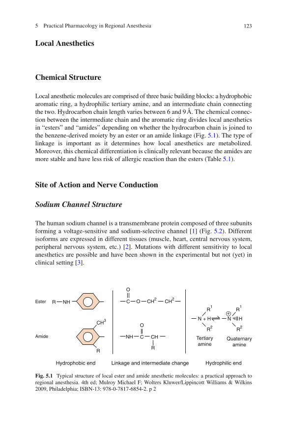
Local anesthetic molecules are comprised of three basic building blocks: a hydrophobic aromatic ring, a hydrophilic tertiary amine, and an intermediate chain connecting the two. Hydrocarbon chain length varies between 6 and 9 Å. The chemical connec-tion between the intermediate chain and the aromatic ring divides local anesthetics in “esters” and “amides” depending on whether the hydrocarbon chain is joined to the benzene-derived moiety by an ester or an amide linkage (Fig. 5.1 ). The type of linkage is important as it determines how local anesthetics are metabolized. Moreover, this chemical differentiation is clinically relevant because the amides are more stable and have less risk of allergic reaction than the esters (Table 5.1 ).
Site of Action and Nerve Conduction
Sodium Channel Structure
The human sodium channel is a transmembrane protein composed of three subunits forming a voltage-sensitive and sodium-selective channel [ 1 ] (Fig. 5.2 ). Different isoforms are expressed in different tissues (muscle, heart, central nervous system, peripheral nervous system, etc.) [ 2 ] . Mutations with different sensitivity to local anesthetics are possible and have been shown in the experimental but not (yet) in clinical setting [ 3 ] .
Ester
Amide
Hydrophobic end Hydrophilic endLinkage and intermediate change
R
R
NH
O
O
C CH Tertiaryamine
Quaternaryamine
R
O CH2
CH3
CH2
R2 R2
R1
R1
C
NH
N+
H+ N H
Fig. 5.1 Typical structure of local ester and amide anesthetic molecules: a practical approach to regional anesthesia. 4th ed; Mulroy Michael F; Wolters Kluwer/Lippincott Williams & Wilkins 2009, Philadelphia; ISBN-13: 978-0-7817-6854-2. p 2

124 J.A. Aguirre et al.
Tabl
e 5.
1 Ph
ysio
chem
ical
pro
pert
ies
of lo
cal a
nest
hetic
s: a
pra
ctic
al a
ppro
ach
to r
egio
nal a
nest
hesi
a, 4
th e
d. M
ulro
y M
icha
el F
; Wol
ters
Klu
wer
/Lip
pinc
ott
Will
iam
s &
Wilk
ins
2009
, Phi
lade
lphi
a; I
SBN
-13:
978-
0-78
17-6
854-
2. p
3
Rel
ativ
e in
vitr
o po
tenc
y
Dru
g (b
rand
nam
e)
Type
(ye
ar
intr
oduc
ed)
Che
mic
al s
truc
ture
R
at s
ciat
ic n
erve
p K
a Pa
rtiti
on c
oeffi
cie
nt a
Plas
ma
prot
ein
bind
ing
Coc
aine
E
ster
CH
2
CH2
CH2
CH
CH
NCH3
CHCOOCH3
CHOOC6H
5
–
8.6
– 92
Proc
aine
(N
ovoc
aine
) E
ster
(19
05)
H2N
COOCH2CH2N
C2H
5
C2H
5 1
8.9
1.7
5.8
Ben
zoca
ine
Est
er (
1900
) H2N
COOC2H
5 –
3.5
81
–
Tetr
acai
ne (
Pont
ocai
ne)
Est
er (
1930
)
CH3
H9C
4
N
N
COOCH2N
CH3
8 8.
5 22
1 75
.6
2-C
hlor
opro
cain
e (N
esac
aine
) E
ster
(19
52)
C2H
5
C2H
5
H2N
Cl COOCH2N
1
8.7
9.0
NA
Lid
ocai
ne (
Xyl
ocai
ne)
Am
ide
(194
4)
C2H
5
C2H
5
CH3
CH3NHCOCH2N
2
7.72
2.
4 64
.3
Mep
ivac
aine
(C
arbo
cain
e,
Polo
cain
e)
Am
ide
(195
7)
CH3
CH3
CH3NHCO
N
2
7.6
21
77.5

1255 Practical Pharmacology in Regional Anesthesia
Rel
ativ
e in
vitr
o po
tenc
y
Dru
g (b
rand
nam
e)
Type
(ye
ar
intr
oduc
ed)
Che
mic
al s
truc
ture
R
at s
ciat
ic n
erve
p K
a Pa
rtiti
on c
oeffi
cie
nt a
Plas
ma
prot
ein
bind
ing
Prilo
cain
e (C
itane
st)
Am
ide
(196
0)
CH3
CH3
NHCOCH
C3H
7NH
2
7.7
25
55
Rop
ivac
ine
(Nar
opin
) A
mid
e (1
995)
A
mid
e (1
995)
CH3
C3H
7
CH3
CO
NH
N
H
4 8.
1 11
5 95
Bup
ivac
aine
(M
arca
ine,
Am
ide
(196
3) S
enso
rcai
ne)
Lev
obup
ivac
aine
(C
hiro
cain
e)
Am
ide
(196
3)
CH3
CH3NHCO
C4H
9
N
8
8.1
346
95.6
Etid
ocai
ne (
Dur
nest
) A
mid
e (1
972)
CH3
CH3
C2H
5
NHCOCHN
C2H
5
C3H
7
8
7.74
80
0 94
a Ota
nol:
buff
er p
H 7
.4

126 J.A. Aguirre et al.
Conduction
With electrical excitation of the neuron, a depolarizing stimulus is conducted down an axon. A stimulus of signifi cant magnitude changes the negative resting potential from −70 mV toward −55 mV, the threshold required for complete depolarization: sodium channels in the cell membrane are activated and open permitting Na + ions to
a b
c d
Vol
tage
Time
Sodium channel
Na
KIntracellular
Extracellular
Potassium channel
+
–
Action potential:resting membranepotential
Vol
tage
Time
–
+
Action potential:depolarization
Vol
tage
Time
Intracellular
Extracellular+
–
Action potential:repolarization
Vol
tage
Time
+
ATP ADP + P
–
Action potential:Na+/K+ exchange
Fig. 5.2 Sodium and potassium channel function and ion movements during nerve depolarization: a practical approach to regional anesthesia. 4th ed; Mulroy Michael F; Wolters Kluwer/Lippincott Williams & Wilkins 2009, Philadelphia; ISBN-13: 978-0-7817-6854-2. p 6
1275 Practical Pharmacology in Regional Anesthesia
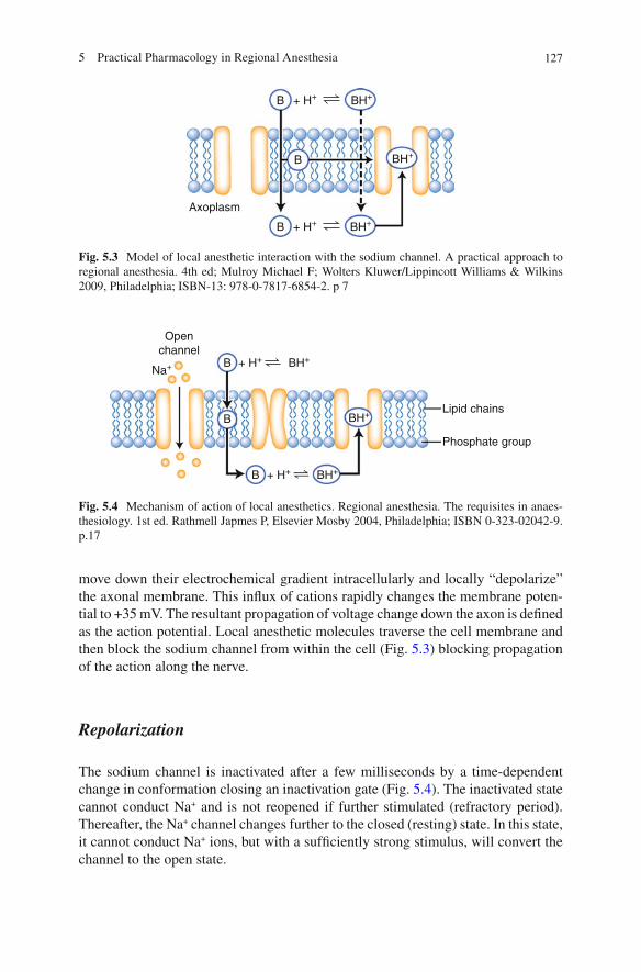
move down their electrochemical gradient intracellularly and locally “depolarize” the axonal membrane. This infl ux of cations rapidly changes the membrane poten-tial to +35 mV. The resultant propagation of voltage change down the axon is defi ned as the action potential. Local anesthetic molecules traverse the cell membrane and then block the sodium channel from within the cell (Fig. 5.3 ) blocking propagation of the action along the nerve.
Repolarization
The sodium channel is inactivated after a few milliseconds by a time-dependent change in conformation closing an inactivation gate (Fig. 5.4 ). The inactivated state cannot conduct Na + and is not reopened if further stimulated (refractory period). Thereafter, the Na + channel changes further to the closed (resting) state. In this state, it cannot conduct Na + ions, but with a suffi ciently strong stimulus, will convert the channel to the open state.
Axoplasm
B
B
+ H+ BH+
B + H+ BH+
BH+
Fig. 5.3 Model of local anesthetic interaction with the sodium channel. A practical approach to regional anesthesia. 4th ed; Mulroy Michael F; Wolters Kluwer/Lippincott Williams & Wilkins 2009, Philadelphia; ISBN-13: 978-0-7817-6854-2. p 7
Openchannel
B
Lipid chains
Phosphate group
+ H+ BH+
B + H+ Na+
BH+
B BH+
Fig. 5.4 Mechanism of action of local anesthetics. Regional anesthesia. The requisites in anaes-thesiology. 1st ed. Rathmell Japmes P, Elsevier Mosby 2004, Philadelphia; ISBN 0-323-02042-9. p.17

128 J.A. Aguirre et al.
Binding of Local Anesthetics
Local anesthetics do not bind to a classical “receptor”; it is more a “binding” site which is located within the sodium channel near its intracellular opening [ 3 ] . It is, on the one hand, a hydrophobic region to which the hydrophobic part of the local anesthetic molecule “binds,” on the other hand, a hydrophilic region with which the quaternary amine interacts. Any change in amino acid sequence can prevent local anesthetics from being effective.
Action potentials are blocked due to an inhibition of Na + movement through the Na + channel by a direct blocking or infl uencing the Na + channel conformation.
Pharmacodynamics and Physiochemical Properties of Local Anesthetics
Potency
The minimal local anesthetic concentration required to produce neural blockade is defi ned as potency. Lipophilicity correlates in in vitro settings well with local anes-thetic potency. In vivo, this correlation exists but is less stable.
Phasic Block
The faster a nerve is stimulated, the lower the concentration of local anesthetic is needed to produce a blockade (in vitro). This observation is called phasic block or rate-dependent block. Typically, phasic block occurs with more hydrophobic (potent) local anesthetics. They show a greater difference in their binding affi nity in dependence of the different channel states compared to the less potent local anes-thetics. There is no clear data about phasic block in the in vivo model, but phasic block seems to explain why hydrophobic local anesthetics are more cardiotoxic than hydrophilic local anesthetics.
Anesthetic Block in Dependency of Nerve/Axon Exposed
Axons are classifi ed with respect to their structure (myelinated, unmyelinated), diameter, conduction velocity, and function. The characteristics of local anesthetic blockade vary among different axon types, but the exact role of size, myelination, or function in axonal blockade is, to date, not entirely clear (Table 5.2 ).

1295 Practical Pharmacology in Regional Anesthesia
Tabl
e 5.
2 A
xon
clas
sifi c
atio
n a .
A p
ract
ical
app
roac
h to
reg
iona
l ane
sthe
sia.
4th
ed.
Mul
roy
Mic
hael
F; W
olte
rs K
luw
er/L
ippi
ncot
t Will
iam
s &
Wilk
ins
2009
, Ph
ilade
lphi
a; I
SBN
-13:
978-
0-78
17-6
854-
2. p
9
Fibe
r ty
pe
Size
( m m
) Fu
nctio
n L
ocal
ane
sthe
tic
sens
itivi
ty (
in v
itro)
Il
lust
ratio
ns
A
a
12–2
0 So
mat
ic m
otor
, pro
prio
cept
ion
++
b 5–
12
Touc
h, p
ress
ure
Mot
or to
mus
cle
spin
dles
+
+
g 3–
6 M
otor
to m
uscl
e sp
indl
es
++
+
D 2–
5 Pa
in, t
empe
ratu
re, t
ouch
+
++
B
<
3 A
uton
omic
(pr
egan
glio
nic)
+
+
C
0.3–
1.4
Pain
, refl
ex
resp
onse
s +
A
uton
omic
(po
stga
nglio
nic
a Hum
an a
xons
are
cla
ssifi
ed b
y si
ze, p
rese
nce
or a
bsen
ce o
f m
yelin
, and
fun
ctio
n, i
n vi
tro,
sm
all
unm
yelli
nate
d ax
ons
are
mos
t re
sist
ant
to l
ocal
ane
sthe
tic
bloc
kade
, whe
reas
larg
e m
yelin
ated
axo
ns a
re th
e m
ost s
ensi
tive.
In
vivo
, how
ever
, the
sen
sitiv
ity to
loca
l ane
sthe
tic b
lock
is d
iffe
rent
for
rea
sons
that
are
not
fu
lly u
nder
stoo
d (S
ee c
hapt
er c
linic
al p
harm
acol
ogy
of lo
cal a
nest
hetic
s). “
+“
indi
cate
s th
e re
lativ
e se
nsiti
vity
to lo
cal a
nest
hetic
blo
ck.
Mye
linat
edU
nmye
linat
ed
Nod
e of
Ran
vier
Nod
e of
Ran
vier
Axo
n
Loca
l ane
sthe
ticm
olec
ules
Sch
wan
n ce
llnu
cleu
s an
d cy
topl
asm

130 J.A. Aguirre et al.
Unmyelinated axons: the concentration of local anesthetic required to block • conduction of unmyelinated axons decreases with increasing length of nerve exposed to the local anesthetic. Myelinated axons: myelin consists of Schwann cell plasma membranes wrapped • around axons. There are gaps, called nodes of Ranvier, at fi xed intervals between the myelinated areas. Myelination results in much faster conduction velocities because the axonal membrane needs to be only depolarized at the node. This process is called saltatory conduction. Unmyelinated axons (C fi bers) are in vitro the most resistant to local anesthetic • blockade, followed by large (A a , A b fi bers) and small (B fi bers) myelinated axons [ 4 ] . Intermediate-size myelinated axons (A d , A g fi bers) are the easiest axons to block in vitro.
Local anesthetics can gain access to axonal membrane of myelinated axons only at the nodes of Ranvier. In vitro, the Na + channels in approximately three consecu-tive nodes (0.4–4 mm) need to be blocked for axonal conduction to fail.
Acid–Base and p K a
Local anesthetics (except benzocaine) are weak bases (p K a = 7.6–9.0) that are com-
mercially prepared as an acidic solution, typically at pH 4–5. The p K a defi nes the
pH, where half of the drug is ionized (positively charged form, conjugate acid) and half is nonionized (base). The ionized and nonionized forms have different, but important, clinical effects. The nonionized form penetrates the nerve membrane, while the ionized form binds to proteins on the intracellular side of the sodium channel (Fig. 5.5 ). The percentage of each form present in a solution or in the tissue depends on the pH of the solution or tissue and can be calculated from the Henderson-Hasselbalch equation:
-ap = pH log(base)/(acid).K
pH: pH in the solution/tissue; p K a : pH at which half the local anesthetic mole-
cules are in the base form and half in the acid form. The p K
a of each local anesthetic is unique and measures the tendency of the mol-
ecule to accept a proton in the base form or to donate a proton in the acid form. Most local anesthetics have a p K
a between 7.5 and 9.0.
Sodium bicarbonate can be added to local anesthetic solutions to raise the pH of the solution, thereby increasing the nonionized form. Other factors being similar, local anesthetics with more basic p K
a have a slower onset of blockade effect due to
the lesser amount of nonionized local anesthetic molecules at physiologic pH. This relative lack of the nonionized form impairs local anesthetic movement across the cell membrane and thus delays block onset (Fig. 5.5 ).

1315 Practical Pharmacology in Regional Anesthesia
Hydrophobicity
The charged form of all local anesthetics is more hydrophilic than the uncharged form. Hydrophobicity correlates with potency and, to a certain extent, to duration of action: the more hydrophobic the drug, the more potent it is. Hydrophobicity facili-tates penetration of the neuronal cell membrane, which accelerates local anesthetic binding to the intracellular portion of the sodium channel.
Adding local anesthetic to a recipient containing two immiscible liquids like an aqueous buffer and a hydrophobic lipid is needed to determine hydrophobicity. The resultant ratio of the concentrations is called the “distribution coeffi cient” (partition coeffi cient).
Protein Binding
One of the most important clinical characteristics of local anesthetics is its duration of action, which correlates with the degree of local anesthetic protein binding (typi-cally to albumin and a -1-acid-gylcoprotein). Binding to plasma protein varies between 5 and 95%. In general, more hydrophobic drugs have higher protein bind-ing. However, plasma protein binding do not correlate necessarily with tissue pro-tein binding.
Tissue
Cell membrane
pKa = 8.4
pH = 5.4
pH = 7.4
(0.1) (1) (0.1) (0.001)
Na+ channel
–N+
pKa = 9.4
H+ + RN RNH+
(1) (10)H+ + RN RNH+
(1) (1000)H+ + RN RNH+
RN + H+RNH+
(10) (0.1)RN + H+RNH+
(1000) (0.1)RNRNH+
Fig. 5.5 Effect of ionization on activity. Regional anesthesia. The requisites in anaesthesiology. 1st ed. Rathmell Japmes P, Elsevier Mosby 2004, Philadelphia; ISBN 0-323-02042-9. p.18

132 J.A. Aguirre et al.
Normally, short-acting local anesthetics have a fast onset of action, while long-duration local anesthetics have a slower onset of clinical effects. Serum protein binding also protects against drug toxicity because only the free (protein unbound) local anesthetic fraction can induce toxicity. However, once serum proteins are saturated, any additional administration or absorption of local anesthetics rapidly causes toxicity. Therefore, patients show a rapid progression from no signs of local anesthetic toxicity to manifestations of severe toxicity (CNS, cardiac) when highly protein-bound local anesthetics are used inadequately.
Binding to plasma proteins is mainly pH dependent: binding decreases during acidosis due to the decrease of available binding sites in an acidic environment.
Metabolism
Ester local anesthetics are primarily metabolized by ubiquitous plasma cholinest-erases (pseudocholinesterase). These enzymes are synthesized by the liver and are found throughout the vascular system and in the cerebrospinal fl uid (CSF). They are responsible for the metabolism of numerous drugs of relevance to the anesthesiolo-gist, including ester local anesthetics, succinylcholine, and mivacurium. Because of the widespread distribution of these enzymes, plasma degradation of ester local anesthetics is typically rapid. In contrast, amide local anesthetics undergo degrada-tions by hepatic enzymes and typically have a longer serum half-life.
Summary
The comprehension of the principles described in this chapter is essential to under-stand local anesthetic clinical pharmacology. However, one should keep in mind that the clinical setting is much more complicated as there are multiple infl uencing factors not present in vitro studies.
Clinical Pharmacology of Local Anesthetics
Factors Determining Block Quality
Block Onset
The proximity of the injected local anesthetic to the nerve is the most important fac-tor determining block onset; the nearer to the nerve, the shorter the time required to diffuse into the nerve (Fig. 5.6 ).

1335 Practical Pharmacology in Regional Anesthesia
The total local anesthetic dose and not the volume or concentration determines the onset time, the duration, and the intensity of the nerve block [ 5 ] .
The choice of the local anesthetic is a crucial issue since hydrophobic agents are more prone to bind to hydrophobic sites on connective tissue compared to hydro-philic drugs. This explains the slower onset of hydrophobic local anesthetics despite their greater potency.
Block Duration
The main factor infl uencing block duration is the clearance rate of the local anesthetics.
The choice of local anesthetic greatly infl uences block duration; hydrophobic local anesthetics have a slower clearance compared to hydrophilic local anesthetics. Moreover, hydrophobic compounds have a higher potency. These two factors are
Skin Local anesthetic
Axon
Blood stream
Systemic tissue
Liverhepatic metabolism
Excreted
Heart:cardiovascular toxicity
Nonspecific localtissue binding
Metabolism(plasma cholinesterase)
Brain:CNS toxicity
Fig. 5.6 Disposition of sites for local anesthetics following peripheral nerve blocks. A practical approach to regional anesthesia, 4th ed. Mulroy Michael F; Wolters Kluwer/Lippincott Williams & Wilkins 2009, Philadelphia; ISBN-13: 978-0-7817-6854-2. p 12

134 J.A. Aguirre et al.
responsible for a longer-lasting block. Furthermore, local anesthetics show variable vascular effects on local blood vessels. Vasoconstriction will reduce clearance, impairing its transport from the injection site. High concentrations of local anesthet-ics lead to a vasodilation increasing local blood fl ow and consequently their own clearance. But with decreasing concentration, vasoconstriction is present reducing clearance and increasing the duration of the block. Individual differences are listed below.
The dose infl uences duration: larger doses of local anesthetics produce a long-lasting block compared to lower doses. This is explained by the longer time required to clear the higher amount of drug.
Block Potency
Lipophilicity correlates with potency: the more lipid soluble the local anesthetic, the more potent it is. Lipophilicity facilitates penetration through the cell membrane accelerating thereby the binding of the local anesthetic to the intracellular binding site of the Na + channel. Lipophilicity is infl uenced by the lateral chains of the ben-zene ring.
Individual Local Anesthetics
Common local anesthetics used in clinical practice and their applications are shown in Table 5.3 .
Ester Local Anesthetics
Cocaine
Topical mucous membrane applications of cocaine (4% solution) result in very rapid anesthesia and vasoconstriction. At excessive doses, vasoconstrictive proper-ties lead to hypertension, coronary ischemia, and arrhythmias. Mixtures of lidocaine with phenylephrine or oxymetazoline are safer alternatives to cocaine for anesthe-tizing and vasoconstricting mucous membranes. Attention must be paid not to mix cocaine with other vasoconstrictors (phenylephrine) because of the increased risk of acute myocardial infarction [ 6 ] .
Cocaine is metabolized in the liver to active metabolites. The half-life is approxi-mately 45 min. If taken together with alcohol, the metabolic pathway is altered, and the highly toxic cocaethylene is produced.

1355 Practical Pharmacology in Regional Anesthesia
Tabl
e 5.
3 L
ocal
ane
sthe
tic d
rug
clin
ical
dos
es. A
pra
ctic
al a
ppro
ach
to re
gion
al a
nest
hesi
a. 4
th e
d. M
ulro
y M
icha
el F
; Wol
ters
Klu
wer
/Lip
pinc
ott W
illia
ms
&
Wilk
ins
2009
, Phi
lade
lphi
a; I
SBN
-13:
978-
0-78
17-6
854-
2. p
17
Max
imum
rec
omm
ende
d do
ses
Epi
dura
l f Pl
ain
With
epi
neph
rine
Dru
g {b
rand
nam
e}
Topi
cal f
(%)
Spin
al f
(%)
Surg
ical
f (%
) O
bste
tric
f (%
) Pe
riph
eral
ner
ve
bloc
k (%
) In
trav
enou
s re
gion
al (
%)
Tota
l m
g/kg
To
tal
mg/
kg
Coc
aine
4
NA
N
A
– N
A
NA
20
0 1.
5 –
– B
enzo
cain
e 5–
20
NA
N
A
– N
A
NA
—
—
—
—
Shor
t dur
atio
n Pr
ocai
ne (
Nov
ocai
ne)
NA
10
N
I N
I 1
NI
500
—
—
—
2–C
hlor
opro
cain
e (n
esac
aine
) N
I N
A
2–3
2–3
1–2
NI
800
11
1,00
0 14
Inte
rmed
iate
dur
atio
n L
idoc
aine
(X
yloc
aine
) 4
5 1.
5 1.
5 0.
5 0.
5 30
0 4.
5 50
0 7
2 2 a
1 M
epiv
acai
ne (
Car
boca
ine,
Pol
ocai
ne)
NA
N
A
1 N
I 1
NA
40
0 —
55
0 e
1.5
2 Pr
illoc
atin
e (C
itane
st)
NA
N
A
2–3
NI
1 0.
5 —
—
50
0 —
Long
dur
atio
n R
opiv
acai
ne (
Nar
opin
) N
A
0.5 b
0.75
, 1 c
0.2
0.5
NA
25
0 —
25
0 3
Bup
ivac
aine
(M
arca
ine,
Sen
sorc
aine
) N
A
0.5
0.5
0.12
5 e 0.
25
0.25
b 17
5 —
22
5 3
0.5
0.5
0.12
5 e 0.
25
0.5
0.5 a
(con
tinue
d)

136 J.A. Aguirre et al.
Max
imum
rec
omm
ende
d do
ses
Epi
dura
l f Pl
ain
With
epi
neph
rine
Dru
g {b
rand
nam
e}
Topi
cal f
(%)
Spin
al f
(%)
Surg
ical
f (%
) O
bste
tric
f (%
) Pe
riph
eral
ner
ve
bloc
k (%
) In
trav
enou
s re
gion
al (
%)
Tota
l m
g/kg
To
tal
mg/
kg
Lev
obup
lvac
aine
(C
hiro
cain
e)
Etid
ocai
ne (
Dur
anes
t)
NA
N
A
1 N
I 1
NI
300
4 40
0 6
1.5
Tetr
acai
ne (
Pont
ocai
ne)
1–2
1 N
A
NA
N
A
NA
Dru
gs a
re g
roup
ed in
gen
eral
dur
atio
n of
act
ion.
Con
cent
ratio
ns li
sted
are
thos
e re
com
men
ded
for
part
icul
ar a
pplic
atio
n N
A n
ot a
vaila
ble,
NI
not i
ndic
ated
, PD
R P
hysi
cian
s’ D
esk
Ref
eren
ce
a Pro
duce
s m
otor
blo
ckad
e su
itabl
e fo
r ce
sare
an d
eliv
ery
b Not
app
rove
d fo
r th
is u
se
c For
sin
gle
inje
ctio
n on
ly; l
ower
con
cent
ratio
ns s
houl
d be
use
d fo
r fo
llow
-up
inje
ctio
ns o
f ca
thet
ers
d Not
pre
pare
d co
mm
erci
ally
; mus
t be
dilu
ted
at ti
me
of u
se
e Spe
cifi c
dos
e fo
r ep
inep
hri
ne-c
onta
inin
g so
lutio
n no
t ide
ntifi
ed; t
his
is la
rges
t des
crib
ed d
ose
f Pre
serv
ativ
e fr
ee s
olut
ions
onl
y
Tabl
e 5.
3 (c
ontin
ued)

1375 Practical Pharmacology in Regional Anesthesia
The maximum recommended dose of cocaine is 200 mg. Attention must be paid to the use of cocaine for awake fi ber-optic nasal intubation: as local anesthetic toxic-ity is additive, the use of cocaine 4% and lidocaine 4–10% or benzocaine can lead to systemic toxic reaction.
Procaine
Procaine was the fi rst synthetic local anesthetic used clinically. Unfortunately, pro-caine combines a short duration and limited tissue penetration. Procaine is still occasionally used for skin infi ltration (0.25–1.0%) and short duration (30–45 min) spinal anesthesia (50–100 mg), although discharge readiness may be slightly longer than that seen with equipotent doses of spinal lidocaine. The block after spinal anes-thesia is shorter compared to the block induced by lidocaine but has a higher failure rate (inadequate sensory block). On the other hand, less transient neurologic symp-toms (TNS) have been reported [ 7 ] . Procaine is ineffective when used topically and is not reliable for epidural anesthesia. It is not recommended for peripheral block since it has a very slow onset time paired with a short-acting time. Procaine is metabolized in the plasma by the cholinesterase; its elimination half-life is approxi-mately 8 min.
The 10% solution should be diluted to 5% with dextrose or saline. Procaine is metabolized to para -aminobenzoic acid (PABA), which can be associated with allergic reactions.
2-Chloroprocaine
Compared to procaine, it has a more rapid onset and slightly longer duration of action. The principal uses of chloroprocaine are in obstetrics and ambulatory anes-thesia. It has rapid onset when used for epidural anesthesia and is therefore fre-quently chosen for urgent forceps or cesarean deliveries. In the 2–3% concentrations, it is also used for spinal anesthesia and peripheral blocks. Like other ester local anesthetics, chloroprocaine is rapidly metabolized by plasma cholinesterase, and with a duration of action between 30 and 60 min, it is a good drug for outpatient procedures. Since serum half-life is approximately 40 s, fetal accumulation and sys-temic toxicity, in general, are extremely unlikely.
The preservative-free solution should be used for central neuraxial blocks because of the concern regarding potential neurotoxicity.
Tetracaine
Tetracaine is the longest-acting ester local anesthetic. It is used in spinal and oph-thalmic anesthesia and is occasionally used for topical airway anesthesia. The latter application has declined with the recognition that tetracaine has a narrow margin between therapeutic and toxic doses that may lead to serious systemic toxicity after
138 J.A. Aguirre et al.
mucosal application. Metabolism is slower compared to procaine; therefore, the risk of systemic toxicity is greater.
Tetracaine is less chemically stable compared to lidocaine and bupivacaine. This instability may result in an occasional failed spinal anesthetic due to degradation of the local anesthetic during storage.
Benzocaine
Benzocaine was the fi rst developed but not the fi rst clinically used synthetic local anesthetic. Because of its low p K
a (3.5), it only exists in the uncharged form at
physiological pH, and it is hardly soluble in aqueous solutions. Therefore, it is exclusively used as a topical spray or troche for mucous mem-
branes or for topical application (cream and gel) for dermal hypesthesia. Methemoglobinemia seems to be observed more frequently when benzocaine is
used. This high risk and the diffi culty of proper dosage (cream and spray) increase benzocaine potential risk for toxicity.
Amide Local Anesthetics
Lidocaine
Lidocaine is the most widely used local anesthetic. It combines signifi cant potency, fast onset, intermediate duration, good tissue penetration, and minimal cardiac tox-icity. Lidocaine is widely used for infi ltration (1–2%), intravenous regional anesthe-sia (0.5%), peripheral nerve blocks (1 and 1.5%), topical airway (4%), spinal anesthesia (0.2–5%), and epidural anesthesia (2%). It produces moderate vasodila-tion. The allergic potency is very low.
Lidocaine 5% has been implicated in the occurrence of cauda equina syndrome with the use of small-diameter microcatheters for continuous spinal anesthesia. Spinal microcatheters have since then been withdrawn from the US market. Single-shot spinal anesthesia can be associated with TNS, the etiology of which is uncer-tain [ 8, 9 ] .
Mepivacaine
Mepivacaine has similar pharmacokinetic profi le to lidocaine, with slightly longer duration and better tissue penetration. Chemically, it is a cyclic tertiary amine like bupivacaine and ropivacaine. It is used primarily for intermediate-duration infi ltra-tion, peripheral, epidural, and spinal nerve blocks in Europe . It has a mild vasocon-stricting effect which may be responsible for its longer duration compared to

1395 Practical Pharmacology in Regional Anesthesia
lidocaine. Mepivacaine is not used anymore in obstetric epidural anesthesia since this drug is poorly metabolized in the fetus and neonate and may be responsible for lower neurobehavioral score in the fi rst days of life [ 10 ] .
Prilocaine
Prilocaine is similar to lidocaine in its clinical profi le and is widely used for intrave-nous regional anesthesia outside the USA. It is the most rapidly metabolized amide local anesthetic. Within the USA, prilocaine was withdrawn from use following several cases of methemoglobinemia. Prilocaine is metabolized to nitro- and ortho-toluidine, which can oxidize hemoglobin to methemoglobin. Prilocaine is mainly used commercially in topical eutectic mixture of local anesthetics (EMLA) cream, as well as in proprietary mixtures of local anesthetics specifi cally marketed for air-way anesthesia. Signifi cant methemoglobinemia has been reported in both of these applications.
Etidocaine
Etidocaine is a derivate of lidocaine. Different chemical changes in the structure make etidocaine very hydrophilic. It is available in the USA as 1, 1.5, or 2% solu-tions. Thus, it is rarely used in contemporary practice. Its onset is similar to lido-caine, but its high protein binding is similar to bupivacaine, as are its duration of action and cardiac toxicity profi le. Clinical potency is similar to that of mepivacaine with 2.5% solutions commonly used in the epidural space and 1% solutions for the performance of peripheral nerve blocks.
Articaine
A structural local anesthetic that has a fi ve-membered-thiophene ring instead of a benzene ring as its hydrophobic tail, articaine 4% is used only as dental local anes-thetic and is the second most used local anesthetic for dentistry in the USA since its introduction in 2000. It is popular due to its rapid onset and long duration with a low risk of allergy risk despite its ester side chain attached to the thiophene ring.
Bupivacaine
Bupivacaine was the fi rst long-acting amide local anesthetic. Chemical structure makes bupivacaine signifi cantly more hydrophobic than mepivacaine and lidocaine, slower in onset but of longer duration. Bupivacaine is highly protein bound, which is consistent with long duration and potential for cardiotoxicity. Indeed, the cardio-toxicity of bupivacaine prompted the development of ropivacaine and l -bupivacaine.

140 J.A. Aguirre et al.
Bupivacaine is popular for use in a wide array of applications, including infi ltration (0.25%), peripheral nerve blocks (0.375–0.5%), spinal (0.5 and 0.75%), and epidu-ral (0.5 and 0.75%) anesthesia. Because of systemic toxicity, it is not used for IV regional anesthesia.
Bupivacaine has a lower therapeutic index, concerning cardiovascular toxicity compared to lidocaine. Bupivacaine is more slowly absorbed into plasma than lido-caine and produces plasma peak concentrations that are approximately 40% lower.
Clinically used concentrations of bupivacaine vary from 0.05% (epidural con-tinuous infusions for labor analgesia and acute pain management) to 0.5% (spinal anesthesia and peripheral nerve blocks). Peripheral nerve blocks provide sensory block for 4–12 h, sometimes up to 24 h.
The 0.75% concentration is specifi cally contraindicated for obstetric epidural anesthesia due to concerns about cardiotoxicity. Contemporary epidural anesthesia incorporates use of multihole catheters, test dosing regimens, incremental dosing, and low concentrations of local anesthetic via continuous infusion.
Levobupivacaine
Levobupivacaine is the levorotatory enantiomer of bupivacaine. Commercial bupi-vacaine is a racemic mixture of both enantiomers (R and S). Levobupivacaine is approximately equivalent to its racemic mixture for its use in regional anesthesia. Cardiac toxicity and CNS studies in animals and healthy volunteers indicated that levobupivacaine is approximately 35% less cardiotoxic compared to racemic bupi-vacaine [ 11, 12 ] . Levobupivacaine is used in the same concentrations, doses, and applications as racemic bupivacaine.
Ropivacaine
Ropivacaine is derived from mepivacaine. Ropivacaine is a long-acting amide local anesthetic which is supplied commercially like levobupivacaine as a single enantiomer. It is available as 0.2, 0.5, 0.75, and 1% solution.
This drug was specifi cally designed and formulated to minimize cardiotoxicity [ 13, 14 ] . At higher concentration (anesthetic), its potency is equivalent to that of bupivacaine [ 15 ] . At lower concentration (analgesic), ropivacaine was shown to be 40% less potent than bupivacaine [ 16 ] . The clinical experience for peripheral blocks shows that at equivalent doses ropivacaine and bupivacaine produce similar onset and quality of block, but it can be stated that bupivacaine has a signifi cantly longer duration. Ropivacaine is primarily used in epidural anesthesia/analgesia and periph-eral nerve block applications. Ropivacaine appears to be approximately 40% less cardiotoxic as compared to racemic bupivacaine in animal models [ 13 ] . Ropivacaine produces vasoconstriction at clinically used concentrations for peripheral nerve blocks explaining the little advantage of adding epinephrine to additionally prolong peripheral nerve block or epidural analgesia [ 17 ] .

1415 Practical Pharmacology in Regional Anesthesia
Adjuvants
Sodium Bicarbonate
Theoretically, sodium bicarbonate could fasten the onset time. However, results were not convincing, and actually, the practice of mixing sodium bicarbonate with local anesthetics is rarely used.
Hyaluronidase
It is used as adjuvant to local anesthetics to breakdown connective tissue in the extracellular matrix and thereby increase drug dispersion through tissue. Except for peribulbar block (sub-Tenon’s block), it has been abandoned. Allergic reactions have also been described in this setting.
Vasoconstrictors
Adding epinephrine leads to vasoconstriction and thereby local blood fl ow and drug clearance are decreased. This prolongs block duration and decreases local anes-thetic plasma concentration following spinal, epidural, and peripheral nerve blocks [ 18 ] . Lower peak plasma concentration decreases the risk for toxicity. However , epinephrine does not provide protection if accidental intravascular local anesthetic injection occurs [ 19 ] .
Clonidine
Alpha-2-adrenergic agonists are analgesic drugs in their own right and have been shown to inhibit both C fi bers and A fi bers and to modestly inhibit local anesthetic clearance [ 20, 21 ] . When added to local anesthetics, clonidine prolongs sensory block during peripheral, central neuraxial, and intravenous regional anesthesia to a degree comparable to that produced by epinephrine. However, unlike epinephrine, clonidine does not prolong motor block when administered orally, as when added to the intrathecal local anesthetic [ 22 ] .
Opioids
When added to short-duration local anesthetics used for spinal anesthesia, short-acting opioids (fentanyl and sufentanil) prolong and intensify sensory block without prolonging motor block or time to void, which is particularly advantageous for

142 J.A. Aguirre et al.
ambulatory spinal anesthesia [ 23 ] . However, postanesthesia nausea and vomiting, itching can be a problem [ 24 ] . When added to local anesthetics or peripheral nerve block, fentanyl has also been shown to prolong sensory block, but at the expense for signifi cantly slowing onset in some studies [ 25 ] .
When added to intrathecal local anesthetics, the peak plasma concentrations for sufentanil occur between 20 and 30 min and are greater than what is necessary for postoperative analgesia [ 14 ] . This explains the many reports of “early” respiratory depression in mothers [ 15 ] and fetal heart rate abnormalities in infants when sufentanil is added to intrathecal local anesthetics for labor analgesia or cesarean section [ 26 ] .
Depot Local Anesthetic Preparations
Depot preparations of local anesthetics are interesting because they would allow using long-acting anesthetics without the need for catheters and pumps.
Gels, polymer microspheres, liposomes, and oil-water emulsions have been stud-ied in animal models to produce long-acting anesthetic blocks [ 27 ] . To date, clinical convincing results are still lacking.
Complications of Regional Anesthesia
Introduction
Overall incidence of neuropathy after peripheral nerve block varies from 0 to >5%. Studies which used closed claims databases ranked neuropathy at the second place, with 16% of all claims [ 28 ] . In a prospective French study, incidence of major neu-rologic adverse reactions was estimated at 3.5/10,000 [ 29 ] . Peripheral nerve dam-ages following either spinal anesthesia or peripheral nerve blockades represented >50% of severe adverse reactions in this investigation.
Permanent injuries after regional anesthesia are rare [ 30– 32 ] . Most surveys with large cohorts are retrospective [ 33, 34 ] or related to closed claims analysis [ 35, 36 ] . Few studies are prospective but focus on specifi c adverse reactions inducing limita-tion in their interpretation [ 29, 37, 38 ] .
The largest recent clinical study was a voluntary reporting model used in France [ 29 ] . Data of 158,083 different blocks from 487 anesthesiologists were collected and analyzed. The incidence of serious complications such as central or peripheral nerve injury, seizure, death, etc. was described as 3.5/10,000 blocks. The risk of deaths was shown to be 1/400,000 regional blocks. All but one occurred during spinal anesthesia.
It can be concluded that the incidence of severe complications of regional anes-thesia is similar to the one observed after general anesthesia.

1435 Practical Pharmacology in Regional Anesthesia
Systemic Toxicity
Systemic toxicity is a signifi cant and potentially dangerous problem [ 39 ] . Beside a local toxicity, an increase of the local anesthetic plasma concentration may lead to systemic toxicity, mainly neurologic and cardiovascular ones. Such an increase in local anesthetic plasmatic concentration may be related to inadvertent intravascular injection with a consecutive sudden plasmatic peak of concentration. The most fre-quent cause of systemic toxicity is related to a high and rapid resorption of local anesthetics through perinervous vessels. Toxicity occurs fi rst in the CNS and then in the cardiovascular system (Fig. 5.7 ).
CNS Toxicity
The incidence of seizures varies between 0.2 and 1/1,000 cases and according to the anesthetic regional procedure [ 40, 41 ] . The clinical manifestation largely depends on the velocity of plasma concentration increment: a slow increase shows clear and reproducible series of typical CNS signs and symptoms. A rapid increase leads to generalized seizures as fi rst clinical manifestation.
Sedatives and hypnotics such as propofol, benzodiazepines, and barbiturates raise seizure threshold and help protecting the CNS [ 42, 43 ] .
The therapeutic to CNS toxicity ratio is for all local anesthetics, the same indicating that none of them are more or less propense to cause seizures.
The prevention and the treatment of CNS toxicity should be done according to published recommendations [ 44, 45 ] .
Stage of CNS-DepressionComa, Apnea, Depression, Hyoptonsion
Convulsive StageGeneral, tonic-clonic Seizures
First StageNumbness, metalic flavour, dysgeusea
LA
-Co
nce
ntr
atio
n
Preconvulsive StageTremor, Tinnitus, Nystagmus, clouding
of consciousness
Indirect cardiac depressionhypertension, tachycardia, arrhythmia
Direct cardiac depressioncardiac arresthypotension
ischemiaAV-dissociation
arrhythmia, bradycardiaECG-widening
low output
Fig. 5.7 Signs and symptoms of local anesthetics toxicity

144 J.A. Aguirre et al.
Cardiac Toxicity
Estimated incidence of cardiac arrest related to local anesthetics varies between 1.8 and 3.1/10,000 cases [ 40, 46 ] .
High plasma concentration of local anesthetics is needed to cause signifi cant car-diovascular toxicity. This may occur, when the local anesthetic is injected intrave-nously, but a quick resuscitation is also possible. The therapeutic/cardiotoxic ratio is lower for hydrophobic local anesthetics (bupivacaine) compared to hydrophilic local anesthetics. Hydrophilic local anesthetics dissociate only after a greater amount of time from their binding sites; therefore, Na + channels are blocked when the next depolarization arrives. Cardiac toxicity can manifest as either malignant dysrhyth-mias (ventricular fi brillation), pulseless electrical activity, or asystolia [ 19, 42, 47 ] .
Cardiac toxicity should be prevented [ 45 ] , but in case of patients experiencing signs or symptoms of local anesthetic systemic toxicity (LAST), treatment should be done according to the ASRA guidelines 2010 [ 44, 48 ] .
Often, the doses of epinephrine in this setting are higher [ 19, 42, 47, 49 ] . Intralipid seems to be effective mainly in case of bupivacaine toxicity. A review about models and mechanisms of local anesthetic cardiac toxicity and a review of clinical presen-tations of local anesthetic systemic toxicity over the last 30 years have recently been published [ 50, 51 ] .
Prevention of Toxicity
Toxicity depends on total dose of local anesthetic injected, type of local anesthetic, speed and site of injection, combination with adjuncts, patient’s medical history, and concomitant use of other drugs leading to dangerous interactions, particularly with drugs presenting a hepatic metabolism action (hepatic blood fl ow modifi cation, cyto-chrome P450 action, etc.). Interactions have been described among local anesthetics
Table 5.4 Classifi cation of nerve injuries
Seddon
Neuropraxia ( Sunderland 1 ) Myelin damage, conduction block Axonotmesis ( Sunderland 2 ) Loss of axonal continuity, endoneurium intact, no conduction Neurotmesis ( Sunderland 3 ) Loss of axonal and endoneurial continuity, perineurium intact, no
conduction ( Sunderland 4 ) Loss of axonal, endoneurial and perineurial continuity;
epineurium intact; no conduction ( Sunderland 5 ) Entire nerve trunk separated; no conduction
Based on data from Seddon H, Three types of nerve injury. Brain 1943;66:236–88; Sunderland S: A classifi cation of peripheral nerve injuries producing loss of function. Brain 1951;74:491–516; and Lundborg G. Nerve injury and repair. Churchill Livingstone; 1988

1455 Practical Pharmacology in Regional Anesthesia
and b -blockers, amiodarone, cimetidine, and volatile agents [ 52– 56 ] . Calculation of the optimal dose taking into account patient’s age, pharmacokinetic and pharmaco-dynamic interactions with concomitant disease, and other drugs could be probably useful [ 57 ] . Development of nerve localization by ultrasonographic technique is thought to help reaching such objectives by limiting the volume of local anesthetic needed to block nerves [ 58 ] . However, clinical practice has shown that such a tech-nique cannot always prevent intravascular injection or quick reabsorption [ 59 ] .
Recently, a good summary about prevention of local anesthetic systemic toxicity (LAST) has been published in a series of articles dealing with LAST [ 45 ] .
Local Tissue Toxicity
Nerve Injury/Transient Neurologic Syndrome
Direct nerve injury from local anesthetic is receiving increased scrutiny, particularly with regard to spinal anesthesia [ 60, 61 ] . Toxicity can result from either local anes-thetics themselves or from additives, preservatives, antiseptics, or the pH of the formulations. The mechanism of local anesthetic-induced neurotoxicity is multifac-torial [ 60, 62 ] . Direct nerve injury is evident when isolated nerves are exposed to high concentration of local anesthetics, particularly lidocaine and tetracaine. Local anesthetics also change the biologic milieu surrounding neurons, including localized alteration of prostaglandin production, altering ionic permeability and changes in neural blood fl ow.
Compared with bupivacaine, lidocaine has a signifi cantly greater potential for direct neurotoxicity, particularly when isolated nerves are exposed to high concen-trations of lidocaine over long periods of time. Hyperbaric 5% lidocaine and tetra-caine have been associated with cauda equina syndrome after continuous spinal anesthesia. In these cases, spinal microcatheters were used to administer supernor-mal doses (up to 300 mg) of hyperbaric 5% lidocaine. Because spinal microcathe-ters (25–32 gauge) greatly limit the speed of drug administration, badly distributed local anesthetics presumably pooled near the catheter tip. As a result of the lordotic lumbar spine curvature, higher concentration of lidocaine remained in the lum-bosacral cistern [ 62, 63 ] .
Single-shot spinal anesthesia can cause transient pain (TNS), manifest as back and posterior leg discomfort with radicular symptoms lasting 1–3 days after spinal anesthesia. The etiology of TNS is unclear, but some have speculated that this syn-drome represents a form of neurotoxicity. Transient neurologic symptoms occur more frequently with lidocaine than bupivacaine, which may relate to lidocaines greater neurotoxicity in isolated nerve preparations [ 36, 64– 67 ] . Additionally, sev-eral risk factors (lidocaine, lithotomy position, out-patient status, arthroscopic knee surgery, and obesity) for developing TNS have been identifi ed [ 64, 65 ] .

146 J.A. Aguirre et al.
Needle Trauma
Recent ultrasonographic data have shown that injections between epineurium and perineurium did not produce signifi cant neural injury [ 68 ] . If injection pressure is low (less than 12 psi), intraneural injection does not necessarily result in permanent injury but can lead to severe injury if pressures are high [ 69 ] .
Studies over the last years have demonstrated that the correlation between nee-dle-nerve proximity and the current necessary to elicit a motor response is poor and not always reliable, despite the high success rate of neurostimulation and its low complication rate [ 70, 71 ] . Moreover, also eliciting paresthesia has surprisingly poor correlation with nerve proximity [ 72, 73 ] . Case reports of intraneural, intravasal, and other complications despite the use of ultrasound have shown that also this promising technique does not guarantee a complete visualization of the targeted nerve to avoid further complications [ 74 ] . The best way to avoid needle-induced nerve trauma is to avoid long bevel needle and perpendicular needle approaches to the nerve.
Clinical symptomatology of perimedullar complication following central ner-vous block is variable. Spinal cord injury can occur even while a patient did not complain of any paresthesia during puncture [ 75, 76 ] . Different risk factors have been identifi ed to explain the occurrence of this complication [ 60 ] . Epidural hema-toma can cause paraplegia following neuraxial anesthesia in patients concomitantly anticoagulated with low-molecular-weight heparin. Other causes of neural injury include positioning injuries, surgical trauma, and injuries related to the use of a limb tourniquet.
Guidelines on management of such complications following both central and peripheral nerve blocks have recently been published by the American Society of Regional Anesthesia [ 60 ] . Decision-making algorithms have been proposed to help the clinician in case of neuropathy occurrence [ 61, 77 ] (Fig. 5.8 ).
Myotoxicity
Skeletal muscle toxicity is a rare and uncommon side effect of local anesthetic drugs. Intramuscular injections of these agents regularly result in reversible myone-crosis [ 78 ] . The extent of muscle damage is dose dependent and worsens with serial or continuous administration. This problem is probably underestimated as incidence of symptomatic clinical forms is unknown. Experimental studies have concluded that all LA cause muscular damages with concentration use in daily practice. The extent of such damage depends on pharmacological properties of each local anes-thetic, dose injected, and site of injection [ 79 ] .
Animal studies in pigs showed lower mean damage score in muscles exposed to ropivacaine compared to exposure to bupivacaine [ 80, 81 ] . Stereospecifi city of the drug seems also to play an important role in Ca 2+ metabolism, which has been shown

1475 Practical Pharmacology in Regional Anesthesia
to be important in myotoxicity [ 82 ] . First reports of muscular dysfunction were related to retrobulbar injection of local anesthetics.
Bupivacaine seems to be the most toxic local anesthetic. Phenomena of apoptosis have been described only with bupivacaine but not with other LA [ 81, 83 ] . Interactions with the Ca 2+ metabolism seem to be a key pathway and explain most damage [ 82, 84 ] . Also, changes in the mitochondrial metabolism induced by local anesthetics have been reported [ 83, 85, 86 ] . These effects are less pronounced with ropivacaine, a less lipophilic local anesthetic, compared with bupivacaine on heart cell preparation [ 87 ] , but this was not shown in rat psoas muscle [ 88 ] . A recent study has concluded that mitochondrial bioenergetics alterations with bupivacaine were more severe in young rats compared to adults [ 89 ] .
Chondrotoxicity
Complications from the use of pain pumps in orthopedic surgery have recently received considerable interest. Human and animal studies have reported on the chondrotoxicity of intra-articular application of bupivacaine [ 90– 92 ] . Postarthro-scopic glenohumeral chondrolysis is a noninfectious entity associated with factors
Suspectedplexus lesion / nerve
Clinical examinationNormal
Normal
Stop
Stop Abnormal
Abnormal
Pathological
Abnormal
Repeat at3-4 weeks
Repeat at1 month
Repeat at1 month
Normal
Normal
SSEP*
MEP*
EMG*
NCS*
Observe Repeat at3 months
Ultrasonography : should beperformed immediately
Sudomotor test optional
Should be performed asearly as possible
* The choice of the examination will be done according to clinical condition and neuro physiologist ‘s recommendations
Fig. 5.8 Algorithm recommended to be performed in case of suspected plexus/nerve lesion

148 J.A. Aguirre et al.
including use of radiofrequency tumoral instruments and intra-articular pain pumps that administer bupivacaine [ 93 ] . Also, the viability of bovine articular chondro-cytes after exposure to corticosteroids alone or with lidocaine in a simulated infl am-matory environment was assessed. The results showed a dose-dependent and time-dependent decrease in chondrocyte viability after exposure to methylpredniso-lone. The combination with lidocaine was toxic, with virtually no cells surviving the treatment [ 94 ] . Continuous 0.5% bupivacaine exposure was shown to have a clear detrimental effect on chondrocytes in an in vitro model [ 95 ] . There is a growing amount of evidence that intra-articular administration of bupivacaine is chondro-toxic, especially at a higher concentration and with a prolonged exposure. More studies are needed to clarify this issue.
Allergy
Allergic reactions may occur from preservatives added to some local anesthetics (sulfi tes and methylparaben). Actual allergic reactions to local anesthetics are quite rare but are more common with ester local anesthetics compared to amides [ 96 ] . This is likely due to the breakdown products of ester local anesthetics, such as PABA. There are only a few convincing reports of allergic reactions to preservative-free amide local anesthetics.
If there is a history suggestive of true allergy, it may be worthwhile to perform allergy testing to preservative-free local anesthetics. Measurement of plasma esterase, which is increased in the event of “true” allergy, is useful. Skin testing is often per-formed to prospectively identify patients with local anesthetic allergy [ 97 ] .
Bleeding Complications
This issue deals mainly with neuraxial blocks. Epidural (1:150,000 cases) or intrath-ecal (1:200,000 cases) hematomas can cause devastating neurologic injury. The increased use of antithrombotic prophylaxis has increased this risk after epidural/spinal anesthesia to 1:1,000–1:10,000. The ASRA has recently reviewed the risks attendant to performance of regional blocks in the anticoagulated patient and refreshed its guidelines [ 98, 99 ] which are also to be found in their website ( www.asra.com ). Patients may develop sensory changes, progressive weakness and/or back pain. Confi rmatory diagnosis with neuraxial imaging (CT and MRI) must be obtained in conjunction with immediate neurosurgical consultation. If more than 8 h pass between symptom onset and decompression, the likelihood of a full or partial recovery decreases dramatically.

1495 Practical Pharmacology in Regional Anesthesia
Medicamentous Coagulopathy
In fully anticoagulated patients (heparin and coumadin), epidural and spinal anes-thesia should be avoided unless clear benefi t outweighs the added risks.
Recently, the ASRA published new guidelines for regional anesthesia in the patient receiving antithrombotic or thrombolytic therapy [ 98 ] .
Infection
Infection is a seldom complication in regional anesthesia. Risk factors are indwell-ing catheters left in place for more than 5 days, immunocompromised patients, catheters in trauma patients, and lack of perioperative antibiotics [ 30 ] .
Peripheral Nerve Blocks
Single-shot peripheral nerve blocks have a low risk of infection. The risk of colonization and infection increases when indwelling catheters are used. Despite the high colonization rate (70% primarily Staphylococcus epidermidis ), clinical evidence of infection is uncommon: less than 3%.
Central Neuraxial Blocks
Single-shot spinal and epidural anesthesia have a low risk of infection, but this risk seems to be higher than for peripheral nerve blocks. The incidence of meningitis after spinal anesthesia is estimated at less than 1:40,000; the risk of abscess after epidural anesthesia is less than 1:10,000 (Lit 2). Risk factors are the use of indwell-ing catheters and bacteremia [ 100 ] .
Clinical Pearls
Nerve-blocking potency of local anesthetics increases with increasing molecular • weight and lipid solubility [ 101 ] . The effectiveness of local anesthetics is infl uenced by the dose, site of adminis-• tration, additives, temperature, and pregnancy [ 101 ] . The plasma [ • 101 ] concentrations of local anesthetics is depending on the injec-tion technique, place of injection, and addition of adjuvants to local anesthetics.

150 J.A. Aguirre et al.
In laboratory experiments, most local anesthetics will only produce cardiovascular • toxicity after the blood concentration has exceeded three times that necessary to produce seizures [ 50 ] . True allergic reactions to preservative-free amide-type local anesthetics are • rare [ 96 ] . True anaphylaxis is more common with ester local anesthetics that are metabo-• lized directly to PABA than to amide local anesthetics [ 96 ] . Some patients may react to preservatives, such as methylparaben, used in local • anesthetics. In contrast to other shorter-acting amide local anesthetics, bupivacaine, levobupi-• vacaine, and ropivacaine have a motor-sparing effect; they produce less motor block for a comparable degree of sensory analgesia. It is well accepted that lipid solubility usually goes hand in hand with local • anesthetic potency. All things being equal, greater lipid solubility is related to increasing length of the aliphatic chain on the amino ring. Intraepidurally administered opioids reduce intraoperative requirements for • volatile anesthetics signifi cantly more compared to their intravenous administra-tion. This proves site-specifi c action in the epidural space. Exceeding a total dose of 0.25 mg of epinephrine may be associated with cardiac • arrhythmias. Adding epinephrine to spinal anesthetics will prolong motor blockade and delay • the return of bladder function, thus preventing patients from achieving discharge criteria. When clonidine is used in combination with opiates, the analgesic effects are • additive, but not synergistic. Thus, patients require a smaller total dose of narcot-ics and have a decreased incidence of oxygen desaturation with equivalent analgesia. Generally, the bigger the size of the nerve fi bers, the greater the amount of local • anesthetic solution required to block conduction. Thus, fi bers of small size are blocked sooner than those of larger diameter. The B fi bers of the autonomic system constitute an exception of this rule: even • though they are myelinated fi bers, a minimum concentration of local anesthetic solution is required to produce an effective blockade. This explains why the sympathetic blockade is observed before the onset of sen-• sory or motor blockade. The onset time of local anesthetic is infl uenced by the molecules p • K
a (the higher
the p K a , the slower the onset time of the nerve block in a physiologic environ-
ment) and diffusibility [ 101 ] . The ability to cross cell membrane depends on the molecular weight and the • liposolubility of the molecule. The nonionized form of the molecule is more lipid soluble than the ionized one; • therefore, it can cross more readily the cell membrane but diffuses less easily. The duration of the action of local anesthetic solutions depends on the protein • binding as well as the clearance from the injection site.

1515 Practical Pharmacology in Regional Anesthesia
The closer the p • K a of local anesthetic is to physiologic pH, the shorter the onset
time of the nerve block [ 101 ] . Increasing the lipophilicity of local anesthetic increases its potency and toxicity, • whereas protein binding is proportional to the duration of action of the local anesthetic. Sensory-motor differentiation is based on the different size and myelinization of • the nerve fi bers involved in pain conduction (A d and C) as compared to those involved in motor function (A a ). Postoperative maintenance is best performed with low concentration of a long-• acting agent, like 0.2% ropivacaine, 0.125–0.2% levobupivacaine. Local toxicity with neurotoxicity primarily occurs in cases of intraneural injec-• tion rather than normal applications of clinically relevant concentrations of local anesthetics [ 102 ] . To decrease the risk of nerve injury, utmost care should be taken during nerve • localization; excessively high concentrations of local anesthetic and high injec-tion pressures should be avoided [ 102 ] . The larger the fascicle, the greater is the risk of accidental intraneural injection • because large fascicles are easily speared by the needle. Injections into epineurium or perineural tissue do not result in signifi cant injection • resistance. When injection is diffi cult (injection pressures >20 psi), the injection should be • stopped because of the risk of intraneural needle position [ 102 ] . It is suggested that nerve stimulation with current intensity of 0.2–0.5 mA • (0.1 ms) indicates close needle-nerve placement [ 103 ] . Stimulation with current intensity of • £ 0.2 mA may be associated with intraneural needle placement. Motor response to nerve stimulation may be absent even when the needle is • inserted intraneurally [ 68 ] .
References
1. Nguyen HM, Goldin AL. Sodium channel carboxyl-terminal residue regulates fast inactiva-tion. J Biol Chem. 2010;285:9077–89.
2. Goldberg YP, MacFarlane J, MacDonald ML, Thompson J, Dube MP, Mattice M, et al. Loss-of-function mutations in the Nav1.7 gene underlie congenital indifference to pain in multiple human populations. Clin Genet. 2007;71:311–9.
3. Yarov-Yarovoy V, Brown J, Sharp EM, Clare JJ, Scheuer T, Catterall WA. Molecular deter-minants of voltage-dependent gating and binding of pore-blocking drugs in transmembrane segment IIIS6 of the Na(+) channel alpha subunit. J Biol Chem. 2001;276:20–7.
4. Huang JH, Thalhammer JG, Raymond SA, Strichartz GR. Susceptibility to lidocaine of impulses in different somatosensory afferent fi bers of rat sciatic nerve. J Pharmacol Exp Ther. 1997;282:802–11.
5. Ilfeld BM, Moeller LK, Mariano ER, Loland VJ, Stevens-Lapsley JE, Fleisher AS, et al. Continuous peripheral nerve blocks: is local anesthetic dose the only factor, or do concentra-tion and volume infl uence infusion effects as well? Anesthesiology. 2010;112:347–54.

152 J.A. Aguirre et al.
6. Ashchi M, Wiedemann HP, James KB. Cardiac complication from use of cocaine and phenylephrine in nasal septoplasty. Arch Otolaryngol Head Neck Surg. 1995;121:681–4.
7. Le Truong HH, Girard M, Drolet P, Grenier Y, Boucher C, Bergeron L. Spinal anesthesia: a comparison of procaine and lidocaine. Can J Anaesth. 2001;48:470–3.
8. Hodgson PS, Neal JM, Pollock JE, Liu SS. The neurotoxicity of drugs given intrathecally (spinal). Anesth Analg. 1999;88:797–809.
9. Duque S, Fernandez L. Delayed-type hypersensitivity to amide local anesthetics. Allergol Immunopathol (Madr). 2004;32:233–4.
10. Meffi n P, Long GJ, Thomas J. Clearance and metabolism of mepivacaine in the human neo-nate. Clin Pharmacol Ther. 1973;14:218–25.
11. Gristwood RW. Cardiac and CNS toxicity of levobupivacaine: strengths of evidence for advantage over bupivacaine. Drug Saf. 2002;25:153–63.
12. Mather LE, Chang DH. Cardiotoxicity with modern local anaesthetics: is there a safer choice? Drugs. 2001;61:333–42.
13. Casati A, Putzu M. Bupivacaine, levobupivacaine and ropivacaine: are they clinically differ-ent? Best Pract Res Clin Anaesthesiol. 2005;19:247–68.
14. Zink W, Graf BM. Benefi t-risk assessment of ropivacaine in the management of postoperative pain. Drug Saf. 2004;27:1093–114.
15. Fanelli G, Casati A, Beccaria P, Aldegheri G, Berti M, Tarantino F, et al. A double-blind comparison of ropivacaine, bupivacaine, and mepivacaine during sciatic and femoral nerve blockade. Anesth Analg. 1998;87:597–600.
16. Camorcia M, Capogna G, Columb MO. Minimum local analgesic doses of ropivacaine, levobupivacaine, and bupivacaine for intrathecal labor analgesia. Anesthesiology. 2005;102:646–50.
17. Weber A, Fournier R, Van Gessel E, Riand N, Gamulin Z. Epinephrine does not prolong the analgesia of 20 mL ropivacaine 0.5% or 0.2% in a femoral three-in-one block. Anesth Analg. 2001;93:1327–31.
18. Buckenmaier 3rd CC, Bleckner LL. Anaesthetic agents for advanced regional anaesthesia: a North American perspective. Drugs. 2005;65:745–59.
19. Bernards CM, Carpenter RL, Kenter ME, Brown DL, Rupp SM, Thompson GE. Effect of epinephrine on central nervous system and cardiovascular system toxicity of bupivacaine in pigs. Anesthesiology. 1989;71:711–7.
20. Butterworth JFT, Strichartz GR. The alpha 2-adrenergic agonists clonidine and guanfacine produce tonic and phasic block of conduction in rat sciatic nerve fi bers. Anesth Analg. 1993;76:295–301.
21. Kopacz DJ, Bernards CM. Effect of clonidine on lidocaine clearance in vivo: a microdialysis study in humans. Anesthesiology. 2001;95:1371–6.
22. Ota K, Namiki A, Iwasaki H, Takahashi I. Dose-related prolongation of tetracaine spinal anesthesia by oral clonidine in humans. Anesth Analg. 1994;79:1121–5.
23. Liu S, Chiu AA, Carpenter RL, Mulroy MF, Allen HW, Neal JM, et al. Fentanyl prolongs lidocaine spinal anesthesia without prolonging recovery. Anesth Analg. 1995;80:730–4.
24. Mulroy MF, Larkin KL, Siddiqui A. Intrathecal fentanyl-induced pruritus is more severe in combi-nation with procaine than with lidocaine or bupivacaine. Reg Anesth Pain Med. 2001;26:252–6.
25. Nishikawa K, Kanaya N, Nakayama M, Igarashi M, Tsunoda K, Namiki A. Fentanyl improves analgesia but prolongs the onset of axillary brachial plexus block by peripheral mechanism. Anesth Analg. 2000;91:384–7.
26. Van de Velde M, Teunkens A, Hanssens M, Vandermeersch E, Verhaeghe J. Intrathecal sufen-tanil and fetal heart rate abnormalities: a double-blind, double placebo-controlled trial com-paring two forms of combined spinal epidural analgesia with epidural analgesia in labor. Anesth Analg. 2004;98:1153–9 (table of contents).
27. Grant SA. The Holy Grail: long-acting local anaesthetics and liposomes. Best Pract Res Clin Anaesthesiol. 2002;16:345–52.
28. Cheney FW, Domino KB, Caplan RA, Posner KL. Nerve injury associated with anesthesia: a closed claims analysis. Anesthesiology. 1999;90:1062–9.
1535 Practical Pharmacology in Regional Anesthesia
29. Auroy Y, Benhamou D, Bargues L, Ecoffey C, Falissard B, Mercier FJ, et al. Major complica-tions of regional anesthesia in France: the SOS Regional Anesthesia Hotline Service. Anesthesiology. 2002;97:1274–80.
30. Capdevila X, Bringuier S, Borgeat A. Infectious risk of continuous peripheral nerve blocks. Anesthesiology. 2009;110:182–8.
31. Popping DM, Zahn PK, Van Aken HK, Dasch B, Boche R, Pogatzki-Zahn EM. Effectiveness and safety of postoperative pain management: a survey of 18 925 consecutive patients between 1998 and 2006 (2nd revision): a database analysis of prospectively raised data. Br J Anaesth. 2008;101:832–40.
32. Neuburger M, Buttner J, Blumenthal S, Breitbarth J, Borgeat A. Infl ammation and infection complications of 2285 perineural catheters: a prospective study. Acta Anaesthesiol Scand. 2007;51:108–14.
33. Horlocker TT. Complications of spinal and epidural anesthesia. Anesthesiol Clin N Am. 2000;18:461–85.
34. Horlocker TT, McGregor DG, Matsushige DK, Schroeder DR, Besse JA. A retrospective review of 4767 consecutive spinal anesthetics: central nervous system complications. Perioperative Outcomes Group. Anesth Analg. 1997;84:578–84.
35. Lee LA, Posner KL, Domino KB, Caplan RA, Cheney FW. Injuries associated with regional anesthesia in the 1980s and 1990s: a closed claims analysis. Anesthesiology. 2004;101:143–52.
36. Ben-David B. Complications of regional anesthesia: an overview. Anesthesiol Clin N Am. 2002;20:665–7 (ix).
37. Capdevila X, Pirat P, Bringuier S, Gaertner E, Singelyn F, Bernard N, et al. Continuous peripheral nerve blocks in hospital wards after orthopedic surgery: a multicenter prospective analysis of the quality of postoperative analgesia and complications in 1,416 patients. Anesthesiology. 2005;103:1035–45.
38. Phillips OC, Ebner H, Nelson AT, Black MH. Neurologic complications following spinal anes-thesia with lidocaine: a prospective review of 10,440 cases. Anesthesiology. 1969;30:284–9.
39. Drasner K. Local anesthetic systemic toxicity: a historical perspective. Reg Anesth Pain Med. 2010;35:162–6.
40. Auroy Y, Narchi P, Messiah A, Litt L, Rouvier B, Samii K. Serious complications related to regional anesthesia: results of a prospective survey in France. Anesthesiology. 1997;87:479–86.
41. Brown DL, Ransom DM, Hall JA, Leicht CH, Schroeder DR, Offord KP. Regional anesthesia and local anesthetic-induced systemic toxicity: seizure frequency and accompanying cardio-vascular changes. Anesth Analg. 1995;81:321–8.
42. Bernards CM, Carpenter RL, Rupp SM, Brown DL, Morse BV, Morell RC, et al. Effect of midazolam and diazepam premedication on central nervous system and cardiovascular toxic-ity of bupivacaine in pigs. Anesthesiology. 1989;70:318–23.
43. Mather LE, Copeland SE, Ladd LA. Acute toxicity of local anesthetics: underlying pharma-cokinetic and pharmacodynamic concepts. Reg Anesth Pain Med. 2005;30:553–66.
44. Weinberg GL. Treatment of local anesthetic systemic toxicity (LAST). Reg Anesth Pain Med. 2010;35:188–93.
45. Mulroy MF, Hejtmanek MR. Prevention of local anesthetic systemic toxicity. Reg Anesth Pain Med. 2010;35:177–80.
46. Kopp SL, Horlocker TT, Warner ME, Hebl JR, Vachon CA, Schroeder DR, et al. Cardiac arrest during neuraxial anesthesia: frequency and predisposing factors associated with sur-vival. Anesth Analg. 2005;100:855–65 (table of contents).
47. Kasten GW, Martin ST. Comparison of resuscitation of sheep and dogs after bupivacaine-induced cardiovascular collapse. Anesth Analg. 1986;65:1029–32.
48. Neal JM, Bernards CM, Butterworth JFt, Di Gregorio G, Drasner K, Hejtmanek MR, et al. ASRA practice advisory on local anesthetic systemic toxicity. Reg Anesth Pain Med. 2010;35:152–61.
49. Chadwick HS. Toxicity and resuscitation in lidocaine- or bupivacaine-infused cats. Anesthesiology. 1985;63:385–90.

154 J.A. Aguirre et al.
50. Butterworth JFt. Models and mechanisms of local anesthetic cardiac toxicity: a review. Reg Anesth Pain Med. 2010;35:167–76.
51. Di Gregorio G, Neal JM, Rosenquist RW, Weinberg GL. Clinical presentation of local anes-thetic systemic toxicity: a review of published cases, 1979 to 2009. Reg Anesth Pain Med. 2010;35:181–7.
52. Freysz M, Beal JL, Timour Q, Bertrix L, Faucon G. Systemic toxicity of local anesthetics. Pharmacokinetic and pharmacodynamic factors. Ann Fr Anesth Reanim. 1988;7:181–8.
53. Siegmund JB, Wilson JH, Imhoff TE. Amiodarone interaction with lidocaine. J Cardiovasc Pharmacol. 1993;21:513–5.
54. Kuhnert BR, Zuspan KJ, Kuhnert PM, Syracuse CD, Brashear WT, Brown DE. Lack of infl u-ence of cimetidine on bupivacaine levels during parturition. Anesth Analg. 1987;66:986–90.
55. Mather LE, Runciman WB, Carapetis RJ, Ilsley AH, Upton RN. Hepatic and renal clearances of lidocaine in conscious and anesthetized sheep. Anesth Analg. 1986;65:943–9.
56. Bax ND, Tucker GT, Lennard MS, Woods HF. The impairment of lignocaine clearance by propranolol – major contribution from enzyme inhibition. Br J Clin Pharmacol. 1985;19:597–603.
57. Rosenberg PH, Veering BT, Urmey WF. Maximum recommended doses of local anesthetics: a multifactorial concept. Reg Anesth Pain Med. 2004;29:564–75 (discussion 24).
58. O’Donnell BD, Iohom G. An estimation of the minimum effective anesthetic volume of 2% lidocaine in ultrasound-guided axillary brachial plexus block. Anesthesiology. 2009;111:25–9.
59. Baciarello M, Danelli G, Fanelli G. Real-time ultrasound visualization of intravascular injection of local anesthetic during a peripheral nerve block. Reg Anesth Pain Med. 2009;34:278–9.
60. Neal JM, Bernards CM, Hadzic A, Hebl JR, Hogan QH, Horlocker TT, et al. ASRA practice advisory on neurologic complications in Regional Anesthesia and Pain Medicine. Reg Anesth Pain Med. 2008;33:404–15.
61. Sorenson EJ. Neurological injuries associated with regional anesthesia. Reg Anesth Pain Med. 2008;33:442–8.
62. Welch MB, Brummett CM, Welch TD, Tremper KK, Shanks AM, Guglani P, et al. Perioperative peripheral nerve injuries: a retrospective study of 380,680 cases during a 10-year period at a single institution. Anesthesiology. 2009;111:490–7.
63. Faccenda KA, Finucane BT. Complications of regional anaesthesia incidence and prevention. Drug Saf. 2001;24:413–42.
64. Freedman JM, Li DK, Drasner K, Jaskela MC, Larsen B, Wi S. Transient neurologic symptoms after spinal anesthesia: an epidemiologic study of 1,863 patients. Anesthesiology. 1998;89:633–41.
65. Pollock JE. Transient neurologic symptoms: etiology, risk factors, and management. Reg Anesth Pain Med. 2002;27:581–6.
66. Liu SS, McDonald SB. Current issues in spinal anesthesia. Anesthesiology. 2001;94:888–906. 67. Zaric D, Pace NL. Transient neurologic symptoms (TNS) following spinal anaesthesia with
lidocaine versus other local anaesthetics. Cochrane Database Syst Rev. 2009;(2):CD003006 68. Bigeleisen PE. Nerve puncture and apparent intraneural injection during ultrasound-guided axil-
lary block does not invariably result in neurologic injury. Anesthesiology. 2006;105:779–83. 69. Kapur E, Vuckovic I, Dilberovic F, Zaciragic A, Cosovic E, Divanovic KA, et al. Neurologic
and histologic outcome after intraneural injections of lidocaine in canine sciatic nerves. Acta Anaesthesiol Scand. 2007;51:101–7.
70. Choyce A, Chan VW, Middleton WJ, Knight PR, Peng P, McCartney CJ. What is the relation-ship between paresthesia and nerve stimulation for axillary brachial plexus block? Reg Anesth Pain Med. 2001;26:100–4.
71. Perlas A, Niazi A, McCartney C, Chan V, Xu D, Abbas S. The sensitivity of motor response to nerve stimulation and paresthesia for nerve localization as evaluated by ultrasound. Reg Anesth Pain Med. 2006;31:445–50.
72. Urmey WF, Stanton J. Inability to consistently elicit a motor response following sensory paresthesia during interscalene block administration. Anesthesiology. 2002;96:552–4.

1555 Practical Pharmacology in Regional Anesthesia
73. Bollini CA, Urmey WF, Vascello L, Cacheiro F. Relationship between evoked motor response and sensory paresthesia in interscalene brachial plexus block. Reg Anesth Pain Med. 2003;28:384–8.
74. Liu SS, Ngeow JE, Yadeau JT. Ultrasound-guided regional anesthesia and analgesia: a quali-tative systematic review. Reg Anesth Pain Med. 2009;34:47–59.
75. Tripathi M, Nath SS, Gupta RK. Paraplegia after intracord injection during attempted epidural steroid injection in an awake-patient. Anesth Analg. 2005;101:1209–11 (table of contents).
76. Tsui BC, Armstrong K. Can direct spinal cord injury occur without paresthesia? A report of delayed spinal cord injury after epidural placement in an awake patient. Anesth Analg. 2005;101:1212–4 (table of contents).
77. Borgeat A, Aguirre J, Curt A. Case scenario: neurologic complication after continuous inter-scalene block. Anesthesiology. 2010;112:742–5.
78. Hogan Q, Dotson R, Erickson S, Kettler R, Hogan K. Local anesthetic myotoxicity: a case and review. Anesthesiology. 1994;80:942–7.
79. Zink W, Graf BM. Local anesthetic myotoxicity. Reg Anesth Pain Med. 2004;29:333–40. 80. Zink W, Bohl JR, Hacke N, Sinner B, Martin E, Graf BM. The long term myotoxic effects of
bupivacaine and ropivacaine after continuous peripheral nerve blocks. Anesth Analg. 2005;101:548–54 (table of contents).
81. Zink W, Seif C, Bohl JR, Hacke N, Braun PM, Sinner B, et al. The acute myotoxic effects of bupivacaine and ropivacaine after continuous peripheral nerve blockades. Anesth Analg. 2003;97:1173–9 (table of contents).
82. Zink W, Missler G, Sinner B, Martin E, Fink RH, Graf BM. Differential effects of bupiva-caine and ropivacaine enantiomers on intracellular Ca2+ regulation in murine skeletal muscle fi bers. Anesthesiology. 2005;102:793–8.
83. Irwin W, Fontaine E, Agnolucci L, Penzo D, Betto R, Bortolotto S, et al. Bupivacaine myo-toxicity is mediated by mitochondria. J Biol Chem. 2002;277:12221–7.
84. Zink W, Graf BM, Sinner B, Martin E, Fink RH, Kunst G. Differential effects of bupivacaine on intracellular Ca2+ regulation: potential mechanisms of its myotoxicity. Anesthesiology. 2002;97:710–6.
85. Wakata N, Sugimoto H, Iguchi H, Nomoto N, Kinoshita M. Bupivacaine hydrochloride induces muscle fi ber necrosis and hydroxyl radical formation-dimethyl sulphoxide reduces hydroxyl radical formation. Neurochem Res. 2001;26:841–4.
86. Nouette-Gaulain K, Bellance N, Prevost B, Passerieux E, Pertuiset C, Galbes O, et al. Erythropoietin protects against local anesthetic myotoxicity during continuous regional anal-gesia. Anesthesiology. 2009;110:648–59.
87. Sztark F, Malgat M, Dabadie P, Mazat JP. Comparison of the effects of bupivacaine and ropi-vacaine on heart cell mitochondrial bioenergetics. Anesthesiology. 1998;88:1340–9.
88. Nouette-Gaulain K, Sirvent P, Canal-Raffi n M, Morau D, Malgat M, Molimard M, et al. Effects of intermittent femoral nerve injections of bupivacaine, levobupivacaine, and ropiva-caine on mitochondrial energy metabolism and intracellular calcium homeostasis in rat psoas muscle. Anesthesiology. 2007;106:1026–34.
89. Nouette-Gaulain K, Dadure C, Morau D, Pertuiset C, Galbes O, Hayot M, et al. Age-dependent bupivacaine-induced muscle toxicity during continuous peripheral nerve block in rats. Anesthesiology. 2009;111:1120–7.
90. Bailie DS, Ellenbecker TS. Severe chondrolysis after shoulder arthroscopy: a case series. J Shoulder Elbow Surg. 2009;18:742–7.
91. Chu CR, Izzo NJ, Papas NE, Fu FH. In vitro exposure to 0.5% bupivacaine is cytotoxic to bovine articular chondrocytes. Arthroscopy. 2006;22:693–9.
92. Rapley JH, Beavis RC, Barber FA. Glenohumeral chondrolysis after shoulder arthroscopy associated with continuous bupivacaine infusion. Arthroscopy. 2009;25:1367–73.
93. Busfi eld BT, Romero DM. Pain pump use after shoulder arthroscopy as a cause of gle-nohumeral chondrolysis. Arthroscopy. 2009;25:647–52.
94. Seshadri V, Coyle CH, Chu CR. Lidocaine potentiates the chondrotoxicity of methylpredni-solone. Arthroscopy. 2009;25:337–47.

156 J.A. Aguirre et al.
95. Anz A, Smith MJ, Stoker A, Linville C, Markway H, Branson K, et al. The effect of bupivacaine and morphine in a coculture model of diarthrodial joints. Arthroscopy. 2009;25:225–31.
96. Amsler E, Flahault A, Mathelier-Fusade P, Aractingi S. Evaluation of re-challenge in patients with suspected lidocaine allergy. Dermatology. 2004;208:109–11.
97. Hein UR, Chantraine-Hess S, Worm M, Zuberbier T, Henz BM. Evaluation of systemic prov-ocation tests in patients with suspected allergic and pseudoallergic drug reactions. Acta Derm Venereol. 1999;79:139–42.
98. Horlocker TT, Wedel DJ, Rowlingson JC, Enneking FK, Kopp SL, Benzon HT, et al. Regional anesthesia in the patient receiving antithrombotic or thrombolytic therapy: American Society of Regional Anesthesia and Pain Medicine Evidence-Based Guidelines (Third Edition). Reg Anesth Pain Med. 2010;35:64–101.
99. Horlocker TT, Wedel DJ, Rowlingson JC, Enneking FK. Executive summary: regional anes-thesia in the patient receiving antithrombotic or thrombolytic therapy: American Society of Regional Anesthesia and Pain Medicine Evidence-Based Guidelines (Third Edition). Reg Anesth Pain Med. 2010;35:102–5.
100. Carp H, Bailey S. The association between meningitis and dural puncture in bacteremic rats. Anesthesiology. 1992;76:739–42.
101. Heavner JE. Local anesthetics. Curr Opin Anaesthesiol. 2007;20:336–42. 102. Hadzic A, Dilberovic F, Shah S, Kulenovic A, Kapur E, Zaciragic A, et al. Combination of
intraneural injection and high injection pressure leads to fascicular injury and neurologic defi -cits in dogs. Reg Anesth Pain Med. 2004;29:417–23.
103. Jankovic D, Wells C. Brachial plexus. In: Jankovic D, Wells C, editors. Regional Nerve Blocks. Berlin: Blackwell Science; 2001. p. 58–86.