Practical Management of Asymptomatic PVC · (PVC) • The first recorded description of...
26
Practical Management of Asymptomatic PVC Maria J Salvador, Barcelona Les 9èmes Journées Tuniso‐Européennes de Cardiologie Pratique JTECP Sousse 2012 Tunis_2012-CCP lecture
Transcript of Practical Management of Asymptomatic PVC · (PVC) • The first recorded description of...

Practical Management of Asymptomatic PVC
Maria J Salvador, BarcelonaLes 9èmes Journées Tuniso‐Européennes de
Cardiologie PratiqueJTECP
Sousse 2012
Tunis_2012-CCP lecture
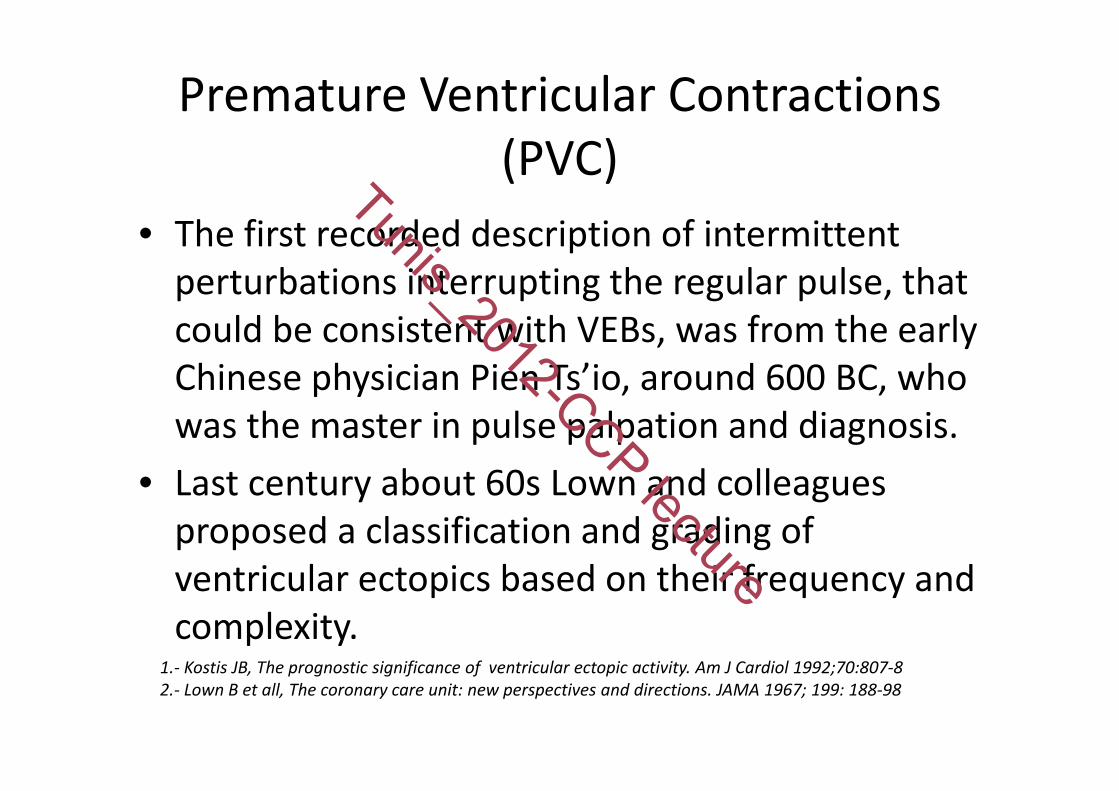
Premature Ventricular Contractions (PVC)
• The first recorded description of intermittent perturbations interrupting the regular pulse, that could be consistent with VEBs, was from the early Chinese physician Pien Ts’io, around 600 BC, who was the master in pulse palpation and diagnosis.
• Last century about 60s Lown and colleagues proposed a classification and grading of ventricular ectopics based on their frequency and complexity.
1.‐ Kostis JB, The prognostic significance of ventricular ectopic activity. Am J Cardiol 1992;70:807‐82.‐ Lown B et all, The coronary care unit: new perspectives and directions. JAMA 1967; 199: 188‐98
Tunis_2012-CCP lecture

VEBs Incidence
• Clinical normal or apparently healthy people– 1% by standard EKG– 40‐75% by 24‐48 hours ambulatory EKG (Holter)
• Increase with age– more prevalence... but more heart diseases
Kennedy HL et al., N Engl J Med 1985; Abdalla.IS et al., Am J Cardiol 1987; CAST investigators, N Engl J Med 1989
Tunis_2012-CCP lecture

VA Classification by Clinical PresentationACC/AHA/ESC 2006 Guidelines for Management of Patients With Ventricular
Arrhythmias and the Prevention of Sudden Cardiac Death
• Hemodynamically stable– Asymptomatic– Minimal symptoms, e.g., palpitations
• Hemodynamically unstable– Presyncope– Syncope– Sudden cardiac death – Sudden cardiac arrest
ACC/AHA/ESC Practice Guidelines Zipes et al. JACC Vol. 48, No. 5, 2006
Tunis_2012-CCP lecture
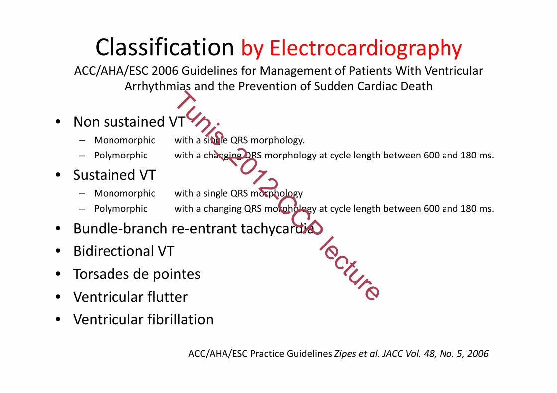
Classification by ElectrocardiographyACC/AHA/ESC 2006 Guidelines for Management of Patients With Ventricular
Arrhythmias and the Prevention of Sudden Cardiac Death
• Non sustained VT – Monomorphic with a single QRS morphology.– Polymorphic with a changing QRS morphology at cycle length between 600 and 180 ms.
• Sustained VT – Monomorphic with a single QRS morphology– Polymorphic with a changing QRS morphology at cycle length between 600 and 180 ms.
• Bundle‐branch re‐entrant tachycardia• Bidirectional VT • Torsades de pointes • Ventricular flutter • Ventricular fibrillation
ACC/AHA/ESC Practice Guidelines Zipes et al. JACC Vol. 48, No. 5, 2006
Tunis_2012-CCP lecture

VA Classification in Absence of Structural Heart Disease (1)
• Non life threatening (typically monomorphic)– Outflow tract:
• Right and Left ventricle outflow• Aortic sinus of Valsalva• Peri‐His bundle
– Idiopathic left ventricular tachycardia• Left posterior and left anterior fascicle• High septal fascicle
– Other• Mitral and Tricuspid annulus• Papillary muscle• Perivascular epicardial
PRYSTOWSKY,EN et al, J Am Coll Cardiol 2012;59:1733–44
Tunis_2012-CCP lecture

VA Classification in Absence of Structural Heart Disease (and 2)
• Life threatening (typically polymorphic)– Genetic Syndromes
• Long QT• Brugada• Catecholaminergic polymorphic ventricular tachycardia• Short QT
– Idiopathic ventricular fibrillation
PRYSTOWSKY,EN et al, J Am Coll Cardiol 2012;59:1733–44
Tunis_2012-CCP lecture
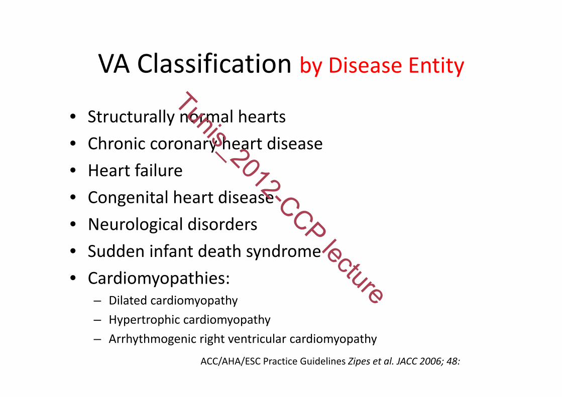
VA Classification by Disease Entity
• Structurally normal hearts • Chronic coronary heart disease • Heart failure • Congenital heart disease • Neurological disorders • Sudden infant death syndrome • Cardiomyopathies:
– Dilated cardiomyopathy – Hypertrophic cardiomyopathy – Arrhythmogenic right ventricular cardiomyopathy
ACC/AHA/ESC Practice Guidelines Zipes et al. JACC 2006; 48:
Tunis_2012-CCP lecture

• Hypertension• Caffeine intake• Exercise• Right Ventricular Outflow Tract• Sleep apnea
Trigger Evaluation of VEBs/VT
Tunis_2012-CCP lecture

VEBs and Hypertension
• MRFIT number of VEBs was related with systolic blood pressure (1)
• ARIC VEBs were related to hypertension/left ventricular mass (2)
• …but the debate about the significance of VEBs in hypertension persists
1‐ Abdalla.IS et al., Am J Cardiol 1987; 2‐ Simpson RJ Jr et al, Am Heart J 2002;
Tunis_2012-CCP lecture

VEBs and Caffeine Intake
• Caffeine is an alkaloid of xantines group, is central stimulant which can increase sympathetic activity (1)
• Live average : 4 hours (2 to 10 h)• Moderate caffeine consumption (200‐300mg/day) does not increase arrhythmias, blood pressure, LDL colesterol or incidence of coronary heart diseases (2,3)
(1) G André NG. (2006) Heart 92:1707‐12. (2) Myers M.G. (2004) Hypertension 43:724‐5. (3) López‐García L. et al (2006) Circulation 113:2045‐53.
Tunis_2012-CCP lecture

VEBs and Exercise
• Exercise is used to evaluate VEBs from 1927• VEBs are “benign” if disappear during exercise but…
– Jouven et al 2000: Frequent VEBs during exercise are related to higher incidence of cardiovascular death (not only for coronary heart diseases) in a 23 y follow up
– Frolkis et al 2003: VEBs during recovery time better predictor of increase risk of cardiovascular death than VEBs occurred during exercise
• VEBs associated to exercise could be a new prognostic criterion in addition to ischemia
Jouven X et al. New Engl J Med 2000;93:826‐33Frolkis JP et al New Engl J Med 2003;348:781‐90
Tunis_2012-CCP lecture

RVT outflow tract• Good prognosis:
– Apparently normal heart, without or mild symptoms, non ischemic exercise induced/monomorphic VT
• “Malignant” ventricular arrhythmias: – Ventricular fibrillation mapped from RVOT or distal Purkinje system from both ventricles
• Test to diagnose:– EKG to evaluate sinus rhythm– MRI to exclude ARVC
Takemoto M et al.J Am Coll Cardiol 2005;45:1259.65/Zipes et al. JACC 2006; 48:e247‐e346
Tunis_2012-CCP lecture

VEBs and Obstructive Sleep Apnea
Nocturnal arrhythmias have been shown to occur in up to 50% of OSA patients. The most common arrhythmias during sleep include:
nonsustained ventricular tachycardia sinus arrest second‐degree atrioventricular conduction blockfrequent (>2 bpm) premature ventricular contractions
AHA/ACC Sleep Apnea and Cardiovascular Disease, Somers et al.2008; 8:686–717
Tunis_2012-CCP lecture

How to evaluate PVCs
• Complete clinical history– Palpitations, presyncope, syncope– Family history of SCD– Drugs and dosage
• Physical examination• 12 leads EKG • Exercise Test• Ambulatory EKG recording (Holter)• Echocardiography
Tunis_2012-CCP lecture

• Identification of – Congenital abnormalities– Electrolyte disturbances– Underlying structural diseases– QRS duration (>120‐130ms)– Repolarization abnormalities– QTc interval
Resting 12 lead EKG (1)(Class I Level of Evidence A)
ACC/AHA/ESC Practice Guidelines Zipes et al. JACC 2006; 48:e247‐e346
Tunis_2012-CCP lecture
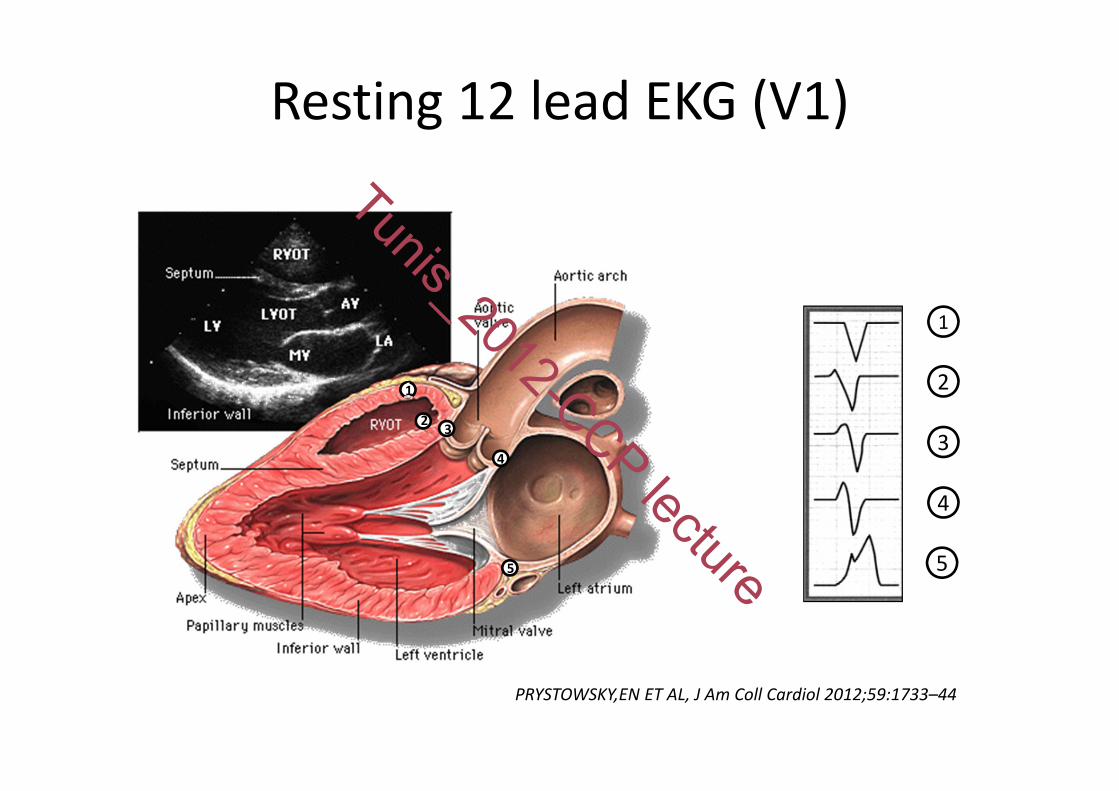
Resting 12 lead EKG (V1)
112 3
4
5
1
2
3
4
5
PRYSTOWSKY,EN ET AL, J Am Coll Cardiol 2012;59:1733–44
Tunis_2012-CCP lecture

Exercise TestClass I Level of Evidence B– Adult with VA and probability intermediate or greater of having CHD
– With known or suspected exercise induced VA
Class IIa Level of Evidence B– To evaluate medical treatment or ablation
Class IIb Level of Evidence C– VA and low probability of CHD by age, gender or symptoms – Isolated PVCs in middle‐aged or older patients without other evidence of CHD
ACC/AHA/ESC Practice Guidelines Zipes et al JACC 2006; 48:e247‐e346
Tunis_2012-CCP lecture
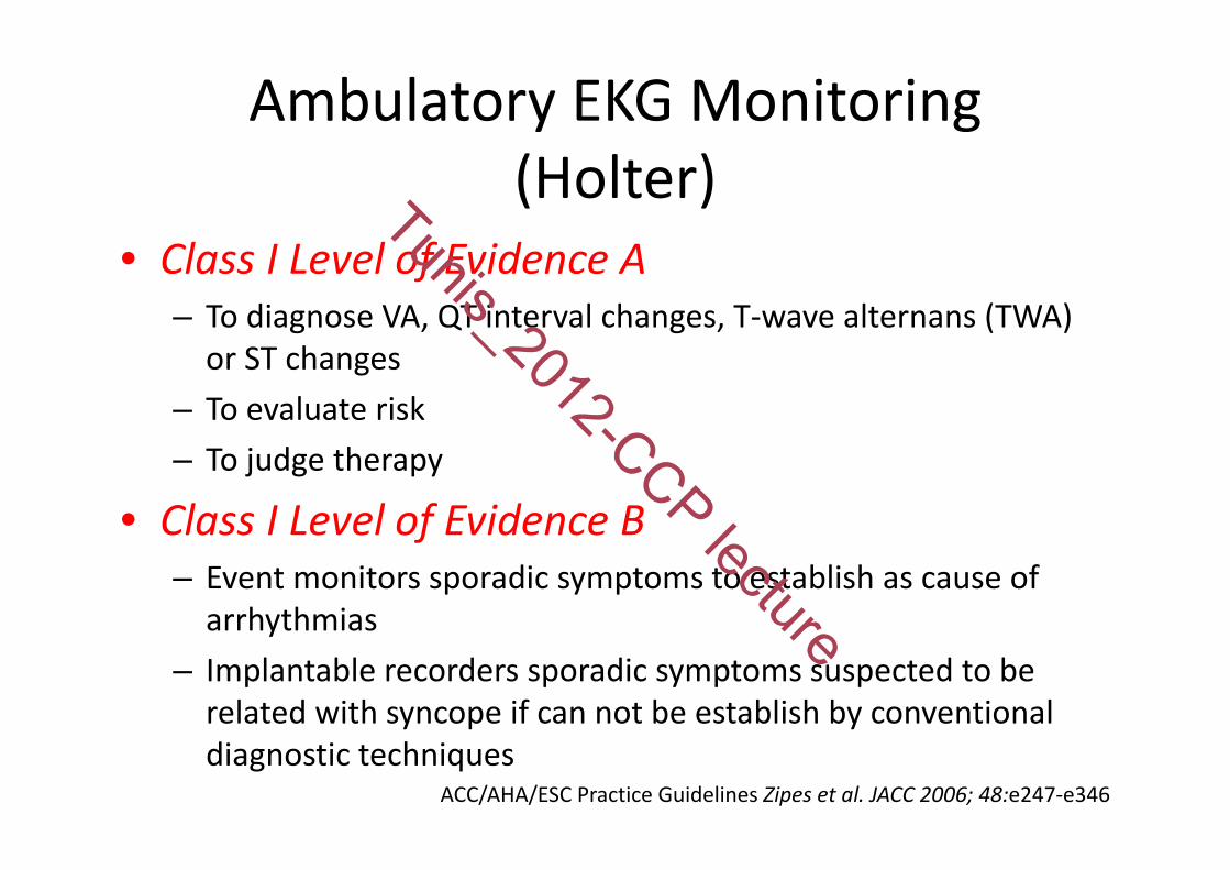
Ambulatory EKG Monitoring(Holter)
• Class I Level of Evidence A– To diagnose VA, QT interval changes, T‐wave alternans (TWA) or ST changes
– To evaluate risk– To judge therapy
• Class I Level of Evidence B– Event monitors sporadic symptoms to establish as cause of arrhythmias
– Implantable recorders sporadic symptoms suspected to be related with syncope if can not be establish by conventional diagnostic techniques
ACC/AHA/ESC Practice Guidelines Zipes et al. JACC 2006; 48:e247‐e346
Tunis_2012-CCP lecture

LVF and Imagining (1)
• Class I Level of Evidence B– Echocardiography
• For VA and structural heart disease• For high probability of SCD
– Exercise Echocardiography or SPECT• VA and intermediated risk of CHD
ACC/AHA/ESC Practice Guidelines Zipes et al. JACC 2006; 48:e247‐e346
Tunis_2012-CCP lecture

LVF and Imagining (and 2)• Class IIa
–MRI, Cardiac Computed Tomography, Radio‐nuclide angiography
• for VA if echo doesn’t provide accurate assessment of ventricular function and/or evaluation structural changes (Level of Evidence B)
–Coronary Angiography• For VA or survivors SCD and greater probability of CHD (Level of Evidence C)
– LF Imaging• In patients with biventricular pacing (Level of Evidence C)
ACC/AHA/ESC Practice Guidelines Zipes et al. JACC 2006; 48:e247‐e346
Tunis_2012-CCP lecture

An approach to the treatment of patients with ventricular ectopic beats
Structural Heart Disease
Frequent VEBs or VT
Frequent symptoms Treatment
‐ ‐( on exercise)
‐ reassure
‐ ‐ + β blocker
‐ + (monomorphic) ±
1. Catheter ablation
+ ‐ ±1. Assess SCD risk
2. β blocker
+ + ±1. β blocker
2. ICD if high SCD risk
ICD, Implantable Cardioverter Defibrillator; SCD ,Sudden Cardiac Death; VEB, Ventricular Ectopic Beat; VT, Ventricular tachycardia
Tunis_2012-CCP lecture

VEBs TreatmentKey points
• Ventricular ectopic beats are frequently seen in clinical practice and are usually benign
• Presence of heart disease should be sought and, if absent, indicate good prognosis in patients with VEBs
• There is not clear evidence that caffeine restriction is efficacy in reducing VEBs but patients with excessive caffeine intake should be cautioned and appropriately advised if symptomatic with VEB
Tunis_2012-CCP lecture
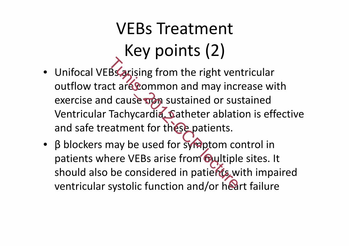
VEBs TreatmentKey points (2)
• Unifocal VEBs arising from the right ventricular outflow tract are common and may increase with exercise and cause non sustained or sustained Ventricular Tachycardia. Catheter ablation is effective and safe treatment for these patients.
• β blockers may be used for symptom control in patients where VEBs arise from multiple sites. It should also be considered in patients with impaired ventricular systolic function and/or heart failure
Tunis_2012-CCP lecture
VEBs TreatmentKey points (and 3)
• Risk of Sudden Cardiac Death from malignant ventricular arrhythmia should be considered in patients with heart disease who have frequent VEBs. Implantable cardioverter‐defibrillator may be indicated if risk stratification criteria are met.
• VEBs have also been shown to trigger malignant ventricular arrhythmias in certain patients with idiopathic ventricular fibrillation and other syndromes. Catheter ablation must be considered in some patients as adjunctive treatment.
Tunis_2012-CCP lecture

Thank you for your attention!
Tunis_2012-CCP lecture