Pr François Tison, Bordeaux, France - movementdisorders.org · Hemichorea-Hemiballism •...
30
Hyperkinetic Emergencies Movement Disorders emergencies: « Any movement disorder which evolves over hours to days, in which failure of appropriately diagnose and manage patients can result in morbidity or even mortality » Poston and Frucht (J Neurol 2008;255:2-13) • Parkinonism • Hyperkinetic: dystonia, chorea, tics, myoclonus Pr François Tison, Bordeaux, France
Transcript of Pr François Tison, Bordeaux, France - movementdisorders.org · Hemichorea-Hemiballism •...

Hyperkinetic Emergencies
Movement Disorders emergencies:« Any movement disorder which evolves over hours to days, in which failure of appropriately diagnose and manage patients can result in morbidity or even mortality »Poston and Frucht (J Neurol 2008;255:2-13)
• Parkinonism
• Hyperkinetic: dystonia, chorea, tics, myoclonus
Pr François Tison, Bordeaux, France

General clinical approach (1)1. To observe: localisation, speed, frequency,
permanent or intermittent, repetitive or chaotic ?rythm and amplitude
jerk brief slow
myoclonic choreic dystonic
2. The phenomenology :� dystonia, � ballism, � chorea,� myoclonus, � tics
duration
Robottom BJ et al.Arch Neurol 2011; 68:719-24

General clinical approach (2)
�Ask: time course ?, triggering/ameliorating factors ? continuous/paroxystic, wake/sleep ?
�Review: medical context, past and present medications, toxins, family history ?
�Examine: focal deficit, consciousness, meningitis, fever ?
�Order: lab tests (blood, CSF?), MRI

Dystonic emergencies
• « Status dystonicus »• Acute drug -induced dystonia• Breathing and swallowing dystonias
Pseudo-dystonic emergencies:� Pseudo-torticollis (kids +++): Atlanto-axial subluxation
inflammatory head and neck processes, posterior fossa and craniocervical jonction tumors
� Tetanus� Seizures (partial seizures with motor manifestations =
frontal)

« Status dystonicus »
• = « Increasingly frequent and severe episodes of generalized dystonia which necessitate urgent hospital admission (Manji H et al. Brain 1998; 121,243-252) (Jankovic and Penn, 1982; « desperate dystonics »- Marsden, 1984, « dystonicstorm »- Vaamonde, 1994 )
• Context of poorly controlled generalized idiopathicdystonia
• OR context of secondary dystonias: Wilson’sdisease, post-traumatic, post-anoxic, PKAN …

Triggering factors : Infection =51.7%, drugs =30%, surgery= 6.7%, metabolic disorder= 5%, DBS failure =5%No apparent cause= 32.6%

« Status Dystonicus » life threatening
• Stiffness and pain• Rhabdomyolysis• Dysphagia/aphagia• Aspiration pneumonia• Impaired ventilation• Infections, fever • Dehydratation• Multi-organ failure (renal)
« movement disorder emergency characterized by severe episodes of generalized hyperkinetic movement disoders that had necessitated urgent hospital admission because of life-threatening complication regardless of the patient neurological condition at baseline »

« Status dystonicus » differential diagnosis
• Neuroleptic malignant syndrome (neuroleptics, fever, autonomic, CK +++)
• Serotonin syndrome (SSRI, myoclonus, confusion and agitation)
• Malignant hyperthermia (anesthesia, fever, rhabdomyolysis)
• Baclophen pump withdrawal

« Status Dystonicus » = management
Deep sedation
Mariotti P et al. Movement Disorders2007; 22(7):963-968

« Status Dystonicus »= management
�Bilateral GPI DBS

Acute drug -induced dystonia
�Neuroleptics +++ (typical:6%, atypical: 1-2%) including anti-emetics
� Introduction or dose increase (usually <24h, 90 % within 5 days)
�Young males (tardive dyskinesias and parkinsonism in older women, acute choreic reactions in childs)
�Self-limiting but distressing may be life-threatening (laryngeal dystonia)

Acute drug -induced dystonia
More frequent clinical forms: head and neck !• Oculogyric crisis: eye deviation/head rotation,
opisthotonus, rigidity, autonomic symptoms, dysarthria, anxiety
• Oculo-cephalic dystonia:trismus, torticollis, blepharospasm, laryngeal dystonia
• Any form of dystonia
� Management :• Parenteral anticholinergics (trihexhyphenidil
diphenylhydramine 25-50 mg, benztropine 1-2mgoften repeated + 5 -7 day oral course),
• Clonazepam or diazepam if resistant

Breathing and swallowing dystonias
• Spasmodic dystonia : usually no airway obstruction unless botulinum toxin-induced weakness
• Adductor laryngeal dystonias (Gerhardt’s syndrome), adductor spasms with stridor in focal dystonias, X-linked-dystonia parkinsonism (Lubag) and MSA
• Tardive dystonias (laryngeal spasms)
• HD and chorea-acanthocytosis

Choreic emergencies
Main causes of acute chorea: Piccolo I. et al. J Neurol 2003; 250: 429-435, Robottom BJ et al.Arch Neurol 2011; 68:719-24
– Vascular (50%)– Drug -induced (16%)– Metabolic (14%)– AIDS-related (12%)– Infectious and inflammatory (8%)
• Hemichorea-Hemiballism• Severe levodopa induced-dyskinesias • Acute generalized chorea (secondary causes)

Hemichorea -Hemiballism
• Hemiballism: Acute onset, dramatic flinging rotatory movements of proximal muscles on one side (arm and/or leg)Increased by action, stress, absent during sleep
• Hemichorea: less ample, more distal

Hemichorea -Hemiballism
• Share the same pathophysiology: historically a lesion of the controlateral STN (15%) but anywhere whithin the BG (GPi, putamen, caudate) and adjacent WM (85%).Postuma RB and Lang AE Lancet Neurology 2003;2:661-68
• Any type of lesion, stroke far more common (>75%)
• Postpump chorea following CP bypass (1.2%) in childrens

Hemichorea -Hemiballism• Distressing, exhausting, self-injuries,
dehydration and rhabdomyolysis in most severe cases
• Usually subsides within hours or days, but may be prolonged in a minority
• Long-term prognosis depends on the prognosis of the underlying disease
• Management: of the cause (stroke, hyperglycaemia), protection of limbs (pads), rehydratation
• If severe and/or prolonged consider neuroleptics (haloperidol) or tetrabenazine
• GPi or thalamus DBS (or lesion)

Hemichorea -Hemiballism
�May also be metabolic: « hyperglycaemic hemiballism »
�Women >65 y, diabetes type 2, more frequent in Asians
�Severe hyperglycaemia, subsides after correction
�Characteristic = high signal on T1 MRI sequences in the putamenPostuma RB and Lang AE
Lancet Neurology 2003; 2:661-68

Drug -induced chorea/ballismMore frequent = L -Dopa in PD : L-Dopa dose increase or ICOMT introduction
• Risk of dehydratation and rhabdomyolysis• Management : decrease dosage or transient
withdrawal, rehydratation, sedation (diazepam)
Other drugs: lithium, anti-epileptic drugs(lamotrigine), opioids and methadone

Acute/subacute generalized choreas
�Choreas of rapid onset, often associated with other neurological or neuropsychiatric features:
�Disclose secondary causes +++– Auto-immune diseases = Post-streptococcal
neurological disorders (Sydenham chorea), Systemic Lupus erythematosus (SLE) and anti-phospholipid syndrome (APLS)
– Metabolic =glucose, thyreotoxicosis
– Encephalitis

Myoclonic emergencies
Generalized myoclonus and/or asterixis (« negative myoclonus ») are common in the setting of metabolic, toxic and drug-induced encephalopathies:
• Liver, kidney and respiratory failure• Serotoninergic drugs « serotoninergic syndrome »,
tricyclic anti-depressants, lithium
• Opiates and benzodiazepinewithdrawal,amphetamine,cocaine ecstasy
• Post-anoxic myoclonus

Opsoclonus-myoclonusSubacute Triad :- Opsoclonus- Myoclonus: Craniocervical and trunk, extremities (adults)- Ataxia
• Paraneoplastic (Anti Ri, HU, others)Neuroblastoma in childrensOthers (Melanoma, non-Hodgkin
• Auto-immune (NMDA, AMPA, GLUR5, GABA-B, GAD…)• Infections (VZV, EBV, Coksackie, West Nile, Lyme …
Traitement : IVIg, corticosteroids, plasmapheresisimmunosupressants
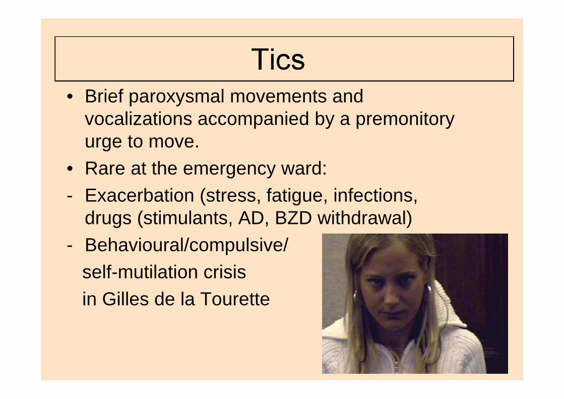
Tics• Brief paroxysmal movements and
vocalizations accompanied by a premonitoryurge to move.
• Rare at the emergency ward:- Exacerbation (stress, fatigue, infections,
drugs (stimulants, AD, BZD withdrawal)- Behavioural/compulsive/
self-mutilation crisisin Gilles de la Tourette

PsychogenicJerks and tremors
Context :- Psychiatric illness(rare)- History of psychogenic disorder- Physical or emotional stress/trauma
Clinical picture :- variable, complex, - inconsistent phenomenology, - suggestibility, distractibility, - Absence of urge to moveExplorations ?- Muscle activation pattern- Bereitschaftspotential preceding the
movement

… and two diagnosis and therapeutic emergencies
#1 Wilson’s disease�Any emergent movement disorder in the young
(< 40 y +++): tremor (any type, midbrain+++), dystonia (face +++ risus sardonicus) and choreo-athethosis
�Urgent to diagnose : serum and urine copper, plasma ceruleoplasmin, slit-lamp examination (KF ring), brain MRI.
�Urgent to treat by decoppering drugs: irreversible brain and liver damage !

#1 Wilson’s disease
Chelation drama and Liver grafting « miracle »

… and two diagnosis and therapeutic emergencies
#2 Whipple’s disease
�Oculo-masticatory or oculo-facio-skeletal myorythmias
�Progressive supranuclear palsy and/or cognitive and behavioral changes
� Jejunal biopsy for histological and PCR analysis
�PCR (T. Whippelii) in the CSF

Whipple’s disease :to treat (sulfamethoxazole-trimethoprime and/ or ceftriaxone) and cure

Conclusion
�Recognize:the movement disorderthe emergency situation
�Diagnose the more common causes:cerebrovascular diseasesacute drug reactions
�Urgent to « cure »: the « 2W »
Wilson’s disease, Whipple’s disease

Acknowledgements
• Pr Coubes, Dr Biolsi, Montpellier• Dr Delberghe, Dr Gonce, Belgium• Dr Fénélon, Créteil• Dr Marion, London• Pr Meissner, Bordeaux• Pr Vidailhet, Paris• Dr Woimant, Paris• MDS Video collection
Frucht SJ and Fahn S, Movement disorders emergenciesHumana Press, 2005