Ppt parietal lobe
100
PARIETAL LOBE Presented by Abdul Qavi ANATOMY AND FUNCTIONAL CORRELATION
-
Upload
qavi786 -
Category
Health & Medicine
-
view
247 -
download
36
Transcript of Ppt parietal lobe

PARIETAL LOBE
Presented by
Abdul Qavi
ANATOMY AND FUNCTIONAL CORRELATION

INTRODUCTION
The parietal cortex is considered as one of the most complex region of human brain which is responsible for the integration of various stimuli .
Have undergone a major expansion in the course of human evolution, largely in the inferior parietal region
Receives, correlates, analyze primary sensory information to interpret stimulus and aid in discrimination and recognition.

Lobe of the hand

Defining the lobes
central (rolandic) sulcus
sylvyan (lateral) sulcus
frontal lobe
temporal lobe
occipitallobe
parietal lobe

BOUNDARIES OF THE PARIETAL LOBE

BOUNDARIES OF THE PARIETAL LOBE
– Anterior border - Central Fissure– Ventral border - Sylvian Fissure– Dorsally by the cingulate gyrus– Posterior border - Parieto-occipital sulcus

SULCI AND GYRI ON THE SUPERO LATERAL SURFACE
superolateralmedial

SULCI AND GYRI ON THE VARIOUS SURFACE OF PARIETAL LOBE
SUPERO LATERAL SURACE • Post central gyrus( area 1,2,3)• Superior parietal lobule (area 5,7) • Inferior parietal lobule• Supra marginal gyrus - lies around the upturned end of sylvian fissure.• Angular gyrus – lies around the upturned end of superior temporal gyrus.MEDIAL SURFACE • Supra splenial sulcus – separate the precuneus from cingulate gyrus • Precuneus - lies between parieto occipital sulcus and paracentral lobule • Isthmus – Separates the splenium of corpus callosum from calcarine
sulcus


Parietal lobe sulci and gyri
Post central sulcus – posterior boundary of somatosensory cortex.
Intraparietal sulcus behind post central sulcus which divides the parietal lobe into sup. & inf. Parietal lobule
Posterior end of sylvian fissure curves upwards to terminates into inf.parietal lobule – surrounding cortex supramarginal gyrus[SMG 40]
Parietal lobe

Parietal lobe sulci and gyri• Posterior end of sup. Temporal
sulcus – angular gyrus[AG 39]• SMG & AG =Ecker’s inf Parietal
Lobule• Ecker’s IPL & post. Third of first
temporal gyrus constitute the wernicke’language area
• 3,1,2-primary sensory areas• 5- somatosensory association
area• 7-somatosensory or
somatosensory/visual

Parietal Topography
•Postcentral Gyrus (1,2,3)
•Superior Parietal Lobule (5 ,7)
•Supramarginal Gyrus (40) •Angular Gyrus ( 39)

Subdivisions of the Parietal Lobes
Functional zones
Anterior zone -1,2,3, •Somatosensory cortex
Posterior zone -remaining areas•Posterior parietal cortex
von Economo:
Posterior parietal areas•PE (5)•PF(7b)•PG -Polymodal and asymmetric larger in right hemisphere

• Visual processing areas– Intraparietal sulcus (cIPS)• Control of saccadic eye movements
– Saccade - involuntary abrupt and rapid small movements made by the eyes when changing the fixation point
• Visual control of grasping
– Parietal reach regions (PRR)• Visually guided grasping movements
Subdivisions of the Parietal Lobes


Somatosensory strip To area PE -Tactile recognitionTo motor regions -sensory information about limb position and movement•Area PE is somatosensory–Inputs from the somatosensory strip–Outputs to primary motor cortex, supplementary motor cortex, premotor regions, and area PF •Area PF Input from somatosensory, primary motor cortex, premotor cortex, and small visual input through area PG•Area PG–Receives complex connections including visual, somesthetic, proprioceptive, auditory, vestibular, oculomotor, and cingulate connections–Parieto-temporo-occipital crossroads–Part of the Dorsal Stream•Close relation between the posterior parietal connections and the prefrontal
Connections of the Parietal Lobes

Connections of the Parietal Lobes
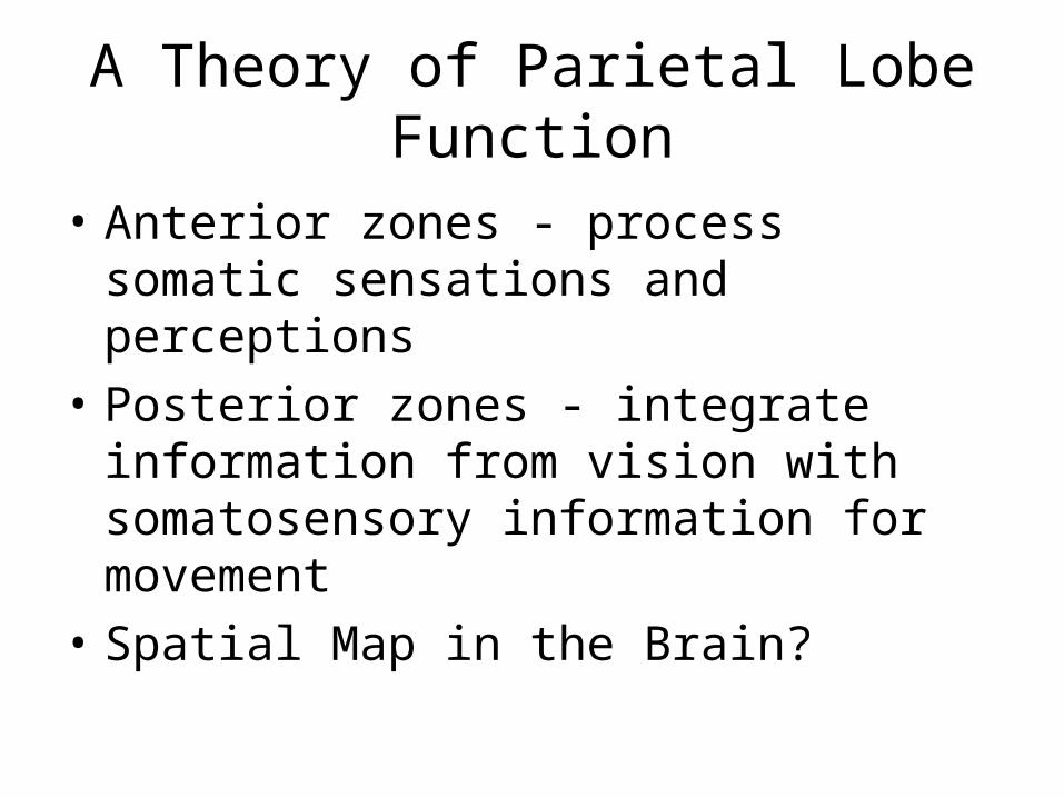
• Anterior zones - process somatic sensations and perceptions
• Posterior zones - integrate information from vision with somatosensory information for movement
• Spatial Map in the Brain?
A Theory of Parietal Lobe Function

NP/MGH
NEURO-IMAGING
NORMAL CORTICAL ANATOMY • The Central Sulcus• Sagittal• Axial• Coronal

NP/MGH
The Central The Central SulcusSulcus

NP/MGH
• superior frontal sulcus - pre CS sign• sigmoidal Hook sign• pars bracket sign• Bifid post-CS sign• thin postcentral gyrus sign• intraparital sulcus - post-CS• midline sulcus sign
The Central Sulcus (CS)*
*Naidich & Brightbill. Int J Neurorad 1996;2:313-338*Naidich & Brightbill. Int J Neurorad 1996;2:313-338

NP/MGH
• Superior frontal sulcus - preCS sign– the posterior end of the superior frontal sulcus joins
the precentral sulcus in 85%
The Central Sulcus (CS)
Precentral sulcus
Superior frontal sulcus
Precentral gyrus
Central sulcus
Superior frontal gyrus
Superior frontal sulcus
Precentral sulcus
Precentral gyrus

NP/MGH
• pars bracket sign– The paired pars
marginalis form a “bracket” to each side of the interhemispheric fissure at or behind the central sulcus (96%).
The Central Sulcus (CS)
Precentral sulcus
Superior frontal sulcus
Precentral gyrus
Central sulcus
Pars bracket Paracentral lobule

NP/MGH
Sigmoid “Hook”
– hooklike configuration of the posterior surface of the precentral gyrus
– the “hook” corresponds to the motor hand area.
– The “hook” is well seen on CT (89%) and MRI (98%).
The Central Sulcus (CS)The Central Sulcus (CS)
Precentral sulcus
Central sulcus

NP/MGH
• Bifid post-CS sign– the post-CS is bifid (85%).– The bifid post-CS encloses the lateral end of the pars
marginalis (88%).
The Central Sulcus (CS)
Precentral sulcus
Precentral gyrus
Central sulcus
Postcentral sulcus
Pars bracket

NP/MGH
Central sulcus
Postcentral sulcus
Central sulcus Central sulcus
Postcentral sulcus
Postcentral sulcus
Pars bracketPars bracket

NP/MGH
Thin post-CG sign
– the postcentral gyrus is thinner than the precentral gyrus (98%).
The Central Sulcus (CS)The Central Sulcus (CS)Precentral
gyrus
Postcentral gyrus

NP/MGH
Intraparietal Sulcus (IPS) and the post-CS
– in axial MRI, the IPS intersects the post-CS (99%).
The Central Sulcus (CS)The Central Sulcus (CS)
Pars bracket
IPS
Postcentral sulcus
IPS
Pars bracket

NP/MGH
Postcentral sulcus
IPS
Postcentral sulcus
IPS
Postcentral sulcus
IPS

NP/MGH
SFS-preCS sign
Hook sign
Pars bracket sign
Bifid post-CS sign
Thin postcentral gyrus sign
IPS - postCS sign
The Central Sulcus (CS)The Central Sulcus (CS)

NP/MGH
Axial Axial NeuroanatomyNeuroanatomy

NP/MGH
Superior Temporal gyrus
Middle Temporal gyrus
Inferior Temporal gyrus
Fusiform gyrus

NP/MGH
Superior occipital gyrusIntra-occipital sulcus
Middle occipital gyrus
Cingulate gyrus
Parieto-occipital fissure
Calcarine sulcus
Cuneus
Middle temporal gyrus
Superior temporal sulcus
Superior temporal gyrus
Insula
Inferior frontal gyrus,pars orbitalis
Superior frontal gyrus Middle frontal gyrus
Inferior frontal gyrus,pars opercularis
Lateral fissure
Lateral fissure
Inferior parietal gyrus

NP/MGH
Middle occipital gyrus
Superior temporal gyrus
Intra-occipital sulcus
Superior frontal gyrus
Central sulcus
Superior occipital gyrusParieto-occipital sulcus
Superior temporal sulcus
Lateral fissure
Inferior parietal gyrus
Postcentral gyrus
Lateral fissure
Middle frontal gyrus
Inferior frontal gyrus

NP/MGH
Postcentral sulcus
Superior frontal sulcus
Central sulcus
Intraparietal sulcus
Superior frontal gyrus
Middle frontal gyrus
Superior parietal gyrus
Centrum semiovale
Parietooccipital sulcus
Precuneus
Angular gyrus
Central sulcus
Inferior frontal gyrus
Supramarginal gyrus
Postcentral sulcus

NP/MGH
Central sulcus
Postcentral sulcus
Superior frontal sulcus
Precentral sulcus
Pars marginalisIntraparietal sulcus
Superior frontal gyrus
Middle frontal gyrus
Precuneus
Paracentral lobule
Superior parietal gyrus

NP/MGH
Coronal Coronal NeuroanatomyNeuroanatomy

NP/MGH
Olfactory bulb
Gyrus rectusMedial Orbital gyrus
Inferior Frontal gyrusSuperior Frontal gyrus
Middle Frontal gyrus
Interhemispheric Fissure
Inferior Frontal gyrus

NP/MGH
Superior Frontal gyrusSuperior Frontal sulcus
Middle Frontal gyrus
Superior Temporal Sulcus
Sylvian Fissure
Amygdala
Precentral sulcus
Anterior commissure
Cingulate sulcus
Superior Temporal gyrus
Middle Temporal gyrus
Inferior Temporal gyrus
Precentral gyrus
NP/MGH
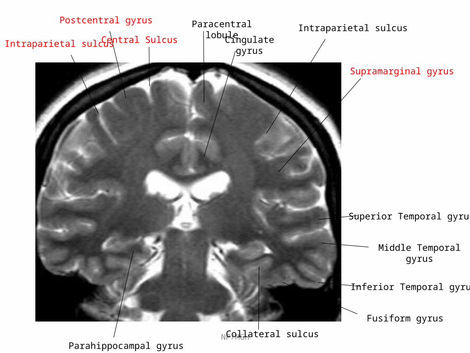
Paracentral lobule
Superior Temporal gyrus
Middle Temporal gyrus
Inferior Temporal gyrus
Central Sulcus
Postcentral gyrus
Cingulate gyrusIntraparietal sulcus
Fusiform gyrus
Collateral sulcusParahippocampal gyrus
Supramarginal gyrus
Intraparietal sulcus

NP/MGH
Superior Temporal gyrus
Middle Temporal gyrus
Inferior temporal gyrus
Fusiform gyrus
Central sulcusParacentral lobule

NP/MGH
Lingual gyrus
Calcarine sulcus
Superior parietal lobuleprecuneus
Cingulate gyrus
Tentorium cerebelli
Fusiform gyrus
Inferior parietal lobule
Middle occipital gyrus
Inferior occipital
gyrus
Lingual gyrus
Collateral sulcus

NP/MGH
Sagittal Sagittal NeuroanatomyNeuroanatomy

NP/MGHSubcallosal gyrus
Gyrus rectus
Parietooccipital sulcus
Fastigium, fourth ventricle
Cingulate gyrus
Calcarine sulcus
Marginal ramus of Cingulate sulcus
precuneus
Paracentral lobule
Cingulate sulcusSuperior frontal gyrus
Cuneus
Lingual gyrus
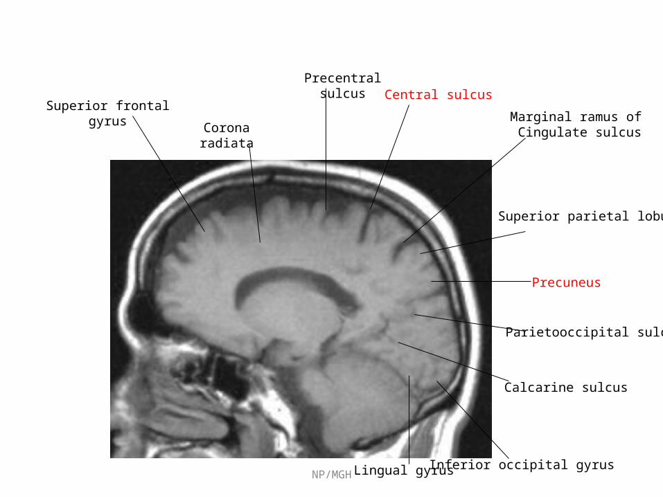
NP/MGH
Parietooccipital sulcus
Calcarine sulcus
Superior parietal lobule
Marginal ramus of Cingulate sulcus
Central sulcusPrecentral
sulcus
Precuneus
Corona radiata
Superior frontal gyrus
Lingual gyrus Inferior occipital gyrus

NP/MGH
Central sulcus
Inferior Temporal gyrus
Middle Temporal gyrus
Superior Temporal gyrus

NP/MGHGyrus rectus
Parietooccipital sulcus
Cingulate gyrus
Calcarine sulcus
Lingual gyrus
Marginal ramus of Cingulate sulcus
Superior parietal lobule
Cingulate sulcus
Caudothallamic groove
Precuneus
Central sulcus
Cuneus
Precentral gyrus
Frontomarginal gyrus
Superior frontal gyrus

NP/MGH
Inferior Temporal gyrus
Superior Temporal sulcus
Superior Temporal gyrus
Anterior occipital sulcus
Superior frontal sulcus
Precentral sulcus
Central sulcus
Postcentral sulcus
Angular gyrus
Lateral fissure, posterior segment
Inferior frontal gyrus,pars orbitalis
Middle Temporal gyrus
Inferior occipital gyrus
Middle occipital gyrus
Inferior frontal gyrus,pars triangularis

BLOOD SUPPLY

BLOOD SUPPLY

Primary somatosensory area Location : Post central gyrus(ant parietal lobule) on lateral surface and dorsal aspect of paracentral lobule on medial serface. Broadman area (3 ,1, 2)Representation : contralatral half of body invertedFunction: initial reception center for afferent impulses, especially for tactile, pressure, and position sensations. necessary for discriminating finer, more critical grades of sensation and for recognizing intensity.Afferent connections: VP nucleaus of thalamus
Outputs: primary motor cortex, contralateral S1,association somatosensory cortex(area 5 & 7), thalamus
Deficit: Postural sensation (proprioception), passive movement (kinesthesis), Tactile sensation, Two point discrimination, Astereognosis,High sensory thresholds
Functional areas

Somatosensory Homunculus Body presentation

Secondary Somatosensory area
Location : superior lip of lateral fissure (parietal operculam)
Representation :contralateral side dominant, Bilateral representation
Afferent: Intralaminar nuclei and posterior group of nuclei of thalamus
Function: not well described, ? Involved in less discriminative aspects of sensation.
Lesions: none ascribed, rarely inability to appreciate pain(asymbolia)

Somato-sensory association area: Location : superior parietal lobule. broadmann’s area(5,7)Function : interpretation; similarities and differences, spatial relationships
and 2D qualities, variations in form and weight, and localization of sensation
• Area 5-, Manipulation of objects Tool use/body image
• Area 7- Integration of visual and somato-sensory stimuli, Hand-eye coordination, reaching and grasping,,
• Afferent: primary somato-sensory area
• Deficit : Impair gnostic (knowing, recognition) aspects of sensation , stereognosis, graphesthesia, two-point discrimination, and tactile localization , poor hand eye coordination,
(appreciation of primary sensations remains, but assoc. functions impaired)

Inferior parietal lobule• Location: supramarginal gyrus (40) and angular gyrus (39)
• Function:. Left hemisphere – language ,maths, reading, writing, understanding of symbols. Right hemisphere—visuo-spatial orientation.
• Lesions Aphasia, agnosia, and apraxia and visuspatial defects
• A deeply placed parietal lesion may cause either an inferior quadrantic or hemianopic visual field defect

Clinical assesment and testing

Post-Central Gyrus,Dominant or Non-Dominant
1. Impaired Postural sensation (proprioception), passive movement (kinesthesis).
2. Astereognosis
3. Impaired Two point discrimination
4. Agraphesthesia
5. Weight discrimination

Inability to discriminate size and shape of objects and identify them by touch alone.
Tests
Patient identifies by touch such common objects as a coin, paperclip, pencil, or key (each hand tested separately)
Patient judges the relative size of a series of coins
Patient judges the texture of a series of objects, such as cloth, wire, sandpaper
Astereognosis (tactile agnosia)
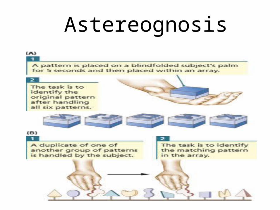
Astereognosis

Graphesthesia Ability to recognise letters or numbers written on skin with
pencil,or dull pin Testing is often done over the finger pads, palms, or dorsum of the
feet Letters or numbers about 1 cm in height are written on the finger
pads, larger elsewhere clear figures as 8, 4 5 used first, more difficult 6, 9 ,3 can be used as
finer tests Tactile movement sense, directional cutaneous kinesthesia- Ability
to tell the direction of movement of a light scratch stimulus drawn for 2 cm to 3 cm across the skin which may be a sensitive indicator of function of the posterior columns and primary somatosensory cortex
Loss of graphesthesia or the sense of tactile movement with intact peripheral sensation implies a cortical lesion, particularly when the loss is unilateral.

Two point discrimination Ability to differentiate, eyes closed, cutaneous stimulation by one
point from stimulation by two points.Instruments: two-point discriminator, electrocardiogram
calipers,compass, paper clip bent into “v,” adjusting the two points to different distances.
Method Either one-point or two-point stimuli are delivered randomly, and
the minimal distance that can be discerned as two points is determined.
The result is taken as the minimum distance between two points that can be consistently felt separately.
Normal 2-point discrimination - 1 mm (tip of the tongue), 2 mm to 3 mm ( lips), 2 mm to 4 mm ( fingertips), 4 mm to 6 mm (dorsum of the fingers), 8 mm to 12 mm( palm), 20 mm to 30 mm( back of the hand), and 30 mm to 40 mm ( dorsum of the foot).
The findings on the two sides of the body must always be compared.

Superior Parietal Lobule,Dominant or Non-Dominant
cannot reach for objects (optic ataxia) -Balint syndrome
Poor visual guidance of hands, fingers, eyes, and limbs, head (hard time catching a ball)
Hard time directing movement in space (trouble flying a kite)
Hard time distinguishing left from right

Dominant inferior parietal lobule
1. Acalculia2. Agraphia3. Left-right confusion4. Finger agnosia5. Conductive aphasia6. Alexia7. Ideomotor apraxia
Gerstmann’s syndrome

Ideomotor apraxia: failure to perform previously learned motor acts accurately.
Results from left hemisphere lesion Usually affects both sides, may be worse on right side Can affect the face (buccofacial) and/or the limbs
Tests Carrying out motor acts to command: Buccofacial (blow out a match, protrude tongue, drink through a
straw)
Limb (salute, use a toothbrush, flip a coin, hammer a nail, comb hair,,snap fingers, kick a ball, crush out a cigarette)
Whole body commands(stand like a boxer, swing a baseball bat)

1. wernicke area
2. Arcuate fasciculus
3. Lt premotor area
4. Lt motor cortex
5. Corpus callosum
6. Rt premotor area
7. Rt motor cortex
Ideomotor apraxia:

Ideational apraxia: Able to carryout individual components of a complex motor act but
can not perform the entire sequence properly leading to a goal.Results from left hemisphere lesion ( temporo-parietal) also seen in generalised cognitive impairment.
Tests Carrying out complex motor acts to command: Opening tooth paste, taking tooth brush from holder, and placing
toothpaste on brush.
How to mail a letterHow to drive a car.

Results from left hemisphere supramarginal gyrus lesion if the underlying arcuate fasciculus is cut
Fluent speech with word finding pauses Severely defective repetition Paraphasia in repetition and in spontaneous speech Normal comprehension and reading Impaired writing, spontaneous and to dictation, errors in
spelling, word choice, Naming may be mildly impaired
Tests Repetition of words, phrases, & sentences Write to dictation (letters, words, sentences) Ask patient to write sentences describing a Job, the weather, or
a picture Confrontation naming of objects, clothing, body parts, parts of
objects, colors
Conduction aphasia

Finger agnosia: Inability to recognize, name, and point to individual
fingers on self and others Usually associated with lesion of dominant hemisphere Lt handed pts may have finger agnosia with lesions of
either hemisphere Limited clinical utility for localisation Tests • Non verbal finger recognition: pt eyes closed, touch pt finger,
then ask pt to point same finger on examiner hand• Identification of named fingers on examiner’s hand: examiner’s
hand placed in various positions. Ask pt “point to my middle finger”
• Verbal identification (naming) of finger on self and examiner: hand placed in various positions, ask pt “what is the name of this finger”

Right-left disorientation
Inability to distinguish right from left on self or env. More common with left hemisphere lesion Normal population (9%males, 17% females ) can have difficulty in rt
- lt testing
Tests • Identification on self(show me your rt foot),• Crossed commands on self(With your rt hand touch your lt shoulder)
• Identification on examiner(point to my lt elbow)• Crossed command on examiner(with ur rt hand point to my lt eye)

Acalculia Loss of ability to understand & order numbers More severe with left hemisphere lesion Also note errors in borrowing, alignment , error to particular
calculation,
Tests Verbal examples(addition, subtraction, multiplication, and division) Eg. 4+6, 8-5, 9*7, 9 /3
Verbal complex problems (allow 20 sec for response)Eg. 14+17, 43-38, 21*5, 128/8
Written complex problems(allow 30 sec for response)108 605 108 559+79 -86 *36 /43

Calculation errorsRt hemispheric lesion with lt neglect
Rt parietal bleed , poor alignment, calculation errors
Alzeimers ds, rote multiplication good but basic arithmatic disturbed

Diagnosed when pt demonstrate basic language errors, gross spelling errors, or use of paragraphias (word or syllable substitution)
Test First, ask patient to write letters and numbers to dictation.Second , ask the pt write names of common objects or body partsThird , if pt can successfully write single words , ask them to writesentence describing his job , whether, or picture from magazine
Agraphia
Results from damage to the angular gyrus itself and renders the patient unable to understand the written words and write.
Pt are not appreciably aphasic but anomia may be present
Alexia

Non-dominant inferior parietal lobule
1. Constructional apraxia2. Dressing apraxia3. Contralateral Neglect 4. Topographic disorientation5. Phonagnosia-6. Amusia . 7. Somatoperceptual disorders(Asomatognosia,
Anosagnosia)8. Sensory extinction or inattention

Inability to draw or construct 2 or 3D figures or shapes in presence of normal strength, coordination, sensation , comprehension.
More common and severe with right non dominant parietal lesion than left.
Tests Reproduction drawings (both 2D and 3D drawings as vertical
diamond, 2D cross, 3D block, 3D pipe, triangle within triangle are used). Scoring done from poor (0) to excellent (3)
Drawings to command(clock with numbers and hands, daisy in flowerpot, house with 2 sides and roof).
Constructional apraxia

Constructional apraxia
Scoring Interpretation
Poor (0) Non recognizable,gross distortion
Fair(1) Mod distorted or rotated 2D and loss 3Dimensionality
Good(2) Minimal distortion
Excellent(3) Perfect or near perfect
Rating 0 is 100% probability andRating 1 is 80% prob of brain damage
Vertical diamond

Constructional apraxia Reproduction drawings
2D cross test 3 D cube test 3 D pipe test Triangle within triangle
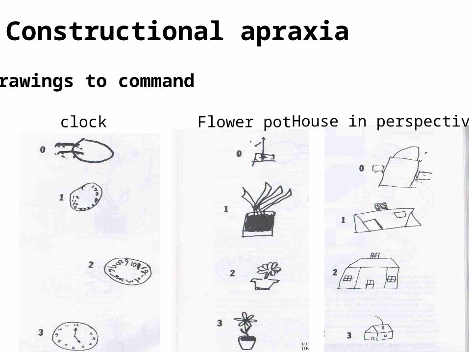
Drawings to command
Constructional apraxia
clock Flower pot House in perspective

Constructional apraxia Block designs Common errors
Rt lt rotation
Near far rotation
Figure ground or color reversal

InterpretationSpecific errors pathognomic of
brain damage (non retarded, age > 10 yrs)
1. Rotation by >45 degree2. Perseveration or repitition of
figure 3. FragmentatIon of design
Constructional apraxia

Dressing apraxia
Unable to properly clothe themselves
Most often leaves lt side partly undressed
MC with Rt nondominant parietal lesions
Associated with impaired tactile and visuospatial coordination
Considered as part of neglect syndrome

Contralateral Neglect and denialNeglect for visual, auditory, and somesthetic stimulation on one side of the body or spaceExamples:1.pt draw clock ,house flower with missing lt side2.If pt asked to read foot ball or ice cream he will read “ ball” and“cream”3.May shave only the right side of his face4.May not use one side of body even if no weakness
May be associated with denial disorder1.Anosognosia-Unawareness or denial of illness in presence of obvious disability

Sensory extinction or inattentionLoss of the ability to perceive two simultaneous sensory stimuli
Double simultaneous light touch stimuli at homologous sites on the two sides of the body.Extinction can also be done on one side. In general the more rostral area is the dominant one; (the face hand test). It may be normal to extinguish the hand stimulus.
Most commonly occurs with lesions of the inferior parietal lobule but may also occur with lesions of the temporoparietaloccipital junction, thalamus, and mesencephalic reticular formation .These areas have shown activation in attentional tasks
Lesions causing hemispatial neglect are similar to those causing inattention and extinction

Sensory extinction or inattention

Topographic disorientationInability to find way to familiar environments, localize places on maps, and find his way to new environmentEvaluationHistory obtained from family- 1.Does pt lost at neighbourhood, or home?2.Has pt lost travelling less frequent location?3.Does pt have difficulty orienting new environment? Localizing places on mapsAsk pt to draw map of India, if pt can’t draw, doctor should draw mapAsk pt to locate cities on map eg. Delhi, mumbai, calcutta1.Are cities located in appropriate states, ? 2.Are cities located on one half of map(either east or west)?
Ability to orient self in hospital environmentask nurses staff regarding pt capacity to find their bed, ward, bathroom

Clinical syndromes Either hemisphere
1. Cortical -sensory syndrome & sensory extinction
2. Total hemi anesthesia may occur
3. Mild hemiparesis, unilateral muscular atrophy in children, hypotonia, slowness of movements, hemiataxia, pseudoathetosis of opposite side
4. Homonymous hemianopia, visual inattention , anosognosia, hemineglect (with right>left lesion)
5. Abolition of optokinetic nystagmus with target moving towards the side of lesion

Right hemisphere Left Hemisphere Topographic disorientation
Visuospatial disorders
Gerstman’s syndrome (Angular gyrus)
Acalculia, Finger agnosia,Lt/rt disorientation,Agraphia
Hemi inattention Tactile agnosia (bimanual asteriognosis)
Anosognosia Bilateral Ideomotor & ideational apraxia
Constructional apraxia, /dressing apraxia Disorder of languageespecially alexia

Take home message Both parietal lobes have equal processing capabilities for light touch, tactile localization, 2-point discrimination, joint position sense, passive movement sense, and stereognosis.
Language and sequential analysis ability are strongly lateralized to the left inferior parietal lobe
Spatial abilities are strongly lateralized than language. Both parietal lobes have substantial spatial abilities, with the right being superior
Lesions to the parietal lobe are seldom localized to one particular quadrant (e.g. inferior, superior), or even restricted to the parietal lobe.
Even after assessment of clinical symptom and signs it is difficult to ascertain all signs to particular area of the parietal lobe.


Questions and Answers

1- Which one is not a part of parietal lobe a) Angular gyrus b) Gyrus rectus c) Supramarginal gyrus d) Precuneus
Ans: b) Gyrus rectus

2- Supramarginal gyrus corresponds to Brodmans area-
a) 39 b) 40 c) 44 d) 42
Ans: b) 40

3- Sigmoid Hook sign denotes- a) Central sulcus b) Precentral sulcus c) Calcarine sulcus d) Parieto-occipital sulcus Ans: a) Central sulcus

4- All are functions of parietal lobe except- a) Stereognosis b) Proprioception c) Two point discrimination d) Prosody
Ans: d) Prosody

5- Inferior quadrantanopia occurs in lesion of-
a) Frontal lobe b) Occipital lobe c) Parietal lobe d) Temporal lobe
Ans: c) Parietal lobe

6- Normal two point discrmination for the ‘tip of tongue’ is-
a) 2-3 mm b) 4-6 mm c) 1 mm d) 6-8 mm
Ans: a) 2-3 mm

7- Gerstman syndrome include all except-
a) Finger agnosia b) Agraphia c) Acalculia d) Aphasia
Ans: d) Aphasia

8- Conduction aphasia occurs in lesion of-
a) Cuneus b) Paracentral lobule c) Angular gyrus d) Arcuate facsiculus
Ans: d) Arcuate facsiculus

9- Which one is not seen in lesion of non-dominant inferior parietal lobule lesion -
a) Ideomotor apraxia b) Dressing apraxia c) Constructional apraxia d) Atopographia
Ans: a) Ideomotor apraxia

10- Unawareness or denial of illness (hemiplegia) is called as-
a) Anosognosia b) Asomatognosia c) Anosodiaphoria d) Autotopagnosia Ans: a) Anosognosia